promus premier dfu - boston scientific · monorail™ stent delivery system promus premier...
TRANSCRIPT
90800463-01
Bo
sto
n S
cien
tifi
c, (
Mas
ter
Bra
nd
DFU
Tem
pla
te 8
.267
7in
x 1
1.69
29in
A4,
901
0591
8AM
), e
DFU
, MB
, Pro
mu
s P
RE
MIE
R, E
N, 9
0800
463-
01B
Black (K) ∆E ≤5.0
ONLYCaution: Federal Law (USA) restricts this device to sale by or on the order of a physician.This device is supplied in sterile condition. All materials inside the sterile barrier pouch (the delivery system and stent, as well as the carrier tube and pouch liner) are sterile. The external surface of the sterile barrier pouch, as well as the product carton, should not be considered sterile.
1 warning:Contents supplied STERILE using an ethylene oxide (EO) process. Do not use if sterile barrier is damaged. If damage is found, call your Boston Scientific representative.For single use only. DO nOT rEUSE, rEPrOCESS Or rESTEriLiZE. Reuse, reprocessing or resterilization may compromise the structural integrity of the device and/or lead to device failure which, in turn, may result in patient injury, illness or death. Reuse, reprocessing or resterilization may also create a risk of contamination of the device and/or cause patient infection or cross-infection, including, but not limited to, the transmission of infectious disease(s) from one patient to another. Contamination of the device may lead to injury, illness or death of the patient.
After use, dispose of product and packaging in accordance with hospital, administrative and/or local government policy.
2 DEViCE DESCriPTiOn:Promus PREMIER Everolimus-Eluting Platinum Chromium Coronary Stent System:The Promus PREMIER Everolimus-Eluting Platinum Chromium Coronary Stent System is a device/drug combination product consisting of a drug/polymer-coated balloon expandable stent, pre-mounted on a Monorail (MR) or Over-The-Wire (OTW) delivery catheter. The stent is made from a platinum chromium alloy (PtCr). The drug-polymer coating consists of a polymer, PVDF-HFP, and the active pharmaceutical ingredient, everolimus. The characteristics of the Promus PREMIER Stent System are described in Table 2.1.
2013-11< EN >
Promus PREMIER™
Everolimus-Eluting Platinum Chromium Coronary Stent System
m o n o r a i l ™
o v e r - t h e - w i r e
TabLE Of COnTEnTS1 warning: ...................................................................................................................................1
2 DEViCE DESCriPTiOn:...............................................................................................................1Table 2.1 Promus PREMIER Stent System Product Description ............................2
2.1 Device Component Description .....................................................................................2Contents for (1) Promus PREMIER Monorail Stent System ............................................2Contents for (1) Promus PREMIER Over-the-Wire Stent System...................................22.2 Drug Component Description ........................................................................................22.2.1 Everolimus ......................................................................................................................2
Figure 2.1 The Chemical Structure of Everolimus ...................................................22.2.2 Primer Polymer and Drug Matrix Copolymer Carrier ..............................................2
Figure 2.2. The Chemical Structure of PBMA ..........................................................2Figure 2.3. The Chemical Structure of PVDF-HFP.....................................................2
2.2.3 Product Matrix and Everolimus Content ...................................................................3Table 2.2 Promus PREMIER Stent System Product Matrix and Everolimus Content ........................................................................................................3
3 inTEnDED USE/inDiCaTiOnS fOr USE:................................................................................3
4 COnTrainDiCaTiOnS: ..............................................................................................................3
5 warningS: .................................................................................................................................3
6 PrECaUTiOnS:............................................................................................................................36.1 General Precautions .......................................................................................................36.2 Pre- and Post-Procedure Antiplatelet Regimen .........................................................36.2.1 Oral Antiplatelet Therapy ............................................................................................36.3 Longitudinal Stent Deformation .....................................................................................46.4 Use of Multiple Stents .....................................................................................................46.5 Brachytherapy ..................................................................................................................46.6 Use in Conjunction with Other Procedures .................................................................46.7 Use in Special Populations ............................................................................................46.7.1 Pregnancy ......................................................................................................................46.7.2 Lactation .........................................................................................................................46.7.3 Gender ............................................................................................................................46.7.4 Ethnicity ..........................................................................................................................46.7.5 Pediatric Use .................................................................................................................46.7.6 Geriatric Use ..................................................................................................................46.8 Lesion/Vessel Characteristics .......................................................................................46.9 Drug Interactions .............................................................................................................46.10 Immune Suppression Potential ...................................................................................46.11 Lipid Elevation Potential ...............................................................................................46.12 Magnetic Resonance Imaging (MRI) Safety Information: ......................................4Medical Registration .............................................................................................................46.13 Stent Handling (also see Section 14, Operational Instructions) ............................4
6.14 Stent Placement .............................................................................................................5Preparation .............................................................................................................................5Placement ...............................................................................................................................56.15 Stent Delivery System Removal ..................................................................................5
Table 6.1. Delivery System Deflation Time Specifications (Seconds) ...................56.16 Post-Procedure ..............................................................................................................5
7 DrUg infOrMaTiOn .................................................................................................................57.1 Mechanism of Action ......................................................................................................57.2 Pharmacokinetics ............................................................................................................5
Table 7.1 Whole Blood Everolimus Pharmacokinetic Parameters (Mean ± SD) for PLATINUM Groups with Three or More Patients Following PROMUS Element Stent Implantation. .....................................................5
7.3 Drug Interactions .............................................................................................................67.4 Carcinogenicity, Genotoxicity, and Reproductive Toxicology ..................................67.5 Pregnancy .........................................................................................................................67.6 Lactation ............................................................................................................................6
8 OVErViEw Of CLiniCaL STUDiES ..........................................................................................68.1 NG PROMUS Clinical Trial ..............................................................................................68.2 PLATINUM Workhorse (WH) Randomized Controlled Trial (RCT) ...........................68.3 PLATINUM Small Vessel (SV) Sub-study .....................................................................68.4 PLATINUM Long Lesion (LL) Sub-study .......................................................................78.5 PLATINUM Pharmacokinetics (PK) Sub-study ...........................................................78.6 PLATINUM Quantitative Coronary Angiography (QCA) Trial ....................................7
Table 8.1 Comparison of PLATINUM Clinical Studies ..............................................7
9 aDVErSE EVEnTS: .....................................................................................................................89.1 Observed Adverse Events ..............................................................................................8
Table 9.1 PLATINUM Workhorse, PLATINUM Small Vessel, PLATINUM Long Lesion, and PLATINUM QCA Major Clinical Events From Post-Procedure to 1-Year Follow-Up and NG PROMUS Major Clinical Events From Post-Procedure to 30-Day Follow-Up..................................................8
9.2 Potential Adverse Events ...............................................................................................9
10 CLiniCaL STUDiES ...................................................................................................................910.1 NG PROMUS Clinical Trial ............................................................................................9
Table 10.1.1. NG PROMUS 30-Day Clinical Results, Intent-to-Treat Patients N= 100 ...........................................................................................................10Table 10.1.2 NG PROMUS Primary Endpoint Results by Gender, Intent-to-Treat, All Patients (N=100 Patients, N=119 Lesions) .............................11Table 10.1.3 NG PROMUS 30-Day Clinical Endpoints, by Gender, Intent-to-Treat, Male and Female Patients (N=100) ...............................................11
10.2 PLATINUM Workhorse (WH) Randomized Controlled Trial (RCT) .......................11Table 10.2.1. PLATINUM Workhorse 12-Month Clinical Results, Intent-to-Treat Patients ..............................................................................................12Table 10.2.2 PLATINUM Workhorse Primary Endpoint ..........................................12Table 10.2.3 PLATINUM Workhorse Post-Procedure Angiographic Results by Lesion .........................................................................................................13Table 10.2.4 PLATINUM Workhorse ARC Definite and Probable Stent Thrombosis .........................................................................................................13Figure 10.2.1 PLATINUM Workhorse Cumulative Rate of Target Lesion Failure to 12 Months, Intent-to-Treat, Event Rate ± 1.5 SE, All Patients (N=1530) ...................................................................................................13Table 10.2.5 PLATINUM Workhorse 12-Month Clinical Results in Patients with Medically Treated Diabetes, Intent-to-Treat Patients ..................................13Table 10.2.6 PLATINUM Workhorse 12-Month Clinical Results in Patients without Medically Treated Diabetes, Intent-to-Treat Patients ............................14Table 10.2.7 PLATINUM Workhorse Primary Endpoint Results by Gender, Intent-to-Treat, All Patients (N=1530) .......................................................................14Table 10.2.8 PLATINUM Workhorse 12-Month Clinical Endpoints by Gender, Intent-to-Treat, PROMUS Element Male and Female Patients (N=768) .............14Figure 10.2.2 PLATINUM Workhorse Cumulative Rate of Target Lesion Failure to 12 Months, Intent-to-Treat, Event Rate ± 1.5 SE, All Male Patients (N=1092) .........................................................................................15Figure 10.2.3 PLATINUM Workhorse Cumulative Rate of Target Lesion Failure to 12 Months, Intent-to-Treat, Event Rate ± 1.5 SE, All Female Patients (N=438) .......................................................................................15
10.3 PLATINUM Small Vessel (SV) Sub-study .................................................................15Table 10.3.1 PLATINUM Small Vessel 12-Month Clinical Results, Intent-to-Treat, All Patients ........................................................................................15Table 10.3.2 PLATINUM Small Vessel Primary Endpoint .......................................16Table 10.3.3 PLATINUM Small Vessel Post-Procedure Angiographic Results ..................................................................................................16Table 10.3.4 PLATINUM Small Vessel ARC Definite and Probable Stent Thrombosis .........................................................................................................16Table 10.3.5 PLATINUM Small Vessel Primary Endpoint Results by Gender, Intent-to-Treat, All Patients (N=94) ............................................................16Table 10.3.6 PLATINUM Small Vessel 12-Month Clinical Endpoints by Gender, Intent-to-Treat, PROMUS Element Male and Female Patients (N=94) .............................................................................................................17
10.4 PLATINUM Long Lesion (LL) Sub-study ...................................................................17Table 10.4.1 PLATINUM Long Lesion 12-Month Clinical Results Intent-to-Treat, All Patients ........................................................................................17Table 10.4.2 PLATINUM Long Lesion Primary Endpoint ........................................17Table 10.4.3 PLATINUM Long Lesion Post-Procedure Angiographic Results .................................................................................................17Table 10.4.4 PLATINUM Long Lesion ARC Definite and Probable Stent Thrombosis .........................................................................................................18Table 10.4.5. PLATINUM Long Lesion Primary Endpoint Results by Gender, Intent-to-Treat, All Patients (N=102) ..........................................................18Table 10.4.6 PLATINUM Long Lesion 12-Month Clinical Endpoints by Gender, Intent-to-Treat, PROMUS Element Male and Female Patients (N=102) .............................................................................................18
10.5 PLATINUM Quantitative Coronary Angiography (QCA) Trial ................................18Table 10.5.1 PLATINUM QCA 12-Month Clinical Results, Intent-to-Treat, All Patients ....................................................................................................................19Table 10.5.2 PLATINUM QCA Primary Endpoint ......................................................19Table 10.5.3 PLATINUM QCA Efficacy Endpoint: 9-Month In-stent Late Loss .......................................................................................................................19Table 10.5.4 PLATINUM QCA Efficacy Endpoint: Post-procedure Incomplete Apposition ................................................................................................19Table 10.5.5 PLATINUM QCA Angiographic and IVUS Results ............................20Table 10.5.6 PLATINUM QCA ARC Definite and Probable Stent Thrombosis .........................................................................................................20Table 10.5.7 PLATINUM QCA 12-Month Target Lesion Failure Results by Gender, Intent-to-Treat, PROMUS Element Male and Female Patients (N=100) ...........................................................................................................20Table 10.5.8 PLATINUM QCA Primary and Efficacy Endpoint Results by Gender, Intent-to-Treat, PROMUS Element Male and Female Patients (N=100) .............................................................................................20
11 inDiViDUaLiZaTiOn Of TrEaTMEnT: ................................................................................20
12 PaTiEnT COUnSELing infOrMaTiOn: .............................................................................20
13 HOw SUPPLiED:......................................................................................................................20HANDLING and STORAGE:. ................................................................................................20
14 OPEraTiOnaL inSTrUCTiOnS:...........................................................................................2114.1 Inspection Prior to Use ...............................................................................................2114.2 Materials Required (not included in Stent Delivery System package) ...............2114.3 Preparation ...................................................................................................................2114.3.1 Packaging Removal ..................................................................................................2114.3.2 Guidewire Lumen Flush ...........................................................................................2114.3.3 Balloon Preparation .................................................................................................2114.3.4 Delivery Procedure ...................................................................................................2114.3.5 Deployment Procedure ............................................................................................2114.3.6 Removal Procedure ..................................................................................................2114.4 Post-Deployment Dilatation of Stented Segments .................................................2214.5 In Vitro Information ......................................................................................................22
Table 14.1 Typical Promus PREMIER Stent System Compliance .........................22
15 warranTY STaTEMEnT: .....................................................................................................22
2
Bo
sto
n S
cien
tifi
c, (
Mas
ter
Bra
nd
DFU
Tem
pla
te 8
.267
7in
x 1
1.69
29in
A4,
901
0591
8AM
), e
DFU
, MB
, Pro
mu
s P
RE
MIE
R, E
N, 9
0800
463-
01B
Black (K) ∆E ≤5.0
Table 2.1 Promus PrEMiEr™ Stent System Product Description
Promus PrEMiErMonorail™ Stent Delivery System
Promus PrEMiErOver-the-wire Stent Delivery System
Drug Coated Stent
Available Stent Lengths (mm) 8, 12, 16, 20, 24, 28, 32, 38*
Available Stent Diameters (mm) 2.25*, 2.50, 2.75, 3.00, 3.50, 4.00
Stent Material Platinum Chromium Alloy (PtCr)
Stent Strut Thickness 0.0032 inches (0.081 mm) for diameters 2.25 mm to 3.50 mm0.0034 inches (0.086 mm) for diameter 4.00 mm
Drug Product A conformal coating of a polymer carrier loaded with 100 μg/cm² everolimus applied to the stent with a maximum nominal drug content of 243.0 μg on the largest stent (4.00 x 38 mm).
Delivery System
Effective Length 144 cm
Delivery System Ports
Single access port to inflation lumen. Guidewire exit port is located approximately 25 cm from tip. Designed for guidewire ≤0.014 inches (0.36 mm)
Y-Connector (Side arm for access to balloon inflation/deflation lumen. Straight arm is continuous with shaft inner lumen). Designed for guidewire ≤0.014 inches (0.36 mm)
Stent Delivery A balloon, with two radiopaque balloon markers, nominally placed 0.4 mm (0.016 inches) beyond the stent at each end.
Balloon Inflation Pressure
Nominal Inflation Pressure:• Diameters 2.25 mm, 2.50 mm, 2.75 mm, 3.00 mm, 3.50 mm, 4.00 mm: 11 atm (1117 kPa)
Rated Burst Inflation Pressure:• Diameters 2.25 mm – 2.75 mm: 18 atm (1827 kPa)• Diameters 3.00 mm – 4.00 mm: 16 atm (1620 kPa)
Catheter Shaft Outer Diameter 2.1F (0.70 mm) proximal and 2.7F (≤0.95 mm) distal
3.4F (≤1.20 mm) proximal for 2.25 to 4.00 mm sizes2.4F (≤0.85 mm) distal for 2.25 to 2.75 mm sizes2.7F (≤0.95 mm) distal for 3.00 to 4.00 mm sizes
Guide Catheter Minimum Inner Diameter Requirement
≥0.056 inches (1.42 mm) ≥ 0.066 inches (1.68 mm)
* 38 mm length is not available in 2.25 mm diameter size
2.1 Device Component DescriptionThe Promus PREMIER Stent System is available in four stent models, each engineered for specific diameters to provide consistent stent-to-artery ratios across the range of reference vessel diameters indicated:
- Small Vessel (SV): 2.25 mm- Small Workhorse (SWH): 2.50, 2.75 mm- Workhorse (WH): 3.00, 3.50 mm- Large Vessel (LV): 4.00 mm
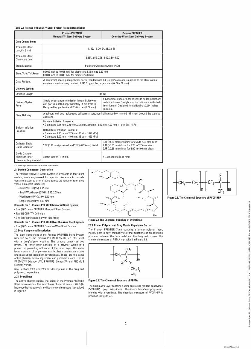
Contents for (1) Promus PrEMiEr Monorail Stent System• One (1) Promus PREMIER Monorail Stent System• Two (2) CLIPIT™ Coil clips• One (1) Flushing needle with luer fittingContents for (1) Promus PrEMiEr Over-the-wire Stent System• One (1) Promus PREMIER Over-the-Wire Stent System2.2 Drug Component DescriptionThe stent component of the Promus PREMIER Stent System (referred to as the Promus PREMIER Stent) is a PtCr stent with a drug/polymer coating. The coating comprises two layers. The inner layer consists of a polymer which is a primer for promoting adhesion of the outer layer. The outer layer consists of a polymer matrix that contains an active pharmaceutical ingredient (everolimus). These are the same active pharmaceutical ingredient and polymers as are used in PROMUS™ (Xience V™), PROMUS Element™, and PROMUS Element™ Plus.See Sections 2.2.1 and 2.2.2 for descriptions of the drug and polymers, respectively.2.2.1 EverolimusThe active pharmaceutical ingredient in the Promus PREMIER Stent is everolimus. The everolimus chemical name is 40-O-(2-hydroxyethyl)-rapamycin and its chemical structure is provided in Figure 2.1.
figure 2.1 The Chemical Structure of Everolimus
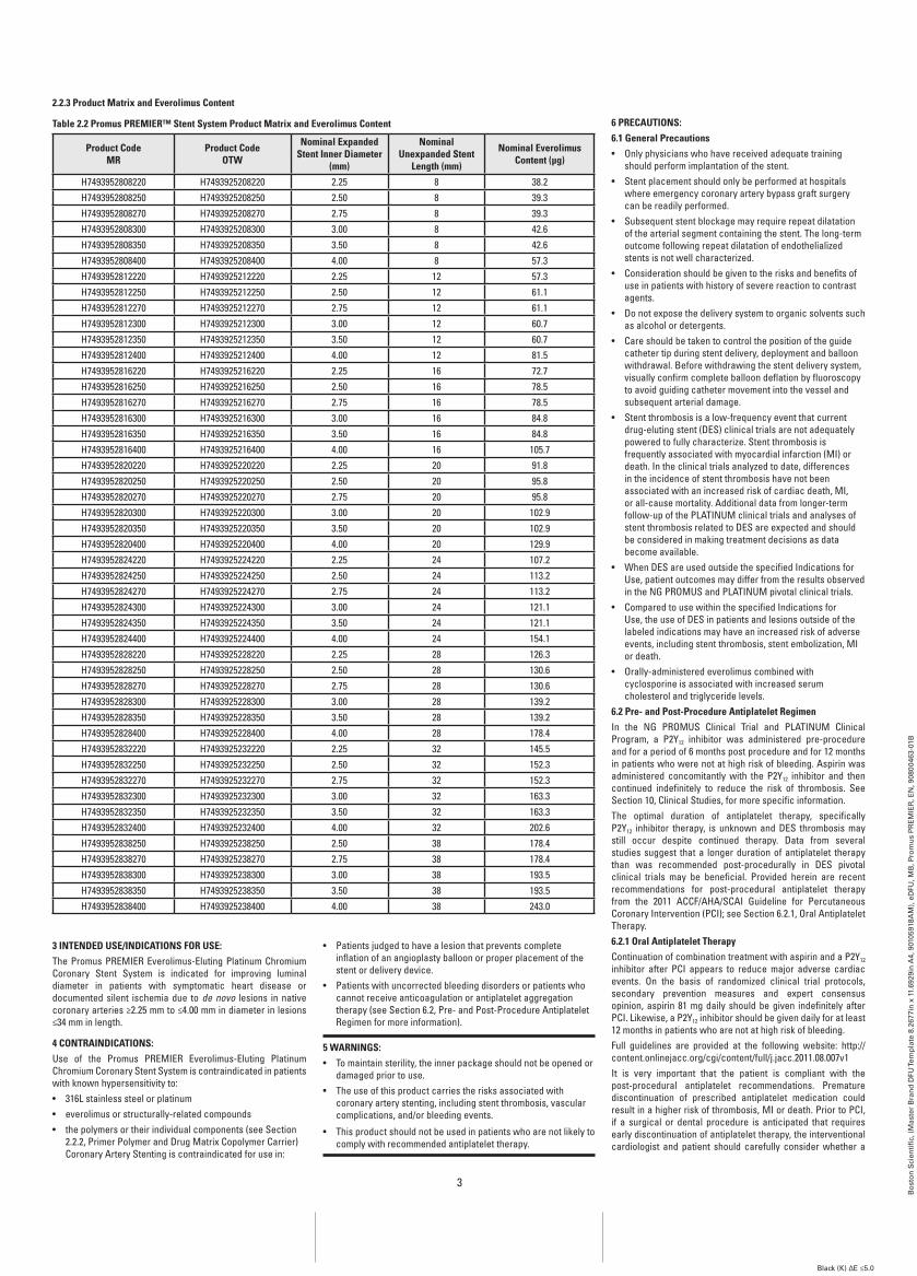
2.2.2 Primer Polymer and Drug Matrix Copolymer CarrierThe Promus PREMIER Stent contains a primer polymer layer, PBMA, poly (n-butyl methacrylate), that functions as an adhesion promoter between the bare metal and the drug matrix layer. The chemical structure of PBMA is provided in Figure 2.2.
figure 2.2. The Chemical Structure of PbMa
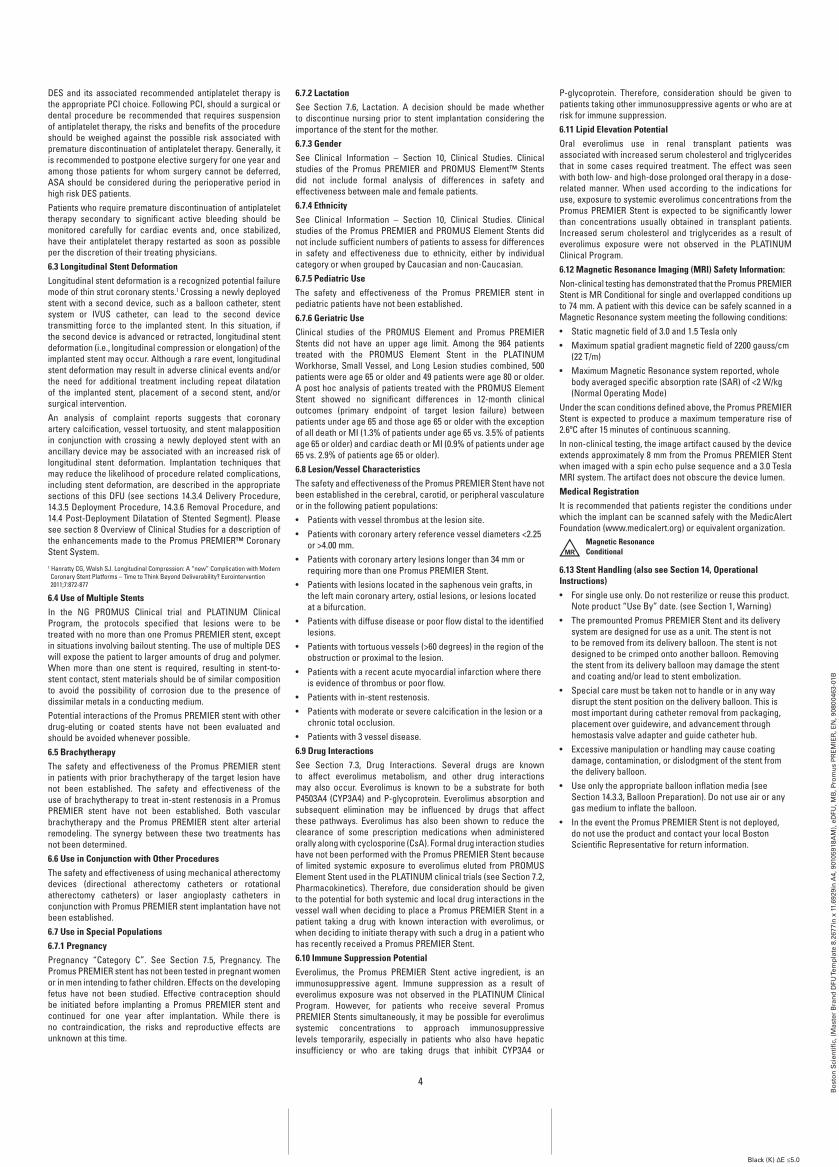
The drug matrix layer contains a semi-crystalline random copolymer, PVDF-HFP, poly (vinylidene fluoride-co-hexafluoropropylene), blended with everolimus. The chemical structure of PVDF-HFP is provided in Figure 2.3.
figure 2.3. The Chemical Structure of PVDf-HfP
3
Bo
sto
n S
cien
tifi
c, (
Mas
ter
Bra
nd
DFU
Tem
pla
te 8
.267
7in
x 1
1.69
29in
A4,
901
0591
8AM
), e
DFU
, MB
, Pro
mu
s P
RE
MIE
R, E
N, 9
0800
463-
01B
Black (K) ∆E ≤5.0
2.2.3 Product Matrix and Everolimus Content
Table 2.2 Promus PrEMiEr™ Stent System Product Matrix and Everolimus Content
Product Code Mr
Product Code OTw
nominal Expanded Stent inner Diameter
(mm)
nominal Unexpanded Stent
Length (mm)
nominal Everolimus Content (μg)
H7493952808220 H7493925208220 2.25 8 38.2H7493952808250 H7493925208250 2.50 8 39.3H7493952808270 H7493925208270 2.75 8 39.3H7493952808300 H7493925208300 3.00 8 42.6H7493952808350 H7493925208350 3.50 8 42.6H7493952808400 H7493925208400 4.00 8 57.3H7493952812220 H7493925212220 2.25 12 57.3H7493952812250 H7493925212250 2.50 12 61.1H7493952812270 H7493925212270 2.75 12 61.1H7493952812300 H7493925212300 3.00 12 60.7H7493952812350 H7493925212350 3.50 12 60.7H7493952812400 H7493925212400 4.00 12 81.5H7493952816220 H7493925216220 2.25 16 72.7H7493952816250 H7493925216250 2.50 16 78.5H7493952816270 H7493925216270 2.75 16 78.5H7493952816300 H7493925216300 3.00 16 84.8H7493952816350 H7493925216350 3.50 16 84.8H7493952816400 H7493925216400 4.00 16 105.7H7493952820220 H7493925220220 2.25 20 91.8H7493952820250 H7493925220250 2.50 20 95.8H7493952820270 H7493925220270 2.75 20 95.8H7493952820300 H7493925220300 3.00 20 102.9H7493952820350 H7493925220350 3.50 20 102.9H7493952820400 H7493925220400 4.00 20 129.9H7493952824220 H7493925224220 2.25 24 107.2H7493952824250 H7493925224250 2.50 24 113.2H7493952824270 H7493925224270 2.75 24 113.2H7493952824300 H7493925224300 3.00 24 121.1H7493952824350 H7493925224350 3.50 24 121.1H7493952824400 H7493925224400 4.00 24 154.1H7493952828220 H7493925228220 2.25 28 126.3H7493952828250 H7493925228250 2.50 28 130.6H7493952828270 H7493925228270 2.75 28 130.6H7493952828300 H7493925228300 3.00 28 139.2H7493952828350 H7493925228350 3.50 28 139.2H7493952828400 H7493925228400 4.00 28 178.4H7493952832220 H7493925232220 2.25 32 145.5H7493952832250 H7493925232250 2.50 32 152.3H7493952832270 H7493925232270 2.75 32 152.3H7493952832300 H7493925232300 3.00 32 163.3H7493952832350 H7493925232350 3.50 32 163.3H7493952832400 H7493925232400 4.00 32 202.6H7493952838250 H7493925238250 2.50 38 178.4H7493952838270 H7493925238270 2.75 38 178.4H7493952838300 H7493925238300 3.00 38 193.5H7493952838350 H7493925238350 3.50 38 193.5H7493952838400 H7493925238400 4.00 38 243.0
3 inTEnDED USE/inDiCaTiOnS fOr USE:The Promus PREMIER Everolimus-Eluting Platinum Chromium Coronary Stent System is indicated for improving luminal diameter in patients with symptomatic heart disease or documented silent ischemia due to de novo lesions in native coronary arteries ≥2.25 mm to ≤4.00 mm in diameter in lesions ≤34 mm in length.
4 COnTrainDiCaTiOnS:Use of the Promus PREMIER Everolimus-Eluting Platinum Chromium Coronary Stent System is contraindicated in patients with known hypersensitivity to:• 316L stainless steel or platinum• everolimus or structurally-related compounds• the polymers or their individual components (see Section
2.2.2, Primer Polymer and Drug Matrix Copolymer Carrier) Coronary Artery Stenting is contraindicated for use in:
• Patients judged to have a lesion that prevents complete inflation of an angioplasty balloon or proper placement of the stent or delivery device.
• Patients with uncorrected bleeding disorders or patients who cannot receive anticoagulation or antiplatelet aggregation therapy (see Section 6.2, Pre- and Post-Procedure Antiplatelet Regimen for more information).
5 warningS:• To maintain sterility, the inner package should not be opened or
damaged prior to use.• The use of this product carries the risks associated with
coronary artery stenting, including stent thrombosis, vascular complications, and/or bleeding events.
• This product should not be used in patients who are not likely to comply with recommended antiplatelet therapy.
6 PrECaUTiOnS:6.1 general Precautions• Only physicians who have received adequate training
should perform implantation of the stent.• Stent placement should only be performed at hospitals
where emergency coronary artery bypass graft surgery can be readily performed.
• Subsequent stent blockage may require repeat dilatation of the arterial segment containing the stent. The long-term outcome following repeat dilatation of endothelialized stents is not well characterized.
• Consideration should be given to the risks and benefits of use in patients with history of severe reaction to contrast agents.
• Do not expose the delivery system to organic solvents such as alcohol or detergents.
• Care should be taken to control the position of the guide catheter tip during stent delivery, deployment and balloon withdrawal. Before withdrawing the stent delivery system, visually confirm complete balloon deflation by fluoroscopy to avoid guiding catheter movement into the vessel and subsequent arterial damage.
• Stent thrombosis is a low-frequency event that current drug-eluting stent (DES) clinical trials are not adequately powered to fully characterize. Stent thrombosis is frequently associated with myocardial infarction (MI) or death. In the clinical trials analyzed to date, differences in the incidence of stent thrombosis have not been associated with an increased risk of cardiac death, MI, or all-cause mortality. Additional data from longer-term follow-up of the PLATINUM clinical trials and analyses of stent thrombosis related to DES are expected and should be considered in making treatment decisions as data become available.
• When DES are used outside the specified Indications for Use, patient outcomes may differ from the results observed in the NG PROMUS and PLATINUM pivotal clinical trials.
• Compared to use within the specified Indications for Use, the use of DES in patients and lesions outside of the labeled indications may have an increased risk of adverse events, including stent thrombosis, stent embolization, MI or death.
• Orally-administered everolimus combined with cyclosporine is associated with increased serum cholesterol and triglyceride levels.
6.2 Pre- and Post-Procedure antiplatelet regimenIn the NG PROMUS Clinical Trial and PLATINUM Clinical Program, a P2Y12 inhibitor was administered pre-procedure and for a period of 6 months post procedure and for 12 months in patients who were not at high risk of bleeding. Aspirin was administered concomitantly with the P2Y12 inhibitor and then continued indefinitely to reduce the risk of thrombosis. See Section 10, Clinical Studies, for more specific information.The optimal duration of antiplatelet therapy, specifically P2Y12 inhibitor therapy, is unknown and DES thrombosis may still occur despite continued therapy. Data from several studies suggest that a longer duration of antiplatelet therapy than was recommended post-procedurally in DES pivotal clinical trials may be beneficial. Provided herein are recent recommendations for post-procedural antiplatelet therapy from the 2011 ACCF/AHA/SCAI Guideline for Percutaneous Coronary Intervention (PCI); see Section 6.2.1, Oral Antiplatelet Therapy.6.2.1 Oral antiplatelet TherapyContinuation of combination treatment with aspirin and a P2Y12 inhibitor after PCI appears to reduce major adverse cardiac events. On the basis of randomized clinical trial protocols, secondary prevention measures and expert consensus opinion, aspirin 81 mg daily should be given indefinitely after PCI. Likewise, a P2Y12 inhibitor should be given daily for at least 12 months in patients who are not at high risk of bleeding.Full guidelines are provided at the following website: http://content.onlinejacc.org/cgi/content/full/j.jacc.2011.08.007v1It is very important that the patient is compliant with the post-procedural antiplatelet recommendations. Premature discontinuation of prescribed antiplatelet medication could result in a higher risk of thrombosis, MI or death. Prior to PCI, if a surgical or dental procedure is anticipated that requires early discontinuation of antiplatelet therapy, the interventional cardiologist and patient should carefully consider whether a
4
Bo
sto
n S
cien
tifi
c, (
Mas
ter
Bra
nd
DFU
Tem
pla
te 8
.267
7in
x 1
1.69
29in
A4,
901
0591
8AM
), e
DFU
, MB
, Pro
mu
s P
RE
MIE
R, E
N, 9
0800
463-
01B
Black (K) ∆E ≤5.0
DES and its associated recommended antiplatelet therapy is the appropriate PCI choice. Following PCI, should a surgical or dental procedure be recommended that requires suspension of antiplatelet therapy, the risks and benefits of the procedure should be weighed against the possible risk associated with premature discontinuation of antiplatelet therapy. Generally, it is recommended to postpone elective surgery for one year and among those patients for whom surgery cannot be deferred, ASA should be considered during the perioperative period in high risk DES patients.Patients who require premature discontinuation of antiplatelet therapy secondary to significant active bleeding should be monitored carefully for cardiac events and, once stabilized, have their antiplatelet therapy restarted as soon as possible per the discretion of their treating physicians.6.3 Longitudinal Stent DeformationLongitudinal stent deformation is a recognized potential failure mode of thin strut coronary stents.1 Crossing a newly deployed stent with a second device, such as a balloon catheter, stent system or IVUS catheter, can lead to the second device transmitting force to the implanted stent. In this situation, if the second device is advanced or retracted, longitudinal stent deformation (i.e., longitudinal compression or elongation) of the implanted stent may occur. Although a rare event, longitudinal stent deformation may result in adverse clinical events and/or the need for additional treatment including repeat dilatation of the implanted stent, placement of a second stent, and/or surgical intervention.An analysis of complaint reports suggests that coronary artery calcification, vessel tortuosity, and stent malapposition in conjunction with crossing a newly deployed stent with an ancillary device may be associated with an increased risk of longitudinal stent deformation. Implantation techniques that may reduce the likelihood of procedure related complications, including stent deformation, are described in the appropriate sections of this DFU (see sections 14.3.4 Delivery Procedure, 14.3.5 Deployment Procedure, 14.3.6 Removal Procedure, and 14.4 Post-Deployment Dilatation of Stented Segment). Please see section 8 Overview of Clinical Studies for a description of the enhancements made to the Promus PREMIER™ Coronary Stent System.1 Hanratty CG, Walsh SJ. Longitudinal Compression: A “new” Complication with Modern Coronary Stent Platforms – Time to Think Beyond Deliverability? Eurointervention 2011;7:872-877
6.4 Use of Multiple StentsIn the NG PROMUS Clinical trial and PLATINUM Clinical Program, the protocols specified that lesions were to be treated with no more than one Promus PREMIER stent, except in situations involving bailout stenting. The use of multiple DES will expose the patient to larger amounts of drug and polymer. When more than one stent is required, resulting in stent-to-stent contact, stent materials should be of similar composition to avoid the possibility of corrosion due to the presence of dissimilar metals in a conducting medium.Potential interactions of the Promus PREMIER stent with other drug-eluting or coated stents have not been evaluated and should be avoided whenever possible.6.5 brachytherapyThe safety and effectiveness of the Promus PREMIER stent in patients with prior brachytherapy of the target lesion have not been established. The safety and effectiveness of the use of brachytherapy to treat in-stent restenosis in a Promus PREMIER stent have not been established. Both vascular brachytherapy and the Promus PREMIER stent alter arterial remodeling. The synergy between these two treatments has not been determined.6.6 Use in Conjunction with Other ProceduresThe safety and effectiveness of using mechanical atherectomy devices (directional atherectomy catheters or rotational atherectomy catheters) or laser angioplasty catheters in conjunction with Promus PREMIER stent implantation have not been established.6.7 Use in Special Populations6.7.1 PregnancyPregnancy “Category C”. See Section 7.5, Pregnancy. The Promus PREMIER stent has not been tested in pregnant women or in men intending to father children. Effects on the developing fetus have not been studied. Effective contraception should be initiated before implanting a Promus PREMIER stent and continued for one year after implantation. While there is no contraindication, the risks and reproductive effects are unknown at this time.
6.7.2 LactationSee Section 7.6, Lactation. A decision should be made whether to discontinue nursing prior to stent implantation considering the importance of the stent for the mother.6.7.3 genderSee Clinical Information – Section 10, Clinical Studies. Clinical studies of the Promus PREMIER and PROMUS Element™ Stents did not include formal analysis of differences in safety and effectiveness between male and female patients.6.7.4 EthnicitySee Clinical Information – Section 10, Clinical Studies. Clinical studies of the Promus PREMIER and PROMUS Element Stents did not include sufficient numbers of patients to assess for differences in safety and effectiveness due to ethnicity, either by individual category or when grouped by Caucasian and non-Caucasian.6.7.5 Pediatric UseThe safety and effectiveness of the Promus PREMIER stent in pediatric patients have not been established.6.7.6 geriatric UseClinical studies of the PROMUS Element and Promus PREMIER Stents did not have an upper age limit. Among the 964 patients treated with the PROMUS Element Stent in the PLATINUM Workhorse, Small Vessel, and Long Lesion studies combined, 500 patients were age 65 or older and 49 patients were age 80 or older. A post hoc analysis of patients treated with the PROMUS Element Stent showed no significant differences in 12-month clinical outcomes (primary endpoint of target lesion failure) between patients under age 65 and those age 65 or older with the exception of all death or MI (1.3% of patients under age 65 vs. 3.5% of patients age 65 or older) and cardiac death or MI (0.9% of patients under age 65 vs. 2.9% of patients age 65 or older).6.8 Lesion/Vessel CharacteristicsThe safety and effectiveness of the Promus PREMIER Stent have not been established in the cerebral, carotid, or peripheral vasculature or in the following patient populations:• Patients with vessel thrombus at the lesion site.• Patients with coronary artery reference vessel diameters <2.25
or >4.00 mm.• Patients with coronary artery lesions longer than 34 mm or
requiring more than one Promus PREMIER Stent.• Patients with lesions located in the saphenous vein grafts, in
the left main coronary artery, ostial lesions, or lesions located at a bifurcation.
• Patients with diffuse disease or poor flow distal to the identified lesions.
• Patients with tortuous vessels (>60 degrees) in the region of the obstruction or proximal to the lesion.
• Patients with a recent acute myocardial infarction where there is evidence of thrombus or poor flow.
• Patients with in-stent restenosis.• Patients with moderate or severe calcification in the lesion or a
chronic total occlusion.• Patients with 3 vessel disease.6.9 Drug interactionsSee Section 7.3, Drug Interactions. Several drugs are known to affect everolimus metabolism, and other drug interactions may also occur. Everolimus is known to be a substrate for both P4503A4 (CYP3A4) and P-glycoprotein. Everolimus absorption and subsequent elimination may be influenced by drugs that affect these pathways. Everolimus has also been shown to reduce the clearance of some prescription medications when administered orally along with cyclosporine (CsA). Formal drug interaction studies have not been performed with the Promus PREMIER Stent because of limited systemic exposure to everolimus eluted from PROMUS Element Stent used in the PLATINUM clinical trials (see Section 7.2, Pharmacokinetics). Therefore, due consideration should be given to the potential for both systemic and local drug interactions in the vessel wall when deciding to place a Promus PREMIER Stent in a patient taking a drug with known interaction with everolimus, or when deciding to initiate therapy with such a drug in a patient who has recently received a Promus PREMIER Stent.6.10 immune Suppression PotentialEverolimus, the Promus PREMIER Stent active ingredient, is an immunosuppressive agent. Immune suppression as a result of everolimus exposure was not observed in the PLATINUM Clinical Program. However, for patients who receive several Promus PREMIER Stents simultaneously, it may be possible for everolimus systemic concentrations to approach immunosuppressive levels temporarily, especially in patients who also have hepatic insufficiency or who are taking drugs that inhibit CYP3A4 or
P-glycoprotein. Therefore, consideration should be given to patients taking other immunosuppressive agents or who are at risk for immune suppression.6.11 Lipid Elevation PotentialOral everolimus use in renal transplant patients was associated with increased serum cholesterol and triglycerides that in some cases required treatment. The effect was seen with both low- and high-dose prolonged oral therapy in a dose-related manner. When used according to the indications for use, exposure to systemic everolimus concentrations from the Promus PREMIER Stent is expected to be significantly lower than concentrations usually obtained in transplant patients. Increased serum cholesterol and triglycerides as a result of everolimus exposure were not observed in the PLATINUM Clinical Program.6.12 Magnetic resonance imaging (Mri) Safety information:Non-clinical testing has demonstrated that the Promus PREMIER Stent is MR Conditional for single and overlapped conditions up to 74 mm. A patient with this device can be safely scanned in a Magnetic Resonance system meeting the following conditions:• Static magnetic field of 3.0 and 1.5 Tesla only • Maximum spatial gradient magnetic field of 2200 gauss/cm
(22 T/m)• Maximum Magnetic Resonance system reported, whole
body averaged specific absorption rate (SAR) of <2 W/kg (Normal Operating Mode)
Under the scan conditions defined above, the Promus PREMIER Stent is expected to produce a maximum temperature rise of 2.6ºC after 15 minutes of continuous scanning.In non-clinical testing, the image artifact caused by the device extends approximately 8 mm from the Promus PREMIER Stent when imaged with a spin echo pulse sequence and a 3.0 Tesla MRI system. The artifact does not obscure the device lumen.Medical registrationIt is recommended that patients register the conditions under which the implant can be scanned safely with the MedicAlert Foundation (www.medicalert.org) or equivalent organization.
Magnetic ResonanceConditionalMR
6.13 Stent Handling (also see Section 14, Operational instructions)• For single use only. Do not resterilize or reuse this product.
Note product “Use By” date. (see Section 1, Warning)• The premounted Promus PREMIER Stent and its delivery
system are designed for use as a unit. The stent is not to be removed from its delivery balloon. The stent is not designed to be crimped onto another balloon. Removing the stent from its delivery balloon may damage the stent and coating and/or lead to stent embolization.
• Special care must be taken not to handle or in any way disrupt the stent position on the delivery balloon. This is most important during catheter removal from packaging, placement over guidewire, and advancement through hemostasis valve adapter and guide catheter hub.
• Excessive manipulation or handling may cause coating damage, contamination, or dislodgment of the stent from the delivery balloon.
• Use only the appropriate balloon inflation media (see Section 14.3.3, Balloon Preparation). Do not use air or any gas medium to inflate the balloon.
• In the event the Promus PREMIER Stent is not deployed, do not use the product and contact your local Boston Scientific Representative for return information.
5
Bo
sto
n S
cien
tifi
c, (
Mas
ter
Bra
nd
DFU
Tem
pla
te 8
.267
7in
x 1
1.69
29in
A4,
901
0591
8AM
), e
DFU
, MB
, Pro
mu
s P
RE
MIE
R, E
N, 9
0800
463-
01B
Black (K) ∆E ≤5.0
6.14 Stent PlacementPreparation• Do not prepare or pre-inflate balloon prior to stent
deployment other than as directed. Use the balloon purging technique described in Section 14.3.3, Balloon Preparation.
• If unusual resistance is felt at any time during lesion access before stent implantation, the stent delivery system and the guide catheter should be removed as a single unit (see Section 6.15, Stent Delivery System Removal).
• An unexpanded stent should be introduced into the coronary arteries one time only. An unexpanded stent should not be subsequently moved in and out through the distal end of the guide catheter as stent or coating damage or stent dislodgment from the balloon may occur.
Placement• The vessel should be pre-dilated with an appropriate
sized balloon. Failure to do so may increase the risk of placement difficulty and procedural complications.
• Do not expand the stent if it is not properly positioned in the vessel (see Section 6.15, Stent Delivery System Removal).
• Balloon pressures should be monitored during inflation. Do not exceed rated burst pressure as indicated on product label (see Section 14.5, In Vitro Information, Table 14.1, Typical Promus PREMIER™ Stent System Compliance). Use of pressures higher than specified on product label may result in a ruptured balloon and intimal damage and dissection. The stent inner diameter should approximate 1.1 times the reference diameter of the vessel.
• Placement of the stent has the potential to compromise side branch patency (see Section 14.4, Post-Deployment Dilatation of Stented Segments).
• Implanting a stent may lead to dissection of the vessel distal and/or proximal to the stented portion, and may cause acute closure of the vessel requiring additional intervention (e.g., CABG, further dilation, placement of additional stents, or other).
• When treating multiple lesions, the distal lesion should generally be stented first, followed by stenting of the more proximal lesion(s). Stenting in this order alleviates the need to cross the proximal stent in placement of the distal stent and reduces the chances of dislodging the proximal stent.
6.15 Stent Delivery System removal• If unusual resistance is felt at any time during lesion
access before stent implantation, the stent delivery system and the guide catheter should be removed as a single unit.
• Do not attempt to pull an unexpanded stent back into the guide catheter, as stent or coating damage or stent dislodgment from the balloon may occur.
• Stent retrieval methods (use of additional wires, snares and/ or forceps) may result in additional trauma to the vascular site. Complications can include bleeding, hematoma, or pseudoaneurysm.
When removing the entire stent delivery system and guide catheter as a single unit, the following steps should be executed under direct visualization using fluoroscopy:• Following stent placement, confirm complete balloon
deflation (See Table 6.1, Delivery System Deflation Time Specifications). If greater than usual resistance is felt during delivery system withdrawal, pay particular attention to guide catheter position. In some cases it may be necessary to pull back slightly on the guide catheter in order to prevent deep seating (unplanned advancement) of the guide catheter and subsequent vessel damage. In cases where unplanned guide catheter movement has occurred, angiographic assessment of the coronary tree should be undertaken to ensure that there is no damage to the coronary vasculature.
• Maintain guidewire placement across the lesion during the entire removal process.
• Carefully pull back the stent delivery system until the proximal balloon marker of the stent delivery system is just distal to the guide catheter distal tip.
• The stent delivery system and the guide catheter should be pulled back until the tip of the guide catheter is just distal to the arterial sheath, allowing the guide catheter to straighten. Carefully retract the stent delivery system into the guide catheter and remove the stent delivery system and the guide catheter from the patient as a single unit while leaving the guidewire across the lesion.
Failure to follow these steps, and/or applying excessive force to the stent delivery system, can potentially result in stent or coating damage, stent dislodgment from the balloon, and/or damage to the delivery system.
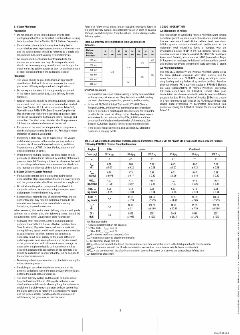
Table 6.1. Delivery System Deflation Time Specifications (Seconds)
balloon Length/Diameter
8mm
12mm
16mm
20mm
24mm
28mm
32mm
38mm
2.25 mm
≤16
≤16
≤16N/A
2.50 mm
≤30
2.75 mm
≤213.00 mm
3.50 mm≤21
4.00 mm
6.16 Post-Procedure• Care must be exercised when crossing a newly deployed stent
with any wire, catheter or ancillary device to avoid disrupting the stent placement, apposition, geometry, and/or coating.
• In the NG PROMUS Clinical Trial and PLATINUM Clinical Program, a P2Y12 inhibitor was administered pre-procedure and for a period of 6 months post-procedure and for 12 months in patients who were not at high risk of bleeding. Aspirin was administered concomitantly with a P2Y12 inhibitor and then continued indefinitely to reduce the risk of thrombosis. See Section 10, Clinical Studies, for more specific information.
• If the patient requires imaging, see Section 6.12, Magnetic Resonance Imaging (MRI).
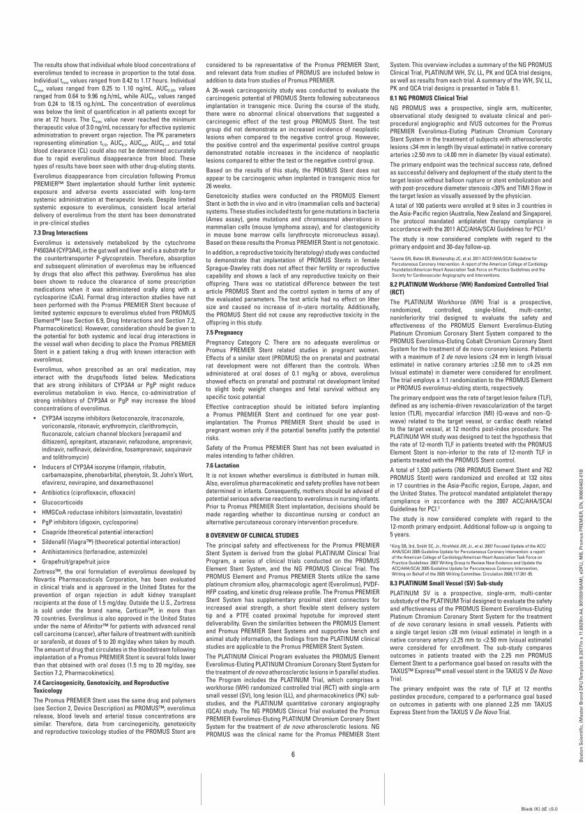
7 DrUg infOrMaTiOn7.1 Mechanism of actionThe mechanism by which the Promus PREMIER Stent inhibits neointimal growth as seen in pre-clinical and clinical studies has not been established. At the cellular level, everolimus inhibits growth factor-stimulated cell proliferation. At the molecular level, everolimus forms a complex with the cytoplasmic protein FKBP-12 (FK 506 Binding Protein). This complex binds to and interferes with FRAP (FKBP-12 Rapamycin Associated Protein), also known as mTOR (mammalian Target Of Rapamycin), leading to inhibition of cell metabolism, growth and proliferation by arresting the cell cycle at the late G1 stage.7.2 PharmacokineticsThe PROMUS Element™ and Promus PREMIER Stents utilize the same platinum chromium alloy stent material and the same Everolimus and PVDF-HFP coating, resulting in similar drug loading and equivalent drug elution profiles, therefore pharmacokinetic (PK) data from studies of PROMUS Element are also representative of Promus PREMIER. Everolimus PK when eluted from the PROMUS Element Stent post-implantation have been evaluated in patients from two different geographies (the United States of America [USA] and Japan) in a non-randomized sub-study of the PLATINUM clinical trial. Whole blood everolimus PK parameters determined from patients receiving the PROMUS Element Stent are provided in Table 7.1.
Table 7.1 whole blood Everolimus Pharmacokinetic Parameters (Mean ± SD) for PLaTinUM groups with Three or More Patients following PrOMUS Element Stent implantation.
region USa Japan Combined
Dose (µg) 102.4 µg 102.4 µg 138.6 µg 95.4 µg 102.4 µg 138.6 µg
n 3 4 b 3 b 4 c 7 b 3 b
tmax
(h)0.66
± 0.270.60
± 0.220.52
± 0.090.47
± 0.030.62
± 0.230.52
± 0.09
Cmax
(ng/mL)0.58
± 0.0780.73
± 0.170.91
± 0.200.71
± 0.090.67
± 0.150.91
± 0.20
aUC0-t
(ng.h/mL)4.77
± 1.707.71
± 6.9710.87± 7.36
7.27± 4.97
6.45± 5.26
10.87± 7.36
aUC0-24h
(ng.h/mL)5.76
± 0.856.42
± 1.309.51
± 0.646.83
± 2.036.14
± 1.109.51
± 0.64
aUC0-∞a
(ng.h/mL) NA 11.91± 1.39
60.74± 25.95
19.26± 11.69
12.95± 2.05
60.74± 25.95
t1/2 a
(h) NA 18.77± 2.11
136.06± 62.08
34.19± 20.81
22.83± 7.20
136.06± 62.08
CL a
(L/h) NA 8656± 1005
2511± 1073
6445± 3924
8044± 1276
2511± 1073
NA: Not assessablea Accurate determination not possibleb n=2 for AUC0-∞, t1/2term and CLc n=3 for AUC0-∞, t1/2term and CLtmax (h)= time to maximum concentrationCmax= maximum observed blood concentrationt1/2 (h)= terminal phase half-lifeAUC0-t = the area beneath the blood concentration versus time curve: time zero to the final quantifiable concentrationAUC0-24h = the area beneath the blood concentration versus time curve: time zero to 24 hours post-implantAUC0-∞ = the area beneath the blood concentration versus time curve: time zero to the extrapolated infinite timeCL= total blood clearance
6
Bo
sto
n S
cien
tifi
c, (
Mas
ter
Bra
nd
DFU
Tem
pla
te 8
.267
7in
x 1
1.69
29in
A4,
901
0591
8AM
), e
DFU
, MB
, Pro
mu
s P
RE
MIE
R, E
N, 9
0800
463-
01B
Black (K) ∆E ≤5.0
The results show that individual whole blood concentrations of everolimus tended to increase in proportion to the total dose. Individual tmax values ranged from 0.42 to 1.17 hours. Individual Cmax values ranged from 0.25 to 1.10 ng/mL. AUC0-24h values ranged from 0.64 to 9.96 ng.h/mL, while AUC0-t values ranged from 0.24 to 18.15 ng.h/mL. The concentration of everolimus was below the limit of quantification in all patients except for one at 72 hours. The Cmax value never reached the minimum therapeutic value of 3.0 ng/mL necessary for effective systemic administration to prevent organ rejection. The PK parameters representing elimination t1/2, AUC0-t, AUClast, AUC0-∞, and total blood clearance (CL) could also not be determined accurately due to rapid everolimus disappearance from blood. These types of results have been seen with other drug-eluting stents.Everolimus disappearance from circulation following Promus PREMIER™ Stent implantation should further limit systemic exposure and adverse events associated with long-term systemic administration at therapeutic levels. Despite limited systemic exposure to everolimus, consistent local arterial delivery of everolimus from the stent has been demonstrated in pre-clinical studies7.3 Drug interactionsEverolimus is extensively metabolized by the cytochrome P4503A4 (CYP3A4), in the gut wall and liver and is a substrate for the countertransporter P-glycoprotein. Therefore, absorption and subsequent elimination of everolimus may be influenced by drugs that also affect this pathway. Everolimus has also been shown to reduce the clearance of some prescription medications when it was administered orally along with a cyclosporine (CsA). Formal drug interaction studies have not been performed with the Promus PREMIER Stent because of limited systemic exposure to everolimus eluted from PROMUS Element™ (see Section 6.9, Drug Interactions and Section 7.2, Pharmacokinetics). However, consideration should be given to the potential for both systemic and local drug interactions in the vessel wall when deciding to place the Promus PREMIER Stent in a patient taking a drug with known interaction with everolimus.Everolimus, when prescribed as an oral medication, may interact with the drugs/foods listed below. Medications that are strong inhibitors of CYP3A4 or PgP might reduce everolimus metabolism in vivo. Hence, co-administration of strong inhibitors of CYP3A4 or PgP may increase the blood concentrations of everolimus.• CYP3A4 isozyme inhibitors (ketoconazole, itraconazole,
voriconazole, ritonavir, erythromycin, clarithromycin, fluconazole, calcium channel blockers [verapamil and diltiazem], aprepitant, atazanavir, nefazodone, amprenavir, indinavir, nelfinavir, delavirdine, fosamprenavir, saquinavir and telithromycin)
• Inducers of CYP3A4 isozyme (rifampin, rifabutin, carbamazepine, phenobarbital, phenytoin, St. John’s Wort, efavirenz, nevirapine, and dexamethasone)
• Antibiotics (ciprofloxacin, ofloxacin)• Glucocorticoids• HMGCoA reductase inhibitors (simvastatin, lovastatin)• PgP inhibitors (digoxin, cyclosporine)• Cisapride (theoretical potential interaction)• Sildenafil (Viagra™) (theoretical potential interaction)• Antihistaminics (terfenadine, astemizole)• Grapefruit/grapefruit juiceZortress™, the oral formulation of everolimus developed by Novartis Pharmaceuticals Corporation, has been evaluated in clinical trials and is approved in the United States for the prevention of organ rejection in adult kidney transplant recipients at the dose of 1.5 mg/day. Outside the U.S., Zortress is sold under the brand name, Certican™, in more than 70 countries. Everolimus is also approved in the United States under the name of Afinitor™ for patients with advanced renal cell carcinoma (cancer), after failure of treatment with sunitinib or sorafenib, at doses of 5 to 20 mg/day when taken by mouth. The amount of drug that circulates in the bloodstream following implantation of a Promus PREMIER Stent is several folds lower than that obtained with oral doses (1.5 mg to 20 mg/day, see Section 7.2, Pharmacokinetics).7.4 Carcinogenicity, genotoxicity, and reproductive ToxicologyThe Promus PREMIER Stent uses the same drug and polymers (see Section 2, Device Description) as PROMUS™; everolimus release, blood levels and arterial tissue concentrations are similar. Therefore, data from carcinogenicity, genotoxicity and reproductive toxicology studies of the PROMUS Stent are
considered to be representative of the Promus PREMIER Stent, and relevant data from studies of PROMUS are included below in addition to data from studies of Promus PREMIER.A 26-week carcinogenicity study was conducted to evaluate the carcinogenic potential of PROMUS Stents following subcutaneous implantation in transgenic mice. During the course of the study, there were no abnormal clinical observations that suggested a carcinogenic effect of the test group PROMUS Stent. The test group did not demonstrate an increased incidence of neoplastic lesions when compared to the negative control group. However, the positive control and the experimental positive control groups demonstrated notable increases in the incidence of neoplastic lesions compared to either the test or the negative control group.Based on the results of this study, the PROMUS Stent does not appear to be carcinogenic when implanted in transgenic mice for 26 weeks.Genotoxicity studies were conducted on the PROMUS Element Stent in both the in vivo and in vitro (mammalian cells and bacteria) systems. These studies included tests for gene mutations in bacteria (Ames assay), gene mutations and chromosomal aberrations in mammalian cells (mouse lymphoma assay), and for clastogenicity in mouse bone marrow cells (erythrocyte micronucleus assay). Based on these results the Promus PREMIER Stent is not genotoxic.In addition, a reproductive toxicity (teratology) study was conducted to demonstrate that implantation of PROMUS Stents in female Sprague-Dawley rats does not affect their fertility or reproductive capability and shows a lack of any reproductive toxicity on their offspring. There was no statistical difference between the test article PROMUS Stent and the control system in terms of any of the evaluated parameters. The test article had no effect on litter size and caused no increase of in-utero mortality. Additionally, the PROMUS Stent did not cause any reproductive toxicity in the offspring in this study.7.5 PregnancyPregnancy Category C: There are no adequate everolimus or Promus PREMIER Stent related studies in pregnant women. Effects of a similar stent (PROMUS) the on prenatal and postnatal rat development were not different than the controls. When administered at oral doses of 0.1 mg/kg or above, everolimus showed effects on prenatal and postnatal rat development limited to slight body weight changes and fetal survival without any specific toxic potentialEffective contraception should be initiated before implanting a Promus PREMIER Stent and continued for one year post-implantation. The Promus PREMIER Stent should be used in pregnant women only if the potential benefits justify the potential risks.Safety of the Promus PREMIER Stent has not been evaluated in males intending to father children.7.6 LactationIt is not known whether everolimus is distributed in human milk. Also, everolimus pharmacokinetic and safety profiles have not been determined in infants. Consequently, mothers should be advised of potential serious adverse reactions to everolimus in nursing infants. Prior to Promus PREMIER Stent implantation, decisions should be made regarding whether to discontinue nursing or conduct an alternative percutaneous coronary intervention procedure.
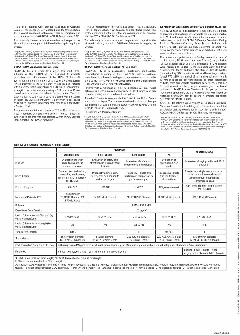
8 OVErViEw Of CLiniCaL STUDiESThe principal safety and effectiveness for the Promus PREMIER Stent System is derived from the global PLATINUM Clinical Trial Program, a series of clinical trials conducted on the PROMUS Element Stent System, and the NG PROMUS Clinical Trial. The PROMUS Element and Promus PREMIER Stents utilize the same platinum chromium alloy, pharmacologic agent (Everolimus), PVDF-HFP coating, and kinetic drug release profile. The Promus PREMIER Stent System has supplementary proximal stent connectors for increased axial strength, a short flexible stent delivery system tip and a PTFE coated proximal hypotube for improved stent deliverability. Given the similarities between the PROMUS Element and Promus PREMIER Stent Systems and supportive bench and animal study information, the findings from the PLATINUM clinical studies are applicable to the Promus PREMIER Stent System.The PLATINUM Clinical Program evaluates the PROMUS Element Everolimus-Eluting PLATINUM Chromium Coronary Stent System for the treatment of de novo atherosclerotic lesions in 5 parallel studies. The Program includes the PLATINUM Trial, which comprises a workhorse (WH) randomized controlled trial (RCT) with single-arm small vessel (SV), long lesion (LL), and pharmacokinetics (PK) sub-studies, and the PLATINUM quantitative coronary angiography (QCA) study. The NG PROMUS Clinical Trial evaluated the Promus PREMIER Everolimus-Eluting PLATINUM Chromium Coronary Stent System for the treatment of de novo atherosclerotic lesions. NG PROMUS was the clinical name for the Promus PREMIER Stent
System. This overview includes a summary of the NG PROMUS Clinical Trial, PLATINUM WH, SV, LL, PK and QCA trial designs, as well as results from each trial. A summary of the WH, SV, LL, PK and QCA trial designs is presented in Table 8.1.8.1 ng PrOMUS Clinical TrialNG PROMUS was a prospective, single arm, multicenter, observational study designed to evaluate clinical and peri-procedural angiographic and IVUS outcomes for the Promus PREMIER Everolimus-Eluting Platinum Chromium Coronary Stent System in the treatment of subjects with atherosclerotic lesions ≤34 mm in length (by visual estimate) in native coronary arteries ≥2.50 mm to ≤4.00 mm in diameter (by visual estimate).The primary endpoint was the technical success rate, defined as successful delivery and deployment of the study stent to the target lesion without balloon rupture or stent embolization and with post-procedure diameter stenosis <30% and TIMI 3 flow in the target lesion as visually assessed by the physician. A total of 100 patients were enrolled at 9 sites in 3 countries in the Asia-Pacific region (Australia, New Zealand and Singapore). The protocol mandated antiplatelet therapy compliance in accordance with the 2011 ACC/AHA/SCAI Guidelines for PCI.2
The study is now considered complete with regard to the primary endpoint and 30-day follow-up.2 Levine GN, Bates ER, Blankenship JC, et al. 2011 ACCF/AHA/SCAI Guideline for Percutaneous Coronary Intervention. A report of the American College of Cardiology Foundation/American Heart Association Task Force on Practice Guidelines and the Society for Cardiovascular Angiography and Interventions.
8.2 PLaTinUM workhorse (wH) randomized Controlled Trial (rCT)The PLATINUM Workhorse (WH) Trial is a prospective, randomized, controlled, single-blind, multi-center, noninferiority trial designed to evaluate the safety and effectiveness of the PROMUS Element Everolimus-Eluting Platinum Chromium Coronary Stent System compared to the PROMUS Everolimus-Eluting Cobalt Chromium Coronary Stent System for the treatment of de novo coronary lesions. Patients with a maximum of 2 de novo lesions ≤24 mm in length (visual estimate) in native coronary arteries ≥2.50 mm to ≤4.25 mm (visual estimate) in diameter were considered for enrollment. The trial employs a 1:1 randomization to the PROMUS Element or PROMUS everolimus-eluting stents, respectively.The primary endpoint was the rate of target lesion failure (TLF), defined as any ischemia-driven revascularization of the target lesion (TLR), myocardial infarction (MI) (Q-wave and non–Q-wave) related to the target vessel, or cardiac death related to the target vessel, at 12 months post-index procedure. The PLATINUM WH study was designed to test the hypothesis that the rate of 12-month TLF in patients treated with the PROMUS Element Stent is non-inferior to the rate of 12-month TLF in patients treated with the PROMUS Stent control.A total of 1,530 patients (768 PROMUS Element Stent and 762 PROMUS Stent) were randomized and enrolled at 132 sites in 17 countries in the Asia-Pacific region, Europe, Japan, and the United States. The protocol mandated antiplatelet therapy compliance in accordance with the 2007 ACC/AHA/SCAI Guidelines for PCI.3
The study is now considered complete with regard to the 12-month primary endpoint. Additional follow-up is ongoing to 5 years.3 King SB, 3rd, Smith SC, Jr., Hirshfeld JW, Jr., et al. 2007 Focused Update of the ACC/AHA/SCAI 2005 Guideline Update for Percutaneous Coronary Intervention: a report of the American College of Cardiology/American Heart Association Task Force on Practice Guidelines: 2007 Writing Group to Review New Evidence and Update the ACC/AHA/SCAI 2005 Guideline Update for Percutaneous Coronary Intervention, Writing on Behalf of the 2005 Writing Committee. Circulation 2008;117:261-95.
8.3 PLaTinUM Small Vessel (SV) Sub-studyPLATINUM SV is a prospective, single-arm, multi-center substudy of the PLATINUM Trial designed to evaluate the safety and effectiveness of the PROMUS Element Everolimus-Eluting Platinum Chromium Coronary Stent System for the treatment of de novo coronary lesions in small vessels. Patients with a single target lesion ≤28 mm (visual estimate) in length in a native coronary artery ≥2.25 mm to <2.50 mm (visual estimate) were considered for enrollment. The sub-study compares outcomes in patients treated with the 2.25 mm PROMUS Element Stent to a performance goal based on results with the TAXUS™ Express™ small vessel stent in the TAXUS V De Novo Trial.The primary endpoint was the rate of TLF at 12 months postindex procedure, compared to a performance goal based on outcomes in patients with one planned 2.25 mm TAXUS Express Stent from the TAXUS V De Novo Trial.
7
Bo
sto
n S
cien
tifi
c, (
Mas
ter
Bra
nd
DFU
Tem
pla
te 8
.267
7in
x 1
1.69
29in
A4,
901
0591
8AM
), e
DFU
, MB
, Pro
mu
s P
RE
MIE
R, E
N, 9
0800
463-
01B
Black (K) ∆E ≤5.0
A total of 94 patients were enrolled at 23 sites in Australia, Belgium, France, Japan, New Zealand, and the United States. The protocol mandated antiplatelet therapy compliance in accordance with the 2007 ACC/AHA/SCAI Guidelines for PCI.4 The sub-study is now considered complete with regard to the 12-month primary endpoint. Additional follow-up is ongoing to 5 years.4 King SB, 3rd, Smith SC, Jr., Hirshfeld JW, Jr., et al. 2007 Focused Update of the ACC/AHA/SCAI 2005 Guideline Update for Percutaneous Coronary Intervention: a report of the American College of Cardiology/American Heart Association Task Force on Practice Guidelines: 2007 Writing Group to Review New Evidence and Update the ACC/AHA/SCAI 2005 Guideline Update for Percutaneous Coronary Intervention, Writing on Behalf of the 2005 Writing Committee. Circulation 2008;117:261-95.
8.4 PLaTinUM Long Lesion (LL) Sub-studyPLATINUM LL is a prospective, single-arm, multi-center substudy of the PLATINUM Trial designed to evaluate the safety and effectiveness of the PROMUS Element™ Everolimus-Eluting Platinum Chromium Coronary Stent System for the treatment of de novo coronary long lesions. Patients with a single target lesion >24 mm and ≤34 mm (visual estimate) in length in a native coronary artery ≥2.50 mm to ≤4.25 mm (visual estimate) were considered for enrollment. The sub-study compares outcomes in patients treated with the 32 mm or 38 mm PROMUS Element Stent to a performance goal based on TAXUS™ Express™ long lesion stent results from the TAXUS V De Novo Trial. The primary endpoint was the rate of TLF at 12 months post-index procedure, compared to a performance goal based on outcomes in patients with one planned 32 mm TAXUS Express Stent from the TAXUS V De Novo Trial.
A total of 102 patients were enrolled at 30 sites in Australia, Belgium, France, Japan, Latvia, New Zealand, and the United States. The protocol mandated antiplatelet therapy compliance in accordance with the 2007 ACC/AHA/SCAI Guidelines for PCI.5 The sub-study is now considered complete with regard to the 12-month primary endpoint. Additional follow-up is ongoing to 5 years.5 King SB, 3rd, Smith SC, Jr., Hirshfeld JW, Jr., et al. 2007 Focused Update of the ACC/AHA/ SCAI 2005 Guideline Update for Percutaneous Coronary Intervention: a report of the American College of Cardiology/American Heart Association Task Force on Practice Guidelines: 2007 Writing Group to Review New Evidence and Update the ACC/AHA/SCAI 2005 Guideline Update for Percutaneous Coronary Intervention, Writing on Behalf of the 2005 Writing Committee. Circulation 2008;117:261-95.
8.5 PLaTinUM Pharmacokinetics (PK) Sub-studyPLATINUM PK is a prospective, single-arm, multi-center, observational sub-study of the PLATINUM Trial to evaluate everolimus blood levels following stent implantation in patients who undergo treatment with the PROMUS Element Everolimus-Eluting Platinum Chromium Coronary Stent System.Patients with a maximum of 2 de novo lesions ≤24 mm (visual estimate) in length in native coronary arteries ≥2.50 mm to ≤4.25 mm (visual estimate) were considered for enrollment. A total of 22 patients were enrolled at 2 sites in the United States and 3 sites in Japan. The protocol mandated antiplatelet therapy compliance in accordance with the 2007 ACC/AHA/SCAI Guidelines for PCI.6 Follow-up is ongoing to 5 years. See Section 7.2, Pharmacokinetics.6 King SB, 3rd, Smith SC, Jr., Hirshfeld JW, Jr., et al. 2007 Focused Update of the ACC/ AHA/SCAI 2005 Guideline Update for Percutaneous Coronary Intervention: a report of the American College of Cardiology/American Heart Association Task Force on Practice Guidelines: 2007 Writing Group to Review New Evidence and Update the ACC/AHA/SCAI 2005 Guideline Update for Percutaneous Coronary Intervention, Writing on Behalf of the 2005 Writing Committee. Circulation 2008;117:261-95.
8.6 PLaTinUM Quantitative Coronary angiography (QCa) TrialPLATINUM QCA is a prospective, single-arm, multi-center, observational study designed to evaluate clinical, angiographic and IVUS outcomes in de novo atherosclerotic coronary lesions treated with the PROMUS Element Everolimus-Eluting Platinum Chromium Coronary Stent System. Patients with a single target lesion ≤34 mm (visual estimate) in length in a native coronary artery ≥2.25 mm and ≤4.25 mm (visual estimate) were considered for enrollment.The primary endpoint was the 30-day composite rate of cardiac death, MI (Q-wave and non−Q-wave), target lesion revascularization (TLR), and stent thrombosis (ST). All patients were required to undergo 9-month angiography and IVUS assessments. Efficacy endpoints of in-stent late loss at 9 months (determined by QCA) in patients with workhorse target lesions (visual RVD ≥2.50 mm and ≤4.25 mm and visual lesion length ≤24 mm) and post-procedure incomplete apposition (determined by IVUS) were compared to predefined performance goals. For 9-month in-stent late loss, the performance goal was based on historical TAXUS Express Stent results. For post-procedure incomplete apposition, the performance goal was based on historical PROMUS™ post-procedure incomplete apposition data from the SPIRIT III study.A total of 100 patients were enrolled at 14 sites in Australia, Malaysia, New Zealand, and Singapore. The protocol mandated antiplatelet therapy compliance in accordance with the 2007 ACC/AHA/SCAI Guidelines for PCI.7 The study is complete. 7 King SB, 3rd, Smith SC, Jr., Hirshfeld JW, Jr., et al. 2007 Focused Update of the ACC/ AHA/SCAI 2005 Guideline Update for Percutaneous Coronary Intervention: a report of the American College of Cardiology/American Heart Association Task Force on Practice Guidelines: 2007 Writing Group to Review New Evidence and Update the ACC/AHA/SCAI 2005 Guideline Update for Percutaneous Coronary Intervention, Writing on Behalf of the 2005 Writing Committee. Circulation 2008;117:261-95.
Table 8.1 Comparison of PLaTinUM Clinical Studies
PLaTinUMPLaTinUM QCa
workhorse rCT Small Vessel Long Lesion PK
PurposeEvaluation of safety and effectiveness in workhorse lesions
Evaluation of safety and effectiveness in small vessel
lesions
Evaluation of safety and effectiveness in long lesions
Evaluation of everolimus blood
levels
Evaluation of angiographic and IVUS outcomes
Study Design
Prospective, randomized, controlled, multi-center,
single-blind non-inferiority to PROMUS
Prospective, single arm, multicenter, comparison to
performance goal
Prospective, single arm, multicenter, comparison to
performance goal
Prospective, single arm, multicenter,
observational
Prospective, single arm, multicenter, observational; comparisons of 2 effectiveness endpoints to
performance goals
Primary Endpoint 12M TLF 12M TLF 12M TLF N/A, observational 30D composite rate (cardiac death, MI, TLR, ST)
Number of Patients (ITT)1530 enrolled;
PROMUS Element: 768PROMUS: 762
94 PROMUS Element 102 PROMUS Element 22 PROMUS Element 100 PROMUS Element
Polymer PBMA, PVDF-HFP
Everolimus Dose Density 100 μg/cm2
Lesion Criteria: Vessel Diameter (by visual estimate), mm ≥2.50 to ≤4.25 ≥2.25 to <2.50 ≥2.50 to ≤4.25 ≥2.50 to ≤4.25 ≥2.25 to ≤4.25
Lesion Criteria: Lesion Length (by visual estimate), mm ≤24 ≤28 >24 to ≤34 ≤24 ≤34
Total Target Lesions Up to 2 1 1 Up to 2 1
Stent Matrix 2.50-4.00 mm diameter12, 18/201, 28 mm length
2.25 mm diameter12, 20, 28, 32 mm length
2.50-4.00 mm diameter 32, 38 mm length
2.50-4.00 mm diameter12, 20, 28 mm length
2.25-4.00 mm diameter12, 20, 28, 32, 382 mm length
Post-Procedure Antiplatelet Therapy A thienopyridine P2Y12 inhibitor for at least 6 months, ideally for 12 months in patients who were not at high risk of bleeding. ASA: indefinitely
Follow-Up Clinical: 30 days, 6 months, 1 year, 18 months, annually 2-5 years Clinical: 30 day, 9 month, 1 year; Angiographic: 9 month; IVUS: 9 month
1PROMUS available in 18 mm length; PROMUS Element available in 20 mm length.2 2.25 mm stent not available in 38 mm length.Abbreviations: ASA=aspirin; ITT=intent-to-treat; IVUS=intravascular ultrasound; MI=myocardial infarction; PK=pharmacokinetics; PBMA=poly (n-butyl methacrylate); PVDF-HFP=poly (vinylidene fluoride-co-hexafluoropropylene); QCA=quantitative coronary angiography; RCT=randomized controlled trial; ST=stent thrombosis; TLF=target lesion failure; TLR=target lesion revascularization
8
Bo
sto
n S
cien
tifi
c, (
Mas
ter
Bra
nd
DFU
Tem
pla
te 8
.267
7in
x 1
1.69
29in
A4,
901
0591
8AM
), e
DFU
, MB
, Pro
mu
s P
RE
MIE
R, E
N, 9
0800
463-
01B
Black (K) ∆E ≤5.0
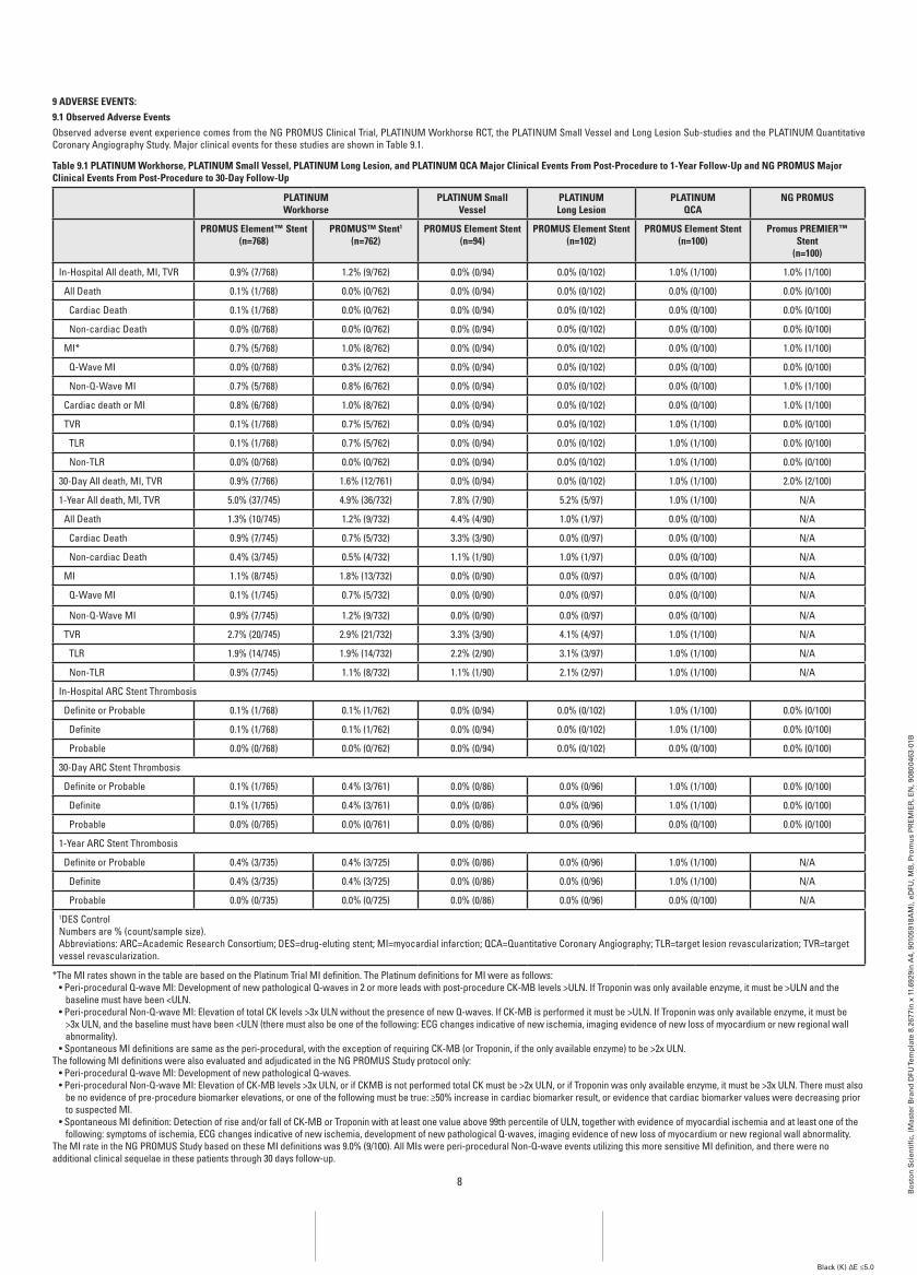
9 aDVErSE EVEnTS:9.1 Observed adverse EventsObserved adverse event experience comes from the NG PROMUS Clinical Trial, PLATINUM Workhorse RCT, the PLATINUM Small Vessel and Long Lesion Sub-studies and the PLATINUM Quantitative Coronary Angiography Study. Major clinical events for these studies are shown in Table 9.1.
Table 9.1 PLaTinUM workhorse, PLaTinUM Small Vessel, PLaTinUM Long Lesion, and PLaTinUM QCa Major Clinical Events from Post-Procedure to 1-Year follow-Up and ng PrOMUS Major Clinical Events from Post-Procedure to 30-Day follow-Up
PLaTinUM workhorse
PLaTinUM Small Vessel
PLaTinUM Long Lesion
PLaTinUMQCa
ng PrOMUS
PrOMUS Element™ Stent(n=768)
PrOMUS™ Stent1
(n=762)PrOMUS Element Stent
(n=94)PrOMUS Element Stent
(n=102)PrOMUS Element Stent
(n=100)Promus PrEMiEr™
Stent(n=100)
In-Hospital All death, MI, TVR 0.9% (7/768) 1.2% (9/762) 0.0% (0/94) 0.0% (0/102) 1.0% (1/100) 1.0% (1/100)
All Death 0.1% (1/768) 0.0% (0/762) 0.0% (0/94) 0.0% (0/102) 0.0% (0/100) 0.0% (0/100)
Cardiac Death 0.1% (1/768) 0.0% (0/762) 0.0% (0/94) 0.0% (0/102) 0.0% (0/100) 0.0% (0/100)
Non-cardiac Death 0.0% (0/768) 0.0% (0/762) 0.0% (0/94) 0.0% (0/102) 0.0% (0/100) 0.0% (0/100)
MI* 0.7% (5/768) 1.0% (8/762) 0.0% (0/94) 0.0% (0/102) 0.0% (0/100) 1.0% (1/100)
Q-Wave MI 0.0% (0/768) 0.3% (2/762) 0.0% (0/94) 0.0% (0/102) 0.0% (0/100) 0.0% (0/100)
Non-Q-Wave MI 0.7% (5/768) 0.8% (6/762) 0.0% (0/94) 0.0% (0/102) 0.0% (0/100) 1.0% (1/100)
Cardiac death or MI 0.8% (6/768) 1.0% (8/762) 0.0% (0/94) 0.0% (0/102) 0.0% (0/100) 1.0% (1/100)
TVR 0.1% (1/768) 0.7% (5/762) 0.0% (0/94) 0.0% (0/102) 1.0% (1/100) 0.0% (0/100)
TLR 0.1% (1/768) 0.7% (5/762) 0.0% (0/94) 0.0% (0/102) 1.0% (1/100) 0.0% (0/100)
Non-TLR 0.0% (0/768) 0.0% (0/762) 0.0% (0/94) 0.0% (0/102) 1.0% (1/100) 0.0% (0/100)
30-Day All death, MI, TVR 0.9% (7/766) 1.6% (12/761) 0.0% (0/94) 0.0% (0/102) 1.0% (1/100) 2.0% (2/100)
1-Year All death, MI, TVR 5.0% (37/745) 4.9% (36/732) 7.8% (7/90) 5.2% (5/97) 1.0% (1/100) N/A
All Death 1.3% (10/745) 1.2% (9/732) 4.4% (4/90) 1.0% (1/97) 0.0% (0/100) N/A
Cardiac Death 0.9% (7/745) 0.7% (5/732) 3.3% (3/90) 0.0% (0/97) 0.0% (0/100) N/A
Non-cardiac Death 0.4% (3/745) 0.5% (4/732) 1.1% (1/90) 1.0% (1/97) 0.0% (0/100) N/A
MI 1.1% (8/745) 1.8% (13/732) 0.0% (0/90) 0.0% (0/97) 0.0% (0/100) N/A
Q-Wave MI 0.1% (1/745) 0.7% (5/732) 0.0% (0/90) 0.0% (0/97) 0.0% (0/100) N/A
Non-Q-Wave MI 0.9% (7/745) 1.2% (9/732) 0.0% (0/90) 0.0% (0/97) 0.0% (0/100) N/A
TVR 2.7% (20/745) 2.9% (21/732) 3.3% (3/90) 4.1% (4/97) 1.0% (1/100) N/A
TLR 1.9% (14/745) 1.9% (14/732) 2.2% (2/90) 3.1% (3/97) 1.0% (1/100) N/A
Non-TLR 0.9% (7/745) 1.1% (8/732) 1.1% (1/90) 2.1% (2/97) 1.0% (1/100) N/A
In-Hospital ARC Stent Thrombosis
Definite or Probable 0.1% (1/768) 0.1% (1/762) 0.0% (0/94) 0.0% (0/102) 1.0% (1/100) 0.0% (0/100)
Definite 0.1% (1/768) 0.1% (1/762) 0.0% (0/94) 0.0% (0/102) 1.0% (1/100) 0.0% (0/100)
Probable 0.0% (0/768) 0.0% (0/762) 0.0% (0/94) 0.0% (0/102) 0.0% (0/100) 0.0% (0/100)
30-Day ARC Stent Thrombosis
Definite or Probable 0.1% (1/765) 0.4% (3/761) 0.0% (0/86) 0.0% (0/96) 1.0% (1/100) 0.0% (0/100)
Definite 0.1% (1/765) 0.4% (3/761) 0.0% (0/86) 0.0% (0/96) 1.0% (1/100) 0.0% (0/100)
Probable 0.0% (0/765) 0.0% (0/761) 0.0% (0/86) 0.0% (0/96) 0.0% (0/100) 0.0% (0/100)
1-Year ARC Stent Thrombosis
Definite or Probable 0.4% (3/735) 0.4% (3/725) 0.0% (0/86) 0.0% (0/96) 1.0% (1/100) N/A
Definite 0.4% (3/735) 0.4% (3/725) 0.0% (0/86) 0.0% (0/96) 1.0% (1/100) N/A
Probable 0.0% (0/735) 0.0% (0/725) 0.0% (0/86) 0.0% (0/96) 0.0% (0/100) N/A1DES ControlNumbers are % (count/sample size).Abbreviations: ARC=Academic Research Consortium; DES=drug-eluting stent; MI=myocardial infarction; QCA=Quantitative Coronary Angiography; TLR=target lesion revascularization; TVR=target vessel revascularization.
*The MI rates shown in the table are based on the Platinum Trial MI definition. The Platinum definitions for MI were as follows:• Peri-procedural Q-wave MI: Development of new pathological Q-waves in 2 or more leads with post-procedure CK-MB levels >ULN. If Troponin was only available enzyme, it must be >ULN and the
baseline must have been <ULN. • Peri-procedural Non-Q-wave MI: Elevation of total CK levels >3x ULN without the presence of new Q-waves. If CK-MB is performed it must be >ULN. If Troponin was only available enzyme, it must be
>3x ULN, and the baseline must have been <ULN (there must also be one of the following: ECG changes indicative of new ischemia, imaging evidence of new loss of myocardium or new regional wall abnormality).
• Spontaneous MI definitions are same as the peri-procedural, with the exception of requiring CK-MB (or Troponin, if the only available enzyme) to be >2x ULN.The following MI definitions were also evaluated and adjudicated in the NG PROMUS Study protocol only:
• Peri-procedural Q-wave MI: Development of new pathological Q-waves. • Peri-procedural Non-Q-wave MI: Elevation of CK-MB levels >3x ULN, or if CKMB is not performed total CK must be >2x ULN, or if Troponin was only available enzyme, it must be >3x ULN. There must also
be no evidence of pre-procedure biomarker elevations, or one of the following must be true: ≥50% increase in cardiac biomarker result, or evidence that cardiac biomarker values were decreasing prior to suspected MI.
• Spontaneous MI definition: Detection of rise and/or fall of CK-MB or Troponin with at least one value above 99th percentile of ULN, together with evidence of myocardial ischemia and at least one of the following: symptoms of ischemia, ECG changes indicative of new ischemia, development of new pathological Q-waves, imaging evidence of new loss of myocardium or new regional wall abnormality.
The MI rate in the NG PROMUS Study based on these MI definitions was 9.0% (9/100). All MIs were peri-procedural Non-Q-wave events utilizing this more sensitive MI definition, and there were no additional clinical sequelae in these patients through 30 days follow-up.
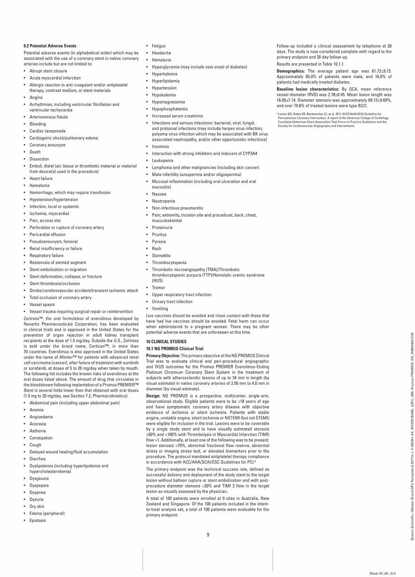
9
Bo
sto
n S
cien
tifi
c, (
Mas
ter
Bra
nd
DFU
Tem
pla
te 8
.267
7in
x 1
1.69
29in
A4,
901
0591
8AM
), e
DFU
, MB
, Pro
mu
s P
RE
MIE
R, E
N, 9
0800
463-
01B
Black (K) ∆E ≤5.0
9.2 Potential adverse EventsPotential adverse events (in alphabetical order) which may be associated with the use of a coronary stent in native coronary arteries include but are not limited to:• Abrupt stent closure• Acute myocardial infarction• Allergic reaction to anti-coagulant and/or antiplatelet
therapy, contrast medium, or stent materials• Angina• Arrhythmias, including ventricular fibrillation and
ventricular tachycardia• Arteriovenous fistula• Bleeding• Cardiac tamponade• Cardiogenic shock/pulmonary edema• Coronary aneurysm• Death• Dissection• Emboli, distal (air, tissue or thrombotic material or material
from device(s) used in the procedure)• Heart failure• Hematoma• Hemorrhage, which may require transfusion• Hypotension/hypertension• Infection, local or systemic• Ischemia, myocardial• Pain, access site• Perforation or rupture of coronary artery• Pericardial effusion• Pseudoaneurysm, femoral• Renal insufficiency or failure• Respiratory failure• Restenosis of stented segment• Stent embolization or migration• Stent deformation, collapse, or fracture• Stent thrombosis/occlusion• Stroke/cerebrovascular accident/transient ischemic attack• Total occlusion of coronary artery• Vessel spasm• Vessel trauma requiring surgical repair or reinterventionZortress™, the oral formulation of everolimus developed by Novartis Pharmaceuticals Corporation, has been evaluated in clinical trials and is approved in the United States for the prevention of organ rejection in adult kidney transplant recipients at the dose of 1.5 mg/day. Outside the U.S., Zortress is sold under the brand name, Certican™, in more than 70 countries. Everolimus is also approved in the United States under the name of Afinitor™ for patients with advanced renal cell carcinoma (cancer), after failure of treatment with sunitinib or sorafenib, at doses of 5 to 20 mg/day when taken by mouth. The following list includes the known risks of everolimus at the oral doses listed above. The amount of drug that circulates in the bloodstream following implantation of a Promus PREMIER™ Stent is several folds lower than that obtained with oral doses (1.5 mg to 20 mg/day, see Section 7.2, Pharmacokinetics).• Abdominal pain (including upper abdominal pain)• Anemia• Angioedema• Anorexia• Asthenia• Constipation• Cough• Delayed wound healing/fluid accumulation• Diarrhea• Dyslipidemia (including hyperlipidemia and
hypercholesterolemia)• Dysgeusia• Dyspepsia• Dyspnea• Dysuria• Dry skin• Edema (peripheral)• Epistaxis
• Fatigue• Headache• Hematuria• Hyperglycemia (may include new onset of diabetes)• Hyperkalemia• Hyperlipidemia• Hypertension• Hypokalemia• Hypomagnesemia• Hypophosphatemia• Increased serum creatinine• Infections and serious infections: bacterial, viral, fungal,
and protozoal infections (may include herpes virus infection, polyoma virus infection which may be associated with BK virus associated nephropathy, and/or other opportunistic infections)
• Insomnia• Interaction with strong inhibitors and inducers of CYP3A4• Leukopenia• Lymphoma and other malignancies (including skin cancer)• Male infertility (azospermia and/or oligospermia)• Mucosal inflammation (including oral ulceration and oral
mucositis)• Nausea• Neutropenia• Non-infectious pneumonitis• Pain; extremity, incision site and procedural, back, chest,
musculoskeletal• Proteinuria• Pruritus• Pyrexia• Rash• Stomatitis• Thrombocytopenia• Thrombotic microangiopathy (TMA)/Thrombotic
thrombocytopenic purpura (TTP)/Hemolytic uremic syndrome (HUS)
• Tremor• Upper respiratory tract infection• Urinary tract infection• VomitingLive vaccines should be avoided and close contact with those that have had live vaccines should be avoided. Fetal harm can occur when administered to a pregnant woman. There may be other potential adverse events that are unforeseen at this time.
10 CLiniCaL STUDiES10.1 ng PrOMUS Clinical TrialPrimary Objective: The primary objective of the NG PROMUS Clinical Trial was to evaluate clinical and peri-procedural angiographic and IVUS outcomes for the Promus PREMIER Everolimus-Eluting Platinum Chromium Coronary Stent System in the treatment of subjects with atherosclerotic lesions of up to 34 mm in length (by visual estimate) in native coronary arteries of 2.50 mm to 4.0 mm in diameter (by visual estimate). Design: NG PROMUS is a prospective, multicenter, single-arm, observational study. Eligible patients were to be ≥18 years of age and have symptomatic coronary artery disease with objective evidence of ischemia or silent ischemia. Patients with stable angina, unstable angina, silent ischemia or NSTEMI (but not STEMI) were eligible for inclusion in the trial. Lesions were to be coverable by a single study stent and to have visually estimated stenosis ≥50% and <100% with Thrombolysis in Myocardial Infarction (TIMI) flow >1. Additionally, at least one of the following was to be present: lesion stenosis ≥70%, abnormal fractional flow reserve, abnormal stress or imaging stress test, or elevated biomarkers prior to the procedure. The protocol mandated antiplatelet therapy compliance in accordance with ACC/AHA/SCAI/ESC Guidelines for PCI.8
The primary endpoint was the technical success rate, defined as successful delivery and deployment of the study stent to the target lesion without balloon rupture or stent embolization and with post-procedure diameter stenosis <30% and TIMI 3 flow in the target lesion as visually assessed by the physician. A total of 100 patients were enrolled at 9 sites in Australia, New Zealand and Singapore. Of the 100 patients included in the intent-to-treat analysis set, a total of 100 patients were evaluable for the primary endpoint.
Follow-up included a clinical assessment by telephone at 30 days. The study is now considered complete with regard to the primary endpoint and 30 day follow-up.Results are presented in Table 10.1.1.Demographics: The average patient age was 61.72±9.73. Approximately 85.0% of patients were male, and 16.0% of patients had medically treated diabetes.baseline lesion characteristics: By QCA, mean reference vessel diameter (RVD) was 2.78±0.45. Mean lesion length was 16.05±7.14. Diameter stenosis was approximately 69.12±9.69%, and over 79.8% of treated lesions were type B2/C.8 Levine GN, Bates ER, Blankenship JC, et al. 2011 ACCF/AHA/SCAI Guideline for Percutaneous Coronary Intervention. A report of the American College of Cardiology Foundation/American Heart Association Task Force on Practice Guidelines and the Society for Cardiovascular Angiography and Interventions.
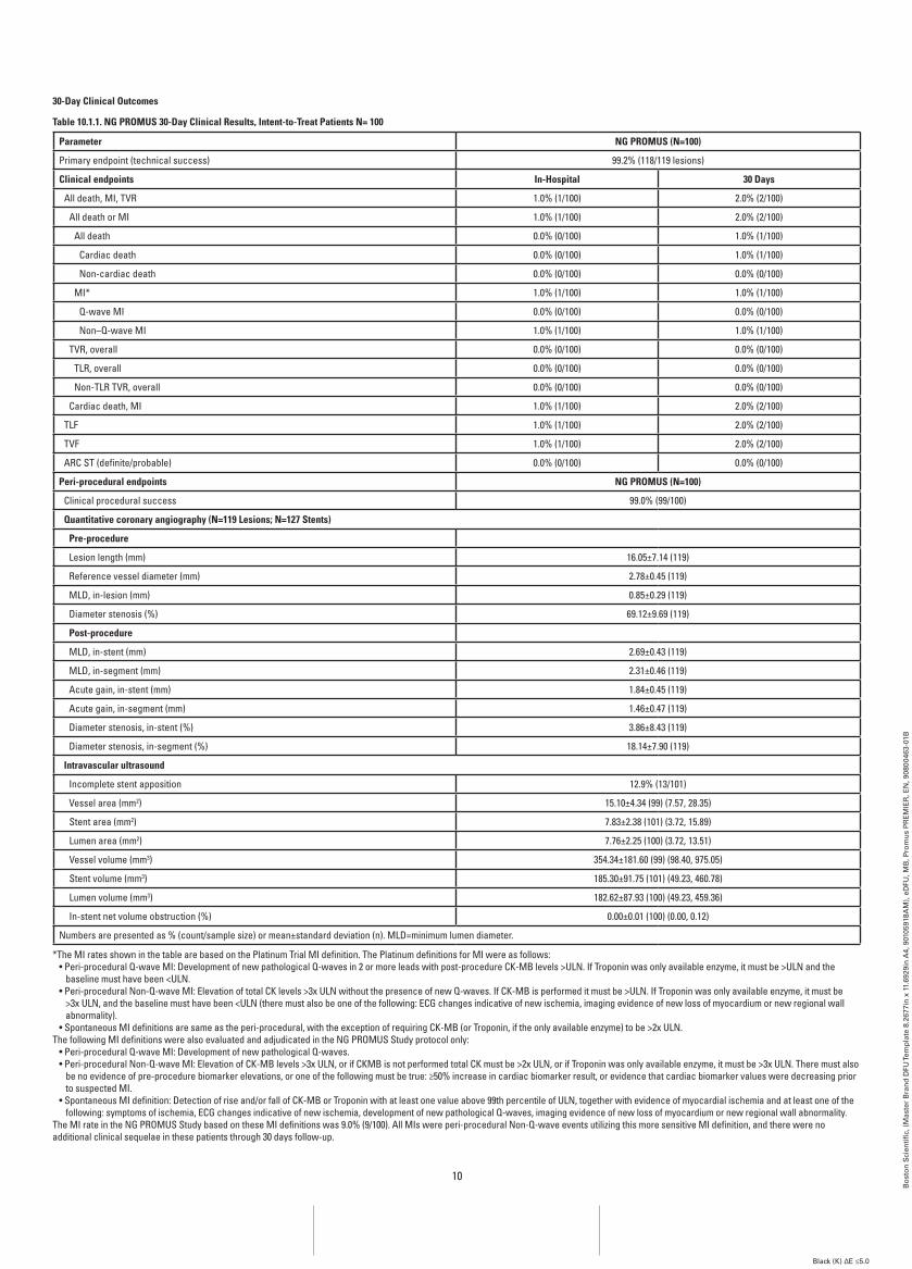
10
Bo
sto
n S
cien
tifi
c, (
Mas
ter
Bra
nd
DFU
Tem
pla
te 8
.267
7in
x 1
1.69
29in
A4,
901
0591
8AM
), e
DFU
, MB
, Pro
mu
s P
RE
MIE
R, E
N, 9
0800
463-
01B
Black (K) ∆E ≤5.0
30-Day Clinical Outcomes
Table 10.1.1. ng PrOMUS 30-Day Clinical results, intent-to-Treat Patients n= 100
Parameter ng PrOMUS (n=100)
Primary endpoint (technical success) 99.2% (118/119 lesions)
Clinical endpoints in-Hospital 30 Days
All death, MI, TVR 1.0% (1/100) 2.0% (2/100)
All death or MI 1.0% (1/100) 2.0% (2/100)
All death 0.0% (0/100) 1.0% (1/100)
Cardiac death 0.0% (0/100) 1.0% (1/100)
Non-cardiac death 0.0% (0/100) 0.0% (0/100)
MI* 1.0% (1/100) 1.0% (1/100)
Q-wave MI 0.0% (0/100) 0.0% (0/100)
Non–Q-wave MI 1.0% (1/100) 1.0% (1/100)
TVR, overall 0.0% (0/100) 0.0% (0/100)
TLR, overall 0.0% (0/100) 0.0% (0/100)
Non-TLR TVR, overall 0.0% (0/100) 0.0% (0/100)
Cardiac death, MI 1.0% (1/100) 2.0% (2/100)
TLF 1.0% (1/100) 2.0% (2/100)
TVF 1.0% (1/100) 2.0% (2/100)
ARC ST (definite/probable) 0.0% (0/100) 0.0% (0/100)
Peri-procedural endpoints ng PrOMUS (n=100)
Clinical procedural success 99.0% (99/100)
Quantitative coronary angiography (n=119 Lesions; n=127 Stents)
Pre-procedure
Lesion length (mm) 16.05±7.14 (119)
Reference vessel diameter (mm) 2.78±0.45 (119)
MLD, in-lesion (mm) 0.85±0.29 (119)
Diameter stenosis (%) 69.12±9.69 (119)
Post-procedure
MLD, in-stent (mm) 2.69±0.43 (119)
MLD, in-segment (mm) 2.31±0.46 (119)
Acute gain, in-stent (mm) 1.84±0.45 (119)
Acute gain, in-segment (mm) 1.46±0.47 (119)
Diameter stenosis, in-stent (%) 3.86±8.43 (119)
Diameter stenosis, in-segment (%) 18.14±7.90 (119)
intravascular ultrasound
Incomplete stent apposition 12.9% (13/101)
Vessel area (mm2) 15.10±4.34 (99) (7.57, 28.35)
Stent area (mm2) 7.83±2.38 (101) (3.72, 15.89)
Lumen area (mm2) 7.76±2.25 (100) (3.72, 13.51)
Vessel volume (mm3) 354.34±181.60 (99) (98.40, 975.05)
Stent volume (mm3) 185.30±91.75 (101) (49.23, 460.78)
Lumen volume (mm3) 182.62±87.93 (100) (49.23, 459.36)
In-stent net volume obstruction (%) 0.00±0.01 (100) (0.00, 0.12)
Numbers are presented as % (count/sample size) or mean±standard deviation (n). MLD=minimum lumen diameter.
*The MI rates shown in the table are based on the Platinum Trial MI definition. The Platinum definitions for MI were as follows:• Peri-procedural Q-wave MI: Development of new pathological Q-waves in 2 or more leads with post-procedure CK-MB levels >ULN. If Troponin was only available enzyme, it must be >ULN and the
baseline must have been <ULN. • Peri-procedural Non-Q-wave MI: Elevation of total CK levels >3x ULN without the presence of new Q-waves. If CK-MB is performed it must be >ULN. If Troponin was only available enzyme, it must be
>3x ULN, and the baseline must have been <ULN (there must also be one of the following: ECG changes indicative of new ischemia, imaging evidence of new loss of myocardium or new regional wall abnormality).
• Spontaneous MI definitions are same as the peri-procedural, with the exception of requiring CK-MB (or Troponin, if the only available enzyme) to be >2x ULN.The following MI definitions were also evaluated and adjudicated in the NG PROMUS Study protocol only:
• Peri-procedural Q-wave MI: Development of new pathological Q-waves. • Peri-procedural Non-Q-wave MI: Elevation of CK-MB levels >3x ULN, or if CKMB is not performed total CK must be >2x ULN, or if Troponin was only available enzyme, it must be >3x ULN. There must also
be no evidence of pre-procedure biomarker elevations, or one of the following must be true: ≥50% increase in cardiac biomarker result, or evidence that cardiac biomarker values were decreasing prior to suspected MI.
• Spontaneous MI definition: Detection of rise and/or fall of CK-MB or Troponin with at least one value above 99th percentile of ULN, together with evidence of myocardial ischemia and at least one of the following: symptoms of ischemia, ECG changes indicative of new ischemia, development of new pathological Q-waves, imaging evidence of new loss of myocardium or new regional wall abnormality.
The MI rate in the NG PROMUS Study based on these MI definitions was 9.0% (9/100). All MIs were peri-procedural Non-Q-wave events utilizing this more sensitive MI definition, and there were no additional clinical sequelae in these patients through 30 days follow-up.
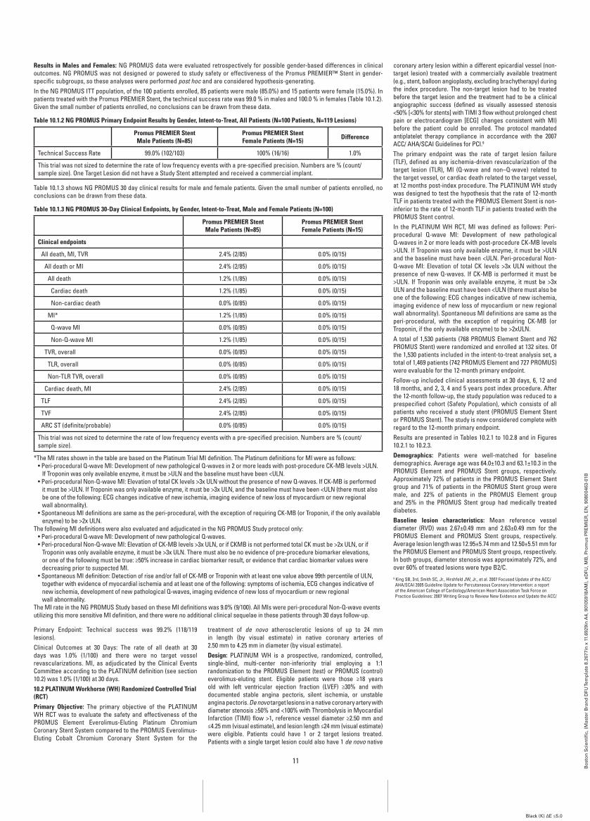
11
Bo
sto
n S
cien
tifi
c, (
Mas
ter
Bra
nd
DFU
Tem
pla
te 8
.267
7in
x 1
1.69
29in
A4,
901
0591
8AM
), e
DFU
, MB
, Pro
mu
s P
RE
MIE
R, E
N, 9
0800
463-
01B
Black (K) ∆E ≤5.0
results in Males and females: NG PROMUS data were evaluated retrospectively for possible gender-based differences in clinical outcomes. NG PROMUS was not designed or powered to study safety or effectiveness of the Promus PREMIER™ Stent in gender-specific subgroups, so these analyses were performed post hoc and are considered hypothesis-generating.In the NG PROMUS ITT population, of the 100 patients enrolled, 85 patients were male (85.0%) and 15 patients were female (15.0%). In patients treated with the Promus PREMIER Stent, the technical success rate was 99.0 % in males and 100.0 % in females (Table 10.1.2). Given the small number of patients enrolled, no conclusions can be drawn from these data.
Table 10.1.2 ng PrOMUS Primary Endpoint results by gender, intent-to-Treat, all Patients (n=100 Patients, n=119 Lesions)
Promus PrEMiEr Stent Male Patients (n=85)
Promus PrEMiEr Stent female Patients (n=15) Difference
Technical Success Rate 99.0% (102/103) 100% (16/16) 1.0%
This trial was not sized to determine the rate of low frequency events with a pre-specified precision. Numbers are % (count/sample size). One Target Lesion did not have a Study Stent attempted and received a commercial implant.
Table 10.1.3 shows NG PROMUS 30 day clinical results for male and female patients. Given the small number of patients enrolled, no conclusions can be drawn from these data.
Table 10.1.3 ng PrOMUS 30-Day Clinical Endpoints, by gender, intent-to-Treat, Male and female Patients (n=100)
Promus PrEMiEr Stent Male Patients (n=85)
Promus PrEMiEr Stent female Patients (n=15)
Clinical endpoints
All death, MI, TVR 2.4% (2/85) 0.0% (0/15)
All death or MI 2.4% (2/85) 0.0% (0/15)
All death 1.2% (1/85) 0.0% (0/15)
Cardiac death 1.2% (1/85) 0.0% (0/15)
Non-cardiac death 0.0% (0/85) 0.0% (0/15)
MI* 1.2% (1/85) 0.0% (0/15)
Q-wave MI 0.0% (0/85) 0.0% (0/15)
Non-Q-wave MI 1.2% (1/85) 0.0% (0/15)
TVR, overall 0.0% (0/85) 0.0% (0/15)
TLR, overall 0.0% (0/85) 0.0% (0/15)
Non-TLR TVR, overall 0.0% (0/85) 0.0% (0/15)
Cardiac death, MI 2.4% (2/85) 0.0% (0/15)
TLF 2.4% (2/85) 0.0% (0/15)
TVF 2.4% (2/85) 0.0% (0/15)
ARC ST (definite/probable) 0.0% (0/85) 0.0% (0/15)
This trial was not sized to determine the rate of low frequency events with a pre-specified precision. Numbers are % (count/sample size).
*The MI rates shown in the table are based on the Platinum Trial MI definition. The Platinum definitions for MI were as follows:• Peri-procedural Q-wave MI: Development of new pathological Q-waves in 2 or more leads with post-procedure CK-MB levels >ULN.
If Troponin was only available enzyme, it must be >ULN and the baseline must have been <ULN. • Peri-procedural Non-Q-wave MI: Elevation of total CK levels >3x ULN without the presence of new Q-waves. If CK-MB is performed
it must be >ULN. If Troponin was only available enzyme, it must be >3x ULN, and the baseline must have been <ULN (there must also be one of the following: ECG changes indicative of new ischemia, imaging evidence of new loss of myocardium or new regional wall abnormality).
• Spontaneous MI definitions are same as the peri-procedural, with the exception of requiring CK-MB (or Troponin, if the only available enzyme) to be >2x ULN.
The following MI definitions were also evaluated and adjudicated in the NG PROMUS Study protocol only:• Peri-procedural Q-wave MI: Development of new pathological Q-waves. • Peri-procedural Non-Q-wave MI: Elevation of CK-MB levels >3x ULN, or if CKMB is not performed total CK must be >2x ULN, or if
Troponin was only available enzyme, it must be >3x ULN. There must also be no evidence of pre-procedure biomarker elevations, or one of the following must be true: ≥50% increase in cardiac biomarker result, or evidence that cardiac biomarker values were decreasing prior to suspected MI.
• Spontaneous MI definition: Detection of rise and/or fall of CK-MB or Troponin with at least one value above 99th percentile of ULN, together with evidence of myocardial ischemia and at least one of the following: symptoms of ischemia, ECG changes indicative of new ischemia, development of new pathological Q-waves, imaging evidence of new loss of myocardium or new regional wall abnormality.
The MI rate in the NG PROMUS Study based on these MI definitions was 9.0% (9/100). All MIs were peri-procedural Non-Q-wave events utilizing this more sensitive MI definition, and there were no additional clinical sequelae in these patients through 30 days follow-up.
Primary Endpoint: Technical success was 99.2% (118/119 lesions).Clinical Outcomes at 30 Days: The rate of all death at 30 days was 1.0% (1/100) and there were no target vessel revascularizations. MI, as adjudicated by the Clinical Events Committee according to the PLATINUM definition (see section 10.2) was 1.0% (1/100) at 30 days. 10.2 PLaTinUM workhorse (wH) randomized Controlled Trial (rCT)Primary Objective: The primary objective of the PLATINUM WH RCT was to evaluate the safety and effectiveness of the PROMUS Element Everolimus-Eluting Platinum Chromium Coronary Stent System compared to the PROMUS Everolimus-Eluting Cobalt Chromium Coronary Stent System for the
treatment of de novo atherosclerotic lesions of up to 24 mm in length (by visual estimate) in native coronary arteries of 2.50 mm to 4.25 mm in diameter (by visual estimate).Design: PLATINUM WH is a prospective, randomized, controlled, single-blind, multi-center non-inferiority trial employing a 1:1 randomization to the PROMUS Element (test) or PROMUS (control) everolimus-eluting stent. Eligible patients were those ≥18 years old with left ventricular ejection fraction (LVEF) ≥30% and with documented stable angina pectoris, silent ischemia, or unstable angina pectoris. De novo target lesions in a native coronary artery with diameter stenosis ≥50% and <100% with Thrombolysis in Myocardial Infarction (TIMI) flow >1, reference vessel diameter ≥2.50 mm and ≤4.25 mm (visual estimate), and lesion length ≤24 mm (visual estimate) were eligible. Patients could have 1 or 2 target lesions treated. Patients with a single target lesion could also have 1 de novo native
coronary artery lesion within a different epicardial vessel (non-target lesion) treated with a commercially available treatment (e.g., stent, balloon angioplasty, excluding brachytherapy) during the index procedure. The non-target lesion had to be treated before the target lesion and the treatment had to be a clinical angiographic success (defined as visually assessed stenosis <50% [<30% for stents] with TIMI 3 flow without prolonged chest pain or electrocardiogram [ECG] changes consistent with MI) before the patient could be enrolled. The protocol mandated antiplatelet therapy compliance in accordance with the 2007 ACC/ AHA/SCAI Guidelines for PCI.9 The primary endpoint was the rate of target lesion failure (TLF), defined as any ischemia-driven revascularization of the target lesion (TLR), MI (Q-wave and non–Q-wave) related to the target vessel, or cardiac death related to the target vessel, at 12 months post-index procedure. The PLATINUM WH study was designed to test the hypothesis that the rate of 12-month TLF in patients treated with the PROMUS Element Stent is non-inferior to the rate of 12-month TLF in patients treated with the PROMUS Stent control.In the PLATINUM WH RCT, MI was defined as follows: Peri-procedural Q-wave MI: Development of new pathological Q-waves in 2 or more leads with post-procedure CK-MB levels >ULN. If Troponin was only available enzyme, it must be >ULN and the baseline must have been <ULN. Peri-procedural Non-Q-wave MI: Elevation of total CK levels >3x ULN without the presence of new Q-waves. If CK-MB is performed it must be >ULN. If Troponin was only available enzyme, it must be >3x ULN and the baseline must have been <ULN (there must also be one of the following: ECG changes indicative of new ischemia, imaging evidence of new loss of myocardium or new regional wall abnormality). Spontaneous MI definitions are same as the peri-procedural, with the exception of requiring CK-MB (or Troponin, if the only available enzyme) to be >2xULN.A total of 1,530 patients (768 PROMUS Element Stent and 762 PROMUS Stent) were randomized and enrolled at 132 sites. Of the 1,530 patients included in the intent-to-treat analysis set, a total of 1,469 patients (742 PROMUS Element and 727 PROMUS) were evaluable for the 12-month primary endpoint. Follow-up included clinical assessments at 30 days, 6, 12 and 18 months, and 2, 3, 4 and 5 years post index procedure. After the 12-month follow-up, the study population was reduced to a prespecified cohort (Safety Population), which consists of all patients who received a study stent (PROMUS Element Stent or PROMUS Stent). The study is now considered complete with regard to the 12-month primary endpoint.Results are presented in Tables 10.2.1 to 10.2.8 and in Figures 10.2.1 to 10.2.3.Demographics: Patients were well-matched for baseline demographics. Average age was 64.0±10.3 and 63.1±10.3 in the PROMUS Element and PROMUS Stent groups, respectively. Approximately 72% of patients in the PROMUS Element Stent group and 71% of patients in the PROMUS Stent group were male, and 22% of patients in the PROMUS Element group and 25% in the PROMUS Stent group had medically treated diabetes.baseline lesion characteristics: Mean reference vessel diameter (RVD) was 2.67±0.49 mm and 2.63±0.49 mm for the PROMUS Element and PROMUS Stent groups, respectively. Average lesion length was 12.95±5.74 mm and 12.50±5.51 mm for the PROMUS Element and PROMUS Stent groups, respectively. In both groups, diameter stenosis was approximately 72%, and over 60% of treated lesions were type B2/C.9 King SB, 3rd, Smith SC, Jr., Hirshfeld JW, Jr., et al. 2007 Focused Update of the ACC/ AHA/SCAI 2005 Guideline Update for Percutaneous Coronary Intervention: a report of the American College of Cardiology/American Heart Association Task Force on Practice Guidelines: 2007 Writing Group to Review New Evidence and Update the ACC/
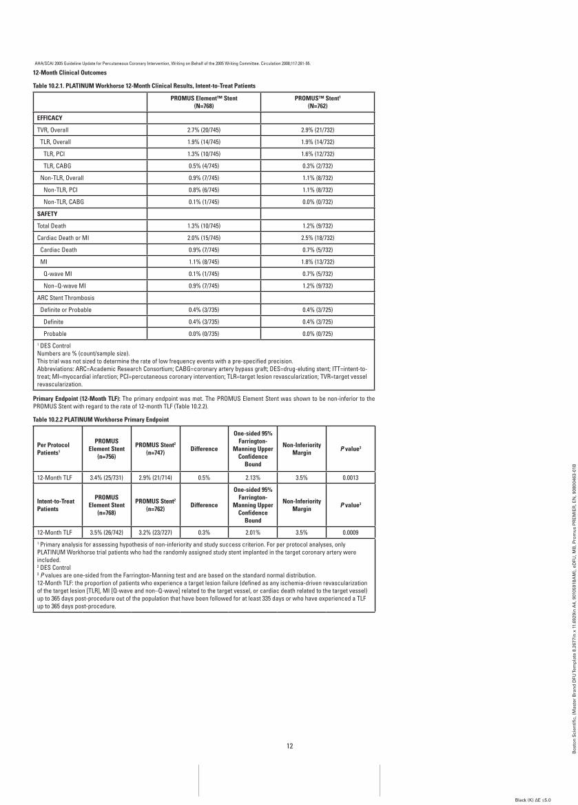
12
Bo
sto
n S
cien
tifi
c, (
Mas
ter
Bra
nd
DFU
Tem
pla
te 8
.267
7in
x 1
1.69
29in
A4,
901
0591
8AM
), e
DFU
, MB
, Pro
mu
s P
RE
MIE
R, E
N, 9
0800
463-
01B
Black (K) ∆E ≤5.0
AHA/SCAI 2005 Guideline Update for Percutaneous Coronary Intervention, Writing on Behalf of the 2005 Writing Committee. Circulation 2008;117:261-95.
12-Month Clinical Outcomes
Table 10.2.1. PLaTinUM workhorse 12-Month Clinical results, intent-to-Treat Patients
PrOMUS Element™ Stent(n=768)
PrOMUS™ Stent1
(n=762)
EffiCaCY
TVR, Overall 2.7% (20/745) 2.9% (21/732)
TLR, Overall 1.9% (14/745) 1.9% (14/732)
TLR, PCI 1.3% (10/745) 1.6% (12/732)
TLR, CABG 0.5% (4/745) 0.3% (2/732)
Non-TLR, Overall 0.9% (7/745) 1.1% (8/732)
Non-TLR, PCI 0.8% (6/745) 1.1% (8/732)
Non-TLR, CABG 0.1% (1/745) 0.0% (0/732)
SafETY
Total Death 1.3% (10/745) 1.2% (9/732)
Cardiac Death or MI 2.0% (15/745) 2.5% (18/732)
Cardiac Death 0.9% (7/745) 0.7% (5/732)
MI 1.1% (8/745) 1.8% (13/732)
Q-wave MI 0.1% (1/745) 0.7% (5/732)
Non−Q-wave MI 0.9% (7/745) 1.2% (9/732)
ARC Stent Thrombosis
Definite or Probable 0.4% (3/735) 0.4% (3/725)
Definite 0.4% (3/735) 0.4% (3/725)
Probable 0.0% (0/735) 0.0% (0/725)1 DES ControlNumbers are % (count/sample size).This trial was not sized to determine the rate of low frequency events with a pre-specified precision.Abbreviations: ARC=Academic Research Consortium; CABG=coronary artery bypass graft; DES=drug-eluting stent; ITT=intent-to-treat; MI=myocardial infarction; PCI=percutaneous coronary intervention; TLR=target lesion revascularization; TVR=target vessel revascularization.
Primary Endpoint (12-Month TLf): The primary endpoint was met. The PROMUS Element Stent was shown to be non-inferior to the PROMUS Stent with regard to the rate of 12-month TLF (Table 10.2.2).
Table 10.2.2 PLaTinUM workhorse Primary Endpoint
Per Protocol Patients1
PrOMUS Element Stent
(n=756)
PrOMUS Stent2
(n=747) Difference
One-sided 95% farrington-
Manning Upper Confidence
bound
non-inferiority Margin P value3
12-Month TLF 3.4% (25/731) 2.9% (21/714) 0.5% 2.13% 3.5% 0.0013
intent-to-Treat Patients
PrOMUS Element Stent
(n=768)
PrOMUS Stent2
(n=762) Difference
One-sided 95% farrington-
Manning Upper Confidence
bound
non-inferiority Margin P value3
12-Month TLF 3.5% (26/742) 3.2% (23/727) 0.3% 2.01% 3.5% 0.00091 Primary analysis for assessing hypothesis of non-inferiority and study success criterion. For per protocol analyses, only PLATINUM Workhorse trial patients who had the randomly assigned study stent implanted in the target coronary artery were included.2 DES Control3 P values are one-sided from the Farrington-Manning test and are based on the standard normal distribution.12-Month TLF: the proportion of patients who experience a target lesion failure (defined as any ischemia-driven revascularization of the target lesion [TLR], MI [Q-wave and non–Q-wave] related to the target vessel, or cardiac death related to the target vessel) up to 365 days post-procedure out of the population that have been followed for at least 335 days or who have experienced a TLF up to 365 days post-procedure.
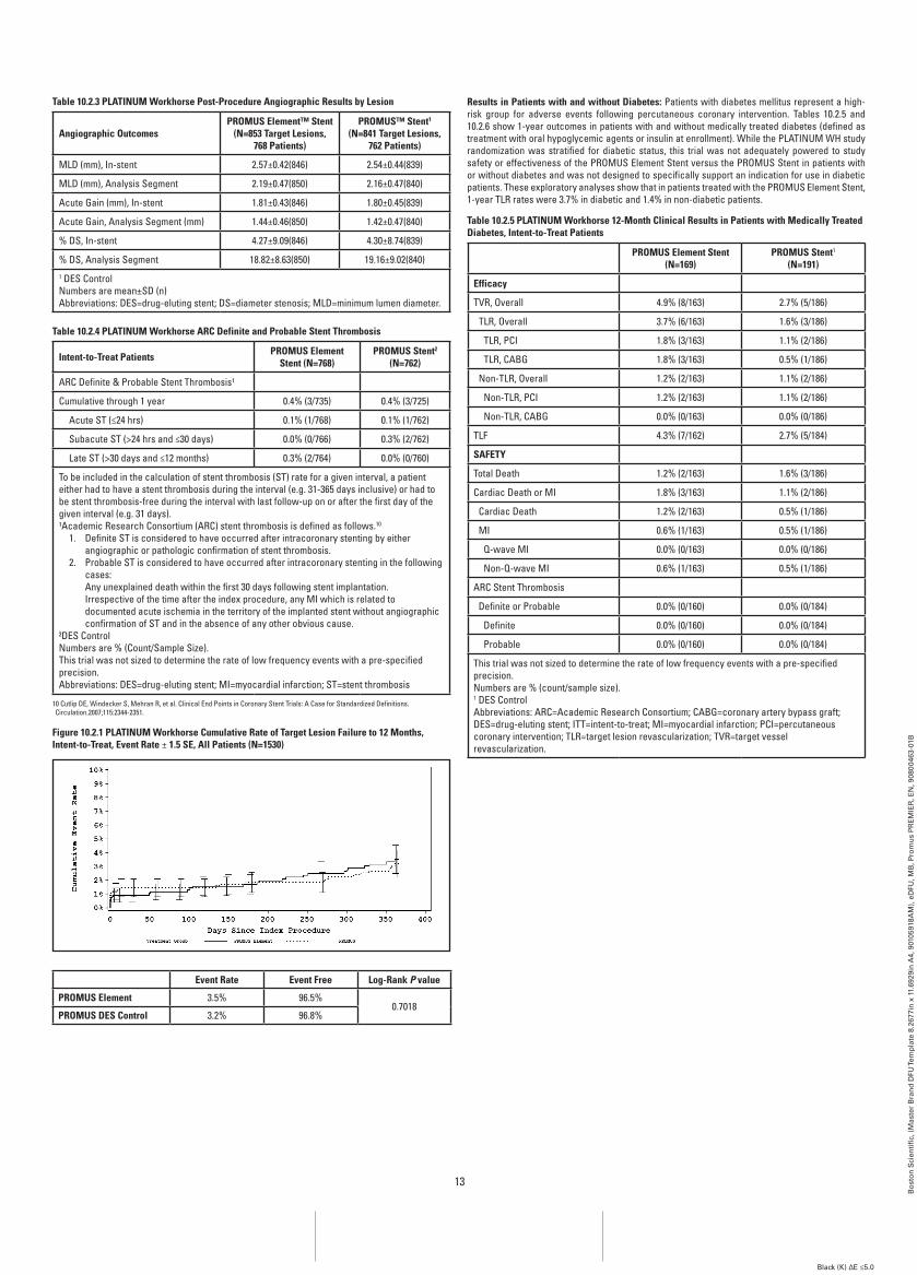
13
Bo
sto
n S
cien
tifi
c, (
Mas
ter
Bra
nd
DFU
Tem
pla
te 8
.267
7in
x 1
1.69
29in
A4,
901
0591
8AM
), e
DFU
, MB
, Pro
mu
s P
RE
MIE
R, E
N, 9
0800
463-
01B
Black (K) ∆E ≤5.0
Table 10.2.3 PLaTinUM workhorse Post-Procedure angiographic results by Lesion
angiographic OutcomesPrOMUS Element™ Stent
(n=853 Target Lesions,768 Patients)
PrOMUS™ Stent1
(n=841 Target Lesions,762 Patients)
MLD (mm), In-stent 2.57±0.42(846) 2.54±0.44(839)
MLD (mm), Analysis Segment 2.19±0.47(850) 2.16±0.47(840)
Acute Gain (mm), In-stent 1.81±0.43(846) 1.80±0.45(839)
Acute Gain, Analysis Segment (mm) 1.44±0.46(850) 1.42±0.47(840)
% DS, In-stent 4.27±9.09(846) 4.30±8.74(839)
% DS, Analysis Segment 18.82±8.63(850) 19.16±9.02(840)1 DES ControlNumbers are mean±SD (n)Abbreviations: DES=drug-eluting stent; DS=diameter stenosis; MLD=minimum lumen diameter.
Table 10.2.4 PLaTinUM workhorse arC Definite and Probable Stent Thrombosis
intent-to-Treat Patients PrOMUS Element Stent (n=768)
PrOMUS Stent2 (n=762)
ARC Definite & Probable Stent Thrombosis¹
Cumulative through 1 year 0.4% (3/735) 0.4% (3/725)
Acute ST (≤24 hrs) 0.1% (1/768) 0.1% (1/762)
Subacute ST (>24 hrs and ≤30 days) 0.0% (0/766) 0.3% (2/762)
Late ST (>30 days and ≤12 months) 0.3% (2/764) 0.0% (0/760)
To be included in the calculation of stent thrombosis (ST) rate for a given interval, a patient either had to have a stent thrombosis during the interval (e.g. 31-365 days inclusive) or had to be stent thrombosis-free during the interval with last follow-up on or after the first day of the given interval (e.g. 31 days).¹Academic Research Consortium (ARC) stent thrombosis is defined as follows.10
1. Definite ST is considered to have occurred after intracoronary stenting by either angiographic or pathologic confirmation of stent thrombosis.
2. Probable ST is considered to have occurred after intracoronary stenting in the following cases:
Any unexplained death within the first 30 days following stent implantation. Irrespective of the time after the index procedure, any MI which is related to
documented acute ischemia in the territory of the implanted stent without angiographic confirmation of ST and in the absence of any other obvious cause.
²DES ControlNumbers are % (Count/Sample Size).This trial was not sized to determine the rate of low frequency events with a pre-specified precision.Abbreviations: DES=drug-eluting stent; MI=myocardial infarction; ST=stent thrombosis
10 Cutlip DE, Windecker S, Mehran R, et al. Clinical End Points in Coronary Stent Trials: A Case for Standardized Definitions. Circulation.2007;115:2344-2351.
figure 10.2.1 PLaTinUM workhorse Cumulative rate of Target Lesion failure to 12 Months, intent-to-Treat, Event rate ± 1.5 SE, all Patients (n=1530)
Event rate Event free Log-rank P value
PrOMUS Element 3.5% 96.5%0.7018
PrOMUS DES Control 3.2% 96.8%
results in Patients with and without Diabetes: Patients with diabetes mellitus represent a high-risk group for adverse events following percutaneous coronary intervention. Tables 10.2.5 and 10.2.6 show 1-year outcomes in patients with and without medically treated diabetes (defined as treatment with oral hypoglycemic agents or insulin at enrollment). While the PLATINUM WH study randomization was stratified for diabetic status, this trial was not adequately powered to study safety or effectiveness of the PROMUS Element Stent versus the PROMUS Stent in patients with or without diabetes and was not designed to specifically support an indication for use in diabetic patients. These exploratory analyses show that in patients treated with the PROMUS Element Stent, 1-year TLR rates were 3.7% in diabetic and 1.4% in non-diabetic patients.
Table 10.2.5 PLaTinUM workhorse 12-Month Clinical results in Patients with Medically Treated Diabetes, intent-to-Treat Patients
PrOMUS Element Stent(n=169)
PrOMUS Stent1
(n=191)
Efficacy
TVR, Overall 4.9% (8/163) 2.7% (5/186)
TLR, Overall 3.7% (6/163) 1.6% (3/186)
TLR, PCI 1.8% (3/163) 1.1% (2/186)
TLR, CABG 1.8% (3/163) 0.5% (1/186)
Non-TLR, Overall 1.2% (2/163) 1.1% (2/186)
Non-TLR, PCI 1.2% (2/163) 1.1% (2/186)
Non-TLR, CABG 0.0% (0/163) 0.0% (0/186)
TLF 4.3% (7/162) 2.7% (5/184)
SafETY
Total Death 1.2% (2/163) 1.6% (3/186)
Cardiac Death or MI 1.8% (3/163) 1.1% (2/186)
Cardiac Death 1.2% (2/163) 0.5% (1/186)
MI 0.6% (1/163) 0.5% (1/186)
Q-wave MI 0.0% (0/163) 0.0% (0/186)
Non-Q-wave MI 0.6% (1/163) 0.5% (1/186)
ARC Stent Thrombosis
Definite or Probable 0.0% (0/160) 0.0% (0/184)
Definite 0.0% (0/160) 0.0% (0/184)
Probable 0.0% (0/160) 0.0% (0/184)
This trial was not sized to determine the rate of low frequency events with a pre-specified precision.Numbers are % (count/sample size).1 DES ControlAbbreviations: ARC=Academic Research Consortium; CABG=coronary artery bypass graft; DES=drug-eluting stent; ITT=intent-to-treat; MI=myocardial infarction; PCI=percutaneous coronary intervention; TLR=target lesion revascularization; TVR=target vessel revascularization.
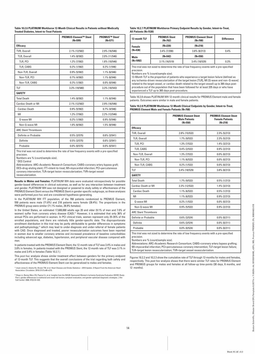
14
Bo
sto
n S
cien
tifi
c, (
Mas
ter
Bra
nd
DFU
Tem
pla
te 8
.267
7in
x 1
1.69
29in
A4,
901
0591
8AM
), e
DFU
, MB
, Pro
mu
s P
RE
MIE
R, E
N, 9
0800
463-
01B
Black (K) ∆E ≤5.0
Table 10.2.6 PLaTinUM workhorse 12-Month Clinical results in Patients without Medically Treated Diabetes, intent-to-Treat Patients
PrOMUS Element™ Stent(n=599)
PrOMUS™ Stent1
(n=571)
Efficacy
TVR, Overall 2.1% (12/582) 2.9% (16/546)
TLR, Overall 1.4% (8/582) 2.0% (11/546)
TLR, PCI 1.2% (7/582) 1.8% (10/546)
TLR, CABG 0.2% (1/582) 0.2% (1/546)
Non-TLR, Overall 0.9% (5/582) 1.1% (6/546)
Non-TLR, PCI 0.7% (4/582) 1.1% (6/546)
Non-TLR, CABG 0.2% (1/582) 0.0% (0/546)
TLF 3.3% (19/580) 3.3% (18/543)
SafETY
Total Death 1.4% (8/582) 1.1% (6/546)
Cardiac Death or MI 2.1% (12/582) 2.9% (16/546)
Cardiac Death 0.9% (5/582) 0.7% (4/546)
MI 1.2% (7/582) 2.2% (12/546)
Q-wave MI 0.2% (1/582) 0.9% (5/546)
Non-Q-wave MI 1.0% (6/582) 1.5% (8/546)
ARC Stent Thrombosis
Definite or Probable 0.5% (3/575) 0.6% (3/541)
Definite 0.5% (3/575) 0.6% (3/541)
Probable 0.0% (0/575) 0.0% (0/541)
This trial was not sized to determine the rate of low frequency events with a pre-specified precision.Numbers are % (count/sample size).1 DES ControlAbbreviations: ARC=Academic Research Consortium; CABG=coronary artery bypass graft; DES=drug-eluting stent; ITT=intent-to-treat; MI=myocardial infarction; PCI=percutaneous coronary intervention; TLR=target lesion revascularization; TVR=target vessel revascularization.
results in Males and females: PLATINUM WH data were evaluated retrospectively for possible gender-based differences in clinical outcomes, as well as for any interaction between treatment and gender. PLATINUM WH was not designed or powered to study safety or effectiveness of the PROMUS Element Stent versus the PROMUS Stent in gender-specific subgroups, so these analyses were performed post hoc and are considered hypothesis-generating.In the PLATINUM WH ITT population, of the 768 patients randomized to PROMUS Element, 550 patients were male (71.6%) and 218 patients were female (28.4%). The proportions in the PROMUS group were similar (71.1% males, 28.9% females).In the United States, an estimated 17,600,000 adults age 20 and older (9.1% of men and 7.0% of women) suffer from coronary artery disease (CAD).11 However, it is estimated that only 36% of annual PCIs are performed in women. In PCI clinical trials, women represent only 25-35% of the enrolled populations, and there are relatively little gender-specific data. The disproportionate enrollment distribution in this trial may be partly attributable to gender differences in symptoms and pathophysiology,12 which may lead to under-diagnosis and under-referral of female patients with CAD. Once diagnosed and treated, poorer revascularization outcomes have been reported in women due to smaller coronary arteries and increased prevalence of baseline comorbidities including advanced age, diabetes, hypertension, and peripheral vascular disease compared with men.In patients treated with the PROMUS Element Stent, the 12-month rate of TLF was 3.4% in males and 3.8% in females. In patients treated with the PROMUS Stent, the 12-month rate of TLF was 3.1% in males and 3.4% in females (Table 10.2.7.).This post hoc analysis shows similar treatment effect between genders for the primary endpoint of 12-month TLF. This suggests that the overall conclusions of the trial regarding both safety and effectiveness of the PROMUS Element Stent can be generalized to males and females. 11 Lloyd-Jones D, Adams RJ, Brown TM, et al. Heart Disease and Stroke Statistics—2010 Update. A Report From the American Heart Association. Circulation. 2010;121(7):e46-e215.
12 Shaw LJ, Bairey Merz CN, Pepine CJ, et al. Insights from the NHLBI-Sponsored Women’s Ischemia Syndrome Evaluation (WISE) Study: Part I: gender differences in traditional and novel risk factors, symptom evaluation, and gender-optimized diagnostic strategies. J Am Coll Cardiol. 2006; 47(3):S4-S20.
Table 10.2.7 PLaTinUM workhorse Primary Endpoint results by gender, intent-to-Treat, all Patients (n=1530)
12-month TLf PrOMUS Stent(n=762)
PrOMUS Element Stent (n=768) Difference
female (n=438)
(n=220) (n=218)
3.4% (7/208) 3.8% (8/213) 0.4%
Male(n=1092)
(n=542) (n=550)
3.1% (16/519) 3.4% (18/529) 0.3%
This trial was not sized to determine the rate of low frequency events with a pre-specified precision.Numbers are % (count/sample size).12-Month TLF is the proportion of patients who experience a target lesion failure (defined as any ischemia-driven revascularization of the target lesion [TLR], MI [Q-wave and non–Q-wave] related to the target vessel, or cardiac death related to the target vessel) up to 365 days post-procedure out of the population that have been followed for at least 335 days or who have experienced a TLF up to 365 days post-procedure.
Table 10.2.8 shows PLATINUM WH 12-month clinical results for PROMUS Element male and female patients. Outcomes were similar in male and female patients.
Table 10.2.8 PLaTinUM workhorse 12-Month Clinical Endpoints by gender, intent-to-Treat, PrOMUS Element Male and female Patients (n=768)
PrOMUS Element Stent Male Patients
(n=550)
PrOMUS Element Stent female Patients
(n=218)
Efficacy
TVR, Overall 2.8% (15/532) 2.3% (5/213)
TLR, Overall 1.7% (9/532) 2.3% (5/213)
TLR, PCI 1.3% (7/532) 1.4% (3/213)
TLR, CABG 0.4% (2/532) 0.9% (2/213)
Non-TLR, Overall 1.3% (7/532) 0.0% (0/213)
Non-TLR, PCI 1.1% (6/532) 0.0% (0/213)
Non-TLR, CABG 0.2% (1/532) 0.0% (0/213)
TLF 3.4% (18/529) 3.8% (8/213)
SafETY
Total Death 1.7% (9/532) 0.5% (1/213)
Cardiac Death or MI 2.3% (12/532) 1.4% (3/213)
Cardiac Death 1.1% (6/532) 0.5% (1/213)
MI 1.1% (6/532) 0.9% (2/213)
Q-wave MI 0.2% (1/532) 0.0% (0/213)
Non-Q-wave MI 0.9% (5/532) 0.9% (2/213)
ARC Stent Thrombosis
Definite or Probable 0.6% (3/524) 0.0% (0/211)
Definite 0.6% (3/524) 0.0% (0/211)
Probable 0.0% (0/524) 0.0% (0/211)
This trial was not sized to determine the rate of low frequency events with a pre-specified precision.Numbers are % (count/sample size).Abbreviations: ARC=Academic Research Consortium; CABG=coronary artery bypass grafting; MI=myocardial infarction; PCI=percutaneous coronary intervention; TLF=target lesion failure; TLR=target lesion revascularization; TVR=target vessel revascularization.
Figures 10.2.2 and 10.2.3 show the cumulative rate of TLF through 12-months for males and females, respectively. This post hoc analysis shows that there were similar TLF rates for PROMUS Element and PROMUS groups for males and females at all follow-up time-points (30 days, 6 months, and 12 months).
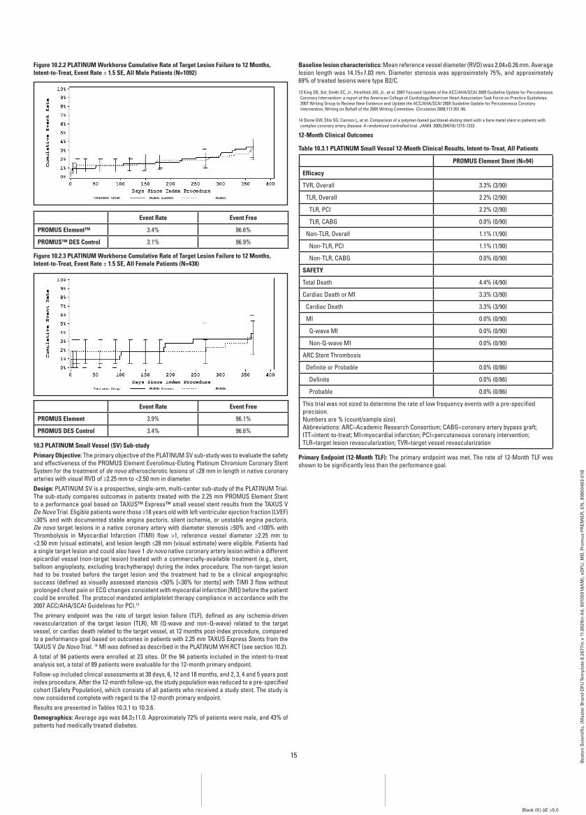
15
Bo
sto
n S
cien
tifi
c, (
Mas
ter
Bra
nd
DFU
Tem
pla
te 8
.267
7in
x 1
1.69
29in
A4,
901
0591
8AM
), e
DFU
, MB
, Pro
mu
s P
RE
MIE
R, E
N, 9
0800
463-
01B
Black (K) ∆E ≤5.0
figure 10.2.2 PLaTinUM workhorse Cumulative rate of Target Lesion failure to 12 Months, intent-to-Treat, Event rate ± 1.5 SE, all Male Patients (n=1092)
Event rate Event free
PrOMUS Element™ 3.4% 96.6%
PrOMUS™ DES Control 3.1% 96.9%
figure 10.2.3 PLaTinUM workhorse Cumulative rate of Target Lesion failure to 12 Months, intent-to-Treat, Event rate ± 1.5 SE, all female Patients (n=438)
Event rate Event free
PrOMUS Element 3.9% 96.1%
PrOMUS DES Control 3.4% 96.6%
10.3 PLaTinUM Small Vessel (SV) Sub-studyPrimary Objective: The primary objective of the PLATINUM SV sub-study was to evaluate the safety and effectiveness of the PROMUS Element Everolimus-Eluting Platinum Chromium Coronary Stent System for the treatment of de novo atherosclerotic lesions of ≤28 mm in length in native coronary arteries with visual RVD of ≥2.25 mm to <2.50 mm in diameter.Design: PLATINUM SV is a prospective, single-arm, multi-center sub-study of the PLATINUM Trial. The sub-study compares outcomes in patients treated with the 2.25 mm PROMUS Element Stent to a performance goal based on TAXUS™ Express™ small vessel stent results from the TAXUS V De Novo Trial. Eligible patients were those ≥18 years old with left ventricular ejection fraction (LVEF) ≥30% and with documented stable angina pectoris, silent ischemia, or unstable angina pectoris. De novo target lesions in a native coronary artery with diameter stenosis ≥50% and <100% with Thrombolysis in Myocardial Infarction (TIMI) flow >1, reference vessel diameter ≥2.25 mm to <2.50 mm (visual estimate), and lesion length ≤28 mm (visual estimate) were eligible. Patients had a single target lesion and could also have 1 de novo native coronary artery lesion within a different epicardial vessel (non-target lesion) treated with a commercially-available treatment (e.g., stent, balloon angioplasty, excluding brachytherapy) during the index procedure. The non-target lesion had to be treated before the target lesion and the treatment had to be a clinical angiographic success (defined as visually assessed stenosis <50% [<30% for stents] with TIMI 3 flow without prolonged chest pain or ECG changes consistent with myocardial infarction [MI]) before the patient could be enrolled. The protocol mandated antiplatelet therapy compliance in accordance with the 2007 ACC/AHA/SCAI Guidelines for PCI.13
The primary endpoint was the rate of target lesion failure (TLF), defined as any ischemia-driven revascularization of the target lesion (TLR), MI (Q-wave and non–Q-wave) related to the target vessel, or cardiac death related to the target vessel, at 12 months post-index procedure, compared to a performance goal based on outcomes in patients with 2.25 mm TAXUS Express Stents from the TAXUS V De Novo Trial. 14 MI was defined as described in the PLATINUM WH RCT (see section 10.2).A total of 94 patients were enrolled at 23 sites. Of the 94 patients included in the intent-to-treat analysis set, a total of 89 patients were evaluable for the 12-month primary endpoint.Follow-up included clinical assessments at 30 days, 6, 12 and 18 months, and 2, 3, 4 and 5 years post index procedure. After the 12-month follow-up, the study population was reduced to a pre-specified cohort (Safety Population), which consists of all patients who received a study stent. The study is now considered complete with regard to the 12-month primary endpoint.Results are presented in Tables 10.3.1 to 10.3.6.Demographics: Average age was 64.3±11.0. Approximately 72% of patients were male, and 43% of patients had medically treated diabetes.
baseline lesion characteristics: Mean reference vessel diameter (RVD) was 2.04±0.26 mm. Average lesion length was 14.15±7.03 mm. Diameter stenosis was approximately 75%, and approximately 69% of treated lesions were type B2/C.
13 King SB, 3rd, Smith SC, Jr., Hirshfeld JW, Jr., et al. 2007 Focused Update of the ACC/AHA/SCAI 2005 Guideline Update for Percutaneous Coronary Intervention: a report of the American College of Cardiology/American Heart Association Task Force on Practice Guidelines: 2007 Writing Group to Review New Evidence and Update the ACC/AHA/SCAI 2005 Guideline Update for Percutaneous Coronary Intervention, Writing on Behalf of the 2005 Writing Committee. Circulation 2008;117:261-95.
14 Stone GW, Ellis SG, Cannon L, et at. Comparison of a polymer-based paclitaxel-eluting stent with a bare metal stent in patients with complex coronary artery disease: A randomized controlled trial. JAMA. 2005;294(10):1215-1223.
12-Month Clinical Outcomes
Table 10.3.1 PLaTinUM Small Vessel 12-Month Clinical results, intent-to-Treat, all Patients
PrOMUS Element Stent (n=94)
Efficacy
TVR, Overall 3.3% (3/90)
TLR, Overall 2.2% (2/90)
TLR, PCI 2.2% (2/90)
TLR, CABG 0.0% (0/90)
Non-TLR, Overall 1.1% (1/90)
Non-TLR, PCI 1.1% (1/90)
Non-TLR, CABG 0.0% (0/90)
SafETY
Total Death 4.4% (4/90)
Cardiac Death or MI 3.3% (3/90)
Cardiac Death 3.3% (3/90)
MI 0.0% (0/90)
Q-wave MI 0.0% (0/90)
Non-Q-wave MI 0.0% (0/90)
ARC Stent Thrombosis
Definite or Probable 0.0% (0/86)
Definite 0.0% (0/86)
Probable 0.0% (0/86)
This trial was not sized to determine the rate of low frequency events with a pre-specified precision.Numbers are % (count/sample size).Abbreviations: ARC=Academic Research Consortium; CABG=coronary artery bypass graft; ITT=intent-to-treat; MI=myocardial infarction; PCI=percutaneous coronary intervention; TLR=target lesion revascularization; TVR=target vessel revascularization
Primary Endpoint (12-Month TLf): The primary endpoint was met. The rate of 12-Month TLF was shown to be significantly less than the performance goal.
16
Bo
sto
n S
cien
tifi
c, (
Mas
ter
Bra
nd
DFU
Tem
pla
te 8
.267
7in
x 1
1.69
29in
A4,
901
0591
8AM
), e
DFU
, MB
, Pro
mu
s P
RE
MIE
R, E
N, 9
0800
463-
01B
Black (K) ∆E ≤5.0
Table 10.3.2 PLaTinUM Small Vessel Primary Endpoint
Per Protocol Patients1
PrOMUS Element™ Stent
(n=89)[95% Ci]
One-sided 95% Clopper-Pearson
Upper Confidence bound
Performance goal P value2
12-Month TLF 2.4% (2/84) [0.3%, 8.3%] 7.31% 21.1% <0.0001
intent-to-Treat Patients
PrOMUS Element Stent(n=94)
[95% Ci]
One-sided 95% Clopper-Pearson
Upper Confidence bound
Performance goal P value2
12-Month TLF 5.6% (5/89) [1.8%, 12.6%] 11.45% 21.1% <0.00011 Primary analysis for comparing to the performance goal and study success criterion.2 P values are one-sided from the exact binomial test.12-Month TLF: the proportion of patients who experience a target lesion failure (defined as any ischemia-driven revascularization of the target lesion [TLR], MI [Q-wave and non–Q-wave] related to the target vessel, or cardiac death related to the target vessel) up to 365 days post-procedure out of the population that have been followed for at least 335 days or who have experienced a TLF up to 365 days post-procedure.
Table 10.3.3 PLaTinUM Small Vessel Post-Procedure angiographic results
angiographic Outcomes PrOMUS Element Stent (n=94 Patients)
MLD (mm), In-stent 1.98±0.19(91)
MLD (mm), Analysis Segment 1.64±0.32(94)
Acute Gain (mm), In-stent 1.47±0.27(91)
Acute Gain, Analysis Segment (mm) 1.13±0.35(94)
% DS, In-stent 3.95±10.95(91)
% DS, Analysis Segment 21.29±10.17(94)
Abbreviations: DS=diameter stenosis; MLD=minimum lumen diameter.Numbers are mean±SD (n)
Table 10.3.4 PLaTinUM Small Vessel arC Definite and Probable Stent Thrombosis
intent-to-Treat Patients PrOMUS Element Stent (n=94)
ARC Definite & Probable Stent Thrombosis¹
Cumulative through 1 year 0.0% (0/86)
Acute ST (≤24 hrs) 0.0% (0/94)
Subacute ST (>24 hrs and ≤30 days) 0.0% (0/94)
Late ST (>30 days and ≤12 months) 0.0% (0/94)
To be included in the calculation of stent thrombosis (ST) rate for a given interval, a patient either had to have a stent thrombosis during the interval (e.g. 31-365 days inclusive) or had to be stent thrombosis-free during the interval with last follow-up on or after the first day of the given interval (e.g. 31 days).¹Academic Research Consortium (ARC) stent thrombosis is defined as follows.15
1. Definite ST is considered to have occurred after intracoronary stenting by either angiographic or pathologic confirmation of stent thrombosis.
2. Probable ST is considered to have occurred after intracoronary stenting in the following cases: Any unexplained death within the first 30 days following stent implantation. Irrespective of the time after the index procedure, any MI which is related to documented acute ischemia in the territory of
the implanted stent without angiographic confirmation of ST and in the absence of any other obvious cause.Numbers are % (Count/Sample Size).This trial was not sized to determine the rate of low frequency events with a pre-specified precision.Abbreviations: MI=myocardial infarction; ST=stent thrombosis
15 Cutlip DE, Windecker S, Mehran R, et al. Clinical End Points in Coronary Stent Trials: A Case for Standardized Definitions. Circulation. 2007;115:2344-2351.
results in Males and females: PLATINUM SV data were evaluated retrospectively for possible gender-based differences in clinical outcomes. PLATINUM SV was not designed or powered to study safety or effectiveness of the PROMUS Element Stent in gender-specific subgroups, so these analyses were performed post hoc and are considered hypothesis-generating.In the PLATINUM SV ITT population, of the 94 patients enrolled, 68 patients were male (72.3%) and 26 patients were female (27.7%). In patients treated with the PROMUS Element Stent, the 12 Month rate of TLF was 7.9% in males and 0% in females (Table 10.2.5). Given the small number of patients enrolled, no conclusions can be drawn from these data.
Table 10.3.5 PLaTinUM Small Vessel Primary Endpoint results by gender, intent-to-Treat, all Patients (n=94)
PrOMUS Element Stent Male Patients
(n=68)
PrOMUS Element Stent female Patients
(n=26)Difference
12-Month TLF 7.9% (5/63) 0.0% (0/26) 7.9%
This trial was not sized to determine the rate of low frequency events with a pre-specified precision.Numbers are % (count/sample size).12-Month TLF is the proportion of patients who experience a target lesion failure (defined as any ischemia-driven revascularization of the target lesion [TLR], MI [Q-wave and non–Q-wave] related to the target vessel, or cardiac death related to the target vessel) up to 365 days post-procedure out of the population that have been followed for at least 335 days or who have experienced a TLF up to 365 days post-procedure.
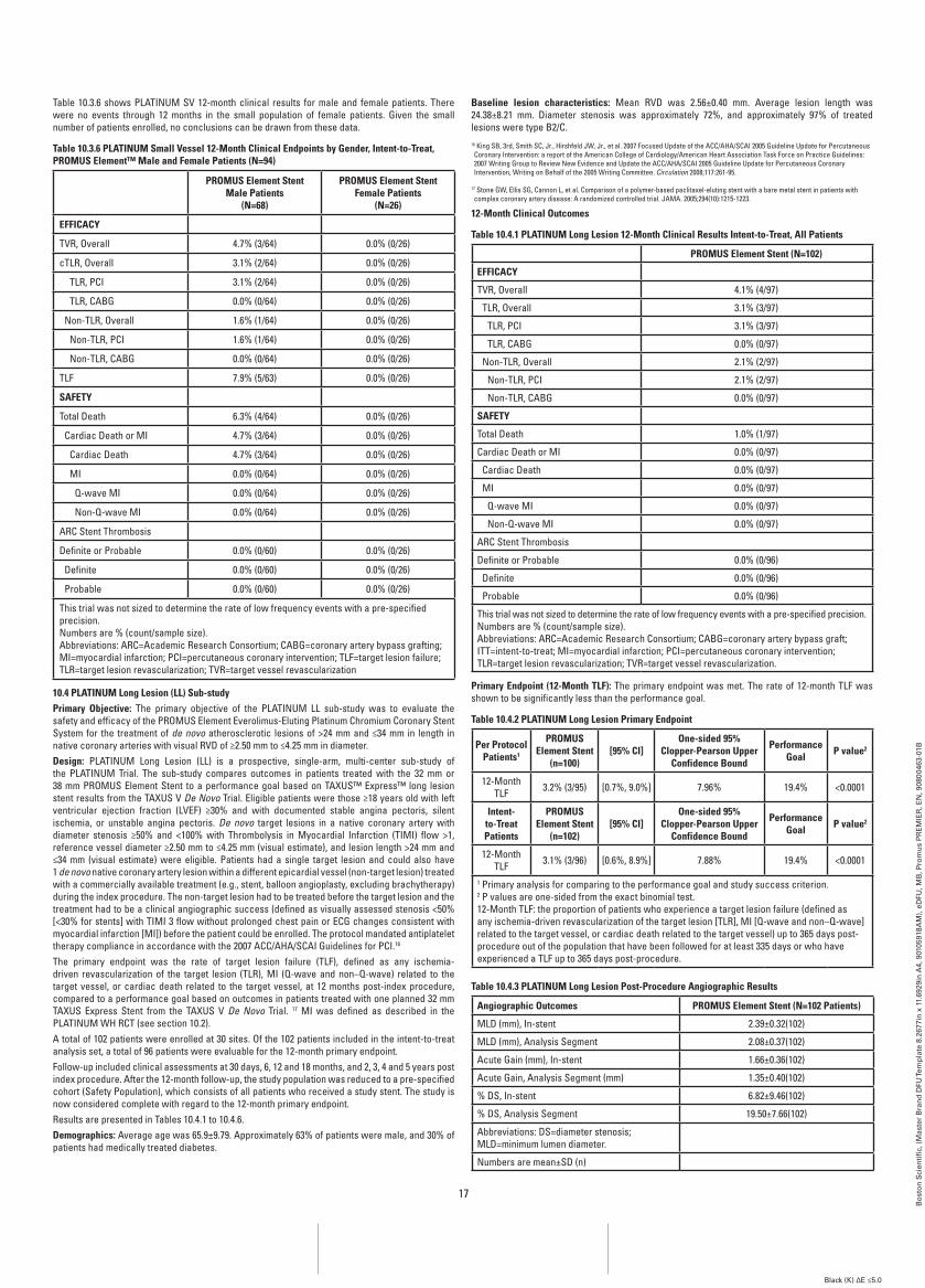
17
Bo
sto
n S
cien
tifi
c, (
Mas
ter
Bra
nd
DFU
Tem
pla
te 8
.267
7in
x 1
1.69
29in
A4,
901
0591
8AM
), e
DFU
, MB
, Pro
mu
s P
RE
MIE
R, E
N, 9
0800
463-
01B
Black (K) ∆E ≤5.0
Table 10.3.6 shows PLATINUM SV 12-month clinical results for male and female patients. There were no events through 12 months in the small population of female patients. Given the small number of patients enrolled, no conclusions can be drawn from these data.
Table 10.3.6 PLaTinUM Small Vessel 12-Month Clinical Endpoints by gender, intent-to-Treat, PrOMUS Element™ Male and female Patients (n=94)
PrOMUS Element Stent Male Patients
(n=68)
PrOMUS Element Stent female Patients
(n=26)
EffiCaCY
TVR, Overall 4.7% (3/64) 0.0% (0/26)
cTLR, Overall 3.1% (2/64) 0.0% (0/26)
TLR, PCI 3.1% (2/64) 0.0% (0/26)
TLR, CABG 0.0% (0/64) 0.0% (0/26)
Non-TLR, Overall 1.6% (1/64) 0.0% (0/26)
Non-TLR, PCI 1.6% (1/64) 0.0% (0/26)
Non-TLR, CABG 0.0% (0/64) 0.0% (0/26)
TLF 7.9% (5/63) 0.0% (0/26)
SafETY
Total Death 6.3% (4/64) 0.0% (0/26)
Cardiac Death or MI 4.7% (3/64) 0.0% (0/26)
Cardiac Death 4.7% (3/64) 0.0% (0/26)
MI 0.0% (0/64) 0.0% (0/26)
Q-wave MI 0.0% (0/64) 0.0% (0/26)
Non-Q-wave MI 0.0% (0/64) 0.0% (0/26)
ARC Stent Thrombosis
Definite or Probable 0.0% (0/60) 0.0% (0/26)
Definite 0.0% (0/60) 0.0% (0/26)
Probable 0.0% (0/60) 0.0% (0/26)
This trial was not sized to determine the rate of low frequency events with a pre-specified precision.Numbers are % (count/sample size).Abbreviations: ARC=Academic Research Consortium; CABG=coronary artery bypass grafting; MI=myocardial infarction; PCI=percutaneous coronary intervention; TLF=target lesion failure; TLR=target lesion revascularization; TVR=target vessel revascularization
10.4 PLaTinUM Long Lesion (LL) Sub-studyPrimary Objective: The primary objective of the PLATINUM LL sub-study was to evaluate the safety and efficacy of the PROMUS Element Everolimus-Eluting Platinum Chromium Coronary Stent System for the treatment of de novo atherosclerotic lesions of >24 mm and ≤34 mm in length in native coronary arteries with visual RVD of ≥2.50 mm to ≤4.25 mm in diameter.Design: PLATINUM Long Lesion (LL) is a prospective, single-arm, multi-center sub-study of the PLATINUM Trial. The sub-study compares outcomes in patients treated with the 32 mm or 38 mm PROMUS Element Stent to a performance goal based on TAXUS™ Express™ long lesion stent results from the TAXUS V De Novo Trial. Eligible patients were those ≥18 years old with left ventricular ejection fraction (LVEF) ≥30% and with documented stable angina pectoris, silent ischemia, or unstable angina pectoris. De novo target lesions in a native coronary artery with diameter stenosis ≥50% and <100% with Thrombolysis in Myocardial Infarction (TIMI) flow >1, reference vessel diameter ≥2.50 mm to ≤4.25 mm (visual estimate), and lesion length >24 mm and ≤34 mm (visual estimate) were eligible. Patients had a single target lesion and could also have 1 de novo native coronary artery lesion within a different epicardial vessel (non-target lesion) treated with a commercially available treatment (e.g., stent, balloon angioplasty, excluding brachytherapy) during the index procedure. The non-target lesion had to be treated before the target lesion and the treatment had to be a clinical angiographic success (defined as visually assessed stenosis <50% [<30% for stents] with TIMI 3 flow without prolonged chest pain or ECG changes consistent with myocardial infarction [MI]) before the patient could be enrolled. The protocol mandated antiplatelet therapy compliance in accordance with the 2007 ACC/AHA/SCAI Guidelines for PCI.16
The primary endpoint was the rate of target lesion failure (TLF), defined as any ischemia-driven revascularization of the target lesion (TLR), MI (Q-wave and non–Q-wave) related to the target vessel, or cardiac death related to the target vessel, at 12 months post-index procedure, compared to a performance goal based on outcomes in patients treated with one planned 32 mm TAXUS Express Stent from the TAXUS V De Novo Trial. 17 MI was defined as described in the PLATINUM WH RCT (see section 10.2).A total of 102 patients were enrolled at 30 sites. Of the 102 patients included in the intent-to-treat analysis set, a total of 96 patients were evaluable for the 12-month primary endpoint.Follow-up included clinical assessments at 30 days, 6, 12 and 18 months, and 2, 3, 4 and 5 years post index procedure. After the 12-month follow-up, the study population was reduced to a pre-specified cohort (Safety Population), which consists of all patients who received a study stent. The study is now considered complete with regard to the 12-month primary endpoint.Results are presented in Tables 10.4.1 to 10.4.6. Demographics: Average age was 65.9±9.79. Approximately 63% of patients were male, and 30% of patients had medically treated diabetes.
baseline lesion characteristics: Mean RVD was 2.56±0.40 mm. Average lesion length was 24.38±8.21 mm. Diameter stenosis was approximately 72%, and approximately 97% of treated lesions were type B2/C.16 King SB, 3rd, Smith SC, Jr., Hirshfeld JW, Jr., et al. 2007 Focused Update of the ACC/AHA/SCAI 2005 Guideline Update for Percutaneous Coronary Intervention: a report of the American College of Cardiology/American Heart Association Task Force on Practice Guidelines: 2007 Writing Group to Review New Evidence and Update the ACC/AHA/SCAI 2005 Guideline Update for Percutaneous Coronary Intervention, Writing on Behalf of the 2005 Writing Committee. Circulation 2008;117:261-95.
17 Stone GW, Ellis SG, Cannon L, et al. Comparison of a polymer-based paclitaxel-eluting stent with a bare metal stent in patients with complex coronary artery disease: A randomized controlled trial. JAMA. 2005;294(10):1215-1223.
12-Month Clinical Outcomes
Table 10.4.1 PLaTinUM Long Lesion 12-Month Clinical results intent-to-Treat, all Patients
PrOMUS Element Stent (n=102)
EffiCaCY
TVR, Overall 4.1% (4/97)
TLR, Overall 3.1% (3/97)
TLR, PCI 3.1% (3/97)
TLR, CABG 0.0% (0/97)
Non-TLR, Overall 2.1% (2/97)
Non-TLR, PCI 2.1% (2/97)
Non-TLR, CABG 0.0% (0/97)
SafETY
Total Death 1.0% (1/97)
Cardiac Death or MI 0.0% (0/97)
Cardiac Death 0.0% (0/97)
MI 0.0% (0/97)
Q-wave MI 0.0% (0/97)
Non-Q-wave MI 0.0% (0/97)
ARC Stent Thrombosis
Definite or Probable 0.0% (0/96)
Definite 0.0% (0/96)
Probable 0.0% (0/96)
This trial was not sized to determine the rate of low frequency events with a pre-specified precision.Numbers are % (count/sample size).Abbreviations: ARC=Academic Research Consortium; CABG=coronary artery bypass graft; ITT=intent-to-treat; MI=myocardial infarction; PCI=percutaneous coronary intervention; TLR=target lesion revascularization; TVR=target vessel revascularization.
Primary Endpoint (12-Month TLf): The primary endpoint was met. The rate of 12-month TLF was shown to be significantly less than the performance goal.
Table 10.4.2 PLaTinUM Long Lesion Primary Endpoint
Per Protocol Patients1
PrOMUS Element Stent
(n=100)[95% Ci]
One-sided 95% Clopper-Pearson Upper
Confidence bound
Performance goal P value2
12-Month TLF 3.2% (3/95) [0.7%, 9.0%] 7.96% 19.4% <0.0001
intent-to-Treat Patients
PrOMUS Element Stent
(n=102)[95% Ci]
One-sided 95% Clopper-Pearson Upper
Confidence bound
Performance goal P value2
12-Month TLF 3.1% (3/96) [0.6%, 8.9%] 7.88% 19.4% <0.0001
1 Primary analysis for comparing to the performance goal and study success criterion.2 P values are one-sided from the exact binomial test.12-Month TLF: the proportion of patients who experience a target lesion failure (defined as any ischemia-driven revascularization of the target lesion [TLR], MI [Q-wave and non–Q-wave] related to the target vessel, or cardiac death related to the target vessel) up to 365 days post-procedure out of the population that have been followed for at least 335 days or who have experienced a TLF up to 365 days post-procedure.
Table 10.4.3 PLaTinUM Long Lesion Post-Procedure angiographic results
angiographic Outcomes PrOMUS Element Stent (n=102 Patients)
MLD (mm), In-stent 2.39±0.32(102)
MLD (mm), Analysis Segment 2.08±0.37(102)
Acute Gain (mm), In-stent 1.66±0.36(102)
Acute Gain, Analysis Segment (mm) 1.35±0.40(102)
% DS, In-stent 6.82±9.46(102)
% DS, Analysis Segment 19.50±7.66(102)
Abbreviations: DS=diameter stenosis; MLD=minimum lumen diameter.
Numbers are mean±SD (n)
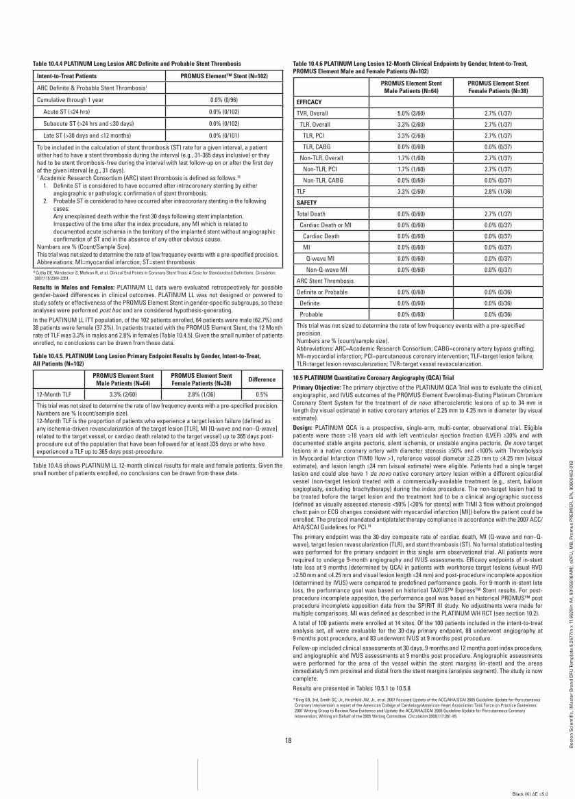
18
Bo
sto
n S
cien
tifi
c, (
Mas
ter
Bra
nd
DFU
Tem
pla
te 8
.267
7in
x 1
1.69
29in
A4,
901
0591
8AM
), e
DFU
, MB
, Pro
mu
s P
RE
MIE
R, E
N, 9
0800
463-
01B
Black (K) ∆E ≤5.0
Table 10.4.4 PLaTinUM Long Lesion arC Definite and Probable Stent Thrombosis
intent-to-Treat Patients PrOMUS Element™ Stent (n=102)
ARC Definite & Probable Stent Thrombosis1
Cumulative through 1 year 0.0% (0/96)
Acute ST (≤24 hrs) 0.0% (0/102)
Subacute ST (>24 hrs and ≤30 days) 0.0% (0/102)
Late ST (>30 days and ≤12 months) 0.0% (0/101)
To be included in the calculation of stent thrombosis (ST) rate for a given interval, a patient either had to have a stent thrombosis during the interval (e.g., 31-365 days inclusive) or they had to be stent thrombosis-free during the interval with last follow-up on or after the first day of the given interval (e.g., 31 days).1 Academic Research Consortium (ARC) stent thrombosis is defined as follows.18
1. Definite ST is considered to have occurred after intracoronary stenting by either angiographic or pathologic confirmation of stent thrombosis.
2. Probable ST is considered to have occurred after intracoronary stenting in the following cases: Any unexplained death within the first 30 days following stent implantation. Irrespective of the time after the index procedure, any MI which is related to documented acute ischemia in the territory of the implanted stent without angiographic confirmation of ST and in the absence of any other obvious cause.
Numbers are % (Count/Sample Size).This trial was not sized to determine the rate of low frequency events with a pre-specified precision.Abbreviations: MI=myocardial infarction; ST=stent thrombosis
18 Cutlip DE, Windecker S, Mehran R, et al. Clinical End Points in Coronary Stent Trials: A Case for Standardized Definitions. Circulation. 2007;115:2344-2351.
results in Males and females: PLATINUM LL data were evaluated retrospectively for possible gender-based differences in clinical outcomes. PLATINUM LL was not designed or powered to study safety or effectiveness of the PROMUS Element Stent in gender-specific subgroups, so these analyses were performed post hoc and are considered hypothesis-generating.In the PLATINUM LL ITT population, of the 102 patients enrolled, 64 patients were male (62.7%) and 38 patients were female (37.3%). In patients treated with the PROMUS Element Stent, the 12 Month rate of TLF was 3.3% in males and 2.8% in females (Table 10.4.5). Given the small number of patients enrolled, no conclusions can be drawn from these data.
Table 10.4.5. PLaTinUM Long Lesion Primary Endpoint results by gender, intent-to-Treat, all Patients (n=102)
PrOMUS Element Stent Male Patients (n=64)
PrOMUS Element Stent female Patients (n=38) Difference
12-Month TLF 3.3% (2/60) 2.8% (1/36) 0.5%
This trial was not sized to determine the rate of low frequency events with a pre-specified precision.Numbers are % (count/sample size).12-Month TLF is the proportion of patients who experience a target lesion failure (defined as any ischemia-driven revascularization of the target lesion [TLR], MI [Q-wave and non–Q-wave] related to the target vessel, or cardiac death related to the target vessel) up to 365 days post-procedure out of the population that have been followed for at least 335 days or who have experienced a TLF up to 365 days post-procedure.
Table 10.4.6 shows PLATINUM LL 12-month clinical results for male and female patients. Given the small number of patients enrolled, no conclusions can be drawn from these data.
Table 10.4.6 PLaTinUM Long Lesion 12-Month Clinical Endpoints by gender, intent-to-Treat, PrOMUS Element Male and female Patients (n=102)
PrOMUS Element Stent Male Patients (n=64)
PrOMUS Element Stent female Patients (n=38)
EffiCaCY
TVR, Overall 5.0% (3/60) 2.7% (1/37)
TLR, Overall 3.3% (2/60) 2.7% (1/37)
TLR, PCI 3.3% (2/60) 2.7% (1/37)
TLR, CABG 0.0% (0/60) 0.0% (0/37)
Non-TLR, Overall 1.7% (1/60) 2.7% (1/37)
Non-TLR, PCI 1.7% (1/60) 2.7% (1/37)
Non-TLR, CABG 0.0% (0/60) 0.0% (0/37)
TLF 3.3% (2/60) 2.8% (1/36)
SafETY
Total Death 0.0% (0/60) 2.7% (1/37)
Cardiac Death or MI 0.0% (0/60) 0.0% (0/37)
Cardiac Death 0.0% (0/60) 0.0% (0/37)
MI 0.0% (0/60) 0.0% (0/37)
Q-wave MI 0.0% (0/60) 0.0% (0/37)
Non-Q-wave MI 0.0% (0/60) 0.0% (0/37)
ARC Stent Thrombosis
Definite or Probable 0.0% (0/60) 0.0% (0/36)
Definite 0.0% (0/60) 0.0% (0/36)
Probable 0.0% (0/60) 0.0% (0/36)
This trial was not sized to determine the rate of low frequency events with a pre-specified precision.Numbers are % (count/sample size).Abbreviations: ARC=Academic Research Consortium; CABG=coronary artery bypass grafting; MI=myocardial infarction; PCI=percutaneous coronary intervention; TLF=target lesion failure; TLR=target lesion revascularization; TVR=target vessel revascularization.
10.5 PLaTinUM Quantitative Coronary angiography (QCa) TrialPrimary Objective: The primary objective of the PLATINUM QCA Trial was to evaluate the clinical, angiographic, and IVUS outcomes of the PROMUS Element Everolimus-Eluting Platinum Chromium Coronary Stent System for the treatment of de novo atherosclerotic lesions of up to 34 mm in length (by visual estimate) in native coronary arteries of 2.25 mm to 4.25 mm in diameter (by visual estimate).Design: PLATINUM QCA is a prospective, single-arm, multi-center, observational trial. Eligible patients were those ≥18 years old with left ventricular ejection fraction (LVEF) ≥30% and with documented stable angina pectoris, silent ischemia, or unstable angina pectoris. De novo target lesions in a native coronary artery with diameter stenosis ≥50% and <100% with Thrombolysis in Myocardial Infarction (TIMI) flow >1, reference vessel diameter ≥2.25 mm to ≤4.25 mm (visual estimate), and lesion length ≤34 mm (visual estimate) were eligible. Patients had a single target lesion and could also have 1 de novo native coronary artery lesion within a different epicardial vessel (non-target lesion) treated with a commercially-available treatment (e.g., stent, balloon angioplasty, excluding brachytherapy) during the index procedure. The non-target lesion had to be treated before the target lesion and the treatment had to be a clinical angiographic success (defined as visually assessed stenosis <50% [<30% for stents] with TIMI 3 flow without prolonged chest pain or ECG changes consistent with myocardial infarction [MI]) before the patient could be enrolled. The protocol mandated antiplatelet therapy compliance in accordance with the 2007 ACC/ AHA/SCAI Guidelines for PCI.19 The primary endpoint was the 30-day composite rate of cardiac death, MI (Q-wave and non−Q-wave), target lesion revascularization (TLR), and stent thrombosis (ST). No formal statistical testing was performed for the primary endpoint in this single arm observational trial. All patients were required to undergo 9-month angiography and IVUS assessments. Efficacy endpoints of in-stent late loss at 9 months (determined by QCA) in patients with workhorse target lesions (visual RVD ≥2.50 mm and ≤4.25 mm and visual lesion length ≤24 mm) and post-procedure incomplete apposition (determined by IVUS) were compared to predefined performance goals. For 9-month in-stent late loss, the performance goal was based on historical TAXUS™ Express™ Stent results. For post-procedure incomplete apposition, the performance goal was based on historical PROMUS™ post procedure incomplete apposition data from the SPIRIT III study. No adjustments were made for multiple comparisons. MI was defined as described in the PLATINUM WH RCT (see section 10.2).A total of 100 patients were enrolled at 14 sites. Of the 100 patients included in the intent-to-treat analysis set, all were evaluable for the 30-day primary endpoint, 88 underwent angiography at 9 months post procedure, and 83 underwent IVUS at 9 months post procedure.Follow-up included clinical assessments at 30 days, 9 months and 12 months post index procedure, and angiographic and IVUS assessments at 9 months post procedure. Angiographic assessments were performed for the area of the vessel within the stent margins (in-stent) and the areas immediately 5 mm proximal and distal from the stent margins (analysis segment). The study is now complete.Results are presented in Tables 10.5.1 to 10.5.8.19 King SB, 3rd, Smith SC, Jr., Hirshfeld JW, Jr., et al. 2007 Focused Update of the ACC/AHA/SCAI 2005 Guideline Update for Percutaneous Coronary Intervention: a report of the American College of Cardiology/American Heart Association Task Force on Practice Guidelines: 2007 Writing Group to Review New Evidence and Update the ACC/AHA/SCAI 2005 Guideline Update for Percutaneous Coronary Intervention, Writing on Behalf of the 2005 Writing Committee. Circulation 2008;117:261-95.
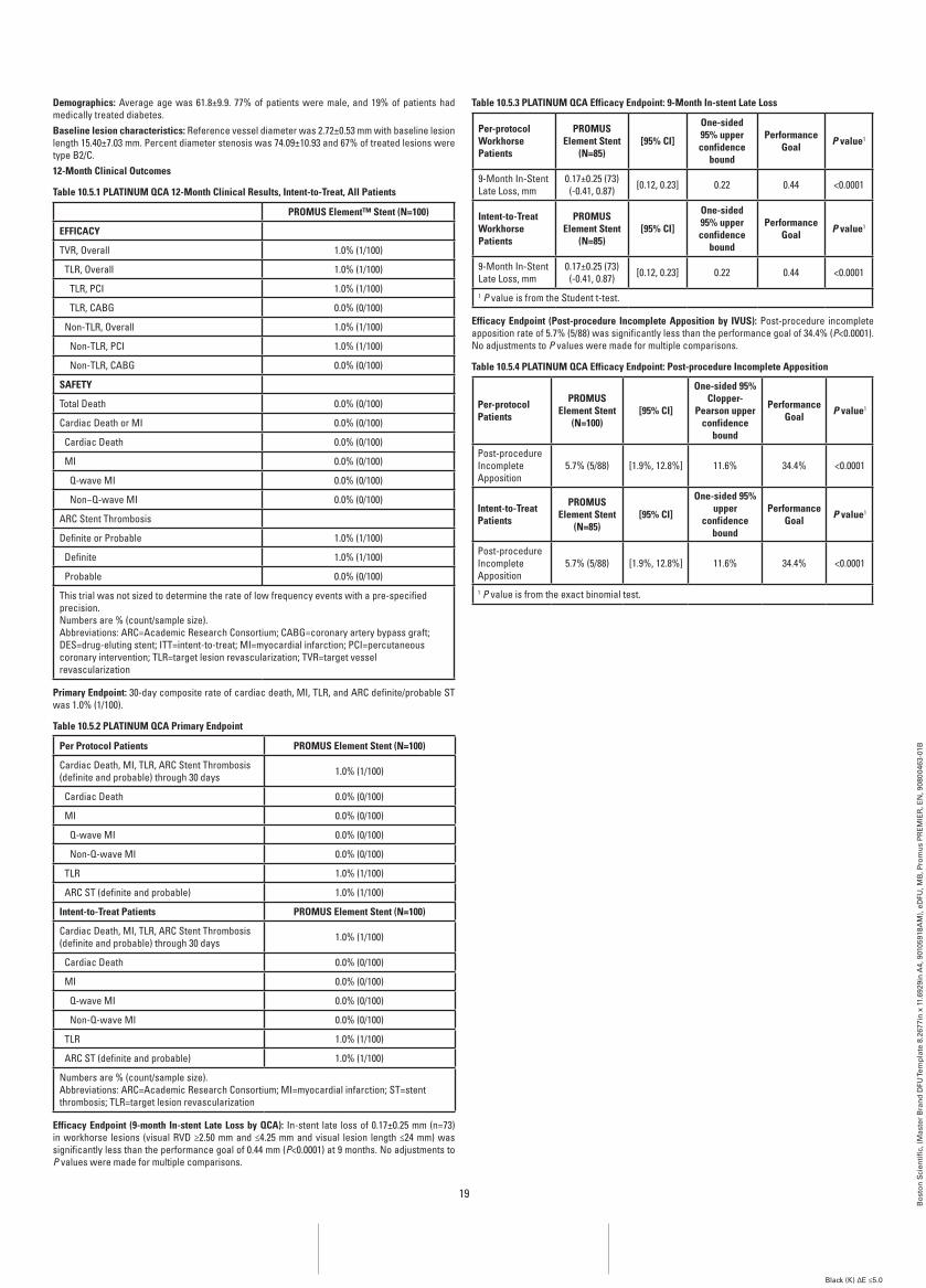
19
Bo
sto
n S
cien
tifi
c, (
Mas
ter
Bra
nd
DFU
Tem
pla
te 8
.267
7in
x 1
1.69
29in
A4,
901
0591
8AM
), e
DFU
, MB
, Pro
mu
s P
RE
MIE
R, E
N, 9
0800
463-
01B
Black (K) ∆E ≤5.0
Demographics: Average age was 61.8±9.9. 77% of patients were male, and 19% of patients had medically treated diabetes.baseline lesion characteristics: Reference vessel diameter was 2.72±0.53 mm with baseline lesion length 15.40±7.03 mm. Percent diameter stenosis was 74.09±10.93 and 67% of treated lesions were type B2/C.12-Month Clinical Outcomes
Table 10.5.1 PLaTinUM QCa 12-Month Clinical results, intent-to-Treat, all Patients
PrOMUS Element™ Stent (n=100)
EffiCaCY
TVR, Overall 1.0% (1/100)
TLR, Overall 1.0% (1/100)
TLR, PCI 1.0% (1/100)
TLR, CABG 0.0% (0/100)
Non-TLR, Overall 1.0% (1/100)
Non-TLR, PCI 1.0% (1/100)
Non-TLR, CABG 0.0% (0/100)
SafETY
Total Death 0.0% (0/100)
Cardiac Death or MI 0.0% (0/100)
Cardiac Death 0.0% (0/100)
MI 0.0% (0/100)
Q-wave MI 0.0% (0/100)
Non−Q-wave MI 0.0% (0/100)
ARC Stent Thrombosis
Definite or Probable 1.0% (1/100)
Definite 1.0% (1/100)
Probable 0.0% (0/100)
This trial was not sized to determine the rate of low frequency events with a pre-specified precision.Numbers are % (count/sample size).Abbreviations: ARC=Academic Research Consortium; CABG=coronary artery bypass graft; DES=drug-eluting stent; ITT=intent-to-treat; MI=myocardial infarction; PCI=percutaneous coronary intervention; TLR=target lesion revascularization; TVR=target vessel revascularization
Primary Endpoint: 30-day composite rate of cardiac death, MI, TLR, and ARC definite/probable ST was 1.0% (1/100).
Table 10.5.2 PLaTinUM QCa Primary Endpoint
Per Protocol Patients PrOMUS Element Stent (n=100)
Cardiac Death, MI, TLR, ARC Stent Thrombosis (definite and probable) through 30 days 1.0% (1/100)
Cardiac Death 0.0% (0/100)
MI 0.0% (0/100)
Q-wave MI 0.0% (0/100)
Non-Q-wave MI 0.0% (0/100)
TLR 1.0% (1/100)
ARC ST (definite and probable) 1.0% (1/100)
intent-to-Treat Patients PrOMUS Element Stent (n=100)
Cardiac Death, MI, TLR, ARC Stent Thrombosis (definite and probable) through 30 days 1.0% (1/100)
Cardiac Death 0.0% (0/100)
MI 0.0% (0/100)
Q-wave MI 0.0% (0/100)
Non-Q-wave MI 0.0% (0/100)
TLR 1.0% (1/100)
ARC ST (definite and probable) 1.0% (1/100)
Numbers are % (count/sample size).Abbreviations: ARC=Academic Research Consortium; MI=myocardial infarction; ST=stent thrombosis; TLR=target lesion revascularization
Efficacy Endpoint (9-month in-stent Late Loss by QCa): In-stent late loss of 0.17±0.25 mm (n=73) in workhorse lesions (visual RVD ≥2.50 mm and ≤4.25 mm and visual lesion length ≤24 mm) was significantly less than the performance goal of 0.44 mm (P<0.0001) at 9 months. No adjustments to P values were made for multiple comparisons.
Table 10.5.3 PLaTinUM QCa Efficacy Endpoint: 9-Month in-stent Late Loss
Per-protocol workhorse Patients
PrOMUS Element Stent
(n=85)[95% Ci]
One-sided 95% upper confidence
bound
Performance goal P value1
9-Month In-Stent Late Loss, mm
0.17±0.25 (73) (-0.41, 0.87) [0.12, 0.23] 0.22 0.44 <0.0001
intent-to-Treat workhorse Patients
PrOMUS Element Stent
(n=85)[95% Ci]
One-sided 95% upper confidence
bound
Performance goal P value1
9-Month In-Stent Late Loss, mm
0.17±0.25 (73) (-0.41, 0.87) [0.12, 0.23] 0.22 0.44 <0.0001
1 P value is from the Student t-test.
Efficacy Endpoint (Post-procedure incomplete apposition by iVUS): Post-procedure incomplete apposition rate of 5.7% (5/88) was significantly less than the performance goal of 34.4% (P<0.0001). No adjustments to P values were made for multiple comparisons.
Table 10.5.4 PLaTinUM QCa Efficacy Endpoint: Post-procedure incomplete apposition
Per-protocol Patients
PrOMUS Element Stent
(n=100)[95% Ci]
One-sided 95% Clopper-
Pearson upper confidence
bound
Performance goal P value1
Post-procedure Incomplete Apposition
5.7% (5/88) [1.9%, 12.8%] 11.6% 34.4% <0.0001
intent-to-Treat Patients
PrOMUS Element Stent
(n=85)[95% Ci]
One-sided 95% upper
confidence bound
Performance goal P value1
Post-procedure Incomplete Apposition
5.7% (5/88) [1.9%, 12.8%] 11.6% 34.4% <0.0001
1 P value is from the exact binomial test.
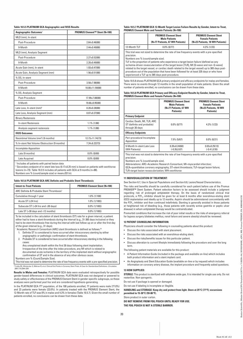
20
Bo
sto
n S
cien
tifi
c, (
Mas
ter
Bra
nd
DFU
Tem
pla
te 8
.267
7in
x 1
1.69
29in
A4,
901
0591
8AM
), e
DFU
, MB
, Pro
mu
s P
RE
MIE
R, E
N, 9
0800
463-
01B
Black (K) ∆E ≤5.0
Table 10.5.5 PLaTinUM QCa angiographic and iVUS results
angiographic Outcomes1 PrOMUS Element™ Stent (n=100)
MLD (mm), In-stent
Post-Procedure 2.64±0.46(88)
9-Month 2.44±0.49(88)
MLD (mm), Analysis Segment
Post-Procedure 2.27±0.52(88)
9-Month 2.20±0.49(88)
Acute Gain (mm), In-stent 1.93±0.47(88)
Acute Gain, Analysis Segment (mm) 1.56±0.51(88)
% DS, In-stent
Post-Procedure 3.58±7.98(88)
9-Month 10.00±11.59(88)
% DS, Analysis Segment
Post-Procedure 17.99±7.88(88)
9-Month 19.66±8.95(88)
Late Loss, In-stent (mm)2 0.20±0.28(88)
Late Loss, Analysis Segment (mm) 0.07±0.27(88)
Binary Restenosis
In-stent Restenosis 1.1% (1/88)
Analysis segment restenosis 1.1% (1/88)
iVUS Outcomes
Neointimal Volume (mm3) (9 months) 12.73±11.74(73)
% In-stent Net Volume Obstruction (9 months) 7.24±6.22(73)
Incomplete Apposition
Late (9 months) 0.0% (0/69)
Late Acquired 0.0% (0/69)1 Includes all patients with paired lesion data2 Secondary endpoint of in-stent late loss (0.17±0.25 mm) is based on patients with workhorse lesions (n=73). Table 10.3.5 includes all patients with QCA at 9 months (n=88).Numbers are % (count/sample size) or mean±SD (n).
Table 10.5.6 PLaTinUM QCa arC Definite and Probable Stent Thrombosis
intent-to-Treat Patients PrOMUS Element Stent (n=100)
ARC Definite & Probable Stent Thrombosis1
Cumulative through 1 year 1.0% (1/100)
Acute ST (≤24 hrs) 1.0% (1/100))
Subacute ST (>24 hrs and ≤30 days) 0.0% (1/100))
Late ST (>30 days and ≤12 months) 0.0% (1/100))
To be included in the calculation of stent thrombosis (ST) rate for a given interval, a patient either had to have a stent thrombosis during the interval (e.g., 31-365 days inclusive) or they had to be stent thrombosis-free during the interval with last follow-up on or after the first day of the given interval (e.g., 31 days).1 Academic Research Consortium (ARC) stent thrombosis is defined as follows.20
1. Definite ST is considered to have occurred after intracoronary stenting by either angiographic or pathologic confirmation of stent thrombosis.
2. Probable ST is considered to have occurred after intracoronary stenting in the following cases: Any unexplained death within the first 30 days following stent implantation. Irrespective of the time after the index procedure, any MI which is related to documented acute ischemia in the territory of the implanted stent without angiographic confirmation of ST and in the absence of any other obvious cause.
Numbers are % (Count/Sample Size).This trial was not sized to determine the rate of low frequency events with a pre-specified precision.
20 Cutlip DE, Windecker S, Mehran R, et al. Clinical End Points in Coronary Stent Trials: A Case for Standardized Definitions. Circulation. 2007;115:2344-2351
results in Males and females: PLATINUM QCA data were evaluated retrospectively for possible gender-based differences in clinical outcomes. PLATINUM QCA was not designed or powered to study safety or effectiveness of the PROMUS Element Stent in gender-specific subgroups, so these analyses were performed post hoc and are considered hypothesis-generating.In the PLATINUM QCA ITT population, of the 100 patients enrolled, 77 patients were male (77.0%) and 23 patients were female (23.0%). In patients treated with the PROMUS Element Stent, the 12-Month rate of TLF was 0% in males and 4.3% in females (Table 10.5.7). Given the small number of patients enrolled, no conclusions can be drawn from these data.
Table 10.5.7 PLaTinUM QCa 12-Month Target Lesion failure results by gender, intent-to-Treat, PrOMUS Element Male and female Patients (n=100)
PrOMUS Element Stent Male Patients
(n=77 Patients, 67 wH Patients)
PrOMUS Element Stent female Patients
(n=23 Patients, 18 wH Patients)
12-Month TLF 0.0% (0/77) 4.3% (1/23)
This trial was not sized to determine the rate of low frequency events with a pre-specified precision.Numbers are % (count/sample size).TLF is the proportion of patients who experience a target lesion failure (defined as any ischemia-driven revascularization of the target lesion [TLR], MI [Q-wave and non–Q-wave] related to the target vessel, or cardiac death related to the target vessel) up to 365 days post-procedure out of the population that have been followed for at least 335 days or who have experienced a TLF up to 365 days post-procedure.
Table 10.5.8 shows PLATINUM QCA primary endpoint and efficacy endpoints for males and females. There were no events through 12 months in the small population of male patients. Given the small number of patients enrolled, no conclusions can be drawn from these data.
Table 10.5.8 PLaTinUM QCa Primary and Efficacy Endpoint results by gender, intent-to-Treat, PrOMUS Element Male and female Patients (n=100)
PrOMUS Element Stent Male Patients
(n=77 Patients, 67 wH Patients)
PrOMUS Element Stent female Patients
(n=23 Patients, 18 wH Patients)
Primary Endpoint
Cardiac Death, MI, TLR, ARC ST (definite and probable) through 30 days
0.0% (0/77) 4.3% (1/23)
Efficacy Endpoints
Peri-procedural Incomplete Apposition 7.5% (5/67) 0.0% (0/21)
9-Month In-stent Late Loss (WH Patients)
0.20±0.24(60)(-0.26,0.87)
0.03±0.25(13)(-0.41,0.55)
This trial was not sized to determine the rate of low frequency events with a pre-specified precision.Numbers are % (count/sample size).Abbreviations: ARC=Academic Research Consortium; MI=myocardial infarction; QCA=quantitative coronary angiography; ST=stent thrombosis; TLF=target lesion failure; TLR=target lesion revascularization; WH=workhorse
11 inDiViDUaLiZaTiOn Of TrEaTMEnT:See Section 6.7, Use in Special Populations and Section 6.8, Lesion/Vessel Characteristics.The risks and benefits should be carefully considered for each patient before use of the Promus PREMIER™ Stent System. Patient selection factors to be assessed should include a judgment regarding risk of prolonged antiplatelet therapy. On the basis of randomized clinical trial protocols, a P2Y12 inhibitor should be given for at least 6 months after everolimus-eluting stent (EES) implantation and ideally up to 12 months. Aspirin should be administered concomitantly with the P2Y12 inhibitor and then continued indefinitely. Stenting is generally avoided in those patients at heightened risk of bleeding (e.g., those patients with recently active gastritis or peptic ulcer disease) in whom antiplatelet therapy would be contraindicated.Premorbid conditions that increase the risk of poor initial results or the risks of emergency referral for bypass surgery (diabetes mellitus, renal failure and severe obesity) should be reviewed.
12 PaTiEnT COUnSELing infOrMaTiOn:Physicians should consider the following in counseling patients about this product:• Discuss the risks associated with stent placement.• Discuss the risks associated with an everolimus-eluting stent.• Discuss the risks/benefits issues for this particular patient.• Discuss alteration to current lifestyle immediately following the procedure and over the long
term.The following patient materials are available for this product:• A Patient Information Guide (included in the package and available on-line) which includes
both product information and a stent implant card.• An Angioplasty and Stent Education Guide (available on-line or by request) which includes
information on coronary artery disease, the implant procedure and frequently asked questions.
13 HOw SUPPLiED:STEriLE: This product is sterilized with ethylene oxide gas. It is intended for single use only. Do not resterilize. Non-pyrogenic.Do not use if package is opened or damaged.Do not use if labeling is incomplete or illegible.HanDLing and STOragE: Keep dry and protect from light. Store at 25°C (77°f); excursions permitted to 15-30°C (59-86°f).Store product in outer carton.DO nOT rEMOVE frOM fOiL POUCH UnTiL rEaDY fOr USE.THE fOiL POUCH iS nOT a STEriLE barriEr.
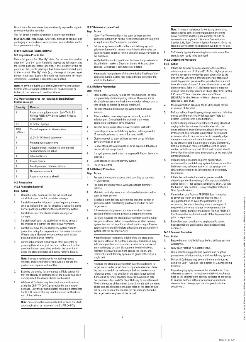
21
Bo
sto
n S
cien
tifi
c, (
Mas
ter
Bra
nd
DFU
Tem
pla
te 8
.267
7in
x 1
1.69
29in
A4,
901
0591
8AM
), e
DFU
, MB
, Pro
mu
s P
RE
MIE
R, E
N, 9
0800
463-
01B
Black (K) ∆E ≤5.0
Do not store devices where they are directly exposed to organic solvents or ionizing radiation.The foil pouch contains Argon (Ar) as a storage medium.DiSPOSaL inSTrUCTiOnS: After use, dispose of product and packaging in accordance with hospital, administrative and/or local government policy.
14 OPEraTiOnaL inSTrUCTiOnS:14.1 inspection Prior to UseCheck foil pouch for ‘‘Use By’’ date. Do not use the product after the ‘‘Use By’’ date. Carefully inspect the foil pouch and the sterile package before opening. If the integrity of the foil pouch or the sterile package has been compromised prior to the product ‘‘Use By’’ date (e.g., damage of the package), contact your local Boston Scientific representative for return information. Do not use if any defects are noted.
note: At any time during use of the Monorail™ Stent Delivery System, if the proximal shaft (hypotube) has been bent or kinked, do not continue to use the catheter.
14.2 Materials required (not included in Stent Delivery System package)
Quantity Material
1 Appropriate guide catheter (see Table 2.1, Promus PREMIER™ Stent System Product Description)
2-3 20 ml (cc) syringe
1000 u/500 cc
Normal heparinized sterile saline
1 ≤0.014 in (0.36 mm) guidewire
1 Rotating hemostatic valve
1 Diluted contrast medium 1:1 with normal heparinized sterile saline
1 Inflation Device
1 Torque Device
1 Pre-deployment dilation catheter
1 Three-way stopcock
1 Appropriate arterial sheath
14.3 Preparation14.3.1 Packaging removalStep action1. Open the outer box to reveal the foil pouch and
carefully inspect the foil pouch for damage.2. Carefully open the foil pouch by tearing along the tear
strip as indicated on the foil pouch to access the sterile barrier package containing the stent delivery system.
3. Carefully inspect the sterile barrier package for damage.
4. Carefully peel open the sterile barrier using aseptic techniques and extract the stent delivery system.
5. Carefully remove the stent delivery system from its protective tubing for preparation of the delivery system. When using a Monorail system, do not bend or kink proximal shaft during removal.
6. Remove the product mandrel and stent protector by grasping the catheter just proximal to the stent (at the proximal balloon bond site), and with the other hand, grasp the stent protector and gently remove distally.
note: If unusual resistance is felt during product mandrel and stent protector removal, do not use the product and replace with another.
7. Examine the device for any damage. If it is suspected that the sterility or performance of the device has been compromised, the device should not be used.
8. A Monorail Catheter may be coiled once and secured using the CLIPIT™ Coil Clips provided in the catheter package. Only the proximal shaft should be inserted into the CLIPIT device; the clip is not intended for the distal end of the catheter.
note: Care should be taken not to kink or bend the shaft upon application or removal of the CLIPIT Coil Clip.
14.3.2 guidewire Lumen flushStep action1. (Over-The-Wire only) Flush the stent delivery system
guidewire lumen with normal heparinized saline through the straight arm of the Y connector manifold.
2. (Monorail system only) Flush the stent delivery system guidewire lumen with normal heparinized saline using the flushing needle supplied for the Monorail delivery system at the distal end.
3. Verify that the stent is positioned between the proximal and distal balloon markers. Check for bends, kinks and other damage. Do not use if any defects are noted.
note: Avoid manipulation of the stent during flushing of the guidewire lumen, as this may disrupt the placement of the stent on the balloon.
14.3.3 balloon PreparationStep action1. Stent contact with any fluid is not recommended, as there
is a possibility of initiating drug release. However, if it is absolutely necessary to flush the stent with saline, contact time should be limited (1 minute maximum).
2. Prepare inflation device/syringe with diluted contrast medium.
3. Attach inflation device/syringe to stopcock; attach to inflation port. Do not bend the proximal shaft when connecting to inflation device/syringe.
4. With tip down, orient stent delivery system vertically.5. Open stopcock to stent delivery system; pull negative for
15 seconds; release to neutral for contrast fill.6. Close stopcock to stent delivery system; purge inflation
device/ syringe of all air.7. Repeat steps 4 through 6 until all air is expelled. If bubbles
persist, do not use product.8. If a syringe was used, attach a prepared inflation device to
stopcock.9. Open stopcock to stent delivery system.10. Leave on neutral.14.3.4 Delivery ProcedureStep action1. Prepare the vascular access site according to standard
PTCA practice.2. Predilate the lesion/vessel with appropriate diameter
balloon.3. Maintain neutral pressure on inflation device attached to
stent delivery system.4. Backload stent delivery system onto proximal portion of
guidewire while maintaining guidewire position across target lesion.
5. Fully open rotating hemostatic valve to allow for easy passage of the stent and prevent damage to the stent.
6. Carefully advance the stent delivery system into the hub of the guide catheter. When using a Monorail stent delivery system be sure to keep the proximal shaft straight. Ensure guide catheter stability before advancing the stent delivery system into the coronary artery.
note: If unusual resistance is felt before the stent exits the guide catheter, do not force passage. Resistance may indicate a problem, and use of excessive force may result in stent damage or stent dislodgment from the balloon. Maintain guidewire placement across the lesion, and remove the stent delivery system and guide catheter as a single unit.
7. Advance the stent delivery system over the guidewire to target lesion under direct fluoroscopic visualization. Utilize the proximal and distal radiopaque balloon markers as a reference point. If the position of the stent is not optimal, it should be carefully repositioned or removed (See also Precautions – Section 6.15, Stent Delivery System Removal). The inside edges of the marker bands indicate both the stent edges and balloon shoulders. Expansion of the stent should not be undertaken if the stent is not properly positioned in the target lesion segment of the vessel.
note: If unusual resistance is felt at any time during lesion access before stent implantation, the stent delivery system and the guide catheter should be removed as a single unit. (See also Precautions – Section 6.15, Stent Delivery System Removal). Once the stent delivery system has been removed do not re-use.
8. Sufficiently tighten the rotating hemostatic valve. The stent is now ready to be deployed.
14.3.5 Deployment ProcedureStep action1. Inflate the delivery system expanding the stent to a
minimum pressure of 11 atm (1117 kPa). Higher pressure may be necessary to optimize stent apposition to the arterial wall. Accepted practice generally targets an initial deployment pressure that would achieve a stent inner diameter of about 1.1 times the reference vessel diameter (see Table 14.1). Balloon pressure must not exceed rated burst pressure of 18 atm (1827 kPa) for the 2.25 mm – 2.75 mm diameter stents and 16 atm (1620 kPa) for the 3.00 mm - 4.00 mm diameter stent sizes (see Table 14.1).
2. Maintain inflation pressure for 15-30 seconds for full expansion of the stent.
3. Deflate balloon by pulling negative pressure on inflation device until balloon is fully deflated (see Table 6.1, System Deflation Time Specifications).
4. Confirm stent position and deployment using standard angiographic techniques. For optimal results, the entire stenosed arterial segment should be covered by the stent. Fluoroscopic visualization during stent expansion should be used in order to properly judge the optimum expanded stent diameter as compared to the proximal and distal coronary artery diameter(s). Optimal expansion requires that the stent be in full contact with the artery wall. Stent wall contact should be verified through routine angiography or intravascular ultrasound (IVUS).
5. If stent sizing/apposition requires optimization, readvance the stent delivery system balloon, or another high-pressure, balloon catheter of the appropriate size, to the stented area using standard angioplasty techniques.
6. Inflate the balloon to the desired pressure while observing under fluoroscopy (refer to product labeling and/or Table 14.1 for balloon compliance chart). Deflate the balloon (see Table 6.1, Delivery System Deflation Time Specifications).
7. If more than one Promus PREMIER Stent is needed to cover the lesion and balloon treated area, it is suggested that, to avoid the potential for gap restenosis, the stents be adequately overlapped. To ensure that there are no gaps between stents, the balloon marker bands of the second Promus PREMIER Stent should be positioned inside of the deployed stent prior to expansion.
8. Reconfirm stent position and angiographic result. Repeat inflations until optimal stent deployment is achieved.
14.3.6 removal ProcedureStep action1. Ensure balloon is fully deflated before delivery system
withdrawal.2. Fully open rotating hemostatic valve.3. While maintaining guidewire position and negative
pressure on inflation device, withdraw delivery system. 4. Monorail Catheters may be coiled once and secured
using the CLIPIT Coil Clip (see Section 14.3.1, Packaging Removal).
5. Repeat angiography to assess the stented area. If an adequate expansion has not been obtained, exchange back to the original stent delivery catheter or exchange to another balloon catheter of appropriate balloon diameter to achieve proper stent apposition to the vessel wall.
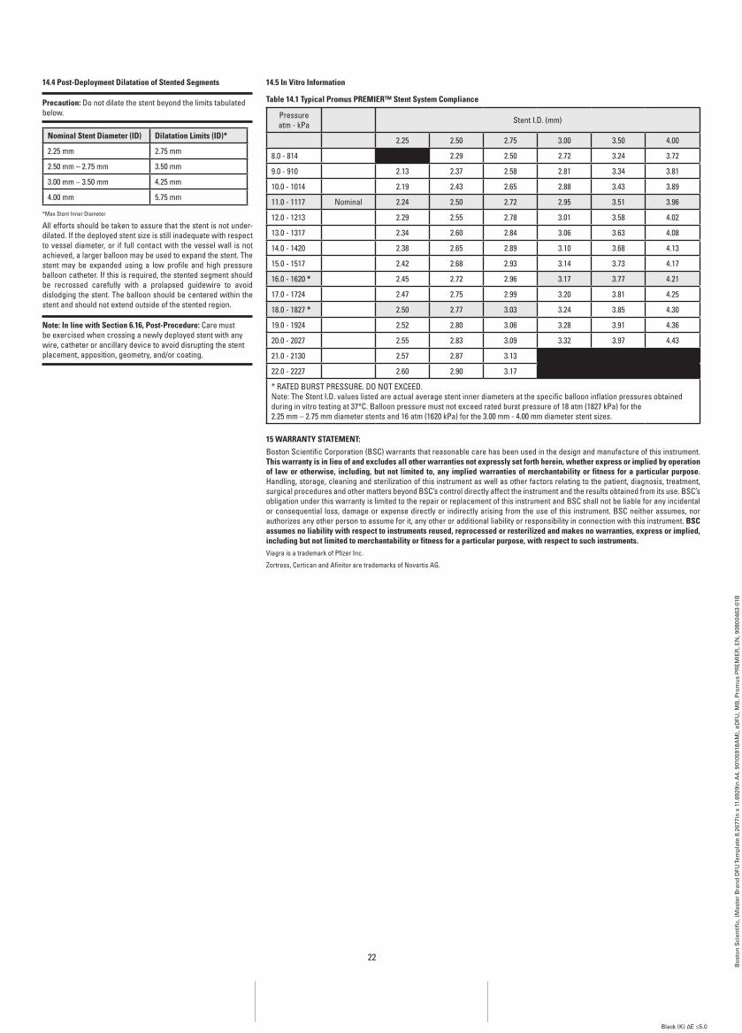
22
Bo
sto
n S
cien
tifi
c, (
Mas
ter
Bra
nd
DFU
Tem
pla
te 8
.267
7in
x 1
1.69
29in
A4,
901
0591
8AM
), e
DFU
, MB
, Pro
mu
s P
RE
MIE
R, E
N, 9
0800
463-
01B
Black (K) ∆E ≤5.0
14.4 Post-Deployment Dilatation of Stented Segments
Precaution: Do not dilate the stent beyond the limits tabulated below.
nominal Stent Diameter (iD) Dilatation Limits (iD)*
2.25 mm 2.75 mm
2.50 mm – 2.75 mm 3.50 mm
3.00 mm – 3.50 mm 4.25 mm
4.00 mm 5.75 mm
*Max Stent Inner Diameter
All efforts should be taken to assure that the stent is not under-dilated. If the deployed stent size is still inadequate with respect to vessel diameter, or if full contact with the vessel wall is not achieved, a larger balloon may be used to expand the stent. The stent may be expanded using a low profile and high pressure balloon catheter. If this is required, the stented segment should be recrossed carefully with a prolapsed guidewire to avoid dislodging the stent. The balloon should be centered within the stent and should not extend outside of the stented region.
note: in line with Section 6.16, Post-Procedure: Care must be exercised when crossing a newly deployed stent with any wire, catheter or ancillary device to avoid disrupting the stent placement, apposition, geometry, and/or coating.
14.5 in Vitro information
Table 14.1 Typical Promus PrEMiEr™ Stent System Compliance
Pressure atm - kPa Stent I.D. (mm)
2.25 2.50 2.75 3.00 3.50 4.00
8.0 - 814 2.29 2.50 2.72 3.24 3.72
9.0 - 910 2.13 2.37 2.58 2.81 3.34 3.81
10.0 - 1014 2.19 2.43 2.65 2.88 3.43 3.89
11.0 - 1117 Nominal 2.24 2.50 2.72 2.95 3.51 3.96
12.0 - 1213 2.29 2.55 2.78 3.01 3.58 4.02
13.0 - 1317 2.34 2.60 2.84 3.06 3.63 4.08
14.0 - 1420 2.38 2.65 2.89 3.10 3.68 4.13
15.0 - 1517 2.42 2.68 2.93 3.14 3.73 4.17
16.0 - 1620 * 2.45 2.72 2.96 3.17 3.77 4.21
17.0 - 1724 2.47 2.75 2.99 3.20 3.81 4.25
18.0 - 1827 * 2.50 2.77 3.03 3.24 3.85 4.30
19.0 - 1924 2.52 2.80 3.06 3.28 3.91 4.36
20.0 - 2027 2.55 2.83 3.09 3.32 3.97 4.43
21.0 - 2130 2.57 2.87 3.13
22.0 - 2227 2.60 2.90 3.17
* RATED BURST PRESSURE. DO NOT EXCEED.Note: The Stent I.D. values listed are actual average stent inner diameters at the specific balloon inflation pressures obtained during in vitro testing at 37°C. Balloon pressure must not exceed rated burst pressure of 18 atm (1827 kPa) for the 2.25 mm – 2.75 mm diameter stents and 16 atm (1620 kPa) for the 3.00 mm - 4.00 mm diameter stent sizes.
15 warranTY STaTEMEnT:Boston Scientific Corporation (BSC) warrants that reasonable care has been used in the design and manufacture of this instrument. This warranty is in lieu of and excludes all other warranties not expressly set forth herein, whether express or implied by operation of law or otherwise, including, but not limited to, any implied warranties of merchantability or fitness for a particular purpose. Handling, storage, cleaning and sterilization of this instrument as well as other factors relating to the patient, diagnosis, treatment, surgical procedures and other matters beyond BSC’s control directly affect the instrument and the results obtained from its use. BSC’s obligation under this warranty is limited to the repair or replacement of this instrument and BSC shall not be liable for any incidental or consequential loss, damage or expense directly or indirectly arising from the use of this instrument. BSC neither assumes, nor authorizes any other person to assume for it, any other or additional liability or responsibility in connection with this instrument. bSC assumes no liability with respect to instruments reused, reprocessed or resterilized and makes no warranties, express or implied, including but not limited to merchantability or fitness for a particular purpose, with respect to such instruments.Viagra is a trademark of Pfizer Inc.
Zortress, Certican and Afinitor are trademarks of Novartis AG.
EU AuthorizedRepresentative
Boston Scientific International S.A.55 avenue des Champs PierreuxTSA 5110192729 NANTERRE CEDEXFRANCE
EC REP
LegalManufacturer
Boston Scientific CorporationOne Boston Scientific PlaceNatick, MA 01760-1537USAUSA Customer Service 888-272-1001
RecyclablePackage
© 2013 Boston Scientific Corporation or its affiliates. All rights reserved.
AustralianSponsor Address
Boston Scientific (Australia) Pty LtdPO Box 332BOTANYNSW 1455AustraliaFree Phone 1800 676 133Free Fax 1800 836 666
AUS
Do not use if packageis damaged.
Bo
sto
n S
cien
tifi
c, (
Mas
ter
Bra
nd
DFU
Tem
pla
te 8
.267
7in
x 1
1.69
29in
A4,
901
0591
8AM
), e
DFU
, MB
, Pro
mu
s P
RE
MIE
R, E
N, 9
0800
463-
01B
Black (K) ∆E ≤5.0