promiscuous interactions and protein disaggregases ... · elifesciences.org research article...
TRANSCRIPT
elifesciences.org
RESEARCH ARTICLE
Promiscuous interactions and proteindisaggregases determine the materialstate of stress-inducible RNP granulesSonja Kroschwald, Shovamayee Maharana, Daniel Mateju, Liliana Malinovska,Elisabeth Nuske, Ina Poser, Doris Richter, Simon Alberti*
Max Planck Institute of Molecular Cell Biology and Genetics, Dresden, Germany
Abstract RNA-protein (RNP) granules have been proposed to assemble by forming solid RNA/
protein aggregates or through phase separation into a liquid RNA/protein phase. Which model
describes RNP granules in living cells is still unclear. In this study, we analyze P bodies in budding
yeast and find that they have liquid-like properties. Surprisingly, yeast stress granules adopt
a different material state, which is reminiscent of solid protein aggregates and controlled by protein
disaggregases. By using an assay to ectopically nucleate RNP granules, we further establish that RNP
granule formation does not depend on amyloid-like aggregation but rather involves many
promiscuous interactions. Finally, we show that stress granules have different properties in
mammalian cells, where they show liquid-like behavior. Thus, we propose that the material state of
RNP granules is flexible and that the solid state of yeast stress granules is an adaptation to extreme
environments, made possible by the presence of a powerful disaggregation machine.
DOI: 10.7554/eLife.06807.001
IntroductionThe organization of the intracellular space into compartments is fundamental to life; it allows cells to
perform complex biochemical reactions in a confined and controlled manner. In addition to membrane-
delimited organelles, such as the Golgi apparatus or the endoplasmic reticulum, the cytoplasm contains
compartments that are not surrounded by membranes. These non-membrane-bound compartments
are frequently linked to gene expression pathways and often contain substantial amounts of RNAs
(Hyman and Brangwynne, 2011; Weber and Brangwynne, 2012; Brangwynne, 2013; Hyman et al.,
2014). Examples in the nucleus include chromatin domains, the nucleolus, and Cajal bodies; examples
in the cytoplasm are P bodies, stress granules, and germ granules. Although their functions are not
always clear, it is assumed that they facilitate or suppress specific biochemical reactions.
Recent studies proposed that P granules—RNA-protein (RNP) granules in germ cells of
Caenorhabditis elegans—form by liquid–liquid demixing or phase separation from the cytoplasm,
so that two phases, a liquid droplet phase and the cytoplasm coexist (Brangwynne et al., 2009; Lee
et al., 2013). Such demixed phases of proteins and RNAs may turn out to be a unifying principle of
subcellular organization (Weber and Brangwynne, 2012; Brangwynne, 2013; Hyman et al., 2014).
Indeed, recent findings show that liquid phase separation is not limited to P granules (Brangwynne
et al., 2011; Aggarwal et al., 2013; Feric and Brangwynne, 2013; Hubstenberger et al., 2013;
Wippich et al., 2013; Banjade and Rosen, 2014). Importantly, defined mixtures of RNAs and proteins
can also phase separate in a cell-free system, driven by multivalent interactions (Li et al., 2012). This
suggests that many cellular components may have an intrinsic ability to phase separate and assemble
into structures with liquid-like properties.
Despite recent evidence for a liquid-like state, others have argued that RNP granules have more
solid material characteristics, similar to protein aggregates (Gilks et al., 2004; Vessey et al., 2006;
*For correspondence: alberti@
mpi-cbg.de
Competing interests: The
authors declare that no
competing interests exist.
Funding: See page 28
Received: 02 February 2015
Accepted: 25 June 2015
Published: 04 August 2015
Reviewing editor: Jeffery W
Kelly, Scripps Research Institute,
United States
Copyright Kroschwald et al.
This article is distributed under
the terms of the Creative
Commons Attribution License,
which permits unrestricted use
and redistribution provided that
the original author and source are
credited.
Kroschwald et al. eLife 2015;4:e06807. DOI: 10.7554/eLife.06807 1 of 32
Decker et al., 2007; Si et al., 2010; Decker and Parker, 2012; Ramaswami et al., 2013). This seems
particularly likely for P bodies and stress granules, which are stress-inducible RNP granules containing
non-translating RNAs and protein factors involved in translation repression or mRNA decay
(Anderson and Kedersha, 2009; Decker and Parker, 2012). P bodies and stress granules form as
a response to acute stress conditions, when a cell has to make arrangements to divert valuable
resources to cellular survival, and their formation coincides with the formation of protein aggregates,
which result from stress-induced protein misfolding.
Proteins contained in P bodies or stress granules are characteristically composed of two types of
domains: RNA-binding domains (RBDs) and domains of low sequence complexity; the latter are also
referred to as prion-like (Gilks et al., 2004; Decker et al., 2007; Reijns et al., 2008; King et al., 2012;
Malinovska et al., 2013). This term is derived from the fact that they have a characteristic amino acid
composition (mostly polar amino acids such as serine, glycine, asparagine, glutamine and tyrosine),
which resembles that of yeast prions. Prion domains (PDs) and prion-like domains (PLDs) have little
structure under normal conditions. However, they can undergo spontaneous conversions into an
aggregated state, which is characterized by a cross-β structure and referred to as amyloid (Alberti
et al., 2009). Once formed, the amyloid state can act as a template for the incorporation of further
proteins, but it can also be reversed through energy-expending protein disaggregation machines
(Doyle et al., 2013).
Despite some recent progress, RNP granule assembly remains a poorly understood molecular
process. In particular, it is not known how RBDs and PLDs cooperate to promote RNP granule
eLife digest Genes consist of long stretches of DNA that code for proteins. The DNA is first
‘transcribed’ to produce an RNA molecule, which is then translated into a protein. In most cells, RNA
molecules are present within a structure called ribonucleoprotein (RNP for short) granules. These
contain the protein machinery needed to transport, store, and break down RNAs.
P bodies and stress granules are two types of RNP granules found in all cells, from yeast to human.
P bodies are present at all times, whereas stress granules assemble when a cell experiences stressful
conditions, such as a lack of nutrients or high temperatures. Once the stress has been overcome, the
stress granules are disassembled.
The precise details of how RNP granules assemble in cells remain poorly understood. One theory
suggests that RNP granules form through a physical process called ‘phase separation’ in which RNA
molecules and proteins above a certain critical concentration condense to form a liquid droplet.
Other research has suggested that RNP granules arise when so-called prion-like proteins
spontaneously clump together and start aggregating to form fibers. These granules would behave
more like solids than liquids.
Kroschwald et al. have now analyzed how P bodies and stress granules form in yeast and human
cells using a chemical compound that can distinguish between liquid-like and solid-like structures.
The results revealed that P bodies and stress granules behave very differently in yeast cells. While P
bodies are indeed liquid droplets, stress granules are more solid in nature and act like protein
aggregates. So why is there a difference between the two? It is known from previous work that when
cells are stressed, many proteins misfold and start aggregating. Kroschwald et al. found that the
formation of stress granules coincides with the formation of aggregates, suggesting that stress
granules themselves are a type of aggregate. Furthermore, stress granule formation does not seem
to involve prion-like fibers, but rather prion-like proteins can easily interact with other proteins in
a promiscuous way, thus promoting the seeding of stress granules and their growth.
Kroschwald et al. next studied human cells and observed that in these cells, both P bodies and
stress granules were liquid droplets. These results together suggest that the physical properties and
method of assembling P bodies and stress granules can vary from one organism to another. Future
work will investigate whether the ability to form solid rather than liquid stress granules provides
extra protection to yeast cells when they are stressed. It also remains to be tested whether and how
stress granules convert into the pathological RNP aggregates that are often seen in neurodegen-
erative diseases.
DOI: 10.7554/eLife.06807.002
Kroschwald et al. eLife 2015;4:e06807. DOI: 10.7554/eLife.06807 2 of 32
Research article Biochemistry | Cell biology
formation in living cells. Recent cell-free reconstitution experiments seem to confirm that PLDs in RNP
granule components can assemble into amyloid-like fibers and undergo sol–gel or liquid–solid phase
transitions (Han et al., 2012; Kato et al., 2012; Kwon et al., 2013, 2014). However, PLDs can also
cause protein-misfolding diseases, which are typically accompanied by solid RNP aggregates (Li et al.,
2013; Ramaswami et al., 2013). These pathological RNP aggregates are similar to those observed in
cell-free reconstitution experiments, raising important questions about the relationship of physiological
and pathological RNP granules in living cells. More insight into this topic is also desirable in light of the
fact that two different models have been proposed to explain RNP granule assembly, which make very
different predictions about the material state of RNP granules (liquid vs solid) and their mode of
assembly (phase separation vs aggregation).
In this study, we analyze P bodies and stress granules in budding yeast and mammalian cells. To our
surprise, we find a high degree of versatility in RNP granule assembly. We show that physiological RNP
granules can have different material properties and behave as liquid-like droplets or solid protein
aggregates. We further establish a key role for RNA in RNP granule assembly and demonstrate that
PLDs in RNA-binding proteins promote RNP granule formation in a manner that does not involve
amyloid-like aggregation. Instead, these domains undergo promiscuous interactions, with other PLDs or
with misfolded proteins. We further reveal a central role for ATP-driven disaggregases in maintaining
the identity and integrity of RNP granules and propose that the presence of the Hsp104 disaggregase in
yeast has enabled the evolution of a unique pathway for RNP granule formation, which resembles
a typical protein aggregation reaction.
Results
PLDs can form two types of assemblies, only one of which is amyloid-likeTo analyze the molecular mechanisms underlying the formation of stress-inducible RNP granules, we
first focused on the role of prion-like proteins. These proteins have been implicated in RNP granule
assembly (Gilks et al., 2004; Vessey et al., 2006; Decker et al., 2007; Reijns et al., 2008; Si et al.,
2010; Kato et al., 2012), but they are also key determinants of protein aggregation (Alberti et al.,
2009; King et al., 2012; Malinovska et al., 2013). Indeed, Decker and colleagues demonstrated in
2007 that the C-terminal domain of the protein Lsm4 is required for P-body formation in yeast. This
domain is aggregation-prone and compositionally similar to yeast prion proteins (Alberti et al.,
2009) (Figure 1—figure supplement 1). Consistent with this, the PD of the yeast prion Rnq1
could functionally replace the PLD of Lsm4, thus restoring P-body formation in certain genetic
backgrounds (Decker et al., 2007). However, it remained undetermined whether the PD of Rnq1
adopted an amyloid-like conformation during P-body assembly or whether other properties of
this domain were required.
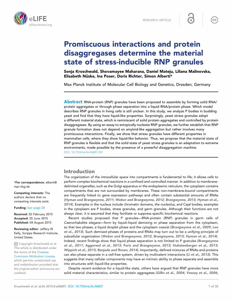
To get insight into this question, we analyzed the PD of Rnq1 in yeast cells. First, we studied its
subcellular localization in yeast carrying the background prion [PIN+], a factor that promotes the
conversion of the Rnq1 protein and other yeast prion proteins into an amyloid state. In these cells,
GFP (green fluorescent protein)-tagged Rnq1PD assembled into punctate structures (Figure 1A). To
determine whether Rnq1PD could also aggregate in the absence of a co-inducing prion, we expressed
it in a [pin−] background. The resulting cells also displayed a punctate fluorescence signal (Figure 1A).
However, the pattern of aggregation was different, because not all the signal was concentrated in foci
(Figure 1—figure supplement 2). Next, we analyzed these cells by semi-denaturing detergent-agarose
gel electrophoresis (SDD-AGE); this method can separate SDS (sodium dodecyl sulfate) -soluble from
SDS-insoluble fractions, SDS insolubility being a hallmark of amyloid polymers (Alberti et al.,
2010). Using this technique, we found that Rnq1PD formed SDS-resistant polymers in [PIN+]cells (Figure 1B). The Rnq1PD structures in [PIN+] cells could also be stained with the amyloid-
specific dye Thioflavin T (ThT) (Figure 1C). In contrast, Rnq1PD expressed in [pin−] cells did not
form SDS-resistant aggregates (Figure 1B) and it could not be stained with ThT (Figure 1C).
Thus, we conclude that Rnq1PD can form two types of assemblies in yeast cells: amyloid-like
aggregates and non-amyloid assemblies.
Hexanediol can disrupt non-amyloid assemblies formed by PLDsOur next goal was to develop a tool that could differentiate between these two assembled states of
Rnq1PD. To do this, we made use of the chemical 1,6-hexanediol, an aliphatic alcohol that
Kroschwald et al. eLife 2015;4:e06807. DOI: 10.7554/eLife.06807 3 of 32
Research article Biochemistry | Cell biology
Figure 1. Prion-like domains can access two distinct aggregated states, only one of which is amyloid-like.
(A) Fluorescence microscopy of yeast cells expressing sfGFP-tagged Rnq1PD in [PIN+] and [pin−] cells. White
lines indicate the cell boundaries. Scale bars: 5 μm. Also see related Figure 1—figure supplements 1–3.
(B) Semi-denaturing detergent-agarose gel electrophoresis (SDD-AGE) of [PIN+] and [pin−] cells containing
a plasmid for expression of Rnq1PD-sfGFP. SDS-resistant amyloid polymers show slower migration in comparison to
SDS-soluble monomers. Proteins were detected by immunoblotting with a GFP-specific antibody. (C) Thioflavin
T (ThT) staining of [PIN+] and [pin−] cells expressing Rnq1PD-mCherry from a plasmid. Note that only amyloid-like
assemblies can be stained with ThT. (D) 1,6-hexanediol treatment specifically disrupts non-amyloid Rnq1PD
assemblies and not amyloids. Fluorescence time-lapse microscopy of [PIN+] (top panel) and [pin−] cells (bottom
panel) expressing Rnq1PD-sfGFP. Time points are before treatment (Before) and 38 min after treatment with 10%
1,6-hexanediol (After). In the control condition (Mock) only media was added. Cells were permeabilized with 10 μg/ml digitonin. See corresponding Video 1. (E) Cells expressing Rnq1PD-sfGFP were treated with 10% 1,6-hexanediol
and digitonin for 1 hr to dissolve non-amyloid Rnq1PD assemblies (Before). Hexanediol was washed out and
replaced with normal growth media (After), and the cells were observed with fluorescence microscopy. Also see
corresponding Video 2.
DOI: 10.7554/eLife.06807.003
The following figure supplements are available for figure 1:
Figure supplement 1. Lsm4 has a prion-like C-terminal domain (underlined) that is enriched for asparagines (N) and
glutamines (Q) and contains hydrophobic residues (L, V, I, M, F).
DOI: 10.7554/eLife.06807.004
Figure supplement 2. Rnq1PD shows a different aggregation pattern in [pin−] and [PIN+] cells.DOI: 10.7554/eLife.06807.005
Figure supplement 3. The amino acid sequence of the prion domain of Rnq1 resembles that of FG repeat-
containing low-complexity domains of nucleoporins (hydrophobic aromatic residues in an asparagine- and
glutamine-rich polar sequence background).
DOI: 10.7554/eLife.06807.006
Kroschwald et al. eLife 2015;4:e06807. DOI: 10.7554/eLife.06807 4 of 32
Research article Biochemistry | Cell biology
has previously been shown to perturb
nucleoporin-mediated transport across the nu-
clear pore (Ribbeck and Gorlich, 2002; Patel
et al., 2007). Many nucleoporins contain domains
of low sequence complexity, which are composi-
tionally similar to yeast PDs and form a sieve-like
matrix that enables the selective passage of cargo
complexes (Frey et al., 2006; Frey and Gorlich,
2007; Hulsmann et al., 2012). Importantly, sieve
formation involves weak hydrophobic interactions
between phenylalanine–glycine repeats that are
embedded in the PLD. When these interactions
are perturbed by 1,6-hexanediol, nucleocytoplas-
mic transport ceases (Ribbeck and Gorlich, 2002;
Patel et al., 2007).
The amino acid composition of the PD of Rnq1 is similar to that of nucleoporins (Figure 1—figure
supplement 3), suggesting that Rnq1PD aggregation may likewise be affected by hexanediol.
Hexanediol indeed triggered the dissolution of non-amyloid Rnq1PD assemblies, whereas the amyloid
form remained unaffected (Figure 1D, Video 1). Importantly, this process was reversible (Figure 1E,
Video 2). This suggests that hexanediol can differentiate between these two types of assemblies and
may thus be a powerful tool to interfere with the formation of structures that depend on weak
interactions between sticky PLDs.
The prion-like protein Lsm4 does not adopt an amyloid-like conformationin P bodiesThe PLD of yeast Lsm4 is required for the assembly of P bodies (Decker et al., 2007). However, the
PLD of Lsm4 has not only been implicated in RNP granule formation, it also assembles into an
amyloid-like state (Alberti et al., 2009). Because the induction of an amyloid state is a concentration-
dependent nucleation process, the conforma-
tional conversion of prion-like proteins into
amyloid can be induced in [PIN+] yeast cells by
raising their concentration. Thus, by expressing
additional Lsm4 from a plasmid in the [PIN+]background, we could genetically drive endoge-
nous Lsm4 into an amyloid state. In this state,
endogenous Lsm4 formed one to a few fluores-
cent puncta in the cytoplasm (Figure 2A, upper
panel). Lsm4 was also present in puncta in
uninduced cells, but they were smaller (Figure 2A,
left lower panel). Previous studies identified these
puncta as P bodies (Decker et al., 2007). Because
P-body formation is strongly enhanced by stress, we
next analyzed the localization of Lsm4 after glucose
depletion. Now, Lsm4 was present in a few large
puncta per cell (Figure 2A, right lower panel), and
this stress-induced localization pattern was reminis-
cent of the pattern formed by Lsm4 in the amyloid
conformation. We further observed that other
P-body components such as Edc3 and Dcp2 co-
localized with amyloid-like Lsm4 structures
(Figure 2B). One potential explanation for this is
that amyloid formation by Lsm4 occurs during the
formation of P bodies.
To investigate this possibility, we analyzed
Lsm4 in the P-body state. First, we generated
lysates from stressed cells and subjected them to
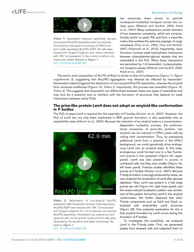
Video 1. Hexanediol treatment specifically disrupts
non-amyloid Rnq1PD assemblies and not amyloids.
Fluorescence time-lapse microscopy of [PIN+] and[pin−] cells expressing Rnq1PD-sfGFP. All cells were
treated with 10 μg/ml digitonin and, where indicated,
with 10% 1,6-hexanediol. In the control condition only
media was added. Related to Figure 1.
DOI: 10.7554/eLife.06807.007
Video 2. Reformation of non-amyloid Rnq1PD
assemblies after hexanediol removal. Cells expressing
Rnq1PD-sfGFP were treated with 10% 1,6-hexanediol
and 10 μg/ml digitonin for 1 hr to dissolve non-amyloid
Rnq1PD assemblies. Hexanediol was washed out and
replaced with normal growth media and the cells were
observed by fluorescence time-lapse microscopy. Re-
lated to Figure 1.
DOI: 10.7554/eLife.06807.008
Kroschwald et al. eLife 2015;4:e06807. DOI: 10.7554/eLife.06807 5 of 32
Research article Biochemistry | Cell biology
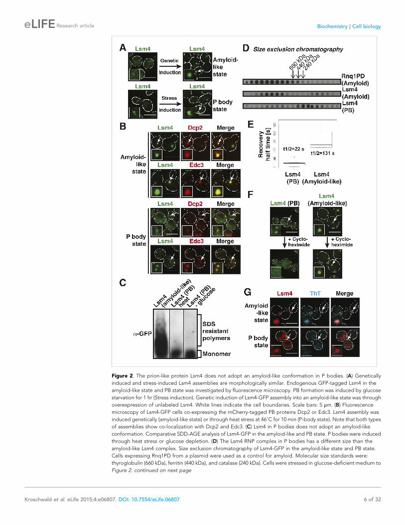
Figure 2. The prion-like protein Lsm4 does not adopt an amyloid-like conformation in P bodies. (A) Genetically
induced and stress-induced Lsm4 assemblies are morphologically similar. Endogenous GFP-tagged Lsm4 in the
amyloid-like state and PB state was investigated by fluorescence microscopy. PB formation was induced by glucose
starvation for 1 hr (Stress induction). Genetic induction of Lsm4-GFP assembly into an amyloid-like state was through
overexpression of unlabeled Lsm4. White lines indicate the cell boundaries. Scale bars: 5 μm. (B) Fluorescence
microscopy of Lsm4-GFP cells co-expressing the mCherry-tagged PB proteins Dcp2 or Edc3. Lsm4 assembly was
induced genetically (amyloid-like state) or through heat stress at 46˚C for 10 min (P-body state). Note that both types
of assemblies show co-localization with Dcp2 and Edc3. (C) Lsm4 in P bodies does not adopt an amyloid-like
conformation. Comparative SDD-AGE analysis of Lsm4-GFP in the amyloid-like and PB state. P bodies were induced
through heat stress or glucose depletion. (D) The Lsm4 RNP complex in P bodies has a different size than the
amyloid-like Lsm4 complex. Size exclusion chromatography of Lsm4-GFP in the amyloid-like state and PB state.
Cells expressing Rnq1PD from a plasmid were used as a control for amyloid. Molecular size standards were:
thyroglobulin (660 kDa), ferritin (440 kDa), and catalase (240 kDa). Cells were stressed in glucose-deficient medium to
Figure 2. continued on next page
Kroschwald et al. eLife 2015;4:e06807. DOI: 10.7554/eLife.06807 6 of 32
Research article Biochemistry | Cell biology
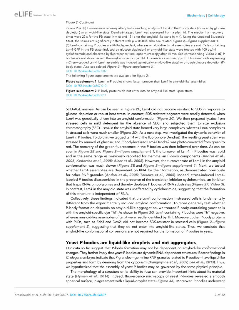
SDD-AGE analysis. As can be seen in Figure 2C, Lsm4 did not become resistant to SDS in response to
glucose depletion or robust heat stress. In contrast, SDS-resistant polymers were readily detected, when
Lsm4 was genetically driven into an amyloid conformation (Figure 2C). We then prepared lysates from
stressed cells in mild detergent (in the absence of SDS) and subjected them to size exclusion
chromatography (SEC). Lsm4 in the amyloid state formed very large complexes, whereas Lsm4 complexes
in stressed cells were much smaller (Figure 2D). As a next step, we investigated the dynamic behavior of
Lsm4 in P bodies. To do this, we tagged Lsm4 with the fluorophore Dendra2. The resulting yeast strain was
stressed by removal of glucose, and P body-localized Lsm4-Dendra2 was photo-converted from green to
red. The recovery of the green fluorescence in the P bodies was then followed over time. As can be
seen in Figure 2E and Figure 2—figure supplement 1, the turnover of Lsm4 in P bodies was rapid
and in the same range as previously reported for mammalian P-body components (Andrei et al.,
2005; Kedersha et al., 2005; Aizer et al., 2008). However, the turnover rate of Lsm4 in the amyloid
conformation was much slower (Figure 2E and Figure 2—figure supplement 1). Next, we tested
whether Lsm4 assemblies are dependent on RNA for their formation, as demonstrated previously
for other RNP granules (Andrei et al., 2005; Teixeira et al., 2005). Indeed, stress-induced Lsm4-
labeled P bodies disassembled in the presence of the translation inhibitor cycloheximide, an inhibitor
that traps RNAs on polysomes and thereby depletes P bodies of RNA substrates (Figure 2F, Video 3).
In contrast, Lsm4 in the amyloid state was unaffected by cycloheximide, suggesting that the formation
of this structure is independent of RNA.
Collectively, these findings indicated that the Lsm4 conformation in stressed cells is fundamentally
different from the experimentally induced amyloid conformation. To more generally test whether
P-body formation depends on amyloid-like aggregation, we treated P body-containing yeast cells
with the amyloid-specific dye ThT. As shown in Figure 2G, Lsm4-containing P bodies were ThT negative,
whereas amyloid-like assemblies of Lsm4 were readily identified by ThT. Moreover, other P-body proteins
with PLDs, such as Edc3 and Dcp2, did not become SDS-resistant in stressed cells (Figure 2—figure
supplement 2), suggesting that they do not enter into amyloid-like states. Thus, we conclude that
amyloid-like conformational conversions are not required for the formation of P bodies in yeast.
Yeast P-bodies are liquid-like droplets and not aggregatesOur data so far suggest that P-body formation may not be dependent on amyloid-like conformational
changes. They further imply that yeast P-bodies are dynamic RNA-dependent structures. Recent findings in
C. elegans embryos indicate that P granules—germ line RNP granules related to P bodies—have liquid-like
properties and form by demixing from the cytoplasm (Brangwynne et al., 2009; Lee et al., 2013). Thus,
we hypothesized that the assembly of yeast P-bodies may be governed by the same physical principle.
The morphology of a structure or its ability to fuse can provide important hints about its material
state (Hyman et al., 2014). Indeed, fluorescence microscopy of yeast P-bodies revealed a smooth
spherical surface, in agreement with a liquid-droplet state (Figure 3A). Moreover, P bodies underwent
Figure 2. Continued
induce PBs. (E) Fluorescence recovery after photobleaching analysis of Lsm4 in the P body state (induced by glucose
depletion) or amyloid-like state. Dendra2-tagged Lsm4 was expressed from a plasmid. The median half-recovery
times were 22 s for the PB state (n = 6) and 131 s for the amyloid-like state (n = 4). Using the unpaired Student’s
t-test, the values are significantly different with p = 0.0018. Also see related Figure 2—figure supplement 1.
(F) Lsm4-containing P bodies are RNA-dependent, whereas amyloid-like Lsm4 assemblies are not. Cells containing
Lsm4-GFP in the PB state (induced by glucose depletion) or amyloid-like state were treated with 100 μg/ml
cycloheximide and observed by fluorescence time-lapse microscopy after 14 min. See corresponding Video 3. (G) P
bodies are not stainable with the amyloid-specific dye ThT. Fluorescence microscopy of ThT-stained cells expressing
mCherry-tagged Lsm4. Lsm4 assembly was induced genetically (amyloid-like state) or through glucose depletion (P-
body state). Also see related Figure 2—figure supplement 2.
DOI: 10.7554/eLife.06807.009
The following figure supplements are available for figure 2:
Figure supplement 1. Lsm4 in P bodies shows faster turnover than Lsm4 in amyloid-like assemblies.
DOI: 10.7554/eLife.06807.010
Figure supplement 2. P-body proteins do not enter into an amyloid-like state upon stress.
DOI: 10.7554/eLife.06807.011
Kroschwald et al. eLife 2015;4:e06807. DOI: 10.7554/eLife.06807 7 of 32
Research article Biochemistry | Cell biology
frequent fusion events (Figure 3A,B). Upon fusion,
the newly formed body rapidly relaxed into a spher-
ical shape (Figure 3A). Such fast relaxation times
indicate that the viscosity of P bodies is relatively
low, in agreement with a dynamic liquid-like state.
A liquid state requires that the molecular
interactions are weak and permanently changing
(Hyman et al., 2014). Solid states instead are based
on tight interactions, which are largely invariant over
time. We reasoned that hexanediol could be a
useful tool to differentiate between liquid-like and
solid-like states, because of its ability to interfere
with weak hydrophobic interactions. In fact, hex-
anediol has been used previously to assess the
liquid-like nature of germ granules in C. elegans
(Updike et al., 2011). Indeed, when we added
hexanediol to yeast cells, it dissolved P bodies (Figure 3C, Video 4). Importantly, this effect was rapid
(Figure 3—figure supplement 1) and reversible, as P bodies reformed after hexanediol washout
(Figure 3D, Video 5). This suggests that P bodies—similar to germ granules in C. elegans—rely on weak
interactions for their formation. Thus, we conclude that yeast P-bodies resemble liquid droplets with
physicochemical properties unlike those of solid amyloid-like aggregates.
Yeast stress granules have different material properties than P bodiesStress granules are related to P bodies; they are induced by stress and function as storage depots for
mRNAs (Anderson and Kedersha, 2008; Decker and Parker, 2012). We therefore wondered
whether yeast stress granules show similar liquid-like behavior as P bodies. To investigate this, we
tested the effect of hexanediol on stress granules. Remarkably, hexanediol did not affect stress
granule integrity, even when applied for extended times or in the presence of digitonin to facilitate
hexanediol entry into the cells (Figure 4A, Figures 4—figure supplement 1, Video 6). Thus, we
conclude that stress granules are distinct from P bodies and may instead have a more solid character.
Like the constituent proteins of P bodies, many stress granule proteins contain PLDs. To investigate
whether these proteins undergo amyloid-like conformational conversions, we initially focused on
one stress granule protein: Nrp1. Nrp1 is a prototypical stress granule protein in that it contains a RNA-
binding domain (RRM - RNA recognition motif) and a PLD (Buchan et al., 2008). However, Nrp1 can
also be converted into an amyloid-like state (Alberti et al., 2009). Moreover, it is one of the proteins
capable of binding to b-isox (Kwon et al., 2013), a chemical that specifically binds to low-complexity
domains, which have the ability to undergo amyloid-like conformational conversions (Kato et al., 2012).
To investigate whether amyloid-like aggregation of Nrp1 is involved in stress granule assembly, we first
converted endogenous Nrp1 into an amyloid state, using a genetic approach (see ‘Materials and methods’
for details). In the resulting cells, Nrp1 localized to one or a few bright cytoplasmic foci (Figure 4B, top
panel). The same localization pattern was observed in stressed cells (Figure 4B, lower panel). Both
structures co-localized with other stress granule proteins such as Pab1 (Figure 4C), which is consistent with
the possibility that genetically induced amyloid-like Nrp1 structures are genuine stress granules. Thus, we
next investigated whether stress triggers an amyloid-like conversion in Nrp1 using SDD-AGE. However,
Nrp1 did not become SDS-resistant in response to stress (Figure 4D). Nrp1 complexes also had a very
different molecular size in stressed cells (Figure 4—figure supplement 2). In addition, we found that the
turnover rate of amyloid-like Nrp1 was much slower (Figure 4E, Figure 4—figure supplement 3). As
a next step, we investigated four additional stress granule proteins with PLDs using SDD-AGE. However,
none of these proteins formed amyloid-like structures in stressed cells (Figure 4—figure supplement 4).
Furthermore, stress-induced Nrp1 assemblies were ThT negative, whereas genetically induced Nrp1
assemblies were stainable with ThT (Figure 4F). Thus, we conclude that stress granules are more solid-like
but like P bodies do not transition into amyloid-like states upon stress.
Yeast stress granules resemble amorphous protein aggregatesWhy do stress granules adopt a different material state than P bodies? A previous study showed that
in heat-stressed cells, the formation of stress granules coincides with the formation of protein
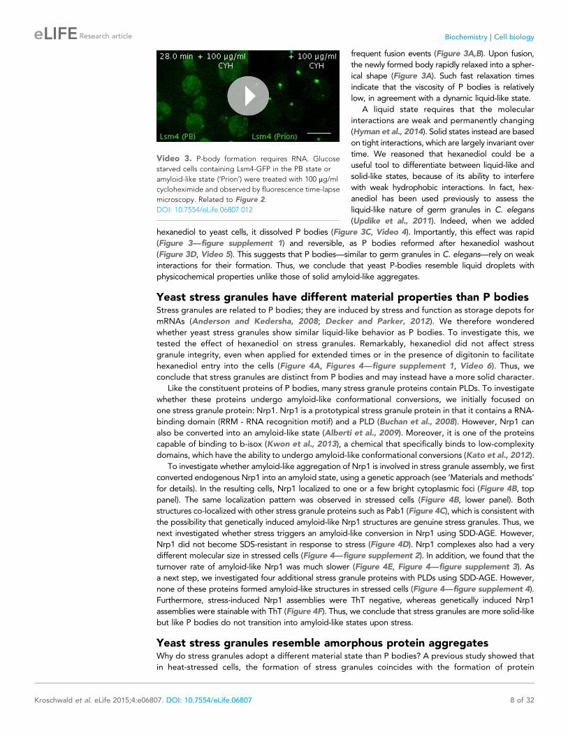
Video 3. P-body formation requires RNA. Glucose
starved cells containing Lsm4-GFP in the PB state or
amyloid-like state (‘Prion’) were treated with 100 μg/ml
cycloheximide and observed by fluorescence time-lapse
microscopy. Related to Figure 2.
DOI: 10.7554/eLife.06807.012
Kroschwald et al. eLife 2015;4:e06807. DOI: 10.7554/eLife.06807 8 of 32
Research article Biochemistry | Cell biology
aggregates, which result from stress-induced protein misfolding (Cherkasov et al., 2013). Thus, we
reasoned that stress granules might be functional aggregates that behave in a similar manner as
aggregates formed by misfolded proteins. To investigate this, we compared the aggregation
behavior of stress granule components and misfolded proteins by time-lapse microscopy. For this
purpose, we used yeast strains co-expressing GFP-tagged stress granule proteins (Pbp1 or Nrp1) and
mCherry-tagged misfolding-prone proteins (a mutant variant of luciferase or a thermo-labile variant of
Ubc9, Ubc9ts) (Kaganovich et al., 2008; Gupta et al., 2011). Indeed, in cells that were exposed to
a robust heat shock of 46˚C, stress granule proteins co-localized with luciferase or Ubc9ts in punctate
structures (Figure 5A, Figure 5—figure supplement 1). Co-localization was also observed between
Nrp1 and several chaperones, such as Hsp42, Ssa1, and Hsp104 (Figure 5—figure supplement 2).
This indicates that stress granule proteins and misfolded proteins are co-deposited, as previously
suggested (Cherkasov et al., 2013).
Despite the fact that protein aggregates and stress granule components showed strong co-
localization, stress granules dissolved faster than misfolded proteins during recovery from stress
(Figure 5—figure supplement 3; Videos 7–9). Importantly, cells that had dissolved their stress
granules re-entered into the cell cycle (Videos 7–9), although they still contained aggregated
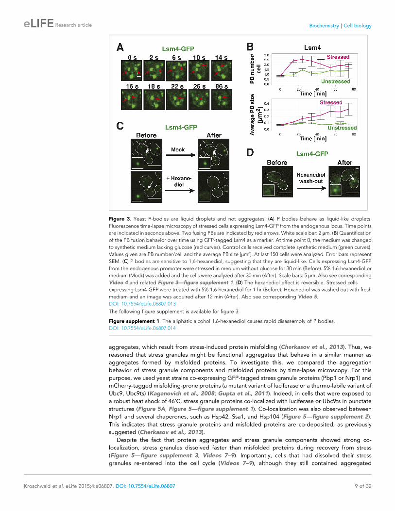
Figure 3. Yeast P-bodies are liquid droplets and not aggregates. (A) P bodies behave as liquid-like droplets.
Fluorescence time-lapse microscopy of stressed cells expressing Lsm4-GFP from the endogenous locus. Time points
are indicated in seconds above. Two fusing PBs are indicated by red arrows. White scale bar: 2 μm. (B) Quantification
of the PB fusion behavior over time using GFP-tagged Lsm4 as a marker. At time point 0, the medium was changed
to synthetic medium lacking glucose (red curves). Control cells received complete synthetic medium (green curves).
Values given are PB number/cell and the average PB size [μm2]. At last 150 cells were analyzed. Error bars represent
SEM. (C) P bodies are sensitive to 1,6-hexanediol, suggesting that they are liquid-like. Cells expressing Lsm4-GFP
from the endogenous promoter were stressed in medium without glucose for 30 min (Before). 5% 1,6-hexanediol or
medium (Mock) was added and the cells were analyzed after 30 min (After). Scale bars: 5 μm. Also see corresponding
Video 4 and related Figure 3—figure supplement 1. (D) The hexanediol effect is reversible. Stressed cells
expressing Lsm4-GFP were treated with 5% 1,6-hexanediol for 1 hr (Before). Hexanediol was washed out with fresh
medium and an image was acquired after 12 min (After). Also see corresponding Video 5.
DOI: 10.7554/eLife.06807.013
The following figure supplement is available for figure 3:
Figure supplement 1. The aliphatic alcohol 1,6-hexanediol causes rapid disassembly of P bodies.
DOI: 10.7554/eLife.06807.014
Kroschwald et al. eLife 2015;4:e06807. DOI: 10.7554/eLife.06807 9 of 32
Research article Biochemistry | Cell biology
misfolded proteins. This suggests that the presence
of misfolded proteins does not prevent re-entry
into the cell cycle and that resumption of growth
may be coupled to stress granule dissolution. It also
implies that stress granule components are less
aggregation-prone than the misfolding-prone
model proteins used in our study. To investigate
this possibility, we repeated the heat shock
experiment at 42˚C. Indeed, under mild heat-
shock conditions only luciferase and Ubc9ts formed
visible aggregates, whereas the stress granule
component Nrp1 remained diffusely localized
(Figure 5B and Figure 5—figure supplement 4).
These findings suggest that stress granule compo-
nents behave like misfolding-prone proteins, which
reversibly aggregate into stress granules, when
cells are exposed to robust environmental stress.
Stress granules form under a variety of con-
ditions, and they show different compositions depending on the nature of the inducing stress (Hoyle
et al., 2007; Grousl et al., 2009; Buchan et al., 2011). To determine whether stress granules induced
by other stresses behave in a similar way, we exposed yeast to glucose depletion. Glucose removal also
caused stress granule formation, and this was accompanied by a low level of luciferase aggregation (see
Video 10). However, in contrast to heat stress conditions, luciferase and stress granule components
coalesced into largely distinct structures (Figure 5C and Figure 5—figure supplement 5). Thus, we
conclude that stress granule proteins aggregate under a variety of conditions but that co-aggregation
with misfolded proteins is most pronounced during robust heat shock.
A previous report proposed a role for disaggregating chaperones in stress granule dissolution
(Cherkasov et al., 2013). Therefore, we next tested how protein disaggregases affect stress granule
formation. Three different proteins promote the disaggregation of protein aggregates in yeast:
Hsp104 and two members of the Hsp110 family, called Sse1 and Sse2 (Glover and Lindquist, 1998;
Shorter, 2011; Duennwald et al., 2012;
Rampelt et al., 2012; Doyle et al., 2013).
We compared wild-type cells and cells in which
these proteins had been inactivated genetically.
Deletion of either of the disaggregases led to
more pronounced stress granule assembly and a
delay in stress granule disassembly (Figure 5D,E,
Video 11). However, inactivation of Hsp104 had
the strongest effect. Similar findings were
obtained for stress granules induced by glucose
depletion (Figure 5F). Thus, we conclude that
stress granules are functional aggregates and
that the components contained in stress granules
need to be reactivated by disaggregation before
cells can re-enter into the cell cycle. Consistent
with this, a recent report showed that cell
cycle-associated RNP granules in the multinu-
clear fungus Ashbya gossypii are functional
aggregates that are remodeled by chaperones
(Lee et al., 2015).
Misfolded proteins can nucleateyeast stress granulesOur findings indicate that during severe stress
conditions both misfolded proteins and
stress granule components co-aggregate.
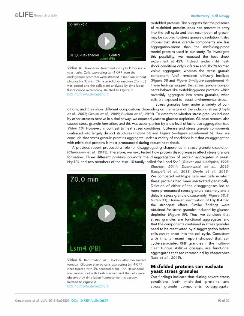
Video 4. Hexanediol treatment disrupts P bodies in
yeast cells. Cells expressing Lsm4-GFP from the
endogenous promoter were stressed in medium without
glucose for 30 min. 5% hexanediol or medium (Control)
was added and the cells were analyzed by time-lapse
fluorescence microscopy. Related to Figure 3.
DOI: 10.7554/eLife.06807.015
Video 5. Reformation of P bodies after hexanediol
removal. Glucose starved cells expressing Lsm4-GFP
were treated with 5% hexanediol for 1 hr. Hexanediol
was washed out with fresh medium and the cells were
observed by time-lapse fluorescence microscopy.
Related to Figure 3.
DOI: 10.7554/eLife.06807.016
Kroschwald et al. eLife 2015;4:e06807. DOI: 10.7554/eLife.06807 10 of 32
Research article Biochemistry | Cell biology
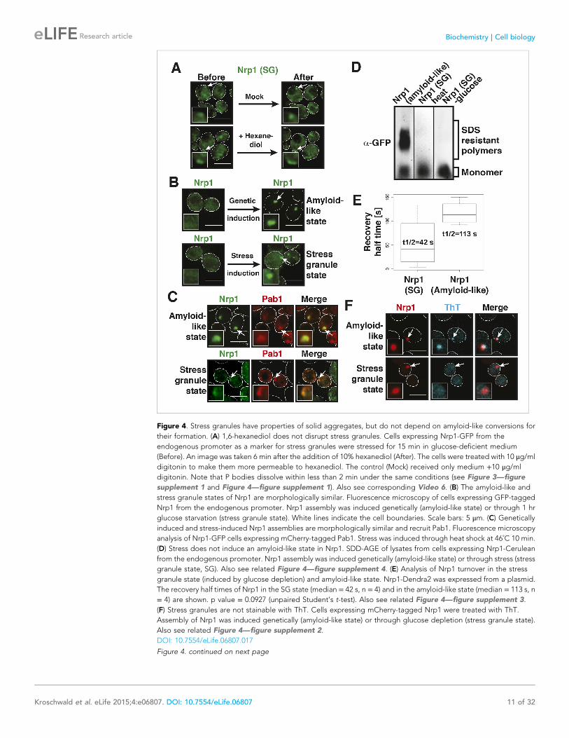
Figure 4. Stress granules have properties of solid aggregates, but do not depend on amyloid-like conversions for
their formation. (A) 1,6-hexanediol does not disrupt stress granules. Cells expressing Nrp1-GFP from the
endogenous promoter as a marker for stress granules were stressed for 15 min in glucose-deficient medium
(Before). An image was taken 6 min after the addition of 10% hexanediol (After). The cells were treated with 10 μg/ml
digitonin to make them more permeable to hexanediol. The control (Mock) received only medium +10 μg/ml
digitonin. Note that P bodies dissolve within less than 2 min under the same conditions (see Figure 3—figure
supplement 1 and Figure 4—figure supplement 1). Also see corresponding Video 6. (B) The amyloid-like and
stress granule states of Nrp1 are morphologically similar. Fluorescence microscopy of cells expressing GFP-tagged
Nrp1 from the endogenous promoter. Nrp1 assembly was induced genetically (amyloid-like state) or through 1 hr
glucose starvation (stress granule state). White lines indicate the cell boundaries. Scale bars: 5 μm. (C) Genetically
induced and stress-induced Nrp1 assemblies are morphologically similar and recruit Pab1. Fluorescence microscopy
analysis of Nrp1-GFP cells expressing mCherry-tagged Pab1. Stress was induced through heat shock at 46˚C 10 min.
(D) Stress does not induce an amyloid-like state in Nrp1. SDD-AGE of lysates from cells expressing Nrp1-Cerulean
from the endogenous promoter. Nrp1 assembly was induced genetically (amyloid-like state) or through stress (stress
granule state, SG). Also see related Figure 4—figure supplement 4. (E) Analysis of Nrp1 turnover in the stress
granule state (induced by glucose depletion) and amyloid-like state. Nrp1-Dendra2 was expressed from a plasmid.
The recovery half times of Nrp1 in the SG state (median = 42 s, n = 4) and in the amyloid-like state (median = 113 s, n
= 4) are shown. p value = 0.0927 (unpaired Student’s t-test). Also see related Figure 4—figure supplement 3.
(F) Stress granules are not stainable with ThT. Cells expressing mCherry-tagged Nrp1 were treated with ThT.
Assembly of Nrp1 was induced genetically (amyloid-like state) or through glucose depletion (stress granule state).
Also see related Figure 4—figure supplement 2.
DOI: 10.7554/eLife.06807.017
Figure 4. continued on next page
Kroschwald et al. eLife 2015;4:e06807. DOI: 10.7554/eLife.06807 11 of 32
Research article Biochemistry | Cell biology
However, despite being deposited in spatial proximity, the two types of proteins do not seem to form
mixed aggregates, in particular when exposed to mild stress condition such as glucose depletion.
A previous study suggested that misfolded proteins seed the formation of stress granules
(Cherkasov et al., 2013). However, it remained unclear whether this is a general mode of stress granule
formation or only applies to conditions of robust heat stress. Investigation of a seeding function requires
experimental control over the aggregated state of a protein in the context of a living cell. To achieve
this, we developed a method based on a self-assembling protein fragment, derived from a viral capsid
protein (μNS). This fragment assembles into large spherical particles in cells, and these particles could
be visualized by adding GFP to the N terminus (Figure 6A). To exclude that these particles are
aggregates of misfolded protein, we first tested whether they co-localize with chaperones (Ssa1 and
Hsp104). However, chaperones did not associate with μNS particles (Figure 6—figure supplement 1),
suggesting that the particles are invisible to the protein quality control system.
An earlier study used μNS particles to test for protein–protein interactions in budding yeast
(Schmitz et al., 2009). In this study, bait proteins were genetically fused to the μNS fragment, which
resulted in the presentation of the bait protein on the particle surface. Using this approach, we
generated particles that carried either mutated luciferase or Ubc9ts on the surface. We found that
both types of particles were recognized by chaperones (Figure 6B and Figure 6—figure supplement 2),
indicating that the misfolded proteins are accessible on the particle surface. Using these particles, we
next tested whether misfolded Ubc9ts or luciferase could recruit stress granule components. Under
normal growth conditions (25˚C), the two stress granule proteins Pab1 or Pub1 were not enriched on the
particle surface (Figure 6C and Figure 6—figure supplement 3, upper panels). However, during
a robust heat shock Pub1 and Pab1 accumulated on μNS particles (Figure 6C and Figure 6—figure
supplement 3, lower panels). Naked particles on
the other hand did not recruit Pab1 or Pub1
(Figure 6—figure supplement 4), indicating that
stress granule assembly was specifically triggered by
the misfolded proteins. Furthermore, interaction of
Pub1 or Pab1 with luciferase was only observed
under robust heat shock and not under glucose
depletion conditions (Figure 6—figure supplement
5). Thus, we conclude that misfolded proteins can
nucleate the formation of stress granules under
severe heat stress conditions, but not under mild
stress conditions or during normal growth.
How do stress granule proteins interact with
misfolded proteins? To address this question, we
focused on the stress granule protein Nrp1. Nrp1
has an intrinsically disordered PLD, which can
undergo amyloid-like conformational conversions
(Alberti et al., 2009) and shows specific binding
to b-isox (Kwon et al., 2013). We speculated
that the structural flexibility of the PLD could
promote the interaction of Nrp1 with misfolded
proteins. To test this hypothesis, we mildly
Figure 4. Continued
The following figure supplements are available for figure 4:
Figure supplement 1. P bodies but not stress granules are sensitive to hexanediol.
DOI: 10.7554/eLife.06807.018
Figure supplement 2. Stress granule-associated and amyloid-like Nrp1 form different complexes in yeast cell lysate.
DOI: 10.7554/eLife.06807.019
Figure supplement 3. Analysis of the turnover of Nrp1 in the stress granule and the amyloid-like state.
DOI: 10.7554/eLife.06807.020
Figure supplement 4. Prion-like stress granule proteins do not transition into an amyloid-like state.
DOI: 10.7554/eLife.06807.021
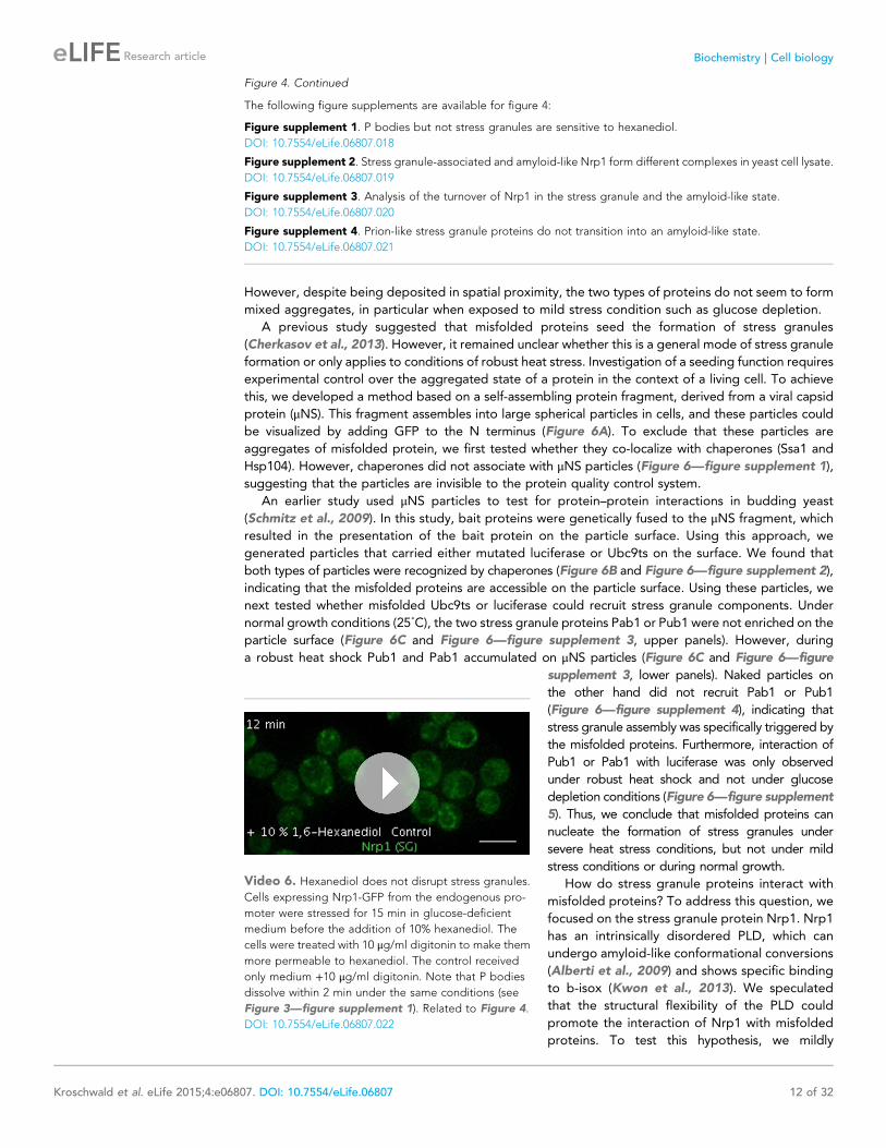
Video 6. Hexanediol does not disrupt stress granules.Cells expressing Nrp1-GFP from the endogenous pro-
moter were stressed for 15 min in glucose-deficient
medium before the addition of 10% hexanediol. The
cells were treated with 10 μg/ml digitonin to make them
more permeable to hexanediol. The control received
only medium +10 μg/ml digitonin. Note that P bodies
dissolve within 2 min under the same conditions (see
Figure 3—figure supplement 1). Related to Figure 4.
DOI: 10.7554/eLife.06807.022
Kroschwald et al. eLife 2015;4:e06807. DOI: 10.7554/eLife.06807 12 of 32
Research article Biochemistry | Cell biology
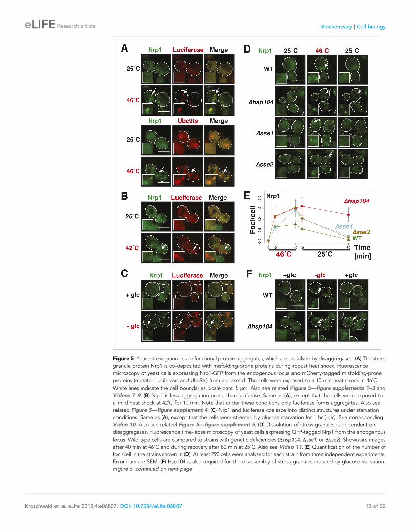
Figure 5. Yeast stress granules are functional protein aggregates, which are dissolved by disaggregases. (A) The stress
granule protein Nrp1 is co-deposited with misfolding-prone proteins during robust heat shock. Fluorescence
microscopy of yeast cells expressing Nrp1-GFP from the endogenous locus and mCherry-tagged misfolding-prone
proteins (mutated luciferase and Ubc9ts) from a plasmid. The cells were exposed to a 10-min heat shock at 46˚C.
White lines indicate the cell boundaries. Scale bars: 5 μm. Also see related Figure 5—figure supplements 1–3 and
Videos 7–9. (B) Nrp1 is less aggregation prone than luciferase. Same as (A), except that the cells were exposed to
a mild heat shock at 42˚C for 10 min. Note that under these conditions only luciferase forms aggregates. Also see
related Figure 5—figure supplement 4. (C) Nrp1 and luciferase coalesce into distinct structures under starvation
conditions. Same as (A), except that the cells were stressed by glucose starvation for 1 hr (-glc). See corresponding
Video 10. Also see related Figure 5—figure supplement 5. (D) Dissolution of stress granules is dependent on
disaggregases. Fluorescence time-lapse microscopy of yeast cells expressing GFP-tagged Nrp1 from the endogenous
locus. Wild-type cells are compared to strains with genetic deficiencies (Δhsp104, Δsse1, or Δsse2). Shown are images
after 40 min at 46˚C and during recovery after 80 min at 25˚C. Also see Video 11. (E) Quantification of the number of
foci/cell in the strains shown in (D). At least 290 cells were analyzed for each strain from three independent experiments.
Error bars are SEM. (F) Hsp104 is also required for the disassembly of stress granules induced by glucose starvation.
Figure 5. continued on next page
Kroschwald et al. eLife 2015;4:e06807. DOI: 10.7554/eLife.06807 13 of 32
Research article Biochemistry | Cell biology
overexpressed Nrp1-mCherry in cells carrying luciferase-coated μNS particles. As can be seen in
Figure 6D, Nrp1 weakly interacted with the particles, even in the absence of stress. Importantly, when
we subsequently applied a robust heat shock, a large amount of Nrp1 accumulated on the surface of the
particle (Figure 6D). This suggests that the interaction between Nrp1 and misfolded proteins is
sufficient to ectopically induce stress granule formation. Next, we tested a variant of Nrp1 lacking the
PLD. Remarkably, this variant was unable to recognize the luciferase-coated particle (Figure 6E). In
contrast, the isolated PLD showed robust binding to luciferase particles (Figure 6E), demonstrating that
the PLD is sufficient for the interaction with misfolded proteins. Thus, PLDs can promiscuously interact
with misfolded proteins, and such promiscuous interactions can nucleate the formation of stress
granules, in particular under conditions of robust heat stress.
Why do misfolded proteins only nucleate stress granules under robust heat-shock conditions? We
reasoned that under mild stress conditions the activity of the cellular chaperone machinery is sufficient
to prevent promiscuous interactions between misfolded proteins and stress granule components.
Indeed, when Hsp104 was inhibited, a mild heat shock was sufficient to induce the co-aggregation of
endogenous Nrp1 and misfolded proteins (Figure 6F and Figure 6—figure supplement 6).
A mild stress stimulus can protect cells from subsequent severe stress, a phenomenon known as
preconditioning (Parsell and Lindquist, 1993). Thus, we hypothesized that preconditioning could prevent
the co-aggregation of stress granule components and misfolded proteins. We preconditioned yeast cells
by increasing the temperatures incrementally from 25˚C to 46˚C. Under these conditions, Pab1 and Pub1
did not co-aggregate with misfolded proteins but
formed assemblies in distinct areas of the cell
(Figure 6G and Figure 6—figure supplement 7).
This shows that molecular chaperones constantly
work to prevent promiscuous interactions with
misfolded proteins and that misfolded proteins
only act as scaffolds for stress granules when the
capacity of the protein quality control machinery is
overrun. It also suggests that preconditioned cells
are protected from promiscuous interactions with
misfolded proteins, presumably because of the up-
regulation of chaperones.
Stress granule assembly isredundant and adaptableOur findings suggest that an interaction with
misfolded proteins is not required for stress
Figure 5. Continued
Wild-type or Δhsp104 cells expressing Nrp1-GFP from the endogenous locus were exposed to glucose starvation for
1 hr (image taken after 40 min at -glc) and observed 80 min after glucose-containing growth medium was added.
DOI: 10.7554/eLife.06807.023
The following figure supplements are available for figure 5:
Figure supplement 1. The stress granule protein Pbp1 is co-deposited with the misfolding-prone protein Ubc9ts
during robust heat shock.
DOI: 10.7554/eLife.06807.024
Figure supplement 2. The stress granule protein Nrp1 co-localizes with chaperones during robust heat shock.
DOI: 10.7554/eLife.06807.025
Figure supplement 3. Stress granules dissolve faster than protein aggregates.
DOI: 10.7554/eLife.06807.026
Figure supplement 4. A mild heat shock does not lead to co-deposition of stress granule components and
misfolding-prone proteins.
DOI: 10.7554/eLife.06807.027
Figure supplement 5. Stress granules and misfolding-prone proteins do not form mixed aggregates under glucose
depletion conditions.
DOI: 10.7554/eLife.06807.028
Video 7. Stress granule and protein aggregate
formation and dissolution in stressed cells. Yeast cells
expressing Nrp1-GFP from the endogenous locus were
transformed with plasmids for the expression of
mCherry-tagged mutant luciferase. Cells were exposed
to a robust heat shock at 46˚C and then transferred back
to 25˚C. Note that stress granules dissolve faster than
protein aggregates. Related to Figure 5.
DOI: 10.7554/eLife.06807.029
Kroschwald et al. eLife 2015;4:e06807. DOI: 10.7554/eLife.06807 14 of 32
Research article Biochemistry | Cell biology
granule formation. To investigate how stress
granules form independently of misfolded pro-
teins, we again focused on Nrp1. We first tested
which domains of Nrp1 are required for re-
cruitment to stress granules. As shown in
Figure 6H, the RBD alone was sufficient for
localization to stress granules, whereas the PLD
was not. This is consistent with many other
studies, which reported that PLDs are dispens-
able for RNP granule localization (Reijns et al.,
2008; Sun et al., 2011; Cai and Futcher, 2013;
Grousl et al., 2013; Kruger et al., 2013; Bley
et al., 2014). One potential explanation for the
observed dispensability of PLDs is that they
mediate interactions that are redundant and
weak and thus only manifest when these domains
are present at high local concentrations. To test
this possibility, we used our particle assay to concentrate stress granule proteins in living cells, thus,
creating a molecular environment similar to that in RNP granules. First, we generated particles that
carried full-length Nrp1 on the surface. After having confirmed that Nrp1-μNS particles are not
recognized by chaperones (Figure 6—figure supplement 8), we tested whether Nrp1 could
ectopically nucleate the formation of stress granules. Indeed, particle-bound Nrp1 was able to recruit
additional Nrp1 molecules from the cytosol, and upon heat shock (Figure 6I, bottom panel) and
glucose depletion (Figure 6—figure supplement 9, bottom panel) stress granules formed on Nrp1
particles. As a next step, we tested which domains of Nrp1 are required for this behavior. We
generated two deletion mutants comprising either the RBD or the PLD. We first confirmed that these
truncation mutants were not misfolded when presented on the particle surface (Figure 6—figure
supplement 10). Using these particles, we found that both the PLD and the RBD were able to recruit
full-length Nrp1 (Figure 6J) and promoted the formation of stress granules (Figure 6—figure
supplement 11 and Figure 6—figure supplement 12). Together, these findings indicate that stress
granule assembly is highly redundant and that nucleation can proceed in multiple ways, through PLDs
or RBDs. Our data further suggest that PLDs work synergistically with RBDs and only function when
present at a high local concentration, as during RNP granule assembly.
Maintenance of yeast P-body integrity requires Hsp104Our findings so far show that P bodies are liquid-like droplets, in contrast to stress granules, which
behave as true aggregates. Because of this distinction, we predicted that P bodies do not co-aggregate
with misfolded proteins. Indeed, we found that P bodies showed only marginal co-localization with
misfolded proteins in cells exposed to robust heat stress (Figure 7A and Figure 7—figure supplement 1).
Co-localization with chaperones, such as Hsp42,
Ssa1 and Hsp104, was also limited (Figure 7B and
Figure 7—figure supplement 2). We next tested
whether P-body formation and dissolution is
affected by protein disaggregases. Under normal
or mild stress conditions, Hsp104 deficiency did not
affect P-body formation. However, upon robust
heat stress (46˚C), the P-body protein Edc3
assembled into irregular aggregate-like struc-
tures (Figure 7C), which persisted for ex-
tended times (Video 12). These aggregate-
like structures co-localized with the stress
granule marker Pub1 and misfolded proteins
(Figure 7D), suggesting that Edc3 is mistargeted
to stress granules in the absence of Hsp104.
Similar findings were made for the P-body protein
Lsm4 (Figure 7—figure supplement 3). Next, we
Video 8. Stress granule and protein aggregate
formation and dissolution in stressed cells. Yeast cells
expressing Nrp1-GFP from the endogenous locus were
transformed with plasmids for the expression of
mCherry-tagged Ubc9ts. Cells were exposed to a robust
heat shock at 46˚C and then transferred back to 25˚C.
Note that stress granules dissolve faster than protein
aggregates. Related to Figure 5.
DOI: 10.7554/eLife.06807.030
Video 9. Stress granule and protein aggregate
formation and dissolution in stressed cells. Yeast cells
expressing Pbp1-GFP from the endogenous locus were
transformed with a plasmid for the expression of
mCherry-tagged Ubc9ts. Cells were exposed to a robust
heat shock at 46˚C and then transferred back to 25˚C.
Note that stress granules dissolve faster than protein
aggregates. Related to Figure 5.
DOI: 10.7554/eLife.06807.031
Kroschwald et al. eLife 2015;4:e06807. DOI: 10.7554/eLife.06807 15 of 32
Research article Biochemistry | Cell biology
generated strains that expressed Hsp104 at
different levels. We found that the amount of
Edc3 that co-aggregated with misfolded proteins
decreased when the expression level of Hsp104
was increased (Figure 7E, Video 13). This
indicates that maintenance of the liquid-like P-
body state requires the continuous action of
Hsp104 during acute stress and that P-body
components are mistargeted to stress granules,
when the disaggregation activity is insufficient.
Mammalian and yeast stressgranules have distinct materialproperties
Do mammalian P bodies and stress granules behave in a similar way as those of yeast?
To investigate the properties of P bodies and stress granules in mammalian cells, we generated stable
HeLa cell lines expressing GFP-tagged G3PB2 or DCP1a as markers for stress granules or P bodies,
respectively, using BAC TransgeneOmics (Poser et al., 2008).
We tested mammalian stress granules and P bodies for three characteristics that define a liquid-like
compartment (Hyman et al., 2014): first, a liquid compartment should be roughly spherical due to
surface tension. Second, the components within the compartment should undergo rapid internal
rearrangement and third, two liquid droplets should fuse and relax into one droplet. Indeed, the
stress granules and P bodies in our cell lines had a characteristic circular shape (Figure 8A), as
expected for a liquid-droplet state. We also noticed that stress granules, in particular in the early
stage of stress exposure, merged and formed larger structures over time (Figure 8B, Video 14), as
did P bodies (Figure 8B, Video 15). In both cases, the structures rapidly relaxed into more spherical
structures, in agreement with a liquid-like state. Next, we applied a technique known as ‘half-bleach’
to test for internal mobility within the compartment. In this method, roughly half a structure is
bleached, and the distribution of the fluorescence within the photo-manipulated structure is then
determined over time (Brangwynne et al., 2009). The analysis of such a half-bleach event showed
that G3BP2 was redistributed rapidly within stress granules from the unbleached to the bleached
area (because of the small size we cannot perform a similar experiment for P bodies) (Figure 8C). To
further investigate the material properties of these RNP granules, we treated HeLa cells harboring P
bodies and stress granules with hexanediol. Hexanediol triggered the disintegration of both types
of compartments, whereas a control amyloid structure, Q103-GFP, was unaffected (Figure 8D).
Thus, we conclude that P bodies and stress granules have liquid-like properties in mammalian cells:
they turn over rapidly, are spherical, and when they fuse they relax into one spherical assembly. This
is consistent with previous fluorescence recovery after photobleaching (FRAP) studies showing that
components within mammalian RNP granules turn over rapidly (Kedersha et al., 2000; Andrei
et al., 2005; Kedersha et al., 2005; Aizer et al.,
2008; Bley et al., 2014).
Our findings demonstrate that mammalian
stress granules are more liquid-like than their
yeast counterparts. We therefore hypothesized
that in mammalian cells, misfolded proteins do
not nucleate stress granules. To investigate this,
we analyzed the subcellular distribution of stress
granules and misfolding-prone luciferase in
stressed HeLa cells. As can be seen in
Figure 8E, stress granules did not overlap with
luciferase aggregates, under conditions of
arsenate stress, proteasome inhibition, or heat
shock. Thus, we conclude that stress granules
are formed in fundamentally different ways in
yeast and mammalian cells.
Video 10. The stress granule protein Nrp1 is not co-
deposited with misfolding-prone proteins during glu-
cose deprivation. Fluorescence microscopy of yeast cells
expressing Nrp1-GFP from the endogenous locus and
mCherry-tagged mutants luciferase from a plasmid.
Related to Figure 5.
DOI: 10.7554/eLife.06807.032
Video 11. Dissolution of stress granules is dependent
on disaggregases. Fluorescence time-lapse microscopy
of yeast cells expressing GFP-tagged Nrp1 from the
endogenous locus. Wild-type cells are compared to
strains with genetic deficiencies (Δhsp104, Δsse1, orΔsse2). Cells were exposed to a robust heat shock at
46˚C and then transferred back to 25˚C. Related to
Figure 5.
DOI: 10.7554/eLife.06807.033
Kroschwald et al. eLife 2015;4:e06807. DOI: 10.7554/eLife.06807 16 of 32
Research article Biochemistry | Cell biology
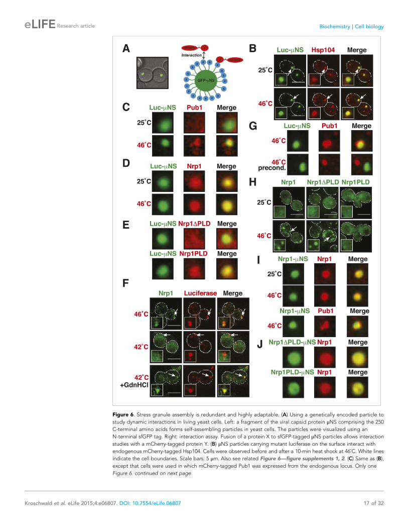
Figure 6. Stress granule assembly is redundant and highly adaptable. (A) Using a genetically encoded particle to
study dynamic interactions in living yeast cells. Left: a fragment of the viral capsid protein μNS comprising the 250
C-terminal amino acids forms self-assembling particles in yeast cells. The particles were visualized using an
N-terminal sfGFP tag. Right: interaction assay. Fusion of a protein X to sfGFP-tagged μNS particles allows interaction
studies with a mCherry-tagged protein Y. (B) μNS particles carrying mutant luciferase on the surface interact with
endogenous mCherry-tagged Hsp104. Cells were observed before and after a 10-min heat shock at 46˚C. White lines
indicate the cell boundaries. Scale bars: 5 μm. Also see related Figure 6—figure supplements 1, 2. (C) Same as (B),
except that cells were used in which mCherry-tagged Pub1 was expressed from the endogenous locus. Only one
Figure 6. continued on next page
Kroschwald et al. eLife 2015;4:e06807. DOI: 10.7554/eLife.06807 17 of 32
Research article Biochemistry | Cell biology
DiscussionIn this study, we investigate two types of stress-inducible RNP granules—P bodies and stress granules.
We find that these RNP granules have distinct material properties in yeast cells: whereas P bodies
show more liquid-like behavior, stress granules exhibit characteristic properties of solid aggregates.
Figure 6. Continued
representative μNS particle is shown at high magnification. Note that Pub1 only interacts with luciferase in cells
exposed to robust heat stress. Also see related Figure 6—figure supplements 3–5. (D) Same as (C), except that
mCherry-tagged Nrp1 was mildly overexpressed from a plasmid carrying an ADH1 promoter. Note that Nrp1 interacts
with luciferase already in unstressed cells, and that the amount of Nrp1 accumulating on the particle is strongly
increased upon heat stress. (E) Same as (D), except that the prion-like domain (PLD) of Nrp1 (Nrp1PLD) or a deletion
mutant lacking the PLD (Nrp1ΔPLD) was observed at 25˚C. (F) The cellular chaperone machinery prevents interactions
between misfolded proteins and stress granule components. Cells expressing Nrp1-GFP from the endogenous locus
and mCherry-tagged mutated luciferase from a plasmid were exposed to a 10-min heat shock at 42˚C or 46˚C. The cells
in the bottom panel were exposed to 3 mM guanidinium hydrochloride (GdnHCl) to inhibit Hsp104. Also see related
Figure 6—figure supplement 6. (G) Same as (C), except that the temperature was increased slowly from 25˚C to 46˚C
(preconditioning). Note that preconditioning prevents co-assembly of stress granules and misfolded proteins. Also see
related Figure 6—figure supplement 7. (H) PLDs mediate interactions only when present in high local concentrations.
Yeast cells were transformed with plasmids for the expression of GFP-tagged wild-type Nrp1 or deletion mutants
lacking the RNA-binding domain (RBD) (Nrp1PLD) or PLD domain (Nrp1ΔPLD). The resulting cells were exposed to
heat shock. (I) Upon heat shock, stress granules form on μNS particles that present Nrp1 on the surface. Same
conditions as (C) and (D). Also see related Figure 6—figure supplements 8–9. (J) Same as (I), except that mutants
lacking the RBD (Nrp1PLD) or PLD domain (Nrp1ΔPLD) were presented on the particle. Note that both mutants are
able to recruit full-length Nrp1 at 25˚C. Also see related Figure 6—figure supplement 10–12.
DOI: 10.7554/eLife.06807.034
The following figure supplements are available for figure 6:
Figure supplement 1. μNS particles do not interact with Hsp104 or Ssa1.
DOI: 10.7554/eLife.06807.035
Figure supplement 2. sfGFP-μNS particles carrying mutant luciferase or Ubc9ts on the surface interact with Hsp104
and Ssa1.
DOI: 10.7554/eLife.06807.036
Figure supplement 3. Misfolded proteins can nucleate stress granule formation under robust heat shock
conditions.
DOI: 10.7554/eLife.06807.037
Figure supplement 4. Control experiment showing that μNS particles do not interact with Pub1 or Pab1.
DOI: 10.7554/eLife.06807.038
Figure supplement 5. Misfolded proteins do not nucleate stress granule formation in glucose-deprived cells.
DOI: 10.7554/eLife.06807.039
Figure supplement 6. Chemical inhibition of Hsp104 leads to co-aggregation of misfolded proteins and stress
granule components even under mild heat shock conditions.
DOI: 10.7554/eLife.06807.040
Figure supplement 7. Misfolded proteins and stress granule components do not co-aggregate in preconditioned
cells.
DOI: 10.7554/eLife.06807.041
Figure supplement 8. Control experiment showing that Nrp1-μNS particles do not interact with Hsp104.
DOI: 10.7554/eLife.06807.042
Figure supplement 9. Nrp1 can nucleate stress granules upon glucose starvation stress.
DOI: 10.7554/eLife.06807.043
Figure supplement 10. Same as Figure 6—figure supplement 8 except that mutants lacking the RBD (Nrp1PLD)
or PLD domain (Nrp1ΔPLD) were presented on the particle.
DOI: 10.7554/eLife.06807.044
Figure supplement 11. The PLD as well as the RBD of Nrp1 can nucleate stress granule formation.
DOI: 10.7554/eLife.06807.045
Figure supplement 12. The PLD or RBD of Nrp1 can nucleate stress granule formation through heterotypic
interactions.
DOI: 10.7554/eLife.06807.046
Kroschwald et al. eLife 2015;4:e06807. DOI: 10.7554/eLife.06807 18 of 32
Research article Biochemistry | Cell biology
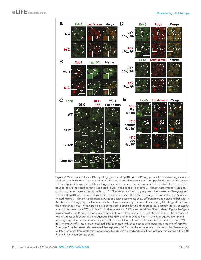
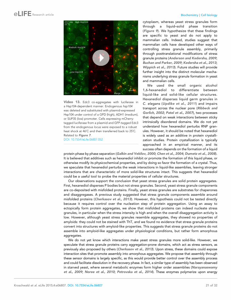
Figure 7. Maintenance of yeast P body integrity requires Hsp104. (A) The P-body protein Edc3 shows only minor co-
localization with misfolded proteins during robust heat stress. Fluorescence microscopy of endogenous GFP-tagged
Edc3 and plasmid-expressed mCherry-tagged mutant luciferase. The cells were stressed at 46˚C for 10 min. Cell
boundaries are indicated in white. Scale bars: 5 μm. Also see related Figure 7—figure supplement 1. (B) Edc3
shows only limited spatial overlap with Hsp104. Fluorescence microscopy of plasmid expressed mCherry-tagged
Edc3 and Hsp104-GFP expressed from the endogenous locus. The cells were subjected to heat stress. Also see
related Figure 7—figure supplement 2. (C) Edc3-positive assemblies show different morphologies and behavior in
the absence of disaggregases. Fluorescence time-lapse microscopy of yeast cells expressing GFP-tagged Edc3 from
the endogenous locus. Wild-type cells are compared to strains lacking disaggregases (Δhsp104, Δsse1, or Δsse2)after 1 hr heat stress at 46˚C and 1 hr 45 min after recovery at 25˚C. Also see Video 12 and related Figure 7—figure
supplement 3. (D) P-body components co-assemble with stress granules in heat-stressed cells in the absence of
Hsp104. Yeast cells expressing endogenous Edc3-GFP and endogenous Pub1-mCherry or aggregation-prone
mCherry-tagged luciferase from a plasmid in Hsp104-deficient cells were subjected to 1 hr heat stress at 46˚C.
(E) The amount of stress granule-localized Edc3 (denoted with S) decreases with increasing amounts of Hsp104.
P denotes P bodies. Yeast cells were used that expressed Edc3 under the endogenous promotor and mCherry-tagged
mutated luciferase from a plasmid. Endogenous hsp104 was deleted and substituted with plasmid-expressed Hsp104
Figure 7. continued on next page
Kroschwald et al. eLife 2015;4:e06807. DOI: 10.7554/eLife.06807 19 of 32
Research article Biochemistry | Cell biology
We further show that the formation of yeast stress granules does not involve amyloid-like
conformational conversions. Rather, yeast stress granules resemble amorphous protein aggregates.
Assembly of these aggregates depends on interactions with RNAs, and PLDs in RNA-binding proteins
contribute to granule formation, by promiscuously interacting with other PLDs or with misfolded
proteins. Finally, we show that stress granules have very different properties in mammalian cells,
where they have liquid-like characteristics and do not behave as aggregates. In summary, these
findings show that RNP granule formation is highly flexible, and that under conditions of acute stress,
disaggregating machines play a key role in maintaining the identity and integrity of RNP granules
(Figure 9).
Cells must respond rapidly to changing environments. This is particularly important for single-
celled organisms such as yeast because they are directly exposed to environmental fluctuations.
How can a cell respond rapidly and efficiently to stress, while at the same time solving the task of
adjusting the activities of numerous proteins and RNAs? An increasing number of studies suggest
that this can be achieved by building compartments. Such compartments are condensed phases of
proteins and RNAs, which exchange components with the surrounding cytoplasm or nucleoplasm
(Brangwynne et al., 2009, 2011; Hyman and Brangwynne, 2011; Hyman and Simons, 2012; Li
et al., 2012; Feric and Brangwynne, 2013; Hubstenberger et al., 2013; Hyman et al., 2014). Two
types of compartments can be distinguished: compartments for localized biochemistry in which
specific chemical reactions occur; and compartments for storage, where macromolecules adopt an
inactive, yet re-activatable state. The prediction would be that these two types of compartments
have different properties.
Indeed, a large body of work shows that P bodies are active compartments, involved in processing
and degrading mRNAs; stress granules on the other hand do not perform biochemical reactions, but
store proteins and RNAs (Anderson and Kedersha, 2009; Decker and Parker, 2012). P bodies are
therefore expected to have different properties than stress granules. A P body should allow for the
continuous entry and exit of RNAs and proteins,
and the components within a P body should be
able to rearrange. A liquid phase meets these
demands. Stress granules on the other hand do
not need to be liquid-like. Their function is to
inactivate proteins and RNAs by removing them
from the cytoplasm. Thus, a solid-like state is
fully compatible with the function of stress
granules. Our findings indeed reveal a remark-
able distinction between yeast P-bodies and
stress granules. Previous studies have also
pointed to differences (Buchan et al., 2008;
Decker and Parker, 2012; Mitchell et al., 2013;
Shah et al., 2013). Based on these findings, we
propose that yeast P-bodies are liquid-like
droplets that form by demixing from the
Figure 7. Continued
under control of a GPD - glyceraldehyde-3-phosphate dehydrogenase (high), ADH1 - alcohol dehydrogenase
1 (medium), or SUP35 (low) promoter. Cells were observed after 1 hr at 46˚C. Also see Video 13.
DOI: 10.7554/eLife.06807.047
The following figure supplements are available for figure 7:
Figure supplement 1. P bodies do not co-aggregate with misfolded proteins under robust heat shock conditions.
DOI: 10.7554/eLife.06807.048
Figure supplement 2. P bodies only show limited co-localization with chaperones under robust heat shock
conditions.
DOI: 10.7554/eLife.06807.049
Figure supplement 3. Lsm4-positive assemblies show different morphologies and behavior in the absence of
disaggregases.
DOI: 10.7554/eLife.06807.050
Video 12. Edc3-positive assemblies show different
morphologies and behavior in the absence of disag-
gregases. Fluorescence time-lapse microscopy of yeast
cells expressing GFP-tagged Edc3 from the endoge-
nous locus. Wild-type cells are compared to strains
lacking functional genes for certain disaggregases
(Δhsp104, Δsse1 or Δsse2). Cells were exposed to
a robust heat shock at 46˚C and then transferred back to
25˚C. Related to Figure 7.
DOI: 10.7554/eLife.06807.051
Kroschwald et al. eLife 2015;4:e06807. DOI: 10.7554/eLife.06807 20 of 32
Research article Biochemistry | Cell biology
cytoplasm, whereas yeast stress granules form
through a liquid–solid phase transition
(Figure 9). We hypothesize that these findings
are specific to yeast and do not apply to
mammalian cells. Indeed, studies suggest that
mammalian cells have developed other ways of
controlling stress granule assembly, primarily
through posttranslational modifications of stress
granule proteins (Anderson and Kedersha, 2009;
Buchan and Parker, 2009; Kedersha et al., 2013;
Wippich et al., 2013). Future studies will provide
further insight into the distinct molecular mecha-
nisms underlying stress granule formation in yeast
and mammalian cells.
We used the small organic alcohol
1,6-hexanediol to differentiate between
liquid-like and solid-like cellular structures.
Hexanediol disperses liquid germ granules in
C. elegans (Updike et al., 2011) and impairs
transport across the nuclear pore (Ribbeck and
Gorlich, 2002; Patel et al., 2007), two processes
that depend on weak interactions between sticky
intrinsically disordered domains. We do not yet
understand how hexanediol perturbs RNP gran-
ules. However, it should be noted that hexanediol
is widely used as an additive in protein crystalli-
zation studies. Protein crystallization is typically
approached in an empirical manner, and its
success often depends on the formation of a liquid
protein phase by phase separation (Galkin and Vekilov, 2000; Chen et al., 2004; Dumetz et al., 2008).
It is believed that additives such as hexanediol inhibit or promote the formation of this liquid phase, or
otherwise modify its physicochemical properties, and by doing so favor the formation of a crystal. Thus,
we speculate that hexanediol perturbs the weak interactions in liquid-like assemblies, leaving stronger
interactions that are characteristic of more solid-like structures intact. This suggests that hexanediol
could be a useful tool to probe the material properties of cellular structures.
Our observations support the conclusion that yeast stress granules are solid protein aggregates.
First, hexanediol disperses P bodies but not stress granules. Second, yeast stress granule components
are co-deposited with misfolded proteins. Finally, yeast stress granules are substrates for chaperones
and disaggregases. A previous study suggested that stress granule components assemble around
misfolded proteins (Cherkasov et al., 2013). However, this hypothesis could not be tested directly
because it requires control over the nucleation step of protein aggregation. Using an assay to
ectopically form protein aggregates, we show that misfolded proteins can indeed nucleate stress
granules, in particular when the stress intensity is high and when the overall disaggregation activity is
low. However, although yeast stress granules resemble aggregates, they showed no properties of
amyloids: they could not be stained with ThT, and we found no evidence that stress granule proteins
convert into structures with amyloid-like properties. This suggests that stress granule proteins do not
assemble into amyloid-like aggregates under physiological conditions, but rather form amorphous
aggregates.
We do not yet know which interactions make yeast stress granules more solid-like. However, we
speculate that stress granule proteins carry aggregation-prone domains, which act as stress sensors, as
previously also proposed by others (Cherkasov et al., 2013). Upon stress, these domains could expose
interaction sites that promote assembly into amorphous aggregates. We propose that assembly through
these sensor domains is largely specific, as this would provide better control over the assembly process
and could facilitate dissolution in the recovery phase. In fact, a similar type of assembly has been observed
in starved yeast, where several metabolic enzymes form higher order assemblies (Narayanaswamy
et al., 2009; Noree et al., 2010; Petrovska et al., 2014). These enzymes polymerize upon energy
Video 13. Edc3 co-aggregates with luciferase in
a Hsp104-dependent manner. Endogenous hsp104
was deleted and substituted with plasmid-expressed
Hsp104 under control of a GPD (high), ADH1 (medium),
or SUP35 (low) promoter. Cells expressing mCherry-
tagged luciferase from a plasmid and GFP-tagged Edc3
from the endogenous locus were exposed to a robust
heat shock at 46˚C and then transferred back to 25˚C.
Related to Figure 7.
DOI: 10.7554/eLife.06807.052
Kroschwald et al. eLife 2015;4:e06807. DOI: 10.7554/eLife.06807 21 of 32
Research article Biochemistry | Cell biology
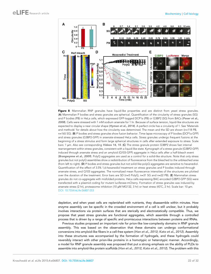
depletion, and when yeast cells are replenished with nutrients, they disassemble within minutes. How
enzyme assembly can be specific in the crowded environment of a cell is still unclear, but it probably
involves interactions via protein surfaces that are sterically and electrostatically compatible. Thus, we
propose that yeast stress granules are functional aggregates, which assemble through a controlled
process that is driven by a range of specific and promiscuous interactions between proteins and RNAs.
Previous studies proposed an important role for prion-like low-complexity domains in RNP granule
assembly. This was based on the observation that these domains can undergo conformational
conversions into amyloid-like fibers in a cell-free system (Han et al., 2012; Kato et al., 2012). Assembly
into these structures was accompanied by the formation of hydrogels, and these hydrogels could
reversibly interact with other prion-like proteins in a homotypic or heterotypic manner. Accordingly,
a model for RNP granule assembly was proposed that put a strong emphasis on the ability of PLDs to
assemble into amyloid-like protein scaffolds (Han et al., 2012; Kato et al., 2012). The problem with this
Figure 8. Mammalian RNP granules have liquid-like properties and are distinct from yeast stress granules.
(A) Mammalian P bodies and stress granules are spherical. Quantification of the circularity of stress granules (SG)
and P bodies (PB) in HeLa cells, which expressed GFP-tagged DCP1a (PB) or G3BP2 (SG) from BACs (Poser et al.,
2008). Cells were stressed with 1 mM sodium arsenate for 1 hr. Because of surface tension, liquid-like structures are
expected to display a near circular shape (Hyman et al., 2014). A perfect circle has a circularity of 1. See ‘Materials
and methods’ for details about how the circularity was determined. The mean and the SD are shown (n=118 PB,
n=165 SG). (B) P bodies and stress granules show fusion behavior. Time-lapse microscopy of P bodies (DCP1a-GFP)
and stress granules (G3BP2-GFP) in arsenate-stressed HeLa cells. Stress granules undergo frequent fusions at the
beginning of a stress stimulus and form large spherical structures in cells after extended exposure to stress. Scale
bars: 1 μm. Also see corresponding Videos 14, 15. (C) The stress granule protein G3BP2 shows fast internal
rearrangement within stress granules, consistent with a liquid-like state. Kymograph of a stress granule (G3BP2-GFP)
induced through arsenate stress and an amyloid (Q103-GFP) aggregate in HeLa cells after a half-bleach event
(Brangwynne et al., 2009). PolyQ aggregates are used as a control for a solid-like structure. Note that only stress
granules but not polyQ assemblies show a redistribution of fluorescence from the bleached to the unbleached area
(from left to right). (D) P bodies and stress granules but not solid-like polyQ aggregates are sensitive to hexanediol.
Quantification of the effect of 3.5% 1,6-hexanediol treatment on stress granules and P bodies induced through
arsenate stress, and Q103 aggregates. The normalized mean fluorescence intensities of the structures are plotted
over the duration of the treatment. Error bars are SD (n=5 PolyQ, n=31 SG and n=22 PB). (E) Mammalian stress
granules do not co-aggregate with misfolded proteins. HeLa cells expressing BAC-encoded G3BP2-GFP (SG) were
transfected with a plasmid coding for mutant luciferase-mCherry. Formation of stress granules was induced by
arsenate stress (2 hr), proteasome inhibition (10 μM MG132, 3 hr) or heat stress (43˚C, 2 hr). Scale bar: 10 μm.
DOI: 10.7554/eLife.06807.053
Kroschwald et al. eLife 2015;4:e06807. DOI: 10.7554/eLife.06807 22 of 32
Research article Biochemistry | Cell biology
model, however, is that hydrogels only rearrange
over long timescales, and thus can hardly provide
the dynamic properties that are characteristic of
RNP granules in living cells (Weber and Brang-
wynne, 2012; Malinovska et al., 2013; Bley
et al., 2014). To account for this, it was suggested
that these amyloid-like fibers are dynamic in vivo,
due to regulation by posttranslational modifica-
tions (Han et al., 2012; Kato et al., 2012; Kwon
et al., 2013). However, evidence for dynamic
fibers in living cells is still lacking.
We used self-assembling particles to locally
concentrate prion-like proteins in living cells,
creating a molecular environment that resembles
that in RNP granules. Using this assay, we find
that prion-like proteins promote RNP granule
formation, but independently of amyloid-like
conversions. Thus, above a certain critical con-
centration, PLDs can promote a phase transition
of RNA-binding proteins into RNP granules.
Based on these findings, we favor the following
two-step model of RNP granule formation: First,
RNA-binding proteins bind to RNAs through
multivalent interactions via RBDs, forming com-
plexes of RNA and protein; these RNP complexes
may already reach a relatively large size. In the second step, these RNP complexes condense into RNP
granules driven by promiscuous interactions between PLDs and further associations between RNAs
and RBDs. In this model, the PLDs primarily assist the final coalescence step, thus, promoting the
condensation of RNPs into large compartments (Figure 9). In agreement with this, recent findings
indicate that PLDs are not essential for RNP granule formation per se but rather promote the fusion
of RNP granules and their enlargement into bigger RNP compartments (Shiina and Nakayama,
2014). However, the function of PLDs may also depend on their specific local environment: In P
bodies, the collective properties of PLDs may be
important for maintaining a dynamic, liquid-like
state, whereas in solid assemblies such as stress
granules, PLDs may act as molecular glue to
connect RNA-binding proteins with other RNA-
binding proteins or misfolded proteins. Thus, PLDs
may function as adaptable interaction domains,
which do not undergo conversions into structurally
well-defined amyloid-like states, but mediate pro-
miscuous interactions with many binding partners
in their local environment. Such behavior would be
highly favorable, because it allows a high degree
of flexibility during compartment formation.
We found that in heavily stressed yeast cells,
the continuous action of protein disaggregases is
required to keep P bodies in a liquid-like state.
Interference with protein disaggregation caused
the mislocalization of P-body components to
stress granules (Figure 7C–E), suggesting that
P-body proteins and potentially many other
proteins can enter into a stress granule state,
but are normally prevented from doing so
through molecular chaperones such as Hsp104.
This implies that the molecular composition of
Video 14. Stress granules show extensive fusion
behavior and form more spherical structures with time.
HeLa cells expressing BAC-encoded G3BP2 were
observed by time-lapse microscopy. The cells were
stressed through arsenate. Related to Figure 8.
DOI: 10.7554/eLife.06807.054
Video 15. Two P bodies fuse and rapidly relax into
a spherical shape. Time-lapse microscopy of P bodies
(labelled by BAC-encoded DCP1a-GFP) in arsenate-
stressed HeLa cells. Related to Figure 8.
DOI: 10.7554/eLife.06807.055
Kroschwald et al. eLife 2015;4:e06807. DOI: 10.7554/eLife.06807 23 of 32
Research article Biochemistry | Cell biology
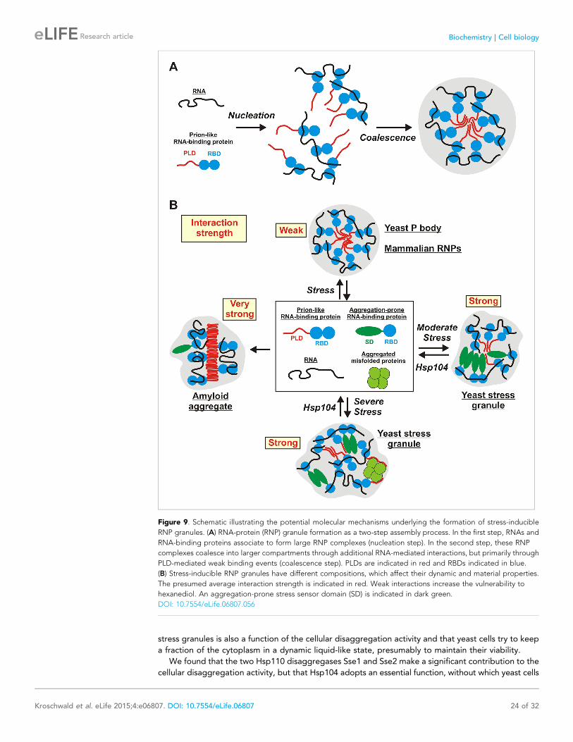
stress granules is also a function of the cellular disaggregation activity and that yeast cells try to keep
a fraction of the cytoplasm in a dynamic liquid-like state, presumably to maintain their viability.
We found that the two Hsp110 disaggregases Sse1 and Sse2 make a significant contribution to the
cellular disaggregation activity, but that Hsp104 adopts an essential function, without which yeast cells
Figure 9. Schematic illustrating the potential molecular mechanisms underlying the formation of stress-inducible
RNP granules. (A) RNA-protein (RNP) granule formation as a two-step assembly process. In the first step, RNAs and
RNA-binding proteins associate to form large RNP complexes (nucleation step). In the second step, these RNP
complexes coalesce into larger compartments through additional RNA-mediated interactions, but primarily through
PLD-mediated weak binding events (coalescence step). PLDs are indicated in red and RBDs indicated in blue.
(B) Stress-inducible RNP granules have different compositions, which affect their dynamic and material properties.
The presumed average interaction strength is indicated in red. Weak interactions increase the vulnerability to
hexanediol. An aggregation-prone stress sensor domain (SD) is indicated in dark green.
DOI: 10.7554/eLife.06807.056
Kroschwald et al. eLife 2015;4:e06807. DOI: 10.7554/eLife.06807 24 of 32
Research article Biochemistry | Cell biology
cannot dissolve stress granules. In contrast to yeast, mammalian cells do not express a Hsp104 homolog
in the cytosol. Therefore, we speculate that the ability of yeast to enter into a solid stress granule state
has co-evolved with Hsp104, providing a potential explanation for why yeast and mammalian stress
granules have different material properties. In contrast to mammalian organisms, sedentary yeast cells
cannot escape from harsh environments. We conjecture that for yeast, formation of solid stress granules
is an effective way of preserving the pre-stress state of the cytosol and that a selected set of yeast
proteins and RNAs has been modified by evolution to aggregate upon stress. Consistent with this,
a recent study showed that the yeast proteome has a higher overall aggregation propensity than the
human proteome (Albu et al., 2014). When the stress subsides, Hsp104 reactivates the proteins and
RNAs preserved in solid stress granules, thus promoting re-entry into the cell cycle. We note that this
situation is reminiscent of dormancy, as for example in spores or seeds, where an entire organism enters
into a resting state by solidifying its macromolecular components (Parry et al., 2014). Future work will
show whether controlled entry into a solid phase can provide general protection to a cell in stressful
environments.
Materials and methods
Cloning proceduresCloning procedures were performed as described previously using the Gateway system (Alberti
et al., 2007, 2009). For a list of plasmids see Supplementary file 1.
Yeast genetic techniques, strains, and mediaThe media used were standard synthetic media or rich media containing 2% D-glucose. The yeast
strain backgrounds were W303 ADE+ (leu2-3,112; his3-11,-15; trp1-1; ura3-1; can1-100; [psi-]; [PIN+])or BY4741 (his3Δ1; leu2Δ0; met15Δ0; ura3Δ0; [psi-]; [PIN+]). Yeast gene deletions were performed
using a PCR-based approach (Gueldener et al., 2002). C-terminal tagging of yeast genes was
performed as described previously (Sheff and Thorn, 2004). The induction of an amyloid state is
a concentration-dependent nucleation process. Thus, the prion-like state in Lsm4 was induced by
overexpressing untagged Lsm4 in a [PIN+] strain that expressed GFP-tagged Lsm4 from the
endogenous promoter. The prion-like state in Nrp1 was induced by using a [PIN+] strain expressing
the Nrp1PLD (Alberti et al., 2009). For a list of strains see Supplementary file 1.
Cell culture of HeLa BAC cell linesHeLa cells were cultured in DMEM (Dulbecco’s modified eagle’s medium) supplemented with 10% FBS
(fetal bovine serum) and penicillin–streptomycin (all Gibco Life Technologies, United Kingdom). Cells
were maintained at 37˚C in a 5% CO2 incubator. HeLa Kyoto cells containing G3BP2-GFP or DCP1a-GFP
BAC constructs were used to visualize stress granules or P bodies, respectively (Poser et al., 2008).
Transient transfection of HeLa cells with pcDNA3.1-Q103 was performed using Jet Prime transfection
reagent (Polyplus, France). Transfection with pcDNA3.1-Luciferase was performed using Lipofectamine
2000 (Invitrogen, Carlsbad, California).
Wide-field fluorescence microscopy of yeast cellsYeast cells were grown in cultures of 50–100 ml at 25˚C to an OD600 not higher than 0.5. The yeast
cells were then immobilized on concanavalin A (Sigma Aldrich, St.Louis, Missouri) -coated precise
glass bottom dishes (MatTek, Ashland, Massachusetts). Microscopy was performed using a DeltaVision
microscope system with softWorx 4.1.2 software (Applied Precision, United Kingdom). The system
was based on an Olympus IX71 microscope (Olympus, Japan), which was used with a UPlanSApo 100
× 1.4 numerical (NA) oil objective. The images were collected with a Cool SnapHQ camera
(Photometrics, Tucson, Arizona) and a pixel size of 0.13 μm. Heat shock experiments were performed
using a Warner heating chamber (Warner instruments, Hamden, Connecticut). When indicated, 5–10%
of 1,6-hexanediol (Merck, Germany) solution or 100 μg/ml cycloheximide (AppliChem, Germany) was
added to the medium to perturb RNP granule integrity. To inhibit Hsp104, guanidinium hydrochloride
(GdnHCl) was added to the medium to a final concentration of 3 mM, 3 hr before imaging. All images
were deconvolved using standard softWorx deconvolution algorithms (enhanced ratio, high-noise
filtering). Shown images are maximum intensity projections of 8–14 individual images. Figures show
Kroschwald et al. eLife 2015;4:e06807. DOI: 10.7554/eLife.06807 25 of 32
Research article Biochemistry | Cell biology
representative cells. The cell boundaries were introduced by thresholding the bright-field image and
overlaying it with the fluorescence image. In all comparative experiments, we used four chamber
dishes (MatTek), allowing us to image four different conditions in the same experiment.
Analysis of the foci-to-cytoplasm ratio of yeast cells (Figure 1B) was performed by manually
defining regions of interest and measuring their fluorescence intensity using Fiji (Schindelin et al.,
2012). The P-body number and size analysis (Figure 3B) was performed using Fiji image analysis tools.
The image was segmented by thresholding and then analyzed using the analyze particle tool (size: 0-
infinity, circularity: 0–1). The number of total cells was determined by manually counting cells in the
bright-field channel using the Fiji cell counter plugin.
Confocal microscopy of ThT-stained yeast cellsCells were grown in 50 ml cultures while shaking at 180 rpm to an OD600 not higher than 0.5. Cultures
were then harvested and resuspended in a 10 mM Tris/EDTA (ethylenediaminetetraacetic acid) buffer
(pH 7) containing 30 μM ThT (Sigma Aldrich) and incubated for 20 min. Subsequently, the cells were
washed three times in Tris/EDTA buffer without ThT and then resuspended in media. Cells were then
immobilized on 1% agar pads. Microscopy was performed on a single photon point scanning confocal
Olympus Fluoview 1000 microscope (Olympus). ThT was excited with a 405-nm diode laser. Emission
of ThT was recorded in the 480–540 nm range. For co-localization studies, we used cells that
expressed mCherry-tagged proteins. mCherry fluorescence was excited with a 561-nm DPSS laser,
and emission was recorded in the 570–670 nm range. Cells were scanned in one z-plane using
a UPlanSApo 60 × 1.35 numerical (NA) oil objective and a scan speed of 8 μs/pixel. Images were
acquired with a pixel resolution of 0.103 μm.
FRAP of yeast cellsYeast cells were grown in cultures of 50–100 ml at 25˚C to an OD600 not higher than 0.5. The cells were
immobilized in precise glass bottom dishes using concanavalin A coating. Images were acquired using
an Olympus IX81 inverted stand microscope (Olympus) with a spinning disk scan head Yokogawa
CSU-X1 (5000 rpm). The system was used with an Olympus UPlanSApo 100 × 1.4 oil objective.
Acquired images had pixel sizes of 0.081 μm. The images were collected with an Andor iXon EM+DU-897 BV back-illuminated EMCCD (Andor, Ireland). For imaging of Dendra2, we used a triple-band
dichromatic mirror: T-405/488/561. Images before and after photoconversion were acquired as z-stacks
with 0.7-μm spacing and a time interval of ∼1 s. The Dendra2 signal was converted in a 1 × 1 pixel area by
using the 405-nm diode laser at 6% intensity in 400 repeats for 100 μs each. For image segmentation, we
used the 3D tracking tool of the Imaris software (Bitplane, Switzerland). The signal from three areas was
obtained for each time point: the photoconverted/bleached foci (IF), a reference focus in a neighboring cell
(IR) and an area of equal size in the background (IB). The fluorescence signal in the foci was normalized as
follows (Carisey et al., 2011):
IF−pre =∑
tbleach−1
t =0
IFðtÞ− IBðtÞfprebleach
;
IR−pre =∑
tbleach−1
t =0
IRðtÞ− IBðtÞfprebleach
;
IF normðtÞ=IF ðtÞ− IBðtÞ
IF−pre
IRðtÞ− IBðtÞIR−pre
:
Since yeast cells are small, we had to ensure that photoconversion in one cell did not affect the
reference foci in the neighboring cell. Therefore, we estimated the decay kinetics of the reference foci
by fitting the following equation to the post-bleach period of IR:
Kroschwald et al. eLife 2015;4:e06807. DOI: 10.7554/eLife.06807 26 of 32
Research article Biochemistry | Cell biology
y =ApeðppxÞ;
where p is the time constant and A is the fluorescence intensity. The average fluorescence level before
the photoconversion was deduced by extrapolating the fitted decay curve, yielding IR_estimate.
If IR_estimate was smaller than IR_measured, it indicated that fluorescence loss in the reference foci occurred
through photoconversion. These cells were excluded from the analysis. To reveal the half-time of
recovery, we fitted the values of IF_norm with:
y =Að1− e−ppxÞ;where p is the time constant and A is the fluorescence intensity.
The half time t1/2 was calculated using:
t1=2 =logð0:5Þ
−p:
Data were analyzed, tested for statistical significance, and plotted using R software. Boxes in
boxplots extend from the 25th to 75th percentiles, with a line at the median.
Fluorescence microscopy of HeLa cellsLive HeLa cells were imaged using the DeltaVision imaging system with softWorx 4.1.2 software
(described above). The system was used with a Plan Apo 60 × 1.42 NA oil immersion objective. 15
sections with 200-nm spacing were acquired and the maximum intensity projections were created in
Fiji. If indicated, 3.5% of hexanediol solution was added to the culture medium to perturb P body and
stress granule integrity. Measurement of the circularity of P bodies and stress granules (Figure 8A)
was performed using a custom-made Matlab routine (see Supplementary file 2) from maximum
intensity projections, according to the following equation:
Circularity =4πArea
ðPerimeterÞ2;
where the value of circularity is 1 for a perfect circle and the value decreases as it deviates from the
circular shape. Microscopy to determine the circularity of granules was performed using an Andor
spinning disk confocal microscope as described above with a UPlanSApo 100 × 1.4 oil objective.
Half-bleach experiment in HeLa cellsThe half-bleach (Brangwynne et al., 2009) in Figure 8C was performed at a pixel resolution of
0.08 μm using a spinning disk confocal microscope as described above. Photo-bleaching and imaging
was performed with the 488-nm laser. Structures were photo-bleached for 1.2 ms within a region of 5 ×5 pixels.
SDD-AGESDD-AGE analysis was used to separate amyloid polymers from monomers in whole cell lysates. Yeast
cells were grown in cultures of 100 ml at 25˚C to an OD600 of 0.5. The cultures were harvested and
washed once with water. The pellets were resuspended in 300 μl ice-cold lysis buffer (50 mM Tris, pH
7.5; 150 mM NaCl; 2.5 mM EDTA; 1% (vol/vol) TritonX-100; 0.33 mM PMSF(phenylmethylsulfonyl
fluoride); 6.7 mM NEM; 1.25 mM benzamidine; 10 μg/ml pepstatin; 10 μg/ml chymostatin; 10 μg/ml aprotinin; 10 μg/ml leupeptin; 10 μg/ml E-64) and added to ice-cold glass beads (425–600 μm)
(Sigma–Aldrich). Cells were lysed using mechanical disruption (Tissue Lyser II, Qiagen,
Netherlands) at 25 Hz for 15 min. Unwanted cell debris and beads were removed by
centrifugation and the supernatant was used for SDD-AGE analysis. The supernatants were
adjusted for equal protein concentrations and mixed 4:1 with 4 × Sample buffer (40 mM Tris
acetic acid, 2 mM EDTA, 20% glycerol, 4% SDS, bromophenol blue). Samples were incubated for
10 min at room temperature and loaded onto a 1.5% agarose gel containing 0.1% SDS in 1 ×TAE/0.1% SDS running buffer. The gel was run at low voltage (less than 80 V) to prevent the gel
Kroschwald et al. eLife 2015;4:e06807. DOI: 10.7554/eLife.06807 27 of 32
Research article Biochemistry | Cell biology
from heating up. Proteins in the gel were detected by immunoblotting with a GFP-specific antibody
(Roche, Switzerland). The exposure time was the same for all immunoblots shown in one experiment.
SECSEC was used to separate cellular complexes according to their size. Cells were grown in cultures
of 50 ml at 25˚C to an OD600 not higher than 0.5. The cultures were harvested and washed once
with water. The pellets were resuspended in 500 μl ice-cold lysis buffer (50 mM Tris, pH 7.5;
150 mM NaCl; 2.5 mM EDTA; 1% (vol/vol) TritonX-100; 0.4 mM PMSF; 8 mM NEM; 1.25 mM
benzamidine; 10 μg/ml pepstatin; 10 μg/ml chymostatin; 10 μg/ml aprotinin; 10 μg/ml leupeptin;
10 μg/ml E-64) and ice-cold glass beads were added (425–600 μm). Cells were lysed using
mechanical disruption (Tissue Lyser II) at 25 Hz for 20 min. To pellet cell debris and beads, samples were
centrifuged and the supernatant was applied to a SpinX-Centrifuge Filter (0.22 μm cellulose acetate,
Sigma-Aldrich). After centrifugation for 5 min at 13.000 rpm at 4˚C, protein concentration of the
supernatant was measured using the Bradford protein assay (BioRad, Germany). Equal amounts were
loaded onto the column. To efficiently resolve the protein complexes, we used the TSK-Gel
G6000PWXL (Tosoh Bioscience, Japan) column with a separation range between 40 kDa and 8.000
kDa. The column was equilibrated in running buffer (2 × PBS (phosphate-buffered saline)) and calibrated
with different standard proteins: Tyroglobulin (660 kDa), Ferritin (440 kDa), Catalase (240 kDa), and BSA
(bovine serum albumin) (67 kDa). Subsequently, a dot blot assay was performed using a vacuum slot blot
device (Whatman, United Kingdom). The fractions were collected on protein-binding nitrocellulose
membrane (Protran B 85, Whatman) and the resulting membrane was used for immunodetection with
an anti-GFP antibody.
AcknowledgementsWe thank several members of the MPI-CBG for critical reading of the manuscript. We thank Daniel
Kaganovich for a plasmid containing mutant luciferase and Cammie Lesser for the GFP-μNS
plasmid. The light microscopy facility and the chromatography facility of the MPI-CBG are
acknowledged for expert technical assistance. We acknowledge funding from the Max Planck
Society, the Dresden International Graduate School (DIGS-BB), and the Alexander von Humboldt
Foundation.
Additional information
Funding
Funder Grant reference Author
Max-Planck-Gesellschaft Sonja Kroschwald,Shovamayee Maharana,Daniel Mateju, LilianaMalinovska, ElisabethNuske, Ina Poser, DorisRichter, Simon Alberti
Alexander von Humboldt-Stiftung
Shovamayee Maharana
Technische UniversitatDresden
Dresden InternationalGraduate School forBiomedicine andBioengineering
Sonja Kroschwald, DanielMateju, Liliana Malinovska,Elisabeth Nuske
The funders had no role in study design, data collection and interpretation, or thedecision to submit the work for publication.
Author contributions
SK, Conception and design, Acquisition of data, Analysis and interpretation of data, Drafting or
revising the article; SM, DM, LM, EN, Conception and design, Acquisition of data, Analysis and
interpretation of data; IP, Acquisition of data, Contributed unpublished essential data or reagents;
DR, Acquisition of data, Analysis and interpretation of data; SA, Conception and design, Analysis and
interpretation of data, Drafting or revising the article
Kroschwald et al. eLife 2015;4:e06807. DOI: 10.7554/eLife.06807 28 of 32
Research article Biochemistry | Cell biology
Additional files
Supplementary files
·Supplementary file 1. List of yeast strains used in this study.DOI: 10.7554/eLife.06807.057
· Supplementary file 2. Matlab routine for measuring the circularity of granules.DOI: 10.7554/eLife.06807.058
ReferencesAggarwal S, Snaidero N, Pahler G, Frey S, Sanchez P, Zweckstetter M, Janshoff A, Schneider A, Weil MT, SchaapIA, Gorlich D, Simons M. 2013. Myelin membrane assembly is driven by a phase transition of myelin basicproteins into a cohesive protein meshwork. PLOS Biology 11:e1001577. doi: 10.1371/journal.pbio.1001577.
Aizer A, Brody Y, Ler LW, Sonenberg N, Singer RH, Shav-Tal Y. 2008. The dynamics of mammalian P body transport,assembly, and disassembly in vivo. Molecular Biology of the Cell 19:4154–4166. doi: 10.1091/mbc.E08-05-0513.
Alberti S, Gitler AD, Lindquist S. 2007. A suite of Gateway cloning vectors for high-throughput genetic analysis inSaccharomyces cerevisiae. Yeast 24:913–919. doi: 10.1002/yea.1502.
Alberti S, Halfmann R, King O, Kapila A, Lindquist S. 2009. A systematic survey identifies prions and illuminatessequence features of prionogenic proteins. Cell 137:146–158. doi: 10.1016/j.cell.2009.02.044.
Alberti S, Halfmann R, Lindquist S. 2010. Biochemical, cell biological, and genetic assays to analyze amyloid andprion aggregation in yeast. Methods in Enzymology 470:709–734. doi: 10.1016/S0076-6879(10)70030-6.
Albu RF, Chan GT, Zhu M, Wong ET, Taghizadeh F, Hu X, Mehran AE, Johnson JD, Gsponer J, Mayor T. 2014. Afeature analysis of lower solubility proteins in three eukaryotic systems. Journal of Proteomics 118:21–38. doi: 10.1016/j.jprot.2014.10.011.
Anderson P, Kedersha N. 2008. Stress granules: the Tao of RNA triage. Trends in Biochemical Sciences 33:141–150. doi: 10.1016/j.tibs.2007.12.003.
Anderson P, Kedersha N. 2009. RNA granules: post-transcriptional and epigenetic modulators of gene expression.Nature Reviews. Molecular Cell Biology 10:430–436. doi: 10.1038/nrm2694.
Andrei MA, Ingelfinger D, Heintzmann R, Achsel T, Rivera-Pomar R, Luhrmann R. 2005. A role for eIF4E and eIF4E-transporter in targeting mRNPs to mammalian processing bodies. RNA 11:717–727. doi: 10.1261/rna.2340405.
Banjade S, Rosen MK. 2014. Phase transitions of multivalent proteins can promote clustering of membranereceptors. eLife 3:e04123. doi: 10.7554/eLife.04123.
Bley N, Lederer M, Pfalz B, Reinke C, Fuchs T, Glass M, Moller B, Huttelmaier S. 2014. Stress granules aredispensable for mRNA stabilization during cellular stress. Nucleic Acids Research 43:e26. doi: 10.1093/nar/gku1275.
Brangwynne CP. 2013. Phase transitions and size scaling of membrane-less organelles. The Journal of Cell Biology203:875–881. doi: 10.1083/jcb.201308087.
Brangwynne CP, Eckmann CR, Courson DS, Rybarska A, Hoege C, Gharakhani J, Julicher F, Hyman AA. 2009.Germline P granules are liquid droplets that localize by controlled dissolution/condensation. Science 324:1729–1732. doi: 10.1126/science.1172046.
Brangwynne CP, Mitchison TJ, Hyman AA. 2011. Active liquid-like behavior of nucleoli determines their size andshape in Xenopus laevis oocytes. Proceedings of the National Academy of Sciences of USA 108:4334–4339.doi: 10.1073/pnas.1017150108.
Buchan JR, Muhlrad D, Parker R. 2008. P bodies promote stress granule assembly in Saccharomyces cerevisiae.The Journal of Cell Biology 183:441–455. doi: 10.1083/jcb.200807043.
Buchan JR, Parker R. 2009. Eukaryotic stress granules: the ins and outs of translation. Molecular Cell 36:932–941.doi: 10.1016/j.molcel.2009.11.020.
Buchan JR, Yoon JH, Parker R. 2011. Stress-specific composition, assembly and kinetics of stress granules inSaccharomyces cerevisiae. Journal of Cell Science 124:228–239. doi: 10.1242/jcs.078444.
Cai Y, Futcher B. 2013. Effects of the yeast RNA-binding protein Whi3 on the half-life and abundance of CLN3mRNA and other targets. PLOS ONE 8:e84630. doi: 10.1371/journal.pone.0084630.
Carisey A, Stroud M, Tsang R, Ballestrem C. 2011. Fluorescence recovery after photobleaching. Methods inMolecular Biology 769:387–402. doi: 10.1007/978-1-61779-207-6_26.
Chen Q, Vekilov PG, Nagel RL, Hirsch RE. 2004. Liquid-liquid phase separation in hemoglobins: distinct aggregationmechanisms of the beta6 mutants. Biophysical Journal 86:1702–1712. doi: 10.1016/S0006-3495(04)74239-3.
Cherkasov V, Hofmann S, Druffel-Augustin S, Mogk A, Tyedmers J, Stoecklin G, Bukau B. 2013. Coordination oftranslational control and protein homeostasis during severe heat stress. Current Biology 23:2452–2462. doi: 10.1016/j.cub.2013.09.058.
Decker CJ, Parker R. 2012. P-bodies and stress granules: possible roles in the control of translation and mRNAdegradation. Cold Spring Harbor Perspectives in Biology 4:a012286. doi: 10.1101/cshperspect.a012286.
Decker CJ, Teixeira D, Parker R. 2007. Edc3p and a glutamine/asparagine-rich domain of Lsm4p function in processingbody assembly in Saccharomyces cerevisiae. The Journal of Cell Biology 179:437–449. doi: 10.1083/jcb.200704147.
Doyle SM, Genest O, Wickner S. 2013. Protein rescue from aggregates by powerful molecular chaperonemachines. Nature Reviews. Molecular Cell Biology 14:617–629. doi: 10.1038/nrm3660.
Kroschwald et al. eLife 2015;4:e06807. DOI: 10.7554/eLife.06807 29 of 32
Research article Biochemistry | Cell biology
Duennwald ML, Echeverria A, Shorter J. 2012. Small heat shock proteins potentiate amyloid dissolution byprotein disaggregases from yeast and humans. PLOS Biology 10:e1001346. doi: 10.1371/journal.pbio.1001346.
Dumetz AC, Chockla AM, Kaler EW, Lenhoff AM. 2008. Protein phase behavior in aqueous solutions:crystallization, liquid-liquid phase separation, gels, and aggregates. Biophysical Journal 94:570–583. doi: 10.1529/biophysj.107.116152.
Feric M, Brangwynne CP. 2013. A nuclear F-actin scaffold stabilizes ribonucleoprotein droplets against gravity inlarge cells. Nature Cell Biology 15:1253–1259. doi: 10.1038/ncb2830.
Frey S, Gorlich D. 2007. A saturated FG-repeat hydrogel can reproduce the permeability properties of nuclearpore complexes. Cell 130:512–523. doi: 10.1016/j.cell.2007.06.024.
Frey S, Richter RP, Gorlich D. 2006. FG-rich repeats of nuclear pore proteins form a three-dimensional meshworkwith hydrogel-like properties. Science 314:815–817. doi: 10.1126/science.1132516.
Galkin O, Vekilov PG. 2000. Control of protein crystal nucleation around the metastable liquid-liquid phaseboundary. Proceedings of the National Academy of Sciences of USA 97:6277–6281. doi: 10.1073/pnas.110000497.
Gilks N, Kedersha N, Ayodele M, Shen L, Stoecklin G, Dember LM, Anderson P. 2004. Stress granuleassembly is mediated by prion-like aggregation of TIA-1. Molecular Biology of the Cell 15:5383–5398.doi: 10.1091/mbc.E04-08-0715.
Glover JR, Lindquist S. 1998. Hsp104, Hsp70, and Hsp40: a novel chaperone system that rescues previouslyaggregated proteins. Cell 94:73–82. doi: 10.1016/S0092-8674(00)81223-4.
Grousl T, Ivanov P, Frydlova I, Vasicova P, Janda F, Vojtova J, Malinska K, Malcova I, Novakova L, Janoskova D,Valasek L, Hasek J. 2009. Robust heat shock induces eIF2alpha-phosphorylation-independent assembly of stressgranules containing eIF3 and 40S ribosomal subunits in budding yeast, Saccharomyces cerevisiae. Journal of CellScience 122:2078–2088. doi: 10.1242/jcs.045104.
Grousl T, Ivanov P, Malcova I, Pompach P, Frydlova I, Slaba R, Senohrabkova L, Novakova L, Hasek J. 2013. Heatshock-induced accumulation of translation elongation and termination factors precedes assembly of stressgranules in S. cerevisiae. PLOS ONE 8:e57083. doi: 10.1371/journal.pone.0057083.
Gueldener U, Heinisch J, Koehler GJ, Voss D, Hegemann JH. 2002. A second set of loxP marker cassettes forCre-mediated multiple gene knockouts in budding yeast. Nucleic Acids Research 30:e23. doi: 10.1093/nar/30.6.e23.
Gupta R, Kasturi P, Bracher A, Loew C, Zheng M, Villella A, Garza D, Hartl FU, Raychaudhuri S. 2011. Fireflyluciferase mutants as sensors of proteome stress. Nature Methods 8:879–884. doi: 10.1038/nmeth.1697.
Han TW, Kato M, Xie S, Wu LC, Mirzaei H, Pei J, Chen M, Xie Y, Allen J, Xiao G, McKnight SL. 2012. Cell-freeformation of RNA granules: bound RNAs identify features and components of cellular assemblies. Cell 149:768–779. doi: 10.1016/j.cell.2012.04.016.
Hoyle NP, Castelli LM, Campbell SG, Holmes LE, Ashe MP. 2007. Stress-dependent relocalization of translationallyprimed mRNPs to cytoplasmic granules that are kinetically and spatially distinct from P-bodies. The Journal ofCell Biology 179:65–74. doi: 10.1083/jcb.200707010.
Hubstenberger A, Noble SL, Cameron C, Evans TC. 2013. Translation repressors, an RNA helicase, anddevelopmental cues control RNP phase transitions during early development. Developmental Cell 27:161–173.doi: 10.1016/j.devcel.2013.09.024.
Hulsmann BB, Labokha AA, Gorlich D. 2012. The permeability of reconstituted nuclear pores provides directevidence for the selective phase model. Cell 150:738–751. doi: 10.1016/j.cell.2012.07.019.
Hyman AA, Brangwynne CP. 2011. Beyond stereospecificity: liquids and mesoscale organization of cytoplasm.Developmental Cell 21:14–16. doi: 10.1016/j.devcel.2011.06.013.
Hyman AA, Simons K. 2012. Cell biology. Beyond oil and water—phase transitions in cells. Science 337:1047–1049. doi: 10.1126/science.1223728.
Hyman AA, Weber CA, Julicher F. 2014. Liquid-liquid phase separation in biology. Annual Review of Cell andDevelopmental Biology 30:39–58. doi: 10.1146/annurev-cellbio-100913-013325.
Kaganovich D, Kopito R, Frydman J. 2008. Misfolded proteins partition between two distinct quality controlcompartments. Nature 454:1088–1095. doi: 10.1038/nature07195.
Kato M, Han TW, Xie S, Shi K, Du X, Wu LC, Mirzaei H, Goldsmith EJ, Longgood J, Pei J, Grishin NV, Frantz DE,Schneider JW, Chen S, Li L, Sawaya MR, Eisenberg D, Tycko R, McKnight SL. 2012. Cell-free formation of RNAgranules: low complexity sequence domains form dynamic fibers within hydrogels. Cell 149:753–767. doi: 10.1016/j.cell.2012.04.017.
Kedersha N, Cho MR, Li W, Yacono PW, Chen S, Gilks N, Golan DE, Anderson P. 2000. Dynamic shuttling of TIA-1accompanies the recruitment of mRNA to mammalian stress granules. The Journal of Cell Biology 151:1257–1268. doi: 10.1083/jcb.151.6.1257.
Kedersha N, Ivanov P, Anderson P. 2013. Stress granules and cell signaling: more than just a passing phase?Trends in Biochemical Sciences 38:494–506. doi: 10.1016/j.tibs.2013.07.004.
Kedersha N, Stoecklin G, Ayodele M, Yacono P, Lykke-Andersen J, Fritzler MJ, Scheuner D, Kaufman RJ, GolanDE, Anderson P. 2005. Stress granules and processing bodies are dynamically linked sites of mRNP remodeling.The Journal of Cell Biology 169:871–884. doi: 10.1083/jcb.200502088.
King OD, Gitler AD, Shorter J. 2012. The tip of the iceberg: RNA-binding proteins with prion-like domains inneurodegenerative disease. Brain Research 1462:61–80. doi: 10.1016/j.brainres.2012.01.016.
Kruger T, Hofweber M, Kramer S. 2013. SCD6 induces ribonucleoprotein granule formation in trypanosomes ina translation-independent manner, regulated by its Lsm and RGG domains. Molecular Biology of the Cell 24:2098–2111. doi: 10.1091/mbc.E13-01-0068.
Kroschwald et al. eLife 2015;4:e06807. DOI: 10.7554/eLife.06807 30 of 32
Research article Biochemistry | Cell biology
Kwon I, Kato M, Xiang S, Wu L, Theodoropoulos P, Mirzaei H, Han T, Xie S, Corden JL, McKnight SL. 2013.Phosphorylation-regulated binding of RNA polymerase II to fibrous polymers of low-complexity domains. Cell155:1049–1060. doi: 10.1016/j.cell.2013.10.033.
Kwon I, Xiang S, Kato M, Wu L, Theodoropoulos P, Wang T, Kim J, Yun J, Xie Y, McKnight SL. 2014. Poly-dipeptides encoded by the C9orf72 repeats bind nucleoli, impede RNA biogenesis, and kill cells. Science 345:1139–1145. doi: 10.1126/science.1254917.
Lee CF, Brangwynne CP, Gharakhani J, Hyman AA, Julicher F. 2013. Spatial organization of the cell cytoplasm byposition-dependent phase separation. Physical Review Letters 111:088101. doi: 10.1103/PhysRevLett.111.088101.
Lee C, Occhipinti P, Gladfelter AS. 2015. PolyQ-dependent RNA-protein assemblies control symmetry breaking.The Journal of Cell Biology 208:533–544. doi: 10.1083/jcb.201407105.
Li P, Banjade S, Cheng HC, Kim S, Chen B, Guo L, Llaguno M, Hollingsworth JV, King DS, Banani SF, Russo PS,Jiang QX, Nixon BT, Rosen MK. 2012. Phase transitions in the assembly of multivalent signalling proteins. Nature483:336–340. doi: 10.1038/nature10879.
Li YR, King OD, Shorter J, Gitler AD. 2013. Stress granules as crucibles of ALS pathogenesis. The Journal of CellBiology 201:361–372. doi: 10.1083/jcb.201302044.
Malinovska L, Kroschwald S, Alberti S. 2013. Protein disorder, prion propensities, and self-organizingmacromolecular collectives. Biochimica et Biophysica Acta 1834:918–931. doi: 10.1016/j.bbapap.2013.01.003.
Mitchell SF, Jain S, She M, Parker R. 2013. Global analysis of yeast mRNPs. Nature Structural & Molecular Biology20:127–133. doi: 10.1038/nsmb.2468.
Narayanaswamy R, Levy M, Tsechansky M, Stovall GM, O’Connell JD, Mirrielees J, Ellington AD, Marcotte EM.2009. Widespread reorganization of metabolic enzymes into reversible assemblies upon nutrient starvation.Proceedings of the National Academy of Sciences of USA 106:10147–10152. doi: 10.1073/pnas.0812771106.
Noree C, Sato BK, Broyer RM, Wilhelm JE. 2010. Identification of novel filament-forming proteins inSaccharomyces cerevisiae and Drosophila melanogaster. The Journal of Cell Biology 190:541–551. doi: 10.1083/jcb.201003001.
Parry BR, Surovtsev IV, Cabeen MT, O’Hern CS, Dufresne ER, Jacobs-Wagner C. 2014. The bacterial cytoplasmhas glass-like properties and is fluidized by metabolic activity. Cell 156:183–194. doi: 10.1016/j.cell.2013.11.028.
Parsell DA, Lindquist S. 1993. The function of heat-shock proteins in stress tolerance: degradation and reactivationof damaged proteins. Annual Review of Genetics 27:437–496. doi: 10.1146/annurev.ge.27.120193.002253.
Patel SS, Belmont BJ, Sante JM, Rexach MF. 2007. Natively unfolded nucleoporins gate protein diffusion acrossthe nuclear pore complex. Cell 129:83–96. doi: 10.1016/j.cell.2007.01.044.
Petrovska I, Nuske E, Munder MC, Kulasegaran G, Malinovska L, Kroschwald S, Richter D, Fahmy K, Gibson K,Verbavatz JM, Alberti S. 2014. Filament formation by metabolic enzymes is a specific adaptation to an advancedstate of cellular starvation. eLife 3:e02409. doi: 10.7554/eLife.02409.
Poser I, Sarov M, Hutchins JR, Heriche JK, Toyoda Y, Pozniakovsky A, Weigl D, Nitzsche A, Hegemann B, Bird AW,Pelletier L, Kittler R, Hua S, Naumann R, Augsburg M, Sykora MM, Hofemeister H, Zhang Y, Nasmyth K, WhiteKP, Dietzel S, Mechtler K, Durbin R, Stewart AF, Peters JM, Buchholz F, Hyman AA. 2008. BAC TransgeneOmics:a high-throughput method for exploration of protein function in mammals. Nature Methods 5:409–415. doi: 10.1038/nmeth.1199.
Ramaswami M, Taylor JP, Parker R. 2013. Altered ribostasis: RNA-protein granules in degenerative disorders. Cell154:727–736. doi: 10.1016/j.cell.2013.07.038.
Rampelt H, Kirstein-Miles J, Nillegoda NB, Chi K, Scholz SR, Morimoto RI, Bukau B. 2012. Metazoan Hsp70machines use Hsp110 to power protein disaggregation. The EMBO Journal 31:4221–4235. doi: 10.1038/emboj.2012.264.
Reijns MA, Alexander RD, Spiller MP, Beggs JD. 2008. A role for Q/N-rich aggregation-prone regions in P-bodylocalization. Journal of Cell Science 121:2463–2472. doi: 10.1242/jcs.024976.
Ribbeck K, Gorlich D. 2002. The permeability barrier of nuclear pore complexes appears to operate viahydrophobic exclusion. The EMBO Journal 21:2664–2671. doi: 10.1093/emboj/21.11.2664.
Schindelin J, Arganda-Carreras I, Frise E, Kaynig V, Longair M, Pietzsch T, Preibisch S, Rueden C, Saalfeld S,Schmid B, Tinevez JY, White DJ, Hartenstein V, Eliceiri K, Tomancak P, Cardona A. 2012. Fiji: an open-sourceplatform for biological-image analysis. Nature Methods 9:676–682. doi: 10.1038/nmeth.2019.
Schmitz AM, Morrison MF, Agunwamba AO, Nibert ML, Lesser CF. 2009. Protein interaction platforms:visualization of interacting proteins in yeast. Nature Methods 6:500–502. doi: 10.1038/nmeth.1337.
Shah KH, Zhang B, Ramachandran V, Herman PK. 2013. Processing body and stress granule assembly occur byindependent and differentially regulated pathways in Saccharomyces cerevisiae. Genetics 193:109–123. doi: 10.1534/genetics.112.146993.
Sheff MA, Thorn KS. 2004. Optimized cassettes for fluorescent protein tagging in Saccharomyces cerevisiae. Yeast21:661–670. doi: 10.1002/yea.1130.
Shiina N, Nakayama K. 2014. RNA granule assembly and disassembly Modulated by nuclear factor associated withDouble-stranded RNA 2 and nuclear factor 45. The Journal of Biological Chemistry 289:21163–21180. doi: 10.1074/jbc.M114.556365.
Shorter J. 2011. The mammalian disaggregase machinery: hsp110 synergizes with Hsp70 and Hsp40 to catalyzeprotein disaggregation and reactivation in a cell-free system. PLOS ONE 6:e26319. doi: 10.1371/journal.pone.0026319.
Si K, Choi YB, White-Grindley E, Majumdar A, Kandel ER. 2010. Aplysia CPEB can form prion-like multimers insensory neurons that contribute to long-term facilitation. Cell 140:421–435. doi: 10.1016/j.cell.2010.01.008.
Kroschwald et al. eLife 2015;4:e06807. DOI: 10.7554/eLife.06807 31 of 32
Research article Biochemistry | Cell biology
Sun Z, Diaz Z, Fang X, Hart MP, Chesi A, Shorter J, Gitler AD. 2011. Molecular determinants and genetic modifiersof aggregation and toxicity for the ALS disease protein FUS/TLS. PLOS Biology 9:e1000614. doi: 10.1371/journal.pbio.1000614.
Teixeira D, Sheth U, Valencia-Sanchez MA, Brengues M, Parker R. 2005. Processing bodies require RNA forassembly and contain nontranslating mRNAs. RNA 11:371–382. doi: 10.1261/rna.7258505.
Updike DL, Hachey SJ, Kreher J, Strome S. 2011. P granules extend the nuclear pore complex environment in theC. elegans germ line. The Journal of Cell Biology 192:939–948. doi: 10.1083/jcb.201010104.
Vessey JP, Vaccani A, Xie Y, Dahm R, Karra D, Kiebler MA, Macchi P. 2006. Dendritic localization of thetranslational repressor Pumilio 2 and its contribution to dendritic stress granules. The Journal of Neuroscience 26:6496–6508. doi: 10.1523/JNEUROSCI.0649-06.2006.
Weber SC, Brangwynne CP. 2012. Getting RNA and protein in phase. Cell 149:1188–1191. doi: 10.1016/j.cell.2012.05.022.
Wippich F, Bodenmiller B, Trajkovska MG, Wanka S, Aebersold R, Pelkmans L. 2013. Dual specificity kinase DYRK3couples stress granule condensation/dissolution to mTORC1 signaling. Cell 152:791–805. doi: 10.1016/j.cell.2013.01.033.
Kroschwald et al. eLife 2015;4:e06807. DOI: 10.7554/eLife.06807 32 of 32
Research article Biochemistry | Cell biology