programme & abstracts · skeletal muscle and tendon through innovation ... skeletal muscle...
TRANSCRIPT
1
PROGRAMME & ABSTRACTS

2
3
2nd MuscleTech Network Workshop PROGRAMME & ABSTRACTS Barcelona, 27th – 29th September 2010
MuscleTech Network Muscletech Network is a scientific platform aiming to generate knowledge and cutting edge research in the field of skeletal muscle and tendon through innovation in the prevention, diagnosis and treatment of illness, injuries, diseases, and myopathies devoted to translate the scientific discoveries into practical applications to improve human health. MuscleTech Network is an initiative of the Fundació Futbol Club Barcelona and Leitat Technological Centre with the support of the General Secretariat for Sport of the Government of Catalonia. www.muscletechnetwork.org International Center for Scientific Debate The International Center for Scientific Debate (ICSD) is an initiative of Biocat, with the support of “la Caixa” Obra Social Foundation, which aims to drive first-rate international scientific events to promote dialogue, collaboration and open exchange of ideas, projects and knowledge among experts of renowned national and international prestige. The ICSD aims to generate advanced debate on the various disciplines that are linked to the life sciences field and their repercussion on society, contributing to Catalonia’s position as a country of scientific excellence. www.biocat.cat/cidc
2nd MuscleTech Network Workshop The "2nd Muscletech Network Workshop" with the motto “From Translational Research to Translational Medicine” strives to encourage cooperation by getting researchers formuniversities and clinical centres to work towards a common goal, improve human health. The "2nd Muscletech Network Workshop" aims to bridge and foster collaboration between MuscleTech Network members, clinical research institutions and leading international research groups via a series of scientific debates; to create new partnerships, promote research, identify opportunities and address the scientific challenges on skeletal muscle and tendon.
2nd MuscleTech Network Workshop 4
Monday 27th 12:00 – 12:30 Opening
Mr. Jordi Monés i Carilla. Director of Sports Area (Medical Area) of FC Barcelona Mr. Eusebi Cima. President of LEITAT TechnologicalCentre Mr. Enric Banda. Director of Science, Research and Environment of “la Caixa” Foundation Mr. Manel Balcells. President of the Executive Committee of Biocat Ms. Marina Geli*. Health Department of the Government of Catalonia Mr. Josep-Lluís Carod-Rovira. Vice-president of the Government of Catalonia *Speaker to be confirmed
12:30 – 12:45 One year after the launch of MuscleTech Network
• Francesc X. Roca Manager of MuscleTech Network, LEITAT Technological Centre. 12:45 – 13:00 Presentation of:
• Ramon Balius Chief Editor of “Apunts, Medicina de l’Esport – On-line sports medicine magazine”.
• Gil Rodas Senior researcher and Sports Medicine Specialist, Physician of the Spanish premier league basketball team Futbol Club Barcelona.
“Clinical Practice Guide for muscular injuries. Epidemiology, diagnosis, treatment and prevention.”
13:00 – 14:30 Lunch 14:30 – 16:30 Key note lecture
• José Luís Terreros Deputy General Director of Sport and Health, National Sports Council.
“From research to medical sport support”.
• Richard Lieber Professor of Orthopaedics and Bioengineering, Veterans Affairs Medical Centre, and Department of Orthopaedics and Bioengineering, University of California at San Diego, San Diego, United States of America.
“Design of Human Muscle: Implications for Muscle Injury”. Skeletal muscle represents a classic example of a structure-function relationship. Skeletal muscle functional properties are primarily determined by muscle architecture. Architecture is defined as the orientation and arrangement of muscle fibres within a muscle and is not obvious from simply inspecting gross anatomy. Detailed architectural properties have been defined for both the upper extremity, the lower extremity, and even portions of the axial skeleton. In addition, the way in which muscles change length has been directly measured in humans using laser diffraction. These studies yield a model of torque generation in which sarcomere length varies with joint angle in a way that defines the biomechanics of the joint. In the context of muscle injury, it has been demonstrated that muscle injury varies as a function of fibre (or sarcomere) strain, rather than the stress imposed on the muscle. As a result, those muscles predisposed to injury are those with short muscle fibers operating on joints with large moment arms. This has been demonstrated experimentally and is consistent with the clinical presentation of sports injuries.
2nd MuscleTech Network Workshop 5
• Carlo Reggiani Professor at the Department of Anatomy and Physiology, University of Padua
“Skeletal Muscle Plasticity: from molecules to training protocols". Skeletal muscles are heterogeneous in nature, being composed of fibres with distinct functional properties. The functional unit of the motor system, the motor unit, is composed a motor neuron and a bunch of muscle fibers with similar, if not identical, structural and functional properties. To form a muscle, many (from tens to hundreds) motor units are assembled together and each brings its own specific and distinct contribution. With the selective recruitment of motor units, a muscle will be able to respond in the best manner to the demand of the nervous system. The heterogeneity of the muscle fibers is the base of the flexibility which allows the same muscle to be used for various tasks from continuous low intensity activity (e.g. posture), to repeated submaximal contractions (for example, locomotion) and to fast and strong maximal contractions (jumping, kicking a ball). In addition, the structural and functional properties of the fibers, which together define the so called, fiber phenotype, can change in response to hormonal and neural influences, nerve-activity being a major determinant of the fiber type profile. This property is defined muscle plasticity or malleability and fibres can change either their size (atrophy/hypertrophy) or their phenotype (slow/fast and oxidative/glycolytic). The molecular mechanisms of the heterogeneity and the plasticity of muscle fibers have been the object of intensive investigation and several signaling pathways have been identified. Cytosolic calcium via calcineurin-NFAT and PGC1-α appears to be a major modulator of phenotype transitions towards slow and oxidative characters, while AKT seems to play an essential role in regulating fibre size. AKT is, in turn, activated by several factors; among them is IGF-1. Load applied on or tension developed by a muscle fibre is relevant to control fibre size, but the mechanism transducing tension into a chemical signal still awaits identification.
• Carl M. Askling Lecturer at the Swedish School of Sport and Health Sciences, Research Leader at the Orthopaedics and Sports Medicine Group, Department of Molecular Medicine and Surgery at the Karolinska Institutet, Stockholm, Sweden
“Different Types of Hamstring Strains in Sports”. Hamstring strain is the most common single injury in elite soccer. To prevent these injuries and to optimize the rehabilitation it is important to analyze in what situations they occur. There are at least two distinctly different types of acute hamstring strains, one occurring during high-speed running and mainly involving the biceps femoris long head, the other during movements leading to extensive lengthening of the hamstrings (such as; high kicking, sliding tackle, sagittal split) often involving the free proximal tendon of semimembranosus. These two injury types will be exemplified with injury situations, MRI-findings and time back to sport from our ongoing study on acute hamstring strains. When the free proximal tendon is involved, there seems to be an extended period of time before full function without symptoms is reached. Injury type, palpation of maximal pain and MRI-findings (tissues involved) can give important information about prognosis. Prevention: It seems to be very important to specifically restore the eccentric hamstring muscle strength after injury before returning to sport to prevent the common hamstring re-injury. Also in the few prospective studies that exist, eccentric training appears to be an essential ingredient. This has been indicated in two rather recent Scandinavian studies, one prospective randomised study on Swedish elite male football players and the other on Icelandic and Norwegian male elite football players. However, both studies suffer from methodological shortcomings, which limit their impact. Rehabilitation: There are no studies describing what kind of rehabilitation program athletes suffering different types of hamstring strains should perform. One study (Sherry et al. 2004) indicated that core stability training is an important part of the rehabilitation program. In our ongoing study we randomized the injured athletes into two different programs. One program with the aim of putting high eccentric demand on the hamstrings in exercises with different degrees of lengthening of the hamstring muscle-tendon complex and the other program consists of more “conventional” exercises. Testing : There is no consensus about a single test, clinical examination or imaging investigation that would provide strict criteria for safe return to sport following hamstring
2nd MuscleTech Network Workshop 6
muscle strain. We are currently evaluating a new active hamstring flexibility test that appears to add discriminative power and provide useful additional information to the common clinical examination before going back to full training/match
16:30 – 16:45 Coffee break 16:45 – 18:00 MTN 2010 projects
Chair: Gil Rodas Senior researcher and Sports Medicine Specialist, Physician of the Spanish premier league basketball team Futbol Club Barcelona.
• Ginés Viscor Professor at the Physiology Department, School of Biological Sciences, University of
Barcelona, Barcelona, Spain.
“Induced Muscle Damage and Induced Intermittent Hypoxia (IHH)”. We have started an exploratory study oriented to determine the possible effectiveness of the intermittent exposure to hypoxia in a hypobaric chamber for the recovery of induced muscular damage in the laboratory rat. Muscular damage is induced in the hind limbs of the experimental animals by means of a session of forced running in a treadmill with negative slope according to protocols widely validated and accepted by the scientific community. Our immediate goal will be to describe to the evolution of plasma markers of muscular damage during the recovery in treated and control rats. Simultaneously, a detailed study of the changes induced at structural and biochemical level of the rat thigh muscles will be also developed. Experimental animals are divided in three groups: control and two experimental. The control animals [CON] will have a passive recovery following a regime of sedentary and normal life. All the experimental animals will be put under a program of intermittent hypobaric hypoxia exposure, although half of them [HIP] will not have any other additional treatment, whereas the third group [EHIP] in addition to the exposure to simulated altitude, will be put under a protocol of light to moderate exercise comparable (on a murine scale) to a physical fitness program in human. In this initial stage, at each week, 2 animals in each group will be sacrificed for a morphofunctional examination of muscular samples. Histochemical characterization, fiber typing, capillarization and expression of several transcription factors (HIF-1, VEGF, EGF, PDFG, among others) will be made to assess the structural damage and their evolution.
• Roser Cussó Professor at the Department of Physiological Sciences I. Biochemistry and Molecular
Biology Unit. Metabolic Regulation and Molecular Pathology Group. School of Medicine, University of Barcelona, Barcelona, Spain.
“Muscle and Tendon Injury Biomarkers”. The Project has two aims: A: Muscle biomarkers: To design an ELISA method able to differentiate injuries by measuring myosins from fast and slow muscle fibres. To study the loss of elastic muscle properties by measuring the serum markers from the titin elastic protein. B: Tendon biomarkers: To study the molecular characteristics of tendon injuries To search the presence of tendon markers in tendon. To apply the result of this search to find specific serum markers that would help diagnose, prognostic and consequences of a tendon injury. The analysis of fast and slow myosin in serum from humans with different grade of injury is shown. The same analysis has been applied to football players and athletes after an eccentric training.
2nd MuscleTech Network Workshop 7
• Ramon Balius Research Leader at the High Performance Sports Research Centre (CEARE), Catalan Sports Council, Barcelona, Spain.
“Longitudinal Studies of Hamstring Muscle Injuries in Athletes”. Muscle injury ranges amongst the most common injuries in sport. Within the muscle injuries, one of the most common injuries is located in the hamstring muscles. It is very important in sport injuries to know depending on the injury type the return to play time and prognosis. In the case of muscle injuries is not referenced with clarity in the literature whether there is any sign trough image diagnostics that can be used for prognostic factors. For most of the studies the sample is small and biased, mixing different types of hamstring injuries. In addition to this, there are few longitudinal studies making this subject area not thoroughly documented and a good research field. The study presented here is longitudinal study with 150 to 200 cases of hamstring rupture. Several variables have been assessed – sectional volume, sectional area of maximum lesion, lesion length, proximity to the insertion area and existence of hematoma. The study also contemplates monitoring their recovery; keep track of the injuries and recurrences that may exist for at least two years. With all data collected and trough a biostatistics analysis will aim to clearly establish clearly the following data: • Time off sport / professional physical activity return ("return to play) • Existence of factors for recurrence. • Existence of MRI criteria of injury healing
• Xavier Peirau Senior Researcher at the Regenerative Tissue Therapy Institute (ITRT), Teknon Medical Centre Barcelona, Spain.
“Research on the Use of Platelet-rich Plasma and Mesenchymal Cells”.
• “FEM - Forum EGARSAT del Músculo” Initiative created by EGARSAT in conjunction with MuscleTech Network dedicated to educate and make society aware of the importance of the muscle in our daily life, with a special interest in the workplace; focusing on communicating best practices and knowledge from scientific research on muscle and tendons to general population to improve quality of people’s life.
2nd MuscleTech Network Workshop 8
Tuesday 28th
9:00 – 10:30 From molecular genetics to protein expression.
Molecular Genetics - Polymorphisms / Physical Performance / Injuries.
The molecular genetics complexity and findings on polymorphism muscular variations are reshaping the way physical performance and injuries were understood. We would like to discuss the causes and expression of muscular variations, and the possibilities of tissue engineering and the potentiality of the so called gene therapy and adult stem cell research. The debate will revolve on the possibility to transfer the scientific research findings into real developments and effective treatments for damaged or diseased tissues and for the improvement of public health in general.
Chairs: Josep Potau Unit of Human Anatomy and Embryology, School of Medicine, University of Barcelona, Barcelona, Spain.
Gil Rodas Senior researcher and Sports Medicine Specialist, Physician of the Spanish premier league basketball team Futbol Club Barcelona.
• Johnny Huard Henry J. Mankin Professor and Vice-chair for Musculoskeletal Cellular Therapeutics in the Department of Orthopaedic Surgery and in the Departments of Microbiology and Molecular Genetics, Pathology and Physical Medicine, Bioengineering, and Rehabilitation. Director of the Stem Cell Research Centre of Children’s Hospital of Pittsburgh. Deputy Director of the McGowan Institute for Regenerative Medicine, United States of America.
“Tissue Engineering Based on Muscle-derived Stem Cells: Potential Applications for Tissue Regeneration” Members of my laboratory have isolated various populations of myogenic cells from the postnatal skeletal muscle of normal mice on the basis of the cells’ adhesion characteristics, proliferation behavior, and myogenic and stem cell marker expression profiles. Although most of these cell populations have displayed characteristics similar to those of satellite cells, we also have identified a unique population of muscle-derived stem cells (MDSCs). MDSCs exhibit long-term proliferation and high self-renewal rates and can differentiate toward various lineages, both in vitro and in vivo. The transplantation of MDSCs, in contrast to that of other myogenic cells, has improved the efficiency of dystrophic muscle regeneration and the delivery of dystrophin to dystrophic muscle. The ability of MDSCs to proliferate in vivo for an extended period of time, combined with their capacity to exhibit self-renewal, multipotent differentiation, and transplantation.
Recent studies performed by members of my laboratory have shown that transplantation of female MDSCs (F-FMSCs) rather than male MDSCs (M-MDSCs) significantly improves skeletal muscle regeneration despite the similar myogenic and stem cell marker expression by both cell types. I will explain the increased muscle regeneration efficiency exhibited by F-MDSCs. My presentation will also address the influence of environmental cues within dystrophic or injured skeletal muscle on the differentiation of MDSCs into fibrotic cells. I will discuss potential strategies by which to prevent scar tissue formation within injured muscle by blocking TGF-β1 activity. I then will discuss the use of MDSCs in gene therapy and tissue engineering applications designed to improve bone and articular cartilage healing through the genetic modification of MDSCs to express osteogenic proteins (BMP2 and -4) and the angiogenic factor VEGF. I will also outline in my presentation new results obtained with human muscle derived stem cells, which we believe will open new avenues by which researchers could use muscle stem cell-based gene therapy and tissue engineering to improve tissue regeneration.
2nd MuscleTech Network Workshop 9
• Rui Diogo Senior Researcher at the Department of Anthropology, George Washington University, Washington, United States of America.
“Human Muscular Variations: Comparative, Evolutionary and Developmental Perspectives, with Implications for Medicine, Sports and Public Health”. The anatomy of primates and other mammals has been the subject of numerous comparative studies. However, the vast majority of these studies deal with skeletal structures. The lack of detailed comparative works on the head, neck, pectoral and forelimb musculature of primates and their closest living relatives, i.e. tree-shrews and colugos, creates difficulties in the discussion of subjects such as the evolution of primates, the origin of modern humans, and the peculiar muscular variations of both human and non-human mammals. The main aim of the present work is to discuss human muscular variations in a broader comparative, evolutionary and developmental framework, based on our own dissections of numerous vertebrate specimens and on an extensive review of the literature. One of topics that will be explored in the talk is the relationship between ontogeny, genetics, and the so-called (somewhat controversially) ‘muscular atavisms’. The implications for medicine, sports, and public health will be discussed.
• Antoni L. Andreu Research Leader at the Mitochondrial Pathology and Neuromuscular Unit,
Research Institute “Institut de Recerca Hospital Universitari Vall d'Hebron”, Barcelona, Spain.
“MacArdle Disease: from Genetics to Exercise.” McArdle disease (MAD) is the most common muscle glycogenosis and it is caused by deficiency of the muscle glycogen phosphorylase (GP) activity. The disease is commonly characterized by exercise intolerance with premature fatigue, myalgia, cramps, increased levels of serum creatine kinase at rest, and recurrent myoglobinuria. The MAD causing gene is PYGM gene, which mutations reduce or abolish the GP activity in muscle. Nowadays, more than 100 PYGM mutations have been found in MAD patients worldwide, but the most frequent mutation in the Caucasian population is the p.R50X. Two naturally occurring animal models (bovine and ovine) of MAD are known. In both models, there is full disease expression and the molecular defects have been identified, but the difficulty in working with cattle and sheep, has limited the possibilities for research studies. In this work, we present new pathologic mutations in the PYGM gene, which include silent mutations and intronic mutations generating aberrant splicing. Additionally, we have characterized the effect of the mechanism nonsense mediated mRNA decay (NMD) in a series of 28 patients diagnosed with MAD. In these patients we showed that NMD degrades the transcribed PYGM mRNA in more than 90 percent of patients. Finally, we present our results in our projects in models of MAD. In the one hand, we will show new data about the reexpresion of GP activity in McArcle muscle cultures. In the other, we will show our advances in the generation of a knock-in mouse model for the p.R50X mutation, as a model of study for MAD.
• Rosa Artells Senior Researcher at the Unit of Human Anatomy and Human Embryology, School of
Medicine, University of Barcelona, Barcelona, Spain (discussion panel member)
10:30 – 11:00 Coffee break
2nd MuscleTech Network Workshop 10
11:00 – 12:30 From protein expression to sarcomere.
Molecular biology - myosin, titine as biomarkers.
The signalling pathway occurring after injuries, the effect of nerve activity in adult skeletal muscle, and specifically the pathways which control muscle growth and fibre type specification or the analysis of the molecular mechanisms involved in the regulation of glucose transport in muscle and adipose cells will be subjects in the limelight during this session. These studies may reveal the underlying causes of obesity, lead to the creation of a glucose reader like device rating the injury grade or shine some light on the neuromuscular field, particularly in mitochondrial disorders and McArdle disease.
Chair: Roser Cussó Professor at the Department of Physiological Sciences I. Biochemistry and Molecular Biology Unit. Metabolic Regulation and Molecular Pathology Group. School of Medicine, University of Barcelona, Barcelona, Spain.
• Per A. Tesch Professor at the Department of Health Sciences, Mid Sweden University, Östersund
and Department of Physiology & Pharmacology, Karolinska Institutet, Stockholm, Sweden. “Skeletal Muscle Mass and Function with Increased Use or Disuse: Lessons from Space to Benefit Sports Performance and Health Promotion”. Since the early era of the manned spaceflight program in 1960’s it has become evident that space crew suffer from muscle atrophy and deteriorate musculoskeletal function. Hence, numerous studies of real or simulated weightlessness ranging from a few weeks to several months duration, have reported marked decreases in maximal voluntary force mainly due to muscle loss. Weight-bearing muscles are more affected than non-weight bearing muscles. The rate of e.g., triceps surae or quadriceps muscle atrophy during the course of one to six weeks of simulated spaceflight may exceed 2% per week. Decreases in isometric, concentric and eccentric strength are even greater. Regardless of which mechanism(s) are responsible for the loss of skeletal muscle mass, countermeasures to this effect are imperative on the International Space Station (ISS) and other future long-term orbital missions. The most attractive solution to this problem seems to be the implementation of some kind of in-flight exercise program alone or combined with other countermeasures e.g., pharmacological interventions or artificial gravity. Collectively, the results from a myriad of human studies show that resistance type, but not aerobic endurance exercise, promotes increased muscle size and strength. It therefore seems logical that resistance exercise would offer an important stimulus to maintain muscle mass and strength in weightlessness. Designing resistance exercise programs for use in a weightlessness environment is challenging. First, there are several physiological aspects to consider. Any exercise task effective in promoting muscle hypertrophy should comprise both concentric and eccentric muscle actions, and in any particular exercise should muscle tension be near maximal. Moreover, in an environment where there is a shortage of food, water and oxygen, energy expenditure must be kept low. Also, there are certain technical aspects that must be taken into account when designing exercise equipment to be used in space. In the absence of gravity, barbells or dumb-bells for example, cannot be used. It is therefore imperative that the gravitational pull from a lifted weight must be replaced by an alternative force or power source. This paper will discuss how findings from recent space related muscle research has been employed for terrestrial use. More specifically, molecular and global skeletal muscle adaptations to weightlessness or unloading with or without resistance training will be covered. Further, the use of resistance exercise paradigms, designed for space travellers to combat muscle atrophy, to optimize athletic performance and benefit in health promotion will be discussed.
2nd MuscleTech Network Workshop 11
• Antonio Zorzano Research Leader at the Molecular Pathology and Therapy in Heterogenic and Polygenic Diseases Research Group, IRB Barcelona, Barcelona, Spain.
“Search of Targets for the Prevention of Muscle Loss: DOR a Regulator of Autophagy”. Our research group studies novel mechanisms or pathways activated in conditions of insulin resistance namely in skeletal muscle. During recent years, studies have focused on the validation of a target for the therapy of muscle wasting. In this respect, the nuclear cofactor DOR (Diabetes- and Obesity-Regulated gene, also known as Tp53inp2 or C20orf110), is a key regulator of autophagy. Originally identified as a protein highly expressed in muscle, heart and brain, DOR localizes in nuclear bodies in proliferating cells. In agreement with its nuclear localization, DOR functions as an enhancer of the transcriptional activity of the thyroid hormone receptor TRalpha1, and DOR loss-of-function reduces thyroid hormone function in muscle cells. This function is driven mainly by the N-terminal part of the protein, whereas the C-terminal fragment of DOR exhibits inhibitory activity. Moreover, DOR physically interacts with TRalpha1 and to T3-responsive promoters, as assessed by co-immunoprecipitation and ChIP assays. The Drosophila homolog of DOR, dDOR, is a coactivator of ecdysone receptor. dDOR binds ecdysone receptor and is required for maximal ecdysone receptor transcriptional activity. In the absence of dDOR, flies display a number of ecdysone loss-of-function phenotypes such as impaired spiracle eversion, impaired salivary gland degradation, and pupal lethality. Studies on the role of DOR on protein turnover in skeletal muscle will be discussed. In addition, a technology platform oriented to identify modulators of DOR will be presented.
• Eduard Gallardo Senior Researcher at the Neurological Research Group at the Institut de Recerca (Research Institute) of the Hospital de la Santa Creu i Sant Pau, Barcelona, Spain.
“Mechanisms of Muscle Membrane Repair: The Dysferlin Model”. During normal contractions muscle needs efficient mechanisms to repair the continuous damage to the cytoskeletal anchors, the contractile apparatus, and the cellular membrane. When repair is not effective, the muscle fibers damaged die and a new fiber will replace the former. Each of these maintenance and repair processes can be affected by genetic mutations, resulting in a muscular dystrophy phenotype. Mutations in the gene that encodes dysferlin lead to a progressive muscular dystrophy Dysferlin is part of a multimeric protein complex critical for repair of muscle membranes after damage. The diagnosis of this disease is complicated because patients may display a wide range of disease onset and phenotypic variability that includes predominant limb girdle involvement, distal weakness and hyperCKemia, collectively referred to as dysferlinopathies. The translational research of these differences, using molecular approaches ( e.g. immunoprecipitation and blue native electrophoresis) will be helpful to understand the mechanisms of muscle damage and repair in detail. We are developing assays that could be potentially used for high throughput screening of compounds that could improve membrane repair in dysferlin myopathy and in normal muscle. Furthermore we are testing assays to quantify the efficacy of different therapies to improve muscle repair membrane.
• Ginés Viscor Professor at the Physiology Department, School of Biological Sciences, University of Barcelona, Barcelona, Spain. (discussion panel member)
12:30 – 14:00 Lunch
2nd MuscleTech Network Workshop 12
14:00 – 15:30 From sarcomere to muscular fibres.
Regenerative capacity and functional units. The advances in the field of skeletal muscle regeneration that have been made in the recent year will be highlighted and the recent studies increasing our understanding on the contribution of satellite cell-derived muscle precursor cells to skeletal muscle regeneration, the effect of the host muscle environment and the studies showing that muscle regeneration may be optimized by using the best stem cell population and by modifying the host muscle environment will probably be focusing much of the presentations topics and debate subjects. Chair: Ramon Segura Emeritus Professor of Physiology at the School of Medicine at the University of Barcelona. Member of the “Physiology of Exercise” Group and Professor at the Medical School of Physical Education and Sport, Barcelona, Spain.
• Johnny Huard Henry J. Mankin Professor and Vice-chair for Musculoskeletal Cellular Therapeutics in the Department of Orthopaedic Surgery and in the Departments of Microbiology and Molecular Genetics, Pathology and Physical Medicine, Bioengineering, and Rehabilitation. Director of the Stem Cell Research Centre of Children’s Hospital of Pittsburgh. Deputy Director of the McGowan Institute for Regenerative Medicine, United States of America.
“Development of Biological Approaches to Improve Muscle Healing after Injury”. Muscle injuries are extremely common and have a high tendency to reoccur. Both limitations in force production and re-injury frequently are attributed to the development of regeneration-restrictive fibrotic tissue at the original site of injury. Gene therapy may prove useful for the development of techniques to improve the healing of muscle injuries. Transforming growth factor-beta 1 (TGF-�) plays a key role in inducing the formation of fibrotic tissue that limits muscle healing after severe injury. Muscle Stem Cells have exhibited the capacity to differentiate into a myofibroblast-like lineage in vitro and can contribute to scar formation after muscle injury in vivo. Various inhibitors of TGF-�including relaxin, decorin, gamma-interferon, and suramin can improve muscle regeneration and force production by limiting muscle fibrosis. Relaxin and decorin are molecules that can be expressed by cells and delivered to muscle via gene therapy techniques. Gamma-interferon, suramin and losartan represent drugs with other clinical indications, are potentially available for clinical use. However, due to the potential side-effects of these agents, further research must be performed in order to determine clinical safety. Ex vivo gene therapy techniques may provide a method to deliver inhibitors of TGF-�to injured muscle. The proteoglycan, decorin, appears to be beneficial not only in reducing fibrosis, but also in improving muscle regeneration. Delivery of MDSCs expressing decorin or a viral vector carrying decorin to injured muscle may help to improve long-term outcomes by reducing muscle fibrosis and, therefore, recurrence of injury. The proteins, myostatin and follistatin , has been shown to inhibit muscle growth and differentiation and represents good target to produce muscle hypertrophy and improved muscle strength. In this course, we will review the current knowledge concerning the use of gene therapy and tissue engineering applications based on muscle stem cells to improve the recovery of skeletal muscle after injuries and disease.
• Jordi Desola The Hyperbaric Therapy Unit of Barcelona). “Moises Broggi” Hospital. Sant Joan
Despí. Barcelona
“Hyperbaric oxygenation for sports injuries : from the empiricism to the evidence” Hyperbaric Oxygenation (HBO) is an old therapy based on obtaining very high Arterial Oxygen Pressure by means of breathing pure oxygen inside a Hyperbaric Chamber. Once the saturation of Haemoglobin is achieved, oxygen remains in a plasmatic solution up to 23 times higher than on atmospheric conditions, permitting a valid tissue oxygen delivery independently of the haemoglobin transport, thus providing supplementary tissue oxygenation in normal tissues with an effective oxygen delivery in conditions of ischaemic, toxic, traumatic, or metabolic tissue hypoxia when oxyhaemoglobin is not available to the
2nd MuscleTech Network Workshop 13
cell. Additionally HBO is the cause of some ancillary effects as increasing of Nitrous Oxide synthesis, stimulating production of great quantity of antioxidants, and non-hypoxemic vasoconstriction over acutely flogotic previously normal tissues. The combination of these actions results in an active angiogenic effect and positive increase of tissue granulation in cases of a wound disorder and inhibition of critic tissue oedema, thus improving tissue regeneration active even under inflammatory circumstances. Current experimental research has revealed an enhancement of Stem-Cells proliferation up to 8 times over those obtained in normobaric conditions. A double-blind placebo-controlled randomized study proved the efficacy of HBO when applied in traumatic compartmental syndromes of lower limbs. HBO has been widely used in the treatment of Sports Injuries in elite athletes although no control study has ever been produced due to methodological difficulties. In spite of this fact, anecdotal data is wide and stimulating for highest research and clinical practice in critical sport injuries, always under very accurately controlled applications. Side effects of HBO are well defined and easy to avoid under medical surveillance of the patients. HBO therapy follows from the early beginning the principles of Evidence Based Medicine carefully controlled by international bodies like the European Committee for Hyperbaric Medicine and the Undersea & Hyperbaric Medical Society in the USA, that provide effective control of clinical indications and conveniently safe clinical practice. This kind of oxygen therapy is not supported by the pharmaceutical industry, thus frequently supporting aggressive critics from biased sectors of medical practice. HBO therapy was granted by the European programme of Cooperation on Science and Technology (COST) in the period 1998-2004 in which Spanish delegation had a very active participation.
• Mauricio Orozco-Levi Research Leader at the Muscle and Respiratory Research Unit, Municipal
Institute of Medical Research (IMIM), Functional Unit of Exercise and Rehabilitation and Unit of Pulmonary Hypertension. Respiratory Department, Hospital del Mar, Barcelona, Spain, Associate Professor at the CEXS-Universitat Pompeu Fabra, Barcelona, Spain, and visiting researcher at the Fred Hutchinson Cancer Research Centre, Seattle, United States of America.
“Respiratory Muscle Function and Structure: Injury and Repair Capacity.” It is clear that circumstantial, experimental and clinical trial evidences support respiratory muscle training as a beneficial strategy in patients with chronic respiratory disease. In recent years, a number of studies have demonstrated that, when training loads are controlled, inspiratory and expiratory muscle training result in important functional benefits. Nevertheless, despite this relative plethora of information regarding not only respiratory muscle function but also structure, there are critical and valuable questions that still remain to be answered and appear to stimulate controversies around the rationale for respiratory muscle training. These controversies translate into the fact that respiratory muscle training has both detractors and defenders in the context of rehabilitation. One critical point is how detractors and defenders can reach an evidence-based consensus to orientate respiratory muscle training towards clinically and pharmaco-economically relevant decisions. This article focuses in four groups of questions on the fields of physiopathological, basic, clinical, and pharmaco-economic research regarding respiratory muscle training in patients with respiratory diseases and elite sport athletes.
• Xavier Peirau Senior Researcher at the Regenerative Tissue Therapy Institute (ITRT), Teknon
Medical Centre Barcelona, Spain (discussion panel member)
15:30 – 15:45 Coffee break
2nd MuscleTech Network Workshop 14
15:45 – 17:15 From muscular fibres to muscle-tendon junction.
Histology and physiology of muscle and tendon / Image diagnosis
Tendon and muscle lesions appear in manifold forms clinically as well as on magnetic resonance or ultrasound images. However reliable and valid non-invasive measurement strategies that could be employed in a clinical setting have been scarce until nowadays. The promotion and dissemination of knowledge based on histology and physiology interlinked with image diagnosis will be the core subject of this block.
Chairs: Ramon Balius. Research Leader at the High Performance Sports Research Centre (CEARE), Catalan Sports Council, Barcelona, Spain.
Jordi Puigdellivol Senior researcher and Sports Medicine Specialist, Physician of the Futbol Club Barcelona.
• Mario Padrón Chairman of the European Society of Musculoskeletal Radiology (ESMR).
“New Concepts in Biological Muscular Regeneration: MRI Survey”
• Lluis Til Senior Researcher at the Physiological and Functional Evaluation Department, High Performance Centre - CAR - Sant Cugat del Vallès, Spain.
“Application of Elastography in Muscle Injury Diagnosis”. The evaluation and monitoring of muscle injury in athletes is based on clinical aspects -clinical experience, clinical examination and tests to determine subjective feelings of tolerance to load increasing - and based as well on image morphological evidences. The previous muscle injury is amongst the most important risk factors for suffering a second one, and it is related to the overall condition of the secondary muscle next to the first injury. Monitoring the muscle evolution functionally will enable capture of important parameters to evaluate the reinjure risk. The rigidness - muscle elasticity - is an adaptability parameter to workloads (stiffness - compliance). Tensiomiography is able to obtain the values of amplitude of the maximum involuntary muscle contraction, valuable parameter but indirect. Elastography as the behaviour of elastic tissue in the muscle is particularly interesting to explore during the maximum isometric contraction. During isometric muscle contraction ideal behaviour has more elasticity than in the muscle relaxation, which makes it more capable of elongation at high speeds (eccentric contraction), the real mechanism of lesion.
• Mª Isabel Miguel Professor at the Department of Pathology and Experimental Therapeutics, Research Unit on Muscle Anatomy and Pathology, School of Medicine (Campus Bellvitge), University of Barcelona, Barcelona, Spain.
"Morphological, Anatomical, Histological and Medical Imaging Techniques towards the Study of the Soleous Muscle". The gastrocnemius muscle alongside with the soleous muscle forms the calcaneal tendon or Achilles heel. This tendon on the posterior compartment of the leg is the strongest tendon in the body but one of the most frequently broken. Achilles heel tears are above its insertion and are attributed to different reasons. Amongst one find the use of certain antibiotics, although the precise mechanism is unknown, repeated micro-traumatisms, or perhaps one of the most likely would be the poor vascularisation.
2nd MuscleTech Network Workshop 15
On top of the different lesion mechanisms there is no anatomical and histological studies explaining in depth the possible causes of the muscle injuries, nor are works that show that a possible muscle injury is due to the anatomical or histological structure itself. In light of this we believe interesting to promote a study where the evaluation of several aspects of the macroscopic and microscopic anatomy of the gastrocnemius and soleous muscles was investigated and comparisons made with other radiology techniques.
• Franchek Drobnic Research Leader at GIRSANE - Performance and Health Research Group for High Level Sports Department of Sport Physiology, High Performance Centre - CAR - Sant Cugat del Vallès, Sport Medicine Dept. Fútbol Club Barcelona. (discussion panel member)
2nd MuscleTech Network Workshop 16
Wednesday 29th
9:00 – 10:30 Evolution and adaptability of muscles diseases and disorders
The human body readily responds to changing environmental stresses in a variety of ways. When an environmental stress is constant and lasts for many generations, successful adaptation may develop through biological evolution. This could also be seen on the neuromuscular adaptation to resistance training or in the muscle morphology and muscle architecture induced by training and detraining/inactivity for example. In turn we find on the characterisation of genetic neuromuscular diseases in the adult patients and analysis of protein aggregate myopathies including clinical, genetic, and pathogenetic aspects focused on improving diagnostic efficiency and treatment of neuromuscular diseases some unsolved questions may be linked with biological evolution.
Chair: Enric Caceres Head of the Orthopaedic Surgery and Traumatology Unit of the Municipal Institute for Public Health in Barcelona, Chair of Orthopaedics at the Autonomous University of Barcelona and President of SECOT (Spanish Society of Orthopaedic Surgery and Traumatology), Barcelona, Spain.
• Geoffrey Verrall Professor at the University of Adelaide, Australia and Sports Physician Specialist in hip and groin injuries, muscle injuries and sports medicine, Australia.
“Training Adaptation of the Hamstring Muscle: The Key to Injury Prevention. Lessons for all Musculoskeletal Disorders”. Hamstring injuries are problematic in many sports especially those that require interval sprinting such as football. As well as time lost to training and playing from the original hamstring injury there is upon return to sport an immediate reduction in sports performance that is followed by a significant recurrent injury rate that in Australian football, also a football code that requires interval sprinting, approaches 30%. This high recurrent injury rate is in fact common for all muscle injuries such as calf and quadriceps injuries. From the Australian Football League (AFL) data on injuries at the elite level of this code recurrent injuries are also common for other disorders such as groin injuries and knee injuries. Thus most sports, and teams involved in those sports, benefit from injury prevention. Injury prevention for any injury involves understanding the size of the problem, establishing risk factors , implementing preventative measures and then monitoring the results. For hamstring injuries the key points to be covered in this lecture will be ⋅ Understanding the epidemiology of hamstring injuries. ⋅ Establishing risk factors ⋅ Implementing a prevention program and monitoring the results
Following this an analysis of the aspects that occurred that may assisted in the prevention strategy. Subsequent recommendations will be made on how to implement hamstring prevention strategies in the practice of team management. Lessons from this hamstring injury prevention program can be applied to other common sporting injuries such as groin and knee injuries. Examples of this in the context of Australian football will be demonstrated. Finally lessons from hamstring injury prevention programs can be applied to the broader community. This is demonstrated in sports programs but also in rehabilitation from common musculoskeletal disorders. This the important lesson for hamstring injury prevention of “practice how you play” can be applied to common injury afflictions such as common lower back strain so that an applied functional rehabilitation program can be instituted that may allow a more prompt recovery with less recurrent injury.
2nd MuscleTech Network Workshop 17
• Mª. Isabel Illa Sendra Research leader at the Neurological Research Group at the Institut de Recerca (Research Institute) of the Hospital de la Santa Creu i Sant Pau, and Professor at the Barcelona and School of Medicine, Autonomous University of Barcelona, Barcelona, Spain.
“MRI and Proteomic Studies in Normal and Pathological Muscles”. Muscular dystrophies are a genetically heterogeneous group of disorders that includes over thirty different entities to date. Differential diagnosis is complex and is based on the clinical phenotype, genetic analysis and muscle biopsy findings. MRI has become a useful tool in the assessment of muscular dystrophies, both in the clinical setting and in research. The main advantage of MRI over manual testing of muscle strength is its higher sensitivity, which allows a more precise detection of the muscles involved in the disease, even at subclinical stages. It is also non-invasive and therefore it can be used in the assessment and follow-up of patients. The strength of MRI as an outcome measure to monitor therapeutic efficacy lies in its potential to quantify changes in muscle pathology and function over time much more rapidly than can be achieved by manual muscle assessment. The use of high-throughput methods in molecular biology has led to a substantial increase of the knowledge in human biology. We performed a proteomic analysis of specific muscles in healthy controls and in muscles from patients with different muscular dystrophies. We identified a series of muscles that become abnormal early in the disease and others that are involved only at final stages in the muscular dystrophies. We then performed the proteomic studies of a series of these muscles of interest. We were able to identify both, common patterns and more importantly differential patterns of protein expression depending on the muscle analyzed. The results of the integrate data from MRI and the proteomic studies indicate that there are important differences between muscles in terms of protein expression both in normal and pathological situations.. The analysis of these results is important to improve our understanding of the muscles physiology as well as to design possible therapeutic intervention on muscle regeneration.
• Josep Mª Grau Research leader at the Internal Medicine Research Group at the Institute of Clinical Medicine and Dermatology, Hospital Clínic, Barcelona, Barcelona, Spain.
“Studies of the Muscular Research Group at Hospital Clinic”. This presentation will summarize the experience of the Muscle Research Group at Hospital Clínic, supported since the year 2000 by the Generalitat de Catalunya. Fifteen persons are the components of the Group: 7 physicians with active clinical practice in a University-based tertiary hospital (Hospital Clínic de Barcelona), one biologist (PhD), 5 predoc students and 2 technicians. Its is important to understand that the Unit is based in the Department of Internal Medicine, which represents a bias in the development of its activity. In the last memory of activities (2006-2009 period) 6 competitive research projects, 102 original articles, 19 reviews, 34 book chapters and 148 Congress Communications (100 International) were recorded. The main areas of interest of the Group are: - Skeletal and cardiac involvement in chronic alcohol consumption. This is a quite interesting issue and important messages arrive to the general population. We have clearly demonstrated a negative relationship between cumulative alcohol intake and development of clinical myopathy and further, the same relationship with respect to the ejection fraction of the left ventricle. The effect of alcohol is worse in women. - Mediterranean diet:This field arises from the first. Red wine in little amounts in non-abstinent people exerts a protective effect on cardiovascular disease. Furthermore, epidemiological studies (PREDIMED) have now extended to deal with other nutrients such as nuts, olive oil, cocoa, etc. - Chronic fatigue syndrome: We actually do not perform true research in this disease, but we take care of these patients in a multidisciplinary fashion (psychologists, physiotherapists, immunologists, and others). - Mitochondrial dysfunctions: Mitochondrial dysfunctions, either primary (MELAS; MERFF..) or secondary are investigated in depth. The mitochondrial lab is based in the CIBERER and works closely with Dr. Andreu’s lab, also in Barcelona. The lab is responsible
2nd MuscleTech Network Workshop 18
of major contributions in the spectrum of secondary mitochondriopathies, in particular those related to the toxicity of antiretrovirals, by means of either muscle tissue or lymphocytes. - Rare diseases: Although, as clinical practice physicians, we treat some rare diseases, this field will be enhanced since all the pediatric patients followed at Hospital Sant Joan de Deu who have reached the adult age are being transferred to the Internal Medicine Department of the Hospital Clínic. - Adult myopathies and neuropathies: We are currently treating more than 300 patients suffering from 6 main categories. Muscle dystrophies, metabolic myopathies and inflammatory myopathies are the most frequent. As specialists in Internal Medicine, we can offer a comprehensive approach to patients suffering from systemic diseases that sometimes involve muscle and nerve tissues. We perform about 200 biopsies (muscle and nerve) every year and we train our residents in such practice, thus avoiding surgeon’s time. Vasculitis, sarcoidosis and amiloidosis are among the most frequent final diagnosis.
• Mauricio Monaco Senior researcher, Paediatrician, Sports Medicine Specialist, and Physician of the
Fútbol Club Barcelona, Barcelona, Spain. (discussion panel member)
10:30 – 11:00 Coffee break 11:00 – 12:30 The tendon Pathology injuries - new treatments. Tendons are subject to many types of injuries. There are various forms of tendinopathies or tendon injuries due to overuse. These types of injuries generally result in inflammation and degeneration or weakening of the tendons, which may eventually lead to tendon rupture... However there are still many unanswered questions in the field of tendinopathy research for a broad discussion, ranging from preclinical basic science on the pathology of the condition, to clinical research and hands-on management of tendinopathy patients. Chairs: Daniel Medina Senior researcher and Sports Medicine Specialist, Physician of the Futbol Club
Barcelona, Barcelona, Spain. Gil Rodas Senior researcher and Sports Medicine Specialist, Physician of the Spanish premier league basketball team Futbol Club Barcelona, Barcelona, Spain.
• Per Aagaard Professor in Biomechanics at the Institute of Sports Science and Clinical
Biomechanics, University of Southern Denmark. Also affiliated to the Copenhagen Institute of Sports Medicine at Bispebjerg Hospital, University of Copenhagen, Denmark.
“Tendon Hypertrophy; Is it Possible? Do we Want it? How can we Target it?”. It is well documented that resistance training can elicit substantial gains in muscle size (hypertrophy) and cause improvements in neuromuscular function. Notably these effects can be elicited in untrained male and female individuals, highly trained athletes, frail elderly individuals, and post surgical patients. In contrast, the effect of resistance training on the biomechanical properties and morphology of human tendons remains largely unexplored. Findings of elevated tendon cross-sectional area (CSA) in highly trained long-distance runners and side-to-side limb differences in tendon CSA in athletes exposed to asymmetric loading patterns altogether suggest that human tendon tissue posses a long-term adaptive plasticity when exposed to chronically increased loading. Recent longitudinal have demonstrated that CSA, stiffness and Young Modulus of human tendon tissue can be enhanced by means of resistance training using heavy external loadings. In result, tendon strain and tensile loading (force exerted per unit square mm) both are reduced, which likely protect against overuse injury. In addition, the training-induced rise in tendon stiffness has potential benefits for rapid force capacity (i.e. contractile rate of force development; RFD). In terms of functional performance, a high RFD in turn allows for enhanced acceleration capacity and elevated muscle power production during rapid movements.

2nd MuscleTech Network Workshop 19
• Henning Langberg Associate Professor and lecturer at the Institute of Sports Medicine, Bispebjerg
Hospital, School of Health Sciences, University of Copenhagen, Copenhagen, Denmark.
“Is Eccentric Loading Better Than Concentric in Rehab of Tendinopathy?”. Tendons are fibrous, tension-bearing elements interposed between the muscles and bones designed to transmit the forces generated by the muscles to the skeleton effecting limb movement. It is well established that skeletal muscle can adapt to changes in functional requirements and to increases in loading e.g. with exercise through quantitative mechanism based on changes in muscle mass and fibre size through muscle hypertrophy, and a qualitative mechanism based on a change in fibre type distribution. The change in muscle power increases the forces distributed from the muscles through the tendons and increases the stress on the connective tissue within the muscle as well as on the tendons in series with the muscles fibres. This may lead to a situation where the forces on the tendons exceed the strength of the tissue with the risk of sustaining injuries. It may thus be essential for the tendons to precede the potential to adapt to these changed by increases in strength and vascularisation to avoid injury. This is supported by data indicating a close relationship between cross-sectional areas of muscles and their tendons. In order to maintain this relationship and withstand the increase in load the tendons need to adapt to the new situation by increasing tissue strength either by hypertrophy, increased cross-links or increased tissue density. Overuse of tendon tissue resulting in pain and malfunction represents a major problem within sports and ergonomics stressing that adaptation of the tendon tissue is not always sufficient to withstand the increase in loading on the connective tissue during physical activity. However in spite of the high incidence of tendon overuse injuries and compared with muscle tissue, only little is known about the adaptive response of tendons to changes in loading. It has been shown that 12-week of eccentric heavy resistance training can reduce pain in runners suffering from chronic Achilles tendinosis, but the mechanism behind the effectiveness of this treatment is unknown. In a recent study on elite soccer players suffering from chronic Achilles tendinosis we showed that the local effect of a heavy-resistance eccentric training on top of their regular training and soccer activity results in an increased turnover of the peritendinous connective tissue After training collagen synthesis was significant increased in the initially injured tendon whereas no change was found in the healthy tendons in response to training indicating that eccentric rehabilitation has strengthened the tendons. Recent studies have shown that Heavy Slow Resistance training (HSR) is just as effective in the treatment of patella tendinopathy (jumpers’ knee) as eccentric training. On the long run (6 mth) HSR seems to be even more efficient as the compliance to HSR is higher than to the eccentric loading regime. This indicates that HSR may be the drug of choice in the future. At present we don’t know is HSR also can be used for tendinopathy in e.g. Achilles tendons. Unfortunately eccentric training has failed to be use as a tool to prevent tendinopathy in Achilles and patella tendons. At present we are thus still lacking prevention programs to this large clinical problem.
• Peter Malliaras Representative of the European College Of Sports and Exercise Physicians
(ECOSEP), Senior Clinical Lecturer at the Education Centre for Sports and Exercise Medicine, Barts and the London School of Medicine and Dentistry, Queen Mary College, University of London, United Kingdom.
“Doppler Signal in Tendinopathy: Significance and Novel Treatments” Tendinopathy is a common and debilitating injury that can affect both athletes and sedentary people. Although the pathoaetiology has not been fully identified, breakdown of the collagen matrix accompanied by neurovascular in-growth has been well described in chronic tendinopathy. The vascular in-growth can be visualised with Doppler ultrasound. A Doppler signal is thought to be a useful clinical finding as some studies indicate that it has a greater relationship with pain than gray scale ultrasound abnormality. Other studies show no relationship between Doppler signal and pain – several factors such as activity, limb position and Doppler settings may modulate the relationship. The current evidence suggests that although a Doppler signal is a useful finding in tendinopathy, treatment decisions should be based on clinical findings rather than Doppler signal. High volume imaging guided injection is a novel treatment designed to target neurovascular in-growth in tendinopathy. This
2nd MuscleTech Network Workshop 20
injection may have a neurophysiological and/or mechanical effect and outcomes from short and long-term case series are encouraging. Further research is needed to identify the optimal dose and volume of this injection.
• Carles Pedret Senior researcher at the MAPFRE Medicine Tennis Centre, and Centre for Image
Diagnostics, Tarragona, Spain. (discussion panel member) 12:30 – 14:00 Lunch 14:00 – 15:30 From muscle-tendon junction to neuro-biomechanics.
Neuromuscular adaptations. The identification of the underlying mechanisms of the sports-related and to some extent our day to day injuries is a desired goal. Neuromuscular adaptations have been identified as critical in injury prevention and recovery. Research using a combination of human experimentation, musculoskeletal modelling and cadaveric modelling to gain insights into these mechanisms, enabling more effective injury screening and prevention strategies to be formulated, will be presented. Tackling other aspects of the neuromuscular adaptations, studies characterizing the neurophysiologic aspects of motor control and pain, and the pathophysiologic aspects of disease that produce sleep disorders, episodes of epilepsy, and others will be exposed. Lastly, neuromuscular adaptations using treadmill training in infants with Down syndrome will be discussed as an example of translational research. Chair: Mariona Violan Director of the Pla d'Activitat Física, Esport i Salut (PAFES) - Plan for Physical
Activity, Sport and Health – of the General Secretariat of Sport – Secretaria General de l’Esport, Barcelona, Spain. Ricard Pruna Senior Researcher and Sports Medicine Specialist, Physician of the Spanish premier league football team Fútbol Club Barcelona, Barcelona, Spain.
Scott McLean Assistant Professor at the Athletic Training & Movement Science Research Group, School of Kinesiology, University of Michigan, United States of America.
“The Anterior Cruciate Ligament (ACL) Injury Enigma”. Anterior cruciate ligament (ACL) injury continues to present in epidemic proportions, particularly in young women, evoking substantial short and long-term debilitation. Current efforts aimed at preventing ACL injuries focus on modifying neuromuscular control elicited during landings, as it directly influences joint mechanics and is amenable to training. In spite of early successes, however, ACL injury rates and their associated sex-disparity remain alarmingly high. We propose that injury causality may thus only be truly elucidated when the integrative impact of multiple risk factors is considered collectively. Further, assessment of how such factors combine within more realistic movement environments appears critical. The inherent complexities associated with such endeavour render in vivo experimental lab-based risk assessments virtually impossible. Applying and integrating innovative in-vivo experimental, cadaveric and computational modelling methods to the ACL injury question, however, can circumvent these concerns. This presentation will initially demonstrate our ongoing efforts to bridge the gap between the lab and the field, as we utilize game relevant movement strategies, inertial measurement unit (IMU) technologies and forward dynamic simulation methods to provide greater insights into realistic injury risk. It will then highlight our current work focused on the development of a clinical risk screening tool via combined cadaveric and computational modeling methods, that is capable of assessing risk based on individual joint morphologic factors. Its application to a highly susceptible population for ACL injury, that is, the maturing female athlete, will also be discussed. Our long-term research goal is to elucidate non-contact ACL injury mechanisms and their dependence on sex. These current efforts represent a critical step in achieving this goal, whereby we uniquely combine multiple experimental modalities to elucidate injury risk based on individual-specific, and sports relevant risk factors. Through such efforts, more effective injury prevention through targeted neuromuscular modifications should ultimately be possible.
2nd MuscleTech Network Workshop 21
• Geoffrey Verrall Professor at the University of Adelaide, Australia and Sports Physician Specialist in hip and groin injuries, muscle injuries and sports medicine, Australia.
“Mechanisms of Hamstring Muscle Injury: Implications for Return to Sport. Application to Other Musculoskeletal Disorders”. Although hamstring injuries are common in running sports, such as football, they are also seen in other sports such as dance and waterskiing. Indeed hamstring injuries are not uncommon in occupational medicine in middle aged workers. When described most injuries in running sports do actually occur when the athlete is running. However in running sports the hamstring can also be injured by being forcibly stretched, like in a tackle or during a kicking motion. In this manner these forcible stretch injuries are similar to what may be seen in a dance hamstring injury. Recent research has demonstrated that sprinters principally have hamstring injuries to their biceps muscle whereas in dancers the semimembranosus muscle is the primarily injured muscle. However to date little evidence has been presented as to why this observation has been noted and whether this has any implication to hamstring injury rehabilitation and return to sport. By understanding the anatomy and morphology of the hamstring musculature and the movement of the different hamstring muscles during sprinting the reason for different mechanisms of injury can be understood. Thus by using well known physical principals it is apparent that the biceps femoris hamstring muscle is more likely to be injured during running locomotion whereas the semimembranosus hamstring muscle is more likely to be injured during a forced stretch motion. This will be presented in this lecture. Observations of the different mechanisms of injury will be demonstrated. Analysis of how the different mechanisms effects return to sport timing and any subsequent recurrent injury will be presented. From this recommendations will be made for effective and safe injury management of athletes that are based on their mechanism of hamstring injury. There are other examples where differing mechanisms of injury can result in the same injury. In the knee an ACL injury can result from direct contact but can also, more commonly, result from a rotation/twist/landing mechanism. Does this have implications for recovery from this significant sporting injury? Finally in the sporting context understanding the mechanism of injury is very important in developing injury prevention programs. Finally lessons from by understanding the mechanisms of hamstring can be applied to the broader community. This principally applies to significant hamstring injuries that occur during the context of recreation or work. These injuries are generally forced stretch injuries and as such rehabilitation using a functional methodology are more important than using traditional stretching/strengthening regimes. This can be applied in broad terms to many common sporting and musculoskeletal injuries.
• Jaume Coll Associate Professor of Neurology at the Autonomous University of Barcelona and Research Leader at the Unit of Neuromuscular Diseases and Neurology Functional Test, Department of Neurosciences, Hospital Germans Trias i Pujol, Badalona, Spain.
“Muscle Response under Stress and Catabolic Status. The Critical Illness Myopathy (CIM)”. The muscle response under specific situations of catabolic stress as the admitted patients in intensive care units may differ from simple atrophy to muscle necrosis. The most common situation is the selective loss of thick myosin filaments of muscle; it causes an increase in weaning time and hospital stay according approximately on 12 days, affecting more than 50% of patients admitted in the ICU with multiple organ failure. Up to now, there is no easy method of detecting the myopathy while the patient is under sedation and ventilation support, the problem is detected when we try to awake the patient and begin weaning, at this time we found that the patients are weak and unable to support spontaneous ventilation. In our study we detected the CIM can begin within the first 10 days on stay in ICU (80% of patients) and can be easily detected by electromyography (EMG) study. All patients with an altered EMG showed typical alterations in muscle biopsy of CIM. Furthermore analyzing contents of amino acids in serum we found a depletion of glutamine muscle content and an unknown serum factor, observed in a patch clamp study, which seems to alter the functionalism of Natrium Voltage Dependent Channels of muscle (Nav 1.4).
2nd MuscleTech Network Workshop 22
At this moment we do not know the precise molecular mechanism that can trigger the specific proteolysis of thick myosin filaments in these patients, but we identified some factors strongly associated to CIM development as the use of neuromuscular blockers, but not the use of corticosteroids. The knowledge of the pathophysiological mechanisms of CIM will help to design future therapeutic strategies aimed at improving the quality of life and reduce the cost involved in increasing the average hospital stay and rehabilitation time needed to recover these patients.
• Rosa Angulo-Barroso Senior Researcher at the Research Unit of the National Institute of Physical Education (INEF- Catalunya), Barcelona, Spain. (discussion panel member)
15:30 – 15:45 Coffee break 14:45 – 17:15 Biomechanics of muscle injury.
Injury mechanisms
A strong, well formed and developed musculoskeletal system is essential to provide structural support at all ages. Experts on human movement, rehabilitation specialists, and scientist performing advanced research in the area of biomechanics will allow audience to gain an understanding of muscle biomechanics and the implications for producing injury and recovering from injury.
Chairs: Angel Cotorro Senior Researcher and Medical Director of the Medical Services of the Royal Spanish Tennis federation. MAPFRE Tennis Medicine Centre, Barcelona, Spain.
Xavier Valle Senior Researcher and Lecturer at the Sport´s Medicine School at the Universitat de Barcelona, Barcelona, Spain.
• Per Aagaard Professor in Biomechanics at the Institute of Sports Science and Clinical
Biomechanics, University of Southern Denmark. Also affiliated to the Copenhagen Institute of Sports Medicine at Bispebjerg Hospital, University of Copenhagen, Denmark.
“Use of Resistance Training in the Prevention and Rehabilitation of Muscle-Tendon Injury: Biomechanical and Neural Aspects”. During the last decade still more evidence has accumulated to suggest that resistance training can be effectively used to prevent and rehabilitate overuse injury in human muscle and tendon tissue. In particular, exercise with emphasis on eccentric (ECC) muscle contractions seems to prove useful for the rehabilitation and prevention of chronic muscle strain disorders and various tendinopathy conditions. Recent findings have suggested that slow-type heavy-resistance strength training (HRST) may have similar beneficial effects. The use of ECC loading modalities as well as the inclusion of decline board training appear to result in elevated tendon forces and increased magnitudes of tendon strain compared to control conditions. Analysis at the tendon level indicates that type I collagen production is increased following ECC training, reflecting that accelerated tissue remodelling takes place. Recent data have demonstrated that HRST using slow movements in subjects with Jumpers Knee pathology may elicit superior long-term rehabilitation results compared to ECC decline board training or treatment using corticosteroid injections. Resistance exercise using ECC muscle overloading appears to be highly effective for the rehabilitation and prevention of muscle strain injury even in chronic recurrent patients, as consistently demonstrated in elite soccer players, sprinters and martial arts fighters. Similar effects may be achieved for diffuse muscle cramp problems. The adaptive mechanisms elicited by ECC training and slow-type HRST seem to involve elevated type I collagen synthesis and an enhanced anabolic stimulus for myofibrillar protein synthesis, i.e. facilitating remodelling processes in tendon and muscle. This may include elevated paracrine and autocrine IGF-1 signalling, serial sarcomere addition, enhanced capacity for cytoskeletal force transmission, and stimulation of fibroblasts located in tendon tissue and the extracellular matrix.
2nd MuscleTech Network Workshop 23
• Richard Lieber Professor of Orthopaedics and Bioengineering, Veterans Affairs Medical Centre, and Department of Orthopaedics and Bioengineering, University of California at San Diego, United States of America.
“Biomechanics of Skeletal Muscle Injury”. Skeletal muscle represents a classic example of a structure-function relationship. While muscles can generate forces and shorten, they can also slowly elongate while activated and decelerate limbs. Interestingly, while performing this role, muscles are injured and become sore in a delayed fashion. We and others have made detailed mechanical measurements of muscle in both human and animal models to determine morphology and mechanism of injury. In terms of mechanics, it has been demonstrated that muscle injury varies as a function of fiber (or sarcomere) strain, rather than the stress imposed on the muscle. As a result, those muscles predisposed to injury are those with short muscle fibers operating on joints with large moment arms. This has been demonstrated experimentally and is consistent with the clinical presentation of sports injuries. In addtion, the inflammation associated with injury may actually be required for optimal healing and strengthening. Using a combined mechanical and biological approach, active stretching of the muscle results in unique structural and gene expression results. Ultimately, an improved understanding of damage mechanisms may lead to rehabilitative and strengthening prescriptions that have a rational scientific basis.
• Joaquim Chaler Senior Researcher at EGARSAT-SUMA Intermutual and lecturer at the
Psychology, Education and Sport Sciences School at Blanquerna, Universitat Ramon Llull, Barcelona, Spain.
“Muscle Strength Imbalances and Injuries”. Research on muscle strength imbalances in sports participants and its relation to musculoskeletal injuries has shown a positive relationship in some cases. Knee and shoulder muscle muscle imbalances have deserved a particular attention. Studies show that shoulder external rotator/internal rotator imbalances may predispose, among other facts, to injury. Such imbalances are particularly usual in some throwing sports such as baseball, volleyball and tennis. Regarding injuries around the knee joint, flexor/extensor muscle imbalances have also been related to particular sports, such as soccer and running, and musculoskeletal injuries like hamstring sprains or strains incidence. Muscle imbalance evaluation is usually performed by using isokinetic dynamometry devices. Thus, following the evidences found in present research, isokinetic dynamometry performance in sports participants would be highly advisable in order to design training programs and prevent injury. However, practical clinical use of such a tool is not generalized. Difficult availability of isokinetic devices, lack of studies pertaining particular sports or populations (i.e. female sports participants) may be the causes of the limited practical use of the tool. Probably, further studies on clinical validity, widening the span of sports and populations tested would be highly desirable.
• Xavier Balius Research Leader at GIRSANE - Performance and Health Research Group for High Level Sports. Head of the Biomechanics Department Representing GIRSANE, Sport Sciences Unit (Olympic Training Centre) and Health Unit (Health Consortium of Terrassa), Barcelona, Spain. (discussion panel member)
17:15 – 18:00 Conclusions and closing remarks of the “2nd MuscleTech Network Workshop on Muscle and Tendon”.
• Carles Pedret Senior researcher at the MAPFRE Medicine Tennis Centre, and Centre for Image Diagnostics, Tarragona, Spain.
2nd MuscleTech Network Workshop 24
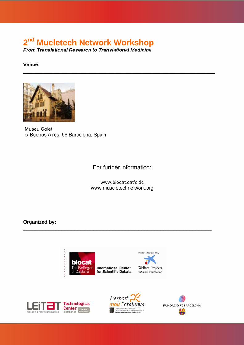
2nd Mucletech Network Workshop From Translational Research to Translational Medicine
Venue: ____________________________________________________________
Museu Colet. c/ Buenos Aires, 56 Barcelona. Spain
For further information:
www.biocat.cat/cidc www.muscletechnetwork.org
Organized by: ___________________________________________________________