principles & practice of light microscopy 3 · principles & practice of light microscopy 3...
TRANSCRIPT

Principles & Practice of Light Microscopy 3 �
• CONTRAST�
Edited by: Zvi Kam, Weizmann�For Advance Light Microscopy course �

Enhancing contrast �In �
Transmitted light microscopy�

Generating contrast in light microscopy
Problem-- many living unstained samples are thin and optically transparent Hard to see by brightfield. Solution-- transmitted light-based techniques for improving contrast (Phase, Darkfield, Polarization, DIC)
Brightfield Phase contrast
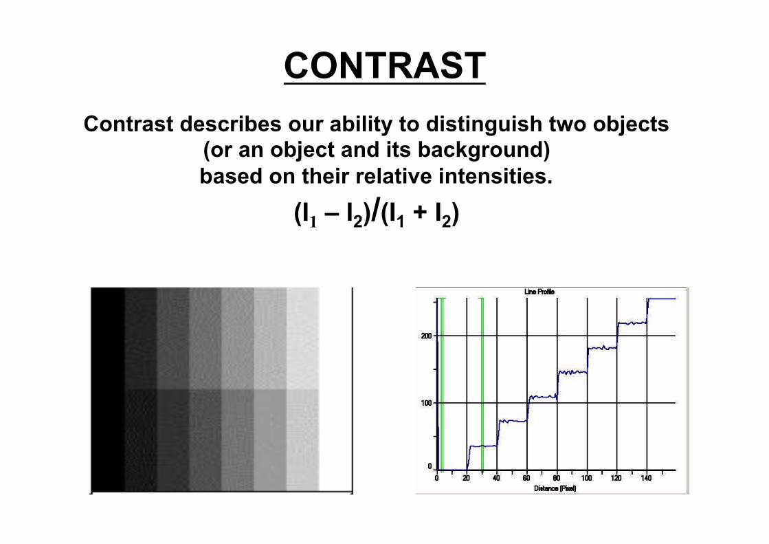
CONTRAST Contrast describes our ability to distinguish two objects
(or an object and its background) based on their relative intensities.
(I1 – I2)/(I1 + I2)

GENERATION OF CONTRAST
• Stains • Darkfield • Rheinberg illumination • Phase contrast microscopy • DIC (Nomarski)
IMPORTANT CONCEPT Microscopists look at thin objects which do not absorb much light.
Therefore, they must find ways to increase constrast.

Absorption is not the only way samples interact with light. (polarization, phase shift) Phase and DIC microscopy convert differences in phase to differences in amplitude
Your eyes are good at seeing differences in amplitude (intensity) and wavelength (color), but not phase or polarization

Optical path difference = D = t (ns-nm)
= 5microns (1.36-1.335) = .125 microns= 125 nm,
which is about 1/4 the wavelength of green light (488 nm)
t = sample thickness. Typical cell in monolayer = 5 microns
n(s) = refractive index of sample. Most cells 1.36
n(m) = refractive index of medium. Cell medium 1.335
Samples of different refractive index change optical path length

What Phase Microscopy accomplishes
Converts differences in optical path length to differences in amplitude

Review-- interference of light waves with same wavelength

surround
Because amplitude of surround and particle waves are almost identical, sample lacks contrast.
Forming an image-- role of diffracted light
S= surround (undiffracted) D= diffracted wave P= particle wave (S+D)

We would rather have D closer to S in amplitude and phase shift to be γ/2 (vs γ/4) for max interference and contrast
S= surround (undiffracted) D= diffracted wave P= particle wave (S+D)
Need way of independently controlling amplitude and phase of S + D.

lamp
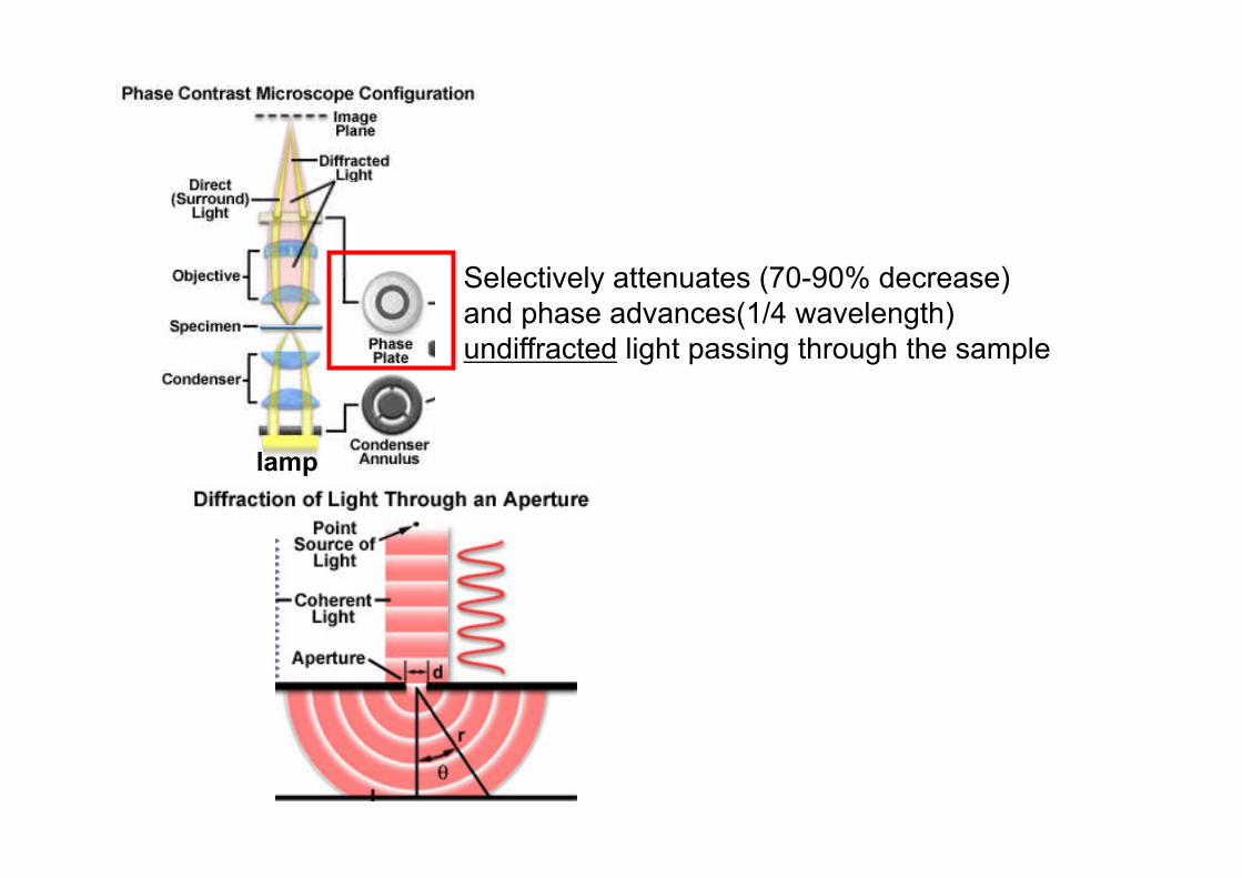
Restricts angles of illumination so diffracted and undiffracted light can be selectively modulated at phase plate

lamp
Selectively attenuates (70-90% decrease) and phase advances(1/4 wavelength) undiffracted light passing through the sample


PHASE CONTRAST MICROSCOPY

Phase contrast:!Interference between"1. directly transmitted"rays attenuated and"phase-shifted in "in the phase plate"and"2. rays diffracted "(scattered) by "refractive index "variations in the "sample"
Special phase

The common plate!in the back-aperture!of phase objectives

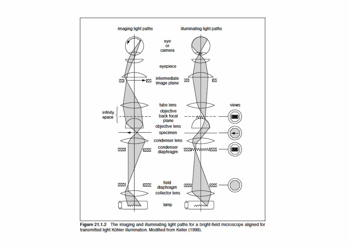
Review-- conjugate image planes in microscope


PHASE CONTRAST ALLIGNMENT

Back focal plane
Front focal plane
Proper alignment of condenser annulus and phase plate are essential for phase microscopy
(separates surround and diffracted light)

Limitations of Phase Contrast
Poor for thick samples for two reasons
1. Poor lateral (z) resolution due to limited aperture
2. Sufficiently thick samples can shift light more than 1 wavelength (so thin and thick sections can have similar brightness for biological samples thicker than about 10 microns)

Limitations of Phase Contrast
Halos -- some diffracted light (esp low spatial frequency and center of objects) also captured by phase plate, leading to localized contrast reversal. Can limit resolution.

PHASE CONTRAST IMAGE

APODIZED PHASE CONTRAST

Halos in phase contrast can be decreased by apodization

Review of Phase
What if we were to increase contrast further by throwing away all non-diffracted light?
Darkfield microscopy

Darkfield images only diffracted light
Similar to phase, projects
Cone of light onto specimen, but
With higher NA than objective, so no
surround light enters objective

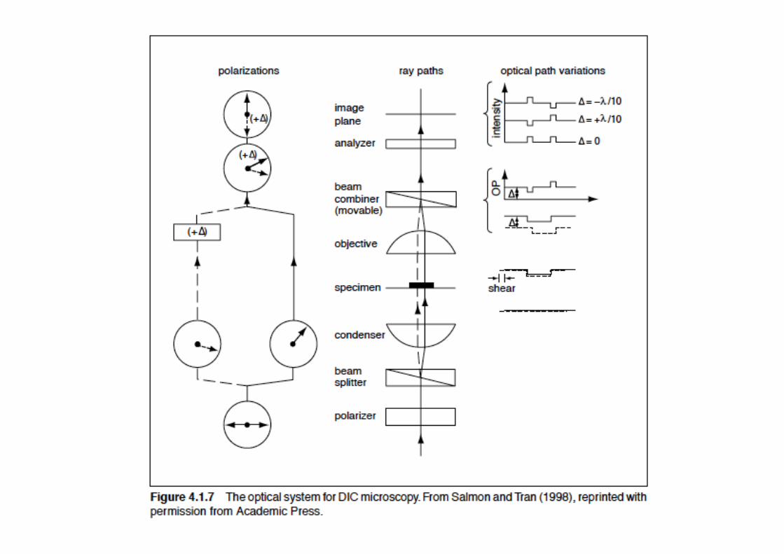
DIC: an alternative technique for enhancing contrast
Also relies on phase shifts, but uses differences in optical path differences (vs absolute optical path for phase contrast)
Uses light polarization, dual beam interferometry
Phase DIC (Differential Interference Contrast)

What DIC accomplishes
Converts relative differences in optical path length to differences in amplitude

Features of a DIC image
1. Contrast is directional
2. Contrast highlights edges
3. One end brighter, other is dimmer than background leading to pseudoshadowed, almost 3d image

Review of light polarity, polarizers

Birefringence
Birefringent materials have two indices of refraction (light travels through at different velocities depending on orientation) and can change polarization state of light.

Consequences of birefringence on light polarity

BIREFRINGENCE
IMPORTANT CONCEPT Index of Refraction (n)
may depend on Polarization

POLARIZERS
Glan-Thompson polarizer
IMPORTANT CONCEPT Absorption may
depend on Polarization


Polarized light microscopy

Polarized Light microscopy Only works with birefringent samples (those that alter polarity of light) -- some polymers such as microtubules Depends on orientation, so rotating stage desirable Compatible with fluorescence microscopy (good way to read out orientation of certain chromophores)
Requires strain-free optics

Can use modification of polarization microscope for non-birefringent samples -- DIC converts optical path difference into polarity changes


Splits parallel waves into mutually perpendicular waves (cannot interfere) with slight shear or separation in one axis

Exactly reverses action of first prism to remove shear and reverse rotation

Differential phase shift of paired waves produces elliptically polarized light that can partially pass through the analyzer

Holzwarth, Webb, Kubinski, and Allen, J. Microscopy, p249-254 (1997)

Nomarski’s!Differential!Interference!Contrast (DIC):""Interference between"righ and left handed"circularly polarized"and slightly SHIFTED"in space images."

DIC IMAGES
X-pol Y-pol


DIC!image!of!cells!


Role of Bias in DIC

Ways to introduce bias in DIC
1. Translate Prisms relative to one another 2. Rotate polarizer (in conjuction with wave retardation plate)

Because of directional contrast, DIC is sensitive to specimen orientation
DIC but not phase is orientation-dependent

DIC not compatible with birefringent samples (can’t plate cells on or or cover cells with plastic).
Phase better than DIC for birefringent samples

DIC is not working well in plastic dishes�

Comparison of Phase Contrast and DIC DIC Phase Contrast
Sensitive to sample orientation yes no Thick samples/optical sectioning good poor Birefringent samples poor good

examine effect of closing down condenser aperture on ability to do optical sectioning (C. elegans)
DIC gives superior lateral and axial resolution

Phase Contrast and DIC often used in conjunction with fluorescence microscopy
To provide cellular or organismal reference. Phase and DIC are much more general (and less toxic detection tools than fluorescence.

DARKFIELD IMAGING

DARKFIELD IMAGES

-Darkfield good for imaging unstained microorganisms,
-even sub-resolution objects such as flagella (20nm diameter) visible with darkfield.
-not good for internal structure
-Dust on sample, optics, bubbles in oil are not tolerated with this technique
First direct visualization of microtubule dynamic instability

RHEINBERG ILLUMINATION

RHEINBERG IMAGE

Review: Phase-- converts optical path length into contrast Darkfield-- images only diffracted light DIC-- contrasts region of sample with local differences in optical path length Polarization-- converts polarity information into contrast,
only works with birefringent samples (polymers, some crystals)

CONTRAST and MTF

MTF

MTF WITH CONTRAST GENERATION

OTHER CONTRAST GENERATION METHODS
• Polarization
• Hoffman modulation/Oblique Illumination
• Interference
• Fluorescence
• QPM (DIC/phase contrast using three BF images).

TAKEHOME MESSAGES
• Numerical aperture determines resolution
• Empty magnification is bad
• Contrast generation often lowers resolution, but it is usually worth it
• Keep dirt off of the image planes
• Use ND filters to adjust illumination intensity

Phase microscopy MicroscopyU microscopyu.com DIC microscopy http://micro.magnet.fsu.edu/primer/techniques/dic/dicintro.html Ted Salmon (Phase and Nomarski alignment handout)