primary mediastinal liposarcoma
TRANSCRIPT
Template provided by: “posters4research.com”
cava, and with its continued growth, caused compression of
this major vessel. On review of literature, superior vena cava
syndrome was the presenting symptom of patients with
mediastinal sarcomas in 37%.
The radiologic features of mediastinal liposarcomas
are nonspecific but are suggestive of the diagnosis. The
predominant finding on conventional chest radiography is a
widened mediastinum. On CT, the appearance of mediastinal
liposarcomas, the same as those located in other sites of
body, varies from a predominantly fat-containing mass to a
solid mass. Low attenuation values between –50 to –150
Hounsfield Unit are consistent with a tissue composed of
fat. Greater values are related to the necrosis, heterogenity
and soft tissue component in liposarcomas. A differential
diagnosis should be made between lipoma, thymolipoma,
teratoma, lymphoma, or germ cell tumor.
Liposarcomas are characterized by their large size
and their variable histologic subtypes, which correlate with
the clinical behavior and the prognosis. The most recent
World Health Organization classification of soft tissue
tumors recognizes 5 categories of liposarcomas: (1) well
differentiated, which includes the adipocytic, sclerosing, and
inflammatory subtypes; (2) dedifferentiated; (3) myxoid; (4)
round cell; and (5) pleomorphic. Patients with
dedifferentiated or pleomorphic liposarcomas have a
significant poor survival and prognosis than those with
myxoid or well differentiated.
Well differentiated low-grade liposarcomas, also
known as atypical lipomatous tumors, have histologic
features in many areas resembling mature adipose tissue.
Evans reported that atypical lipomatous tumors may
transform to dedifferentiated liposarcomas and usually do
not metastasize. The patient had predominantly myxoid with
some round cell component, and these subtypes are
considered to be less aggressive than the dedifferentiated
subtype.
Surgical removal is the optimal treatment for a
mediastinal liposarcoma. According to literature, aggressive
surgical intervention seems to favour the quality of life of
patients and prolong survival, being considered the most
effective treatment. If the entire tumor cannot be resected,
surgical debulking often results in symptomatic relief.
Chemotherapy and radiotherapy are ineffective modalities,
may be added as adjuncts to surgical excision used in
unresectable or incompletely resected tumors, but
liposarcomas seem to have low sensitivity. In the patient’s
case, he underwent surgical debulking. However it took him
almost two months to follow-up so adjuvant chemotherapy
was not started immediately.
Prognosis depends on the histological subtype and on
surgical resection with wide margins of safety. Recurrence is
common in deep-seated liposarcomas and it becomes
apparent within the first 6 months in most cases, but it may
be delayed for 5 or 10 years following the initial excision.
A 32-year old man from the Philippines presented with
a three-month history of right facial and extremity edema He
did not seek consult until he experienced dyspnea. On work-
up, his chest radiograph revealed a right suprahilar mass.
CT-scan revealed lobulated masses on the superior, anterior,
and the right middle mediastinum measuring 7.8 x 9.0 x 7.5
cm and compressing the superior vena cava. He was then
referred to the Philippine General Hospital for further
management. Open biopsy of the mass with frozen section
revealed a high grade liposarcoma. During this time, the
patient was lost to follow-up. After 2 months, he came back
to our Institution dyspneic with worsening edema. Repeat
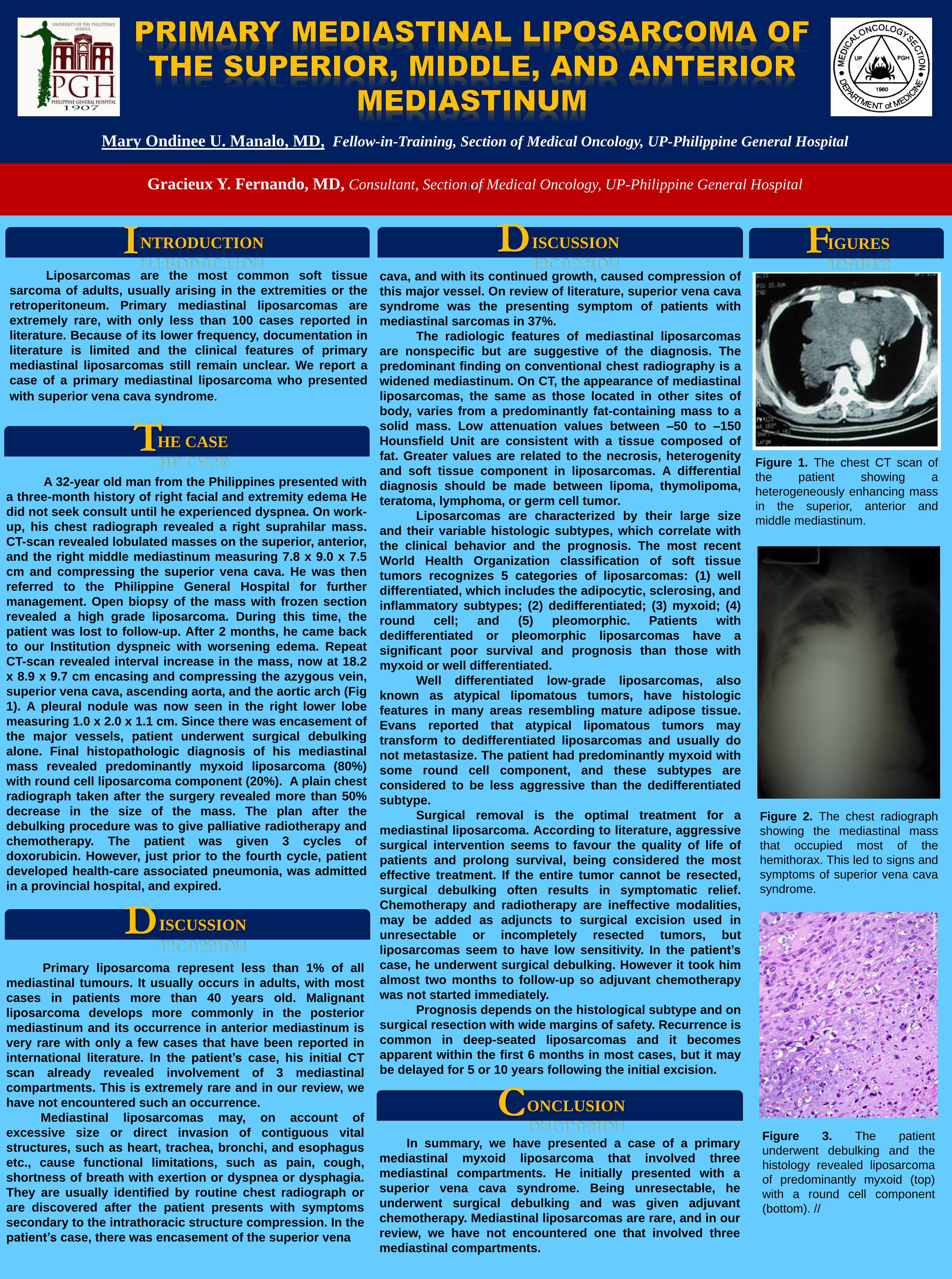
CT-scan revealed interval increase in the mass, now at 18.2
x 8.9 x 9.7 cm encasing and compressing the azygous vein,
superior vena cava, ascending aorta, and the aortic arch (Fig
1). A pleural nodule was now seen in the right lower lobe
measuring 1.0 x 2.0 x 1.1 cm. Since there was encasement of
the major vessels, patient underwent surgical debulking
alone. Final histopathologic diagnosis of his mediastinal
mass revealed predominantly myxoid liposarcoma (80%)
with round cell liposarcoma component (20%). A plain chest
radiograph taken after the surgery revealed more than 50%
decrease in the size of the mass. The plan after the
debulking procedure was to give palliative radiotherapy and
chemotherapy. The patient was given 3 cycles of
doxorubicin. However, just prior to the fourth cycle, patient
developed health-care associated pneumonia, was admitted
in a provincial hospital, and expired.
ltant
Liposarcomas are the most common soft tissue
sarcoma of adults, usually arising in the extremities or the
retroperitoneum. Primary mediastinal liposarcomas are
extremely rare, with only less than 100 cases reported in
literature. Because of its lower frequency, documentation in
literature is limited and the clinical features of primary
mediastinal liposarcomas still remain unclear. We report a
case of a primary mediastinal liposarcoma who presented
with superior vena cava syndrome.
Mary Ondinee U. Manalo, MD, Fellow-in-Training, Section of Medical Oncology, UP-Philippine General Hospital
Gracieux Y. Fernando, MD, Consultant, Section of Medical Oncology, UP-Philippine General Hospital
In summary, we have presented a case of a primary
mediastinal myxoid liposarcoma that involved three
mediastinal compartments. He initially presented with a
superior vena cava syndrome. Being unresectable, he
underwent surgical debulking and was given adjuvant
chemotherapy. Mediastinal liposarcomas are rare, and in our
review, we have not encountered one that involved three
mediastinal compartments.
HE CASE T
NTRODUCTION I ISCUSSION D
ONCLUSION C
IGURES F
ISCUSSION D Primary liposarcoma represent less than 1% of all
mediastinal tumours. It usually occurs in adults, with most
cases in patients more than 40 years old. Malignant
liposarcoma develops more commonly in the posterior
mediastinum and its occurrence in anterior mediastinum is
very rare with only a few cases that have been reported in
international literature. In the patient’s case, his initial CT
scan already revealed involvement of 3 mediastinal
compartments. This is extremely rare and in our review, we
have not encountered such an occurrence.
Mediastinal liposarcomas may, on account of
excessive size or direct invasion of contiguous vital
structures, such as heart, trachea, bronchi, and esophagus
etc., cause functional limitations, such as pain, cough,
shortness of breath with exertion or dyspnea or dysphagia.
They are usually identified by routine chest radiograph or
are discovered after the patient presents with symptoms
secondary to the intrathoracic structure compression. In the
patient’s case, there was encasement of the superior vena
Fig 1
Figure 1. The chest CT scan of
the patient showing a
heterogeneously enhancing mass
in the superior, anterior and
middle mediastinum.
Figure 2. The chest radiograph
showing the mediastinal mass
that occupied most of the
hemithorax. This led to signs and
symptoms of superior vena cava
syndrome.
Figure 3. The patient
underwent debulking and the
histology revealed liposarcoma
of predominantly myxoid (top)
with a round cell component
(bottom). //