prevalence and characteristics of ectopic atrial tachycardia and...
TRANSCRIPT

PREVALENCE AND CHARACTERISTICS OF ECTOPIC ATRIAL TACHYCARDIA AND INAPPROPRIATE SINUS TACHYCARDIA
AINO-MAIJASTILL
Department of Internal Medicine,University of Oulu
OULU 2004


AINO-MAIJA STILL
PREVALENCE AND CHARACTERISTICS OF ECTOPIC ATRIAL TACHYCARDIA AND INAPPROPRIATE SINUS TACHYCARDIA
Academic Dissertation to be presented with the assent ofthe Faculty of Medicine, University of Oulu, for publicdiscussion in the Auditorium 8 of Oulu UniversityHospital, on May 28th, 2004, at 12 noon.
OULUN YLIOPISTO, OULU 2004

Copyright © 2004University of Oulu, 2004
Supervised byProfessor Heikki HuikuriDocent Pekka RaatikainenDocent Juhani Airaksinen
Reviewed byDocent Juha MustonenDocent Markku Mäkijärvi
ISBN 951-42-7380-X (nid.)ISBN 951-42-7381-8 (PDF) http://herkules.oulu.fi/isbn9514273818/
ISSN 0355-3221 http://herkules.oulu.fi/issn03553221/
OULU UNIVERSITY PRESSOULU 2004

Still, Aino-Maija, Prevalence and characteristics of ectopic atrial tachycardia andinappropriate sinus tachycardia Department of Internal Medicine, University of Oulu, P.O.Box 5000, FIN-90014 University ofOulu, Finland 2004Oulu, Finland
AbstractThis research was designed to assess the prevalence, characteristics, natural course and autonomicregulation of ectopic atrial tachycardia (EAT) and inappropriate sinus tachycardia (IST) and theresponse of IST to adenosine.
The prevalence of EAT, as estimated from the electrocardiograms (ECG) of males applying for apilot's licence, was 0.34%. During a mean follow-up time of 8 years among 10 asymptomatic subjectsand 7 years among 17 symptomatic patients, a majority of the subjects showed a reduction of the heartrate (HR), either with restoration of sinus rhythm (SR) (37%) or with a change in P wave morphology(37%).
The prevalence of IST in a random sample of 604 middle-aged subjects was 1.16%. The systolicand diastolic ambulatory blood pressures were higher among the subjects with IST than subjects withSR (P < 0.001). The other laboratory, echocardiographic and personality measurements, with theexception of the hostility score (P < 0.001), revealed no differences between the groups. During amean follow-up of 6 years, none of the subjects with IST developed any evidence of structural heartdisease despite ongoing palpitations, and there was no significant reduction of the 24-hour averageHR.
In an analysis of R-R interval variability from 24-hour ECG recordings in 12 patients withincessant EAT, 12 subjects with IST and 24 subjects with SR, the time- and frequency-domainmeasures of HR variability did not differ between the subjects with EAT and IST. However, the short-term fractal HR behaviour differed between EAT and IST.
In studies of the effects of adenosine in 18 patients with IST and 18 subjects with SR, adenosineprolonged significantly the sinus interval (P < 0.001) in the control subjects, but did not cause anysignificant changes of atrial cycle length in the patients with IST.
Conclusions: 1. EAT has a tendency towards gradual degeneration over time. 2. The prevalenceof IST is higher than previously assumed, but the overall prognosis is good. 3. EAT and IST seem tobe under similar autonomic regulation as the sinus node, but the firing of ectopic atrial foci showsmore random behaviour. 4. The usual negative chronotropic effect of adenosine is impaired insubjects with IST.
Keywords: adenosine, arrhythmia, electrocardiography, prognosis


To my family


Acknowledgements
This work was carried out at the Department of Internal Medicine, University of Oulu.Part of the patients were gathered from Kuopio University Hospital, Kuopio, Finland,Central Military Hospital, Helsinki, Finland, and University of Miami School ofMedicine, Miami, Florida, USA.
I wish to express my gratitude to Professor Antero Kesäniemi, M.D., Ph.D., who bothas the head of the Department and as a co-worker has supported me during my work forthis thesis.
I am deeply grateful to Docent Juha Mustonen, M.D., Ph.D., and Docent MarkkuMäkijärvi, M.D., Ph.D., for their valuable comments and constructive criticism during thepreparation of the final manuscript.
My warmest and deepest gratitude is due to my doctoral supervisor Professor HeikkiHuikuri, M.D., Ph.D., for introducing to me this subject and for his excellent guidanceduring the process. I am also grateful for the possibility to be a member of his researchteam.
I sincerely thank Docent Pekka Raatikainen, M.D., Ph.D., my second supervisor, forhis enthusiastic and valuable contribution to this work during the recent years.
I owe my special thanks to Docent Juhani Airaksinen, M.D., Ph.D., my other supervi-sor, for his warm and positive attitude towards research work and colleagues.
My sincere gratitude is also due to Professor Agustin Castellanos, M.D., Docent JuhaHartikainen, M.D., Ph.D., Professor Markku Ikäheimo, M.D., Ph.D., Docent Jouko Kar-jalainen, M.D., Ph.D., Docent Heikki Kauma, M.D., Ph.D., Docent Raimo Kettunen,M.D., Ph.D., Docent Juhani Koistinen, M.D., Ph.D., Associate Professor Raul Mitrani,M.D., Professor Robert Myerburg, M.D., Docent Timo Mäkikallio, M.D., Ph.D., andAntti Ylitalo, M.D., Ph.D. who were my co-workers.
I want to thank our research team: Vesa Jokinen, M.D., Ph.D., Kai Lindgren, M.D.,Juha Perkiömäki, M.D., Ph.D., Sirkku Pikkujämsä M.D., Ph.D., Jari Tapanainen, M.D.,Ph.D., and Mikko Tulppo, Ph.D., for their existence and help. Pirkko Huikuri, R.N., PäiviKarjalainen, R.N., Marja Hietaniemi, R.N., Maire Kukkonen, R.N., Ms. Anne Lehtinen,Ms. Anne Salovaara and Mirja Peltola, M.Sc. are most sincerely acknowledged for theirtechnical assistance.

I wish to express my warmest thanks to all colleagues and the staff in the Cardiovascu-lar Laboratory for both clinical and scientific help. Especially Markku Linnaluoto, M.Sc.,is acknowledged.
I express my thanks to Mrs. Sirkka-Liisa Leinonen for her prompt revision of theEnglish language of this thesis.
My deepest gratitude is due to my mother and my late father for their love and continu-ous encouragement during my life and their love and important help to my family. I wishto thank my sister Minna and her family for love and support. I thank my aunt Arja forher love towards our family and for her help in our home to make this thesis possible. Iwant to thank all of my relatives and friends for their support and encouragement duringthese years.
Finally, I owe my most loving thanks to my husband Jari especially for his love andsupport. I also thank him for giving me new interesting perspectives into life. Our won-derful children Emil-Aleksi, 4, and Jenni-Julia, 3, are really the centre of my life.
This study was financially supported by AstraZeneca, Masala, Finland, The FinnishFoundation for Cardiovascular Research, The Commemorative Foundation of MaudKuistila, Orion Corporation Research Foundation, Oulu University Hospital and the OuluUniversity Scholarship Foundation.
Oulu, April 2004 Aino-Maija Still

Abbreviations
ACL atrial cycle lengthAT atrial tachycardiaAV atrioventricularBMI body mass indexBP blood pressureBPM beats per minuteEAT ectopic atrial tachycardiaECG electrocardiographyHR heart rateHRV heart rate variabilityIKAch,Ado acetylcholine- and adenosine-sensitive potassium channelIHR intrinsic heart rateIST inappropriate sinus tachycardiaLF low frequencyHF high frequencyLVH left ventricular hypertrophyRF radiofrequencyR-R interval interval between consecutive ventricular beats SD standard deviationSR sinus rhythmSVT supraventricular tachycardia


List of original articles
This thesis is based on the following publications, which are referred to in the text bytheir Roman numerals:
I Poutiainen A-M, Koistinen MJ, Airaksinen KEJ, Hartikainen JEK, Kettunen RVJ,Karjalainen JE & Huikuri HV (1999) Prevalence and natural course of ectopic atrialtachycardia. European Heart Journal 20:694–700.
II Huikuri HV, Poutiainen A-M, Mäkikallio TH, Koistinen MJ, Airaksinen KEJ,Mitrani R, Myerburg RJ & Castellanos A (1999) Dynamic behavior and autonomicregulation of ectopic atrial pacemakers. Circulation 100:1416–1422.
III Still A-M, Huikuri HV, Airaksinen KEJ, Koistinen MJ, Kettunen R, Hartikainen J,Mitrani RD, Castellanos A, Myerburg RJ & Raatikainen MJP (2002) Impaired nega-tive chronotropic response to adenosine in patients with inappropriate sinus tachy-cardia. Journal of Cardiovascular Electrophysiology 13:557–562.
IV Still A-M, Raatikainen MJP, Ylitalo A, Kauma H, Ikäheimo M, Kesäniemi YA &Huikuri HV (2003) Prevalence, characteristics and natural course of inappropriatesinus tachycardia. Submitted for publication.


Contents
Abstract Acknowledgements Abbreviations List of original articlesContents1 Introduction . . . . . . . . . . . . . . . . . . . . . . . . . . . . . . . . . . . . . . . . . . . . . . . . . . . . . . . . 172 Review of the literature . . . . . . . . . . . . . . . . . . . . . . . . . . . . . . . . . . . . . . . . . . . . . . . 19
2.1 General aspects of the nervous regulation of heart rate . . . . . . . . . . . . . . . . . . 192.2 Ectopic atrial tachycardia . . . . . . . . . . . . . . . . . . . . . . . . . . . . . . . . . . . . . . . . . 20
2.2.1 History . . . . . . . . . . . . . . . . . . . . . . . . . . . . . . . . . . . . . . . . . . . . . . . . . . . 202.2.2 Prevalence . . . . . . . . . . . . . . . . . . . . . . . . . . . . . . . . . . . . . . . . . . . . . . . . 212.2.3 Pathophysiology . . . . . . . . . . . . . . . . . . . . . . . . . . . . . . . . . . . . . . . . . . . 21
2.2.3.1 The etiology of ectopic atrial tachycardia . . . . . . . . . . . . . . . . . 232.2.3.2 Autonomic regulation of ectopic atrial tachycardia . . . . . . . . . . 23
2.2.4 Symptoms and characteristics . . . . . . . . . . . . . . . . . . . . . . . . . . . . . . . . . 252.2.5 Diagnosis and differential diagnosis . . . . . . . . . . . . . . . . . . . . . . . . . . . . 26
2.2.5.1 Clinical evaluation . . . . . . . . . . . . . . . . . . . . . . . . . . . . . . . . . . . 262.2.5.2 Electrophysiologic evaluation . . . . . . . . . . . . . . . . . . . . . . . . . . 302.2.5.3 Differential diagnosis . . . . . . . . . . . . . . . . . . . . . . . . . . . . . . . . . 32
2.2.6 Natural course . . . . . . . . . . . . . . . . . . . . . . . . . . . . . . . . . . . . . . . . . . . . . 332.2.7 Treatment and outcome . . . . . . . . . . . . . . . . . . . . . . . . . . . . . . . . . . . . . . 34
2.2.7.1 Medical therapy . . . . . . . . . . . . . . . . . . . . . . . . . . . . . . . . . . . . . 342.2.7.2 Invasive therapy . . . . . . . . . . . . . . . . . . . . . . . . . . . . . . . . . . . . . 36
2.3 Inappropriate sinus tachycardia . . . . . . . . . . . . . . . . . . . . . . . . . . . . . . . . . . . . 372.3.1 History . . . . . . . . . . . . . . . . . . . . . . . . . . . . . . . . . . . . . . . . . . . . . . . . . . . 372.3.2 Prevalence and characteristics . . . . . . . . . . . . . . . . . . . . . . . . . . . . . . . . . 372.3.3 Pathophysiology . . . . . . . . . . . . . . . . . . . . . . . . . . . . . . . . . . . . . . . . . . . 38
2.3.3.1 Sinus node anatomy and physiology . . . . . . . . . . . . . . . . . . . . . 382.3.3.2 Pathophysiology of inappropriate sinus tachycardia . . . . . . . . . 382.3.3.3 Autonomic regulation . . . . . . . . . . . . . . . . . . . . . . . . . . . . . . . . 40
2.3.4 Symptoms . . . . . . . . . . . . . . . . . . . . . . . . . . . . . . . . . . . . . . . . . . . . . . . . 41

2.3.5 Diagnosis and differential diagnosis . . . . . . . . . . . . . . . . . . . . . . . . . . . . 412.3.5.1 Clinical evaluation . . . . . . . . . . . . . . . . . . . . . . . . . . . . . . . . . . . 412.3.5.2 Electrophysiologic evaluation . . . . . . . . . . . . . . . . . . . . . . . . . . 442.3.5.3 Differential diagnosis . . . . . . . . . . . . . . . . . . . . . . . . . . . . . . . . . 45
2.3.6 Natural course . . . . . . . . . . . . . . . . . . . . . . . . . . . . . . . . . . . . . . . . . . . . . 462.3.7 Treatment and outcome . . . . . . . . . . . . . . . . . . . . . . . . . . . . . . . . . . . . . . 46
2.3.7.1 Medical and other therapies . . . . . . . . . . . . . . . . . . . . . . . . . . . . 462.3.7.2 Invasive therapy . . . . . . . . . . . . . . . . . . . . . . . . . . . . . . . . . . . . . 47
3 Aims of the study . . . . . . . . . . . . . . . . . . . . . . . . . . . . . . . . . . . . . . . . . . . . . . . . . . . 504 Subjects and methods . . . . . . . . . . . . . . . . . . . . . . . . . . . . . . . . . . . . . . . . . . . . . . . . 51
4.1 Subjects . . . . . . . . . . . . . . . . . . . . . . . . . . . . . . . . . . . . . . . . . . . . . . . . . . . . . . . 514.2 Definitions of tachycardias . . . . . . . . . . . . . . . . . . . . . . . . . . . . . . . . . . . . . . . . 534.3 Collection of clinical data and follow-up . . . . . . . . . . . . . . . . . . . . . . . . . . . . . 534.4 Electrocardiographic measurements . . . . . . . . . . . . . . . . . . . . . . . . . . . . . . . . . 54
4.4.1 12-lead ECG . . . . . . . . . . . . . . . . . . . . . . . . . . . . . . . . . . . . . . . . . . . . . . 544.4.2 Ambulatory ECG recordings . . . . . . . . . . . . . . . . . . . . . . . . . . . . . . . . . . 54
4.4.2.1 Heart rate variabilility . . . . . . . . . . . . . . . . . . . . . . . . . . . . . . . . 544.4.3 Ambulatory blood pressure and heart rate recordings . . . . . . . . . . . . . . . 56
4.5 Electrophysiological studies . . . . . . . . . . . . . . . . . . . . . . . . . . . . . . . . . . . . . . . 564.6 Echocardiographic measurements . . . . . . . . . . . . . . . . . . . . . . . . . . . . . . . . . . 574.7 Pharmacological maneuvers . . . . . . . . . . . . . . . . . . . . . . . . . . . . . . . . . . . . . . . 57
4.7.1 Autonomic blockade . . . . . . . . . . . . . . . . . . . . . . . . . . . . . . . . . . . . . . . . 574.7.2 Atropine . . . . . . . . . . . . . . . . . . . . . . . . . . . . . . . . . . . . . . . . . . . . . . . . . . 584.7.3 Adenosine . . . . . . . . . . . . . . . . . . . . . . . . . . . . . . . . . . . . . . . . . . . . . . . . 58
4.8 Laboratory methods . . . . . . . . . . . . . . . . . . . . . . . . . . . . . . . . . . . . . . . . . . . . . 584.9 Statistical analyses . . . . . . . . . . . . . . . . . . . . . . . . . . . . . . . . . . . . . . . . . . . . . . 58
5 Results . . . . . . . . . . . . . . . . . . . . . . . . . . . . . . . . . . . . . . . . . . . . . . . . . . . . . . . . . . . . 605.1 Ectopic atrial tachycardia . . . . . . . . . . . . . . . . . . . . . . . . . . . . . . . . . . . . . . . . . 60
5.1.1 Prevalence . . . . . . . . . . . . . . . . . . . . . . . . . . . . . . . . . . . . . . . . . . . . . . . . 605.1.2 Natural course . . . . . . . . . . . . . . . . . . . . . . . . . . . . . . . . . . . . . . . . . . . . . 605.1.3 Heart rate variability . . . . . . . . . . . . . . . . . . . . . . . . . . . . . . . . . . . . . . . . 62
5.1.3.1 Time- and frequency-domain measures of heart rate variability 625.1.3.2 Fractal and complexity measures of R-R interval variability . . 64
5.1.4 Heart rate responses of ectopic atrial tachycardia to intravenous atropine . . . . . . . . . . . . . . . . . . . . . . . . . . . . . . . . . . . . . . . . 65
5.2 Inappropriate sinus tachycardia . . . . . . . . . . . . . . . . . . . . . . . . . . . . . . . . . . . . 655.2.1 Prevalence . . . . . . . . . . . . . . . . . . . . . . . . . . . . . . . . . . . . . . . . . . . . . . . . 655.2.2 Characteristics of the subjects with inappropriate sinus tachycardia . . . 665.2.3 Natural course . . . . . . . . . . . . . . . . . . . . . . . . . . . . . . . . . . . . . . . . . . . . . 695.2.4 Heart rate variability . . . . . . . . . . . . . . . . . . . . . . . . . . . . . . . . . . . . . . . . 69
5.2.4.1 Time- and frequency-domain measures of heart rate variability 695.2.4.2 Fractal and complexity measures of R-R interval variability . . 70
5.2.5 Heart rate responses of inappropriate sinus tachycardia to intravenous atropine . . . . . . . . . . . . . . . . . . . . . . . . . . . . . . . . . . . . . . . . 70
5.2.6 Electrophysiological effects of adenosine in inappropriate sinus tachycardia . . . . . . . . . . . . . . . . . . . . . . . . . . . . . . . . . . . . . . . . . . . 70

6 Discussion . . . . . . . . . . . . . . . . . . . . . . . . . . . . . . . . . . . . . . . . . . . . . . . . . . . . . . . . . 746.1 Ectopic atrial tachycardia . . . . . . . . . . . . . . . . . . . . . . . . . . . . . . . . . . . . . . . . . 74
6.1.1 Prevalence . . . . . . . . . . . . . . . . . . . . . . . . . . . . . . . . . . . . . . . . . . . . . . . 746.1.2 Natural course . . . . . . . . . . . . . . . . . . . . . . . . . . . . . . . . . . . . . . . . . . . . . 746.1.3 Heart rate variability . . . . . . . . . . . . . . . . . . . . . . . . . . . . . . . . . . . . . . . . 75
6.1.3.1 Abnormalities in time- and frequency-domain heart rate variability . . . . . . . . . . . . . . . . . . . . . . . . . . . . . . . . . . . . . . 75
6.1.3.2 Abnormalities in short-term correlation properties of R-R interval dynamics . . . . . . . . . . . . . . . . . . . . . . . . . . . . . . . . 75
6.2 Inappropriate sinus tachycardia . . . . . . . . . . . . . . . . . . . . . . . . . . . . . . . . . . . . 766.2.1 Prevalence . . . . . . . . . . . . . . . . . . . . . . . . . . . . . . . . . . . . . . . . . . . . . . . 766.2.2 Characteristics of subjects with inappropriate sinus tachycardia . . . . . . 776.2.3 Natural course . . . . . . . . . . . . . . . . . . . . . . . . . . . . . . . . . . . . . . . . . . . . . 786.2.4 Heart rate variability . . . . . . . . . . . . . . . . . . . . . . . . . . . . . . . . . . . . . . . . 78
6.2.4.1 Abnormalities in time- and frequency-domain heart rate variability . . . . . . . . . . . . . . . . . . . . . . . . . . . . . . . . . . . . . . 78
6.2.4.2 Short-term correlation properties of R-R interval dynamics . . . 786.2.5 Pharmacological maneuvers . . . . . . . . . . . . . . . . . . . . . . . . . . . . . . . . . . 78
6.2.5.1 Autonomic blockade . . . . . . . . . . . . . . . . . . . . . . . . . . . . . . . . . 786.2.5.2 Adenosine . . . . . . . . . . . . . . . . . . . . . . . . . . . . . . . . . . . . . . . . . 79
6.2.6 Potential mechanism(s) of inappropriate sinus tachycardia . . . . . . . . . . 806.3 Limitations of the study . . . . . . . . . . . . . . . . . . . . . . . . . . . . . . . . . . . . . . . . . . 816.4 Implications of the study . . . . . . . . . . . . . . . . . . . . . . . . . . . . . . . . . . . . . . . . . 816.5 Future perspectives . . . . . . . . . . . . . . . . . . . . . . . . . . . . . . . . . . . . . . . . . . . . . . 82
7 Conclusions . . . . . . . . . . . . . . . . . . . . . . . . . . . . . . . . . . . . . . . . . . . . . . . . . . . . . . . . 83References


1 Introduction
Supraventricular tachycardias (SVTs) are common rhythm disturbances and were previ-ously clustered together into one diagnostic category. Based on the knowledge gainedfrom invasive electrophysiological studies, it has become possible to differentiatebetween the various SVT subtypes, mainly by the mechanisms involved (1). Clinically,part of these arrhythmias can be diagnosed by means of 12-lead ECG, and new and effec-tive methods of treatment can be devised, as necessary. The precipitants of the subtypesof SVTs vary with age, sex and associated comorbidity. This study was designed to focuson ectopic atrial tachycardia (EAT) and inappropriate sinus tachycardia (IST).
EAT is often manifested as incessant tachycardia that originates outside the sinus node,showing abnormal P waves in ECG (2). IST is non-paroxysmal tachycardia characterizedby an increased resting heart rate (HR) or an exaggerated HR response to minimal exer-tion (3) without an appropriate relationship with metabolic or physiologic demands (4).
Even though ambulatory electrocardiographic monitoring has been increasingly usedto provide data concerning arrhythmias in different age groups, there are no earlier studieson the prevalence of EAT or IST. The clinical importance of EAT and IST is that theymay cause severe symptoms and be resistant to traditional antiarrhythmic medications. Inaddition the increasing prevalence of congestive heart failure has highlighted the impor-tance of the search for potentially reversible etiologies of cardiomyopathy, which hasbeen associated with incessant EAT, especially in children (5–8). There are some earlierstudies on the natural course of EAT with varying spontaneous resolutions (2,9–16). Theprognosis of IST is largely unknown, and the mechanism of this arrhythmia is poorlyunderstood.
Experimental and clinical evidence suggests a strong relationship between the auto-nomic nervous system and the occurrence of atrial tachyarrhythmias (17). The analysis ofheart rate variability (HRV) is a useful tool for indirect assessment of autonomic effectson the heart and may have implications for understanding the arrhythmogenic mecha-nism. HRV reflects the neural regulation of normal pacemaker tissue and is well charac-terized (18–28), but there is little information of the autonomic nervous regulation ofabnormal atrial foci originating outside the sinus node.
Adenosine is an endogenous purine nucleoside that has become an important agent forthe diagnosis and treatment of cardiac arrhythmias (29). The efficacy of intravenous ade-

18
nosine for the termination of tachycardias requiring the atrioventricular (AV) node as partof their reentrant circuit is well established (30,31), but there is much less data on theresponse to adenosine in patients with arrhythmias involving the sinus node.
This study was designed to evaluate the prevalence and natural course of EAT insymptomatic inpatients and in asymptomatic subjects. We also estimated the prevalenceand characteristics of subjects fulfilling the criteria of IST in a random sample of hyper-tensive and normotensive middle-aged subjects and assessed the natural course of ISTboth in the random sample and in symptomatic hospital patients with documented IST.HRV analyses were used to determine the possible differences in HR behaviour betweensinus rhythm (SR), IST and EAT. Finally, the response of IST to adenosine was systemati-cally assessed.

2 Review of the literature
2.1 General aspects of the nervous regulation of heart rate
The sinus node, atrioventricular node and specialized conduction system of the heart pos-sess the inherent ability for spontaneous, rhythmic initiation of the cardiac excitationimpulse. Autonomic nervous system also influences the rate of spread of the excitationimpulse, the depolarization and repolarization of the myocardium, and the contractility ofboth the atria and the ventricles (32).
The parasympathetic innervation of the heart originates in the medulla and passesthrough the right and left cardiac nerves. Two sets of cardiac nerves arise from each vagusnerve: the superior (superior and inferior cervical) cardiac nerves, which arise from thevagi in the neck, and the inferior (thoracic) cardiac nerves, which arise from either thevagus nerves or the recurrent branches of the vagi. The sympathetic innervation of theheart passes from the spinal cord to the upper four or five thoracic ganglia. Some fibersfrom the upper thoracic ganglia pass up the cervical sympathetic to the superior, middle,or inferior cervical ganglia. The superior (cervical), middle (cervical), and inferior (cervi-cal) cardiac nerves originate from their respective ganglia and pass downward through thedeep and superficial parts of the cardiac plexus to the heart (32).
Most of the parasympathetic nerves are distributed near the sinus node and AV con-duction tissue, to a lesser extent to muscle of the two atria, and even less to the ventricularmuscle. The right and left vagi are distributed differentially to the various cardiac struc-tures. The right vagus nerve affects the sinus node predominantly. Stimulation slows sinusnodal firing or may even stop it for several seconds. The left vagus nerve mainly inhibitsAV conduction tissue to produce various degrees of AV block. However, the effent vagalfibers overlap. Cardiac parasympathetic impulses are transmitted by acetylcholine(33,34).
The postganglionic cardiac sympathetic fibers approach the base of the heart along theadventitial surface of the great vessels. On reaching the base of the heart, these fibers aredistributed to the various chambers as an extensive epicardial plexus. They then penetratethe myocardium, usually accompanying the coronary vessels. Sympathetic fibers aredense to the epicardial coronary arteries and veins and are moderate to intramural ves-

20
sels. The sympathetic nerves are distributed to the same areas as parasympathetic nerves,but with a strong presentation to the ventricular muscle as well as to the other parts ofheart. Sympathetic stimulation to the heart is largely mediated by the release of norepi-nephrine. The effect of stimulating these nerves is to increase the rate at which the heartbeats. Maximal sympathetic stimulation can almost triple the resting heart rate. Conduc-tion time through the AV node is also reduced (32–34).
Both sympathetic and parasympathetic fibers influence the sinus node, the AV node,and both the atrial and ventricular myocardium. The sinus node is usually under the tonicinfluence of both divisions of the autonomic nervous system. The sympathetic systemenchances automaticity, whereas the parasympathetic system inhibits it. Changes in heartrate usually involve a reciprocal action of the two divisions of the autonomic nervous sys-tem. Thus an increased heart rate is produced by a diminution of parasympathetic activityand concomitant increase in sympathetic activity; deceleration is usually achieved by theopposite mechanisms. Under certain conditions the heart rate may change by selectiveaction of just one division of the autonomic nervous system, rather than by reciprocalchanges in both divisions. In healthy individuals the heart rate at rest is dominated by theparasympathetic innervation. When physiological circumstances require the heart to beatmore rapidly, as in exercise, the activity of the parasympathetic nerves is inhibited, whilethat of the sympathetic nerves is enhanced. Sympathetic activity alters heart rate and AVconduction much more slowly than does vagal activity. Therefore, vagal activity can exertbeat-by-beat control of cardiac function, whereas sympathetic activity cannot. In general,sympathetic stimulation increases the strength of heart muscle contraction, whereas para-sympathetic stimulation decreases it (32–34).
2.2 Ectopic atrial tachycardia
2.2.1 History
EAT was first described as early as 1922 by Gallavardin (35). A variety of names, includ-ing automatic atrial tachycardia, (uni)focal atrial tachycardia and ectopic atrial tachycar-dia, have been used, but because of the uncertainty concerning the mechanism of thistachycardia, the term EAT is preferred. In 1947, Parkinson and Papp (36) introduced theattribute "repetitive" to describe this type of tachycardia, and they also described a persis-tent variety of arrhythmia (37,38), which is referred to as “sustained” by other authors(39). In children with chronic EAT or atrial tachycardia (AT) with AV block, the condi-tion most probably also includes forms of tachycardia other than EAT, such as the perma-nent form of junctional reciprocating tachycardia, multifocal AT and atrial flutter, andthus there is no homogeneous entity (39–44).
The development of invasive electrophysiological techniques has enabled better classi-fication, diagnostic and therapeutic possibilities of ATs. The electrophysiological proper-ties of EAT have been elucidated by Goldreyer et al (45) and Gillette and Garson (46).Later on, the literature on EAT in children and adults has been enriched by reports on

21
clinical aspects and medical, surgical and catheter ablative treatments (10,12,13,16,47–55).
The results of antiarrhythmic drug therapy, and even cardiac surgery, have been subop-timal in EAT. Since chronic tachycardias, such as EAT, are among the few reversiblecauses of cardiomyopathy, alternative treatment options have been explored aggressively.Radiofrequency (RF) catheter ablation was adopted quickly for the management of EAT,and its efficacy has been proven during the past decade (50–55). A new interesting elec-trophysiological issue is the association between EAT and focal atrial fibrillation (56,57).
2.2.2 Prevalence
EAT may occur as a primary disturbance of cardiac rhythm in a structurally normal heartor develop as a complication of other cardiac or pulmonary disorders. Although an asso-ciation with atrial neoplasms has been described, patients with EAT typically show noevidence of any defined organic heart disease (58). Even though EAT may present at anyage, it is predominantly seen in young infants, children and young adults. Onset may evenbe prenatal (7,11,12).
EAT is thought to be an uncommon rhythm disorder, but its true prevalence in the gen-eral population has not earlier been assessed. In the previous studies, approximately 20%of SVTs in children (5) and 25% in adults have been nonparoxysmal (59). EAT has beenelectrophysiologically identified in 5–20% of children (60,61) and approximately 5% ofadult patients with symptomatic SVTs (62). The prevalence of asymptomatic subjectswith EAT and the occurrence of EAT in patients with structurally normal hearts is notknown. A positive family history of tachycardia has been reported with this arrhythmia(46). Pooling of the results of the previously published sixteen series indicates that thedistribution between the genders is quite equal (198 females and 192 males) (2,10,12–14,16,45,46,53,55,63–68)
2.2.3 Pathophysiology
EAT is defined as a SVT arising from the atrial muscle that does not include the sinusnode. It is characterized by inappropriate rapid frequency of atrial activation, which startsrhythmically in a very small area (focus) and spreads centrifugally (46,69–72). When thefiring rate of the ectopic focus exceeds that of the sinus node, it becomes the predomi-nant pacemaker of the heart. The mechanism of focal ectopic activity has been a subjectof debate. Most data suggest that EAT is caused by abnormal automaticity of atrial cellsto undergo spontaneous diastolic depolarization and to initiate an electrical impulse in theabsence of external electrical stimulation (62,73–75). In one study, microelectroderecordings from left atrial appendage tissue obtained at the time of surgical excision of anEAT focus revealed a high resting membrane potential and spontaneous phase 4 automa-ticity (73). Further support for automaticity as the mechanism may be found from the useof RF catheter therapy. It has been observed that EAT almost always involves a small

22
area of tissue, since it is eliminated within several seconds of the onset of RF energyapplication (54,60). Secondly, just prior to successful elimination of the foci, the ectopicrate often accelerates, consistently with the enhanced automaticity observed in responseto RF energy in other tissues (76). Finally, the foci seem to cluster in a few specific areas.These findings all suggest that EAT involves subtle electrical changes in otherwise nor-mal tissue from trabeculated atria or connections between the atria and the systemic veins(54). Other possible mechanisms of EAT include triggered activity (early and delayedafter potentials) and micro re-entry (70,74,77,78). Most of the above hypotheses arebased on imperfect surrogate markers, such as the response to pacing or pharmacologicalmaneuvers, and are fraught with problems. Further work is needed to evaluate the cellu-lar mechanism of EAT.
EAT may arise from any site in the right or left atrium. The foci are not randomly dis-tributed, but rather tend to cluster in certain anatomical zones (7,51,52,60,77,79,80). Inthe right atrium, the most frequent locations of the foci are along the crista terminalis (2/3), in the para-Hisian region and around the ostium of the coronary sinus (80). In addi-tion, ATs originating from around the tricuspid annulus have been characterized (81). Inthe left atrium, the most frequent location of the foci seems to be at the junction (orinside) the pulmonary veins. There are also isolated reports of left atrial tachycardia origi-nating from the mitral annulus (68,82). The distribution of EAT foci may differ, depend-ing on the patient population (75). In several studies, the ectopic foci have been reportedto occur in the right atrium (2,12,49). One of the primary differences between adult andpediatric patients with EAT is the predominance of right-sided foci in adults, whereasboth left- and right-sided ones are seen in children (15,60).
There are some studies of histological features of the ectopic foci. Most cases havebeen not associated with any specific pathologic abnormalities of cardiac or skeletal mus-cle (83) or resected atrial tissue near the focus (15). Moro et al found fibrous plaque withsome degenerated myocytes (84). However, they suggested it could also be a fibroticlesion due to a previous inflammatory process. In other studies, the occasional pathologi-cal findings have been restricted to nonspecific fibrosis, cellular hypertrophy and patchyfatty infiltrates. All of these changes may be secondary to tachycardia-induced myopathy,but in some cases the tissue examined was completely normal (85).
What most of these foci seem to have in common is a region of structural inhomogene-ity. An interesting question is why the large bulk of surrounding myocardium does notelectronically inhibit the firing of a small focus. Lesh (75) speculates that, in addition toabnormal automaticity, a region of relatively poor cell-to-cell coupling is required for theinitiation and perpetuation of EAT. Accordingly, a cell or a small cluster of cells withabnormal automaticity well coupled with the surrounding normal atrium could not be ableto manifest that tendency due to electrotonic interactions. However, if the region ofabnormal automaticity exhibits poor cell-to-cell coupling, a reduced electrotonic influ-ence allows these cells to manifest and maintain their abnormal firing. Fractionated elec-trograms often seen at a successful EAT ablation site may be markers of the requisite non-uniformly anisotropic substrate (75). For example, the crista teminalis contains cells thathave very sparsely distributed transverse gap junctions as well as cells with automaticproperties (86).

23
One expanding field of research is the identification of patients with focal atrial ectopywhich triggers atrial fibrillation. This special type of atrial fibrillation is initiated by aburst of rapid EAT with subsequent degeneration to atrial fibrillation (56,57).
2.2.3.1 The etiology of ectopic atrial tachycardia
The etiology of EAT is unknown. Most reports support the hypothesis that myocardialdysfunction is a result rather than the cause of incessant EAT (7,12,87,88). The exactpathophysiological mechanisms of this dysfunction are not fully understood. The mostnotable feature of tachycardia-induced myopathy is a prompt reversal of ventricular dys-function when tachycardia is eliminated (89). In animal models, dilated cardiomyopathycan be induced within 30 days when the atrium is paced chronically at rates that areapproximately 3 times normal, and recovery occurs in about 2 weeks when pacing is ter-minated (90). It is now known that ventricular dysfunction is truly related to depressedcontractile performance of individual myocytes instead of being simply a phenomenondue to excess preload or high afterload. Multiple factors, such as alterations in cellularcalcium homeostasis, changes in cell ultrastructure including decreased myofibril content,alteration in the Na+, K+-ATPase system and changes in beta-adrenergic receptor func-tion, have been implicated as causes of tachycardia-induced myopathy (85). The centralbiochemical derangement that includes these changes has not yet been identified. Onepossible explanation is that long-standing tachycardia causes chronic depletion of myo-cardial high-energy phosphate (5,6,87,88,91–93). Although AV dyssynchrony may playsome role, normal AV synchrony with a normal PR interval may still lead to ventriculardysfunction. Immature or developing myocardium may be more vulnerable to the delete-rious effects of chronic EAT than mature myocardium, possibly because the rate of AVconduction tends to be quite brisk through a younger patient's AV node, although theexact explanation for this age-related difference is uncertain (94).
2.2.3.2 Autonomic regulation of ectopic atrial tachycardia
Pharmacological maneuvers
Several pharmacological agents have been used to aid the diagnosis of EAT (Table 1).Isoproterenol. During an invasive electrophysiological examination, administration of
isoproterenol as an adrenergic stimulator is commonly used to initiate or accelerate EATin a similar way as it accelerates SR. The intravenous infusion rate of isoproterenolshould increase HR by at least 20–40 % or above 100 bpm to ensure arrhythmia induction(54,66). Typically, this requires an infusion rate of 0.01 to 0.04 µg/kg/min (62,66). Epi-nephrine and aminophylline can also be used to provoke ectopic focus (62).

24
Table 1. Pharmacological agents in the diagnosis of ectopic atrial tachycardia.
Adenosine. Adenosine is an endogenous purine nucleoside that has become an impor-tant agent for the diagnosis and treatment of cardiac arrhythmias (29). Adenosine acts onsupraventricular tissue via two cellular mechanism: 1) It activates the acetylcholine-sensi-tive and adenosine-sensitive potassium channel (IKACh,Ado), which results in hyperpolar-ization of the membrane potential towards the potassium equilibrium potential (≈-90 mV)and shortening of the action potential duration by increasing potassium conductance (95).2) Adenosine also has indirect antiadrenergic effects. It decreases intracellular cAMP,which results in inhibition of the L-type calcium current (ICa(L)) as well as the transientinward current (ITi) (30). The effect of adenosine on EAT depends on the mechanism ofthe arrhythmia. These effects are consistent with transient suppression of EAT attributableto enhanced automaticity (70,96,97). The findings on the effects of adenosine on EAT arecomplex. Previously reported responses of EAT to adenosine include an absence of effect(98–100), transient slowing and termination (101–104). These differences may be due tothe different mechanisms of tachycardia or related to the tachycardia focus. Chen et alshowed septal AT to be more sensitive to adenosine than free-wall AT (66). Marrouche etal (105) reported insensitivity of left septal AT to adenosine. In the study of Engelstein etal, all patients showed suppression of tachycardia after an adenosine-induced transientAV nodal block (96). A rebound effect of adenosine with acceleration of AT rate has alsobeen reported in EAT (70). Thus, adenosine may help to confirm the diagnosis of EAT asthe ACL (atrial cycle length) will remain unchanged in the face of a transiently increasedAV block or marked shortening of ACL (106), and the P wave axis and morphology canbe evaluated (107). In clinical practice, the potent negative dromotropic action of adenos-ine helps the diagnosis of EAT.
Calcium channel blockers. Intravenous administration of verapamil may cause an AVblock but does neither affect the rate of AT nor terminate EAT (62,96).
Atropine. In their earlier studies, Morton & Thomas and Jose & Taylor (108,109)found that intravenous atropine (0.04 mg/kg) increased sinus rate by 20% to 50%. Thereare no published studies of the anticholinergic effects of atropine on EAT.
Heart rate variability. HR is normally determined by the rate of depolarization of thefastest cardiac pacemaker. Because temporal changes in cardiac interbeat intervals aremediated by phasic vagal and sympathetic outflow, measurement of HRV from ECG
Medication Mechanism of effect Dose Clinical effectsIsoproterenol β-receptor agonist 0.01–0.04 µg/kg/min
infusionHR increase
Adenosine IKAch,Ado-channel activa-tion
Indirect antiadrenergic effects
0.1–0.15 mg/kg i.v. quick bolus
Transient slowing or ter-mination of EAT
No effectTransient AV block
Calcium channel blockers (verapamil)
L-type calcium current blocker
Verapamil 5 mg i.v. slow bolus
AV blockNo effect on EAT rateNo termination of EAT
Atropine Muscarin receptor antago-nist
0.04 mg/kg i.v. bolus HR increaseAV conduction time
decreases

25
recordings has considerable potential to assess the role of the autonomic nervous systemin the regulation of pacemaker tissue (18–23). Beginning with the original observations ofAkselrod et al (18) on the typical spectral patterns of HRV during normal SR, a largeamount of data have been published on the characteristics of HRV in healthy subjects andpatients with various cardiac diseases (19–23).
The neural regulation and dynamics of the richly innervated sinus node are well char-acterized (18–28), but there is little information on the dynamic behaviour of pacemakersoriginating outside the sinus node.
Both experimental and clinical evidence suggests that there is a strong relationshipbetween the autonomic nervous system and the occurrence of atrial tachyarrhythmias.Either vagal or sympathetic stimulation can produce arrhythmogenic effects on atrial tis-sues. Therefore, any fluctuations in autonomic tone or sympathovagal imbalance mayproduce atrial tachyarrhythmias. In addition, there is heterogeneous autonomic innerva-tion, and there are frequent and complex interactions between the sympathetic and para-sympathetic nervous systems, which may produce spatially and temporally inhomoge-neous autonomic effects. These spatial and temporal inhomogeneities of autonomiceffects may play important roles in the genesis of various atrial tachyarrhythmias. Fur-thermore, persistent atrial tachyarrhythmias may result in remodelling of the autonomicnervous system and lead to a vicious circle. Development of new methods to explore thesubtle modulation of the autonomic nervous system may have implications for under-standing the arrhythmogenic mechanism and providing effective therapy (17).
HRV can be analyzed from short-term and long-term (usually 24-hour) ECGrecordings. Long-term recordings are better reproducible than short-term recordings.Short-term recording of HRV should be done in controlled situations, in order to improvereproducibility. These recordings are often applied with maneuvers affecting the activityof the autonomic nervous system, e.g. during the Valsalva maneuver, orthostatic test ordeep breathing. Huikuri et al (110) showed in their study that 45-minute ECG recording(15 minutes while quietly lying down and breathing normally, 15 minutes in the sittingposition and 15 minutes while walking) gives a reasonable correlation with the 24-hourmeasures of HRV.
The variation in HR may be evaluated by a number of methods. Recently, methods ofanalysis based on nonlinear dynamics have been developed to describe the complexbehaviour of cardiac interbeat intervals (24–26,111,112). The studies using these newmethods have shown typical self-similar or fractal-like features in the R-R intervaldynamics of normal SR (24–28) and have also implicated abnormal HR behaviour inpatients with structural heart disease (25–28).
2.2.4 Symptoms and characteristics
Clinical features of EAT have been described by few investigators (2,12,13). Symptomsconsist of tachypnea and feeding problems in infants and toddlers, while older patientsexperience fatigue, syncope, shortness of breath, palpitation and exercise intolerance.There are also two reports of patients with chronic EAT and stroke (39,41). AlthoughEAT is usually more significant symptomatically than hemodynamically, a case of cardi-

26
ac arrest due to hemodynamic derangement during EAT with extremely rapid AV conduc-tion has been reported (58). Another case report suggested that incessant EAT resulted inthe occurrence of aborted sudden cardiac death by spontaneous ventricular fibrillation(113), but in that particular case, it was not possible to exclude the residual action of ami-odarone and flecainide or the residual peripartum cardiomyopathy.
In contrast to most re-entrant SVTs, EAT often presents as an incessant or frequentlyrepetitive tachycardia. In the repetitive type, the activity from the atrial focus is episodicand the tachycardia is frequently interrupted by short periods of SR, but the ectopicrhythm still accounts for more than 50 % of the atrial rhythm over a 24-hour period (62).With incessant/sustained tachycardia, although the firing rate of the ectopic focus mayvary, sinoatrial activity is not apparent for long periods of time, tachycardia being con-stantly present for months or years without evidence of sinus activity (39). Transitionfrom one form to another may occur (39). The repetitive type may be tolerated well foryears, causing symptoms only in cases of exclusive HR during phases of tachycardia andonly rarely inducing dilated cardiomyopathy. In contrast, the incessant type is usuallyseverely symptomatic, though some patients may be asymptomatic for many years.Symptoms of heart failure may appear very late, only after the left ventricular shorteningfraction has reduced to less than 15% (64) or when ventricular dysfunction eventuallyleads to low cardiac output and pulmonary edema (62). Left ventricular end-diastolicdimension is the first echocardiographic index to become abnormal (114), and then thedecreased left ventricular shortening fraction is seen in echocardiography, suggestingdilated cardiomyopathy (2).
2.2.5 Diagnosis and differential diagnosis
2.2.5.1 Clinical evaluation
Clinical evaluation of the patient with suspected EAT includes a careful medical history,cardiovascular examination, 12-lead ECG, echocardiography and 24-hour ECG recording.Antiarrhythmic medication should be discontinued for at least two days before the exami-nations. The diagnosis of EAT may be suspected from the surface ECG. The ECG charac-teristics of EAT are summarized in Table 2. A typical example of EAT is presented inFigure 1. Atrial rate is often between 130 and 240 bpm, but possibly as low as 90 bpm oras high as 300 bpm (4). In children, the 24-hour average HR is often within 140–170 bpm(62). Atrial rate may increase during exercise (115) and is accelerated by adrenergic stim-ulation (2,62). In HR trend analysis, different patterns can be observed, depending on thetype of EAT (Figure 2, 3A and 3B).

27
Table 2. The ECG and diagnostic characteristics of EAT and IST.
EAT ISTHeart rate Inappropriate for age and activity
Highly variable rate of atrial tachycardia depending on the state of activity and autonomic stateNarrow complex SVT with distinctly visible P waves, sometimes aberrant ventricular conduc-tion
≥ 100 bpm at rest or with minimal stress
P wave Abnormal P wave morphology and inferior, right or left deviation axis, a normal or mini-mally prolonged PR interval, but PR<RP, a clearly defined isoelectric baseline between P waves in all leads
P wave axis and morphology during tachycardia similar or identical to that during SR or P wave positive in ECG leads I, II and aVF
Onset/termination
Onset of tachycardia with a P wave identical to subsequent P wavesGradual shift of the pacemaker from the sinus node to the ectopic focus Warm-up (gradual shortening of the P wave CL over the first few beats after the onset of tachy-cardia Cool-down (gradual rate decrease before tachy-cardia termination sometimes associated with exit block at termination)
Gradual over 1–3 minutes
Second-degree AV block without interruption of the tachycardia, occurring especially at rest or at asleep
Type Incessant/repetitive Chronic, nonparoxysmalNotable Exclusion of secondary causes of
sinus tachycardia (e.g. physical deconditioning, heart failure, hyper-thyreoidism, anemia, hypovolemia, infection, diabetes mellitus with evi-dence of autonomic dysfunction, phe-ochromocytoma, orthostatic hypotension or drug abuse including thyroid hormone, diuretics and stimu-lants)Exclusion of atrial tachycardiasCorrelation of symptoms with docu-mented tachycardia
CL=cycle length

28
Fig.
1. E
CG
of a
sym
ptom
atic
42-
year
-old
wom
an w
ith in
cess
ant e
ctop
ic a
tria
l tac
hyca
rdia
. Her
hea
rt r
ate
was
110
bpm
. Not
ice
nega
tive
Pw
aves
in le
ads I
I, II
I, aV
F an
d V
2-6.
In th
e el
ectr
ophy
siol
ogic
stud
y th
e ec
topi
c fo
cus w
as lo
caliz
ed in
the
righ
t ven
tric
le, n
ear
atri
oven
tric
ular
node
.

29
Fig. 2. 24-hour heart rate trend analysis of a symptomatic 42-year-old woman with incessantectopic atrial tachycardia. Average heart rate was 104 bpm (range 93-164 bpm) and the profileof the curve is monotonic.
Fig. 3. (A) 24-hour heart rate trend analysis of a 41-year-old woman with repetitive type of ec-topic atrial tachycardia with average heart rate 84 bpm (range 57-163 bpm). During EAT pa-roxysm heart rate increases clearly.
Fig. 3. (B) The closer view of the EAT paroxysm shows gradual increase in heart rate.
Evaluation of the surface P wave configuration during EAT can provide only a generalguide to AT localization (Table 3). Tang et al. showed that a positive biphasic P wave inaVL suggests a right atrial origin with 88% sensitivity and 79% specificity, whereas apositive P wave in V1 and a negative P wave in lead aVL suggest a left atrial origin with93% sensitivity and 88% specificity (116). A very low amplitude (isoelectric) P wave inthe leads aVL and I suggests a focus close to the septum (68). Lateral right EAT demon-strates upright P waves in the leads I and aVL (81), and left atrial or pulmonary vein foci

30
tend to be inverted in these leads (although they may also be isoelectric in lead I) (117).However, it is well recognized that a positive P wave in lead aVL is also consistent with aright pulmonary vein origin (117). Negative P waves in the inferior leads II, III and aVFare suggestive of a caudal origin, whereas positive P waves in the inferior leads are seenwhen the origin is cranial (116). Chen et al (66) reported that a negative or biphasic Pwave in lead V1 and a positive or biphasic P wave in all inferior leads were related toanteroseptal AT. A negative or biphasic P wave in lead V1 and a negative P wave in atleast two of the three inferior leads suggested mid-septal AT, and a positive P wave inlead V1 and a negative P wave in all three inferior leads indicated posteroseptal atrialtachycardia. Recently, Marrouche et al found out that no specific 12-lead ECG P wavemorphology appears to be associated with left septal atrial tachycardia (118). It has alsobeen suggested that multi-lead body surface potential recording can be used to help local-ize the site of origin of the tachycardia (119).
Table 3. Evaluation of the surface P wave configuration during ectopic atrial tachycardia.
2.2.5.2 Electrophysiologic evaluation
An invasive electrophysiological test is often needed to confirm the diagnosis of EAT. Ifpossible, the index arrhythmia should be well documented before invasive testing,because aggressive programmed atrial stimulation will at times induce non-clinical tachy-cardias (75). The electrophysiological evaluation should be made in a non-medicatedstate, and sedation should be minimized. EAT is sensitive to autonomic state, and seda-tion may suppress inducibility in a way that cannot always be overcome with the infu-sion of isoproterenol (75).
Baseline electrophysiological study consists of measurement of conduction intervals,followed by determination of atrial and AV nodal refractory periods. The diagnosticcatheters are positioned in the lateral portion of the high right atrium, the His position, the
Focus location P wave morphology and leadLeft atrial EAT Positive V1 and negative aVL
Left atrial or pulmonary vein: negative I and aVL, or isoelec-tric in IRight pulmonary vein origin: positive aVL
Right atrial EAT Positive, biphasic in aVLLateral : positive I and aVL
Septal EAT Isoelectric aVL and IAnteroseptal EAT Negative or biphasic V1 and positive or biphasic in all inferior
leadsMid-septal EAT Negative or biphasic V1 and negative in at least 2/3 inferior
leadsPosteroseptal EAT Positive V1 and negative in all 3 inferior leadsLeft septal EAT No specific changesCaudal origin EAT Negative II, III and aVFCranial origin EAT Positive II, III and aVF

31
coronary sinus and the right ventricular apex (Figure 4). The electrophysiological proper-ties of EAT have been elucidated by Goldreyer et al in adults (45) and confirmed byGillette and Garson in children (46). ACL during tachycardia is usually > 250 ms, but canbe as short as < 200 ms (4). Over a prolonged period of observation, ACL may exhibitimportant variations (45). EAT does not terminate with external direct current (DC) car-dioversion, and vagal maneuvers do not terminate the tachycardia because it is not AVjunction-dependent (62). EAT is independent of intra-atrial conduction delay. Usually,EAT cannot be initiated or terminated with atrial electrical stimulation, not even by over-driving, but there is one report of the inducibility of EAT with programmed stimulation(97). The reset behaviour of the EAT focus in response to single premature extrastimulican be nearly identical to that of the normal sinus node. VA dissociation during ventricu-lar pacing easily excludes the possibility of AV reentrant tachycardia (66).
Fig. 4. In the electrophysiologic study the diagnostic catheters are positioned in the lateral por-tion of the high right atrium (RA), the His position, the coronary sinus (CS) and the right vent-ricular apex (RVA). In the right corner the figures show intracardiac registration curves; A =atrial, H = His, V = ventricle.
As described earlier, P wave morphology on ECG can be used to target the endocardialmapping to specific anatomic structures determined before electrophysiological study(68,116). Endocardial mapping is started by placing specific multipolar catheters alongthe regions of most likely tachycardia origin (120–122). In the clinical setting, bipolarrecordings with short interelectrode distance (< 2 mm) offer sufficient spatial resolutionfor most tachycardias. Activation mapping with a rowing catheter is used to determine theexact site of tachycardia by recording the earliest presystolic onset of local atrial activa-tion during the tachycardia. Atrial activation preceding the onset of the P wave by 20–60ms in the bipolar recording mode is used to locate the zone of origin (60). Endocardialmapping can trace the origin of activation to a specific area, from which it spreads centrif-

32
ugally to both atria (4). Unipolar recordings are helpful by showing negative (QS) patternwith sharp initial deflection at the location of the focus. The spread of activation from thefocus or origin is not always uniformly radial, as conduction can be directed by anatomi-cal or functional pathways and barriers (123). During EAT there is generally an electri-cally silent period in ACL in surface ECG, and intra-cardiac mapping will show signifi-cant portions of the cycle length without recorded activity. Left atrial mapping is best per-formed via transseptal approach when the foramen ovale is closed (124). The atrial acti-vation sequence will vary, depending on the site of the atrial focus. When the focus islocated high in the crista terminalis, the atrial activation sequence will not be very differ-ent from that during SR or IST. In these cases, only sharp changes in the rate with minor,but significant, changes in the activation sequence will permit EAT diagnosis (80). Pacemapping to match the surface P wave during tachycardia can be used adjunctively, butbecause of the difficulty in clearly discerning the surface P wave morphology, activationmapping is more accurate (52).
There are also novel computerized approaches to the mapping of EAT. The electro-magnetic mapping system (CARTOTM Biosense, Biosense Ltd., Israel) is based on asequential mapping technology allowing detailed 3-dimensional reconstruction of thechamber geometry and the exact endocardial activation sequence (55,125–127). In theCARTO system, the endocardial activation sequence must be constructed point by point(128), whereas the non-contact mapping system (129,130) (EnSiteTM, Endocardial Solu-tions, USA) allows simultaneous acquisition of electrical signals of the whole chamber bya single-beat analysis. The use of intracardiac echocardiography might be helpful in dis-cerning the anatomical substrate for EAT (80,131).
2.2.5.3 Differential diagnosis
While most left atrial foci are usually easy to diagnose based on P wave morphology,right atrial foci and right pulmonary vein foci are sometimes difficult to distinguish fromsinus tachycardia. Whereas most SVTs have a sudden onset and termination, in EAT theshift of the pacemaker from the sinus node to the ectopic focus is more gradual. In earlystages this may evade detection and be misinterpreted as sinus tachycardia (132). Thevariability of the tachycardia rate from minute to minute and from hour to hour isextremely helpful in differentiating EAT from reentrant SVT. If the P wave axis and mor-phology are normal, the differentiation of sinus tachycardia is difficult. On the otherhand, any change in the P wave axis or morphology from normal SR should suggest anectopic focus (132). In cases with negative P waves in the standard leads II, III and aVFand a PR/RR ratio < 0.5, the differential diagnosis between EAT and the permanent formof junctional reciprocating tachycardia or uncommon AV nodal reentrant tachycardia maybe difficult (2). Normally intermittent second-degree AV block effectively differentiatesthese arrhythmias from EAT. Other forms of SVT possibly confounded with EAT areintra-atrial or sinus node re-entry tachycardia, which can be excluded with certainty onlyby invasive electrophysiologic study (2) (Table 4). They are, however, clinically charac-terized, intermittent rather than chronic, initiated by atrial extra-stimuli and usually with-out a "warm-up phenomenon". In the electrophysiological examination, it is noteworthy

33
that relatively small re-entry circuits may resemble focal AT, especially if a limited num-ber of endocardial recordings are collected (4).
It is important to differentiate EAT with secondary left ventricular dysfunction fromprimary dilated cardiomyopathy with sinus tachycardia (133). The rate of tachycardia(faster in patients with EAT), the P wave axis in the horizontal plane (more posterior inpatients with EAT), the presence of a second-degree AV block (more frequent in patientswith EAT) and a decreased shortening fraction (more severe in patients with idiopathiccardiomyopathy) may be helpful in differentiating between these two entities (134).
Table 4. Electrophysiologic differentation of EAT, IST and sinus node re-entry.
2.2.6 Natural course
Spontaneous resolution of EAT has been reported after successful pharmacologic controlof the arrhythmia. In nine previous studies, the average rate of spontaneous resolution ofEAT was 31% (2,9–16). Spontaneous resolution is common in infants less than 6 monthsold (up to 90%) (11), but quite rare in elderly patients (9). The wide variation in theserates appears to be due to a combination of factors, but is probably most influenced byage at presentation, with 72% of the spontaneous resolutions occurring in patients < 6months of age. Low spontaneous resolution rates were not associated with shorter follow-up in nine studies, but overall, the arrhythmia was long-term in two thirds of patients(2,9–16). Klersy et al (16) reported in their study that age at the onset of arrhythmia wasthe single covariate able to predict the possibility of remission independently. Disappear-ance of arrhythmia was noted in 55% of the patients aged less than 25, compared to 14%of the patients aged 26 or older.
EAT is usually chronic tachycardia persisting for months and years. Untreated inces-sant type of EAT, especially with rapid ventricular reponse may cause cardiomyopathyimpairing the patient's prognosis (91,135) and these patients need urgent treatment (2).Functional left ventricular myopathy is also present in up to 50–75% of cases (5,9,15,62)
EAT IST SNRTInitiation Adrenergic stimulation Adrenergic stimulation Extrastimulation
Atropine Easily induced
Rate response at onset
Warm-up over a few beats Gradual over seconds/minutes
Immediate
Shift in focus Gradual Gradual SuddenLocal electrogram Fractionated Normal NormalTermination Cool down Gradual SuddenVagal maneuvers Usually no effect Slowing/inferior shift Abrupt terminationInducible by pacing Slowing without change in focus No YesTerminated by pacing
No No Yes
SNRT = sinus node re-entry

34
especially in children, but appears to be less common in the older adult population whofirst present with EAT (52). The onset of dysfunction occurs over a variable period,depending on the rate of tachycardia, the percentage of time the patient has tachycardiaand the patient’s age.
The ventricular rate that causes tachycardia-induced cardiomyopathy has not beendetermined, although any prolonged episode of HR greater than 100 bpm may be impor-tant in adults (93), and in the published pediatric series, mean heart rates of about 150–176 bpm were seen in the patients with depressed cardiac function, compared to about90–136 bpm in those with well preserved ventricular performance (15,62,89). Childrenwith chronic tachycardia rates of more than 140 bpm should be followed up closely (136).If the tachycardia is intermittent and present for less than 50 % of the day, ventricular dys-function usually does not occur (64). When the tachycardia is terminated, symptoms areoften relieved immediately (137). Ventricular function normalizes over weeks to months.The shortening fraction normalizes first, but left ventricular size lags behind by months(114). Normalization of ventricular function occurs regardless of whether tachycardia isstopped by pharmacological or nonpharmacological means (138). There are no earlierreports of the mortality of patients with EAT.
2.2.7 Treatment and outcome
2.2.7.1 Medical therapy
Acute treatment. In acute treatment of hemodynamically stable EAT adenosine is usuallyused and other antiarrhythmic medications can also be attempted. Electrical cardiover-sion can be tried in hemodynamically unstable EAT (Table 5).
Long-term treatment. There is widespread agreement that the traditional antiarrhyth-mic drugs are of little help in EAT. Virtually every class of antiarrhythmic agents has beenused, but no single class or agent appears to be universally effective. Digoxin may slowdown the atrial rate in some cases, but it usually has no permanent effect (12,13). The cat-echolamine sensitivity of EAT suggested that beta-blocking agents should be therapeutic.They indeed slow down atrial tachycardia in some patients (12), but are considered inef-fective in others (13). Class I A antiarrhythmic agents, particularly quinidine and procain-amide, have been disappointing in the treatment of EAT. In contrast, they may evenincrease the rate of EAT in some cases (12). Class I C agents, encainide, propafenone andflecainide, have shown their usefulness in EAT in several studies (13,48,139–141).Finally, amiodarone, a complex class III agent, seems to be the most effective agent in thetreatment of EAT (12,47). The value of amiodarone is limited by the frequency of undes-ired side-effects, which may lead to discontinuation of the drug. Severe side-effects, how-ever, appear to be less frequent in infants and children than in adults (47,142). Naheed etal reported that the most successful drug combinations were amiodarone with beta-block-ers or verapamil (10). One report describes effective EAT treatment in children with race-mic sotalol, which associates class III with beta-blocking properties (143). In a series of

35
54 young patients treated for EAT, Garson et al found an effective drug in only 50% ofcases (15). At best, medication slows the rate by 5 to 10 % and prevents some of thesymptoms of myocardial failure (136). Table 5 describes the latest ACC/AHA/ESCguidelines for the management of EAT (144).
Table 5. ACC/AHA/ESC recommendations for treatment of focal atrial tachycardiaa (141).
Clinical situation Recommendation Classification Level of evidence
Acute treatmentb
A. ConversionHemodynamically unstable patient DC cardioversion I BHemodynamically stable patient Adenosine IIa C
Beta blockers IIa CVerapamil, diltiatzem IIa CProcainamide IIa CFlecainide/propafenone IIa CAmiodarone, sotalol IIa C
B. Rate regulation (in absence of digitalis therapy)
Beta blockers I C
Verapamil, diltiazem I CDigoxin IIb C
Prophylactic therapyRecurrent symptomatic AT Catheter ablation I B
Betablockers, calcium-chan-nel blockers
I C
Disopyramidec IIa CFlecainide/propafenonec IIa CSotalol, amiodarone IIa C
Asymptomatic or symptomatic incessant ATs
Catheter ablation I B
Nonsustained and asymptomatic No therapy I CCatheter ablation III C
See references in detail ref. 141aExcluded are patients with multifocal atrial tachycardia, in whom beta-blockers and sotalol are often contrain-dicated due to pulmonary disease. bAll listed drugs for acute treatment are administered intravenously.cFlecai-nide, propafenone and disopyramide should not be used unless combined with an AV-nodal-blocking agent.AT indicates atrial tachycardia; DC, direct current.The levels of the evidence are ranked as follows: Level A (highest): derived from multiple randomized clinicaltrials; Level B (intermediate): data on the basis of a limited number of randomized trials, nonrandomizedstudies, or observational registries; Level C (lowest): the primary basis for the recommendation was expertconsensus.The format for classifying indications, summarizing both evidence and expert opinion:Class I: Conditions for which there is evidence and/or general agreement that the procedure or treatment is use-ful and effective; Class II: Conditions for which there is conflicting evidence and/or a divergence of opinionabout the usefulness/efficacy of a procedure or treatment; Class IIa: The weight of evidence or opinion is infavour of the procedure or treatment; Class IIb: Usefulness/efficacy is less well established by evidence oropinion; Class III: Conditions for which there is evidence and/or general agreement that the procedure or treat-ment is not useful/effective and may even be harmful in some cases.

36
2.2.7.2 Invasive therapy
Surgical resection or ablation of the ectopic focus was first attempted in 1973 (145).Since then, different surgical procedures (electrocoagulation, excision, cryoablation of theectopic focus or electric isolation of an atrial portion) with variable success rates havebeen described (7,39,49,79,123,137,146–151). The problems related to open heart sur-gery and intra-operative mapping have shifted the interest in the invasive therapy of EATfrom arrhythmia surgery to catheter ablation.
The earliest catheter ablation method involved direct current (DC) ablation of the nor-mal AV conducting system with permanent VVIR pacemaker implantation. This approachcontrolled the ventricular rate, but permitted the atrial tachyarrhythmia to persist (152).Direct ablation of the EAT focus was first reported by Silka et al 1984 using DC energy(153). DC ablation did not gain wide acceptance as a treatment of EAT, primarily due tothe risk of perforation from barotrauma in the relatively thin-walled atrium. The introduc-tion of RF current as an ablation energy source caused a resurgence of interest in cathetertherapy for EAT. Studies by Lee et al (154) and other investigators (155,156) in animalmodels demonstrated the transmural nature of RF lesions at the atrial level, which healedwith well-organized fibrous scars and did not result in acute or late perforation. The tech-nique was quickly applied to both adult (157) and pediatric patients (60,158) with EATduring the last decade.
The response of the EAT focus to successful RF application is rapid and similar to thatseen with a correctly positioned catheter in patients undergoing ablation for accessorypathways (62). The most common response to successful ablation is transient accelerationof the atrial rate followed by abrupt termination of the tachycardia, but occasionally heatmay also increase the firing rate of the ectopic focus (62,75). Once tachycardia termi-nates, energy application should be continued for 60–100 seconds (75), and power can betitrated to achieve a tip/tissue interface temperature of 65–70°C, which is high enough toensure proper lesion formation without boiling, coagulum formation and impedance rises(159,160).
Following a successful RF application, the patient is usually observed in the EP labo-ratory with the catheters in place for another 30 to 60 minutes. Isoproterenol infusion andsometimes programmed stimulation are used to ensure that there is no ectopic activity left(62).
The acute success rate of catheter ablation of EAT guided by conventional fluoro-scopic methods is 70–90% (51,53,54,78). A single focus and right atrial origin are impor-tant predictors of successful ablation. Mapping targeted to the anatomic structures mostcommonly involved in EAT has yielded high ablation success rate even without the use ofsophisticated three-dimensional mapping techniques (62,80,81). On the other hand, Weisset al reported a 100 % acute success rate by using the CARTO system in the managementof EAT (67). The reported recurrence rates after successful ablation vary between 9 and14 % in different studies with different follow-up time (54,62,67,68).
Catheter ablation of EAT is safe. The rate of minor complications in different studies isapproximately 3% (51–54,60,70–72,78,161,162). Despite the generally thin atrial wall,cardiac perforation is uncommon and probably no more frequent than with the ablation ofother SVTs. However, the thin atrial wall frequently does not limit lesion growth beyondthe myocardium, so damage to noncardiac structures may occur, most notably damage to

37
the right or left phrenic nerve with ablation of the right or left lateral atrial freewall, supe-rior vena cava, and left pulmonary vein tachycardias. Other site-specific ablation risksinclude the risk of sinus node dysfunction after ablation of EAT arising from the sinusnode region and complete heart block after ablation in the anteroseptal region. Recentfindings suggest that the ablation of pulmonary vein tachycardias may result in pulmo-nary vein stenosis and occlusion (163). In children, the size of an atrial lesion producedby RF energy may increase with age. Whether this lesion expansion creates clinical prob-lems is unknown (124).
Our knowledge of the sequence of onset of ventricular dysfunction allows us to tailorour treatment: According to earlier comments (64), medical treatment could be carriedout until ventricular dilatation occurs. The shortening fraction should not be allowed tofall below normal. Ablation therapy is also a treatment option for patients who are intoler-ant of drug treatment or who do not want to long-term therapy.
2.3 Inappropriate sinus tachycardia
2.3.1 History
Inappropriate sinus tachycardia, which has also been called chronic nonparoxysmal sinustachycardia or permanent sinus tachycardia, was first described in 1939. Codvelle andBoucher reported a case of an apparently healthy young man who, for two years, had rest-ing sinus rates in the range of 160 bpm (164). Subsequently, Wising (165) reported fourmembers of a single family who had unexplained sinus tachycardia of long duration. In1979, the syndrome of IST was defined by Bauernfeind and colleagues (166) as chronicnonparoxysmal sinus tachycardia in otherwise healthy individuals, and they further stud-ied the mechanism of IST. This arrhythmia received little attention until 1994, whenMorillo and colleagues (167) published their studies of the mechanism of IST. In recentyears, IST has been increasingly recognized and studied as a clinical entity, and the thera-peutic possibilities of IST have been a subject of great interest lately (1,3,105,168–180).
2.3.2 Prevalence and characteristics
IST has been thought to be relatively uncommon among the general population, but thereare no earlier studies concerning the prevalence of IST. The syndrome is not associatedwith structural heart disease. The majority of patients with this clinical syndrome areyoung, 20–45 years old (105,171,175) and over 90% of patients in the earlier publishedseries have been female (3,105,166,167,170,171,174,177,181). In addition, there is a dis-proportionate occurrence of IST in health care professionals, such as cardiac nurses andphysiotherapists (167,170). The explanation for these findings is lacking, but it can beselection bias. Recently, Lopera et al. (180) reported four elderly females (aged 61–71

38
years) with long-standing IST, representing them as a unique subgroup within the ISTpatient population. Thus, IST can be present in a wide spectrum of patient populations,including elderly as well as younger subjects.
2.3.3 Pathophysiology
2.3.3.1 Sinus node anatomy and physiology
The sinus node is a collection of morphologically and electrically distinct cells (182,183).The central portion of the sinus node, which houses the dominant pacemaking function,contains cells with longer action potentials and faster rates of phase 4 diastolic depolar-ization than other cardiac cells (183,184). As early as 1911, Lewis (185) et al. and subse-quently Meek and Eyster (186) showed that the site of origin of the impulse is located inthe region between the superior vena cava and the right atrial appendage. Boineau et al(187,188) later noted, both in animal and in human studies, that the sinus node actuallycomprises a wide complex of pacemaker cells distributed along the lateral aspect of theright atrium. Using extensive intraoperative mapping techniques, they elegantly showedthat the sinus node in humans is a structure approximately 7.5x1.5 cm in size and distrib-uted along the long axis of the crista terminalis, extending from its superior aspect at thejunction of the superior vena cava and the right atrium almost to its inferior extent at thejunction of the right atrium and the inferior vena cava. Furthermore, Anderson et al (189)demonstrated that sinus pacemaker tissue may also be found in the superior and medialaspects of the crista terminalis where it crosses anterior to the superior vena cava. Thesinus node complex is anatomically situated subepicardially within the terminal groove(sulcus terminalis), and the analogous endocardial structure is the crista terminalis(171,189).
The sinus node complex responds to changes in autonomic balance with changes in therate of firing. It has also been shown that a change in HR correlates with a change in thesite of origin in atrial depolarization. That is, an increase in HR associated with sympa-thetic activation causes a cranial shift in the pacemaker location, whereas vagal stimula-tion, resulting in slowing down of HR, causes a caudal shift along the crista terminalis(188,190). The sinus pacemaker may be shifted over a distance of up to 3 cm along theregion of the sulcus terminalis (188,191). This dynamic behaviour is helpful when evalu-ating patients suspected of having IST.
2.3.3.2 Pathophysiology of inappropriate sinus tachycardia
IST is focal tachycardia originating in the sinus node at rates above the physiologic range,but without any appropriate relation to the metabolic or physiologic demand. The patho-physiologic mechanisms of IST are not well understood, and the differences between the

39
studies on its mechanism suggest that this may possibly be a heterogeneous condition.Bauernfeind et al (166) originally suggested that the tachycardia results from abnormalautonomic influence on the sinus node, i.e. either excessive sympathetic tone or bluntedvagal activity. In their series of 7 patients, only one had an elevated intrinsic heart rate(IHR). Others have found a decrease in HRV in IST patients and have suggesteddecreased vagal tone (174).
Another possible mechanism for the syndrome of IST is a primary abnormality of thesinus node. Although no causative links have been established, it is suggestive that a pri-mary cellular abnormality may be present. Pathological analysis of sinus nodes surgicallyremoved from patients with medically refractory IST showed an increased number oflipofuscin-laden vacuoles in the transitional cells of the sinus node. This finding is consis-tent with cell degeneration (192). Morillo et al (167) studied 6 patients with IST. Theirautonomic balance as assessed by HRV was normal, but the intrinsic heart rate (IHR) wassignificantly elevated in all patients. In addition, the patients demonstrated beta-adrener-gic hypersensitivity as assessed by dose-response curves to isoproterenol and a depressedefferent cardiovagal reflex in the cold pressor test. These data suggest that, at least inthese patients, the mechanism involved a primary abnormality of sinus node functionmanifested as enhanced automaticity, perhaps worsened by a depressed vagal response oran exaggerated response to catecholamines. More recently, the report by Marrouche et al(105) was consistent with a combined mechanism involving both increased sympathetichypersensitivity and intrinsic automatic behaviour. The fact that women have higher IHRthan men after autonomic blockade (109) also suggests that the difference in HR is notrelated to a gender difference in autonomic tone but to the intrinsic properties of the sinusnode. It is conceivable that very young patients have a different mechanism with predom-inant hyperactivity of the sympathoadrenergic system (105). Some patients with IST mayalso have a psychological component of hypersensitivity to somatic input.
Several investigators have reported IST in patients after RF catheter ablation, particu-larly after atrioventricular node modification (170,193–196). In large series of patients,the incidence appears to be approximately 10 % (193,194). However, the tachycardia usu-ally persists for less than one month and rarely requires therapy (193,194). In someinstances, the tachycardia may require medical or ablative therapy (170). Some studies ofHRV after ablation have suggested vagal withdrawal (194,195). Although sympathovagalimbalance is one possible mechanism, the precise mechanism of this phenomenon isunknown.
Patients with the IST syndrome are likely to represent heterogeneous etiologies ofabnormal sinus automaticity, abnormal autonomic influences and automatic focal ATsarising near the sinus node, and different pathophysiologic mechanisms may show similarclinical pictures (171). One possibility is that IST may represent autonomic neuritis orpart of a spectrum of more general autonomic neuropathy. Differentiation between themechanisms may help to guide therapy in a given patient. Clearly, further research intothe etiology of IST is required.

40
2.3.3.3 Autonomic regulation
Pharmacological maneuvers
Autonomic testing in inappropriate sinus tachycardia. Autonomic testing is useful inindividual patients to clarify the mechanism of IST and may assist in the choice of thera-py. This includes determination of IHR during complete beta-adrenergic and cholinergicblockade (109,197). Jose and Taylor have demonstrated that complete cardiac denerva-tion can be produced chemically by bolus injections of propranolol (0.2 mg/kg) and atro-pine (0.04 mg/kg) (109). The IHR measured is compared with the predicted value foreach patient by the formula IHR=118.1–(0.57 x age) (197). Because vagal influences pre-dominate, administration of a large dose of atropine normally results in a relatively large(mean 53 bpm) increment in resting HR. Normally, there is relatively little resting betasympathetic tone, and administration of a large dose of propranolol results in only a small(mean 9 bpm) decrement in resting heart rate (198). A bolus injection of atropine (1 mg)alone can also be administered to assess the maximum HR (199).
Assessment of the HR response to graded isoproterenol infusion is a part of auto-nomic testing (167). Adrenergic stimulation by isoproterenol is occasionally required forthe initiation of tachycardia during electrophysiological testing. Intravenous infusion ofisoproterenol is administered, starting at 0.5 to 1 µg per minute and titrated every 3 to 5minutes to a maximum dose of 6 µg /minute with serial assessment of sinus cycle length.
Adenosine. The efficacy of intravenous adenosine for the termination of tachycardiasrequiring the AV node as a part of their re-entrant circuit is well established, but there aremuch less data on the response to adenosine in patients with arrhythmias involving thesinus node. Adenosine is known to cause transient slowing of physiologic sinus tachycar-dia (200) and to terminate sinus node re-entrant tachycardia (96,201). Earlier, only Glatteret al have studied the effects of adenosine on IST. In their study, adenosine failed torelieve tachycardia, although some increase in the tachycardia cycle length was seen in allpatients (106).
Heart rate variability
There are few studies dealing with autonomic regulation of IST. Morillo et al (167)attempted to evaluate sympathovagal balance by measuring the LH/HF ratio for only 10minutes before and 10 minutes after upright tilt (i.e., a total of 20 minutes). They foundthat the ratio was normal and no difference existed between the control and studypatients. The results of the studies of Castellanos et al (179) confirmed this, but they alsoreported that 24-hour ECG recording-based time- and frequency-domain indexes of HRVwere markedly reduced in a population of young, mainly female patients with IST. It isnoteworthy that the LF/HF ratio only expresses the relation between the numerator andthe denominator regardless of their absolute values (179). Also, Sgarbossa et al (174) andrecently Lopera and colleagues (180) found similar changes in the traditional indices ofHRV. These changes have been traditionally considered to be due to abnormally lowparasympathetic tone or increased sympathetic tone and/or to indicate, as several studieshave suggested, that secondary increases in sinus rates by themselves, regardless of theircauses (high-dose isoproterenol, exercise and so forth), can decrease overall HRV (202–206). Castellanos et al (179) also observed the decrease in traditional HRV parameters

41
after attempted normalization to a rate of 75 bpm, suggesting an overall 24-hour decreasein parasympathetic activity and possible short periods of increased vagal tone seen at theAV nodal level in IST.
It is possible that analysis of traditional HRV is not able to support a primary sympa-thetic or parasympathetic mechanism. Non-linear indices, such as approximate entropyand detrended fluctuation analysis, may be more useful in this respect (180).
2.3.4 Symptoms
The symptoms of IST syndrome include palpitations with or without dizziness, (pre)syn-cope, light-headedness, chest pain/discomfort, shortness of breath, anxiety and reductionin exercise capacity. Patients with IST have a spectrum of clinical presentations rangingfrom minimally symptomatic palpitations to incessant and incapacitating tachycardia. Inmany cases, the symptoms are disproportionate to the degree of tachycardia (199). Occa-sionally, the patient may be completely asymptomatic and is found to have resting tachy-cardia during a routine physical examination (1).
2.3.5 Diagnosis and differential diagnosis
2.3.5.1 Clinical evaluation
The diagnostic criteria of IST have been in a state of evolution. The diagnosis must bebased on the complete clinical picture and exclusion of other atrial tachycardias. Becausesymptoms are frequently intermittent, serial evaluation may be required (3).
The clinical evaluation of the patient includes a careful medical history, cardiovascu-lar examination, 12-lead ECG, echocardiography, 24-hour ECG recording and a treadmilltest. Antiarrhythmic medication should be discontinued at least two days before the eval-uation. The ECG characteristics of IST are summarized in Table 2, and a typical ECGexample of IST is shown in Figure 5.

42
Fig.
5. E
CG
of a
36-
year
-old
wom
an w
ith in
appr
opri
ate s
inus
tach
ycar
dia.
Her
hea
rt ra
te a
t res
t was
101
bpm
. P w
ave a
xis a
nd m
orph
olog
yar
e si
mila
r as
in si
nus r
hyth
m.
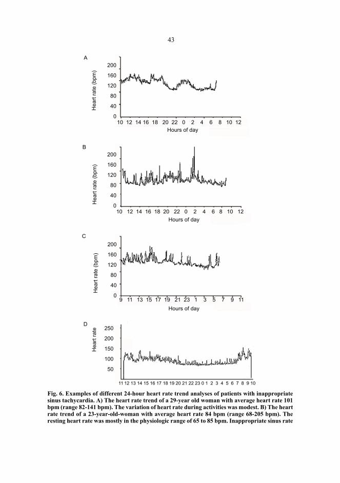
43
Fig. 6. Examples of different 24-hour heart rate trend analyses of patients with inappropriatesinus tachycardia. A) The heart rate trend of a 29-year old woman with average heart rate 101bpm (range 82-141 bpm). The variation of heart rate during activities was modest. B) The heartrate trend of a 23-year-old-woman with average heart rate 84 bpm (range 68-205 bpm). Theresting heart rate was mostly in the physiologic range of 65 to 85 bpm. Inappropriate sinus rate
Hea
rt ra
te (b
pm)
Hea
rt ra
te (b
pm)
Hea
rt ra
te (b
pm)
200
160
120
80
40
0
200
160
120
80
40
0
200
160
120
80
40
010 12 14 16 18 20 22 0 2 4 6 8 10 12
10 12 14 16 18 20 22 0 2 4 6 8 10 12
9 11 13 15 17 19 21 23 1 3 5 7 9 11Hours of day
Hours of day
Hours of day
A
B
C
Hea
rt ra
te 250
200
150
100
50
11 12 13 14 15 16 17 18 19 20 21 22 23 0 1 2 3 4 5 6 7 8 9 10
D

44
response was seen with modest activity. C) The heart rate trend of a 26-year-old woman withaverage heart rate 109 bpm (range 96-165 bpm). The resting heart rate was elevated, and inap-propriate sinus rate response was associated with mild exertion. D) The heart rate trend of a36-year old woman. The average heart rate was 97 bpm (range 70-157 bpm). An exaggeratedheart rate response was seen during the waking hours and a near-normal heart rate duringsleep.
24-hour ambulatory ECG recording characteristically demonstrates, in a non-medi-cated state, a mean HR above 90–95 bpm (169,179). A daytime resting HR is usuallyhigher than 95 bpm, and an increase in sinus rate from supine to upright position of morethan 25–30 bpm have been reported (179). Generally, patients have a near-normal restingHR during sleep and an exaggerated HR response during the waking hours. However, the24-hour HR trend analysis in patients with IST may have a wide spectrum (178) (Figure6). The bicycle test reflects the exaggerated HR response to minimal exercise. Within thefirst 90 seconds of a standard Bruce protocol in a bicycle test, patients with IST have aHR response greater than 130 bpm to this minimal workload (169). Echocardiography isusually normal, although there have been occasional instances of mild tachycardia-induced cardiomyopathy in patients with long-standing IST (169). The syndrome of ISTis rarely associated with mitral valve prolapse (170).
2.3.5.2 Electrophysiologic evaluation
The sole purpose of the diagnostic invasive electrophysiological examination is the exclu-sion of other atrial arrhythmias in a patient who has a clear documentation of excessiveHRs with concomitant symptoms and is considered for nonpharmacologic therapies(199). In patients who are mildly symptomatic and do not require therapy or are respon-sive to medical therapy alone, electrophysiological evaluation is not necessary.
The electrophysiological diagnosis of IST includes (1) exclusion of arrhythmias initi-ated or terminated by programmed atrial stimulation or overdrive pacing, (2) intracardiacatrial electrogram recordings that reveal a cranio-caudal pattern of activation, with theearliest recordings occurring at the superior aspect of the crista terminalis despite auto-nomic manipulation (atropine or isoproterenol), (3) a gradual increase and decrease in HRat the initiation and termination of tachycardia, and (4) shifts at the earliest site of activa-tion along the crista terminalis in response to changes in the tachycardia rate (170).
The basic electrophysiological examination is done in the way described in the sectionon EAT (see pages 30 and 31). The normal physiology of the sinus node distinguishes ISTfrom other atrial arrhythmias. Sinus node function, as assessed by automaticity of thesinus node, is within the normal limits. This is determined by overdrive suppression of thesinus node and by noting the recovery time after atrial pacing at high rates. Intra-atrial,AV nodal and His-Purkinje conduction times are also normal (1). The approach describedby Kalman et al and Lee and co-workers involves a combination of electrophysiologicalmapping and an anatomic approach. For activation sequence mapping, a multipolar cathe-ter is placed along the crista terminalis in addition to the catheter used for a routine elec-trophysiological evaluation (coronary sinus, His bundle region and right ventricularapex). Intracardiac echocardiography is useful because the crista terminalis not visible by

45
fluoroscopy and has a varied course in patient populations. Intracardiac echocardio-graphy readily identifies the crista terminalis and its proximity to a catheter. HR and thesite of the earliest crista terminalis activation are noted at the baseline and during gradu-ated isoproterenol infusion (199). Under maximal adrenergic stimulation with isoprotere-nol and parasympathetic blockade with atropine, the superior portions of the crista termi-nalis are revealed as the earliest site of activation. Programmed atrial stimulation can beattempted to induce tachycardia at the baseline and after catecholamine infusion. Detailedendocardial atrial activation mapping during tachycardia should reveal the earliest activa-tion in this region, showing a high-to-low and right-to-left sequence and thus suggestingorigin in or around the sinus node (1,171). In some laboratories, aminophylline is alsoused in initiating IST (105). Advanced mapping technologies, such as the 3-dimensionalelectro-anatomical mapping system (105) or the non-contact mapping system (207), mayhave advantages in identifying and tracking the RF application site and reducing proce-dural time.
2.3.5.3 Differential diagnosis
In the differential diagnosis, it is important to exclude sinus node re-entrant tachycardia,an AT which originates in the sinus node region, physiologic sinus tachycardia and pos-tural tachycardia syndrome. The inclusion of symptoms as the criteria of IST differenti-ates this syndrome from severely deconditioned individuals who might fulfill the criteriafor "inappropriate" sinus tachycardia based on rate alone (170). Secondary causes ofsinus tachycardia, as described earlier (Table 2), should be excluded. The differentiationof sinus tachycardia from AT or sinus node re-entry tachycardia is usually made in theelectrophysiological laboratory, but it may be difficult even there. Documentation of thearrhythmia on 12-lead ECG often enables differentiation of IST from EAT. Programmedstimulation is useful to exclude sinoatrial re-entrant tachycardia, because, in contrast tosinoatrial re-entrant tachycardia, atrial pacing neither initiates nor terminates IST. Table 4shows the electrophysiological differentiation of IST, EAT and sinus node re-entry. Ingeneral, in IST the site of origin of activation changes and moves down along the cristaterminalis as a function of changes in autonomic tone, whereas in focal tachycardia site oforigin remains fixed. The borderline between IST and focal AT originating in crista ter-minalis is relatively imprecise. During ablation, AT appears to be a focal disorder, whichmay be successfully ablated with a single well-targeted RF application. IST is a diffusedisorder, and successful ablation requires targeting of the tissue over a considerableregion of the pacemaker complex. The intracardiac atrial electrograms associated with ATare generally fractionated, whereas the intracardiac atrial electrograms of IST are notfractionated (3).
Postural tachycardia syndrome is characterized by a high sinus rate and disablingsymptoms, such as palpitations, fatigue, light-headedness and exercise intolerance, relatedto orthostatic intolerance caused by mild autonomic dysfunction (208). The characteristicfeature in these patients is postural sinus tachycardia, i.e. the patients exhibit markedincrease in HR upon rising up from a supine position (208) in the absence of orthostatichypotension and improved by fludrocortisone (209). In some patients, there may be an

46
overlap between IST and postural tachycardia syndrome or neurocardiogenic (vasovagal)syncope and chronic fatigue syndrome. The similarities to panic and anxiety disorder mayprevent a proper diagnosis. In fact, many patients with IST referred for electrophysiologi-cal evaluation carry a primary psychiatric diagnosis, being managed with various anxi-olytic agents (1).
2.3.6 Natural course
Only one previous report suggests that the prognosis of these patients may be excellent(165), although dilated cardiomyopathy may develop in some patients with chronicuntreated ATs, impairing the patient's prognosis (91,135). The risk of tachycardia-inducedcardiomyopathy in treated patients with IST is unknown, but is likely to be small (144).
Reported cases of IST deal predominantly with young females and with symptoms ofrelatively short-to-intermediate duration (167,169,171,174,179). Recently, Lopera et al(180) published a study of four elderly females with long-standing (at least 15 years) IST.All these patients had developed systemic hypertension after many years of sustained car-diac symptoms of palpitations and documented high resting HRs. The investigators didnot observe dilated cardiomyopathy in any of their patients. Although a cause-effect rela-tion between chronic IST and systemic hypertension cannot be established based on theresults of that study, it is possible that treatment of the IST could facilitate the treatmentof the underlying systemic hypertension. There are no earlier studies on the mortality ofpatients with IST.
2.3.7 Treatment and outcome
2.3.7.1 Medical and other therapies
According to the ACC/AHA/ESC guidelines for the management of patients with IST(Table 6) (144), initial therapy should be pharmacological, and the agents most frequent-ly used as first-line therapy are beta-blockers. Because no comparative studies ofmanagement exist for IST, therapy is empirical and tailored to individual patients. Inces-sant forms of IST are particularly problematic to treat. The patient's response to exerciseor isoproterenol infusion may be useful in both guiding the choice of therapy and assess-ing its efficacy. Nondihydropyridine calcium-channel blockers, such as verapamil and dil-tiazem, are an alternative and may be more useful in patients with an elevated IHR and anormal response to isoproterenol (210). In general, these drugs have no serious sideeffects. More aggressive medical therapy with drugs that affect sinus node automaticity,such as amiodarone or propafenone, may be indicated in selected patients. However, theirpotentially serious side effects must be considered before initiating this therapy in apatient population comprising mostly young people (210). Newer agents that slow HR by

47
inhibiting the hyperpolarization-activated (If) current are investigational and have notbeen used in patients with IST (211–218).
Table 6. ACC/AHA/ESC recommendations for treatment of inappropriate sinus tachycar-dia (141).
Earlier studies have indicated that teenagers may represent a selected group of subjectswith IST who may respond to aerobic exercise programs (219), but in adults monitoredaerobic exercise reducing the resting HR did not abolish inappropriate paroxysms ofsinus tachycardia (105).
2.3.7.2 Invasive therapy
Surgical and nonelectric ablation. Asymptomatic or minimally symptomatic patients whoare not seeking treatment or respond to pharmacologic therapy do not usually requireinvasive investigations and therapy. Medically refractory patients have been managedsurgically with the use of a right atrial exclusion technique, leaving the patient with ajunctional escape rhythm (220). Likewise, reports of sinus node ablation with the use ofintracoronary ethanol have documented success in eradicating IST (221). Intraoperativecryoablation or percutaneous catheter ablation of the His bundle have also been used inthe management of patients refractory to medical therapy. The main limitation of thesetechniques is that most patients are rendered pacemaker-dependent (78,169,222). Further-more, in the case of His bundle ablation, the atria continue to beat at a fast rate, whichmight cause the patient to continue experiencing symptoms as well as create dual-cham-ber pacemaker problems with the ventricles "tracking" the high atrial rate or cause symp-toms during mode switching or a high-rate block (1,199).
Radiofrequency catheter ablation. The recognition that the sinus node region is a largecomplex exhibiting rate-dependent site differentiation has allowed targeted ablation toeliminate the fastest sinus rates while maintaining the pacemaker function of the sinusnode (199). Animal studies have shown the feasibility of performing sinus node modifica-tion with high-frequency current (223) and Nd:YAG laser photocoagulation (224). Morerecently, RF energy was found to be quite effective in patients with IST (191,225). Theappropriate site for lesion delivery is dictated by activation mapping. By maneuvering amapping/ablation catheter throughout the high right atrium, it is possible to record elec-trograms from the distal portion of the ablation catheter that precede the surface P wave,as well as all other recorded electrograms, during the tachycardia. The local endocardial
Treatment Recommendation Classification Level of evidenceMedical Beta-blockers I C
Verapamil, diltiazem IIa CInterventional Catheter ablation - sinus node
modification/elimination*IIb C
*used as a last resort. See references in detail ref. 141. The levels of the evidence are ranked and the formats for classifying indications are as in Table 5.

48
activation time preceding the onset of the surface P wave by 25 to 45 ms has been associ-ated with an increased probability of HR being decreased after the application of RFenergy (178). This portion of the crista terminalis is then targeted for ablation. Progres-sively superior portions of the crista terminalis are then ablated until the target HR reduc-tion is achieved. Power should be adjusted to achieve a catheter tip temperature of 55° to70°C. Speeding up of the sinus HR followed by a marked subsequent HR reduction or theappearance of a junctional rhythm during ablation are indicators for successful ablationsites, at which the delivery of ablation energy should be continued for at least 60–90seconds (199). Because of the complex sinus node structure, the sinus node ablation pro-cedure is usually time-consuming, and several RF applications are normally required(178). The clinician must take care to ablate the superomedial portion of the crista termi-nalis as it courses anterior to the superior vena cava to insert on the intra-atrial septum. Byusing 3-dimensional electroanatomical mapping, Marrouche and colleagues (105) esti-mated the ablated area to measure 12±4mm x 195mm. After ablation, the site of the earli-est activation along the crista terminalis, even during maximal adrenergic stimulation, isinferior. This is also shown by the superiorly directed P wave axis on the 12-lead ECG.This approach has been demonstrated to be acutely effective to achieve a significantreduction in sinus rate and yet to maintain chronotropic competence (170,191).
HR control may have been achieved by either destruction of pacemaker tissue or inter-ruption of autonomic neural inputs or both. Histological examination of the canine sinusnode after RF catheter ablation demonstrated an area of fibrosis with complete loss ofboth nodal cells and sympathetic innervation (191). The demonstration of decreased IHRafter RF ablation is consistent with the destruction of sinus node cells and/or sympatheticinnervation to the sinus node complex.
Results of sinus node modification. The definitions of successful short-term outcomesare not uniform. The baseline sinus rate usually varies greatly from patient to patient andsometimes varies in a given patient from time to time during the procedure. In some labo-ratories, they titrate isoproterenol infusion to raise HR above 120 bpm, and a sinus nodemodification is defined as successful when HR decreases by 30 bpm during isoproterenolinfusion (178) and atropine (199). Other investigators have defined short-term success asreduction of the baseline sinus rate to less than 90 bpm and the sinus rate during iso-proterenol infusion by more than 20% (177) or by 25% (170). In the total sinus node abla-tion, a reduction of HR by more than 50% of tachycardia HR with a junctional escaperhythm has been defined as a successful outcome (170). Short-term clinical benefits havenot been demonstrated clearly by ICE (178). Overall, sinus node modification/ablationcan achieve short-term success in 70% to 100% of treated patients(105,170,175,177,226).
The long-term clinical outcomes have been disappointing in these patients, althoughno precise definition of successful long-term clinical outcome has been established. Out-comes could be measured objectively by the HR response or subjectively by the patient'ssymptoms during follow-up. Previously, studies on RF catheter ablation of IST havereported a wide range of long-term success rates (23–83%) and a sizable risk of completesinus node abolition (105,169,170,175,177,227). One of the factors contributing to lim-ited long-term cure was the inability to target with precision the sites of impulse originusing conventional mapping (177). Recently, RF ablation guided by intracardiac echocar-diography has also appeared to reduce the risk of inadvertent sinus node damage (227–

49
229). However, this technique seems to be associated with a poor success rate (23%) atlong-term follow-up (227). Ren and colleagues (181) got 70% predictive value of thecharacteristic intracardiac echocardiography signature, suggesting transmural/epicardialdamage for long-term IST control. One must realize that sinus node modification is not afocal ablation, but requires complete abolition of the cranial portion of the sinus nodecomplex, which seems to show automatic behaviour. Although a successful long-termoutcome may be achieved in a few patients, symptoms of palpitations and autonomiccharacteristics frequently persist.
There was a high rate (30–77%) of symptom recurrence in the series with sinus nodemodification (3,227,230). Most recurrences occurred 1–6 months after the procedure fol-lowing apparent early success (3,227). Recurrence may be classified into 1 of 2 generalcategories. Symptomatic recurrence or persistence of symptoms in the absence of docu-mented IST and despite persisting evidence of a successful electrophysiological outcomehas been observed in some cases. The second category is electrophysiological and tachy-cardic recurrence after an initially successful procedure (3). It appears that complete abla-tion of the sinus node resulting in junctional rhythm has better long-term success (72%),but requires pacemaker insertion (227,230). Also, the important recognition is the rela-tively frequent association with other atrial tachycardias, which require ablation to maxi-mize procedural success (105).
Complications of sinus node modification. Early complications of RF modification ofsinus node function include the need for permanent pacemaker implantation due to exten-sive total sinus node ablation, transient junctional rhythm persisting for up to approxi-mately 48 hours, transient right phrenic nerve palsy and transient superior vena cava syn-drome caused by ablation around the junction of the superior vena cava and right atrium(210). This complication could be diminished by using intracardiac echocardiography foraccurate guidance of the procedure (228).
Indications of sinus node modification. Sinus node modification could be considered insymptomatic patients with IST with persistently increased HR. Definition of autonomicfeatures is critical before any attempt at sinus node modification. In certain laboratories,sinus node modification or total sinus node ablation is not recommended for patients withIST and evidence of postural orthostatic tachycardia (178).
The personality of patients with IST is complex, and it is therefore important to discussthoroughly the realistic expectations and beneficial effects of the procedure. Marrouche etal (105) had many study patients with psychiatric disorders, which improved followingablation in about 66 % of them. They also showed that patients with no symptoms otherthan palpitation and early ablation after the onset of the rhythm disorder were more likelyto perceive the procedure as beneficial and to experience complete symptom resolution.After ablation, counselling and easy access to experienced allied professionals increasethe positive impact of this procedure on the use of health care resources. Apparently, ear-lier identification and treatment could improve the quality of life of the patients with IST(180). Given the relatively low rate of long-term success in relieving symptoms despiteelectrophysiological modification of sinus node function, further studies are needed toidentify the optimal extent of ablation and the cohort of patients most likely to benefitfrom such procedure (199).
3 Aims of the study
The purposes of the present study were to assess the prevalence, characteristics and natu-ral course of EAT and IST.
The specific aims were:1. to assess the prevalence and natural course of EAT in an asymptomatic, healthy male
population and in symptomatic hospital patients (I).2. to assess the prevalence and characteristics of subjects fulfilling the diagnostic criteria
of IST in a random sample of middle-aged subjects and to assess the natural courseand prognosis of IST during long-term follow-up (IV).
3. to determine whether there are differences in HR behaviour between normal SR, ISTand EAT (II).
4. to systematically study the response of IST to adenosine (III).

4 Subjects and methods
4.1 Subjects
A total of 41 patients with EAT and 46 patients with IST and 42 control patients with SRwere recruited (Articles I–IV). Some of the patients took part in more than one study. Thestudy protocols were approved by the local institutional ethical committees. All patientsand control subjects gave their written informed consent for the study. Demographic dataof the patients are presented in Table 7.
The resting 12-lead ECGs of consecutive young males applying for a pilot's licenceduring the years 1981–1993 were analysed retrospectively at the Central Military Hospi-tal in Helsinki, Finland, to assess the prevalence of EAT in an asymptomatic population.To assess the prevalence of EAT in symptomatic patients, all the ECGs of in and outpa-tients with arrhythmia at the Oulu and Kuopio University Hospitals in the years 1987–1995 were analysed, using five different arrhythmia diagnosis codes according to theICD9-classification (4270A, 4272A, 4278X, 7850A, 7851A) to cover all possible EATpatients. The patients with ECG evidence of EAT were invited for a control visit. (I)
To evaluate the HRV of tachycardias originating from the ectopic foci and the sinusnode, inpatients and outpatients of Oulu University Hospital (n=19) and the University ofMiami School of Medicine (n=5) with fast HR in standard 12-lead ECG and in 24-hourambulatory recordings were included in the study. All patients had elevated HRs observedincidentally (n=3) on a routine examination or brought to attention because of their symp-toms (n=21), causing referral to a hospital. Twelve patients had EAT and 12 IST. The con-trol group with normal SR consisted of 24 age- and sex-matched healthy subjects withoutclinical or echocardiographic evidence of structural heart disease. These subjects wereenrolled from the cross-sectional study undertaken earlier and described in detail previ-ously (231–233). (II)

52
Table 7. Characteristics of study populations.
The effects of adenosine were studied in a population consisting of 18 patients referredfor evaluation of IST to Oulu University Hospital, Kuopio University Hospital or MiamiUniversity Medical Center. Most of the patients presented with incapacitating symptoms,such as palpitations, weakness and light-headedness. The mean duration of the symptomsattributable to IST was 6.9±5.6 years. The age- and sex-matched control group consistedof 18 patients with SR who were referred for an elective electrophysiological examinationbecause of a narrow complex SVT. In the IST and control groups, 7 and 12 patientsunderwent an invasive electrophysiological study, respectively. (III)
The prevalence of IST was assessed in a randomly assigned population of 604 middle-aged subjects (335 males and 265 females). The population consisted of 290 hyperten-sive and 314 normotensive middle-aged subjects, who were originally enrolled for theOulu Project Elucidating Risk of Atherosclerosis (OPERA) study. The OPERA project isa population-based, epidemiological case-control study addressing the risk factors anddisease end-points of cardiovascular diseases (234). The entire OPERA study populationconsisted of 1200 subjects aged between 40 and 59 years at the time of enrolment. Thesubjects with hypertension (300 men and 300 women) were randomly selected by agestratification (15 male and 15 female subjects for each year of birth) from the nationalSocial Insurance Institute register for the reimbursement of hypertension medication. Thenormotensive age- and sex-matched controls were picked from the social insurance regis-
Setting Prevalence and natural
course of EAT(asympto-
matic)
Prevalence and natural
course of EAT(sympto-
matic)
Preva-lence and
natural course of
IST
Preva-lence and
natural course of
IST
HRV in EAT and IST
Adenosinesensitiveness of
IST
Study I I IV IV II IIIEAT/IST EAT EAT IST IST EAT/IST ISTNumber of patients
12 17 7 9 12 EAT12 IST24 SR
18 IST18 SR
5/5 autonom.bl.Females/males 0/12 8/9 4/3 1/8 EAT 6/6
IST 9/3SR 12/12
IST 13/5SR 11/7
Mean age (y) 19±1(17–21 y)
26±17(19d–56y)
47±7 46±7 EAT 30±12IST 32±8SR 34±12
IST 46±15SR 46±11
Heart rate (bpm) at the baseline
109±17
Heart rate (bpm) at the follow-up
81±19
24-hour heart rate (bpm) at the base-line
94±2
24-hour heart rate (bpm) at the fol-low-up
89±8
Follow-up time (y) 8±3 7±3 6±2
Hypertension No No 5 pts 3 pts

53
ter covering the whole population of the City of Oulu (235). In the current analysis, allsubjects with abnormal P wave morphology or axis in 12-lead ECG, structural heart dis-ease, diabetes or any pathological condition (e.g., anemia, hypovolemia, thyroid disease)or medication (e.g., thyroid hormone substitution, antiasthmatic drugs) known to acceler-ate HR were excluded. Patients with hypertension continued their antihypertensive medi-cation at the time of the primary recordings. (IV)
During the years 1996–1998, nine of the patients referred to Oulu University Hospitalfor evaluation of symptomatic SVTs were diagnosed with IST. For the evaluation of thenatural course and prognosis of IST, these 9 patients were pooled together with the 7 sub-jects from the random population who fulfilled the diagnostic criteria of IST. (IV)
4.2 Definitions of tachycardias
Incessant EAT was defined according to the following criteria: (1) resting daytime HR ≥90 bpm, (2) average HR > 90 bpm on 24-hour ECG recordings, and (3) an abnormal Pwave axis and/or abnormal P wave morphology in ≥ 2 precordial leads and in ≥ 1 bipolarlimb lead on 12-lead ECG (2,236). P wave morphology was described biphasic if P wavehad two positive phases. P wave was diphasic if P wave had one negative and one posi-tive phase or one positive and one negative phase. During the follow-up, the rhythm wasdefined as EAT if the P wave morphology was similar and the rate > 90 bpm in the repeatECG without a change (<10°) in the frontal P wave axis. If the P wave morphology wasabnormal, but the rate < 90 bpm, the rhythm was defined as slow ectopic rhythm. If the Pwave morphology and/or axis was clearly different in the repeat ECG, the rhythm wasdefined as a fusion of ectopic and sinus rhythms. The criteria of normal SR were com-plete normalization of P wave morphology and axis, with a rate < 90 bpm. The ECGs ofall subjects were inspected by the author and by another experienced cardiologist.
IST was defined as a resting daytime HR > 100 bpm with a normal P wave morphol-ogy and axis obtained from standard 12-lead ECGs recorded on ≥ 2 separate days and anaverage HR of > 90 bpm on 24-hour ECG recording (169). In the middle-aged randomsample (IV) only the subjects with an average HR > 90 bpm during the ambulatory 24-hour HR measurement and resting HR > 100 bpm in either a supine or a sitting positionwere considered to fulfil the criteria of IST.
4.3 Collection of clinical data and follow-up
The study subjects were interviewed by the same physician. In the medical history, spe-cial attention was given to arrhythmia symptoms, previous diseases and medications. Inthe middle-aged random sample (III) a standardized health questionnaire on smokinghabits, alcohol consumption, physical activity and personality type was also filled in.Alcohol intake was converted into grams of absolute ethanol per week. On the basis ofleisure-time physical activity, four groups were formed, using a modification of the meth-od described by Grimby (237). Personality type was evaluated on (1) the Framingham

54
type A behaviour pattern scale (238), (2) Bortner's short rating scale of behaviour pattern(239) and (3) the hostility scale (240).
All the patients with evidence of EAT or IST were invited for a control visit, duringwhich the following parameters were checked: clinical history, physical examination, 12-lead ECG, 24-hour ambulatory ECG recording and echocardiography (I–IV). Antiarrhyth-mic medications were stopped for at least four half-lives before the control visit.
4.4 Electrocardiographic measurements
4.4.1 12-lead ECG
Resting HR was measured and P wave morphology and axis were evaluated from stan-dard 12-lead ECG recordings at a paper speed of 50 mm/s after at least 15 minutes’ rest.
4.4.2 Ambulatory ECG recordings
The short-term ambulatory ECGs (IV) were recorded with a Dynacord Holter Recorder(Model 420, DM, Scientific, Irvine, CA, USA) with a sample frequency of 256 Hz. Eachsubject was monitored for 15 minutes while quietly lying down and breathing normally,for 15 minutes in a sitting position and for 15 minutes while walking (IV).
Twenty-four-hour HR ambulatory ECGs were recorded using an Oxford Medilog 4500Holter recorder (Oxford Medical Ltd., England) (II–IV). All patients were encouraged tocontinue their normal daily activities during the recordings. The data were sampled digi-tally and transferred to a microcomputer for an analysis of the average 24-hour HR andHRV. Before the HRV analysis, all recordings were carefully edited in order to eliminatesegments with intermittent AV block and premature ectopic beats in the patients with ISTand sinus beats in the patients with EAT. For details, see Article II.
4.4.2.1 Heart rate variabilility
Only recordings with qualified beats for at least a 16-hour period and with > 80 % quali-fied ectopic or sinus beats, respectively, were included in the analyses of HRV. HRV mea-sures were analysed with custom-made analysis programs (Hearts, Hearts Signal Co,Kempele, Finland) as described in detail the previously published papers (27,28,231–233).
Time- and Frequency -Domain Analysis of HRV. The time- and frequency-domainmeasures of HRV were analysed by the methods recommended by the Task Force of theEuropean Society of Cardiology (22). The SD of all normal-to-normal R-R intervals

55
(SDNN) and the difference between the maximum and minimum hourly HR (circadianrhythm) were computed as standard time-domain measures of HRV. Spectral power wasquantified both by fast Fourier transform analysis and by autoregressive analysis in 4 fre-quency bands (232,233): <0.0033 Hz (ultra low frequency [ULF]), 0.0033 to 0.04 Hz(very-low frequency [VLF]), 0.04 to 0.15 Hz (low frequency [LF]), and 0.15 to 0.40 Hz(high frequency [HF]). ULF and VLF spectral components were computed over the entirerecording interval by the fast Fourier method (233). LF and HF components were com-puted from the segments of 512 R-R intervals by the autoregressive method, and the aver-age values of the entire recording interval were calculated for the components. The LFand HF components were calculated as both absolute and normalized units, which weobtained by dividing the power of these components by the total variance from which theDC component had been subtracted and multiplying this value by 100 (21). In the 45-minute ECG recordings the LF and HF components during the 15-minute period at supineand sitting position and standardized walking were calculated as absolute units and LF/HF ratio was used.
Time domain measures of HRV correlate well with vagal activity (20,241–244). Inspectral HRV analysis, respiration is related to the high frequency (HF) power (21).Reflecting the predominantly parasympathetic origin of the HF component, this band canbe minimized with atropine or vagotomy (18,19,21,241,243,245). The LF peak of HRVhas been attributed to sympathetic activity with vagal modulation, but there are discrepantfindings dividing the opinions on the genesis of LF oscillation (18,19,21,243,245,246).Studies on muscle sympathetic nerve activity have suggested that LF and HF may, respec-tively, reflect sympathetic and vagal outflow (247,248). As a result, the relationshipbetween LF power and HF power (LF/HF ratio) has often been referred to as a specificmarker of sympathovagal balance (21,23), but this idea has been seriously criticized(249). The origin of the ULF and VLF components is not clear. Vagal activity (18,21),thermoregulation and peripheral vascular resistance have been suggested as the cause forfluctuation in the VLF region (250–253).
Poincaré plot analysis. A Poincaré plot analysis is a diagram in which each R-R inter-val is plotted as a function of the previous R-R interval. Both visual analysis and of thegraphic display and quantitative analysis of the plots can be used to describe R-R intervaldynamics. The quantitative 2-D analysis of these plots has been described in detail previ-ously (232). The Scattergrams of successive R-R intervals were plotted for the entire 24-hour period, and the SD of instantaneous R-R interval variability (SD1) and of continuousvariability (SD2) were then analysed. The shape of the plot was also classified as com-plex, torpedo-shaped, or normal, as described previously (232).
Fractal and complexity analysis of R-R interval variability. The power-law relation-ship of R-R interval variability was calculated from the frequency range 10–4 to 10–2 Hz.The point power spectrum was logarithmically smoothed in the frequency domain and thepower integrated into bins spaced 0.0167 log (Hz) apart. A robust line-fitting algorithm oflog (power) on log (frequency) was then applied to the power spectrum between 10–4 and10–2 Hz, and the slope of this line was calculated (β). This spectral range was chosen onthe basis of previous observations regarding the linear relationship between log (power)and log (frequency) in this frequency band in human HR time series data (25). The detailsof this method have been described previously (25,233).

56
The detrended fluctuation analysis technique was used to quantify the fractal scalingproperties of short-term and intermediate-term R-R interval time series. This method is amodified root-mean-square analysis of random walk that quantifies the presence orabsence of fractal correlation properties and has been validated for nonstationary timeseries (111). In this method, the root-mean-square fluctuation of integrated and detrendedtime series is measured at each observation window and plotted against the size of theobservation window on a log-log scale. The fractal-like signal (1/f signal spectrum)results in an exponent value of 1 (α=1.0). The details of this method have been describedelsewhere (26,28,111). In the present study, the HR correlations were defined separatelyfor short-term (<11 beats, α1) and long-term (>11 beats, α2) R-R interval data (scalingexponents) on the basis of the previous finding of a crossover point on the log-log plot.Both α1 and α2 were analysed from segments of 8000 R-R intervals and averaged toobtain mean values for the entire recording period.
Approximate entropy. Approximate entropy is a measure that quantifies the regularityor predictability of time series. The details of the method used have been described earlier(27,112). The parameters m and r of the method must be fixed to calculate approximateentropy, and m=2 and r=20% of the SD of the data sets were chosen on the basis of previ-ous findings of statistical validity. Approximate entropy values were computed from 8000R-R interval segments and averaged to obtain a mean value of approximate entropy char-acterizing the entire recording (27).
4.4.3 Ambulatory blood pressure and heart rate recordings
Ambulatory 24-hour BP and HR were recorded using the fully automatic SpaceLabs90207 oscillometric unit (SpaceLabs Inc., Redmond, New Jersey) during normal dailyactivities (IV). Measurements were performed automatically at every 15 minutes from4:00 AM till midnight and at every 20 minutes from midnight till 4:00 AM. The meanvalue of HR during the ambulatory measurement was used in the analysis.
In order to avoid biases caused by temporary changes in sinus frequency, the HR in therandom sample (IV) was measured using three different methods on three separate dayswithin one month: 1) ambulatory 24-hour HR recording, 2) 45-minute ECG recording and3) standard 12-lead ECG .
4.5 Electrophysiological studies
Electrophysiological examinations were performed based on clinical indications. Quadri-polar catheters were positioned in the high right atrium, the His bundle position, in thecoronary sinus and in the right ventricular apex. In some patients with IST, a 20-polecatheter was also positioned along the tricuspid valve annulus in the right atrium, with thetip of the catheter at the coronary sinus ostium. Intracardiac electrograms, filtered from30 to 250 Hz, were printed on an electrostatic paper at a paper speed of 100 mm/s orstored on an optical disk using a digitized amplifier/recorder system.

57
Detailed mapping and ablation of the EATs were performed using a 7 F deflectableablation catheter with a 4-mm distal tip electrode. The criteria of incessant EAT duringthe electrophysiological examination were: (1) an endocardial activation sequence incon-sistent with a sinus node origin, (2) inability to terminate or reset the tachycardia withappropriately timed premature atrial extrastimuli or rapid atrial pacing, (3) exclusion ofintra-atrial, AV and AV nodal re-entry and (4) sudden restoration of SR after RF ablationof tachycardia. The earliest local intracardiac activation in relation to the P wave on thesurface ECG was used to select the target site for ablation. RF energy was applied at theappropriate target sites in a range of 20 to 40 W power for 40 to 60 seconds until termina-tion of EAT.
IST was defined by (1) an activation sequence consistent with a sinus origin (i.e., earli-est endocardial activation in the lateral high right atrium obtained with the halo catheter)and (2) exclusion of re-entry by inability to terminate or reset tachycardia by atrial extra-stimuli or rapid atrial pacing. None of the IST patients were treated by sinus node modifi-cation/ablation.
4.6 Echocardiographic measurements
A Hewlett Packard Sonos 500 ultrasound system (Hewlett Packard Company, Massachu-setts, USA) was used for the echocardiographic examinations. M-mode measurementswere obtained under 2-D guidance according to the recommendations of the AmericanSociety of Echocardiography (254). The left ventricular mass was calculated according tothe formula of Troy et al (255). Fractional shortening was calculated by dividing the dif-ference between the left ventricular dimensions in diastole and systole by the diastolicdimension and then multiplying it by 100.
4.7 Pharmacological maneuvers
4.7.1 Autonomic blockade
Because the electrophysiological effects of adenosine are influenced by autonomic tone(116,256,257), in a subset of 5 patients with IST and 5 patients with SR, the direct effectsof adenosine were assessed under complete beta-adrenergic and cholinergic blockade.Autonomic blockade was achieved pharmacologically by abolishing all beta-adrenergicand cholinergic receptor input to the heart with intravenous propranolol (0.2 mg/kg) andatropine (0.04 mg/kg), respectively. The adenosine sensitivity test during autonomicblockade was carried out 10 minutes after the drugs were injected. The expected IHR wascalculated according to a previously described regression equation (197): IHR=118,1–(0,57 x age). The IHR was compared to the actual atrial rate during autonomic blockade.

58
4.7.2 Atropine
An intravenous atropine bolus injection was given continuously and gradually at a doseof 0.02 mg/kg to the patients with EAT and IST. HR was continuously recorded at a paperspeed of 25 mm/s until no increase in the average HR was observed during the 2-minuteperiod. The difference between baseline HR and maximum HR after atropine was calcu-lated and compared with the normal reference values obtained in the previous studies(108,109).
4.7.3 Adenosine
During IST, adenosine was given as a rapid bolus injection (0.1 and 0.15 mg/kg) followedby a 10-mL bolus of normal saline into a large antecubital vein at bedside or into the fem-oral vein during the electrophysiological studies. The higher dosage was used only if thefirst bolus did not cause a high-degree AV block. During each study, 6- or 12-channelECG was recorded continuously at a paper speed of 50 mm/s or using a computer-baseddigitized recording system (CardioLab EP System; Prucka Engineering, Inc., Houston,TX, USA), until the effect of adenosine disappeared (minimum of 3 min). Following theadenosine bolus, ACL was measured beat-to-beat until it and the AV nodal conductiontime returned to baseline. Statistical comparisons within and between the groups weremade before the adenosine injection (=baseline), at the time of the maximal effect(=peak) and during the maximal reflex acceleration of sinus rate (=rebound). In the eventthat no apparent effect on ACL was detected, the time of the maximal AV delay was usedto pinpoint the peak effect of adenosine. In the control subjects, adenosine was given dur-ing normal SR either after successful catheter ablation or during bedside testing tounmask latent pre-excitation.
4.8 Laboratory methods
All the blood samples were drawn after a 12-hour requested fast. A two-hour oral glu-cose tolerance test was performed to identify latent diabetes, and the levels of haemoglo-bin, thyreotropin, glutamyl transferase and glucose were analyzed using standard labora-tory methods (IV). In the other study populations, laboratory tests were made by the clini-cal indications.
4.9 Statistical analyses
All parametric data are reported as mean ± SD (I–IV). Statistical analyses were per-formed using SigmaStat 2.03 (SPSS Science, Chicago, IL)(II) or SPSS for Windows ver-

59
sion 7.5–10.1 (I, III, IV). Paired Student's t-test was used to compare HR at baseline andat follow-up (I, IV). Analysis of variance by Bonferroni’s post hoc test was used to com-pare the duration of follow-up time (I). Within-subject differences between the groupswere assessed with ANOVA for repeated measurements followed by a post hoc t-test withBonferroni correction (II, III). Continuous variables were compared with Student’s t-testfor unpaired samples, and comparisons of categorical data were made with the Mann-Whitney Rank Sum test (III). The differences of frequencies between the class variableswere tested by the Chi-square test and those between the non-normally distributed vari-ables by the Mann-Whitney test (IV). A value of P < 0.05 was considered statistically sig-nificant.

5 Results
5.1 Ectopic atrial tachycardia
5.1.1 Prevalence
Out of a total of 3554 12-lead ECGs of healthy asymptomatic males aged between 17 and21 years (mean 19±1 years), which were analysed retrospectively at the Central MilitaryHospital, 12 showed evidence of EAT (prevalence 0.34%). All ECGs had been interpret-ed as normal by the primary physician. The ECGs of 17 out of the 3700 symptomaticinpatients with arrhythmia (prevalence 0.46%) examined between the years 1980 and1993 pointed to EAT.
5.1.2 Natural course
A repeat ECG was obtained in 10 males after a follow-up time of 8±3 years (range 3–11years). Nine of the subjects were still asymptomatic, but one had developed symptoms ofpalpitation. Two subjects were lost to follow-up. None of the subjects had ever had anyantiarrhythmic medication. In all symptomatic arrhythmia patients, a repeat ECG wasrecorded after a mean follow-up of 7±3 years (range 3–16 years) at a mean age of 26±17years (eight females and nine males).
Seven of the 27 patients (26%), both asymptomatic (n=10) and symptomatic (n=17),continued to have similar or different ectopic atrial rhythm at follow-up, and 10 (37%)had SR. Ten patients (37%) showed a clear change in P wave morphology in at least twoleads and in the frontal axis of the P wave without its complete normalization, suggestiveof a fusion of ectopic atrial and sinus rhythms (Figure 7). HR was significantly slower onthe repeat ECG (81±19 bpm vs. 109±17 bpm, P<0.001). Only three patients (11%) hadfaster EAT.

61
Prim
ary
ECG
Nov
embe
r 12,
199
6H
eart
rate
87
beat
s.min
–1
Prim
ary
ECG
May
27,
199
2H
eart
rate
97
beat
s.min
–1
II aVL
V1
V2
V3
II aVL
V1
V2
V3
Fig.
7. L
eft:
the
prim
ary
EC
G o
f an
asym
ptom
atic
18-
year
-old
mal
e ap
plyi
ng fo
r a
pilo
t’s l
icen
ce. T
he h
eart
rat
e w
as 9
7 bp
m; i
n le
ad a
VL
the
Pw
ave
is n
egat
ive,
in le
ads V
1-2
diph
asic
and
in le
ad V
3 bi
phas
ic. F
our
and
a ha
lf ye
ars l
ater
he
is st
ill a
sym
ptom
atic
and
in th
e re
peat
EC
G h
eart
rate
is sl
ower
87
bpm
. The
P w
ave
mor
phol
ogy
has c
hang
ed; i
n le
ad a
VL
the
P w
ave
is p
ositi
ve, i
soel
ectr
ic in
lead
s II a
nd V
1 an
d no
tche
d in
V2
sugg
estiv
e of
fusi
on o
f ect
opic
atr
ial a
nd si
nus r
hyth
ms.
Bip
hasi
c=P
wav
e w
ith tw
o po
sitiv
e ph
ases
, dip
hasi
c=P
wav
e w
ith o
ne n
egat
ive
and
one
posi
tive
phas
e or
with
one
pos
itive
and
one
neg
ativ
e ph
ase.

62
The follow-up time tended to be longer in the patients who where in SR (mean 92±41months) compared to those with fusion rhythm or EAT (81±34 months), but the differ-ence was not statistically significant. Five patients had used sotalol and one patient pro-pranolol regularly. One patient had used metoprolol and one propranolol if needed. EATwas the only arrhythmia documented in each case, and none of the patients developedheart failure during the follow-up time. None of the 27 patients had a positive family his-tory of EAT.
5.1.3 Heart rate variability
5.1.3.1 Time- and frequency-domain measures of heart rate variability
Table 8 shows the time- and frequency-domain measures of HRV. None of these measuresdiffered between the patients with EAT and IST. However, all traditional HRV measuresexpressed in absolute units, including the measures obtained by quantitative analysis ofPoincaré plots, were reduced in the patients with EAT compared to those with normal SR(see examples in Figure 8). Despite the reduced overall HRV, the Poincaré plots showednormal comet-shaped characteristics in the cases with EAT. The LF/HF ratio or the LFand HF components analysed in normalised units did not differ from normal in EAT.There was no difference between the maximum and minimum HRs (amplitude of the cir-cadian rhythm) between the study groups (Table 8).

63
Table 8. Twenty-four-hour heart rate variability data.
Parameter EAT(n=12)
IST(n=12)
Normal sinus rhythm(n=24)
Mean 24-h HR (bpm) 109±12† 106±9† 72±8Minimum HR (bpm) 95±15 90±10 56±6Maximum HR (bpm) 130±13 121±15 95±14Difference between maximum and minimum HR (bpm) 35±14 31±8 40±14SD1 (ms) 17±12† 15±6† 37±12SD2 (ms) 56±33* 67±14† 146±29SDNN (ms) 75±28† 77±16† 176±39SDNNi 13±4† 13±3† 21±5HF power (ms2) 208±256 162±137 1101±750
Ln (ms2) 4.4±1.3† 4.2±0.8† 6.7±0.9NU 44.7±7 39±6 38±7
LF power (ms2) 247±286 344±158 1611±960
Ln (ms2) 4.5±1.6† 4.7±1.1† 7.2±0.7NU 55±8 60±7 61±6
LF/HF ratio 1.5±1.2 1.9±1.6 1.9±0.9VLF power (ms2) 517±472 556±289 2792±1358
Ln (ms2) 5.3±1.3† 5.8±0.6† 7.8±0.6ApEn 1.13±0.22 1.05±0.33 1.15±0.16α1 0.71±0.16†§ 1.16±0.13 1.19±0.11α2 1.06±0.21 1.04±0.19 1.08±0.16β –1.39±0.22 1.39±0.22 –1.26±0.15SD1=short-term SD and SD2=long-term SD analysed from Poincare plots; SDNNi=SDNN normalized withR-R interval length; Ln=natural logarithmic of absolute values, NU=normalized units; ApEn=approximateentropy. SDNN= standard deviations of all R-R intervals. β=slope of regression line of log(power)-log(fre-quency) of HR variability between frequencies 10-2 and 10-4. HF=high-frequency power component of heartrate variability. LnHF=logarithmic high-frequency power component of heart rate variability. LF=low-fre-quency power component. LnLF=logarithmic low-frequency power component. VLF=very-low-frequencypower component. LnVLF=logarithmic very-low-frequency power component of heart rate variability.α1=short-term scaling exponent. α2=long-term scaling exponent.Values are mean ± SD. †P value <0.001 and *P<0.01 compared with normal sinus rhythm with ANOVA andBonferroni post hoc analysis, §P<0.001 between EAT and IST.See details in Article II.

64
Fig. 8. Examples of power spectral analysis of R-R-interval variability of patient with ectopicatrial tachycardia (EAT) without random beat-to-beat R-R-interval dynamics, patient withinappropriate sinus tachycardia (IST) and subject with normal sinus rhythm (SR). Typicalspectral pattern with LF and HF oscillations of R-R-interval variability can be observed in EATand IST, but absolute values of spectral components are smaller in magnitude in EAT and ISTthan in SR. Power-law slope of R-R-interval dynamics does not differ between EAT, IST andSR.
5.1.3.2 Fractal and complexity measures of R-R interval variability
Approximate entropy, the long-term scaling exponent α2 and the power-law slope incases with EAT did not differ from those with SR. However, the short-term scaling com-ponent α1 was significantly lower in EAT than in either IST or normal SR (Table 8 andFigures 8 and 9).
Fig. 9. Individual values of short-term scaling exponent of patients with ectopic atrial tachycar-dia (EAT), inappropriate sinus tachycardia (IST), and normal sinus rhythm (SR). Short-termscaling exponent was reduced (α1<0.95) in all but 1 of the patients with EAT.

65
Full disclosures of Holter recordings and ECG printouts of patients with EAT showedsudden, abrupt prolongations in R-R intervals (exit blocks from ectopic foci) without evi-dence of a change in P wave morphology or AV block in 6 patients throughout the 24-hour recordings. In 5 cases with low α1 value, no abrupt changes in R-R intervals wereobserved, but the ectopic foci showed continuous instability in beat-to-beat behaviourduring the entire recording period. Unstable beat-to-beat R-R interval behaviour and exitblocks were more commonly observed during sleep than in the daytime.
5.1.4 Heart rate responses of ectopic atrial tachycardia to intravenous atropine
HR increased from 104±16 bpm to 134±11 bpm (p<0.001) after intravenous atropineadministration in the patients with EAT. The mean increase of HR was 30±14 beats(range 11–65 bpm). In 2 patients, the increase in HR was smaller than the reference val-ues reported for healthy subjects with normal SR (>20% increase from baseline).
5.2 Inappropriate sinus tachycardia
5.2.1 Prevalence
The distribution of the average 24-hour HR in the random middle-aged population isshown in Figure 10. The distribution is clearly skewed to the right after a cutoff point ofapproximately 85–90 bpm. The average 24-hour HR was over 90 bpm in 19 of the 604subjects (3.2%). Ten patients (1.7%) had HR > 100 bpm in a supine position and 15patients (2.5%) in a sitting position, and 70 patients (11.6%) had HR > 100 bpm duringwalking. However, only seven of these subjects had both daytime HR >100 bpm in asupine or sitting position and 24-hour average HR > 90 bpm (95±4 bpm), giving an over-all prevalence of 1.16 % for IST in the random middle-aged population.

66
Fig. 10. Distribution of the average 24-hour heart rate in a random middle-aged population.
5.2.2 Characteristics of the subjects with inappropriate sinus tachycardia
The characteristics of the subjects fulfilling the diagnostic criteria of IST and the controlsin the random middle-aged population are shown summarized in Table 9. The only demo-graphic difference between the groups was personality type on the hostility scale (10±2vs. 8±3, P<0.001). In the random population, 4 subjects fulfilling the diagnostic criteriaof IST were women (57%) and 3 were men. The mean age of the subjects was 47±7years. Five subjects (71%) had previously diagnosed hypertension, and 2 subjects (29%)were health care workers. The ambulatory systolic (147±11 mmHg vs. 130±13 mmHg,P<0.001) and diastolic (92±7 mmHg vs. 81±8 mmHg, P<0.001) blood pressures weresignificantly higher among those with IST. No significant differences were observedbetween the groups in the echocardiographic, laboratory and HRV measurements,although there was a non-significant trend towards reduction of the LF/HF ratio duringwalking as compared to the supine position (Table 10).

67
Table 9. Characteristics of a random middle-aged population.
IST (n=7) Control (n=597)Age (years) 47±7 51±6Sex (female/male) 4/3 332/265BMI (kg/m2) 27.1±3.3 27.4±4.4Waist/hip ratio 0.86±0.97 0.87±0.86Current smoking status
No smoking 4 (57%) 421 (71%)Moderate smoking (<20/day) 2 (29%) 154 (25%)Heavy smoking (>20/day) 1 (14%) 22 (4%)
Alcohol consumptionNo drinking 1 (14%) 89 (15%)1–100 g/week 4 (57%) 369 (61%)>100 g/week 2 (29%) 143 (24%)
Leisure time physical activityNo activity 2 (29%) 24 (4%)Mild activity 2 (29%) 166 (28%)Moderate activity 1 (14%) 198 (33%)Heavy activity 2 (29%) 208 (35%)
Personality typeFramingham 27±7 26±5Bortner 22±3 22±3Hostility 10±2 * 8±3
*P < 0.001. See details in Article IV.

68
Table 10. Haemodynamic and laboratory parameters in the random middle-aged popula-tion.
Among the hospital population, the mean HR in the subjects with IST was 110±11 (range101–129) bpm in the 12-lead ECG at rest and 94±3 (range 91–98) bpm in the 24-hourECG recording. The mean age of the patients was 46±7 years, and eight (89%) of themwere female. Three patients had hypertension (33%), and one was a health care worker(11%). The results of the blood tests and M-mode and 2-D echocardiography were nor-mal in all patients. When these patients were compared to those from the random samplewho fulfilled the diagnostic criteria of IST, several parallel features were identified. Therewas no significant difference in age (47±7 vs. 46±7 years) or the 24-hour average HR(95±4 bpm vs. 94±36 bpm) between the random and hospital populations. Likewise,hypertension was a relatively common finding in both populations. However, in contrastto the random population, almost all the symptomatic inpatients with IST were female(57% vs. 89%).
Parameter IST (n=7) Control (n=597) SignificanceHeart rate (bpm) 12-lead ECG at rest 87±8 73±13 0.00524-hour ambulatory recording 95±4 70±10 <0.001During a 15-minute supine period 94±8 67±10 <0.001During a 15-minute sitting period 100±8 71±10 <0.001During a 15-minute walking period 110±4 83±12 <0.001Heart rate variabilityLF/HF ratio at rest 2.03±1.22 2.33±1.45 NSLF/HF ratio at sitting 1.99±1.17 2.9±2.27 NSLF/HF ratio at walking 1.55±1.45 2.47±2.12 NSBlood pressure (mmHg)24-hour ambulatory systolic pressure 147±11 130±13 <0.0124-hour ambulatory diastolic pressure 92±7 81±8 <0.01Echocardiographic measurementsLeft atrial diameter (mm) 40±5 39±5 NSLeft ventricular diastolic diameter (mm) 49±5 52±5 NSFractional shortening (%) 35±6 35±6 NSLeft ventricular mass (g) 175±46 207±62 NSLaboratory measurementsSerum thyreotropin (mU/l) 1.7±0.8 2.1±1.9 NSSerum haemoglobin (g/l) 137±15 143±13 NSFasting blood glucose (mmol/l) 4.6±0.4 4.4±0.5 NS2-hour blood glucose (mmol/l) 4.6±1.1 5.4±1.5 NSTotal serum cholesterol (mmol/l) 5.4±0.97 5.7±1.0 NSSerum triglycerides (mmol/l) 1.1±0.3 1.5±0.9 NSLF=low-frequency and HF=high-frequency domain in the spectral analysis of heart rate variability, respec-tively. See details in Article IV.

69
5.2.3 Natural course
In the evaluation of the natural course and prognosis of IST, the subjects fulfilling the ISTcriteria in the random population (n=7) and the symptomatic inpatients (n=9) were com-bined. Eleven of the 16 subjects consented to participate in a follow-up examination at72±29 months (range 36–133). Two subjects (one in each population) were excludedbecause of asthma medication at the time of the control visit, and two subjects in the ran-dom and one in the symptomatic hospital population were lost to follow-up. All subjectswho consented to attend the follow-up examinations had symptoms of rapid HR at thetime of the control visit. In the random population, two subjects felt an increased HR dur-ing exercise, one had palpitations at rest, and one patient suffered from “strong heartbeats”. In the symptomatic hospital population, all patients had palpitations at rest or dur-ing minor exercise. There was no significant change in HR after a mean follow-up of6.0±2.4 years. The average HR in the 24-hour ambulatory ECG recording was 94±2 bpm(range 91–98 bpm) at baseline and 89±8 bpm (range 80–103 bpm) at follow-up. Like-wise, there were no significant differences in the resting HR measured from the 12-leadECG between the baseline and follow-up visit (104±14 bpm vs. 98±15 bpm, P=0.07).However, despite the persistent symptoms and the relatively high HR at the follow-upexamination, only 4 of the 11 subjects still fulfilled the combined diagnostic criteria ofIST, whereas the other subjects’ average HR in the 24-hour Holter recording had droppedbelow 90 bpm.
The prognosis of IST was benign. All subjects were alive at follow-up, and none hadsymptoms or clinical findings of significant structural heart disease. There were no signif-icant changes in the echocardiographic parameters between the baseline and follow-upexaminations (Table 6), although one subject in the random population had developedhypertension.
5.2.4 Heart rate variability
5.2.4.1 Time- and frequency-domain measures of heart rate variability
All traditional HRV measures were reduced in the patients with IST compared to the sub-jects with normal SR (Table 8). Despite the reduced overall HRV, the Poincaré plotsshowed normal comet-shaped characteristics in cases with IST. The LF/HF ratio or theLF and HF components analysed in normalised units did not differ from normal in IST.

70
5.2.4.2 Fractal and complexity measures of R-R interval variability
The short- and long-term scaling exponents α1 and α2, the power-law slope β andapproximate entropy did not differ between the subjects with IST and those with normalSR (Table 8).
5.2.5 Heart rate responses of inappropriate sinus tachycardia to intravenous atropine
In the subjects with IST, HR increased from 108± 15 to 137±18 bpm after the administra-tion of intravenous atropine. The mean increase of HR was 29±13 bpm (range 15–49bpm). Two subjects with IST had a smaller increase in HR than the normal referencerange.
5.2.6 Electrophysiological effects of adenosine in inappropriate sinus tachycardia
Basic ECG measurements (III) showed that the resting HR was significantly higher in thepatients with IST (114±17 bpm) than in the control subjects (79±11 bpm), P<0.001. Theaverage HR of the IST patients during a 24-hour ECG recording was 94±3 (range 91–104) bpm and showed an unusually high sinus rate during minimal physical activity.
During autonomic blockade, the atrial rate was 132±11 (range 120–145) bpm in theIST patients and 101±13 (range 88–122) bpm in the control patients (P<0.01). Thus, theatrial rate was significantly higher in the IST than in the control group, not only under thebaseline conditions but also during autonomic blockade. Moreover, the atrial rate duringcomplete autonomic blockade was significantly higher than the expected range of IHR inthe patients with IST (132±11 vs. 88±4 bpm, P<0.001), but not in the control subjects(101±13 vs. 93±8 bpm, p=0.28).
IST, unlike many other SVTs, was resistant to the antiarrhythmic actions of adenosine.The nucleoside failed to terminate the tachycardia in all the patients with IST. In thepatients with IST, the first dose of adenosine (0.1 mg/kg) had no significant negativechronotropic action, whereas in the control subjects, it prolonged ACL by 210±159 ms(P<0.001). The highest dose of adenosine increased ACL from 780±128 msec to 984±225msec (P<0.001) in the control group. Although the maximum dose of adenosine was sig-nificantly greater in the IST (0.13±0.03 vs. 0.11±0.02 mg/kg, P=0.014) than in the controlgroup, it caused no significant slowing down of atrial rate. More specifically, in thepatients with IST, there was only a minor increase in ACL (526±69 msec to 590±149 ms,P>0.05) even with the maximum dose of adenosine (Figure 11). A comparison of the ISTpatients to the control subjects indicated that adenosine prolonged ACL significantly lessin the patients with IST. In the IST and control groups, adenosine increased ACL by11±16% and 27±26% (P=0.037), respectively. Adenosine had little or no effect on the

71
atrial activation sequence (Figure 12). In the control subjects, the effect of adenosine onACL was clearly biphasic, i.e., the initial slowing down of the atrial rate was rapidly fol-lowed by a reflex shortening of ACL. Similar to the peak negative chronotropic effect, therebound effect caused by adenosine was significantly greater in the control than the ISTgroup. The maximum change in ACL between the baseline and the rebound effect was47±51 msec in the IST group and 173±96 msec (P<0.001) in the control group (Figure11B).
Fig. 11. Effect of adenosine on atrial cycle length in patients with inappropriate sinus tachycar-dia (IST) and control subjects under baseline conditions. A. Typical example of the beat-to-beatchanges in atrial cycle length after adenosine. B. Mean changes in atrial cycle length. The doseof adenosine was significantly greater in the IST group (0.13±0.03 mg/kg vs. 0.11±0.02 mg/kg,P=0.014) than in the control group. IST vs. control: ***P<0.001. Peak/rebound vs. baseline: ††P<0.01, † P<0.05.
A
B
72
Fig. 12. Effect of adenosine on atrial activation sequence in a patient with inappropriate sinustachycardia (IST). Adenosine had minimal to no effect on atrial activation sequence in patientswith IST. Shown are surface lead I and bipolar intracardiac ECGs from high right atrial (hRA),tricuspid annulus (TA), and coronary sinus (CS) before and after adenosine (0.15 mg/kg) bolus.Electrodes TA1 and TA7 are located at CS ostium and the sinus node area, respectively.
Because the metabolism of adenosine in human blood is extremely rapid, the effectiveamount of the drug delivered to the myocardium may vary according to the injection site.In our study, adenosine was injected into either a large antecubital vein (bedside sub-group) or a femoral vein (electrophysiology subgroup). In the electrophysiology sub-group, there was a statistically significant difference between the groups, i.e. adenosineprolonged ACL significantly more in the control subjects than in the patients with IST(211±213 msec vs. 25±48 msec, P=0.032). In the bedside subgroup, there was a clear ten-dency for adenosine to prolong ACL more in the control subjects than in the patients withIST (194±117 msec vs. 88±117 msec, P=0.142). Thus, the results of the subgroup analy-sis corroborated that the chronotropic response to adenosine was impaired in the patientswith IST.
During pharmacological beta-adrenergic and cholinergic receptor blockade, adenosineprolonged ACL from a baseline value of 523±60 ms to a peak value of 785±243 ms(P=0.048) in the control subjects. In the patients with IST, the adenosine-induced changefrom the baseline was significantly smaller, i.e., from 461±60 to 487±67 ms (P>0.05).Compared with the baseline conditions, there were no significant differences between thepeak effects of adenosine, but the magnitude of the rebound effect tended to be impairedduring the autonomic blockade (p=0.061).
The dromotropic effects of adenosine: The maximum dose of adenosine caused a tran-sient second- or third-degree AV block in 44% of the IST patients and in 77% of the con-

73
trol subjects (P<0.001). Interestingly, adenosine caused apparent prolongation of the AVconduction time or a high-degree AV block without affecting ACL in 8 (44%) of the ISTpatients, whereas in all the control subjects, lengthening of the AV conduction timealways concurred with slowing down of the sinus rate.
There were no significant differences in the incidence of adverse effects between theIST group and the control subjects. No major side effects occurred in either group underthe baseline conditions or during the pharmacological autonomic blockade. Minoradverse effects, such as facial flushing and brief chest discomfort, were rather common inboth groups. In two patients with IST, the highest dose of adenosine (0.15 mg/kg) causeda brief, hemodynamically stable, self-terminating episode of atrial fibrillation.
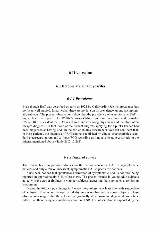
6 Discussion
6.1 Ectopic atrial tachycardia
6.1.1 Prevalence
Even though EAT was described as early as 1922 by Gallavardin (35), its prevalence hasnot been well studied. In particular, there are no data on its prevalence among asymptom-atic subjects. The present observations show that the prevalence of asymptomatic EAT ishigher than that reported for Wolff-Parkinson-White syndrome in young healthy males(258–260). It is evident that EAT is not well known among physicians and therefore oftenescapes diagnosis. In fact, none of the present subjects applying for a pilot's licence hadbeen diagnosed as having EAT. In the earlier studies, researchers have felt confident that,in most patients, the diagnosis of EAT can be established by clinical characteristics, stan-dard electrocardiogram and 24-hour ECG recording as long as one adheres strictly to thecriteria mentioned above (Table 2) (2,12,261).
6.1.2 Natural course
There have been no previous studies on the natural course of EAT in asymptomaticpatients and only a few on incessant, symptomatic EAT in paediatric patients.
It has been noticed that spontaneous remission of symptomatic EAT is not rare, beingreported in approximately 31% of cases (9). The present results in young adult subjectsagree with the earlier findings in younger subjects suggesting that spontaneous remissionis common.
During the follow-up, a change in P wave morphology in at least two leads suggestiveof a fusion of sinus and ectopic atrial rhythms was observed in some subjects. Theseobservations suggest that the ectopic foci gradually slow down and degenerate over timerather than there being any sudden restoration of SR. This observation is supported by the

75
rarity of the diagnosis of EAT in middle-aged or elderly people without concomitantstructural heart disease (262,263).
6.1.3 Heart rate variability
6.1.3.1 Abnormalities in time- and frequency-domain heart rate variability
Autonomic regulation of EAT and IST can be compared by traditional analysis methodsbecause of the similar average HR between the two rhythms. The present results suggestthat vagal outflow both to ectopic foci and to sinoatrial cells and their responsiveness tothe vagal input are similar, because phasic vagal tone is the major factor in the genesis ofHRV (18–23). This concept is also supported by the observation that atropine resulted ina similar increase in average HR during EAT and IST.
HRV analysed in absolute units was reduced in EAT compared with normal SR. Themechanism for differences in overall HRV is difficult to interpret, however, because thetraditional HRV analysis methods may fail to reveal specific autonomic abnormalities incases with markedly elevated HR (203,206). Several factors that increase HR itself,regardless of the cause of increase, reduce the overall HRV (203,264). Therefore, the pat-tern of reduced overall R-R interval variability in patients with EAT may result not onlyfrom a reduced vagal input but also from enhanced intrinsic automaticity of pacemakercells or an increased sympathetic input. The traditional methods of analyzing HRV inabsolute units may not be able to document the specific autonomic disturbance as a mech-anism of arrhythmias in these cases. Notably, the LF/HF ratio and the LF and HF compo-nents expressed in normalized units did not differ from normal in EAT, which suggeststhat there are no marked abnormalities in the phasic influences of the autonomic nervoussystem on ectopic foci. The increase in HR was within the normal limits after atropine inthe majority of patients with ectopic tachycardias, which also suggests that abnormalvagal function may not be the predominant autonomic disturbance in this tachycardia(167).
6.1.3.2 Abnormalities in short-term correlation properties of R-R interval dynamics
An increase in the randomness of short-term HR behaviour, expressed as a reduction ofthe short-term scaling exponent, was a typical feature of the EATs and resulted eitherfrom abrupt prolongations in the R-R intervals or from continuous instability in the beat-to-beat behaviour of R-R intervals. An ECG analysis of Holter recordings revealed thatthe prolongations in R-R intervals resulted from abrupt exit blocks due to the ectopic foci.Anecdotal cases of exit blocks due to ectopic atrial foci have also been reported previous-ly in short-term ECG recordings (265).

76
Two potential mechanisms may explain the unstable or random R-R interval behaviourof EATs. First, it is possible that the exit blocks from the ectopic foci result from a spe-cific electrophysiological abnormality similar to that observed in the diseased sinus node.Second, there may be a specific autonomic mechanism behind this abnormality. Accentu-ated sympathovagal interaction caused by high sympathetic outflow together with a con-comitant increase in phasic vagal outflow may result in uncorrelated short-term HRbehaviour. Experiments in healthy volunteers have shown that incremental doses of nore-pinephrine infusion with baroreflex-mediated vagal activation result in similar abrupt pro-longations of sinus intervals as observed in the present patients with EAT (266). Areduced short-term scaling exponent and random beat-to-beat HR dynamics have alsobeen observed in patients with heart failure with high levels of norepinephrine (26,267).A complex interaction between norepinephrine and acetylcholine at the presynaptic andpostsynaptic levels of the target tissue has been described (268), which may facilitate therandom firing of both normal and abnormal pacemakers.
6.2 Inappropriate sinus tachycardia
6.2.1 Prevalence
Our study was the first to describe the prevalence and characteristics of IST in a randommiddle-aged population and to assess the natural course and prognosis of IST duringlong-term follow-up. Our results indicate that IST, defined as 24-hour average HR >90bpm and HR >100 bpm in a supine or sitting position, is a frequent finding among mid-dle-aged subjects. The prevalence of IST (1.16%) exceeded that reported for the Wolff-Parkinson-White syndrome (0.15–0.31%), paroxysmal SVT (0.23%) and EAT (0.46%)(269–273). In the random population, the distribution of 24-hour average HR was clearlyskewed towards the right. Thus, it is likely that the elevation of HR does not simply rep-resent a continuum of the overall Gaussian distribution of sinus frequency in the generalpopulation.
The diagnosis of IST is difficult. It requires not only typical electrocardiographic find-ings but also careful exclusion of all reversible causes of sinus tachycardia (169,171).Thus, it is obvious that assessment of the actual prevalence of IST in a random populationcannot be based on casual HR measurements. Here, all subjects with an abnormal P waveduring tachycardia, structural heart disease, diabetes and any pathological condition ormedication known to accelerate HR were excluded. In order to avoid biases caused bytemporary changes in the sinus rate, the diagnosis of IST in the random population wasbased on three different HR measurements. Measurement of HR from casual 12-leadECG significantly overestimated the prevalence of IST, and 24-hour and 45-minute ECGrecordings under standard conditions also gave a much higher prevalence for IST than theuse of combined diagnostic criteria. Furthermore, only patients with elevated HR in botha supine and an upright position were included in the current study. If subjects with ortho-static intolerance (postural orthostatic tachycardia syndrome) (208) had been included,

77
the prevalence of IST would have been even higher. These data emphasize the importanceof repeated ambulatory ECG recordings in the diagnosis of IST.
6.2.2 Characteristics of subjects with inappropriate sinus tachycardia
The characteristics of patients fulfilling the diagnostic criteria of IST have not previouslybeen studied in a random population. According to the earlier reports, almost all highlysymptomatic patients with IST have been women, many of them have been hypertensive,and many have been health care workers (166,167,170,171). The results of our study con-firmed the higher prevalence of elevated HR among hypertensive than normotensive sub-jects. In the five series published earlier, about 90% of the patients with IST were females(166,167,170,171,174). In the present study, no evident gender-related difference wasseen in the random population, but most of the patients with IST in the symptomatic hos-pital population were women. The reasons for this difference are not clear, but it is possi-ble that women with elevated HR may become more often symptomatic and/or seek moreeasily diagnostic examinations and treatment. Given the observation that there were noother differences in the patient characteristics, it is unlikely that the subjects fulfilling thediagnostic criteria of IST in the random sample and the symptomatic hospital populationwould have represented different diagnostic entities.
Besides the higher blood pressure among the subjects with IST, there were no otherdifferences in the demographic characteristics, lifestyles, personality types, with theexception of hostility, laboratory values or echocardiographic parameters between the ISTand control subjects. Thus, the elevation of HR could not be explained by abnormal glu-cose metabolism, obesity, higher thyroid activity or lower physical activity. Although ISTmay have some shared features with hypertension, a causal relationship between highblood pressure and elevated HR seems unlikely. The association between the hostilityscore and elevated HR remains speculative.
In some previous studies, high sinus rate and disabling symptoms have been related toorthostatic intolerance caused by mild autonomic dysfunction (208). The characteristicfeature of these patients is postural sinus tachycardia, i.e., they exhibit a marked increasein HR upon rising from a supine position (208). In the present study, there were no differ-ences in the response of HR to postural changes or standard walking between the IST andcontrol subjects. Furthermore, no significant differences were observed in their sympatho-vagal balance (LF/HF ratio) as assessed by spectral analysis of HRV. Hence, altered auto-nomic input to the sinus node and postural changes did not appear to be a predominantcause of IST in this random population. These observations corroborate that the elevationof HR in patients with IST may be related to an intrinsic enhancement rather than alteredautonomic regulation of sinus node activity (274).

78
6.2.3 Natural course
There were only minor changes in the HR of the subjects with IST during a mean follow-up period of 6 years. Although some subjects no longer fulfilled the diagnostic criteria ofIST, the average HR did not differ significantly between the baseline and control mea-surements. Most subjects complained of moderate symptoms of elevated HR at the con-trol visit, but evident aggravation of the symptoms was rare.
It has been reported that nonparoxysmal SVT may cause cardiomyopathy and impairthe patient’s prognosis (135). In the present study, none of the subjects with IST had anyclinical or echocardiographic evidence of left ventricular dysfunction in the control exam-ination. Furthermore, an extensive Medline search yielded only one case report showingthat chronic nonparoxysmal sinus tachycardia had caused severe left ventricular dysfunc-tion (275). These observations confirm that, despite the constantly elevated HR and per-sistent symptoms, the prognosis of IST is good.
6.2.4 Heart rate variability
6.2.4.1 Abnormalities in time- and frequency-domain heart rate variability
HRV analysed in absolute units was reduced in IST compared with normal SR. Reducedoverall HRV in IST has also been reported previously (179) and may result from areduced vagal input and/or from enhanced intrinsic automaticity of pacemaker cells or anelevated sympathetic input. The LF/HF ratio and the LF and HF components analysed innormalized units did not differ from normal in IST, suggesting that there are no markedabnormalities in the phasic influences of the autonomic nervous system on sinoatrial cellscausing IST.
6.2.4.2 Short-term correlation properties of R-R interval dynamics
The randomness of the short-term HR behaviour of IST did not differ from normal SR.
6.2.5 Pharmacological maneuvers
6.2.5.1 Autonomic blockade
The results of the baseline electrophysiological measurements corroborated and expand-ed the observations made previously in other laboratories. It was found that not only the

79
resting HR but also IHR was significantly higher in the patients with IST than in the con-trol subjects. Likewise, in the patients with IST but not in the control subjects, HR afterthe abolition of autonomic input with propranolol and atropine was significantly higherthan IHR calculated according to the equation of Jose and Collison (197).
6.2.5.2 Adenosine
There have been few data on the effects of adenosine on tachyarrhythmias involving thesinus node. To our knowledge, only Glatter et al (106) studied the effects of adenosine onIST. Similar to their study, adenosine failed to terminate the tachycardia in all of the ISTpatients in our study. Thus, in contrast to sinus node re-entrant tachycardia (96,201), ISTwas found to be resistant to the antiarrhythmic actions of adenosine. This is in contrastwith the anticipated greater negative chronotropic effect of adenosine in subjects with ahigher atrial rate (rate-dependent action) (29). Despite a shorter initial cycle length and alarger dose of adenosine, the negative chronotropic response to adenosine was smaller inthe patients with IST than in the control subjects. Glatter (106) et al found slight prolon-gation of the tachycardia cycle, but many of their patients had isoproterenol infusion atthe time of the adenosine test (personal communication). Under such conditions, it is like-ly that the indirect negative chronotropic (antiadrenergic) effect of adenosine is augment-ed. In keeping with their results, we observed that adenosine caused only a modest, if any,shift in the atrial activation sequence.
The electrophysiological effects of adenosine may vary, depending on the autonomicnervous system tone and the injection site (29). The depressant effect of adenosine on thesinus node is accentuated by the heightened vagal (256) and beta-adrenergic tone (257).Therefore, to eliminate the influence of background autonomic tone, we studied theeffects of adenosine during pharmacologic beta-adrenergic and cholinergic receptorblockade. In the control subjects, autonomic blockade had minimal to no effect on thedirect negative chronotropic response to adenosine, although reflex tachycardia tended tobe blunted. These data indicate that, in control subjects, adenosine exerts a significantnegative chronotropic effect regardless of autonomic tone. In contrast, adenosine did nothave any significant effect on IST cycle length either under baseline conditions or duringautonomic blockade. Consequently, the direct (i.e., cAMP-independent) negative chrono-tropic effect of adenosine is expected to be diminished in patients with IST. The unusualpharmacokinetic properties of adenosine (e.g., extremely rapid metabolism and biphasiceffect on sinus rate) (29) preclude steady-state dose-response measurements and maycause site-dependent variation in the response to adenosine injection. Nevertheless, theresults of our subgroup analysis demonstrated that, regardless of the injection site, ade-nosine prolonged ACL more effectively in the control subjects than in the patients withIST, i.e., the negative chronotropic response to adenosine was impaired in the patientswith IST.
Numerous experimental (29,30) and clinical (276) investigations have showed that thenegative dromotropic effect of adenosine is dose- and rate-dependent. In this study, ade-nosine elicited a transient second- or third-degree AV block more frequently in the controlsubjects (i.e., subjects with slower atrial rate) than the IST patients. Hence, not only the

80
negative chronotropic but also the dromotropic response to adenosine were impaired inpatients with IST. On the other hand, several findings suggest that purinergic and para-sympathetic control is more evident in the sinus than in the AV node (277). Consistentwith this notion, we found that adenosine increased the AV nodal conduction time (orcaused a high-degree AV block) with virtually no effect on ACL in eight patients withIST. In another study, it was seen that the phenylephrine-induced increase in vagal tonecan provoke a second-degree AV block without causing any distinct change in the ISTrate (166).
6.2.6 Potential mechanism(s) of inappropriate sinus tachycardia
During the recent years, it has become apparent that inheritable ion channel abnormali-ties play an important role in the pathogenesis of several cardiac tachyarrhythmias, suchas the long QT syndrome (278). In addition, Lerman et al (279) showed that a mutation inthe inhibitory branch of the cAMP signal transduction pathway may result in idiopathicright ventricular outflow tract tachycardia that is unresponsive to adenosine. Wising (165)previously provided evidence that IST is a heritable disorder, and knockout of theIKAch,Ado protein in mice causes a state that closely resembles IST (280,281). Hence, it istempting to speculate that a specific gene mutation controlling the function of theIKAch,Ado protein is the basic abnormality behind IST. Nevertheless, the underlyingcause(s) of IST may be heterogeneous (173).
The present data provide new insight into the possible mechanism(s) of IST. First, theresistance of IST to the antiarrhythmic action of adenosine argues against triggered(96,201) activity as the principal mechanism of IST. Second, the higher than normal IHRsupports the previous notion that the mechanism(s) of IST involve(s) a primary abnormal-ity of the sinus node rather than abnormal autonomic modulation (29). Given the accentu-ation of the effect of adenosine on the sinus node by beta-adrenergic stimulation (257),the impaired response to adenosine makes excessive sympathetic input unlikely as a pri-mary mechanism of IST. Third, at the cellular level, an impaired negative chronotropiceffect to adenosine suggests that either the indirect (anti-β-adrenergic) or preferably thedirect Gi,o -protein-mediated signal transduction pathway is malfunctioning in patientswith IST. Accordingly, we postulate that IST may result from a deficient function of theIKAch,Ado channel. Because the A1-adenosine receptors and the muscarinic cholinergicsystem share the same receptor-effector pathway (277), this could explain not only theimpaired negative chronotropic response to adenosine but also the blunted response tovagal input in the patients with IST (1,171). This interpretation is supported by severalexperimental findings. Clapham’s group showed that IKAch,Ado–deficient GIRK4 knock-out mice exhibited a significantly faster sinus rate than wild-type mice (280). They alsoreported that the bradycardic response to purinergic and cholinergic agonists and HRV aredecreased in mice without functional IKAch,Ado channels (281). As in patients with IST,the effect of adenosine on AV nodal conduction was influenced less than the effect onsinus rate (281).

81
6.3 Limitations of the study
The asymptomatic population sample with EAT comprised only young males. Therefore,the prevalence figures may not be applicable to other populations. It was evident, howev-er, that EAT is not an uncommon finding in asymptomatic young adults, and its preva-lence may be even higher in younger subjects (2,12,13,46,282). A cut-off HR of 90 bpmwas used in this study to define EAT (I). This definition is based on the recommenda-tions by Spodick et al. (236), who defined 50 bpm and 90 bpm as the extremes of normalresting sinus HR in adult populations, which should improve the sensitivity of the tachy-cardia threshold. The definition of normal P wave morphology may also be problematic.We attempted to avoid overestimating the prevalence of EAT by including only patientswith an abnormal P wave in at least three leads, including one bipolar limb lead.
The diagnosis of IST was based on non-invasive measurements of HR by serial ECGand Holter recordings. Nevertheless, although no routine invasive electrophysiologicalstudies were made, we feel that an overlap between IST and other atrial tachyarrhythmiasis unlikely because there was no ECG or clinical evidence of such tachyarrhythmias.Likewise, the lack of any demonstrable orthostatic intolerance makes POTS an unlikelydifferential diagnostic option. On the other hand, it is possible that the present analysisunderestimated the actual prevalence of IST among the hypertensive subjects in the ran-dom population, because 149 of the 604 subjects were on beta-blocking and/or 62 patientson calcium-channel medication at the baseline. The population studied with symptomaticIST is rather small, but confirms the good prognosis of IST recently reported by Lopera etal in elderly females (180).
Finally, we must add that a variety of genetic and other mechanisms may be responsi-ble for the clinical syndrome, and there may be some overlap between IST and chronicorthostatic intolerance (1,171).
6.4 Implications of the study
Our results suggest that invasive therapy, such as catheter ablation, may not be indicatedin asymptomatic or mildly symptomatic patients with EAT because of the higher proba-bility of spontaneous remission of arrhythmia than of the occurrence of symptoms orheart failure.
The results of our study are consistent in suggesting that early reassurance or medicaltreatment is essential, while invasive therapy should be limited only to the most symp-tomatic patients, as suggested in the ACC/AHA/ESC guidelines for the management ofpatients with IST (144).
Analysis of the fractal characteristics of cardiac interbeat dynamics revealed abnormaldynamic behaviour that was not revealed by the traditional methods of HRV analysis,which confirms that the new analysis methods based on fractals and nonlinear dynamicsadd significantly to the diagnostic performance of the conventional methods used toassess abnormal HR behaviour. Long-term ECG recordings may also help to differentiatebetween IST and EAT in cases where 12-lead ECG does not yield a definitive diagnosis.

82
Our data suggest that adenosine may be a useful noninvasive probe in the evaluation ofpatients with suspected IST. Because adenosine markedly decelerates physiological sinustachycardia (200,257) and terminates sinus node re-entrant tachycardia as well as trig-gered atrial tachycardias (96,201), intravenous adenosine is likely to help in differentiat-ing between these arrhythmias and IST.
6.5 Future perspectives
In the future, studies with larger patient populations and application of molecular biologytechniques will be required to validate the exact mechanism and pathophysiology of IST.New electroanatomical techniques (CARTO, Ensite) may also help in assessment of exactlocalization of both IST and EAT and facilitate in the catheter ablation of these arrhyth-mias. Especially symptomatic patients with EAT can perhaps be more easily examinedand treated by new mapping techniques. In IST, Ensite technique can also record theexact intra-atrial activation sequence, which could possibly give new information of thisarrhythmia.
Because these arrhythmias are relatively rare, a national arrhythmia register might helpin further evaluation of these arrhythmias in a larger sample size. Patient follow-up, clini-cal outcomes and given treatment would be easily available for each patient. In addition,national guidelines of SVTs in Finland would expand the knowledge of EAT and ISTamong the medical community. Guidelines in native language would highlight this ratherrare issue and offer information to a larger group of physicians.

7 Conclusions
1. Asymptomatic EAT is not an uncommon finding among young males. In the majority,the atrial rhythm slows over time, leading to either restoration of SR or fusion of ecto-pic atrial and sinus rhythms. This suggests that the ectopic foci gradually degenerateover time.
2. IST appears to be a more common disorder than previously assumed. Despite thechronic nature and long-lasting symptoms of this disorder, its natural course isbenign.
3. HRV values obtained by time- and frequency-domain analysis methods over a 24-hour period did not differ between EAT and IST, which suggests that atrial arrhythmicfoci are under similar long-term phasic regulation as the sinus node. In contrast, short-term correlation properties of R-R interval behaviour were not similar during IST andEAT. These differences may result from a specific autonomic disturbance that cont-rols the discharge of the ectopic foci or from an intrinsic electrophysiological abnor-mality of these foci.
4. IST is resistant to the antiarrhythmic actions of adenosine. Unlike several other SVTs,IST is not terminated by adenosine and, regardless of the autonomic tone, the nega-tive chronotropic response to adenosine is impaired in patients with IST. These fin-dings are expected to have important diagnostic implications and to provide newinsight into the pathophysiologic mechanism(s) of IST.

References
1. Cossu SF & Steinberg JS (1998) Supraventricular tachyarrhythmias involving the sinus node:clinical and electrophysiologic characteristics. Prog Cardiovasc Dis 41:51–63.
2. von Bernuth G, Engelhardt W, Kramer HH, Singer H, Schneider P, Ulmer H, Brodherr-Heber-lein S, Kienast W, Lang D & Lindinger A (1992) Atrial automatic tachycardia in infancy andchildhood. Eur Heart J 13:1410–1415.
3. Kalman JM (1997) Inappropriate sinus tachycardia: An update. Cardiac ElectrophysiologyReview 1/2:61–66.
4. Saoudi N, Cosio F, Waldo A, Chen SA, Iesaka Y, Lesh M, Saksena S, Salerno J & Schoels W(2001) Classification of atrial flutter and regular atrial tachycardia according to electrophysio-logic mechanism and anatomic bases: a statement from a joint expert group from the WorkingGroup of Arrhythmias of the European Society of Cardiology and the North American Societyof Pacing and Electrophysiology. J Cardiovasc Electrophysiol 12:852–866.
5. Gillette PC, Smith RT, Garson A Jr, Mullins CE, Gutgesell HP, Goh TH, Cooley DA & McNa-mara DG (1985) Chronic supraventricular tachycardia. A curable cause of congestive cardio-myopathy. JAMA 253:391–392.
6. Coleman HN, III, Taylor RR, Pool PE, Whipple GH, Covell JW, Ross J Jr & Braunwald E(1971) Congestive heart failure following chronic tachycardia. Am Heart J 81:790–798.
7. Olsson SB, Blomstrom P & Sabel KG (1984) Incessant ectopic atrial tachycardia: successfulsurgical treatment with regression of dilated cardiomyopathy picture. Am J Cardiol 1984:1465–1466.
8. Naccarelli GV, Shih HT & Jalal S (1995) Sinus node reentry and atrial tachycardias. In: ZipesD and Jalife J (eds) Cardiac Electrophysiology From cell to bedside. W.B. Saunders Company,Philadelphia, p.607–619.
9. Walsh EP, Saul JP &Triedman JK (1994) Natural and unnatural history of ectopic atrialtachycardia: One institution's experience (abstract). Pacing Clin Electrophysiol 17:746.
10. Naheed ZJ, Strasburger JF, Benson DW Jr & Deal BJ (1995) Natural history and managementstrategies of automatic atrial tachycardia in children. Am J Cardiol 75:405–407.
11. Bauersfeld U, Gow RM, Hamilton RM & Izukawa T (1995) Treatment of atrial ectopictachycardia in infants < 6 months old. Am Heart J 129:1145–1148.
12. Mehta AV, Sanchez GR, Sacks EJ, Casta A, Dunn JM & Donner RM (1988) Ectopic automaticatrial tachycardia in children: clinical characteristics, management and follow-up. J Am CollCardiol 11:379–385.
13. Koike K, Hesslein PS, Finlay CD, Williams WG, Izukawa T & Freedom RM (1988) Atrialautomatic tachycardia in children. Am J Cardiol 61:1127–1130.

85
14. Dhala AA, Case CL & Gillette PC (1994) Evolving treatment strategies for managing atrialectopic tachycardia in children. Am J Cardiol 74:283–286.
15. Garson A Jr, Smith RT & Moak JP (1990) Atrial automatic tachycardia in children. In: TouboulP & Waldo AL (eds) Atrial arrhythmias: Current concepts and management, Mosby-YearBook, St Louis, p. 282–287.
16. Klersy C, Chimienti M, Marangoni E, Comelli M & Salerno JA (1993) Factors that predictspontaneous remission of ectopic atrial tachycardia. Eur Heart J 14:1654–1656.
17. Tai CT, Chiou CW & Chen SA (2002) Interaction between the autonomic nervous system andatrial tachyarrhythmias. J Cardiovasc Electrophysiol 13:83–87.
18. Akselrod S, Gordon D, Ubel FA, Shannon DC, Berger AC & Cohen RJ (1981) Power spectrumanalysis of heart rate fluctuation: a quantitative probe of beat-to-beat cardiovascular control.Science 213:220–222.
19. Pomeranz B, Macaulay RJ, Caudill MA, Kutz I, Adam D, Gordon D, Kilborn KM, Barger AC,Shannon DC & Cohen RJ (1985) Assessment of autonomic function in humans by heart ratespectral analysis. Am J Physiol 248:H151–H153.
20. Eckberg DL (1983) Human sinus arrhythmia as an index of vagal cardiac outflow. J Appl Phy-siol 54:961–966.
21. Pagani M, Lombardi F, Guzzetti S, Rimoldi O, Furlan R, Pizzinelli P, Sandrone G, Malfatto G,Dell'Orto S & Piccaluga E (1986) Power spectral analysis of heart rate and arterial pressurevariabilities as a marker of sympatho-vagal interaction in man and conscious dog. Circ Res59:178–193.
22. Task Force of the European Society of Cardiology and the North American Society of Pacingand Electrophysiology (1996) Heart rate variability: standards of measurement, physiologicalinterpretation and clinical use. Circulation 93:1043–1065.
23. Malliani A, Pagani M, Lombardi F & Cerutti S (1991) Cardiovascular neural regulation explo-red in the frequency domain. Circulation 84:482–492.
24. Goldberger AL (1996) Non-linear dynamics for clinicians: chaos theory, fractals, and comple-xity at the bedside. Lancet 347:1312–1314.
25. Bigger JT Jr, Steinman RC, Rolnitzky LM, Fleiss JL, Albrecht P & Cohen RJ (1996) Power lawbehavior of RR-interval variability in healthy middle-aged persons, patients with recent acutemyocardial infarction, and patients with heart transplants. Circulation 93:2142–2151.
26. Ho KK, Moody GB, Peng CK, Mietus JE, Larson MG, Levy D & Goldberger AL (1997) Pre-dicting survival in heart failure case and control subjects by use of fully automated methods forderiving nonlinear and conventional indices of heart rate dynamics. Circulation 96:842–848.
27. Mäkikallio TH, Seppänen T, Niemelä M, Airaksinen KE, Tulppo M & Huikuri HV (1996)Abnormalities in beat to beat complexity of heart rate dynamics in patients with a previousmyocardial infarction. J Am Coll Cardiol 28:1005–1011.
28. Mäkikallio TH, Seppänen T, Airaksinen KE, Koistinen J, Tulppo MP, Peng CK, Goldberger AL& Huikuri HV (1997) Dynamic analysis of heart rate may predict subsequent ventriculartachycardia after myocardial infarction. Am J Cardiol 80:779–783.
29. Raatikainen MJP, Dennis DM & Belardinelli L (1998) Cardiac electrophysiology of adenosine:Cellular basis and clinical implications. In: Peller A & Belardinelli L (eds) Effects of Extracel-lular adenosine and ATP on Cardiomyocytes. R.G. Landes Company, Austin, p. 87–132.
30. Lerman BB & Belardinelli L (1991) Cardiac electrophysiology of adenosine. Basic and clinicalconcepts. Circulation 83:1499–1509.
31. Ganz LI & Friedman PL (1995) Supraventricular tachycardia. N Engl J Med 332:162–173.32. Waller BF & Schlant RC (1996) Anatomy of the heart. Innervation of the heart. In: Schlant RC
& Alexander RW (eds) The heart. Mc Graw Hill Inc, NY, p. 82–84.33. Berne RM & Levy MN (1993) Regulation of heartbeat. In: Berne RM & Levy MN (eds) Phy-
siology. Mosby-Year Book, St Louis, p. 417–419.

86
34. Pocock G & Richards CD (1999) The heart and circulation. In: Pocock G & Richards CD (eds)Human Physiology The basis of Medicine. Oxford University Press, NY, p. 291–292.
35. Gallavardin L (1922) De la tachycardie paroxystique a centre excitable. Arch Mal Coeur 15:1.36. Parkinson J & Papp C (1947) Repetitive paroxysmal tachycardia. Br Heart J 9:241.37. Schachnow N, Spellman S & Rubin I (1954) Persistent supraventricular tachycardia. Circula-
tion 42:673.38. Hay JD & Keidan SE (1952) Persistent ectopic auricular tachycardia in children. Br Heart J
14:435.39. Keane JF, Plauth WH & Nadas AS (1972) Chronic ectopic tachycardia of infancy and child-
hood. Am Heart J 84:748–757.40. Ström G, Zetterqvist E & Zetterqvist P (1960) Chronic supraventricular tachycardia of conti-
nuous or repetitive type in children. Acta Paediat 49:827–837.41. Morgan CL & Nadas AS (1964) Chronic ectopic tachycardia in infancy and childhood. Am
Heart J 67:617.42. von Bernuth G, Lang D & Belz GG (1974) Die repetitive paroxysmale Tachycardie im Kindes-
alter. Mschr Kinderheilk 122:183–185.43. Jacobsen JR, Andersen ED, Sandoe E, Videbaek J & Wennevold A (1975) Chronic supraventri-
cular tachycardia in infancy and childhood. Acta Paediatr Scand 64:597–604.44. Wolf D (1978) Vorhoftachykardien mit AV-Leitungsstörungen im Säuglings- und Kindesalter.
Mschr Kinderheilk 126:387–390.45. Goldreyer BN, Gallagher JJ & Damato AN (1973) The electrophysiologic demonstration of
atrial ectopic tachycardia in man. Am Heart J 85:205–215.46. Gillette PC & Garson A Jr (1977) Electrophysiologic and pharmacologic characteristics of
automatic ectopic atrial tachycardia. Circulation 56:571–575.47. Coumel P & Fidelle J (1980) Amiodarone in the treatment of cardiac arrhythmias in children:
one hundred thirty-five cases. Am Heart J 100:1063–1069.48. Kunze KP, Kuck KH, Schluter M & Bleifeld W (1986) Effect of encainide and flecainide on
chronic ectopic atrial tachycardia. J Am Coll Cardiol 7:1121–1126.49. Ott DA, Garson A Jr, Cooley DA, Smith RT & Moak J (1987) Cryoablative techniques in the
treatment of cardiac tachyarrhythmias. Ann Thorac Surg 43:138–143.50. Walsh EP & Saul JP (1991) Transcatheter ablation for pediatric tachyarrhythmias using radio-
frequency electrical energy. Pediatr Ann 20:386–392.51. Kay GN, Chong F, Epstein AE, Dailey SM & Plumb VJ (1993) Radiofrequency ablation for
treatment of primary atrial tachycardias. J Am Coll Cardiol 21:901–909.52. Tracy CM, Swartz JF, Fletcher RD, Hoops HG, Solomon AJ, Karasik PE & Mukherjee D
(1993) Radiofrequency catheter ablation of ectopic atrial tachycardia using paced activationsequence mapping. J Am Coll Cardiol 21:910–917.
53. Goldberger J, Kall J, Ehlert F, Deal B, Olshansky B, Benson DW, Baerman J, Kopp D, KadishA & Wilber D (1993) Effectiveness of radiofrequency catheter ablation for treatment of atrialtachycardia. Am J Cardiol 72:787–793.
54. Lesh MD, Van Hare GF, Epstein LM, Fitzpatrick AP, Scheinman MM, Lee RJ, Kwasman MA,Grogin HR & Griffin JC (1994) Radiofrequency catheter ablation of atrial arrhythmias. Resultsand mechanisms. Circulation 89:1074–1089.
55. Hoffmann E, Reithmann C, Nimmermann P, Elser F, Dorwarth U, Remp T & Steinbeck G(2002) Clinical experience with electroanatomic mapping of ectopic atrial tachycardia. PacingClin Electrophysiol 25:49–56.
56. Jais P, Haissaguerre M, Shah DC, Chouairi S, Gencel L, Hocini M & Clementy J (1997) Afocal source of atrial fibrillation treated by discrete radiofrequency ablation. Circulation95:572–576.

87
57. Haissaguerre M, Shah DC, Jais P & Clementy J (1997) Role of catheter ablation for atrial fibril-lation. Curr Opin Cardiol 12:18–23.
58. Graffigna A, Vigano M, Pagani F & Salerno G (1992) Surgical treatment for ectopic atrialtachycardia. Ann Thorac Surg 54:338–343.
59. Wellens HJ (1994) Atrial tachycardia. How important is the mechanism? Circulation 90:1576–1577.
60. Walsh EP, Saul JP, Hulse JE, Rhodes LA, Hordof AJ, Mayer JE & Lock JE (1992) Transcathe-ter ablation of ectopic atrial tachycardia in young patients using radiofrequency current. Circu-lation 86:1138–1146.
61. Weindling SN, Saul JP & Walsh EP (1996) Efficacy and risks of medical therapy for supravent-ricular tachycardia in neonates and infants. Am Heart J 131:66–72.
62. Walsh EP (2000) Ablation of ectopic atrial tachycardia in children. In: Huang SKS & WilberDJ (eds) Radiofrequency catheter ablation of cardiac arrhythmias: Basic concepts and clinicalapplications. Futura Publishing Company Inc, Armonk, p. 115–136.
63. Hendry PJ, Packer DL, Anstadt MP, Plunkett MD & Lowe JE (1990) Surgical treatment ofautomatic atrial tachycardias. Ann Thorac Surg 49:253–259.
64. Gillette PC (1992) Successful transcatheter ablation of ectopic atrial tachycardia in youngpatients using radiofrequency current. Circulation 86:1339–1340.
65. Prager NA, Cox JL, Lindsay BD, Ferguson TB Jr, Osborn JL & Cain ME (1993) Long-termeffectiveness of surgical treatment of ectopic atrial tachycardia. J Am Coll Cardiol 22:85–92.
66. Chen CC, Tai CT, Chiang CE, Yu WC, Lee SH, Chen YJ, Hsieh MH, Tsai CF, Lee KW, DingYA, Chang MS & Chen SA (2000) Atrial tachycardias originating from the atrial septum:electrophysiologic characteristics and radiofrequency ablation. J Cardiovasc Electrophysiol11:744–749.
67. Weiss C, Willems S, Rueppel R, Hoffmann M & Meinertz T (2001) Electroanatomical Map-ping (CARTO) of ectopic atrial tachycardia: impact of bipolar and unipolar local electrogramannotation for localization the focal origin. J Interv Card Electrophysiol 5:101–107.
68. Kistler PM, Sanders P, Hussin A, Morton JB, Vohra JK, Sparks PB & Kalman JM (2003) Focalatrial tachycardia arising from the mitral annulus: electrocardiographic and electrophysiologiccharacterization. J Am Coll Cardiol 41:2212–2219.
69. Seals AA, Lawrie GM, Magro S, Lin HT, Pacifico A, Roberts R & Wyndham CR (1988) Surgi-cal treatment of right atrial focal tachycardia in adults. J Am Coll Cardiol 11:1111–1117.
70. Chen SA, Chiang CE, Yang CJ, Cheng CC, Wu TJ, Wang SP, Chiang BN & Chang MS (1994)Sustained atrial tachycardia in adult patients. Electrophysiological characteristics, pharmacolo-gical response, possible mechanisms, and effects of radiofrequency ablation. Circulation90:1262–1278.
71. Poty H, Saoudi N, Haissaguerre M, Daou A, Clementy J & Letac B (1996) Radiofrequency cat-heter ablation of atrial tachycardias. Am Heart J 131:481–489.
72. Pappone C, Stabile G, De Simone A, Senatore G, Turco P, Damiano M, Iorio D, Spampinato N& Chiariello M (1996) Role of catheter-induced mechanical trauma in localization of targetsites of radiofrequency ablation in automatic atrial tachycardia. J Am Coll Cardiol 27:1090–1097.
73. de Bakker JM, Coronel R, McGuire MA, Vermeulen JT, Opthof T, Tasseron S, van Hemel NM& Defauw JJ (1994) Slow potentials in the atrioventricular junctional area of patients operatedon for atrioventricular node tachycardias and in isolated porcine hearts. J Am Coll Cardiol23:709–715.
74. Saul JP, Walsh EP & Triedman JK (1995) Mechanisms and therapy of complex arrhythmias inpediatric patients. J Cardiovasc Electrophysiol 6:1129–1148.
75. Lesh MD (2000) Catheter ablation of atrial flutter and tachycardia. In: Zipes DP & Jalife J (eds)Cardiac electrophysiology: from cell to bedside. W.B. Saunders Company, Philadelphia, p.1009–1027.
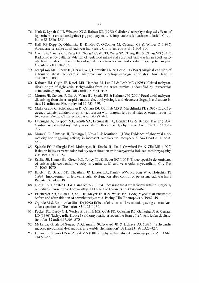
88
76. Nath S, Lynch C III, Whayne JG & Haines DE (1993) Cellular electrophysiological effects ofhyperthermia on isolated guinea pig papillary muscle. Implications for catheter ablation. Circu-lation 88:1826–1831.
77. Kall JG, Kopp D, Olshansky B, Kinder C, O'Connor M, Cadman CS & Wilber D (1995)Adenosine-sensitive atrial tachycardia. Pacing Clin Electrophysiol 18:300–306.
78. Chen SA, Chiang CE, Yang CJ, Cheng CC, Wu TJ, Wang SP, Chiang BN & Chang MS (1993)Radiofrequency catheter ablation of sustained intra-atrial reentrant tachycardia in adult patie-nts. Identification of electrophysiological characteristics and endocardial mapping techniques.Circulation 88:578–587.
79. Josephson ME, Spear JF, Harken AH, Horowitz LN & Dorio RJ (1982) Surgical excision ofautomatic atrial tachycardia: anatomic and electrophysiologic correlates. Am Heart J104:1076–1085.
80. Kalman JM, Olgin JE, Karch MR, Hamdan M, Lee RJ & Lesh MD (1998) "Cristal tachycar-dias": origin of right atrial tachycardias from the crista terminalis identified by intracardiacechocardiography. J Am Coll Cardiol 31:451–459.
81. Morton JB, Sanders P, Das A, Vohra JK, Sparks PB & Kalman JM (2001) Focal atrial tachycar-dia arising from the tricuspid annulus: electrophysiologic and electrocardiographic characteris-tics. J Cardiovasc Electrophysiol 12:653–659.
82. Mallavarapu C, Schwartzman D, Callans DJ, Gottlieb CD & Marchlinski FE (1996) Radiofre-quency catheter ablation of atrial tachycardia with unusual left atrial sites of origin: report oftwo cases. Pacing Clin Electrophysiol 19:988–992.
83. Dunnigan A, Pierpont ME, Smith SA, Breningstall G, Benditt DG & Benson DW Jr (1984)Cardiac and skeletal myopathy associated with cardiac dysrhythmias. Am J Cardiol 53:731–737.
84. Moro C, Rufilanchas JJ, Tamargo J, Novo L & Martinez J (1988) Evidence of abnormal auto-maticity and triggering activity in incessant ectopic atrial tachycardia. Am Heart J 116:550–552.
85. Spinale FG, Fulbright BM, Mukherjee R, Tanaka R, Hu J, Crawford FA & Zile MR (1992)Relation between ventricular and myocyte function with tachycardia-induced cardiomyopathy.Circ Res 71:174–187.
86. Saffitz JE, Kanter HL, Green KG, Tolley TK & Beyer EC (1994) Tissue-specific determinantsof anisotropic conduction velocity in canine atrial and ventricular myocardium. Circ Res74:1065–1070.
87. Kugler JD, Baisch SD, Cheatham JP, Latson LA, Pinsky WW, Norberg W & Hofschire PJ(1984) Improvement of left ventricular dysfunction after control of persistent tachycardia. JPediatr 105:543–548.
88. Giorgi LV, Hartzler GO & Hamaker WR (1984) Incessant focal atrial tachycardia: a surgicallyremediable cause of cardiomyopathy. J Thorac Cardiovasc Surg 87:466–469.
89. Fishberger SB, Colan SD, Saul JP, Mayer JE Jr & Walsh EP (1996) Myocardial mechanicsbefore and after ablation of chronic tachycardia. Pacing Clin Electrophysiol 19:42–49.
90. Ogilvie RI & Zborowska-Sluis D (1992) Effect of chronic rapid ventricular pacing on total vas-cular capacitance. Circulation 85:1524–1530.
91. Packer DL, Bardy GH, Worley SJ, Smith MS, Cobb FR, Coleman RE, Gallagher JJ & GermanLD (1986) Tachycardia-induced cardiomyopathy: a reversible form of left ventricular dysfunc-tion. Am J Cardiol 57:563–570.
92. McLaren, Gersh BJ,Sugrue DD,Hammill SC,Seward JB & Holmes DR (1985) Tachycardiainduced myocardial dysfunction: a revesible phenomenon? Br Heart J 1985:323–327.
93. Umana E, Solares CA & Alpert MA (2003) Tachycardia-induced cardiomyopathy. Am J Med114:51–55.

89
94. Tanaka R, Spinale FG, Crawford FA & Zile MR (1992) Effect of chronic supraventriculartachycardia on left ventricular function and structure in newborn pigs. J Am Coll Cardiol20:1650–1660.
95. Belardinelli L & Isenberg G (1983) Isolated atrial myocytes: adenosine and acetylcholineincrease potassium conductance. Am J Physiol 244:H734–H737.
96. Engelstein ED, Lippman N, Stein KM & Lerman BB (1994) Mechanism-specific effects ofadenosine on atrial tachycardia. Circulation 89:2645–2654.
97. Iwai S, Markowitz SM, Stein KM, Mittal S, Slotwiner DJ, Das MK, Cohen JD, Hao SC & Ler-man BB (2002) Response to adenosine differentiates focal from macroreentrant atrial tachycar-dia: validation using three-dimensional electroanatomic mapping. Circulation 106:2793–2799.
98. Rankin AC, Oldroyd KG, Chong E, Rae AP & Cobbe SM (1989) Value and limitations ofadenosine in the diagnosis and treatment of narrow and broad complex tachycardias. Br Heart J62:195–203.
99. diMarco JP, Sellers TD, Lerman BB, Greenberg ML, Berne RM & Belardinelli L (1985) Diag-nostic and therapeutic use of adenosine in patients with supraventricular tachyarrhythmias. JAm Coll Cardiol 6:417–425.
100. Griffith MJ, Linker NJ, Ward DE & Camm AJ (1988) Adenosine in the diagnosis of broadcomplex tachycardia. Lancet 1:672–675.
101. Till J, Shinebourne EA, Rigby ML, Clarke B, Ward DE & Rowland E (1989) Efficacy andsafety of adenosine in the treatment of supraventricular tachycardia in infants and children. BrHeart J 62:204–211.
102. Perelman MS & Krikler DM (1987) Termination of focal atrial tachycardia by adenosinetriphosphate. Br Heart J 58:528–530.
103. Shenasa H, Kanter RJ, Hamer ME, Sorrentino RA, SandersWE, GreenfieldRA, Page RL &Wharton JM (1993) Reappraisal of the efficacy of adenosine for termination of ectopic atrialtachycardia (abstract). J Am Coll Cardiol 21:456A.
104. Shenasa H, Greenfield RA, Johnson EE, Sorrentino RA & Wharton JM (1993) Does adenosinedifferentiate sinoatrial reentrant tachycardia from ectopic atrial tachycardia? (abstract) Circula-tion 88:I–396.
105. Marrouche NF, Beheiry S, Tomassoni G, Cole C, Bash D, Dresing T, Saliba W, Abdul-Karim A,Tchou P, Schweikert R, Leonelli F & Natale A (2002) Three-dimensional nonfluoroscopicmapping and ablation of inappropriate sinus tachycardia. Procedural strategies and long-termoutcome. J Am Coll Cardiol 39:1046–1054.
106. Glatter KA, Cheng J, Dorostkar P, Modin G, Talwar S, Al Nimri M, Lee RJ, Saxon LA, LeshMD & Scheinman MM (1999) Electrophysiologic effects of adenosine in patients with supra-ventricular tachycardia. Circulation 99:1034–1040.
107. Case CL & Gillette PC (1993) Automatic atrial and junctional tachycardias in the pediatricpatient: strategies for diagnosis and management. Pacing Clin Electrophysiol 16:1323–1335.
108. Morton HJV & Thomas ET (1958) Effects of atropine on the heart rate. Lancet 2:1313–1315.109. Jose AD & Taylor RR (1969) Autonomic blockade by propranolol and atropine to study intrin-
sic myocardial function in man. J Clin Invest 48:2019–2031.110. Huikuri HV, Ylitalo A, Pikkujämsä SM, Ikäheimo MJ, Airaksinen KE, Rantala AO, Lilja M &
Kesäniemi YA (1996) Heart rate variability in systemic hypertension. Am J Cardiol 77:1073–1077.
111. Peng CK, Havlin S, Stanley HE & Goldberger AL (1995) Quantification of scaling exponentsand crossover phenomena in nonstationary heartbeat time series. Chaos 5:82–87.
112. Pincus SM & Viscarello RR (1992) Approximate entropy: a regularity measure for fetal heartrate analysis. Obstet Gynecol 79:249–255.
113. Sosa E, Marcial MB, Scanavacca M, Bellotti G & Pileggi F (1991) Incessant ectopic atrialtachycardia and sudden death. Pacing Clin Electrophysiol 14:764–767.

90
114. Damiano RJ Jr, Tripp HF Jr, Asano T, Small KW, Jones RH & Lowe JE (1987) Left ventriculardysfunction and dilatation resulting from chronic supraventricular tachycardia. J Thorac Car-diovasc Surg 94:135–143.
115. Scheinman MM, Basu D & Hollenberg M (1974) Electrophysiologic studies in patients withpersistent atrial tachycardia. Circulation 50:266–273.
116. Tang CW, Scheinman MM, Van Hare GF, Epstein LM, Fitzpatrick AP, Lee RJ & Lesh MD(1995) Use of P wave configuration during atrial tachycardia to predict site of origin. J Am CollCardiol 26:1315–1324.
117. Yamane T, Shah DC, Peng JT, Jais P, Hocini M, Deisenhofer I, Choi KJ, Macle L, Clementy J& Haissaguerre M (2001) Morphological characteristics of P waves during selective pulmonaryvein pacing. J Am Coll Cardiol 38:1505–1510.
118. Marrouche NF, Sippensgroenewegen A, Yang Y, Dibs S & Scheinman MM (2002) Clinical andelectrophysiologic characteristics of left septal atrial tachycardia. J Am Coll Cardiol 40:1133–1139.
119. Sippensgroenewegen A, Peeters HA, Jessurun ER, Linnenbank AC, Robles de Medina EO,Lesh MD & van Hemel NM (1998) Body surface mapping during pacing at multiple sites in thehuman atrium: P-wave morphology of ectopic right atrial activation. Circulation 97:369–380.
120. Eldar M, Fitzpatrick AP, Ohad D, Smith MF, Hsu S, Whayne JG, Vered Z, Rotstein Z, Kordis T,Swanson DK, Chin M, Scheinman MM, Lesh MD & Greenspon AJ (1996) Percutaneous multi-electrode endocardial mapping during ventricular tachycardia in the swine model. Circulation94:1125–1130.
121. Triedman JK, Jenkins KJ, Colan SD, Van Praagh R, Lock JE & Walsh EP (1997) Multipolarendocardial mapping of the right heart using a basket catheter: acute and chronic animal stu-dies. Pacing Clin Electrophysiol 20:51–59.
122. Schmitt C, Zrenner B, Schneider M, Karch M, Ndrepepa G, Deisenhofer I, Weyerbrock S,Schreieck J & Schomig A (1999) Clinical experience with a novel multielectrode basket cathe-ter in right atrial tachycardias. Circulation 99:2414–2422.
123. Wyndham CR, Arnsdorf MF, Levitsky S, Smith TC, Dhingra RC, Denes P & Rosen KM (1980)Successful surgical excision of focal paroxysmal atrial tachycardia. Observations in vivo and invitro. Circulation 62:1365–1372.
124. Wharton M, Shenasa H, Barold H, Simons G & Vergara I (2000) Ablation of atrial tachycardiain adults. In: Huang SKS & Wilber DJ eds Radiofrequency catheter ablation of cardiac arrhyth-mias: basic concepts and clinical applications. Futura Publishing Company Inc, Armonk, p.139–163.
125. Ben Haim SA, Osadchy D, Schuster I, Gepstein L, Hayam G & Josephson ME (1996) Nonfluo-roscopic, in vivo navigation and mapping technology. Nat Med 2:1393–1395.
126. Gepstein L, Hayam G & Ben Haim SA (1997) A novel method for nonfluoroscopic catheter-based electroanatomical mapping of the heart. In vitro and in vivo accuracy results. Circulation95:1611–1622.
127. Hoffmann E, Reithmann C, Nimmermann P, Remp T & Steinbeck G (1999) Atrial reentranttachycardia after heart transplantation. Circulation 99:326–327.
128. Kottkamp H, Hindricks G, Breithardt G & Borggrefe M (1997) Three-dimensional electromag-netic catheter technology: electroanatomical mapping of the right atrium and ablation of ecto-pic atrial tachycardia. J Cardiovasc Electrophysiol 8:1332–1337.
129. Schmitt H, Weber S, Schwab JO, Voss RM, Kneller R, Tillmanns H & Waldecker B (2001)Diagnosis and ablation of focal right atrial tachycardia using a new high-resolution, non-con-tact mapping system. Am J Cardiol 87:1017–1021.
130. Seidl K, Schwacke H, Rameken M, Drogemuller A, Beatty G & Senges J (2003) Noncontactmapping of ectopic atrial tachycardias: different characteristics of isopotential maps and unipo-lar electrogram. Pacing Clin Electrophysiol 26:16–25.
91
131. Kalman JM, Olgin JE, Karch MR & Lesh MD (1997) Use of intracardiac echocardiography ininterventional electrophysiology. Pacing Clin Electrophysiol 20:2248–2262.
132. Dreifus LS & Hessen SE (1990) Supraventricular tachycardia: diagnosis and treatment. Cardio-logy 77:259–268.
133. Rabbani LE, Wang PJ, Couper GL & Friedman PL (1991) Time course of improvement inventricular function after ablation of incessant automatic atrial tachycardia. Am Heart J121:816–819.
134. Gelb BD & Garson A Jr (1990) Noninvasive discrimination of right atrial ectopic tachycardiafrom sinus tachycardia in "dilated cardiomyopathy". Am Heart J 120:886–891.
135. Shinbane JS, Wood MA, Jensen DN, Ellenbogen KA, Fitzpatrick AP & Scheinman MM (1997)Tachycardia-induced cardiomyopathy: a review of animal models and clinical studies. J AmColl Cardiol 29:709–715.
136. Gillette PC, Wampler DG, Garson A Jr, Zinner A, Ott D & Cooley D (1985) Treatment of atrialautomatic tachycardia by ablation procedures. J Am Coll Cardiol 6:405–409.
137. Gillette PC, Garson A Jr, Hesslein PS, Karpawich PP, Tierney RC, Cooley DA & McNamaraDG (1981) Successful surgical treatment of atrial, junctional, and ventricular tachycardia unas-sociated with accessory connections in infants and children. Am Heart J 102:984–991.
138. Leman RB, Gillette PC & Zinner AJ (1986) Resolution of congestive cardiomyopathy causedby supraventricular tachycardia using amiodarone. Am Heart J 112:622–624.
139. Pool PE & Quart BD (1988) Treatment of ectopic atrial arrhythmias and premature atrial comp-lexes in adults with encainide. Am J Cardiol 62:60L–62L.
140. Kuck KH, Kunze KP, Schluter M & Duckeck W (1988) Encainide versus flecainide for chronicatrial and junctional ectopic tachycardia. Am J Cardiol 62:37L–44L.
141. Perry JC, McQuinn RL, Smith RT Jr, Gothing C, Fredell P & Garson A Jr (1989) Flecainideacetate for resistant arrhythmias in the young: efficacy and pharmacokinetics. J Am Coll Car-diol 14:185–191.
142. Guccione P, Paul T & Garson A Jr (1990) Long-term follow-up of amiodarone therapy in theyoung: continued efficacy, unimpaired growth, moderate side effects. J Am Coll Cardiol15:1118–1124.
143. Colloridi V, Perri C, Ventriglia F & Critelli G (1992) Oral sotalol in pediatric atrial ectopictachycardia. Am Heart J 123:254–256.
144. Blomström-Lundqvist C, Scheinman MM, Aliot EM, Alpert JS, Calkins H, Camm AJ,Campbell WB, Haines DE, Kuck KH, Lerman BB, Miller DD, Shaeffer CW, Stevenson WG,Tomaselli GF, Antman EM, Smith SC Jr, Alpert JS, Faxon DP, Fuster V, Gibbons RJ,Gregoratos G, Hiratzka LF, Hunt SA, Jacobs AK, Russel RO Jr, Priori SG, Blanc JJ, Budaj A,Burgos EF, Cowie M, Deckers JW, Garcia MA, Klein WW, Lekakis J, Lindahl B, Mazzotta G,Morais JC, Oto A, Smiseth O & Trappe HJ (2003) ACC/AHA/ESC guidelines for the manage-ment of patients with supraventricular arrhythmias. J Am Coll Cardiol 42:1493–1531.
145. Coumel P, Aigueperse J, Perrault MA, Fantoni A, Slama R & Bouvrain Y (1973) [Detectionand attempted surgical exeresis of a left auricular ectopic focus with refractory tachycardia.Favorable outcome]Reperage et tentative d'exerese chirurgicale d'un foyer ectopique auriculaire gauche avectachycardie rebelle. Evolution favorable. Ann Cardiol Angeiol (Paris) 22:189–199.
146. Anderson KP, Stinson EB & Mason JW (1982) Surgical exclusion of focal paroxysmal atrialtachycardia. Am J Cardiol 49:869–874.
147. Bredikis JJ, Bukauskas FF, Lekas RI, Sakalauskas JJ, Bredikis AJ, Laurushonis KA, Putelis RA& Turkevichius GS (1986) Cryosurgery in tachycardia treatment. Pacing Clin Electrophysiol9:1403–1406.
148. Frank G, Baumgart D, Klein H, Luhmer I, Kallfelz HC & Borst HG (1986) Successful surgicaltreatment of focal atrial tachycardia-a case report and review of the literature. Thorac Cardio-vasc Surg 34:398–402.

92
149. Guiraudon GM (1987) Cryoablation, a versatile tool in arrhythmia surgery. Ann Thorac Surg43:129–130.
150. Iwa T, Ichihashi T, Hashizume Y, Ishida K & Okada R (1985) Successful surgical treatment ofleft atrial tachycardia. Am Heart J 109:160–162.
151. Williams JM, Ungerleider RM, Lofland GK & Cox JL (1980) Left atrial isolation: new techni-que for the treatment of supraventricular arrhythmias. J Thorac Cardiovasc Surg 80:373–380.
152. Langberg JJ, Chin MC, Rosenqvist M, Cockrell J, Dullet N, Van Hare G, Griffin JC & Schein-man MM (1989) Catheter ablation of the atrioventricular junction with radiofrequency energy.Circulation 80:1527–1535.
153. Silka MJ, Gillette PC, Garson A Jr & Zinner A (1985) Transvenous catheter ablation of a rightatrial automatic ectopic tachycardia. J Am Coll Cardiol 5:999–1001.
154. Lee MA, Huang SK & Graham AR (1987) Radiofrequency catheter ablation of the canineatrium and tricuspid annulus (abstract). Circulation 76:IV–405.
155. Huang SK (1991) Advances in applications of radiofrequency current to catheter ablation the-rapy. Pacing Clin Electrophysiol 14:28–42.
156. Lavergne T, Prunier L, Cuize L, Bruneval P, Von Euw D, Le Heuzey JY & Peronneau P (1989)Transcatheter radiofrequency ablation of atrial tissue using a suction catheter. Pacing ClinElectrophysiol 12:177–186.
157. Margolis PD, Roman CA & Moulton KP (1990) Radiofrequency catheter ablation of left andright ectopic atrial tachycardia (abstract). Circulation 82:III–718.
158. Walsh EP, Saul JP & Hulse JE (1991) Successful transcatheter ablation of ectopic atrialtachycardia using radiofrequency energy (abstract). Pacing Clin Electrophysiol 14:II–656.
159. Langberg JJ, Calkins H, Atassi R, Borganelli M, Leon A, Kalbfleisch SJ & Morady F (1992)Temperature monitoring during radiofrequency catheter ablation of accessory pathways. Circu-lation 86:1469–1474.
160. Haines DE (1993) The biophysics of radiofrequency catheter ablation in the heart: the impor-tance of temperature monitoring. Pacing Clin Electrophysiol 16:586–591.
161. Shenasa H, Sanders W Jr & Pressley J (1993) Safety and efficacy of radiofrequency catheterablation of ectopic atrial tachycardia (abstract). Pacing Clin Electrophysiol 16:850.
162. Weiss C, Willems S, Cappato R, Kuck KH & Meinertz T (1998) [High frequency current abla-tion of ectopic atrial tachycardia. Different mapping strategies for localization of right- and left-sided origin]Hochfrequenzstromablation von ektopen atrialen Tachykardien. Unterschiedliche Mapping-Strategien zur Lokalisation rechts- und linsseitiger Ursprungsorte. Herz 23:269–279.
163. Robbins IM, Colvin EV, Doyle TP, Kemp WE, Loyd JE, McMahon WS & Kay GN (1998) Pul-monary vein stenosis after catheter ablation of atrial fibrillation. Circulation 98:1769–1775.
164. Codvelle MM & Boucher H (1939) Tachycardie sinusale permanente a haute frequence sanstroubles fonctionnels. Bull Mem Soc Med Hop Paris 54:1849–1852.
165. Wising P (1941) Familial, congenital sinus tachycardia. Acta Med Scand. 108:299–305. 166. Bauernfeind RA, Amat-Y-Leon F, Dhingra RC, Kehoe R, Wyndham C & Rosen KM (1979)
Chronic nonparoxysmal sinus tachycardia in otherwise healthy persons. Annals of InternalMedicine 91:702–710.
167. Morillo CA, Klein GJ, Thakur RK, Li H, Zardini M & Yee R (1994) Mechanism of 'inap-propriate' sinus tachycardia. Role of sympathovagal balance. Circulation 90:873–877.
168. Waspe LE, Chien WW, Merillat JC & Stark SI (1994) Sinus node modification using radiofre-quency current in a patient with persistent inappropriate sinus tachycardia. Pacing ClinElectrophysiol 17:1569–1576.
169. Lee RJ & Shinbane JS (1997) Inappropriate sinus tachycardia. Diagnosis and treatment. Car-diol Clin 15:599–605.

93
170. Lee RJ, Kalman JM, Fitzpatrick AP, Epstein LM, Fisher WG, Olgin JE, Lesh MD & ScheinmanMM (1995) Radiofrequency catheter modification of the sinus node for "inappropriate" sinustachycardia. Circulation 92:2919–2928.
171. Krahn AD, Yee R, Klein GJ & Morillo C (1995) Inappropriate sinus tachycardia: evaluationand therapy. J Cardiovasc Electrophysiol 6:1124–1128.
172. Esmailzadeh B, Bernat R, Winkler K, Meybehm M, Pfeiffer D & Kirchhoff PG (1997) Surgicalexcision of the sinus node in a patient with inappropriate sinus tachycardia. J Thorac Cardio-vasc Surg 114:861–864.
173. Leonelli F, Richey M, Beheiry S, Rajkovich K & Natale A (1998) Tridimensional mapping:guided modification of the sinus node. J Cardiovasc Electrophysiol 9:1214–1217.
174. Sgarbossa EB, Yamanouchi Y, Rejna TG, Miller DP, Morant VA & Pinski SL (1995) Autono-mic imbalance in patients with inappropriate sinus tachycardia (abstract). J Am Coll CardiolFeb:193A
175. Shen WK, Low PA, Jahangir A, Munger TM, Friedman PA, Osborn MJ, Stanton MS, PackerDL, Rea RF & Hammill SC (2001) Is sinus node modification appropriate for inappropriatesinus tachycardia with features of postural orthostatic tachycardia syndrome? Pacing ClinElectrophysiol 24:217–230.
176. Sato T, Mitamura H, Murata M, Shinagawa K, Miyoshi S, Kanki H, Takatsuki S, Soejima K,Miyazaki T & Ogawa S (2000) Electrophysiologic findings of a patient with inappropriatesinus tachycardia cured by selective radiofrequency catheter ablation. J Electrocardiol 33:381–386.
177. Man KC, Knight B, Tse HF, Pelosi F, Michaud GF, Flemming M, Strickberger SA & Morady F(2000) Radiofrequency catheter ablation of inappropriate sinus tachycardia guided by activa-tion mapping. J Am Coll Cardiol 35:451–457.
178. Shen WK (2002) Modification and ablation for inappropriate sinus tachycardia: current status.Card Electrophysiol Rev 6:349–355.
179. Castellanos A, Moleiro F, Chakko S, Acosta H, Huikuri H, Mitrani RD & Myerburg RJ (1998)Heart rate variability in inappropriate sinus tachycardia. Am J Cardiol 82:531–534.
180. Lopera G, Castellanos A, Moleiro F, Huikuri HV & Myerburg RJ (2003) Chronic inappropriatesinus tachycardia in elderly females. Ann Noninvasive Electrocardiol 8:139–143.
181. Ren JF, Marchlinski FE, Callans DJ & Zado ES (2001) Echocardiographic lesion characteris-tics associated with successful ablation of inappropriate sinus tachycardia. J CardiovascElectrophysiol 12:814–818.
182. Kwong KF, Schuessler RB, Green KG, Laing JG, Beyer EC, Boineau JP & Saffitz JE (1998)Differential expression of gap junction proteins in the canine sinus node. Circ Res 82:604–612.
183. Wu J, Schuessler RB, Rodefeld MD, Saffitz JE & Boineau JP (2001) Morphological and mem-brane characteristics of spider and spindle cells isolated from rabbit sinus node. Am J PhysiolHeart Circ Physiol 280:H1232–H1240.
184. Boyett MR, Honjo H, Yamamoto M, Nikmaram MR, Niwa R & Kodama I (1999) Downwardgradient in action potential duration along conduction path in and around the sinoatrial node.Am J Physiol 276:H686–H698.
185. Lewis T, Oppenheimer BS & Oppenheimer A (1911) The site of origin of the mammalian heartbeat: pacemaker in the dog. Heart 2:147–169.
186. Meek WJ & Eyster JAE (1914) Experiments on the origin and propagation of the impulse in theheart IV: The effect of vagal stimulation and of cooling on location of the pacemaker within thesino-auricular node. Am J Physiol 34:368–383.
187. Boineau JP, Schuessler RB, Hackel DB, Miller CB, Brockus CW & Wylds AC (1980) Wide-spread distribution and rate differentiation of the atrial pacemaker complex. Am J Physiol239:H406–H415.

94
188. Boineau JP, Canavan TE, Schuessler RB, Cain ME, Corr PB & Cox JL (1988) Demonstrationof a widely distributed atrial pacemaker complex in the human heart. Circulation 77:1221–1237.
189. Anderson KR, Ho SY & Anderson RH (1979) Location and vascular supply of sinus node inhuman heart. Br Heart J 41:28–32.
190. Kalman JM, Olgin JE, Saxon LA, Lee RJ & Lesh MD (1996) Electrophysiology of the cristaterminalis in normal human atria (abstract). Pacing Clin Electrophysiol 19:578.
191. Kalman JM, Lee RJ, Fisher WG, Chin MC, Ursell P, Stillson CA, Lesh MD & Scheinman MM(1995) Radiofrequency catheter modification of sinus pacemaker function guided by intracar-diac echocardiography. Circulation 92:3070–3081.
192. Lowe JE, Hartwich T, Takla M & Schaper J (1988) Ultrastructure of electrophysiologicallyidentified human sinoatrial nodes. Basic Res Cardiol 83:401–409.
193. Skeberis V, Simonis F, Tsakonas K, Celiker A, Andries E & Brugada P (1994) Inappropriatesinus tachycardia following radiofrequency ablation of AV nodal tachycardia: incidence andclinical significance. Pacing Clin Electrophysiol 17:924–927.
194. Madrid AH, Mestre JL, Moro C, Vivas E, Tejero I, Novo L, Marin E & Orellana L (1995) Heartrate variability and inappropriate sinus tachycardia after catheter ablation of supraventriculartachycardia. Eur Heart J 16:1637–1640.
195. Weber H & Frey B (1995) Heart rate variability and inappropriate sinus tachycardia after highfrequency ablation: sign of the injured autonomic nervous system? Eur Heart J 16:1459–1461.
196. Chen SA, Wu TJ, Chiang CE, Tai CT, Chiou CW, Ueng KC, Lee SH, Cheng CC, Wen ZC &Chang MS (1995) Recurrent tachycardia after selective ablation of slow pathway in patientswith atrioventricular nodal reentrant tachycardia. Am J Cardiol 76:131–137.
197. Jose AD & Collison D (1970) The normal range and determinants of the intrinsic heart rate inman. Cardiovasc Res 4:160–167.
198. Goldstein RE, Beiser GD, Stampfer M & Epstein SE (1975) Impairment of autonomicallymediated heart rate control in patients with cardiac dysfunction. Circ Res 36:571–578.
199. Lesh MD, Karch MR, Kalman JM, Roithinger FX, Lee RJ & Shinbane JS (2000) Ablation ofinappropriate sinus tachycardia. In: Huang SKS & Wilber DJ (eds) Radiofrequency catheterablation of cardiac arrhythmias: basic concepts and clinical applications. Futura PublishingCompany Inc, Armonk, p. 165–173.
200. Watt AH & Routledge PA (1986) Transient bradycardia and subsequent sinus tachycardia pro-duced by intravenous adenosine in healthy adult subjects. Br J Clin Pharmacol 21:533–536.
201. Griffith MJ, Garratt CJ, Ward DE & Camm AJ (1989) The effects of adenosine on sinus nodereentrant tachycardia. Clin Cardiol 12:409–411.
202. Kim YH, Ahmed MW, Kadish AH & Goldberger JJ (1997) Characterization of the factors thatdetermine the effect of sympathetic stimulation on heart rate variability. Pacing ClinElectrophysiol 20:1936–1946.
203. Ahmed MW, Kadish AH, Parker MA & Goldberger JJ (1994) Effect of physiologic and phar-macologic adrenergic stimulation on heart rate variability. J Am Coll Cardiol 24:1082–1090.
204. Pagani M, Lucini D, Rimoldi O, Furlan R, Piazza S & Biancardi L (1995) Effects of physicaland mental exercise on heart rate variability. In: Malik M & Camm AJ (eds) Heart rate variabi-lity. Futura Publishing Company Inc, Armonk, p. 245–266.
205. Van Hoogenhuyze D, Weinstein N, Martin GJ, Weiss JS, Schaad JW, Sahyouni XN, Fintel D,Remme WJ & Singer DH (1991) Reproducibility and relation to mean heart rate of heart ratevariability in normal subjects and in patients with congestive heart failure secondary to coro-nary artery disease. Am J Cardiol 68:1668–1676.
206. Tsuji H, Venditti FJ Jr, Manders ES, Evans JC, Larson MG, Feldman CL & Levy D (1996)Determinants of heart rate variability. J Am Coll Cardiol 28:1539–1546.

95
207. Schilling RJ, Peters NS & Davies DW (1998) Simultaneous endocardial mapping in the humanleft ventricle using a noncontact catheter: comparison of contact and reconstructed electro-grams during sinus rhythm. Circulation 98:887–898.
208. Karas B, Grubb BP, Boehm K & Kip K (2000) The postural orthostatic tachycardia syndrome:a potentially treatable cause of chronic fatigue, exercise intolerance, and cognitive impairmentin adolescents. Pacing Clin Electrophysiol 23:344–351.
209. Fouad FM, Tadena-Thome L, Bravo EL & Tarazi RC (1986) Idiopathic hypovolemia. AnnIntern Med 104:298–303.
210. Olgin JE (2000) Sinus tachycardia and sinus node reentry. In: Zipes DP & Jalife J (eds) Cardiacelectrophysiology: from cell to bedside. W.B. Saunders Company, Philadelphia, p. 459–468.
211. Roth W, Bauer E, Heinzel G, Cornelissen PJ, van Tol RG, Jonkman JH & Zuiderwijk PB (1993)Zatebradine: pharmacokinetics of a novel heart-rate-lowering agent after intravenous infusionand oral administration to healthy subjects. J Pharm Sci 82:99–106.
212. Goethals M, Raes A & van Bogaert PP (1993) Use-dependent block of the pacemaker currentI(f) in rabbit sinoatrial node cells by zatebradine (UL-FS 49). On the mode of action of sinusnode inhibitors. Circulation 88:2389–2401.
213. BoSmith RE, Briggs I & Sturgess NC (1993) Inhibitory actions of ZENECA ZD7288 onwhole-cell hyperpolarization activated inward current (If) in guinea-pig dissociated sinoatrialnode cells. Br J Pharmacol 110:343–349.
214. Kalman JM, Tonkin AM & Power JM (1995) Specific effects of zatebradine on sinus node fun-ction: suppression of automaticity, prolongation of sinoatrial conduction and pacemaker shift inthe denervated canine heart. J Pharmacol Exp Ther 272:85–93.
215. Yamazaki K, Furukawa Y, Nakano H, Kasama M, Imamura H & Chiba S (1995) Inhibition ofthe subsidiary pacemaker activity by zatebradine, an If inhibitor, in the anesthetized dog heart. JCardiovasc Pharmacol 26:957–964.
216. Bois P, Bescond J, Renaudon B & Lenfant J (1996) Mode of action of bradycardic agent, S16257, on ionic currents of rabbit sinoatrial node cells. Br J Pharmacol 118:1051–1057.
217. Glasser SP, Michie DD, Thadani U & Baiker WM (1997) Effects of zatebradine (ULFS 49 CL),a sinus node inhibitor, on heart rate and exercise duration in chronic stable angina pectoris.Zatebradine Investigators. Am J Cardiol 79:1401–1405.
218. Yamazaki K, Furukawa Y, Hirose M & Chiba S (1997) Zatebradine inhibits tachycardiainduced by bronchodilators without affecting respiratory resistance in dogs. Eur J Pharmacol320:21–27.
219. Natale A, Beheiry S, Fanelli R, Pisano E & Tomassoni G (1998) Intensive monitored exerciseprogram for treatment of inappropriate sinus tachycardia. Circulation 98:I–706.
220. Yee R, Guiraudon GM, Gardner MJ, Gulamhusein SS & Klein GJ (1984) Refractory paroxys-mal sinus tachycardia: management by subtotal right atrial exclusion. J Am Coll Cardiol3:400–404.
221. de Paola AA, Horowitz LN, Vattimo AC, Marques FB, Miyamoto MH, Ferreira DF, TerzianAB, Cirenza C, De Souza IA & Portugal OP (1992) Sinus node artery occlusion for treatmentof chronic nonparoxysmal sinus tachycardia. Am J Cardiol 70:128–130.
222. Jayaprakash S, Sparks PB & Vohra J (1997) Inappropriate sinus tachycardia (IST): manage-ment by radiofrequency modification of sinus node. Aust N Z J Med 27:391–397.
223. Chorro FJ, Sanchis J, Lopez-Merino V, Such L, Cerda M & Burguera M (1990) Transcatheterablation of the sinus node in dogs using high-frequency current. Eur Heart J 11:82–89.
224. Littmann L, Svenson RH, Gallagher JJ, Bharati S, Lev M, Linder KD, Tatsis GP & Nichelson C(1990) Modification of sinus node function by epicardial laser irradiation in dogs. Circulation81:350–359.
225. Sanchis J, Chorro FJ, Lopez-Merino V, Such L, Cerda M & Valentin V (1990) Closed chestradiofrequency ablation of the sinoatrial node in dogs. Pacing Clin Electrophysiol 13:745–756.

96
226. Shinbane JS, Lee RJ, Evans GT, Saxon LA, Kalman JM, Olgin JE, Scheinman MM & Lesh MD(1996) Electrophysiologic and electrocardiographic manifestations of transcatheter radiofre-quency sinus node modification in humans (abstract). Pacing Clin Electrophysiol 19:114.
227. Shinbane J, Lesh M, Scheinman M, Wood K, Evans GT Jr, Saxon L, Barron H, Kalman J & LeeR (1997) Long-term follow-up after radiofrequency sinus node modification for inappropriatesinus tachycardia. J Am Coll Cardiol 29:199A.
228. Callans DJ, Ren JF, Schwartzman D, Gottlieb CD, Chaudhry FA & Marchlinski FE (1999) Nar-rowing of the superior vena cava-right atrium junction during radiofrequency catheter ablationfor inappropriate sinus tachycardia: analysis with intracardiac echocardiography. J Am CollCardiol 33:1667–1670.
229. Lesh MD, Kalman JM & Karch MR (1998) Use of intracardiac echocardiography duringelectrophysiologic evaluation and therapy of atrial arrhythmias. J Cardiovasc Electrophysiol9:S40–S47.
230. Imai S, Scleinkofer D, Hakagawa H & (1997) Outcome of sinoatrial nodal modification basedon post-ablation rhythm (abstract). Circulation 96:I–451.
231. Huikuri HV, Niemelä MJ, Ojala S, Rantala A, Ikäheimo MJ & Airaksinen KE (1994) Circadianrhythms of frequency domain measures of heart rate variability in healthy subjects and patientswith coronary artery disease. Effects of arousal and upright posture. Circulation 90:121–126.
232. Huikuri HV, Seppanen T, Koistinen MJ, Airaksinen J, Ikäheimo MJ, Castellanos A & Myer-burg RJ (1996) Abnormalities in beat-to-beat dynamics of heart rate before the spontaneousonset of life-threatening ventricular tachyarrhythmias in patients with prior myocardialinfarction. Circulation 93:1836–1844.
233. Huikuri HV, Mäkikallio TH, Airaksinen KE, Seppänen T, Puukka P, Räihä IJ & Sourander LB(1998) Power-law relationship of heart rate variability as a predictor of mortality in the elderly.Circulation 97:2031–2036.
234. Kiema TR, Kauma H, Rantala AO, Lilja M, Reunanen A, Kesäniemi YA & Savolainen MJ(1996) Variation at the angiotensin-converting enzyme gene and angiotensinogen gene loci inrelation to blood pressure. Hypertension 28:1070–1075.
235. Rantala AO, Kauma H, Lilja M, Savolainen MJ, Reunanen A & Kesäniemi YA (1999) Preva-lence of the metabolic syndrome in drug-treated hypertensive patients and control subjects. JIntern Med 245:163–174.
236. Spodick DH (1992) Normal sinus heart rate: sinus tachycardia and sinus bradycardia redefined.Am Heart J 124 :1119–1121.
237. Grimby G (1986) Physical activity and muscle training in the elderly. Acta Med Scand Suppl711:233–237.
238. Haynes SG, Levine S, Scotch N, Feinleib M & Kannel WB (1978) The relationship of psycho-social factors to coronary heart disease in the Framingham study. I. Methods and risk factors.Am J Epidemiol 107:362–383.
239. Bortner RW (1969) A short rating scale as a potential measure of pattern A behavior. J ChronicDis 22:87–91.
240. Koskenvuo M, Kaprio J, Rose RJ, Kesaniemi A, Sarna S, Heikkila K & Langinvainio H (1988)Hostility as a risk factor for mortality and ischemic heart disease in men. Psychosom Med50:330–340.
241. Bailey JR, Fitzgerald DM & Applegate RJ (1996) Effects of constant cardiac autonomic nervestimulation on heart rate variability. Am J Physiol 270:H2081–H2087.
242. Coker R, Koziell A, Oliver C & Smith SE (1984) Does the sympathetic nervous systeminfluence sinus arrhythmia in man? Evidence from combined autonomic blockade. J Physiol356:459–464.
243. Hayano J, Sakakibara Y, Yamada A, Yamada M, Mukai S, Fujinami T, Yokoyama K, WatanabeY & Takata K (1991) Accuracy of assessment of cardiac vagal tone by heart rate variability innormal subjects. Am J Cardiol 67:199–204.

97
244. Katona PG & Jih F (1975) Respiratory sinus arrhythmia: noninvasive measure of parasympat-hetic cardiac control. J Appl Physiol 39:801–805.
245. Akselrod S, Gordon D, Madwed JB, Snidman NC, Shannon DC & Cohen RJ (1985) Hemody-namic regulation: investigation by spectral analysis. Am J Physiol 249:H867–H875.
246. Chess GF, Tam RM & Calaresu FR (1975) Influence of cardiac neural inputs on rhythmic vari-ations of heart period in the cat. Am J Physiol 228:775–780.
247. Pagani M, Montano N, Porta A, Malliani A, Abboud FM, Birkett C & Somers VK (1997) Rela-tionship between spectral components of cardiovascular variabilities and direct measures ofmuscle sympathetic nerve activity in humans. Circulation 95:1441–1448.
248. Montano N, Cogliati C, Porta A, Pagani M, Malliani A, Narkiewicz K, Abboud FM, Birkett C& Somers VK (1998) Central vagotonic effects of atropine modulate spectral oscillations ofsympathetic nerve activity. Circulation 98:1394–1399.
249. Eckberg DL (1997) Sympathovagal balance: a critical appraisal. Circulation 96:3224–3232.250. Rosenbaum M & Race D (1968) Frequency-response characteristics of vascular resistance ves-
sels. Am J Physiol 215:1397–1402.251. Sayers BM (1973) Analysis of heart rate variability. Ergonomics 16:17–32.252. Kitney RI (1975) An analysis of the nonlinear behaviour of the human thermal vasomotor con-
trol system. J Theor Biol 52:231–248.253. Lindqvist A, Parviainen P, Kolari P, Tuominen J, Välimäki I, Antila K & Laitinen LA (1989) A
non-invasive method for testing neural circulatory control in man. Cardiovasc Res 23:262–272.254. Sahn DJ, DeMaria A, Kisslo J & Weyman A (1978) The committee on M-mode standardization
of the American Society of Echocardiography: recommendations regarding quantitation in M-mode echocardiography: results of a survey of echocardiographic measurements. Circulation58:1072–1083.
255. Troy BL, Pombo J & Rackley CE (1972) Measurement of left ventricular wall thickness andmass by echocardiography. Circulation 45:602–611.
256. Pelleg A, Mitsuoka T, Mazgalev T & Michelson EL (1988) Interacting negative chronotropiceffects of adenosine and the vagus nerve on the canine sinus node. Cardiovasc Res 22:55–61.
257. Kou WH, Man KC, Goyal R, Strickberger SA & Morady F (1999) Interaction between autono-mic tone and the negative chronotropic effect of adenosine in humans. Pacing Clin Electrophy-siol 22:1792–1796.
258. Hiss RG & Lamb LE (1962) Electrocardiographic findings in 122,043 individuals. Circulation25:947–961.
259. Smith RF (1964) The Wolff-Parkinson-White syndrome as an aviation risk. Circulation29:672–679.
260. Sorbo MD, Buja GF, Miorelli M, Nistri S, Perrone C, Manca S, Grasso F, Giordano GM &Nava A (1995) [The prevalence of the Wolff-Parkinson-White syndrome in a population of116,542 young males]Prevalenza della sindrome di Wolff-Parkinson-White in una popolazione di 116.452 giovanimaschi. G Ital Cardiol 25:681–687.
261. Garson A Jr & Gillette PC (1981) Electrophysiologic studies of supraventricular tachycardia inchildren. II. Prediction of specific mechanism by noninvasive features. Am Heart J 102:383–388.
262. Haines DE & diMarco JP (1990) Sustained intraatrial reentrant tachycardia: clinical, electrocar-diographic and electrophysiologic characteristics and long-term follow-up. J Am Coll Cardiol15:1345–1354.
263. McGuire MA, Johnson DC, Nunn GR, Yung T, Uther JB & Ross DL (1989) Surgical therapyfor atrial tachycardia in adults. J Am Coll Cardiol 14:1777–1782.
264. Tulppo MP, Mäkikallio TH, Takala TE, Seppänen T & Huikuri HV (1996) Quantitative beat-to-beat analysis of heart rate dynamics during exercise. Am J Physiol 271:H244–H252.

98
265. Calvino JM, Azan L & Castellanos A Jr (1957) Paroxysmal tachycardia with 2:1 exit block.Am Heart J 54:444–447.
266. Tulppo MP, Mäkikallio TH, Seppänen T, Airaksinen JK & Huikuri HV (1998) Heart ratedynamics during accentuated sympathovagal interaction. Am J Physiol 274:H810–H816.
267. Woo MA, Stevenson WG, Moser DK & Middlekauff HR (1994) Complex heart rate variabilityand serum norepinephrine levels in patients with advanced heart failure. J Am Coll Cardiol23:565–569.
268. Levy MN (1971) Sympathetic-parasympathetic interactions in the heart. Circ Res 29:437–445.269. Packard JM, Graettinger JS & Graybiel A (1954) Analysis of the electrocardiograms obtained
from 1000 young healthy aviators: ten year follow-up. Circulation 10:384–400. 270. Hejtmancik MR (1957)The electrocardiographic syndrome of short P-R interval and broad
QRS complexes: a clinical study of 80 cases. Am Heart J 54:708–721. 271. Guize L, Soria R, Chaouat JC, Chretien JM, Houe D & Le Heuzey JY (1985) [Prevalence and
course of Wolf-Parkinson-White syndrome in a population of 138,048 subjects]Prevalence et evolution du syndrome de Wolff-Parkinson-White dans une population de 138048 sujets. Ann Med Interne (Paris) 136:474–478.
272. Orejarena LA, Vidaillet H Jr, DeStefano F, Nordstrom DL, Vierkant RA, Smith PN & Hayes JJ(1998) Paroxysmal supraventricular tachycardia in the general population. J Am Coll Cardiol31:150–157.
273. Poutiainen AM, Koistinen MJ, Airaksinen KEJ, Hartikainen EJ, Kettunen RVJ, Karjalainen JE& Huikuri HV (1999) Prevalence and natural course of ectopic atrial tachycardia. Eur Heart J20: 694–700.
274. Still AM, Huikuri HV, Airaksinen KE, Koistinen MJ, Kettunen R, Hartikainen J, Mitrani RD,Castellanos A, Myerburg RJ & Raatikainen MJ (2002) Impaired negative chronotropic res-ponse to adenosine in patients with inappropriate sinus tachycardia. J Cardiovasc Electrophy-siol 13:557–562.
275. Brandt RR & Shen WK (1995) Bradycardia-induced polymorphic ventricular tachycardia afteratrioventricular junction ablation for sinus tachycardia-induced cardiomyopathy. J CardiovascElectrophysiol 6:630–633.
276. Lai WT, Lee CS & Wu SN (1994) Rate-dependent properties of adenosine-induced negativedromotropism in humans. Circulation 90:1832–1839.
277. Belardinelli L, Song Y & Shryock J (2000) Cholinergic and purinergic control of cardiacelectrical activity. In: Zipes DP & Jalife J (eds) Cardiac electrophysiology: From cell to bed-side. W.B Saunders Company, Philadelphia, p. 294–299.
278. Chiang CE & Roden DM (2000) The long QT syndromes: genetic basis and clinical implica-tions. J Am Coll Cardiol 36:1–12.
279. Lerman BB, Dong B, Stein KM, Markowitz SM, Linden J & Catanzaro DF (1998) Right vent-ricular outflow tract tachycardia due to a somatic cell mutation in G protein subunitαi2. J ClinInvest 101:2862–2868.
280. Kovoor P, Wickman K, Maguire CT, Pu W, Gehrmann J, Berul CI & Clapham DE (2001) Eva-luation of the role of I(KACh) in atrial fibrillation using a mouse knockout model. J Am CollCardiol 37:2136–2143.
281. Wickman K, Nemec J, Gendler SJ & Clapham DE (1998) Abnormal heart rate regulation inGIRK4 knockout mice. Neuron 20:103–114.
282. Bredikis J, Lekas R, Benetis R, Sileikis V, Veikutis V, Krisciukaitis A & Vaskelyte J (1991)Diagnosis and surgical treatment of ectopic atrial tachycardia. Eur J Cardiothorac Surg 5:199–204.