pressure ulcers and incontinence- …...iad versus pressure ulcers – prof. d. beeckman, 2016...
TRANSCRIPT

Prof. dr. Dimitri Beeckman University Centre for Nursing and Midwifery Ghent University, Belgium European Pressure Ulcer Advisory Panel Chair of the Scientific Committee
PRESSURE ULCERS AND INCONTINENCE- ASSOCIATED DERMATITIS (IAD) ETIOLOGY AND PREVENTION

IAD VERSUS PRESSURE ULCERS – PROF. D. BEECKMAN, 2016
INTRODUCTION

IAD VERSUS PRESSURE ULCERS – PROF. D. BEECKMAN, 2016
INTRODUCTION

IAD VERSUS PRESSURE ULCERS – PROF. D. BEECKMAN, 2016
29 year old woman after giving birth …
INTRODUCTION

IAD VERSUS PRESSURE ULCERS – PROF. D. BEECKMAN, 2016
INTRODUCTION

IAD VERSUS PRESSURE ULCERS – PROF. D. BEECKMAN, 2016
A NEW TERMINOLOGY AND A NEW DEFINITION?
Pressure ulcer vs. Pressure injury
A pressure injury is localized damage to the skin and underlying soft tissue usually over a bony prominence or related to a medical or other device.
The injury can present as intact skin or an open ulcer and may be painful.
The injury occurs as a result of intense and/or prolonged pressure or pressure in combination with shear.
The tolerance of soft tissue for pressure and shear may also be affected by microclimate, nutrition, perfusion, co-morbidities and condition of the soft tissue.
(National Pressure Ulcer Advisory Panel, 2016)
INTRODUCTION

IAD VERSUS PRESSURE ULCERS – PROF. D. BEECKMAN, 2016
A NEW TERMINOLOGY AND A NEW DEFINITION?
INTRODUCTION
Maruccia M, Ruggieri M, Onesti MG. Facial skin breakdown in patients with noninvasive ventilation devices: report of two cases and indications for treatment and prevention. Int Wound J 2015; 12:451–455

IAD VERSUS PRESSURE ULCERS – PROF. D. BEECKMAN, 2016
Hip 6%
Other 7%
Ischium 6% Ankell
7% Elbow 9%
Heel 30%
Sacrum 36%
INTRODUCTION
IAD VERSUS PRESSURE ULCERS – PROF. D. BEECKMAN, 2016
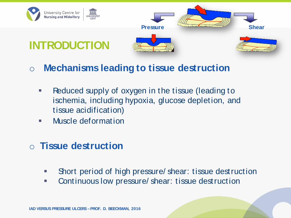
o Mechanisms leading to tissue destruction
Reduced supply of oxygen in the tissue (leading to ischemia, including hypoxia, glucose depletion, and tissue acidification)
Muscle deformation
o Tissue destruction Short period of high pressure/shear: tissue destruction Continuous low pressure/shear: tissue destruction
Pressure Shear
INTRODUCTION

IAD VERSUS PRESSURE ULCERS – PROF. D. BEECKMAN, 2016
INTRODUCTION

IAD VERSUS PRESSURE ULCERS – PROF. D. BEECKMAN, 2016
o New insights?
Reperfusion injury
INTRODUCTION

IAD VERSUS PRESSURE ULCERS – PROF. D. BEECKMAN, 2016
o New insights?
INTRODUCTION

IAD VERSUS PRESSURE ULCERS – PROF. D. BEECKMAN, 2016
o New insights?
o Microclimate Temperature on the skin surface or tissue temperature Air humidity or the humidity at the skin surface between
the body and the support surface Influence on the susceptibility of the skin/soft tissue for
the effects of pressure, shear or friction forces
INTRODUCTION
“Microclimate and its elements remain to be fully defined, and its relationship to pressure ulcer development clearly characterised. Evidence to date suggests that extremes of skin temperature and/or humidity/skin moisture appear to increase
the sensitivity of skin to the damaging effects of pressure, shear stresses and friction”
Clark M. et al. (2010).
Microclimate in context. In: Pressure ulcer prevention: pressure, shear, friction and microclimate in context – a consensus document. Wounds International.

IAD VERSUS PRESSURE ULCERS – PROF. D. BEECKMAN, 2016
o New insights?
o Impaired lymphatic drainage (accumulation of waste products)
o Tissue deformation / direct cell deformation
INTRODUCTION
The concepts involved in understanding pressure, shear, friction and microclimate and their
synergistic actions in the formation of pressure ulcers are complex and not yet fully understood

IAD VERSUS PRESSURE ULCERS – PROF. D. BEECKMAN, 2016
• Friction does not cause pressure ulcers
INTRODUCTION

IAD VERSUS PRESSURE ULCERS – PROF. D. BEECKMAN, 2016
T.Defloor - Verplegingswetenschap - Universiteit Gent
17
NONE of these photographs is a pressure ulcer!
1
2
3

IAD VERSUS PRESSURE ULCERS – PROF. D. BEECKMAN, 2016
Definition A reactive response of the skin to chronic exposure to urine and
faecal material which could be observed as an inflammation and erythema with or without erosion or denudation
INTRODUCTION

IAD VERSUS PRESSURE ULCERS – PROF. D. BEECKMAN, 2016
TERMINOLOGY
o The terminology used in international literature to describe perineal skin breakdown caused by incontinence, is divers.
o Terms that have been used for incontinence-associated dermatitis (IAD) are:
• Diaper/napkin/nappy dermatitis • Diaper/napkin/nappy rash • Irritant dermatitis • Skin maceration • Moisture lesions • Incontinence lesions • Perineal dermatitis • Perineal rash

IAD VERSUS PRESSURE ULCERS – PROF. D. BEECKMAN, 2016
TERMINOLOGY
o International Statistical Classification of Diseases and Related Health Problems (10th Revision Version for 2007) (ICD-10):
• Diseases of the skin and subcutaneous tissue (Chapter XII, L00-L99) in subcategory ‘Dermatitis and eczema’ (L20-L30)
• Contains codes for diaper dermatitis but does not contain separate codes for IAD
o IAD = part of a broader group of skin conditions, referred to as Moisture-Associated Skin Damage (MASD)
o US discussion: Incontinence associated skin damage (IASD) vs. Incontinence-
Associated Dermatitis (IAD): o Most observations of this type of damage are not confirmed by a
physician or nurse practitioner o Dermatitis = medical diagnosis and nurses are not allowed to chart it
in the health record

IAD VERSUS PRESSURE ULCERS – PROF. D. BEECKMAN, 2016
Size of the problem: Incidence
INTRODUCTION

IAD VERSUS PRESSURE ULCERS – PROF. D. BEECKMAN, 2016
Size of the problem: Survey (1911 caregivers) o 90.4% observed IAD in daily practice o Confusion about prevention
• IAD is not a pressure ulcer (73.5%) • … but should be prevented as a
pressure ulcer (61.5%)
INTRODUCTION

IAD VERSUS PRESSURE ULCERS – PROF. D. BEECKMAN, 2016
ETIOLOGY
o Incontinence: water is pulled into and held in the corneocytes
o Overhydration : swelling and disruption of the structure of the stratum corneum, and leads to visible changes in the skin
o Excessive hydration: irritants may more easily penetrate the stratum corneum to exacerbate inflammation
o Overhydrated skin: epidermis more prone to injury from friction

IAD VERSUS PRESSURE ULCERS – PROF. D. BEECKMAN, 2016
ETIOLOGY
o Exposure to urine and/or faeces: skin becomes more alkaline (skin bacteria convert the substance urea to ammonia which is alkaline)
o Increase in skin pH: micro-organisms to thrive and increase the risk of skin infection
o Faeces contain lipolytic (lipid-digesting) and proteolytic (protein-digesting) enzymes capable of damaging the stratum corneum

IAD VERSUS PRESSURE ULCERS – PROF. D. BEECKMAN, 2016
o Skin barrier
ETIOLOGY

IAD VERSUS PRESSURE ULCERS – PROF. D. BEECKMAN, 2016
RISK FACTORS
o Knowledge and awareness of risk factors is helpful to tailor IAD prevention and management
o IAD prevalence studies identified following key risk factors for IAD:
• Incontinence: liquid stool is most irritating, followed by double incontinence, fecal incontinence and urine incontinence
• Health status (critical illness, multimorbidity) • Fever • Diminished perfusion and oxygenation • Poor skin condition (e.g. steroid use/diabetes) • Restricted mobility and activity • Higher score on care dependency • Poor nutritional status • Risk of friction and shear • Restricted cognitive awareness

IAD VERSUS PRESSURE ULCERS – PROF. D. BEECKMAN, 2016
CLINICAL CHARACTERISTICS

IAD VERSUS PRESSURE ULCERS – PROF. D. BEECKMAN, 2016
CLINICAL CHARACTERISTICS

IAD VERSUS PRESSURE ULCERS – PROF. D. BEECKMAN, 2016
IAD Pressure ulcer
1. Cause Moisture (+ friction) Pressure/Shear
2. Location Peri- anal (anal cleft) Bony prominence
3. Shape Diffuse – Kissing ulcer 1 spot
4. Depth Superficial Superfical - deep
5. Necrosis - Possible
6. Edges Diffuse - irregular Distinct edges
7. Colour Redness is not equal Redness is equal
CLINICAL CHARACTERISTICS

IAD VERSUS PRESSURE ULCERS – PROF. D. BEECKMAN, 2016
o Observation Bright red erythema in
persons with light skin tones, more subtle red in persons with darker skin tones Skin fold or underneath diaper Poorly demarcated and
irregular borders Surface of skin may glisten
owing to serous exudates, hyperhydration or denudation
DIAGNOSIS: SKIN INSPECTION

IAD VERSUS PRESSURE ULCERS – PROF. D. BEECKMAN, 2016
o What in dark skin tones?
Inflammation not readily
apparent: often seen as areas of hyper- pigmentation or variable red tones
DIAGNOSIS: SKIN INSPECTION
IAD VERSUS PRESSURE ULCERS – PROF. D. BEECKMAN, 2016
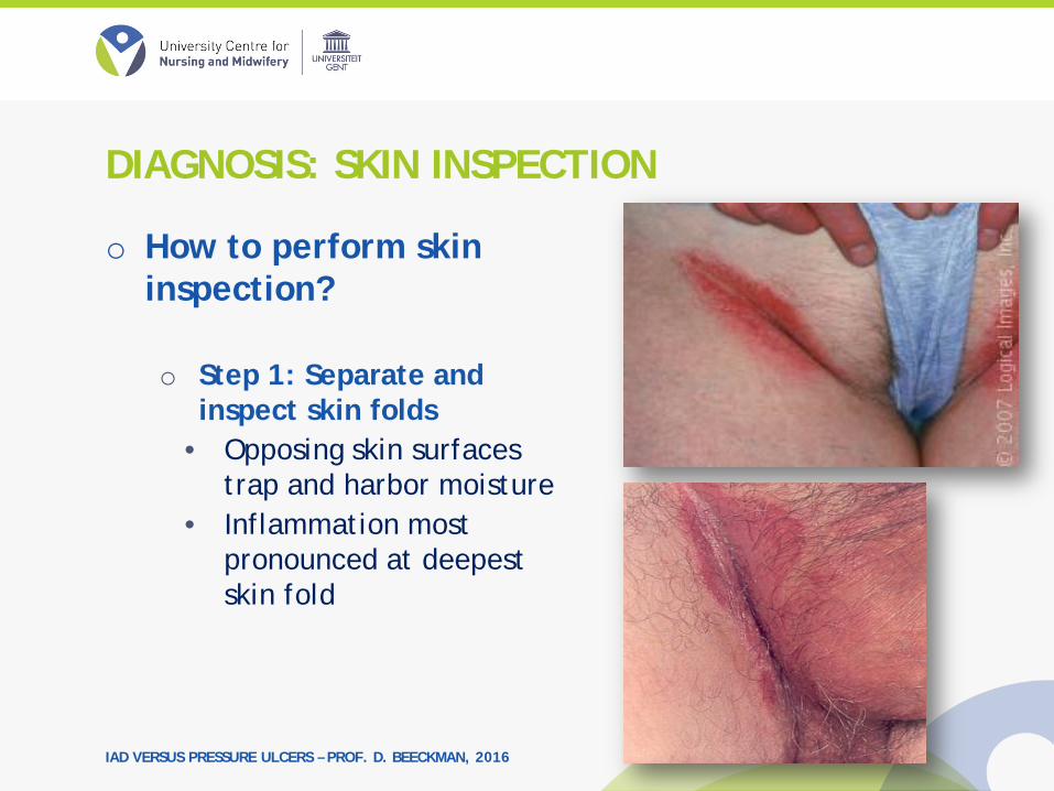
o How to perform skin inspection? o Step 1: Separate and
inspect skin folds • Opposing skin surfaces
trap and harbor moisture • Inflammation most
pronounced at deepest skin fold
DIAGNOSIS: SKIN INSPECTION

IAD VERSUS PRESSURE ULCERS – PROF. D. BEECKMAN, 2016
o How to perform skin inspection? o Step 1: Separate and
inspect skin folds • Opposing skin surfaces
trap and harbor moisture • Inflammation most
pronounced at deepest skin fold
DIAGNOSIS: SKIN INSPECTION

IAD VERSUS PRESSURE ULCERS – PROF. D. BEECKMAN, 2016
o How to perform skin inspection? o Step 2: Assess for skin erosion
• May present initially as “islands of partial thickness erosion”
• Multiple areas of erosion closely spaced • Entire dermis may be eroded in severe cases
DIAGNOSIS: SKIN INSPECTION

IAD VERSUS PRESSURE ULCERS – PROF. D. BEECKMAN, 2016
o How to perform skin inspection? o Step 2: Assess for skin erosion
DIAGNOSIS: SKIN INSPECTION

IAD VERSUS PRESSURE ULCERS – PROF. D. BEECKMAN, 2016
o How to perform skin inspection? o Step 3: Inspect for
secondary infection (such as Candida)
• Thrives in warm, moist environment and damages stratum corneum
• Prevalence of 18% in 976 acute care inpatients (Junkin & Selekof 2006)
DIAGNOSIS: SKIN INSPECTION

IAD VERSUS PRESSURE ULCERS – PROF. D. BEECKMAN, 2016
o How to perform skin inspection? o Step 3: Inspect for secondary infection (such as
Candida)
DIAGNOSIS: SKIN INSPECTION

IAD VERSUS PRESSURE ULCERS – PROF. D. BEECKMAN, 2016
IAD ASSESSMENT
o Existing tools are time-consuming and linguistic complex for use in routine clinical practice in nursing homes
o A simple classification tool, supported by photographs illustrating the severity categories is needed
o Development of the IAD Severity Categorisation Tool:
• To enhance correct identification and classification of IAD • To standardize record keeping • To provide a common description of IAD severity for the purpose of
clinical practice, audit and research

IAD VERSUS PRESSURE ULCERS – PROF. D. BEECKMAN, 2016
IAD ASSESSMENT
o IAD Severity Categorisation Tool:

IAD VERSUS PRESSURE ULCERS – PROF. D. BEECKMAN, 2016
The aim of this systematic review and meta-analysis was to identify the associations between IAD, moisture and incontinence as its most important etiologic factors, and pressure ulcer development. The following research questions were addressed:
1. What is the association between IAD and pressure ulcer development?
2. What is the association between incontinence and pressure ulcer development?
3. What is the association between moisture and pressure ulcer development?
IAD VS. PRESSURE ULCERS

IAD VERSUS PRESSURE ULCERS – PROF. D. BEECKMAN, 2016
IAD VERSUS PRESSURE ULCERS – PROF. D. BEECKMAN, 2016
Data sources o Five databases (Medline, Embase, CINAHL, Web of Science,
and the Cochrane Library) o Conference proceedings o End of study inclusion: March 15, 2013
Study Selection o Original studies with a quantitative design o Persons aged 18 years and older o Reporting an association between incontinence-associated
dermatitis, incontinence or moisture, and the development of PUs.
IAD VS. PRESSURE ULCERS

IAD VERSUS PRESSURE ULCERS – PROF. D. BEECKMAN, 2016
Data Extraction and Synthesis o Quality was assessed using the Quality Assessment Tool for
Quantitative Studies o Data from included studies were extracted and tabulated
using a standardized evidence table o A quality assurance check was independently performed on
10% of the included studies.
IAD VS. PRESSURE ULCERS

IAD VERSUS PRESSURE ULCERS – PROF. D. BEECKMAN, 2016
Results o Fifty-eight studies were included o Measures of relative effect at the univariate level were
meta-analyzed o In most studies (86%), a significant association between
variables of interest was found, with pooled odds ratios in univariate models varied between 1.92 (95% CI 1.54-2.38) for urinary incontinence and 4.99 (95%CI 2.62-9.50) for double incontinence (p<0.05)
IAD VS. PRESSURE ULCERS

IAD VERSUS PRESSURE ULCERS – PROF. D. BEECKMAN, 2016
Conclusion o Despite the methodological
variation in available studies and the heterogeneity of their results, our analysis indicates a likely association between IAD, its most important etiological factors, and the development of pressure ulcers
o Well-designed cohort studies are needed to determine a causal relationship between the variables
IAD VS. PRESSURE ULCERS

IAD VERSUS PRESSURE ULCERS – PROF. D. BEECKMAN, 2016
PREVENTION

IAD VERSUS PRESSURE ULCERS – PROF. D. BEECKMAN, 2016
PREVENTION

IAD VERSUS PRESSURE ULCERS – PROF. D. BEECKMAN, 2016
PREVENTION

IAD VERSUS PRESSURE ULCERS – PROF. D. BEECKMAN, 2016
o Prevention of IAD must include a consistent and well defined skin care regimen, including: Gentle perineal cleansing Moisturization The application of a skin protectant or moisture
barrier
o The use of absorptive or containment products and/or indwelling devices, might be needed in specific situations to support prevention of IAD
PREVENTION

IAD VERSUS PRESSURE ULCERS – PROF. D. BEECKMAN, 2016
o The use of absorptive or containment products and/or indwelling devices, might be needed in specific situations to support prevention of IAD
PREVENTION

IAD VERSUS PRESSURE ULCERS – PROF. D. BEECKMAN, 2016
o Gentle perineal cleansing o Should involve a product whose pH range reflects the
acid mantle of healthy skin (pH between 5.4-5.9) o The pH of normal soap is alkaline and in the range of
9.5–11.0 o Increase of stratum corneum swelling o Alteration in lipid rigidity o Many no-rinse skin cleansers are “pH balanced” in order
to ensure that their pH is closer to that of healthy skin.
PREVENTION

IAD VERSUS PRESSURE ULCERS – PROF. D. BEECKMAN, 2016
o Gentle perineal cleansing
PREVENTION

IAD VERSUS PRESSURE ULCERS – PROF. D. BEECKMAN, 2016
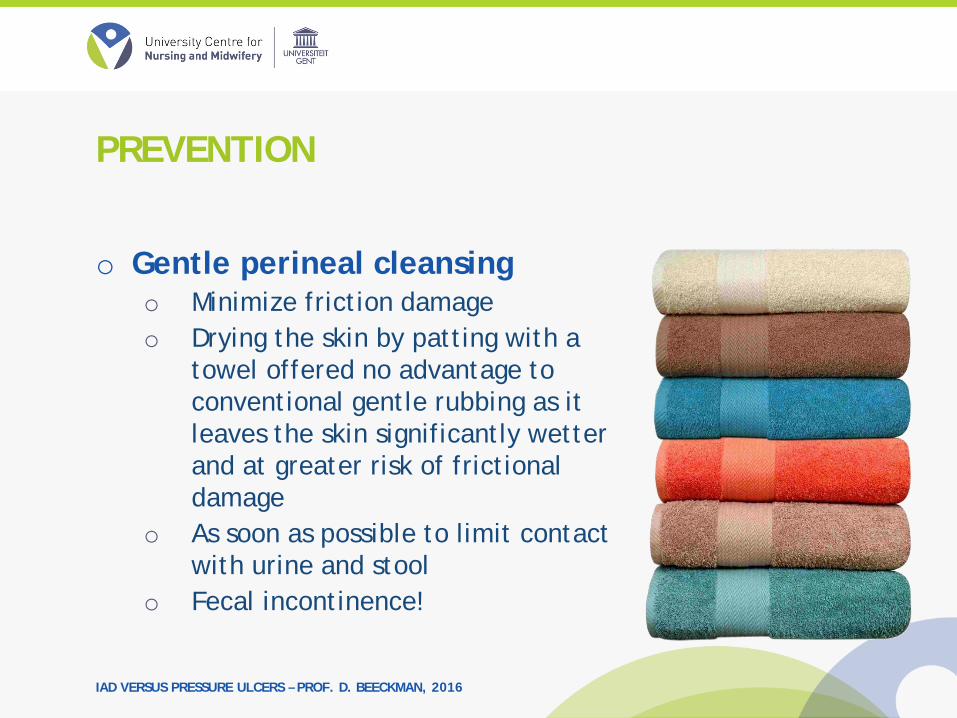
o Gentle perineal cleansing o Gentle cleansing is preferred over scrubbing techniques and
a soft cloth is recommended to minimize friction damage o Drying the skin by patting with a towel offered no
advantage to conventional gentle rubbing as it leaves the skin significantly wetter and at greater risk of frictional damage
o As soon as possible to limit contact with urine and stool o It is particularly important to provide timely cleansing
following an episode of fecal incontinence
PREVENTION
IAD VERSUS PRESSURE ULCERS – PROF. D. BEECKMAN, 2016
o Gentle perineal cleansing o Minimize friction damage o Drying the skin by patting with a
towel offered no advantage to conventional gentle rubbing as it leaves the skin significantly wetter and at greater risk of frictional damage
o As soon as possible to limit contact with urine and stool
o Fecal incontinence!
PREVENTION

IAD VERSUS PRESSURE ULCERS – PROF. D. BEECKMAN, 2016
o Moisturization / skin conditioning o Barrier function = intercellular lipids + intact
keratinocytes o Loss of water at the stratum corneum (TEWL) o Moisturization / skin conditioning involves repairing the
skin barrier o Moisturizers contain varying combinations of emollients,
occlusives, and humectants o The routine use of moisturizers is useful in replacing
intercellular lipids and maintaining the barrier function of the skin
PREVENTION

IAD VERSUS PRESSURE ULCERS – PROF. D. BEECKMAN, 2016
o Skin protecting o To primarily prevent skin
breakdown due to moisture and biological irritants in urine and faeces
o A wide variety of products and formulas with both moisturizing and/or protecting/barrier capability.
o Must allow skin observation!
PREVENTION

IAD VERSUS PRESSURE ULCERS – PROF. D. BEECKMAN, 2016
o Skin protecting o Commercially available skin protectants vary in their
ability to protect the skin from irritants, prevent maceration, and maintain skin health:
• Petrolatum: protection against irritants and maceration and provided some skin hydration
• Dimethicone: varied in protection against irritants and have good skin hydration potential and low barrier efficacy
• Zinc oxide-based products: Good protection against irritants but poor skin hydration to prevent maceration
• Acrylate terpolymer based products: Protection against irritants but no skin hydration
PREVENTION

IAD VERSUS PRESSURE ULCERS – PROF. D. BEECKMAN, 2016
o Treatment of IAD must include a consistent and well defined skin care regimen, including: Gentle perineal cleansing Moisturization The application of a skin protectant or moisture
barrier
o The use of absorptive or containment products and/or indwelling devices, might be needed in specific situations to support treatment of IAD
TREATMENT

IAD VERSUS PRESSURE ULCERS – PROF. D. BEECKMAN, 2016
o The addition of antifungal products, steroidal based topical anti-inflammatory products, and topical antibiotics to treat IAD is only recommended in specific situations.
o Referral to a continence specialist if needed! o Patients who do not respond to treatment within
two weeks should be referred for additional evaluation
TREATMENT

IAD VERSUS PRESSURE ULCERS – PROF. D. BEECKMAN, 2016
Moisture
Urine Faeces Urine/faeces Washing
Ureum/amonium pH
Bacterial load
Enzym. activity pH
Bacterial load
Enzym. activity Ureum/amonium
pH Bacterial load
Chemical irritation
Physical irritation
+
Skin permeability pH
Bacterial growth Cutaneous infections
Friction IAD + Weakened skin
Incontinence Management
Moisturizing Protecting
Gentle perineal cleansing
HEALTHY SKIN
SUMMARY

IAD VERSUS PRESSURE ULCERS – PROF. D. BEECKMAN, 2016
CONCLUSION
o Incontinence = risk factor for pressure ulcers, but IAD can occur in the absence of any other pressure ulcer-associated risk factors and vice versa
o The presence of any urinary and/or faecal incontinence, even in the absence of other risk factors, should trigger implementation of an appropriate IAD prevention protocol
o Skin care should be an essential element in each pressure ulcer prevention protocol

IAD VERSUS PRESSURE ULCERS – PROF. D. BEECKMAN, 2016
Gray M, Beeckman D, Bliss DZ, Fader M, Logan S, Junkin J, Selekof J, Doughty D, Kurz P. Incontinence-associated dermatitis: a comprehensive review and update. J Wound Ostomy Continence Nurs. 2012 Jan-Feb;39(1):61-74. Doughty D, Junkin J, Kurz P, Selekof J, Gray M, Fader M, Bliss DZ, Beeckman D, Logan S. Incontinence-associated dermatitis: consensus statements, evidence-based guidelines for prevention and treatment, and current challenges. J Wound Ostomy Continence Nurs. 2012 May-Jun;39(3):303-15; quiz 316-7. Draelos ZD. Active agents in common skin care products. Plast Reconstr Surg. 2010 Feb;125(2):719-24. Short RW, Chan JL, Choi JM, Egbert BM, Rehmus WE, Kimball AB. Effects of moisturization on epidermal homeostasis and differentiation. Clin Exp Dermatol. 2007 Jan;32(1):88-90.
REFERENCES

IAD VERSUS PRESSURE ULCERS – PROF. D. BEECKMAN, 2016
Nolan K, Marmur E. Moisturizers: reality and the skin benefits. Dermatol Ther. 2012 May-Jun;25(3):229-33. Draelos ZD. New treatments for restoring impaired epidermal barrier permeability: skin barrier repair creams. Clin Dermatol. 2012 May-Jun;30(3):345-8. Beeckman D., Schoonhoven L., Verhaeghe S., Heyneman A., Defloor T. (2009). Prevention and treatment of incontinence associated dermatitis: a review of the literature. Journal of Advanced Nursing, 65(6)1141-1154. Beeckman D., Schoonhoven L., Fletcher J., Furtado K., Heyman H., Paquay L., De Bacquer D., Defloor T. (2010) Pressure ulcers and incontinence-associated dermatitis: effectiveness of the Pressure Ulcer CLASsification education tool on classification by nurses. BMJ Quality & Safety, 19(5):e3.
REFERENCES
IAD VERSUS PRESSURE ULCERS – PROF. D. BEECKMAN, 2016
Beeckman D, Woodward S, Gray M. Incontinence-associated dermatitis: step-by step prevention and treatment. Br J Community Nurs. 2011a Aug;16(8):382-9. Beeckman D, Woodward S, Rajpaul K, Vanderwee K. Clinical challenges of preventing incontinence-associated dermatitis. Br J Nurs. 2011b Jul 15-28;20(13):784-6,788,790. Bliss DZ, Zehrer C, Savik K, Smith G, Hedblom E. An economic evaluation of four skin damage prevention regimens in nursing home residents with incontinence: economics of skin damage prevention. J Wound Ostomy Continence Nurs. 2007 Mar-Apr;34(2):143-52; discussion 152.
Beeckman D, Van Lancker A, Van Hecke A, Verhaeghe S (2014) A Systematic Review and Meta- Analysis of Incontinence- Associated Dermatitis, Incontinence, and Moisture as Risk Factors for Pressure Ulcer Development. Res Nurs Health, In Press.
REFERENCES