presentation1.pptx, ultrasound examination of the liver and gall bladder
TRANSCRIPT
Dr ABD ALLAH NAZEER MD
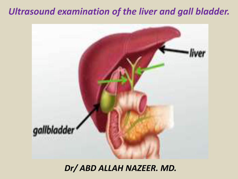
Ultrasound examination of the liver and gall bladder
Ultrasound of the Liver -ProtocolRole of Ultrasound
To assess theSizeCapsular contour (smooth coarse lobulated)Parenchymal echogenicityVascularityBiliary treeMasses or collections
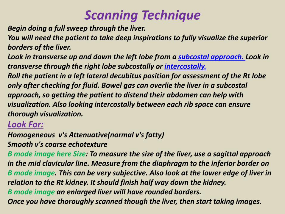
Scanning TechniqueBegin doing a full sweep through the liverYou will need the patient to take deep inspirations to fully visualize the superior borders of the liverLook in transverse up and down the left lobe from a subcostal approach Look in transverse through the right lobe subcostally or intercostallyRoll the patient in a left lateral decubitus position for assessment of the Rt lobe only after checking for fluid Bowel gas can overlie the liver in a subcostal approach so getting the patient to distend their abdomen can help with visualization Also looking intercostally between each rib space can ensure thorough visualization
Look ForHomogeneous vs Attenuative(normal vs fatty)Smooth vs coarse echotextureB mode image here Size To measure the size of the liver use a sagittal approach in the mid clavicular line Measure from the diaphragm to the inferior border on B mode image This can be very subjective Also look at the lower edge of liver in relation to the Rt kidney It should finish half way down the kidney B mode image an enlarged liver will have rounded bordersOnce you have thoroughly scanned though the liver then start taking images
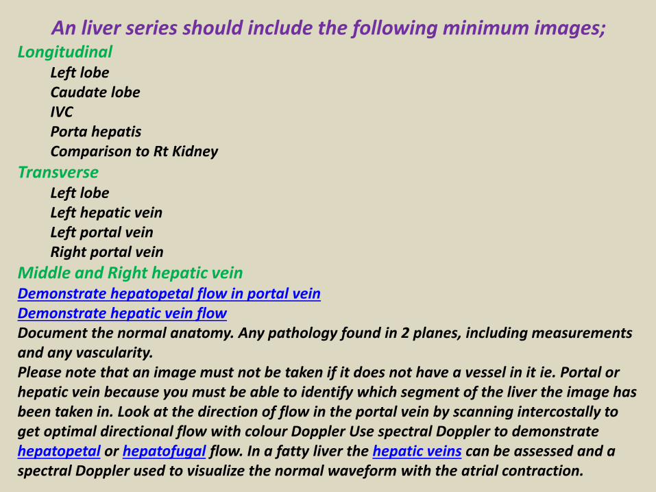
An liver series should include the following minimum imagesLongitudinal
Left lobeCaudate lobeIVCPorta hepatisComparison to Rt Kidney
TransverseLeft lobeLeft hepatic veinLeft portal veinRight portal vein
Middle and Right hepatic veinDemonstrate hepatopetal flow in portal veinDemonstrate hepatic vein flowDocument the normal anatomy Any pathology found in 2 planes including measurements and any vascularityPlease note that an image must not be taken if it does not have a vessel in it ie Portal or hepatic vein because you must be able to identify which segment of the liver the image has been taken in Look at the direction of flow in the portal vein by scanning intercostally to get optimal directional flow with colour Doppler Use spectral Doppler to demonstrate hepatopetal or hepatofugal flow In a fatty liver the hepatic veins can be assessed and a spectral Doppler used to visualize the normal waveform with the atrial contraction
Role of UltrasoundTo assess theSizeCapsular contour (smooth coarse lobulated)Parenchymal echogenicityVascularityBiliary treeMasses or collections
LimitationsObesity and patients with severe cases of metabolic disorders such as haemochromatosis and fatty infiltration will reduce detail and the diagnostic yield of the scan
PreparationIdeally fast the patient for 6hours to reduce bowel gas and prevent gall bladder contraction
Equipment SelectionDepending on the size of the patient a curved linear array 2-6MhzIf there is nodularity of the liver border then a linear array with a 7-12MHZ frequency will better appreciate this Good colour power Doppler capabilities when assessing vessels or vascularity of a structureBe prepared to change focal zone position and frequency output of probe (or probes) to adequately assess both superficial and deeper structures
Parasagittal Scan PlaneThe Liver and Rt Kidney are
visualized in this view
Intercostal Scan PlaneThe Middle and Rt Hepatic Vein
are visualized in this view
Subcostal Scan Plane The probe is angled cephalad under the ribs to avoid any bowel or ribs shadowing over the liver
Rt Portal Vein is shown coursing transversely in this view
Scan Plane Left Lobe of Liver The probe is in the epigastric region just below the sternum It is angled cephalad to view the left lobe in its entirety The probe may need to be angled towards the left side to see the most medial edge of the left lobe
Normal Anatomy seen in the Transverse View of the Left Lobe
The Portal Vein should have constant forward flow into the liver (hepatopetal flow) As seen in this image the colour is red which is set for movement towards the probe If there is flow reversalthis is hepatofugal (tip Fugitive= run away) and represents portal hypertension
Because the hepatic veins drain into the IVC immediately prior to the Right Atrium they have phasic flow reflective of cardiac motion
Normal Liver MeasurementsmdashWe need to be able to determine such conditions as hepatomegally mdashIt needs to be a consistent measurement to be able to compare sizes over timemdashThe calipers need to be positioned in the same position between sonographers for accuracy Ideally the same sonographer should be used in a follow up assessmentmdashThe upper border lies in the right mid-clavicular line at the 5th intercostal space mdashMost people have the lower border extending to the lower costal marginmdashIf it is measured in the mid-hepatic line with a large field of view it should measure lt16cm from the posterior diaphragm to the lower anterior edge However organ size increases with gender age height weight and body surface areamdashIf the measurement is made from the anterior diaphragm to the lower edge of the liver in the mid-clavicular line it should be no gt13cmBe careful not to get confused with a riedelrsquos lobe as it can increase the measurement
MIDCLAVICULARMeasured in the mid-hepatic line with a large field of view it should measure lt16cm from the post diaphragm to the lower anterior edge
MIDHEPATIC
If the measurement is made from the ant diaphragm to the lower edge of the liver in the mid-clavicular line it should be no gt13cm
Common PathologyFatty liverCirrhosisLiver cystsHaemangiomaPortal hypertensionPortal vein thrombosisHepatic vein thrombosisLiver abscesscollectionTraumaMetastasesHCCAbscess
Fatty liver
Fatty liver
Cirrhotic liver
Cirrhotic liver
Bilharzial liver with peri-portal fibrosis
Bilharzial liver with peri-portal fibrosis
A) Sonography of hepatic cysts
Ultrasound images reveal an anechoic lesion in the right lobe of the liver with thin walls and clear fluid content A small projection is seen from the wall This may be thickening of the wall or a fine nodule These findings
suggest a liver cyst Differential diagnosis biliary cystadenoma
Ultrasound Typical liver cyst
Color and Power Doppler images of the hepatic cyst showed no flow in the liver cyst However the spectral Doppler trace showed faint pulsations within this lesion These pulsations appear to be transmitted from the abdominal aorta and appear insignificant Thus we arrived at a diagnosis of a simple cyst of the left lobe of the liver based on these ultrasound and Color Doppler images Absence of flow in this cyst ruled out possibilities like a vascular lesion like aneurysm of the portal vein or AVM The liver also shows moderate fatty changes
Hydatid cyst with multiple hypoechoic internal daughter cysts
Compound picture of four images showing several typical hemangiomas
Hemangioma of liver (hepatic hemangioma)
Images show a large (8 cms) rounded well defined hyperechoic non-calcific mass in the right lobe of liver There is a moderate amount of acoustic enhancement posterior to the lesion These ultrasound images show some of the typical features of hemangioma (cavernous) of the liver In this case the patient is an adult female which is typical of hepatic hemangioma However usually hemangioma are smaller in size (less than 4 cms)
Multiple Hemangioma of the Liver
Multiple hemangioma of the liver the lesions are small in size Such lesions are rather atypical of hemangioma
Hemangioma of the caudate lobe of liver
Large echogenic hemangioma in the caudate lobe of liver
Subcostal scan through the right liver showing a large hemangioma with central hypoechoic area due to thrombosis
Hepatic abscess or abscess of Liver (Amebic liver abscess)
Sonography of the liver was done in this case of pain in the right iliac fossa Ultrasound image on left shows thickened edematous wall of the cecum so typhlitis Image on right shows 2 hypoechoic lesions in the right lobe of liver Both lesions are fairly large and show in homogenous echotexture In view of the underlying disease in the cecum a diagnosis of typhlitis with amebic liver (hepatic) abscess (developing stage) was made
Ruptured liver abscess (rupture or leak of hepatic abscess)
Ultrasound images show a large abscess of the right lobe of liver with rupture of the lower margin of the abscess The leaked fluid is contained within localized adhesions in the peritoneal cavity
Abdominal CT with iv contrast administration shows a large well defined hypodense liver mass B)Abdominal ultrasound show a heterogeneous hyperechoic mass with a hypoechoic area inside consistent with liver abscess
Hyperechoic lesion in the right liver with central calcification proven to be an adenoma
Large heterogeneous lesion in the left liver adenoma with recent bleeding
Irregularly demarcated cirrhotic liver with small hypoechoic HCC A Ascites Hyperechoic HCC
Diffuse metastatic liver involvementTypical metastases with bulls eye aspect
Liver metastasis with bulls eye aspect protruding at the liver surface
Hypoechoic liver metastases
Calcified liver metastases
Blood vessels disorder of the liverThe portal vein provides about two thirds of the blood This blood contains oxygen and many nutrients brought to the liver from the intestines for processing The hepatic artery provides the remaining one third of blood This oxygen-rich blood comes from the heart and provides the liver with about half of its oxygen supply Receiving blood from two blood vessels helps protect the liver If one of these blood vessels is damaged the liver can often continue to function because it receives oxygen and nutrients from the other blood supply Blood leaves the liver through the hepatic veins This blood is a mixture of blood from the hepatic artery and from the portal vein The hepatic veins carry blood to the inferior vena cavamdashthe largest vein in the bodymdashwhich then carries blood from the abdomen and lower parts of the body to the right side of the heartBlood vessel (vascular) disorders of the liver usually result from inadequate blood flowmdashwhether into or out of the liver If the problem is blood flow out of the liver blood backs up in the liver causing congestion In either case liver cells do not receive enough blood (called ischemia) and thus are deprived of oxygen and nutrients Inadequate blood flowmdashinto or out of the livermdashmay result from heart failure or disorders that make blood more likely to clot (clotting disorders) In clotting disorders a clot may block the portal vein or a hepatic vein slowing or blocking blood flow
Normal vascular structures of the liver
Portal hypertension
Portal hypertension
Cavernous transformation of the portal vein
Portal vein thrombosis
Acute thrombosis of the portal vein with echogenic thrombus inside
Partial thrombosis of the portal vein
Ultrasound of liver graft in 56-year-old man with hepatic artery thrombosis Color Doppler ultrasound shows absence of flow in hepatic artery at hilus (arrow)
Ultrasound of liver graft in 56-year-old man with hepatic artery thrombosis Contrast-enhanced ultrasound reveals no arterial
perfusion in early phase at hilus level (arrow) nor at intrahepatic level
Hepatic artery stenosis in 39-year-old man Doppler ultrasound of hepatic artery at intrahepatic level shows prolonged acceleration time of 0153 second
Reversed flow in the hepatic vein due to obstruction
Reversed flow in the hepatic vein due to obstruction
Right hepatic vein thrombosis
Ultrasound of the Gall bladder
Fundus
Body
Normal US ofGall Bladder
Ultrasound of the Gallbladder ndash ProtocolPatient PreparationFast for 6 hours No food or drinkPreferably book the appointment in the morning to reduce bowel gas
Scanning TechniqueLooking supine left lateral decubitus and erectUse the liver as a window especially when rolling the pt onto their left sideMeasure the wall lt3mmIs the gallbladder enlargedgt10cm in lengthCheck with colour Doppler for increased vascularity of the wallAssess the cystic duct neck body and fundus (sometimes there is a phrygian cap)
Normal Scanning Position to take advantage of using the liver as a window and displacing the bowel
A normal Gallbladder should be thin walled (lt3mm) and anechoic It is a pear shaped saccular structure for bile storage in the Right Upper Quadrant Its size varies depending on the amount of bile Fasted it will be approximately 10cm long
Folds are commonly seen and are normal Make note if pathology such as calculi are contained within a
compartment created by a fold
A Phrygian cap is a specific relatively common inversion of the distal fundus of the gallbladder into the body It may become adherent It is an anatomic variant or acquired abnormality
Ultrasonography with the typical technique and the probe placed at the right hypochondrium could not locate the gallbladder at the plane of the liver hilum
Congenital agenesis of the gall bladder
Congenital agenesis of the gall bladder
Multiple gall bladder multiple septations forming a honeycomb appearance
Duplicated gallbladder at US MRI and cholangiography with double cystic ducts
Gall bladder stones and sludgeUS criteria for diagnosis of cholelithiasis1- Echogenic focus2- Cast acoustic shadow3- Seek gravitational dependence
Wall-echo-shadow sign with multiple stonesMultiple stones with confluent shadowsSingle stone with clean shadow
Acute cholecystitis is swelling (inflammation) of the gallbladder It is
a potentially serious condition that usually needs to be treated in hospitalAcute cholecystitis is either calcular(95) or non calcular (5) The main symptom of acute cholecystitis is a sudden sharp pain in the upper right side of your tummy (abdomen) that spreads towards your right shoulderThe affected part of the abdomen is usually extremely tender and breathing deeply can make the pain worseUnlike some others types of abdominal pain the pain associated with acute cholecystitis is usually persistent and doesnt go away within a few hoursSome people may additional symptoms such asa high temperature (fever) nausea and vomiting sweating loss of appetite yellowing of the skin and the whites of the eyes (jaundice) a bulge in the abdomen
US- GB wall in acute cholecystitis appears as a thin hypoechoic line sandwiched between two echogenic lines
CT- thick wall with hypodense outer layer (subserosal edema)
Acute calculus cholecystitis
Acute acalculous cholecystitis LEFT US at the spot of maximum tenderness shows mural thickening of the gallbladder (arrow) that is completely filled with sludge (asterix) without any stones RIGHT Power-Doppler sonography shows hypervascularity of the gallbladder wall (arrowhead) as a supporting sign of inflammation
Acute gangrenous cholecystitis
Xanthogranulomatous cholecystitisIntramural hypoechoichypoattenuating foci of abscess and inflammation
Emphysematous cholecystitis (A) and (B) TS and LS with gas and debris in thegallbladder lumen (C) Gas in the gallbladder lumen completely obscures the content
Emphysematous cholecystitis
CT scan showing markedly enlarged gall bladder with intraluminal blood and thrombus Hemorrhagic cholecystitis
Hemorrhagic cholecystitis with blood within the bladder lumen
Chronic cholecystitis Chronic cholecystitis is gallbladder inflammation that has lasted a long time It almost always results from gallstones and from prior attacks of acute cholecystitis Chronic cholecystitis is characterized by repeated attacks of pain (biliary colic) that occur when gallstones periodically block the cystic ductIn chronic cholecystitis the gallbladder is damaged by repeated attacks of acute inflammation usually due to gallstones and may become thick-walled scarred and small The gallstones may block the opening of the gallbladder into the cystic duct or block the cystic duct itself The gallbladder usually also contains sludge If scarring is extensive calcium may be deposited in the walls of the gallbladder causing them to harden (called porcelain gallbladder)
SymptomsA gallbladder attack whether in acute or chronic cholecystitis begins as pain
Chronic calcular cholecystitis
Chronic calcular cholecystitis
Distended gall bladder containing echogenic gravity dependent mobile biliary sludge with calculi
Porcelain GBRare due to chronic inflammation mural calcification
Biliary sludge is a mixture of particulate solids that have precipitated from bile Such sediment consists of cholesterol crystals calcium bilirubinate pigment and other calcium salts Sludge is usually detected on transabdominal ultrasonography Microscopy of aspirated bile and endoscopic ultrasonography are far more sensitive Biliary sludge is associated with pregnancy with rapid weight loss particularly in the obese with critical illness involving low or absent oral intake and the use of total parenteral nutrition (TPN) Complications caused by biliary sludge include biliary colic acute cholangitis and acute pancreatitis Asymptomatic patients with sludge or microlithiasis require no therapy
Biliary sludge
Biliary sludge
Gallbladder polyps are relatively frequent seen in up to 5 of the
population Over 90 are benign and the majority are cholesterol polypsCholesterol polyps are most frequently identified in patients between 40-50 years of age and are more common in women (FM 291)
Tiny polyp of the gall bladder wall
Multiple gall bladder polyps
Large gall bladder polyp
Adenomyomatosis of the gallbladder is a hyperplastic
cholestosis of the gallbladder wall It is a relatively common and benign cause of focal gallbladder wall thickening It is most easily seen on ultrasound and MRI Adenomyomatosis is relatively common found in ~5 of all cholecystectomy specimens It is typically seen in patients in their 5th decade The incidence increases with age which may be the result of protracted inflammation (see below) There is a female predilection (MF = 13) It is most often an incidental finding has no intrinsic malignant potential and usually requires no treatment Three morphological types of adenomyomatosis are describedfundal (localized)segmental (annular)generalized (diffuse)
Segmental adenomyomatosis of gall bladder
Diffuse adenomyomatosis of gall bladder
Adenomyomatosis of the gallbladder with a thickened wall Color Dopplershows twinkling artefacts caused by cholesterol crystals in the wall
Adenomyomatosis of the gallbladder with a thickened wall Color Doppler shows twinkling artefacts caused by cholesterol crystals in the wall
Gallbladder adenocarcinomas is the most frequent and are usually asymptomatic until it is no longer curable As such early incidental detection is important if the occasional patient is to be successfully treated Less frequent malignancies is squamous cell carcinoma Small cell carcinoma Carcinosarcoma Lymphoma and metastasisEpidemiologyAlthough overall uncommon gallbladder adenocarcinoma is the most common primary hepatobiliary carcinoma and the 5th most common malignancy of the gastrointestinal tract Predominantly affects older persons with long-standing cholecystolithiasis and as such is most common in elderly women (gt 60 years of age FM ratio = 41) Risk factors include chronic cholecystitis gallstones are seen in 70-90 of casesfamilial adenomatous polyposis syndrome (FAP)inflammatory bowel disease (IBD)porcelain gallbladder
US of Gall bladder carcinoma
Gallbladder adenocarcinoma
US shows echogenic mass adherent to gallbladder fundus (arrows) CT shows enhancing mass within the gallbladder (arrows) with involvement of the liver (arrowhead)
Gallbladder carcinoma
B-cell lymphoma of the gallbladder
50-year-old man with renal-cell carcinoma Intraoperative ultrasound image of polypoid gallbladder lesion Postcontrast axial CT image demonstrates
avidly contrast-enhancing polypoid gallbladder lesion (arrow)
Thank You

Ultrasound of the Liver -ProtocolRole of Ultrasound
To assess theSizeCapsular contour (smooth coarse lobulated)Parenchymal echogenicityVascularityBiliary treeMasses or collections
Scanning TechniqueBegin doing a full sweep through the liverYou will need the patient to take deep inspirations to fully visualize the superior borders of the liverLook in transverse up and down the left lobe from a subcostal approach Look in transverse through the right lobe subcostally or intercostallyRoll the patient in a left lateral decubitus position for assessment of the Rt lobe only after checking for fluid Bowel gas can overlie the liver in a subcostal approach so getting the patient to distend their abdomen can help with visualization Also looking intercostally between each rib space can ensure thorough visualization
Look ForHomogeneous vs Attenuative(normal vs fatty)Smooth vs coarse echotextureB mode image here Size To measure the size of the liver use a sagittal approach in the mid clavicular line Measure from the diaphragm to the inferior border on B mode image This can be very subjective Also look at the lower edge of liver in relation to the Rt kidney It should finish half way down the kidney B mode image an enlarged liver will have rounded bordersOnce you have thoroughly scanned though the liver then start taking images
An liver series should include the following minimum imagesLongitudinal
Left lobeCaudate lobeIVCPorta hepatisComparison to Rt Kidney
TransverseLeft lobeLeft hepatic veinLeft portal veinRight portal vein
Middle and Right hepatic veinDemonstrate hepatopetal flow in portal veinDemonstrate hepatic vein flowDocument the normal anatomy Any pathology found in 2 planes including measurements and any vascularityPlease note that an image must not be taken if it does not have a vessel in it ie Portal or hepatic vein because you must be able to identify which segment of the liver the image has been taken in Look at the direction of flow in the portal vein by scanning intercostally to get optimal directional flow with colour Doppler Use spectral Doppler to demonstrate hepatopetal or hepatofugal flow In a fatty liver the hepatic veins can be assessed and a spectral Doppler used to visualize the normal waveform with the atrial contraction
Role of UltrasoundTo assess theSizeCapsular contour (smooth coarse lobulated)Parenchymal echogenicityVascularityBiliary treeMasses or collections
LimitationsObesity and patients with severe cases of metabolic disorders such as haemochromatosis and fatty infiltration will reduce detail and the diagnostic yield of the scan
PreparationIdeally fast the patient for 6hours to reduce bowel gas and prevent gall bladder contraction
Equipment SelectionDepending on the size of the patient a curved linear array 2-6MhzIf there is nodularity of the liver border then a linear array with a 7-12MHZ frequency will better appreciate this Good colour power Doppler capabilities when assessing vessels or vascularity of a structureBe prepared to change focal zone position and frequency output of probe (or probes) to adequately assess both superficial and deeper structures
Parasagittal Scan PlaneThe Liver and Rt Kidney are
visualized in this view
Intercostal Scan PlaneThe Middle and Rt Hepatic Vein
are visualized in this view
Subcostal Scan Plane The probe is angled cephalad under the ribs to avoid any bowel or ribs shadowing over the liver
Rt Portal Vein is shown coursing transversely in this view
Scan Plane Left Lobe of Liver The probe is in the epigastric region just below the sternum It is angled cephalad to view the left lobe in its entirety The probe may need to be angled towards the left side to see the most medial edge of the left lobe
Normal Anatomy seen in the Transverse View of the Left Lobe
The Portal Vein should have constant forward flow into the liver (hepatopetal flow) As seen in this image the colour is red which is set for movement towards the probe If there is flow reversalthis is hepatofugal (tip Fugitive= run away) and represents portal hypertension
Because the hepatic veins drain into the IVC immediately prior to the Right Atrium they have phasic flow reflective of cardiac motion
Normal Liver MeasurementsmdashWe need to be able to determine such conditions as hepatomegally mdashIt needs to be a consistent measurement to be able to compare sizes over timemdashThe calipers need to be positioned in the same position between sonographers for accuracy Ideally the same sonographer should be used in a follow up assessmentmdashThe upper border lies in the right mid-clavicular line at the 5th intercostal space mdashMost people have the lower border extending to the lower costal marginmdashIf it is measured in the mid-hepatic line with a large field of view it should measure lt16cm from the posterior diaphragm to the lower anterior edge However organ size increases with gender age height weight and body surface areamdashIf the measurement is made from the anterior diaphragm to the lower edge of the liver in the mid-clavicular line it should be no gt13cmBe careful not to get confused with a riedelrsquos lobe as it can increase the measurement
MIDCLAVICULARMeasured in the mid-hepatic line with a large field of view it should measure lt16cm from the post diaphragm to the lower anterior edge
MIDHEPATIC
If the measurement is made from the ant diaphragm to the lower edge of the liver in the mid-clavicular line it should be no gt13cm
Common PathologyFatty liverCirrhosisLiver cystsHaemangiomaPortal hypertensionPortal vein thrombosisHepatic vein thrombosisLiver abscesscollectionTraumaMetastasesHCCAbscess
Fatty liver
Fatty liver
Cirrhotic liver
Cirrhotic liver
Bilharzial liver with peri-portal fibrosis
Bilharzial liver with peri-portal fibrosis
A) Sonography of hepatic cysts
Ultrasound images reveal an anechoic lesion in the right lobe of the liver with thin walls and clear fluid content A small projection is seen from the wall This may be thickening of the wall or a fine nodule These findings
suggest a liver cyst Differential diagnosis biliary cystadenoma
Ultrasound Typical liver cyst
Color and Power Doppler images of the hepatic cyst showed no flow in the liver cyst However the spectral Doppler trace showed faint pulsations within this lesion These pulsations appear to be transmitted from the abdominal aorta and appear insignificant Thus we arrived at a diagnosis of a simple cyst of the left lobe of the liver based on these ultrasound and Color Doppler images Absence of flow in this cyst ruled out possibilities like a vascular lesion like aneurysm of the portal vein or AVM The liver also shows moderate fatty changes
Hydatid cyst with multiple hypoechoic internal daughter cysts
Compound picture of four images showing several typical hemangiomas
Hemangioma of liver (hepatic hemangioma)
Images show a large (8 cms) rounded well defined hyperechoic non-calcific mass in the right lobe of liver There is a moderate amount of acoustic enhancement posterior to the lesion These ultrasound images show some of the typical features of hemangioma (cavernous) of the liver In this case the patient is an adult female which is typical of hepatic hemangioma However usually hemangioma are smaller in size (less than 4 cms)
Multiple Hemangioma of the Liver
Multiple hemangioma of the liver the lesions are small in size Such lesions are rather atypical of hemangioma
Hemangioma of the caudate lobe of liver
Large echogenic hemangioma in the caudate lobe of liver
Subcostal scan through the right liver showing a large hemangioma with central hypoechoic area due to thrombosis
Hepatic abscess or abscess of Liver (Amebic liver abscess)
Sonography of the liver was done in this case of pain in the right iliac fossa Ultrasound image on left shows thickened edematous wall of the cecum so typhlitis Image on right shows 2 hypoechoic lesions in the right lobe of liver Both lesions are fairly large and show in homogenous echotexture In view of the underlying disease in the cecum a diagnosis of typhlitis with amebic liver (hepatic) abscess (developing stage) was made
Ruptured liver abscess (rupture or leak of hepatic abscess)
Ultrasound images show a large abscess of the right lobe of liver with rupture of the lower margin of the abscess The leaked fluid is contained within localized adhesions in the peritoneal cavity
Abdominal CT with iv contrast administration shows a large well defined hypodense liver mass B)Abdominal ultrasound show a heterogeneous hyperechoic mass with a hypoechoic area inside consistent with liver abscess
Hyperechoic lesion in the right liver with central calcification proven to be an adenoma
Large heterogeneous lesion in the left liver adenoma with recent bleeding
Irregularly demarcated cirrhotic liver with small hypoechoic HCC A Ascites Hyperechoic HCC
Diffuse metastatic liver involvementTypical metastases with bulls eye aspect
Liver metastasis with bulls eye aspect protruding at the liver surface
Hypoechoic liver metastases
Calcified liver metastases
Blood vessels disorder of the liverThe portal vein provides about two thirds of the blood This blood contains oxygen and many nutrients brought to the liver from the intestines for processing The hepatic artery provides the remaining one third of blood This oxygen-rich blood comes from the heart and provides the liver with about half of its oxygen supply Receiving blood from two blood vessels helps protect the liver If one of these blood vessels is damaged the liver can often continue to function because it receives oxygen and nutrients from the other blood supply Blood leaves the liver through the hepatic veins This blood is a mixture of blood from the hepatic artery and from the portal vein The hepatic veins carry blood to the inferior vena cavamdashthe largest vein in the bodymdashwhich then carries blood from the abdomen and lower parts of the body to the right side of the heartBlood vessel (vascular) disorders of the liver usually result from inadequate blood flowmdashwhether into or out of the liver If the problem is blood flow out of the liver blood backs up in the liver causing congestion In either case liver cells do not receive enough blood (called ischemia) and thus are deprived of oxygen and nutrients Inadequate blood flowmdashinto or out of the livermdashmay result from heart failure or disorders that make blood more likely to clot (clotting disorders) In clotting disorders a clot may block the portal vein or a hepatic vein slowing or blocking blood flow
Normal vascular structures of the liver
Portal hypertension
Portal hypertension
Cavernous transformation of the portal vein
Portal vein thrombosis
Acute thrombosis of the portal vein with echogenic thrombus inside
Partial thrombosis of the portal vein
Ultrasound of liver graft in 56-year-old man with hepatic artery thrombosis Color Doppler ultrasound shows absence of flow in hepatic artery at hilus (arrow)
Ultrasound of liver graft in 56-year-old man with hepatic artery thrombosis Contrast-enhanced ultrasound reveals no arterial
perfusion in early phase at hilus level (arrow) nor at intrahepatic level
Hepatic artery stenosis in 39-year-old man Doppler ultrasound of hepatic artery at intrahepatic level shows prolonged acceleration time of 0153 second
Reversed flow in the hepatic vein due to obstruction
Reversed flow in the hepatic vein due to obstruction
Right hepatic vein thrombosis
Ultrasound of the Gall bladder
Fundus
Body
Normal US ofGall Bladder
Ultrasound of the Gallbladder ndash ProtocolPatient PreparationFast for 6 hours No food or drinkPreferably book the appointment in the morning to reduce bowel gas
Scanning TechniqueLooking supine left lateral decubitus and erectUse the liver as a window especially when rolling the pt onto their left sideMeasure the wall lt3mmIs the gallbladder enlargedgt10cm in lengthCheck with colour Doppler for increased vascularity of the wallAssess the cystic duct neck body and fundus (sometimes there is a phrygian cap)
Normal Scanning Position to take advantage of using the liver as a window and displacing the bowel
A normal Gallbladder should be thin walled (lt3mm) and anechoic It is a pear shaped saccular structure for bile storage in the Right Upper Quadrant Its size varies depending on the amount of bile Fasted it will be approximately 10cm long
Folds are commonly seen and are normal Make note if pathology such as calculi are contained within a
compartment created by a fold
A Phrygian cap is a specific relatively common inversion of the distal fundus of the gallbladder into the body It may become adherent It is an anatomic variant or acquired abnormality
Ultrasonography with the typical technique and the probe placed at the right hypochondrium could not locate the gallbladder at the plane of the liver hilum
Congenital agenesis of the gall bladder
Congenital agenesis of the gall bladder
Multiple gall bladder multiple septations forming a honeycomb appearance
Duplicated gallbladder at US MRI and cholangiography with double cystic ducts
Gall bladder stones and sludgeUS criteria for diagnosis of cholelithiasis1- Echogenic focus2- Cast acoustic shadow3- Seek gravitational dependence
Wall-echo-shadow sign with multiple stonesMultiple stones with confluent shadowsSingle stone with clean shadow
Acute cholecystitis is swelling (inflammation) of the gallbladder It is
a potentially serious condition that usually needs to be treated in hospitalAcute cholecystitis is either calcular(95) or non calcular (5) The main symptom of acute cholecystitis is a sudden sharp pain in the upper right side of your tummy (abdomen) that spreads towards your right shoulderThe affected part of the abdomen is usually extremely tender and breathing deeply can make the pain worseUnlike some others types of abdominal pain the pain associated with acute cholecystitis is usually persistent and doesnt go away within a few hoursSome people may additional symptoms such asa high temperature (fever) nausea and vomiting sweating loss of appetite yellowing of the skin and the whites of the eyes (jaundice) a bulge in the abdomen
US- GB wall in acute cholecystitis appears as a thin hypoechoic line sandwiched between two echogenic lines
CT- thick wall with hypodense outer layer (subserosal edema)
Acute calculus cholecystitis
Acute acalculous cholecystitis LEFT US at the spot of maximum tenderness shows mural thickening of the gallbladder (arrow) that is completely filled with sludge (asterix) without any stones RIGHT Power-Doppler sonography shows hypervascularity of the gallbladder wall (arrowhead) as a supporting sign of inflammation
Acute gangrenous cholecystitis
Xanthogranulomatous cholecystitisIntramural hypoechoichypoattenuating foci of abscess and inflammation
Emphysematous cholecystitis (A) and (B) TS and LS with gas and debris in thegallbladder lumen (C) Gas in the gallbladder lumen completely obscures the content
Emphysematous cholecystitis
CT scan showing markedly enlarged gall bladder with intraluminal blood and thrombus Hemorrhagic cholecystitis
Hemorrhagic cholecystitis with blood within the bladder lumen
Chronic cholecystitis Chronic cholecystitis is gallbladder inflammation that has lasted a long time It almost always results from gallstones and from prior attacks of acute cholecystitis Chronic cholecystitis is characterized by repeated attacks of pain (biliary colic) that occur when gallstones periodically block the cystic ductIn chronic cholecystitis the gallbladder is damaged by repeated attacks of acute inflammation usually due to gallstones and may become thick-walled scarred and small The gallstones may block the opening of the gallbladder into the cystic duct or block the cystic duct itself The gallbladder usually also contains sludge If scarring is extensive calcium may be deposited in the walls of the gallbladder causing them to harden (called porcelain gallbladder)
SymptomsA gallbladder attack whether in acute or chronic cholecystitis begins as pain
Chronic calcular cholecystitis
Chronic calcular cholecystitis
Distended gall bladder containing echogenic gravity dependent mobile biliary sludge with calculi
Porcelain GBRare due to chronic inflammation mural calcification
Biliary sludge is a mixture of particulate solids that have precipitated from bile Such sediment consists of cholesterol crystals calcium bilirubinate pigment and other calcium salts Sludge is usually detected on transabdominal ultrasonography Microscopy of aspirated bile and endoscopic ultrasonography are far more sensitive Biliary sludge is associated with pregnancy with rapid weight loss particularly in the obese with critical illness involving low or absent oral intake and the use of total parenteral nutrition (TPN) Complications caused by biliary sludge include biliary colic acute cholangitis and acute pancreatitis Asymptomatic patients with sludge or microlithiasis require no therapy
Biliary sludge
Biliary sludge
Gallbladder polyps are relatively frequent seen in up to 5 of the
population Over 90 are benign and the majority are cholesterol polypsCholesterol polyps are most frequently identified in patients between 40-50 years of age and are more common in women (FM 291)
Tiny polyp of the gall bladder wall
Multiple gall bladder polyps
Large gall bladder polyp
Adenomyomatosis of the gallbladder is a hyperplastic
cholestosis of the gallbladder wall It is a relatively common and benign cause of focal gallbladder wall thickening It is most easily seen on ultrasound and MRI Adenomyomatosis is relatively common found in ~5 of all cholecystectomy specimens It is typically seen in patients in their 5th decade The incidence increases with age which may be the result of protracted inflammation (see below) There is a female predilection (MF = 13) It is most often an incidental finding has no intrinsic malignant potential and usually requires no treatment Three morphological types of adenomyomatosis are describedfundal (localized)segmental (annular)generalized (diffuse)
Segmental adenomyomatosis of gall bladder
Diffuse adenomyomatosis of gall bladder
Adenomyomatosis of the gallbladder with a thickened wall Color Dopplershows twinkling artefacts caused by cholesterol crystals in the wall
Adenomyomatosis of the gallbladder with a thickened wall Color Doppler shows twinkling artefacts caused by cholesterol crystals in the wall
Gallbladder adenocarcinomas is the most frequent and are usually asymptomatic until it is no longer curable As such early incidental detection is important if the occasional patient is to be successfully treated Less frequent malignancies is squamous cell carcinoma Small cell carcinoma Carcinosarcoma Lymphoma and metastasisEpidemiologyAlthough overall uncommon gallbladder adenocarcinoma is the most common primary hepatobiliary carcinoma and the 5th most common malignancy of the gastrointestinal tract Predominantly affects older persons with long-standing cholecystolithiasis and as such is most common in elderly women (gt 60 years of age FM ratio = 41) Risk factors include chronic cholecystitis gallstones are seen in 70-90 of casesfamilial adenomatous polyposis syndrome (FAP)inflammatory bowel disease (IBD)porcelain gallbladder
US of Gall bladder carcinoma
Gallbladder adenocarcinoma
US shows echogenic mass adherent to gallbladder fundus (arrows) CT shows enhancing mass within the gallbladder (arrows) with involvement of the liver (arrowhead)
Gallbladder carcinoma
B-cell lymphoma of the gallbladder
50-year-old man with renal-cell carcinoma Intraoperative ultrasound image of polypoid gallbladder lesion Postcontrast axial CT image demonstrates
avidly contrast-enhancing polypoid gallbladder lesion (arrow)
Thank You
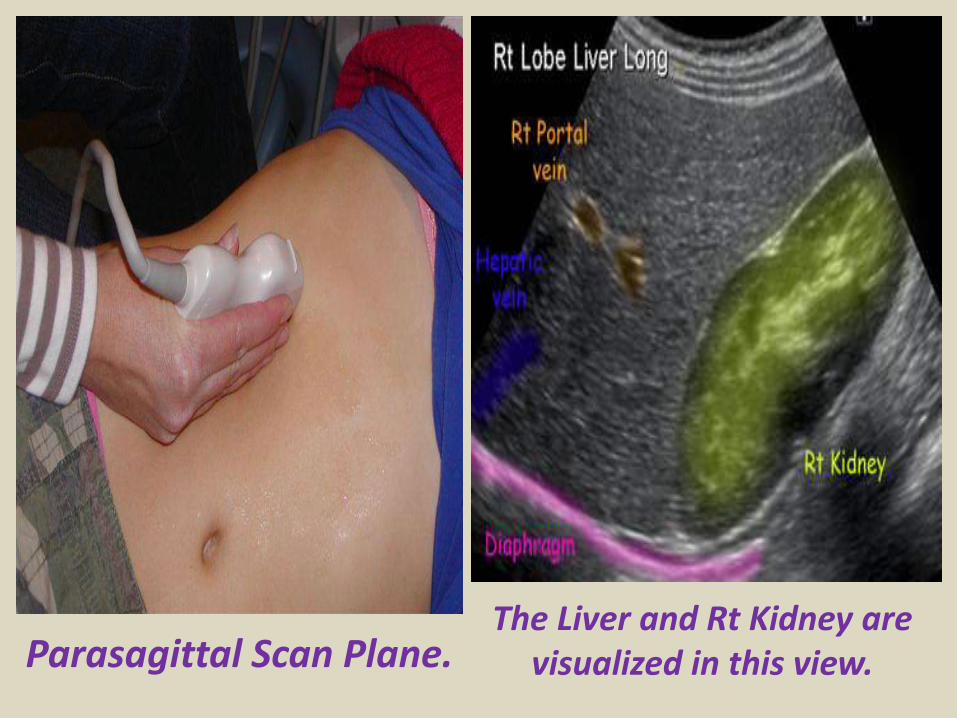
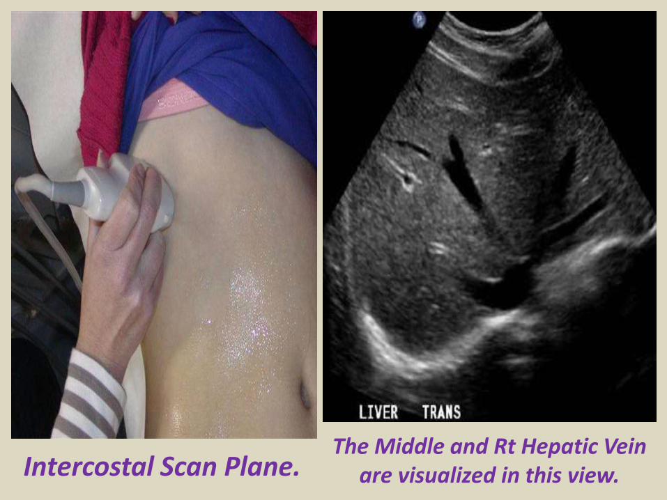
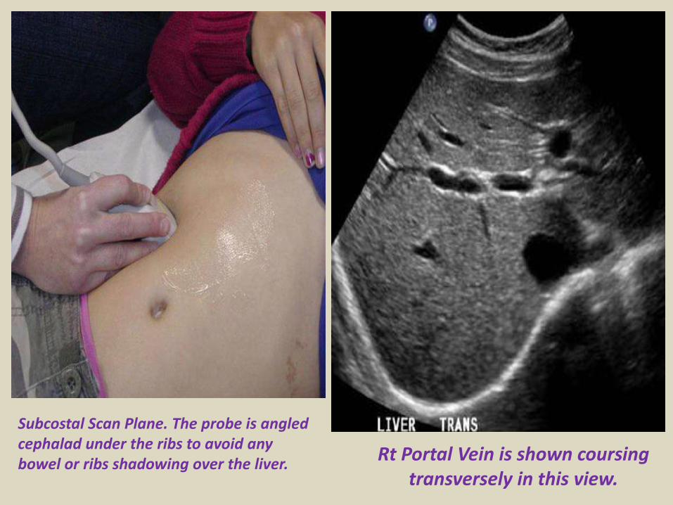
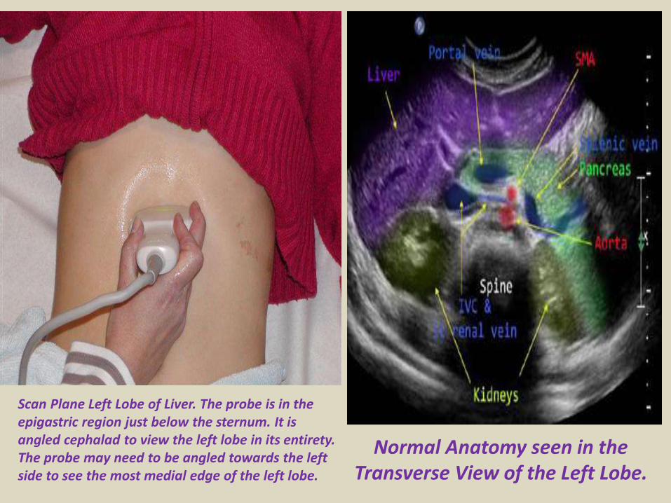
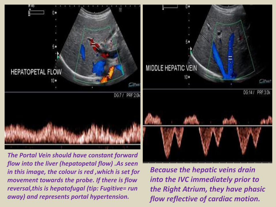
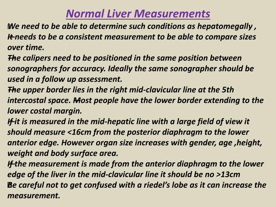
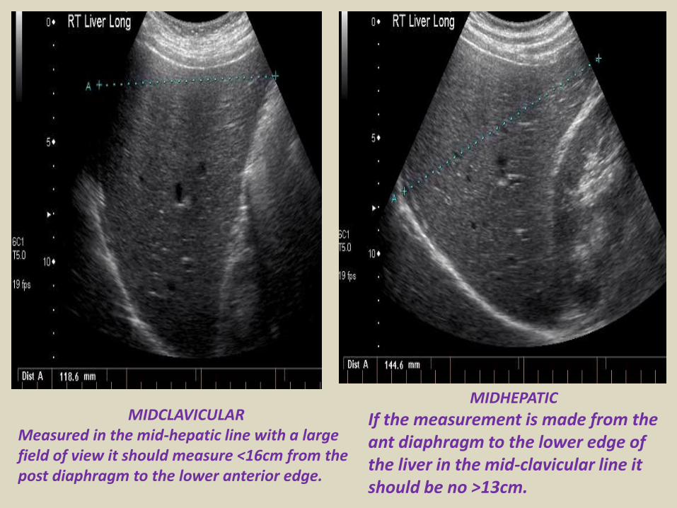
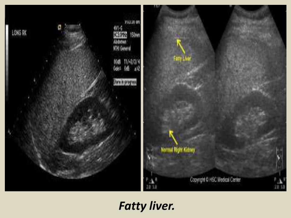
Fatty liver
Fatty liver
Cirrhotic liver
Cirrhotic liver
Bilharzial liver with peri-portal fibrosis
Bilharzial liver with peri-portal fibrosis
A) Sonography of hepatic cysts
Ultrasound images reveal an anechoic lesion in the right lobe of the liver with thin walls and clear fluid content A small projection is seen from the wall This may be thickening of the wall or a fine nodule These findings
suggest a liver cyst Differential diagnosis biliary cystadenoma
Ultrasound Typical liver cyst
Color and Power Doppler images of the hepatic cyst showed no flow in the liver cyst However the spectral Doppler trace showed faint pulsations within this lesion These pulsations appear to be transmitted from the abdominal aorta and appear insignificant Thus we arrived at a diagnosis of a simple cyst of the left lobe of the liver based on these ultrasound and Color Doppler images Absence of flow in this cyst ruled out possibilities like a vascular lesion like aneurysm of the portal vein or AVM The liver also shows moderate fatty changes
Hydatid cyst with multiple hypoechoic internal daughter cysts
Compound picture of four images showing several typical hemangiomas
Hemangioma of liver (hepatic hemangioma)
Images show a large (8 cms) rounded well defined hyperechoic non-calcific mass in the right lobe of liver There is a moderate amount of acoustic enhancement posterior to the lesion These ultrasound images show some of the typical features of hemangioma (cavernous) of the liver In this case the patient is an adult female which is typical of hepatic hemangioma However usually hemangioma are smaller in size (less than 4 cms)
Multiple Hemangioma of the Liver
Multiple hemangioma of the liver the lesions are small in size Such lesions are rather atypical of hemangioma
Hemangioma of the caudate lobe of liver
Large echogenic hemangioma in the caudate lobe of liver
Subcostal scan through the right liver showing a large hemangioma with central hypoechoic area due to thrombosis
Hepatic abscess or abscess of Liver (Amebic liver abscess)
Sonography of the liver was done in this case of pain in the right iliac fossa Ultrasound image on left shows thickened edematous wall of the cecum so typhlitis Image on right shows 2 hypoechoic lesions in the right lobe of liver Both lesions are fairly large and show in homogenous echotexture In view of the underlying disease in the cecum a diagnosis of typhlitis with amebic liver (hepatic) abscess (developing stage) was made
Ruptured liver abscess (rupture or leak of hepatic abscess)
Ultrasound images show a large abscess of the right lobe of liver with rupture of the lower margin of the abscess The leaked fluid is contained within localized adhesions in the peritoneal cavity
Abdominal CT with iv contrast administration shows a large well defined hypodense liver mass B)Abdominal ultrasound show a heterogeneous hyperechoic mass with a hypoechoic area inside consistent with liver abscess
Hyperechoic lesion in the right liver with central calcification proven to be an adenoma
Large heterogeneous lesion in the left liver adenoma with recent bleeding
Irregularly demarcated cirrhotic liver with small hypoechoic HCC A Ascites Hyperechoic HCC
Diffuse metastatic liver involvementTypical metastases with bulls eye aspect
Liver metastasis with bulls eye aspect protruding at the liver surface
Hypoechoic liver metastases
Calcified liver metastases
Blood vessels disorder of the liverThe portal vein provides about two thirds of the blood This blood contains oxygen and many nutrients brought to the liver from the intestines for processing The hepatic artery provides the remaining one third of blood This oxygen-rich blood comes from the heart and provides the liver with about half of its oxygen supply Receiving blood from two blood vessels helps protect the liver If one of these blood vessels is damaged the liver can often continue to function because it receives oxygen and nutrients from the other blood supply Blood leaves the liver through the hepatic veins This blood is a mixture of blood from the hepatic artery and from the portal vein The hepatic veins carry blood to the inferior vena cavamdashthe largest vein in the bodymdashwhich then carries blood from the abdomen and lower parts of the body to the right side of the heartBlood vessel (vascular) disorders of the liver usually result from inadequate blood flowmdashwhether into or out of the liver If the problem is blood flow out of the liver blood backs up in the liver causing congestion In either case liver cells do not receive enough blood (called ischemia) and thus are deprived of oxygen and nutrients Inadequate blood flowmdashinto or out of the livermdashmay result from heart failure or disorders that make blood more likely to clot (clotting disorders) In clotting disorders a clot may block the portal vein or a hepatic vein slowing or blocking blood flow
Normal vascular structures of the liver
Portal hypertension
Portal hypertension
Cavernous transformation of the portal vein
Portal vein thrombosis
Acute thrombosis of the portal vein with echogenic thrombus inside
Partial thrombosis of the portal vein
Ultrasound of liver graft in 56-year-old man with hepatic artery thrombosis Color Doppler ultrasound shows absence of flow in hepatic artery at hilus (arrow)
Ultrasound of liver graft in 56-year-old man with hepatic artery thrombosis Contrast-enhanced ultrasound reveals no arterial
perfusion in early phase at hilus level (arrow) nor at intrahepatic level
Hepatic artery stenosis in 39-year-old man Doppler ultrasound of hepatic artery at intrahepatic level shows prolonged acceleration time of 0153 second
Reversed flow in the hepatic vein due to obstruction
Reversed flow in the hepatic vein due to obstruction
Right hepatic vein thrombosis
Ultrasound of the Gall bladder
Fundus
Body
Normal US ofGall Bladder
Ultrasound of the Gallbladder ndash ProtocolPatient PreparationFast for 6 hours No food or drinkPreferably book the appointment in the morning to reduce bowel gas
Scanning TechniqueLooking supine left lateral decubitus and erectUse the liver as a window especially when rolling the pt onto their left sideMeasure the wall lt3mmIs the gallbladder enlargedgt10cm in lengthCheck with colour Doppler for increased vascularity of the wallAssess the cystic duct neck body and fundus (sometimes there is a phrygian cap)
Normal Scanning Position to take advantage of using the liver as a window and displacing the bowel
A normal Gallbladder should be thin walled (lt3mm) and anechoic It is a pear shaped saccular structure for bile storage in the Right Upper Quadrant Its size varies depending on the amount of bile Fasted it will be approximately 10cm long
Folds are commonly seen and are normal Make note if pathology such as calculi are contained within a
compartment created by a fold
A Phrygian cap is a specific relatively common inversion of the distal fundus of the gallbladder into the body It may become adherent It is an anatomic variant or acquired abnormality
Ultrasonography with the typical technique and the probe placed at the right hypochondrium could not locate the gallbladder at the plane of the liver hilum
Congenital agenesis of the gall bladder
Congenital agenesis of the gall bladder
Multiple gall bladder multiple septations forming a honeycomb appearance
Duplicated gallbladder at US MRI and cholangiography with double cystic ducts
Gall bladder stones and sludgeUS criteria for diagnosis of cholelithiasis1- Echogenic focus2- Cast acoustic shadow3- Seek gravitational dependence
Wall-echo-shadow sign with multiple stonesMultiple stones with confluent shadowsSingle stone with clean shadow
Acute cholecystitis is swelling (inflammation) of the gallbladder It is
a potentially serious condition that usually needs to be treated in hospitalAcute cholecystitis is either calcular(95) or non calcular (5) The main symptom of acute cholecystitis is a sudden sharp pain in the upper right side of your tummy (abdomen) that spreads towards your right shoulderThe affected part of the abdomen is usually extremely tender and breathing deeply can make the pain worseUnlike some others types of abdominal pain the pain associated with acute cholecystitis is usually persistent and doesnt go away within a few hoursSome people may additional symptoms such asa high temperature (fever) nausea and vomiting sweating loss of appetite yellowing of the skin and the whites of the eyes (jaundice) a bulge in the abdomen
US- GB wall in acute cholecystitis appears as a thin hypoechoic line sandwiched between two echogenic lines
CT- thick wall with hypodense outer layer (subserosal edema)
Acute calculus cholecystitis
Acute acalculous cholecystitis LEFT US at the spot of maximum tenderness shows mural thickening of the gallbladder (arrow) that is completely filled with sludge (asterix) without any stones RIGHT Power-Doppler sonography shows hypervascularity of the gallbladder wall (arrowhead) as a supporting sign of inflammation
Acute gangrenous cholecystitis
Xanthogranulomatous cholecystitisIntramural hypoechoichypoattenuating foci of abscess and inflammation
Emphysematous cholecystitis (A) and (B) TS and LS with gas and debris in thegallbladder lumen (C) Gas in the gallbladder lumen completely obscures the content
Emphysematous cholecystitis
CT scan showing markedly enlarged gall bladder with intraluminal blood and thrombus Hemorrhagic cholecystitis
Hemorrhagic cholecystitis with blood within the bladder lumen
Chronic cholecystitis Chronic cholecystitis is gallbladder inflammation that has lasted a long time It almost always results from gallstones and from prior attacks of acute cholecystitis Chronic cholecystitis is characterized by repeated attacks of pain (biliary colic) that occur when gallstones periodically block the cystic ductIn chronic cholecystitis the gallbladder is damaged by repeated attacks of acute inflammation usually due to gallstones and may become thick-walled scarred and small The gallstones may block the opening of the gallbladder into the cystic duct or block the cystic duct itself The gallbladder usually also contains sludge If scarring is extensive calcium may be deposited in the walls of the gallbladder causing them to harden (called porcelain gallbladder)
SymptomsA gallbladder attack whether in acute or chronic cholecystitis begins as pain
Chronic calcular cholecystitis
Chronic calcular cholecystitis
Distended gall bladder containing echogenic gravity dependent mobile biliary sludge with calculi
Porcelain GBRare due to chronic inflammation mural calcification
Biliary sludge is a mixture of particulate solids that have precipitated from bile Such sediment consists of cholesterol crystals calcium bilirubinate pigment and other calcium salts Sludge is usually detected on transabdominal ultrasonography Microscopy of aspirated bile and endoscopic ultrasonography are far more sensitive Biliary sludge is associated with pregnancy with rapid weight loss particularly in the obese with critical illness involving low or absent oral intake and the use of total parenteral nutrition (TPN) Complications caused by biliary sludge include biliary colic acute cholangitis and acute pancreatitis Asymptomatic patients with sludge or microlithiasis require no therapy
Biliary sludge
Biliary sludge
Gallbladder polyps are relatively frequent seen in up to 5 of the
population Over 90 are benign and the majority are cholesterol polypsCholesterol polyps are most frequently identified in patients between 40-50 years of age and are more common in women (FM 291)
Tiny polyp of the gall bladder wall
Multiple gall bladder polyps
Large gall bladder polyp
Adenomyomatosis of the gallbladder is a hyperplastic
cholestosis of the gallbladder wall It is a relatively common and benign cause of focal gallbladder wall thickening It is most easily seen on ultrasound and MRI Adenomyomatosis is relatively common found in ~5 of all cholecystectomy specimens It is typically seen in patients in their 5th decade The incidence increases with age which may be the result of protracted inflammation (see below) There is a female predilection (MF = 13) It is most often an incidental finding has no intrinsic malignant potential and usually requires no treatment Three morphological types of adenomyomatosis are describedfundal (localized)segmental (annular)generalized (diffuse)
Segmental adenomyomatosis of gall bladder
Diffuse adenomyomatosis of gall bladder
Adenomyomatosis of the gallbladder with a thickened wall Color Dopplershows twinkling artefacts caused by cholesterol crystals in the wall
Adenomyomatosis of the gallbladder with a thickened wall Color Doppler shows twinkling artefacts caused by cholesterol crystals in the wall
Gallbladder adenocarcinomas is the most frequent and are usually asymptomatic until it is no longer curable As such early incidental detection is important if the occasional patient is to be successfully treated Less frequent malignancies is squamous cell carcinoma Small cell carcinoma Carcinosarcoma Lymphoma and metastasisEpidemiologyAlthough overall uncommon gallbladder adenocarcinoma is the most common primary hepatobiliary carcinoma and the 5th most common malignancy of the gastrointestinal tract Predominantly affects older persons with long-standing cholecystolithiasis and as such is most common in elderly women (gt 60 years of age FM ratio = 41) Risk factors include chronic cholecystitis gallstones are seen in 70-90 of casesfamilial adenomatous polyposis syndrome (FAP)inflammatory bowel disease (IBD)porcelain gallbladder
US of Gall bladder carcinoma
Gallbladder adenocarcinoma
US shows echogenic mass adherent to gallbladder fundus (arrows) CT shows enhancing mass within the gallbladder (arrows) with involvement of the liver (arrowhead)
Gallbladder carcinoma
B-cell lymphoma of the gallbladder
50-year-old man with renal-cell carcinoma Intraoperative ultrasound image of polypoid gallbladder lesion Postcontrast axial CT image demonstrates
avidly contrast-enhancing polypoid gallbladder lesion (arrow)
Thank You
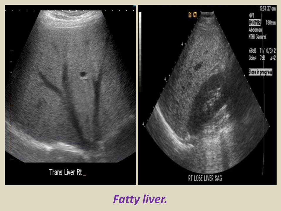
Fatty liver
Cirrhotic liver
Cirrhotic liver
Bilharzial liver with peri-portal fibrosis
Bilharzial liver with peri-portal fibrosis
A) Sonography of hepatic cysts
Ultrasound images reveal an anechoic lesion in the right lobe of the liver with thin walls and clear fluid content A small projection is seen from the wall This may be thickening of the wall or a fine nodule These findings
suggest a liver cyst Differential diagnosis biliary cystadenoma
Ultrasound Typical liver cyst
Color and Power Doppler images of the hepatic cyst showed no flow in the liver cyst However the spectral Doppler trace showed faint pulsations within this lesion These pulsations appear to be transmitted from the abdominal aorta and appear insignificant Thus we arrived at a diagnosis of a simple cyst of the left lobe of the liver based on these ultrasound and Color Doppler images Absence of flow in this cyst ruled out possibilities like a vascular lesion like aneurysm of the portal vein or AVM The liver also shows moderate fatty changes
Hydatid cyst with multiple hypoechoic internal daughter cysts
Compound picture of four images showing several typical hemangiomas
Hemangioma of liver (hepatic hemangioma)
Images show a large (8 cms) rounded well defined hyperechoic non-calcific mass in the right lobe of liver There is a moderate amount of acoustic enhancement posterior to the lesion These ultrasound images show some of the typical features of hemangioma (cavernous) of the liver In this case the patient is an adult female which is typical of hepatic hemangioma However usually hemangioma are smaller in size (less than 4 cms)
Multiple Hemangioma of the Liver
Multiple hemangioma of the liver the lesions are small in size Such lesions are rather atypical of hemangioma
Hemangioma of the caudate lobe of liver
Large echogenic hemangioma in the caudate lobe of liver
Subcostal scan through the right liver showing a large hemangioma with central hypoechoic area due to thrombosis
Hepatic abscess or abscess of Liver (Amebic liver abscess)
Sonography of the liver was done in this case of pain in the right iliac fossa Ultrasound image on left shows thickened edematous wall of the cecum so typhlitis Image on right shows 2 hypoechoic lesions in the right lobe of liver Both lesions are fairly large and show in homogenous echotexture In view of the underlying disease in the cecum a diagnosis of typhlitis with amebic liver (hepatic) abscess (developing stage) was made
Ruptured liver abscess (rupture or leak of hepatic abscess)
Ultrasound images show a large abscess of the right lobe of liver with rupture of the lower margin of the abscess The leaked fluid is contained within localized adhesions in the peritoneal cavity
Abdominal CT with iv contrast administration shows a large well defined hypodense liver mass B)Abdominal ultrasound show a heterogeneous hyperechoic mass with a hypoechoic area inside consistent with liver abscess
Hyperechoic lesion in the right liver with central calcification proven to be an adenoma
Large heterogeneous lesion in the left liver adenoma with recent bleeding
Irregularly demarcated cirrhotic liver with small hypoechoic HCC A Ascites Hyperechoic HCC
Diffuse metastatic liver involvementTypical metastases with bulls eye aspect
Liver metastasis with bulls eye aspect protruding at the liver surface
Hypoechoic liver metastases
Calcified liver metastases
Blood vessels disorder of the liverThe portal vein provides about two thirds of the blood This blood contains oxygen and many nutrients brought to the liver from the intestines for processing The hepatic artery provides the remaining one third of blood This oxygen-rich blood comes from the heart and provides the liver with about half of its oxygen supply Receiving blood from two blood vessels helps protect the liver If one of these blood vessels is damaged the liver can often continue to function because it receives oxygen and nutrients from the other blood supply Blood leaves the liver through the hepatic veins This blood is a mixture of blood from the hepatic artery and from the portal vein The hepatic veins carry blood to the inferior vena cavamdashthe largest vein in the bodymdashwhich then carries blood from the abdomen and lower parts of the body to the right side of the heartBlood vessel (vascular) disorders of the liver usually result from inadequate blood flowmdashwhether into or out of the liver If the problem is blood flow out of the liver blood backs up in the liver causing congestion In either case liver cells do not receive enough blood (called ischemia) and thus are deprived of oxygen and nutrients Inadequate blood flowmdashinto or out of the livermdashmay result from heart failure or disorders that make blood more likely to clot (clotting disorders) In clotting disorders a clot may block the portal vein or a hepatic vein slowing or blocking blood flow
Normal vascular structures of the liver
Portal hypertension
Portal hypertension
Cavernous transformation of the portal vein
Portal vein thrombosis
Acute thrombosis of the portal vein with echogenic thrombus inside
Partial thrombosis of the portal vein
Ultrasound of liver graft in 56-year-old man with hepatic artery thrombosis Color Doppler ultrasound shows absence of flow in hepatic artery at hilus (arrow)
Ultrasound of liver graft in 56-year-old man with hepatic artery thrombosis Contrast-enhanced ultrasound reveals no arterial
perfusion in early phase at hilus level (arrow) nor at intrahepatic level
Hepatic artery stenosis in 39-year-old man Doppler ultrasound of hepatic artery at intrahepatic level shows prolonged acceleration time of 0153 second
Reversed flow in the hepatic vein due to obstruction
Reversed flow in the hepatic vein due to obstruction
Right hepatic vein thrombosis
Ultrasound of the Gall bladder
Fundus
Body
Normal US ofGall Bladder
Ultrasound of the Gallbladder ndash ProtocolPatient PreparationFast for 6 hours No food or drinkPreferably book the appointment in the morning to reduce bowel gas
Scanning TechniqueLooking supine left lateral decubitus and erectUse the liver as a window especially when rolling the pt onto their left sideMeasure the wall lt3mmIs the gallbladder enlargedgt10cm in lengthCheck with colour Doppler for increased vascularity of the wallAssess the cystic duct neck body and fundus (sometimes there is a phrygian cap)
Normal Scanning Position to take advantage of using the liver as a window and displacing the bowel
A normal Gallbladder should be thin walled (lt3mm) and anechoic It is a pear shaped saccular structure for bile storage in the Right Upper Quadrant Its size varies depending on the amount of bile Fasted it will be approximately 10cm long
Folds are commonly seen and are normal Make note if pathology such as calculi are contained within a
compartment created by a fold
A Phrygian cap is a specific relatively common inversion of the distal fundus of the gallbladder into the body It may become adherent It is an anatomic variant or acquired abnormality
Ultrasonography with the typical technique and the probe placed at the right hypochondrium could not locate the gallbladder at the plane of the liver hilum
Congenital agenesis of the gall bladder
Congenital agenesis of the gall bladder
Multiple gall bladder multiple septations forming a honeycomb appearance
Duplicated gallbladder at US MRI and cholangiography with double cystic ducts
Gall bladder stones and sludgeUS criteria for diagnosis of cholelithiasis1- Echogenic focus2- Cast acoustic shadow3- Seek gravitational dependence
Wall-echo-shadow sign with multiple stonesMultiple stones with confluent shadowsSingle stone with clean shadow
Acute cholecystitis is swelling (inflammation) of the gallbladder It is
a potentially serious condition that usually needs to be treated in hospitalAcute cholecystitis is either calcular(95) or non calcular (5) The main symptom of acute cholecystitis is a sudden sharp pain in the upper right side of your tummy (abdomen) that spreads towards your right shoulderThe affected part of the abdomen is usually extremely tender and breathing deeply can make the pain worseUnlike some others types of abdominal pain the pain associated with acute cholecystitis is usually persistent and doesnt go away within a few hoursSome people may additional symptoms such asa high temperature (fever) nausea and vomiting sweating loss of appetite yellowing of the skin and the whites of the eyes (jaundice) a bulge in the abdomen
US- GB wall in acute cholecystitis appears as a thin hypoechoic line sandwiched between two echogenic lines
CT- thick wall with hypodense outer layer (subserosal edema)
Acute calculus cholecystitis
Acute acalculous cholecystitis LEFT US at the spot of maximum tenderness shows mural thickening of the gallbladder (arrow) that is completely filled with sludge (asterix) without any stones RIGHT Power-Doppler sonography shows hypervascularity of the gallbladder wall (arrowhead) as a supporting sign of inflammation
Acute gangrenous cholecystitis
Xanthogranulomatous cholecystitisIntramural hypoechoichypoattenuating foci of abscess and inflammation
Emphysematous cholecystitis (A) and (B) TS and LS with gas and debris in thegallbladder lumen (C) Gas in the gallbladder lumen completely obscures the content
Emphysematous cholecystitis
CT scan showing markedly enlarged gall bladder with intraluminal blood and thrombus Hemorrhagic cholecystitis
Hemorrhagic cholecystitis with blood within the bladder lumen
Chronic cholecystitis Chronic cholecystitis is gallbladder inflammation that has lasted a long time It almost always results from gallstones and from prior attacks of acute cholecystitis Chronic cholecystitis is characterized by repeated attacks of pain (biliary colic) that occur when gallstones periodically block the cystic ductIn chronic cholecystitis the gallbladder is damaged by repeated attacks of acute inflammation usually due to gallstones and may become thick-walled scarred and small The gallstones may block the opening of the gallbladder into the cystic duct or block the cystic duct itself The gallbladder usually also contains sludge If scarring is extensive calcium may be deposited in the walls of the gallbladder causing them to harden (called porcelain gallbladder)
SymptomsA gallbladder attack whether in acute or chronic cholecystitis begins as pain
Chronic calcular cholecystitis
Chronic calcular cholecystitis
Distended gall bladder containing echogenic gravity dependent mobile biliary sludge with calculi
Porcelain GBRare due to chronic inflammation mural calcification
Biliary sludge is a mixture of particulate solids that have precipitated from bile Such sediment consists of cholesterol crystals calcium bilirubinate pigment and other calcium salts Sludge is usually detected on transabdominal ultrasonography Microscopy of aspirated bile and endoscopic ultrasonography are far more sensitive Biliary sludge is associated with pregnancy with rapid weight loss particularly in the obese with critical illness involving low or absent oral intake and the use of total parenteral nutrition (TPN) Complications caused by biliary sludge include biliary colic acute cholangitis and acute pancreatitis Asymptomatic patients with sludge or microlithiasis require no therapy
Biliary sludge
Biliary sludge
Gallbladder polyps are relatively frequent seen in up to 5 of the
population Over 90 are benign and the majority are cholesterol polypsCholesterol polyps are most frequently identified in patients between 40-50 years of age and are more common in women (FM 291)
Tiny polyp of the gall bladder wall
Multiple gall bladder polyps
Large gall bladder polyp
Adenomyomatosis of the gallbladder is a hyperplastic
cholestosis of the gallbladder wall It is a relatively common and benign cause of focal gallbladder wall thickening It is most easily seen on ultrasound and MRI Adenomyomatosis is relatively common found in ~5 of all cholecystectomy specimens It is typically seen in patients in their 5th decade The incidence increases with age which may be the result of protracted inflammation (see below) There is a female predilection (MF = 13) It is most often an incidental finding has no intrinsic malignant potential and usually requires no treatment Three morphological types of adenomyomatosis are describedfundal (localized)segmental (annular)generalized (diffuse)
Segmental adenomyomatosis of gall bladder
Diffuse adenomyomatosis of gall bladder
Adenomyomatosis of the gallbladder with a thickened wall Color Dopplershows twinkling artefacts caused by cholesterol crystals in the wall
Adenomyomatosis of the gallbladder with a thickened wall Color Doppler shows twinkling artefacts caused by cholesterol crystals in the wall
Gallbladder adenocarcinomas is the most frequent and are usually asymptomatic until it is no longer curable As such early incidental detection is important if the occasional patient is to be successfully treated Less frequent malignancies is squamous cell carcinoma Small cell carcinoma Carcinosarcoma Lymphoma and metastasisEpidemiologyAlthough overall uncommon gallbladder adenocarcinoma is the most common primary hepatobiliary carcinoma and the 5th most common malignancy of the gastrointestinal tract Predominantly affects older persons with long-standing cholecystolithiasis and as such is most common in elderly women (gt 60 years of age FM ratio = 41) Risk factors include chronic cholecystitis gallstones are seen in 70-90 of casesfamilial adenomatous polyposis syndrome (FAP)inflammatory bowel disease (IBD)porcelain gallbladder
US of Gall bladder carcinoma
Gallbladder adenocarcinoma
US shows echogenic mass adherent to gallbladder fundus (arrows) CT shows enhancing mass within the gallbladder (arrows) with involvement of the liver (arrowhead)
Gallbladder carcinoma
B-cell lymphoma of the gallbladder
50-year-old man with renal-cell carcinoma Intraoperative ultrasound image of polypoid gallbladder lesion Postcontrast axial CT image demonstrates
avidly contrast-enhancing polypoid gallbladder lesion (arrow)
Thank You
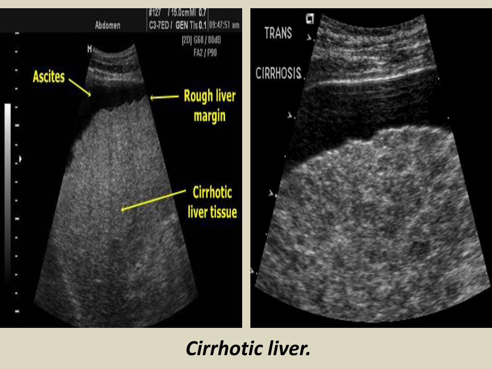
Cirrhotic liver
Cirrhotic liver
Bilharzial liver with peri-portal fibrosis
Bilharzial liver with peri-portal fibrosis
A) Sonography of hepatic cysts
Ultrasound images reveal an anechoic lesion in the right lobe of the liver with thin walls and clear fluid content A small projection is seen from the wall This may be thickening of the wall or a fine nodule These findings
suggest a liver cyst Differential diagnosis biliary cystadenoma
Ultrasound Typical liver cyst
Color and Power Doppler images of the hepatic cyst showed no flow in the liver cyst However the spectral Doppler trace showed faint pulsations within this lesion These pulsations appear to be transmitted from the abdominal aorta and appear insignificant Thus we arrived at a diagnosis of a simple cyst of the left lobe of the liver based on these ultrasound and Color Doppler images Absence of flow in this cyst ruled out possibilities like a vascular lesion like aneurysm of the portal vein or AVM The liver also shows moderate fatty changes
Hydatid cyst with multiple hypoechoic internal daughter cysts
Compound picture of four images showing several typical hemangiomas
Hemangioma of liver (hepatic hemangioma)
Images show a large (8 cms) rounded well defined hyperechoic non-calcific mass in the right lobe of liver There is a moderate amount of acoustic enhancement posterior to the lesion These ultrasound images show some of the typical features of hemangioma (cavernous) of the liver In this case the patient is an adult female which is typical of hepatic hemangioma However usually hemangioma are smaller in size (less than 4 cms)
Multiple Hemangioma of the Liver
Multiple hemangioma of the liver the lesions are small in size Such lesions are rather atypical of hemangioma
Hemangioma of the caudate lobe of liver
Large echogenic hemangioma in the caudate lobe of liver
Subcostal scan through the right liver showing a large hemangioma with central hypoechoic area due to thrombosis
Hepatic abscess or abscess of Liver (Amebic liver abscess)
Sonography of the liver was done in this case of pain in the right iliac fossa Ultrasound image on left shows thickened edematous wall of the cecum so typhlitis Image on right shows 2 hypoechoic lesions in the right lobe of liver Both lesions are fairly large and show in homogenous echotexture In view of the underlying disease in the cecum a diagnosis of typhlitis with amebic liver (hepatic) abscess (developing stage) was made
Ruptured liver abscess (rupture or leak of hepatic abscess)
Ultrasound images show a large abscess of the right lobe of liver with rupture of the lower margin of the abscess The leaked fluid is contained within localized adhesions in the peritoneal cavity
Abdominal CT with iv contrast administration shows a large well defined hypodense liver mass B)Abdominal ultrasound show a heterogeneous hyperechoic mass with a hypoechoic area inside consistent with liver abscess
Hyperechoic lesion in the right liver with central calcification proven to be an adenoma
Large heterogeneous lesion in the left liver adenoma with recent bleeding
Irregularly demarcated cirrhotic liver with small hypoechoic HCC A Ascites Hyperechoic HCC
Diffuse metastatic liver involvementTypical metastases with bulls eye aspect
Liver metastasis with bulls eye aspect protruding at the liver surface
Hypoechoic liver metastases
Calcified liver metastases
Blood vessels disorder of the liverThe portal vein provides about two thirds of the blood This blood contains oxygen and many nutrients brought to the liver from the intestines for processing The hepatic artery provides the remaining one third of blood This oxygen-rich blood comes from the heart and provides the liver with about half of its oxygen supply Receiving blood from two blood vessels helps protect the liver If one of these blood vessels is damaged the liver can often continue to function because it receives oxygen and nutrients from the other blood supply Blood leaves the liver through the hepatic veins This blood is a mixture of blood from the hepatic artery and from the portal vein The hepatic veins carry blood to the inferior vena cavamdashthe largest vein in the bodymdashwhich then carries blood from the abdomen and lower parts of the body to the right side of the heartBlood vessel (vascular) disorders of the liver usually result from inadequate blood flowmdashwhether into or out of the liver If the problem is blood flow out of the liver blood backs up in the liver causing congestion In either case liver cells do not receive enough blood (called ischemia) and thus are deprived of oxygen and nutrients Inadequate blood flowmdashinto or out of the livermdashmay result from heart failure or disorders that make blood more likely to clot (clotting disorders) In clotting disorders a clot may block the portal vein or a hepatic vein slowing or blocking blood flow
Normal vascular structures of the liver
Portal hypertension
Portal hypertension
Cavernous transformation of the portal vein
Portal vein thrombosis
Acute thrombosis of the portal vein with echogenic thrombus inside
Partial thrombosis of the portal vein
Ultrasound of liver graft in 56-year-old man with hepatic artery thrombosis Color Doppler ultrasound shows absence of flow in hepatic artery at hilus (arrow)
Ultrasound of liver graft in 56-year-old man with hepatic artery thrombosis Contrast-enhanced ultrasound reveals no arterial
perfusion in early phase at hilus level (arrow) nor at intrahepatic level
Hepatic artery stenosis in 39-year-old man Doppler ultrasound of hepatic artery at intrahepatic level shows prolonged acceleration time of 0153 second
Reversed flow in the hepatic vein due to obstruction
Reversed flow in the hepatic vein due to obstruction
Right hepatic vein thrombosis
Ultrasound of the Gall bladder
Fundus
Body
Normal US ofGall Bladder
Ultrasound of the Gallbladder ndash ProtocolPatient PreparationFast for 6 hours No food or drinkPreferably book the appointment in the morning to reduce bowel gas
Scanning TechniqueLooking supine left lateral decubitus and erectUse the liver as a window especially when rolling the pt onto their left sideMeasure the wall lt3mmIs the gallbladder enlargedgt10cm in lengthCheck with colour Doppler for increased vascularity of the wallAssess the cystic duct neck body and fundus (sometimes there is a phrygian cap)
Normal Scanning Position to take advantage of using the liver as a window and displacing the bowel
A normal Gallbladder should be thin walled (lt3mm) and anechoic It is a pear shaped saccular structure for bile storage in the Right Upper Quadrant Its size varies depending on the amount of bile Fasted it will be approximately 10cm long
Folds are commonly seen and are normal Make note if pathology such as calculi are contained within a
compartment created by a fold
A Phrygian cap is a specific relatively common inversion of the distal fundus of the gallbladder into the body It may become adherent It is an anatomic variant or acquired abnormality
Ultrasonography with the typical technique and the probe placed at the right hypochondrium could not locate the gallbladder at the plane of the liver hilum
Congenital agenesis of the gall bladder
Congenital agenesis of the gall bladder
Multiple gall bladder multiple septations forming a honeycomb appearance
Duplicated gallbladder at US MRI and cholangiography with double cystic ducts
Gall bladder stones and sludgeUS criteria for diagnosis of cholelithiasis1- Echogenic focus2- Cast acoustic shadow3- Seek gravitational dependence
Wall-echo-shadow sign with multiple stonesMultiple stones with confluent shadowsSingle stone with clean shadow
Acute cholecystitis is swelling (inflammation) of the gallbladder It is
a potentially serious condition that usually needs to be treated in hospitalAcute cholecystitis is either calcular(95) or non calcular (5) The main symptom of acute cholecystitis is a sudden sharp pain in the upper right side of your tummy (abdomen) that spreads towards your right shoulderThe affected part of the abdomen is usually extremely tender and breathing deeply can make the pain worseUnlike some others types of abdominal pain the pain associated with acute cholecystitis is usually persistent and doesnt go away within a few hoursSome people may additional symptoms such asa high temperature (fever) nausea and vomiting sweating loss of appetite yellowing of the skin and the whites of the eyes (jaundice) a bulge in the abdomen
US- GB wall in acute cholecystitis appears as a thin hypoechoic line sandwiched between two echogenic lines
CT- thick wall with hypodense outer layer (subserosal edema)
Acute calculus cholecystitis
Acute acalculous cholecystitis LEFT US at the spot of maximum tenderness shows mural thickening of the gallbladder (arrow) that is completely filled with sludge (asterix) without any stones RIGHT Power-Doppler sonography shows hypervascularity of the gallbladder wall (arrowhead) as a supporting sign of inflammation
Acute gangrenous cholecystitis
Xanthogranulomatous cholecystitisIntramural hypoechoichypoattenuating foci of abscess and inflammation
Emphysematous cholecystitis (A) and (B) TS and LS with gas and debris in thegallbladder lumen (C) Gas in the gallbladder lumen completely obscures the content
Emphysematous cholecystitis
CT scan showing markedly enlarged gall bladder with intraluminal blood and thrombus Hemorrhagic cholecystitis
Hemorrhagic cholecystitis with blood within the bladder lumen
Chronic cholecystitis Chronic cholecystitis is gallbladder inflammation that has lasted a long time It almost always results from gallstones and from prior attacks of acute cholecystitis Chronic cholecystitis is characterized by repeated attacks of pain (biliary colic) that occur when gallstones periodically block the cystic ductIn chronic cholecystitis the gallbladder is damaged by repeated attacks of acute inflammation usually due to gallstones and may become thick-walled scarred and small The gallstones may block the opening of the gallbladder into the cystic duct or block the cystic duct itself The gallbladder usually also contains sludge If scarring is extensive calcium may be deposited in the walls of the gallbladder causing them to harden (called porcelain gallbladder)
SymptomsA gallbladder attack whether in acute or chronic cholecystitis begins as pain
Chronic calcular cholecystitis
Chronic calcular cholecystitis
Distended gall bladder containing echogenic gravity dependent mobile biliary sludge with calculi
Porcelain GBRare due to chronic inflammation mural calcification
Biliary sludge is a mixture of particulate solids that have precipitated from bile Such sediment consists of cholesterol crystals calcium bilirubinate pigment and other calcium salts Sludge is usually detected on transabdominal ultrasonography Microscopy of aspirated bile and endoscopic ultrasonography are far more sensitive Biliary sludge is associated with pregnancy with rapid weight loss particularly in the obese with critical illness involving low or absent oral intake and the use of total parenteral nutrition (TPN) Complications caused by biliary sludge include biliary colic acute cholangitis and acute pancreatitis Asymptomatic patients with sludge or microlithiasis require no therapy
Biliary sludge
Biliary sludge
Gallbladder polyps are relatively frequent seen in up to 5 of the
population Over 90 are benign and the majority are cholesterol polypsCholesterol polyps are most frequently identified in patients between 40-50 years of age and are more common in women (FM 291)
Tiny polyp of the gall bladder wall
Multiple gall bladder polyps
Large gall bladder polyp
Adenomyomatosis of the gallbladder is a hyperplastic
cholestosis of the gallbladder wall It is a relatively common and benign cause of focal gallbladder wall thickening It is most easily seen on ultrasound and MRI Adenomyomatosis is relatively common found in ~5 of all cholecystectomy specimens It is typically seen in patients in their 5th decade The incidence increases with age which may be the result of protracted inflammation (see below) There is a female predilection (MF = 13) It is most often an incidental finding has no intrinsic malignant potential and usually requires no treatment Three morphological types of adenomyomatosis are describedfundal (localized)segmental (annular)generalized (diffuse)
Segmental adenomyomatosis of gall bladder
Diffuse adenomyomatosis of gall bladder
Adenomyomatosis of the gallbladder with a thickened wall Color Dopplershows twinkling artefacts caused by cholesterol crystals in the wall
Adenomyomatosis of the gallbladder with a thickened wall Color Doppler shows twinkling artefacts caused by cholesterol crystals in the wall
Gallbladder adenocarcinomas is the most frequent and are usually asymptomatic until it is no longer curable As such early incidental detection is important if the occasional patient is to be successfully treated Less frequent malignancies is squamous cell carcinoma Small cell carcinoma Carcinosarcoma Lymphoma and metastasisEpidemiologyAlthough overall uncommon gallbladder adenocarcinoma is the most common primary hepatobiliary carcinoma and the 5th most common malignancy of the gastrointestinal tract Predominantly affects older persons with long-standing cholecystolithiasis and as such is most common in elderly women (gt 60 years of age FM ratio = 41) Risk factors include chronic cholecystitis gallstones are seen in 70-90 of casesfamilial adenomatous polyposis syndrome (FAP)inflammatory bowel disease (IBD)porcelain gallbladder
US of Gall bladder carcinoma
Gallbladder adenocarcinoma
US shows echogenic mass adherent to gallbladder fundus (arrows) CT shows enhancing mass within the gallbladder (arrows) with involvement of the liver (arrowhead)
Gallbladder carcinoma
B-cell lymphoma of the gallbladder
50-year-old man with renal-cell carcinoma Intraoperative ultrasound image of polypoid gallbladder lesion Postcontrast axial CT image demonstrates
avidly contrast-enhancing polypoid gallbladder lesion (arrow)
Thank You
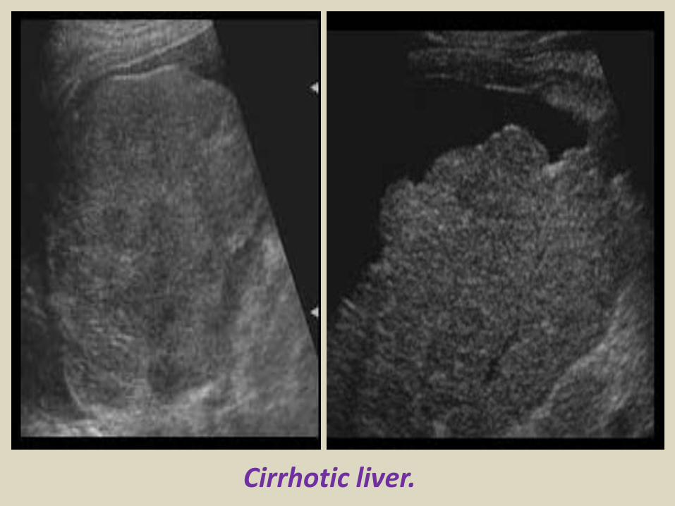
Cirrhotic liver
Bilharzial liver with peri-portal fibrosis
Bilharzial liver with peri-portal fibrosis
A) Sonography of hepatic cysts
Ultrasound images reveal an anechoic lesion in the right lobe of the liver with thin walls and clear fluid content A small projection is seen from the wall This may be thickening of the wall or a fine nodule These findings
suggest a liver cyst Differential diagnosis biliary cystadenoma
Ultrasound Typical liver cyst
Color and Power Doppler images of the hepatic cyst showed no flow in the liver cyst However the spectral Doppler trace showed faint pulsations within this lesion These pulsations appear to be transmitted from the abdominal aorta and appear insignificant Thus we arrived at a diagnosis of a simple cyst of the left lobe of the liver based on these ultrasound and Color Doppler images Absence of flow in this cyst ruled out possibilities like a vascular lesion like aneurysm of the portal vein or AVM The liver also shows moderate fatty changes
Hydatid cyst with multiple hypoechoic internal daughter cysts
Compound picture of four images showing several typical hemangiomas
Hemangioma of liver (hepatic hemangioma)
Images show a large (8 cms) rounded well defined hyperechoic non-calcific mass in the right lobe of liver There is a moderate amount of acoustic enhancement posterior to the lesion These ultrasound images show some of the typical features of hemangioma (cavernous) of the liver In this case the patient is an adult female which is typical of hepatic hemangioma However usually hemangioma are smaller in size (less than 4 cms)
Multiple Hemangioma of the Liver
Multiple hemangioma of the liver the lesions are small in size Such lesions are rather atypical of hemangioma
Hemangioma of the caudate lobe of liver
Large echogenic hemangioma in the caudate lobe of liver
Subcostal scan through the right liver showing a large hemangioma with central hypoechoic area due to thrombosis
Hepatic abscess or abscess of Liver (Amebic liver abscess)
Sonography of the liver was done in this case of pain in the right iliac fossa Ultrasound image on left shows thickened edematous wall of the cecum so typhlitis Image on right shows 2 hypoechoic lesions in the right lobe of liver Both lesions are fairly large and show in homogenous echotexture In view of the underlying disease in the cecum a diagnosis of typhlitis with amebic liver (hepatic) abscess (developing stage) was made
Ruptured liver abscess (rupture or leak of hepatic abscess)
Ultrasound images show a large abscess of the right lobe of liver with rupture of the lower margin of the abscess The leaked fluid is contained within localized adhesions in the peritoneal cavity
Abdominal CT with iv contrast administration shows a large well defined hypodense liver mass B)Abdominal ultrasound show a heterogeneous hyperechoic mass with a hypoechoic area inside consistent with liver abscess
Hyperechoic lesion in the right liver with central calcification proven to be an adenoma
Large heterogeneous lesion in the left liver adenoma with recent bleeding
Irregularly demarcated cirrhotic liver with small hypoechoic HCC A Ascites Hyperechoic HCC
Diffuse metastatic liver involvementTypical metastases with bulls eye aspect
Liver metastasis with bulls eye aspect protruding at the liver surface
Hypoechoic liver metastases
Calcified liver metastases
Blood vessels disorder of the liverThe portal vein provides about two thirds of the blood This blood contains oxygen and many nutrients brought to the liver from the intestines for processing The hepatic artery provides the remaining one third of blood This oxygen-rich blood comes from the heart and provides the liver with about half of its oxygen supply Receiving blood from two blood vessels helps protect the liver If one of these blood vessels is damaged the liver can often continue to function because it receives oxygen and nutrients from the other blood supply Blood leaves the liver through the hepatic veins This blood is a mixture of blood from the hepatic artery and from the portal vein The hepatic veins carry blood to the inferior vena cavamdashthe largest vein in the bodymdashwhich then carries blood from the abdomen and lower parts of the body to the right side of the heartBlood vessel (vascular) disorders of the liver usually result from inadequate blood flowmdashwhether into or out of the liver If the problem is blood flow out of the liver blood backs up in the liver causing congestion In either case liver cells do not receive enough blood (called ischemia) and thus are deprived of oxygen and nutrients Inadequate blood flowmdashinto or out of the livermdashmay result from heart failure or disorders that make blood more likely to clot (clotting disorders) In clotting disorders a clot may block the portal vein or a hepatic vein slowing or blocking blood flow
Normal vascular structures of the liver
Portal hypertension
Portal hypertension
Cavernous transformation of the portal vein
Portal vein thrombosis
Acute thrombosis of the portal vein with echogenic thrombus inside
Partial thrombosis of the portal vein
Ultrasound of liver graft in 56-year-old man with hepatic artery thrombosis Color Doppler ultrasound shows absence of flow in hepatic artery at hilus (arrow)
Ultrasound of liver graft in 56-year-old man with hepatic artery thrombosis Contrast-enhanced ultrasound reveals no arterial
perfusion in early phase at hilus level (arrow) nor at intrahepatic level
Hepatic artery stenosis in 39-year-old man Doppler ultrasound of hepatic artery at intrahepatic level shows prolonged acceleration time of 0153 second
Reversed flow in the hepatic vein due to obstruction
Reversed flow in the hepatic vein due to obstruction
Right hepatic vein thrombosis
Ultrasound of the Gall bladder
Fundus
Body
Normal US ofGall Bladder
Ultrasound of the Gallbladder ndash ProtocolPatient PreparationFast for 6 hours No food or drinkPreferably book the appointment in the morning to reduce bowel gas
Scanning TechniqueLooking supine left lateral decubitus and erectUse the liver as a window especially when rolling the pt onto their left sideMeasure the wall lt3mmIs the gallbladder enlargedgt10cm in lengthCheck with colour Doppler for increased vascularity of the wallAssess the cystic duct neck body and fundus (sometimes there is a phrygian cap)
Normal Scanning Position to take advantage of using the liver as a window and displacing the bowel
A normal Gallbladder should be thin walled (lt3mm) and anechoic It is a pear shaped saccular structure for bile storage in the Right Upper Quadrant Its size varies depending on the amount of bile Fasted it will be approximately 10cm long
Folds are commonly seen and are normal Make note if pathology such as calculi are contained within a
compartment created by a fold
A Phrygian cap is a specific relatively common inversion of the distal fundus of the gallbladder into the body It may become adherent It is an anatomic variant or acquired abnormality
Ultrasonography with the typical technique and the probe placed at the right hypochondrium could not locate the gallbladder at the plane of the liver hilum
Congenital agenesis of the gall bladder
Congenital agenesis of the gall bladder
Multiple gall bladder multiple septations forming a honeycomb appearance
Duplicated gallbladder at US MRI and cholangiography with double cystic ducts
Gall bladder stones and sludgeUS criteria for diagnosis of cholelithiasis1- Echogenic focus2- Cast acoustic shadow3- Seek gravitational dependence
Wall-echo-shadow sign with multiple stonesMultiple stones with confluent shadowsSingle stone with clean shadow
Acute cholecystitis is swelling (inflammation) of the gallbladder It is
a potentially serious condition that usually needs to be treated in hospitalAcute cholecystitis is either calcular(95) or non calcular (5) The main symptom of acute cholecystitis is a sudden sharp pain in the upper right side of your tummy (abdomen) that spreads towards your right shoulderThe affected part of the abdomen is usually extremely tender and breathing deeply can make the pain worseUnlike some others types of abdominal pain the pain associated with acute cholecystitis is usually persistent and doesnt go away within a few hoursSome people may additional symptoms such asa high temperature (fever) nausea and vomiting sweating loss of appetite yellowing of the skin and the whites of the eyes (jaundice) a bulge in the abdomen
US- GB wall in acute cholecystitis appears as a thin hypoechoic line sandwiched between two echogenic lines
CT- thick wall with hypodense outer layer (subserosal edema)
Acute calculus cholecystitis
Acute acalculous cholecystitis LEFT US at the spot of maximum tenderness shows mural thickening of the gallbladder (arrow) that is completely filled with sludge (asterix) without any stones RIGHT Power-Doppler sonography shows hypervascularity of the gallbladder wall (arrowhead) as a supporting sign of inflammation
Acute gangrenous cholecystitis
Xanthogranulomatous cholecystitisIntramural hypoechoichypoattenuating foci of abscess and inflammation
Emphysematous cholecystitis (A) and (B) TS and LS with gas and debris in thegallbladder lumen (C) Gas in the gallbladder lumen completely obscures the content
Emphysematous cholecystitis
CT scan showing markedly enlarged gall bladder with intraluminal blood and thrombus Hemorrhagic cholecystitis
Hemorrhagic cholecystitis with blood within the bladder lumen
Chronic cholecystitis Chronic cholecystitis is gallbladder inflammation that has lasted a long time It almost always results from gallstones and from prior attacks of acute cholecystitis Chronic cholecystitis is characterized by repeated attacks of pain (biliary colic) that occur when gallstones periodically block the cystic ductIn chronic cholecystitis the gallbladder is damaged by repeated attacks of acute inflammation usually due to gallstones and may become thick-walled scarred and small The gallstones may block the opening of the gallbladder into the cystic duct or block the cystic duct itself The gallbladder usually also contains sludge If scarring is extensive calcium may be deposited in the walls of the gallbladder causing them to harden (called porcelain gallbladder)
SymptomsA gallbladder attack whether in acute or chronic cholecystitis begins as pain
Chronic calcular cholecystitis
Chronic calcular cholecystitis
Distended gall bladder containing echogenic gravity dependent mobile biliary sludge with calculi
Porcelain GBRare due to chronic inflammation mural calcification
Biliary sludge is a mixture of particulate solids that have precipitated from bile Such sediment consists of cholesterol crystals calcium bilirubinate pigment and other calcium salts Sludge is usually detected on transabdominal ultrasonography Microscopy of aspirated bile and endoscopic ultrasonography are far more sensitive Biliary sludge is associated with pregnancy with rapid weight loss particularly in the obese with critical illness involving low or absent oral intake and the use of total parenteral nutrition (TPN) Complications caused by biliary sludge include biliary colic acute cholangitis and acute pancreatitis Asymptomatic patients with sludge or microlithiasis require no therapy
Biliary sludge
Biliary sludge
Gallbladder polyps are relatively frequent seen in up to 5 of the
population Over 90 are benign and the majority are cholesterol polypsCholesterol polyps are most frequently identified in patients between 40-50 years of age and are more common in women (FM 291)
Tiny polyp of the gall bladder wall
Multiple gall bladder polyps
Large gall bladder polyp
Adenomyomatosis of the gallbladder is a hyperplastic
cholestosis of the gallbladder wall It is a relatively common and benign cause of focal gallbladder wall thickening It is most easily seen on ultrasound and MRI Adenomyomatosis is relatively common found in ~5 of all cholecystectomy specimens It is typically seen in patients in their 5th decade The incidence increases with age which may be the result of protracted inflammation (see below) There is a female predilection (MF = 13) It is most often an incidental finding has no intrinsic malignant potential and usually requires no treatment Three morphological types of adenomyomatosis are describedfundal (localized)segmental (annular)generalized (diffuse)
Segmental adenomyomatosis of gall bladder
Diffuse adenomyomatosis of gall bladder
Adenomyomatosis of the gallbladder with a thickened wall Color Dopplershows twinkling artefacts caused by cholesterol crystals in the wall
Adenomyomatosis of the gallbladder with a thickened wall Color Doppler shows twinkling artefacts caused by cholesterol crystals in the wall
Gallbladder adenocarcinomas is the most frequent and are usually asymptomatic until it is no longer curable As such early incidental detection is important if the occasional patient is to be successfully treated Less frequent malignancies is squamous cell carcinoma Small cell carcinoma Carcinosarcoma Lymphoma and metastasisEpidemiologyAlthough overall uncommon gallbladder adenocarcinoma is the most common primary hepatobiliary carcinoma and the 5th most common malignancy of the gastrointestinal tract Predominantly affects older persons with long-standing cholecystolithiasis and as such is most common in elderly women (gt 60 years of age FM ratio = 41) Risk factors include chronic cholecystitis gallstones are seen in 70-90 of casesfamilial adenomatous polyposis syndrome (FAP)inflammatory bowel disease (IBD)porcelain gallbladder
US of Gall bladder carcinoma
Gallbladder adenocarcinoma
US shows echogenic mass adherent to gallbladder fundus (arrows) CT shows enhancing mass within the gallbladder (arrows) with involvement of the liver (arrowhead)
Gallbladder carcinoma
B-cell lymphoma of the gallbladder
50-year-old man with renal-cell carcinoma Intraoperative ultrasound image of polypoid gallbladder lesion Postcontrast axial CT image demonstrates
avidly contrast-enhancing polypoid gallbladder lesion (arrow)
Thank You
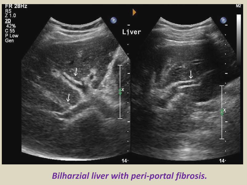
Bilharzial liver with peri-portal fibrosis
Bilharzial liver with peri-portal fibrosis
A) Sonography of hepatic cysts
Ultrasound images reveal an anechoic lesion in the right lobe of the liver with thin walls and clear fluid content A small projection is seen from the wall This may be thickening of the wall or a fine nodule These findings
suggest a liver cyst Differential diagnosis biliary cystadenoma
Ultrasound Typical liver cyst
Color and Power Doppler images of the hepatic cyst showed no flow in the liver cyst However the spectral Doppler trace showed faint pulsations within this lesion These pulsations appear to be transmitted from the abdominal aorta and appear insignificant Thus we arrived at a diagnosis of a simple cyst of the left lobe of the liver based on these ultrasound and Color Doppler images Absence of flow in this cyst ruled out possibilities like a vascular lesion like aneurysm of the portal vein or AVM The liver also shows moderate fatty changes
Hydatid cyst with multiple hypoechoic internal daughter cysts
Compound picture of four images showing several typical hemangiomas
Hemangioma of liver (hepatic hemangioma)
Images show a large (8 cms) rounded well defined hyperechoic non-calcific mass in the right lobe of liver There is a moderate amount of acoustic enhancement posterior to the lesion These ultrasound images show some of the typical features of hemangioma (cavernous) of the liver In this case the patient is an adult female which is typical of hepatic hemangioma However usually hemangioma are smaller in size (less than 4 cms)
Multiple Hemangioma of the Liver
Multiple hemangioma of the liver the lesions are small in size Such lesions are rather atypical of hemangioma
Hemangioma of the caudate lobe of liver
Large echogenic hemangioma in the caudate lobe of liver
Subcostal scan through the right liver showing a large hemangioma with central hypoechoic area due to thrombosis
Hepatic abscess or abscess of Liver (Amebic liver abscess)
Sonography of the liver was done in this case of pain in the right iliac fossa Ultrasound image on left shows thickened edematous wall of the cecum so typhlitis Image on right shows 2 hypoechoic lesions in the right lobe of liver Both lesions are fairly large and show in homogenous echotexture In view of the underlying disease in the cecum a diagnosis of typhlitis with amebic liver (hepatic) abscess (developing stage) was made
Ruptured liver abscess (rupture or leak of hepatic abscess)
Ultrasound images show a large abscess of the right lobe of liver with rupture of the lower margin of the abscess The leaked fluid is contained within localized adhesions in the peritoneal cavity
Abdominal CT with iv contrast administration shows a large well defined hypodense liver mass B)Abdominal ultrasound show a heterogeneous hyperechoic mass with a hypoechoic area inside consistent with liver abscess
Hyperechoic lesion in the right liver with central calcification proven to be an adenoma
Large heterogeneous lesion in the left liver adenoma with recent bleeding
Irregularly demarcated cirrhotic liver with small hypoechoic HCC A Ascites Hyperechoic HCC
Diffuse metastatic liver involvementTypical metastases with bulls eye aspect
Liver metastasis with bulls eye aspect protruding at the liver surface
Hypoechoic liver metastases
Calcified liver metastases
Blood vessels disorder of the liverThe portal vein provides about two thirds of the blood This blood contains oxygen and many nutrients brought to the liver from the intestines for processing The hepatic artery provides the remaining one third of blood This oxygen-rich blood comes from the heart and provides the liver with about half of its oxygen supply Receiving blood from two blood vessels helps protect the liver If one of these blood vessels is damaged the liver can often continue to function because it receives oxygen and nutrients from the other blood supply Blood leaves the liver through the hepatic veins This blood is a mixture of blood from the hepatic artery and from the portal vein The hepatic veins carry blood to the inferior vena cavamdashthe largest vein in the bodymdashwhich then carries blood from the abdomen and lower parts of the body to the right side of the heartBlood vessel (vascular) disorders of the liver usually result from inadequate blood flowmdashwhether into or out of the liver If the problem is blood flow out of the liver blood backs up in the liver causing congestion In either case liver cells do not receive enough blood (called ischemia) and thus are deprived of oxygen and nutrients Inadequate blood flowmdashinto or out of the livermdashmay result from heart failure or disorders that make blood more likely to clot (clotting disorders) In clotting disorders a clot may block the portal vein or a hepatic vein slowing or blocking blood flow
Normal vascular structures of the liver
Portal hypertension
Portal hypertension
Cavernous transformation of the portal vein
Portal vein thrombosis
Acute thrombosis of the portal vein with echogenic thrombus inside
Partial thrombosis of the portal vein
Ultrasound of liver graft in 56-year-old man with hepatic artery thrombosis Color Doppler ultrasound shows absence of flow in hepatic artery at hilus (arrow)
Ultrasound of liver graft in 56-year-old man with hepatic artery thrombosis Contrast-enhanced ultrasound reveals no arterial
perfusion in early phase at hilus level (arrow) nor at intrahepatic level
Hepatic artery stenosis in 39-year-old man Doppler ultrasound of hepatic artery at intrahepatic level shows prolonged acceleration time of 0153 second
Reversed flow in the hepatic vein due to obstruction
Reversed flow in the hepatic vein due to obstruction
Right hepatic vein thrombosis
Ultrasound of the Gall bladder
Fundus
Body
Normal US ofGall Bladder
Ultrasound of the Gallbladder ndash ProtocolPatient PreparationFast for 6 hours No food or drinkPreferably book the appointment in the morning to reduce bowel gas
Scanning TechniqueLooking supine left lateral decubitus and erectUse the liver as a window especially when rolling the pt onto their left sideMeasure the wall lt3mmIs the gallbladder enlargedgt10cm in lengthCheck with colour Doppler for increased vascularity of the wallAssess the cystic duct neck body and fundus (sometimes there is a phrygian cap)
Normal Scanning Position to take advantage of using the liver as a window and displacing the bowel
A normal Gallbladder should be thin walled (lt3mm) and anechoic It is a pear shaped saccular structure for bile storage in the Right Upper Quadrant Its size varies depending on the amount of bile Fasted it will be approximately 10cm long
Folds are commonly seen and are normal Make note if pathology such as calculi are contained within a
compartment created by a fold
A Phrygian cap is a specific relatively common inversion of the distal fundus of the gallbladder into the body It may become adherent It is an anatomic variant or acquired abnormality
Ultrasonography with the typical technique and the probe placed at the right hypochondrium could not locate the gallbladder at the plane of the liver hilum
Congenital agenesis of the gall bladder
Congenital agenesis of the gall bladder
Multiple gall bladder multiple septations forming a honeycomb appearance
Duplicated gallbladder at US MRI and cholangiography with double cystic ducts
Gall bladder stones and sludgeUS criteria for diagnosis of cholelithiasis1- Echogenic focus2- Cast acoustic shadow3- Seek gravitational dependence
Wall-echo-shadow sign with multiple stonesMultiple stones with confluent shadowsSingle stone with clean shadow
Acute cholecystitis is swelling (inflammation) of the gallbladder It is
a potentially serious condition that usually needs to be treated in hospitalAcute cholecystitis is either calcular(95) or non calcular (5) The main symptom of acute cholecystitis is a sudden sharp pain in the upper right side of your tummy (abdomen) that spreads towards your right shoulderThe affected part of the abdomen is usually extremely tender and breathing deeply can make the pain worseUnlike some others types of abdominal pain the pain associated with acute cholecystitis is usually persistent and doesnt go away within a few hoursSome people may additional symptoms such asa high temperature (fever) nausea and vomiting sweating loss of appetite yellowing of the skin and the whites of the eyes (jaundice) a bulge in the abdomen
US- GB wall in acute cholecystitis appears as a thin hypoechoic line sandwiched between two echogenic lines
CT- thick wall with hypodense outer layer (subserosal edema)
Acute calculus cholecystitis
Acute acalculous cholecystitis LEFT US at the spot of maximum tenderness shows mural thickening of the gallbladder (arrow) that is completely filled with sludge (asterix) without any stones RIGHT Power-Doppler sonography shows hypervascularity of the gallbladder wall (arrowhead) as a supporting sign of inflammation
Acute gangrenous cholecystitis
Xanthogranulomatous cholecystitisIntramural hypoechoichypoattenuating foci of abscess and inflammation
Emphysematous cholecystitis (A) and (B) TS and LS with gas and debris in thegallbladder lumen (C) Gas in the gallbladder lumen completely obscures the content
Emphysematous cholecystitis
CT scan showing markedly enlarged gall bladder with intraluminal blood and thrombus Hemorrhagic cholecystitis
Hemorrhagic cholecystitis with blood within the bladder lumen
Chronic cholecystitis Chronic cholecystitis is gallbladder inflammation that has lasted a long time It almost always results from gallstones and from prior attacks of acute cholecystitis Chronic cholecystitis is characterized by repeated attacks of pain (biliary colic) that occur when gallstones periodically block the cystic ductIn chronic cholecystitis the gallbladder is damaged by repeated attacks of acute inflammation usually due to gallstones and may become thick-walled scarred and small The gallstones may block the opening of the gallbladder into the cystic duct or block the cystic duct itself The gallbladder usually also contains sludge If scarring is extensive calcium may be deposited in the walls of the gallbladder causing them to harden (called porcelain gallbladder)
SymptomsA gallbladder attack whether in acute or chronic cholecystitis begins as pain
Chronic calcular cholecystitis
Chronic calcular cholecystitis
Distended gall bladder containing echogenic gravity dependent mobile biliary sludge with calculi
Porcelain GBRare due to chronic inflammation mural calcification
Biliary sludge is a mixture of particulate solids that have precipitated from bile Such sediment consists of cholesterol crystals calcium bilirubinate pigment and other calcium salts Sludge is usually detected on transabdominal ultrasonography Microscopy of aspirated bile and endoscopic ultrasonography are far more sensitive Biliary sludge is associated with pregnancy with rapid weight loss particularly in the obese with critical illness involving low or absent oral intake and the use of total parenteral nutrition (TPN) Complications caused by biliary sludge include biliary colic acute cholangitis and acute pancreatitis Asymptomatic patients with sludge or microlithiasis require no therapy
Biliary sludge
Biliary sludge
Gallbladder polyps are relatively frequent seen in up to 5 of the
population Over 90 are benign and the majority are cholesterol polypsCholesterol polyps are most frequently identified in patients between 40-50 years of age and are more common in women (FM 291)
Tiny polyp of the gall bladder wall
Multiple gall bladder polyps
Large gall bladder polyp
Adenomyomatosis of the gallbladder is a hyperplastic
cholestosis of the gallbladder wall It is a relatively common and benign cause of focal gallbladder wall thickening It is most easily seen on ultrasound and MRI Adenomyomatosis is relatively common found in ~5 of all cholecystectomy specimens It is typically seen in patients in their 5th decade The incidence increases with age which may be the result of protracted inflammation (see below) There is a female predilection (MF = 13) It is most often an incidental finding has no intrinsic malignant potential and usually requires no treatment Three morphological types of adenomyomatosis are describedfundal (localized)segmental (annular)generalized (diffuse)
Segmental adenomyomatosis of gall bladder
Diffuse adenomyomatosis of gall bladder
Adenomyomatosis of the gallbladder with a thickened wall Color Dopplershows twinkling artefacts caused by cholesterol crystals in the wall
Adenomyomatosis of the gallbladder with a thickened wall Color Doppler shows twinkling artefacts caused by cholesterol crystals in the wall
Gallbladder adenocarcinomas is the most frequent and are usually asymptomatic until it is no longer curable As such early incidental detection is important if the occasional patient is to be successfully treated Less frequent malignancies is squamous cell carcinoma Small cell carcinoma Carcinosarcoma Lymphoma and metastasisEpidemiologyAlthough overall uncommon gallbladder adenocarcinoma is the most common primary hepatobiliary carcinoma and the 5th most common malignancy of the gastrointestinal tract Predominantly affects older persons with long-standing cholecystolithiasis and as such is most common in elderly women (gt 60 years of age FM ratio = 41) Risk factors include chronic cholecystitis gallstones are seen in 70-90 of casesfamilial adenomatous polyposis syndrome (FAP)inflammatory bowel disease (IBD)porcelain gallbladder
US of Gall bladder carcinoma
Gallbladder adenocarcinoma
US shows echogenic mass adherent to gallbladder fundus (arrows) CT shows enhancing mass within the gallbladder (arrows) with involvement of the liver (arrowhead)
Gallbladder carcinoma
B-cell lymphoma of the gallbladder
50-year-old man with renal-cell carcinoma Intraoperative ultrasound image of polypoid gallbladder lesion Postcontrast axial CT image demonstrates
avidly contrast-enhancing polypoid gallbladder lesion (arrow)
Thank You
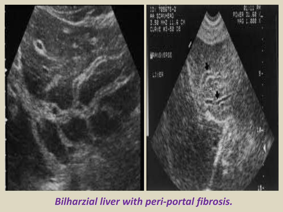
Bilharzial liver with peri-portal fibrosis
A) Sonography of hepatic cysts
Ultrasound images reveal an anechoic lesion in the right lobe of the liver with thin walls and clear fluid content A small projection is seen from the wall This may be thickening of the wall or a fine nodule These findings
suggest a liver cyst Differential diagnosis biliary cystadenoma
Ultrasound Typical liver cyst
Color and Power Doppler images of the hepatic cyst showed no flow in the liver cyst However the spectral Doppler trace showed faint pulsations within this lesion These pulsations appear to be transmitted from the abdominal aorta and appear insignificant Thus we arrived at a diagnosis of a simple cyst of the left lobe of the liver based on these ultrasound and Color Doppler images Absence of flow in this cyst ruled out possibilities like a vascular lesion like aneurysm of the portal vein or AVM The liver also shows moderate fatty changes
Hydatid cyst with multiple hypoechoic internal daughter cysts
Compound picture of four images showing several typical hemangiomas
Hemangioma of liver (hepatic hemangioma)
Images show a large (8 cms) rounded well defined hyperechoic non-calcific mass in the right lobe of liver There is a moderate amount of acoustic enhancement posterior to the lesion These ultrasound images show some of the typical features of hemangioma (cavernous) of the liver In this case the patient is an adult female which is typical of hepatic hemangioma However usually hemangioma are smaller in size (less than 4 cms)
Multiple Hemangioma of the Liver
Multiple hemangioma of the liver the lesions are small in size Such lesions are rather atypical of hemangioma
Hemangioma of the caudate lobe of liver
Large echogenic hemangioma in the caudate lobe of liver
Subcostal scan through the right liver showing a large hemangioma with central hypoechoic area due to thrombosis
Hepatic abscess or abscess of Liver (Amebic liver abscess)
Sonography of the liver was done in this case of pain in the right iliac fossa Ultrasound image on left shows thickened edematous wall of the cecum so typhlitis Image on right shows 2 hypoechoic lesions in the right lobe of liver Both lesions are fairly large and show in homogenous echotexture In view of the underlying disease in the cecum a diagnosis of typhlitis with amebic liver (hepatic) abscess (developing stage) was made
Ruptured liver abscess (rupture or leak of hepatic abscess)
Ultrasound images show a large abscess of the right lobe of liver with rupture of the lower margin of the abscess The leaked fluid is contained within localized adhesions in the peritoneal cavity
Abdominal CT with iv contrast administration shows a large well defined hypodense liver mass B)Abdominal ultrasound show a heterogeneous hyperechoic mass with a hypoechoic area inside consistent with liver abscess
Hyperechoic lesion in the right liver with central calcification proven to be an adenoma
Large heterogeneous lesion in the left liver adenoma with recent bleeding
Irregularly demarcated cirrhotic liver with small hypoechoic HCC A Ascites Hyperechoic HCC
Diffuse metastatic liver involvementTypical metastases with bulls eye aspect
Liver metastasis with bulls eye aspect protruding at the liver surface
Hypoechoic liver metastases
Calcified liver metastases
Blood vessels disorder of the liverThe portal vein provides about two thirds of the blood This blood contains oxygen and many nutrients brought to the liver from the intestines for processing The hepatic artery provides the remaining one third of blood This oxygen-rich blood comes from the heart and provides the liver with about half of its oxygen supply Receiving blood from two blood vessels helps protect the liver If one of these blood vessels is damaged the liver can often continue to function because it receives oxygen and nutrients from the other blood supply Blood leaves the liver through the hepatic veins This blood is a mixture of blood from the hepatic artery and from the portal vein The hepatic veins carry blood to the inferior vena cavamdashthe largest vein in the bodymdashwhich then carries blood from the abdomen and lower parts of the body to the right side of the heartBlood vessel (vascular) disorders of the liver usually result from inadequate blood flowmdashwhether into or out of the liver If the problem is blood flow out of the liver blood backs up in the liver causing congestion In either case liver cells do not receive enough blood (called ischemia) and thus are deprived of oxygen and nutrients Inadequate blood flowmdashinto or out of the livermdashmay result from heart failure or disorders that make blood more likely to clot (clotting disorders) In clotting disorders a clot may block the portal vein or a hepatic vein slowing or blocking blood flow
Normal vascular structures of the liver
Portal hypertension
Portal hypertension
Cavernous transformation of the portal vein
Portal vein thrombosis
Acute thrombosis of the portal vein with echogenic thrombus inside
Partial thrombosis of the portal vein
Ultrasound of liver graft in 56-year-old man with hepatic artery thrombosis Color Doppler ultrasound shows absence of flow in hepatic artery at hilus (arrow)
Ultrasound of liver graft in 56-year-old man with hepatic artery thrombosis Contrast-enhanced ultrasound reveals no arterial
perfusion in early phase at hilus level (arrow) nor at intrahepatic level
Hepatic artery stenosis in 39-year-old man Doppler ultrasound of hepatic artery at intrahepatic level shows prolonged acceleration time of 0153 second
Reversed flow in the hepatic vein due to obstruction
Reversed flow in the hepatic vein due to obstruction
Right hepatic vein thrombosis
Ultrasound of the Gall bladder
Fundus
Body
Normal US ofGall Bladder
Ultrasound of the Gallbladder ndash ProtocolPatient PreparationFast for 6 hours No food or drinkPreferably book the appointment in the morning to reduce bowel gas
Scanning TechniqueLooking supine left lateral decubitus and erectUse the liver as a window especially when rolling the pt onto their left sideMeasure the wall lt3mmIs the gallbladder enlargedgt10cm in lengthCheck with colour Doppler for increased vascularity of the wallAssess the cystic duct neck body and fundus (sometimes there is a phrygian cap)
Normal Scanning Position to take advantage of using the liver as a window and displacing the bowel
A normal Gallbladder should be thin walled (lt3mm) and anechoic It is a pear shaped saccular structure for bile storage in the Right Upper Quadrant Its size varies depending on the amount of bile Fasted it will be approximately 10cm long
Folds are commonly seen and are normal Make note if pathology such as calculi are contained within a
compartment created by a fold
A Phrygian cap is a specific relatively common inversion of the distal fundus of the gallbladder into the body It may become adherent It is an anatomic variant or acquired abnormality
Ultrasonography with the typical technique and the probe placed at the right hypochondrium could not locate the gallbladder at the plane of the liver hilum
Congenital agenesis of the gall bladder
Congenital agenesis of the gall bladder
Multiple gall bladder multiple septations forming a honeycomb appearance
Duplicated gallbladder at US MRI and cholangiography with double cystic ducts
Gall bladder stones and sludgeUS criteria for diagnosis of cholelithiasis1- Echogenic focus2- Cast acoustic shadow3- Seek gravitational dependence
Wall-echo-shadow sign with multiple stonesMultiple stones with confluent shadowsSingle stone with clean shadow
Acute cholecystitis is swelling (inflammation) of the gallbladder It is
a potentially serious condition that usually needs to be treated in hospitalAcute cholecystitis is either calcular(95) or non calcular (5) The main symptom of acute cholecystitis is a sudden sharp pain in the upper right side of your tummy (abdomen) that spreads towards your right shoulderThe affected part of the abdomen is usually extremely tender and breathing deeply can make the pain worseUnlike some others types of abdominal pain the pain associated with acute cholecystitis is usually persistent and doesnt go away within a few hoursSome people may additional symptoms such asa high temperature (fever) nausea and vomiting sweating loss of appetite yellowing of the skin and the whites of the eyes (jaundice) a bulge in the abdomen
US- GB wall in acute cholecystitis appears as a thin hypoechoic line sandwiched between two echogenic lines
CT- thick wall with hypodense outer layer (subserosal edema)
Acute calculus cholecystitis
Acute acalculous cholecystitis LEFT US at the spot of maximum tenderness shows mural thickening of the gallbladder (arrow) that is completely filled with sludge (asterix) without any stones RIGHT Power-Doppler sonography shows hypervascularity of the gallbladder wall (arrowhead) as a supporting sign of inflammation
Acute gangrenous cholecystitis
Xanthogranulomatous cholecystitisIntramural hypoechoichypoattenuating foci of abscess and inflammation
Emphysematous cholecystitis (A) and (B) TS and LS with gas and debris in thegallbladder lumen (C) Gas in the gallbladder lumen completely obscures the content
Emphysematous cholecystitis
CT scan showing markedly enlarged gall bladder with intraluminal blood and thrombus Hemorrhagic cholecystitis
Hemorrhagic cholecystitis with blood within the bladder lumen
Chronic cholecystitis Chronic cholecystitis is gallbladder inflammation that has lasted a long time It almost always results from gallstones and from prior attacks of acute cholecystitis Chronic cholecystitis is characterized by repeated attacks of pain (biliary colic) that occur when gallstones periodically block the cystic ductIn chronic cholecystitis the gallbladder is damaged by repeated attacks of acute inflammation usually due to gallstones and may become thick-walled scarred and small The gallstones may block the opening of the gallbladder into the cystic duct or block the cystic duct itself The gallbladder usually also contains sludge If scarring is extensive calcium may be deposited in the walls of the gallbladder causing them to harden (called porcelain gallbladder)
SymptomsA gallbladder attack whether in acute or chronic cholecystitis begins as pain
Chronic calcular cholecystitis
Chronic calcular cholecystitis
Distended gall bladder containing echogenic gravity dependent mobile biliary sludge with calculi
Porcelain GBRare due to chronic inflammation mural calcification
Biliary sludge is a mixture of particulate solids that have precipitated from bile Such sediment consists of cholesterol crystals calcium bilirubinate pigment and other calcium salts Sludge is usually detected on transabdominal ultrasonography Microscopy of aspirated bile and endoscopic ultrasonography are far more sensitive Biliary sludge is associated with pregnancy with rapid weight loss particularly in the obese with critical illness involving low or absent oral intake and the use of total parenteral nutrition (TPN) Complications caused by biliary sludge include biliary colic acute cholangitis and acute pancreatitis Asymptomatic patients with sludge or microlithiasis require no therapy
Biliary sludge
Biliary sludge
Gallbladder polyps are relatively frequent seen in up to 5 of the
population Over 90 are benign and the majority are cholesterol polypsCholesterol polyps are most frequently identified in patients between 40-50 years of age and are more common in women (FM 291)
Tiny polyp of the gall bladder wall
Multiple gall bladder polyps
Large gall bladder polyp
Adenomyomatosis of the gallbladder is a hyperplastic
cholestosis of the gallbladder wall It is a relatively common and benign cause of focal gallbladder wall thickening It is most easily seen on ultrasound and MRI Adenomyomatosis is relatively common found in ~5 of all cholecystectomy specimens It is typically seen in patients in their 5th decade The incidence increases with age which may be the result of protracted inflammation (see below) There is a female predilection (MF = 13) It is most often an incidental finding has no intrinsic malignant potential and usually requires no treatment Three morphological types of adenomyomatosis are describedfundal (localized)segmental (annular)generalized (diffuse)
Segmental adenomyomatosis of gall bladder
Diffuse adenomyomatosis of gall bladder
Adenomyomatosis of the gallbladder with a thickened wall Color Dopplershows twinkling artefacts caused by cholesterol crystals in the wall
Adenomyomatosis of the gallbladder with a thickened wall Color Doppler shows twinkling artefacts caused by cholesterol crystals in the wall
Gallbladder adenocarcinomas is the most frequent and are usually asymptomatic until it is no longer curable As such early incidental detection is important if the occasional patient is to be successfully treated Less frequent malignancies is squamous cell carcinoma Small cell carcinoma Carcinosarcoma Lymphoma and metastasisEpidemiologyAlthough overall uncommon gallbladder adenocarcinoma is the most common primary hepatobiliary carcinoma and the 5th most common malignancy of the gastrointestinal tract Predominantly affects older persons with long-standing cholecystolithiasis and as such is most common in elderly women (gt 60 years of age FM ratio = 41) Risk factors include chronic cholecystitis gallstones are seen in 70-90 of casesfamilial adenomatous polyposis syndrome (FAP)inflammatory bowel disease (IBD)porcelain gallbladder
US of Gall bladder carcinoma
Gallbladder adenocarcinoma
US shows echogenic mass adherent to gallbladder fundus (arrows) CT shows enhancing mass within the gallbladder (arrows) with involvement of the liver (arrowhead)
Gallbladder carcinoma
B-cell lymphoma of the gallbladder
50-year-old man with renal-cell carcinoma Intraoperative ultrasound image of polypoid gallbladder lesion Postcontrast axial CT image demonstrates
avidly contrast-enhancing polypoid gallbladder lesion (arrow)
Thank You
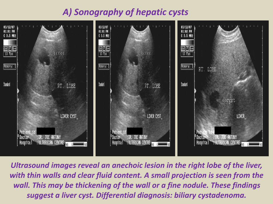
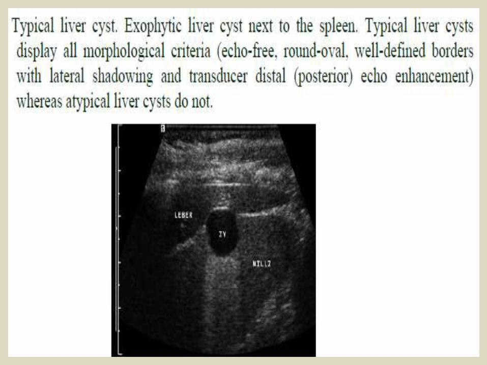
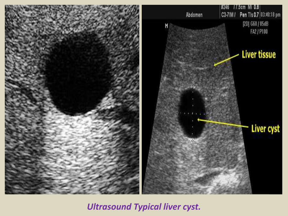
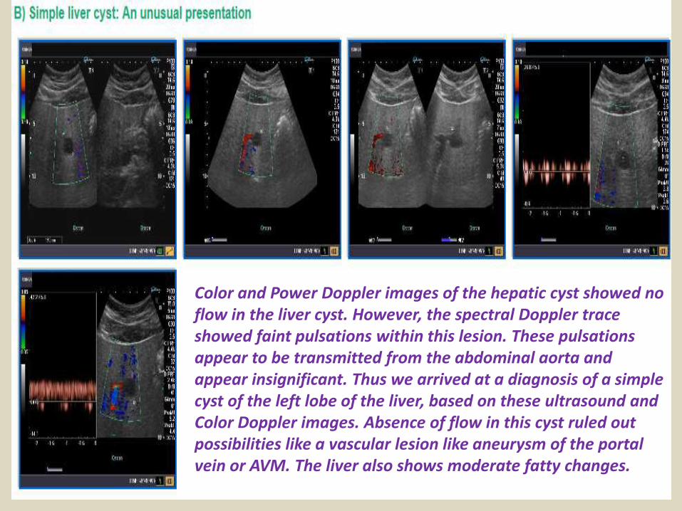
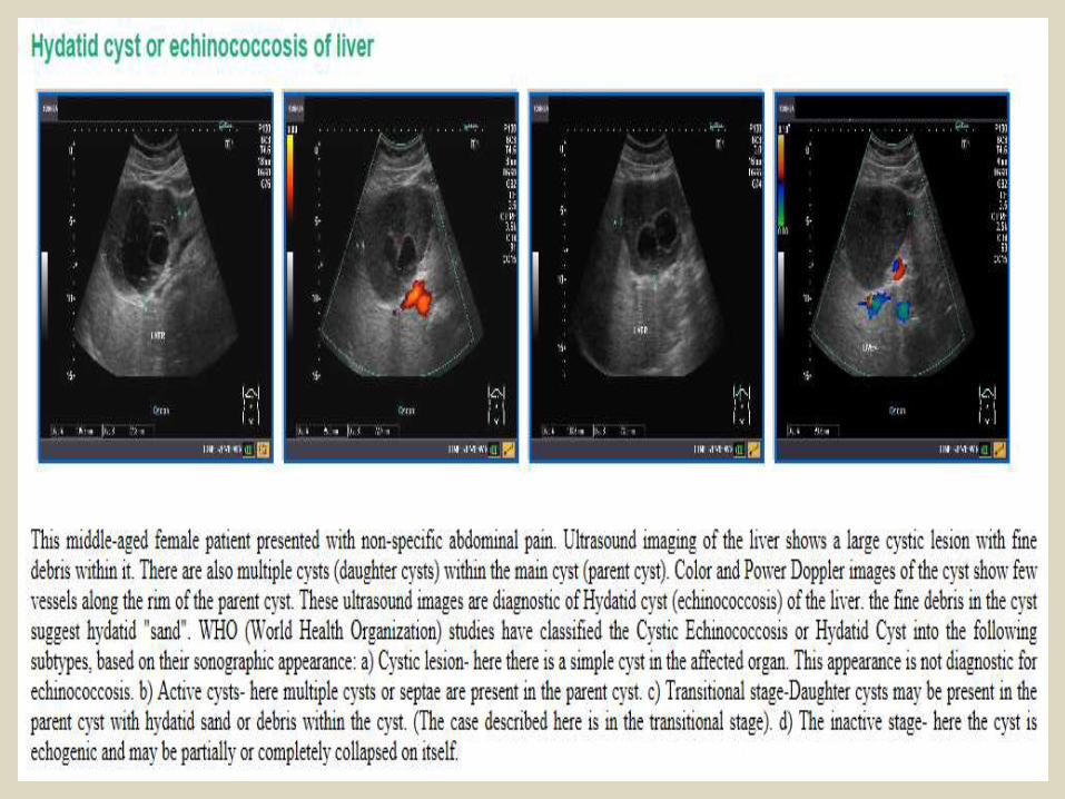
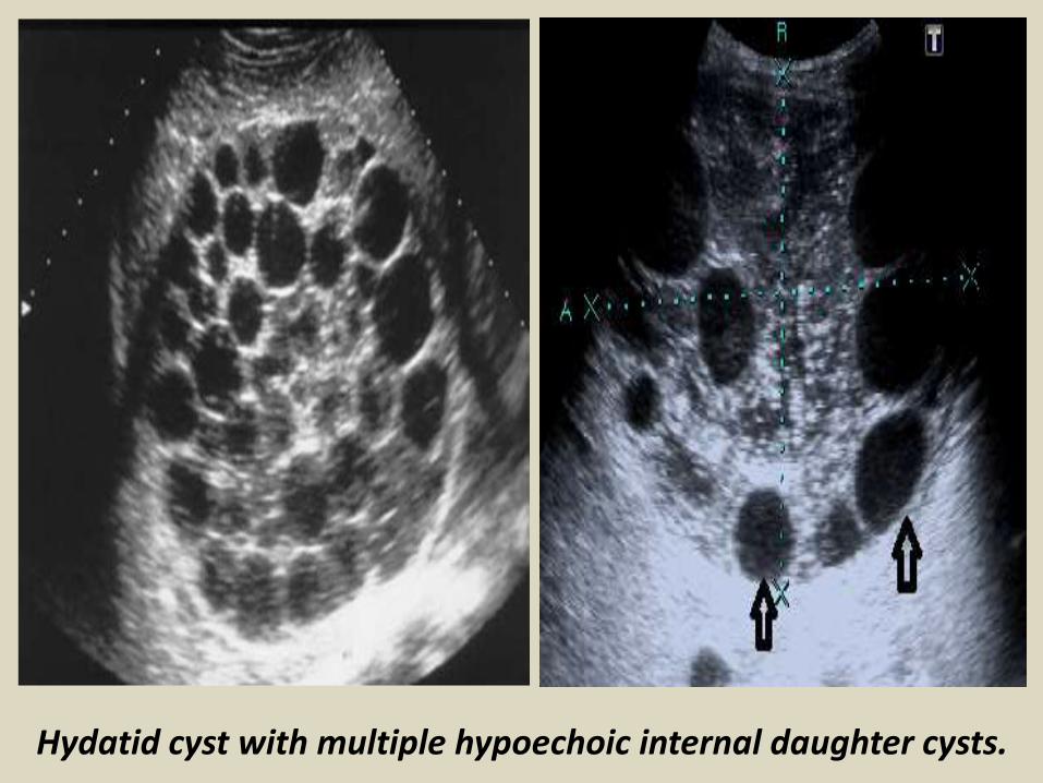
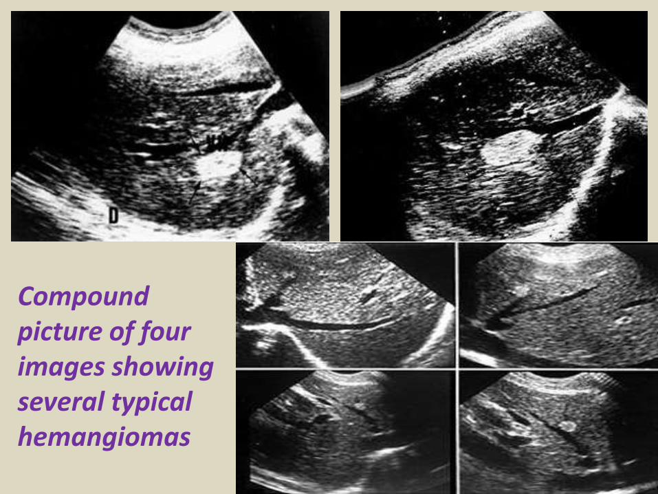
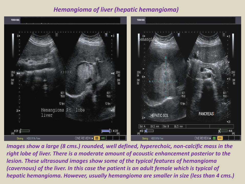
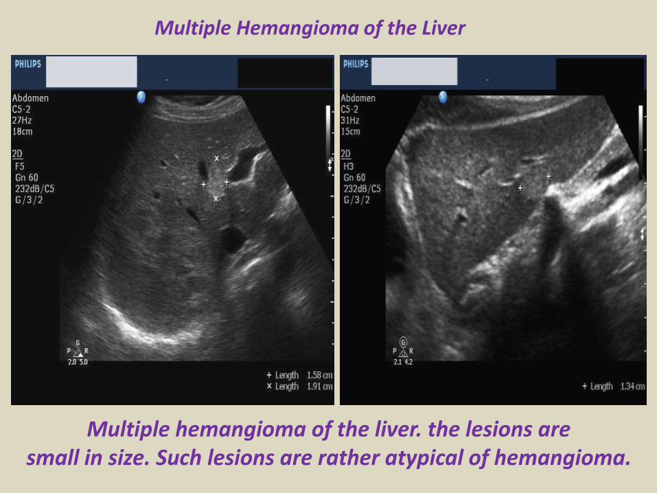
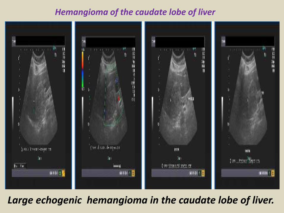
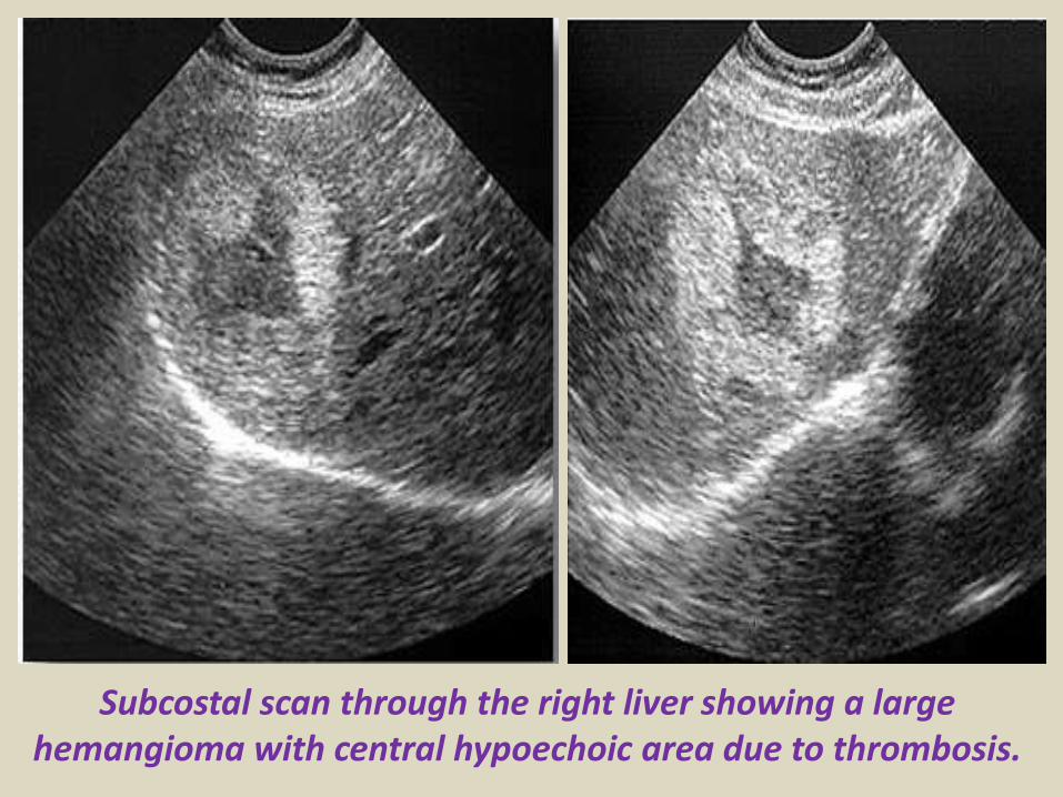
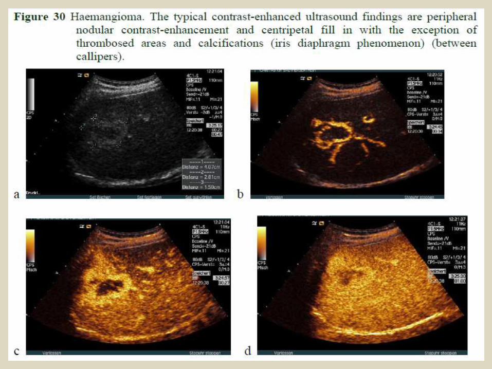
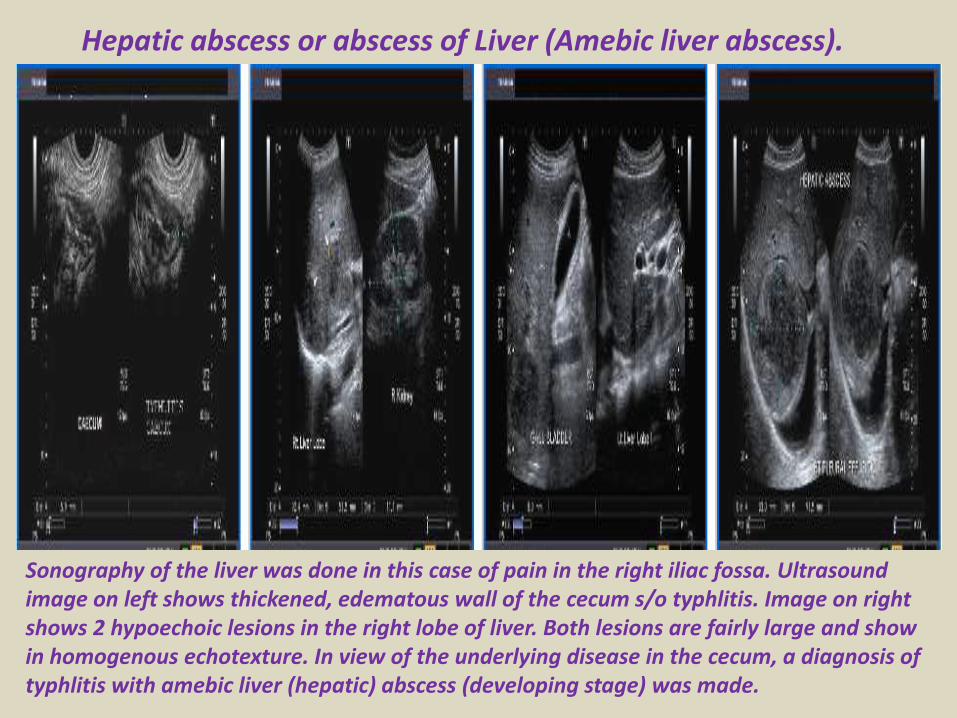
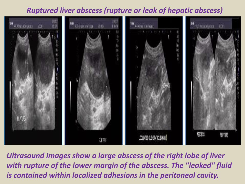
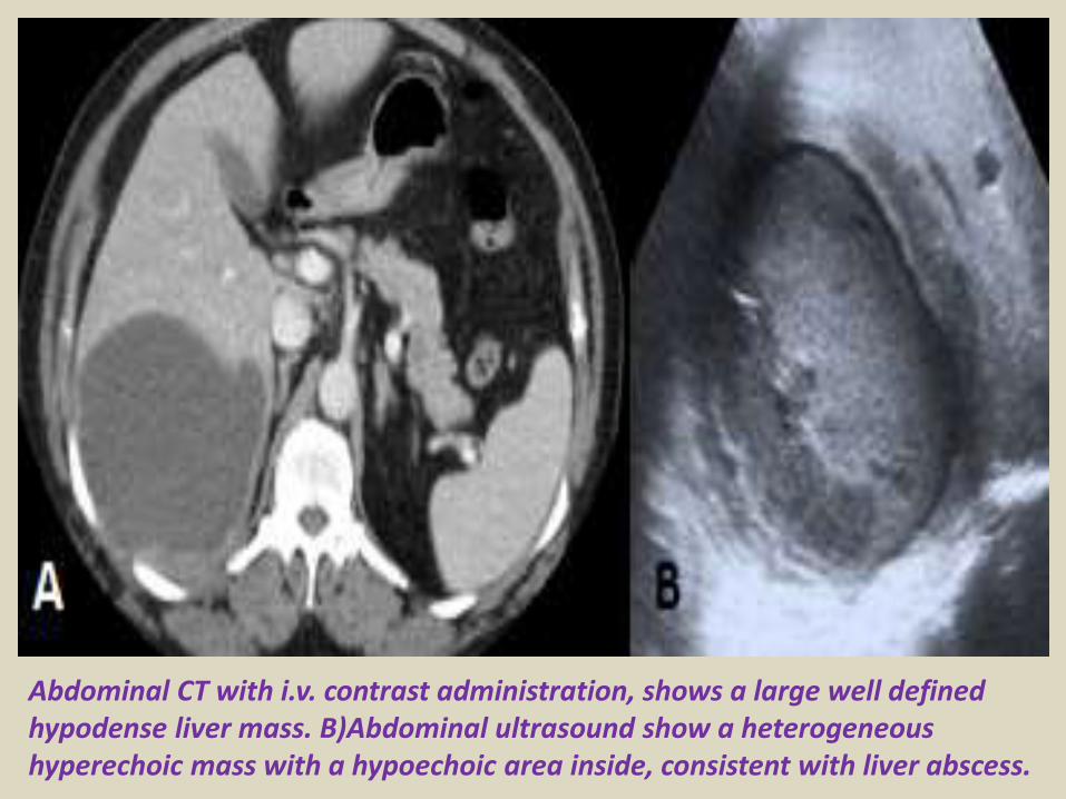
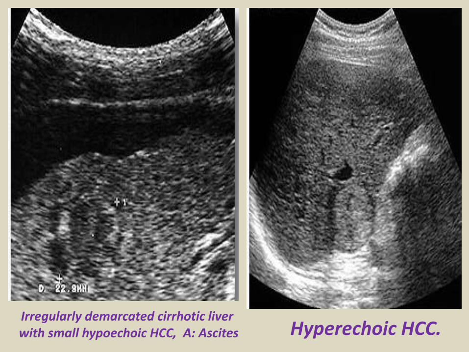
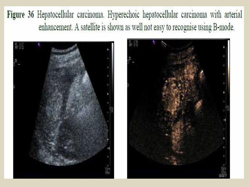
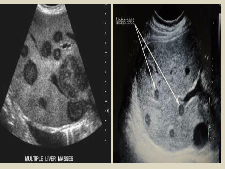
Portal hypertension
Portal hypertension
Cavernous transformation of the portal vein
Portal vein thrombosis
Acute thrombosis of the portal vein with echogenic thrombus inside
Partial thrombosis of the portal vein
Ultrasound of liver graft in 56-year-old man with hepatic artery thrombosis Color Doppler ultrasound shows absence of flow in hepatic artery at hilus (arrow)
Ultrasound of liver graft in 56-year-old man with hepatic artery thrombosis Contrast-enhanced ultrasound reveals no arterial
perfusion in early phase at hilus level (arrow) nor at intrahepatic level
Hepatic artery stenosis in 39-year-old man Doppler ultrasound of hepatic artery at intrahepatic level shows prolonged acceleration time of 0153 second
Reversed flow in the hepatic vein due to obstruction
Reversed flow in the hepatic vein due to obstruction
Right hepatic vein thrombosis
Ultrasound of the Gall bladder
Fundus
Body
Normal US ofGall Bladder
Ultrasound of the Gallbladder ndash ProtocolPatient PreparationFast for 6 hours No food or drinkPreferably book the appointment in the morning to reduce bowel gas
Scanning TechniqueLooking supine left lateral decubitus and erectUse the liver as a window especially when rolling the pt onto their left sideMeasure the wall lt3mmIs the gallbladder enlargedgt10cm in lengthCheck with colour Doppler for increased vascularity of the wallAssess the cystic duct neck body and fundus (sometimes there is a phrygian cap)
Normal Scanning Position to take advantage of using the liver as a window and displacing the bowel
A normal Gallbladder should be thin walled (lt3mm) and anechoic It is a pear shaped saccular structure for bile storage in the Right Upper Quadrant Its size varies depending on the amount of bile Fasted it will be approximately 10cm long
Folds are commonly seen and are normal Make note if pathology such as calculi are contained within a
compartment created by a fold
A Phrygian cap is a specific relatively common inversion of the distal fundus of the gallbladder into the body It may become adherent It is an anatomic variant or acquired abnormality
Ultrasonography with the typical technique and the probe placed at the right hypochondrium could not locate the gallbladder at the plane of the liver hilum
Congenital agenesis of the gall bladder
Congenital agenesis of the gall bladder
Multiple gall bladder multiple septations forming a honeycomb appearance
Duplicated gallbladder at US MRI and cholangiography with double cystic ducts
Gall bladder stones and sludgeUS criteria for diagnosis of cholelithiasis1- Echogenic focus2- Cast acoustic shadow3- Seek gravitational dependence
Wall-echo-shadow sign with multiple stonesMultiple stones with confluent shadowsSingle stone with clean shadow
Acute cholecystitis is swelling (inflammation) of the gallbladder It is
a potentially serious condition that usually needs to be treated in hospitalAcute cholecystitis is either calcular(95) or non calcular (5) The main symptom of acute cholecystitis is a sudden sharp pain in the upper right side of your tummy (abdomen) that spreads towards your right shoulderThe affected part of the abdomen is usually extremely tender and breathing deeply can make the pain worseUnlike some others types of abdominal pain the pain associated with acute cholecystitis is usually persistent and doesnt go away within a few hoursSome people may additional symptoms such asa high temperature (fever) nausea and vomiting sweating loss of appetite yellowing of the skin and the whites of the eyes (jaundice) a bulge in the abdomen
US- GB wall in acute cholecystitis appears as a thin hypoechoic line sandwiched between two echogenic lines
CT- thick wall with hypodense outer layer (subserosal edema)
Acute calculus cholecystitis
Acute acalculous cholecystitis LEFT US at the spot of maximum tenderness shows mural thickening of the gallbladder (arrow) that is completely filled with sludge (asterix) without any stones RIGHT Power-Doppler sonography shows hypervascularity of the gallbladder wall (arrowhead) as a supporting sign of inflammation
Acute gangrenous cholecystitis
Xanthogranulomatous cholecystitisIntramural hypoechoichypoattenuating foci of abscess and inflammation
Emphysematous cholecystitis (A) and (B) TS and LS with gas and debris in thegallbladder lumen (C) Gas in the gallbladder lumen completely obscures the content
Emphysematous cholecystitis
CT scan showing markedly enlarged gall bladder with intraluminal blood and thrombus Hemorrhagic cholecystitis
Hemorrhagic cholecystitis with blood within the bladder lumen
Chronic cholecystitis Chronic cholecystitis is gallbladder inflammation that has lasted a long time It almost always results from gallstones and from prior attacks of acute cholecystitis Chronic cholecystitis is characterized by repeated attacks of pain (biliary colic) that occur when gallstones periodically block the cystic ductIn chronic cholecystitis the gallbladder is damaged by repeated attacks of acute inflammation usually due to gallstones and may become thick-walled scarred and small The gallstones may block the opening of the gallbladder into the cystic duct or block the cystic duct itself The gallbladder usually also contains sludge If scarring is extensive calcium may be deposited in the walls of the gallbladder causing them to harden (called porcelain gallbladder)
SymptomsA gallbladder attack whether in acute or chronic cholecystitis begins as pain
Chronic calcular cholecystitis
Chronic calcular cholecystitis
Distended gall bladder containing echogenic gravity dependent mobile biliary sludge with calculi
Porcelain GBRare due to chronic inflammation mural calcification
Biliary sludge is a mixture of particulate solids that have precipitated from bile Such sediment consists of cholesterol crystals calcium bilirubinate pigment and other calcium salts Sludge is usually detected on transabdominal ultrasonography Microscopy of aspirated bile and endoscopic ultrasonography are far more sensitive Biliary sludge is associated with pregnancy with rapid weight loss particularly in the obese with critical illness involving low or absent oral intake and the use of total parenteral nutrition (TPN) Complications caused by biliary sludge include biliary colic acute cholangitis and acute pancreatitis Asymptomatic patients with sludge or microlithiasis require no therapy
Biliary sludge
Biliary sludge
Gallbladder polyps are relatively frequent seen in up to 5 of the
population Over 90 are benign and the majority are cholesterol polypsCholesterol polyps are most frequently identified in patients between 40-50 years of age and are more common in women (FM 291)
Tiny polyp of the gall bladder wall
Multiple gall bladder polyps
Large gall bladder polyp
Adenomyomatosis of the gallbladder is a hyperplastic
cholestosis of the gallbladder wall It is a relatively common and benign cause of focal gallbladder wall thickening It is most easily seen on ultrasound and MRI Adenomyomatosis is relatively common found in ~5 of all cholecystectomy specimens It is typically seen in patients in their 5th decade The incidence increases with age which may be the result of protracted inflammation (see below) There is a female predilection (MF = 13) It is most often an incidental finding has no intrinsic malignant potential and usually requires no treatment Three morphological types of adenomyomatosis are describedfundal (localized)segmental (annular)generalized (diffuse)
Segmental adenomyomatosis of gall bladder
Diffuse adenomyomatosis of gall bladder
Adenomyomatosis of the gallbladder with a thickened wall Color Dopplershows twinkling artefacts caused by cholesterol crystals in the wall
Adenomyomatosis of the gallbladder with a thickened wall Color Doppler shows twinkling artefacts caused by cholesterol crystals in the wall
Gallbladder adenocarcinomas is the most frequent and are usually asymptomatic until it is no longer curable As such early incidental detection is important if the occasional patient is to be successfully treated Less frequent malignancies is squamous cell carcinoma Small cell carcinoma Carcinosarcoma Lymphoma and metastasisEpidemiologyAlthough overall uncommon gallbladder adenocarcinoma is the most common primary hepatobiliary carcinoma and the 5th most common malignancy of the gastrointestinal tract Predominantly affects older persons with long-standing cholecystolithiasis and as such is most common in elderly women (gt 60 years of age FM ratio = 41) Risk factors include chronic cholecystitis gallstones are seen in 70-90 of casesfamilial adenomatous polyposis syndrome (FAP)inflammatory bowel disease (IBD)porcelain gallbladder
US of Gall bladder carcinoma
Gallbladder adenocarcinoma
US shows echogenic mass adherent to gallbladder fundus (arrows) CT shows enhancing mass within the gallbladder (arrows) with involvement of the liver (arrowhead)
Gallbladder carcinoma
B-cell lymphoma of the gallbladder
50-year-old man with renal-cell carcinoma Intraoperative ultrasound image of polypoid gallbladder lesion Postcontrast axial CT image demonstrates
avidly contrast-enhancing polypoid gallbladder lesion (arrow)
Thank You
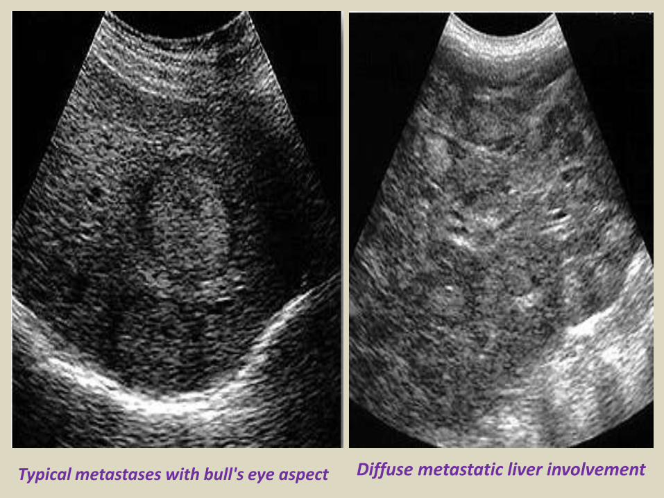
Portal hypertension
Cavernous transformation of the portal vein
Portal vein thrombosis
Acute thrombosis of the portal vein with echogenic thrombus inside
Partial thrombosis of the portal vein
Ultrasound of liver graft in 56-year-old man with hepatic artery thrombosis Color Doppler ultrasound shows absence of flow in hepatic artery at hilus (arrow)
Ultrasound of liver graft in 56-year-old man with hepatic artery thrombosis Contrast-enhanced ultrasound reveals no arterial
perfusion in early phase at hilus level (arrow) nor at intrahepatic level
Hepatic artery stenosis in 39-year-old man Doppler ultrasound of hepatic artery at intrahepatic level shows prolonged acceleration time of 0153 second
Reversed flow in the hepatic vein due to obstruction
Reversed flow in the hepatic vein due to obstruction
Right hepatic vein thrombosis
Ultrasound of the Gall bladder
Fundus
Body
Normal US ofGall Bladder
Ultrasound of the Gallbladder ndash ProtocolPatient PreparationFast for 6 hours No food or drinkPreferably book the appointment in the morning to reduce bowel gas
Scanning TechniqueLooking supine left lateral decubitus and erectUse the liver as a window especially when rolling the pt onto their left sideMeasure the wall lt3mmIs the gallbladder enlargedgt10cm in lengthCheck with colour Doppler for increased vascularity of the wallAssess the cystic duct neck body and fundus (sometimes there is a phrygian cap)
Normal Scanning Position to take advantage of using the liver as a window and displacing the bowel
A normal Gallbladder should be thin walled (lt3mm) and anechoic It is a pear shaped saccular structure for bile storage in the Right Upper Quadrant Its size varies depending on the amount of bile Fasted it will be approximately 10cm long
Folds are commonly seen and are normal Make note if pathology such as calculi are contained within a
compartment created by a fold
A Phrygian cap is a specific relatively common inversion of the distal fundus of the gallbladder into the body It may become adherent It is an anatomic variant or acquired abnormality
Ultrasonography with the typical technique and the probe placed at the right hypochondrium could not locate the gallbladder at the plane of the liver hilum
Congenital agenesis of the gall bladder
Congenital agenesis of the gall bladder
Multiple gall bladder multiple septations forming a honeycomb appearance
Duplicated gallbladder at US MRI and cholangiography with double cystic ducts
Gall bladder stones and sludgeUS criteria for diagnosis of cholelithiasis1- Echogenic focus2- Cast acoustic shadow3- Seek gravitational dependence
Wall-echo-shadow sign with multiple stonesMultiple stones with confluent shadowsSingle stone with clean shadow
Acute cholecystitis is swelling (inflammation) of the gallbladder It is
a potentially serious condition that usually needs to be treated in hospitalAcute cholecystitis is either calcular(95) or non calcular (5) The main symptom of acute cholecystitis is a sudden sharp pain in the upper right side of your tummy (abdomen) that spreads towards your right shoulderThe affected part of the abdomen is usually extremely tender and breathing deeply can make the pain worseUnlike some others types of abdominal pain the pain associated with acute cholecystitis is usually persistent and doesnt go away within a few hoursSome people may additional symptoms such asa high temperature (fever) nausea and vomiting sweating loss of appetite yellowing of the skin and the whites of the eyes (jaundice) a bulge in the abdomen
US- GB wall in acute cholecystitis appears as a thin hypoechoic line sandwiched between two echogenic lines
CT- thick wall with hypodense outer layer (subserosal edema)
Acute calculus cholecystitis
Acute acalculous cholecystitis LEFT US at the spot of maximum tenderness shows mural thickening of the gallbladder (arrow) that is completely filled with sludge (asterix) without any stones RIGHT Power-Doppler sonography shows hypervascularity of the gallbladder wall (arrowhead) as a supporting sign of inflammation
Acute gangrenous cholecystitis
Xanthogranulomatous cholecystitisIntramural hypoechoichypoattenuating foci of abscess and inflammation
Emphysematous cholecystitis (A) and (B) TS and LS with gas and debris in thegallbladder lumen (C) Gas in the gallbladder lumen completely obscures the content
Emphysematous cholecystitis
CT scan showing markedly enlarged gall bladder with intraluminal blood and thrombus Hemorrhagic cholecystitis
Hemorrhagic cholecystitis with blood within the bladder lumen
Chronic cholecystitis Chronic cholecystitis is gallbladder inflammation that has lasted a long time It almost always results from gallstones and from prior attacks of acute cholecystitis Chronic cholecystitis is characterized by repeated attacks of pain (biliary colic) that occur when gallstones periodically block the cystic ductIn chronic cholecystitis the gallbladder is damaged by repeated attacks of acute inflammation usually due to gallstones and may become thick-walled scarred and small The gallstones may block the opening of the gallbladder into the cystic duct or block the cystic duct itself The gallbladder usually also contains sludge If scarring is extensive calcium may be deposited in the walls of the gallbladder causing them to harden (called porcelain gallbladder)
SymptomsA gallbladder attack whether in acute or chronic cholecystitis begins as pain
Chronic calcular cholecystitis
Chronic calcular cholecystitis
Distended gall bladder containing echogenic gravity dependent mobile biliary sludge with calculi
Porcelain GBRare due to chronic inflammation mural calcification
Biliary sludge is a mixture of particulate solids that have precipitated from bile Such sediment consists of cholesterol crystals calcium bilirubinate pigment and other calcium salts Sludge is usually detected on transabdominal ultrasonography Microscopy of aspirated bile and endoscopic ultrasonography are far more sensitive Biliary sludge is associated with pregnancy with rapid weight loss particularly in the obese with critical illness involving low or absent oral intake and the use of total parenteral nutrition (TPN) Complications caused by biliary sludge include biliary colic acute cholangitis and acute pancreatitis Asymptomatic patients with sludge or microlithiasis require no therapy
Biliary sludge
Biliary sludge
Gallbladder polyps are relatively frequent seen in up to 5 of the
population Over 90 are benign and the majority are cholesterol polypsCholesterol polyps are most frequently identified in patients between 40-50 years of age and are more common in women (FM 291)
Tiny polyp of the gall bladder wall
Multiple gall bladder polyps
Large gall bladder polyp
Adenomyomatosis of the gallbladder is a hyperplastic
cholestosis of the gallbladder wall It is a relatively common and benign cause of focal gallbladder wall thickening It is most easily seen on ultrasound and MRI Adenomyomatosis is relatively common found in ~5 of all cholecystectomy specimens It is typically seen in patients in their 5th decade The incidence increases with age which may be the result of protracted inflammation (see below) There is a female predilection (MF = 13) It is most often an incidental finding has no intrinsic malignant potential and usually requires no treatment Three morphological types of adenomyomatosis are describedfundal (localized)segmental (annular)generalized (diffuse)
Segmental adenomyomatosis of gall bladder
Diffuse adenomyomatosis of gall bladder
Adenomyomatosis of the gallbladder with a thickened wall Color Dopplershows twinkling artefacts caused by cholesterol crystals in the wall
Adenomyomatosis of the gallbladder with a thickened wall Color Doppler shows twinkling artefacts caused by cholesterol crystals in the wall
Gallbladder adenocarcinomas is the most frequent and are usually asymptomatic until it is no longer curable As such early incidental detection is important if the occasional patient is to be successfully treated Less frequent malignancies is squamous cell carcinoma Small cell carcinoma Carcinosarcoma Lymphoma and metastasisEpidemiologyAlthough overall uncommon gallbladder adenocarcinoma is the most common primary hepatobiliary carcinoma and the 5th most common malignancy of the gastrointestinal tract Predominantly affects older persons with long-standing cholecystolithiasis and as such is most common in elderly women (gt 60 years of age FM ratio = 41) Risk factors include chronic cholecystitis gallstones are seen in 70-90 of casesfamilial adenomatous polyposis syndrome (FAP)inflammatory bowel disease (IBD)porcelain gallbladder
US of Gall bladder carcinoma
Gallbladder adenocarcinoma
US shows echogenic mass adherent to gallbladder fundus (arrows) CT shows enhancing mass within the gallbladder (arrows) with involvement of the liver (arrowhead)
Gallbladder carcinoma
B-cell lymphoma of the gallbladder
50-year-old man with renal-cell carcinoma Intraoperative ultrasound image of polypoid gallbladder lesion Postcontrast axial CT image demonstrates
avidly contrast-enhancing polypoid gallbladder lesion (arrow)
Thank You
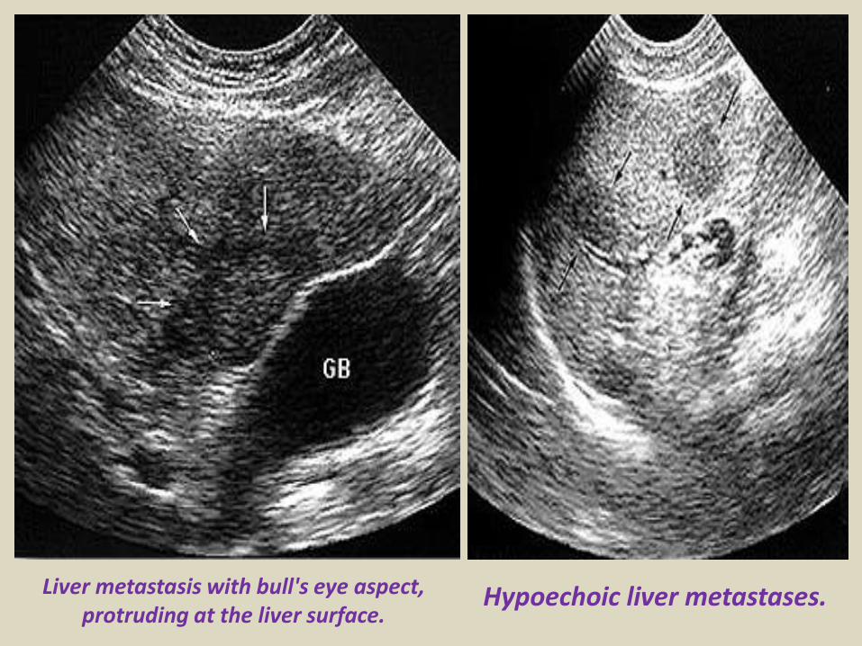
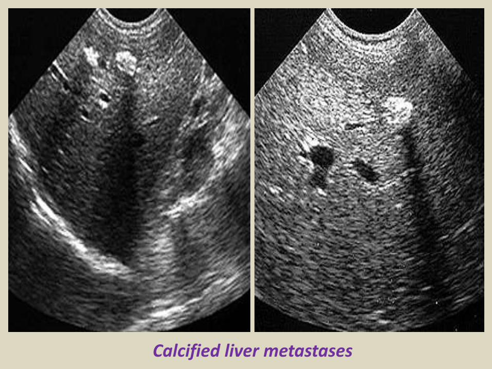
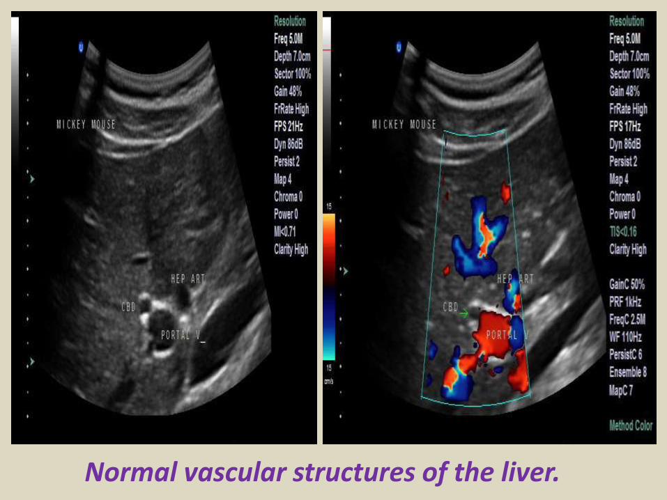
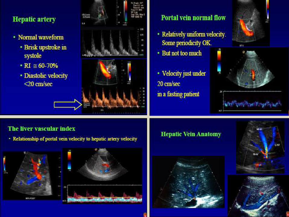
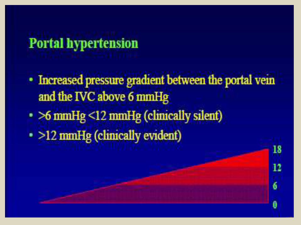
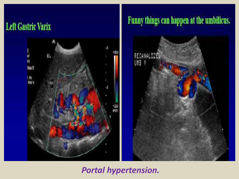
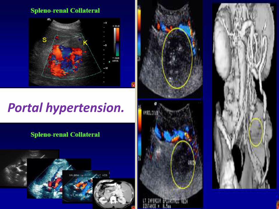
Reversed flow in the hepatic vein due to obstruction
Reversed flow in the hepatic vein due to obstruction
Right hepatic vein thrombosis
Ultrasound of the Gall bladder
Fundus
Body
Normal US ofGall Bladder
Ultrasound of the Gallbladder ndash ProtocolPatient PreparationFast for 6 hours No food or drinkPreferably book the appointment in the morning to reduce bowel gas
Scanning TechniqueLooking supine left lateral decubitus and erectUse the liver as a window especially when rolling the pt onto their left sideMeasure the wall lt3mmIs the gallbladder enlargedgt10cm in lengthCheck with colour Doppler for increased vascularity of the wallAssess the cystic duct neck body and fundus (sometimes there is a phrygian cap)
Normal Scanning Position to take advantage of using the liver as a window and displacing the bowel
A normal Gallbladder should be thin walled (lt3mm) and anechoic It is a pear shaped saccular structure for bile storage in the Right Upper Quadrant Its size varies depending on the amount of bile Fasted it will be approximately 10cm long
Folds are commonly seen and are normal Make note if pathology such as calculi are contained within a
compartment created by a fold
A Phrygian cap is a specific relatively common inversion of the distal fundus of the gallbladder into the body It may become adherent It is an anatomic variant or acquired abnormality
Ultrasonography with the typical technique and the probe placed at the right hypochondrium could not locate the gallbladder at the plane of the liver hilum
Congenital agenesis of the gall bladder
Congenital agenesis of the gall bladder
Multiple gall bladder multiple septations forming a honeycomb appearance
Duplicated gallbladder at US MRI and cholangiography with double cystic ducts
Gall bladder stones and sludgeUS criteria for diagnosis of cholelithiasis1- Echogenic focus2- Cast acoustic shadow3- Seek gravitational dependence
Wall-echo-shadow sign with multiple stonesMultiple stones with confluent shadowsSingle stone with clean shadow
Acute cholecystitis is swelling (inflammation) of the gallbladder It is
a potentially serious condition that usually needs to be treated in hospitalAcute cholecystitis is either calcular(95) or non calcular (5) The main symptom of acute cholecystitis is a sudden sharp pain in the upper right side of your tummy (abdomen) that spreads towards your right shoulderThe affected part of the abdomen is usually extremely tender and breathing deeply can make the pain worseUnlike some others types of abdominal pain the pain associated with acute cholecystitis is usually persistent and doesnt go away within a few hoursSome people may additional symptoms such asa high temperature (fever) nausea and vomiting sweating loss of appetite yellowing of the skin and the whites of the eyes (jaundice) a bulge in the abdomen
US- GB wall in acute cholecystitis appears as a thin hypoechoic line sandwiched between two echogenic lines
CT- thick wall with hypodense outer layer (subserosal edema)
Acute calculus cholecystitis
Acute acalculous cholecystitis LEFT US at the spot of maximum tenderness shows mural thickening of the gallbladder (arrow) that is completely filled with sludge (asterix) without any stones RIGHT Power-Doppler sonography shows hypervascularity of the gallbladder wall (arrowhead) as a supporting sign of inflammation
Acute gangrenous cholecystitis
Xanthogranulomatous cholecystitisIntramural hypoechoichypoattenuating foci of abscess and inflammation
Emphysematous cholecystitis (A) and (B) TS and LS with gas and debris in thegallbladder lumen (C) Gas in the gallbladder lumen completely obscures the content
Emphysematous cholecystitis
CT scan showing markedly enlarged gall bladder with intraluminal blood and thrombus Hemorrhagic cholecystitis
Hemorrhagic cholecystitis with blood within the bladder lumen
Chronic cholecystitis Chronic cholecystitis is gallbladder inflammation that has lasted a long time It almost always results from gallstones and from prior attacks of acute cholecystitis Chronic cholecystitis is characterized by repeated attacks of pain (biliary colic) that occur when gallstones periodically block the cystic ductIn chronic cholecystitis the gallbladder is damaged by repeated attacks of acute inflammation usually due to gallstones and may become thick-walled scarred and small The gallstones may block the opening of the gallbladder into the cystic duct or block the cystic duct itself The gallbladder usually also contains sludge If scarring is extensive calcium may be deposited in the walls of the gallbladder causing them to harden (called porcelain gallbladder)
SymptomsA gallbladder attack whether in acute or chronic cholecystitis begins as pain
Chronic calcular cholecystitis
Chronic calcular cholecystitis
Distended gall bladder containing echogenic gravity dependent mobile biliary sludge with calculi
Porcelain GBRare due to chronic inflammation mural calcification
Biliary sludge is a mixture of particulate solids that have precipitated from bile Such sediment consists of cholesterol crystals calcium bilirubinate pigment and other calcium salts Sludge is usually detected on transabdominal ultrasonography Microscopy of aspirated bile and endoscopic ultrasonography are far more sensitive Biliary sludge is associated with pregnancy with rapid weight loss particularly in the obese with critical illness involving low or absent oral intake and the use of total parenteral nutrition (TPN) Complications caused by biliary sludge include biliary colic acute cholangitis and acute pancreatitis Asymptomatic patients with sludge or microlithiasis require no therapy
Biliary sludge
Biliary sludge
Gallbladder polyps are relatively frequent seen in up to 5 of the
population Over 90 are benign and the majority are cholesterol polypsCholesterol polyps are most frequently identified in patients between 40-50 years of age and are more common in women (FM 291)
Tiny polyp of the gall bladder wall
Multiple gall bladder polyps
Large gall bladder polyp
Adenomyomatosis of the gallbladder is a hyperplastic
cholestosis of the gallbladder wall It is a relatively common and benign cause of focal gallbladder wall thickening It is most easily seen on ultrasound and MRI Adenomyomatosis is relatively common found in ~5 of all cholecystectomy specimens It is typically seen in patients in their 5th decade The incidence increases with age which may be the result of protracted inflammation (see below) There is a female predilection (MF = 13) It is most often an incidental finding has no intrinsic malignant potential and usually requires no treatment Three morphological types of adenomyomatosis are describedfundal (localized)segmental (annular)generalized (diffuse)
Segmental adenomyomatosis of gall bladder
Diffuse adenomyomatosis of gall bladder
Adenomyomatosis of the gallbladder with a thickened wall Color Dopplershows twinkling artefacts caused by cholesterol crystals in the wall
Adenomyomatosis of the gallbladder with a thickened wall Color Doppler shows twinkling artefacts caused by cholesterol crystals in the wall
Gallbladder adenocarcinomas is the most frequent and are usually asymptomatic until it is no longer curable As such early incidental detection is important if the occasional patient is to be successfully treated Less frequent malignancies is squamous cell carcinoma Small cell carcinoma Carcinosarcoma Lymphoma and metastasisEpidemiologyAlthough overall uncommon gallbladder adenocarcinoma is the most common primary hepatobiliary carcinoma and the 5th most common malignancy of the gastrointestinal tract Predominantly affects older persons with long-standing cholecystolithiasis and as such is most common in elderly women (gt 60 years of age FM ratio = 41) Risk factors include chronic cholecystitis gallstones are seen in 70-90 of casesfamilial adenomatous polyposis syndrome (FAP)inflammatory bowel disease (IBD)porcelain gallbladder
US of Gall bladder carcinoma
Gallbladder adenocarcinoma
US shows echogenic mass adherent to gallbladder fundus (arrows) CT shows enhancing mass within the gallbladder (arrows) with involvement of the liver (arrowhead)
Gallbladder carcinoma
B-cell lymphoma of the gallbladder
50-year-old man with renal-cell carcinoma Intraoperative ultrasound image of polypoid gallbladder lesion Postcontrast axial CT image demonstrates
avidly contrast-enhancing polypoid gallbladder lesion (arrow)
Thank You
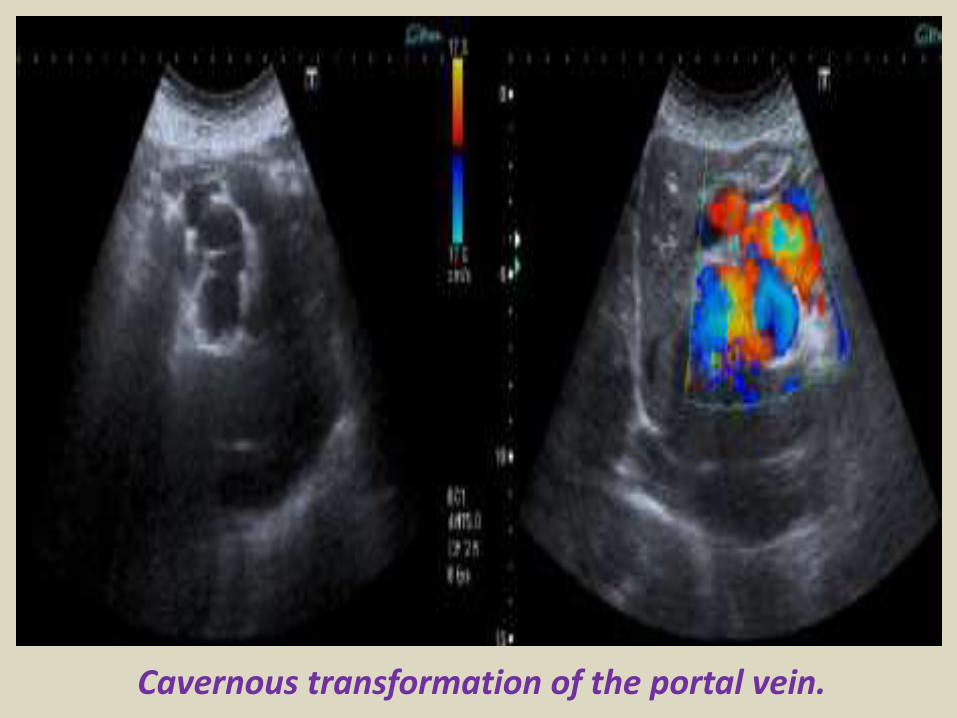
Reversed flow in the hepatic vein due to obstruction
Right hepatic vein thrombosis
Ultrasound of the Gall bladder
Fundus
Body
Normal US ofGall Bladder
Ultrasound of the Gallbladder ndash ProtocolPatient PreparationFast for 6 hours No food or drinkPreferably book the appointment in the morning to reduce bowel gas
Scanning TechniqueLooking supine left lateral decubitus and erectUse the liver as a window especially when rolling the pt onto their left sideMeasure the wall lt3mmIs the gallbladder enlargedgt10cm in lengthCheck with colour Doppler for increased vascularity of the wallAssess the cystic duct neck body and fundus (sometimes there is a phrygian cap)
Normal Scanning Position to take advantage of using the liver as a window and displacing the bowel
A normal Gallbladder should be thin walled (lt3mm) and anechoic It is a pear shaped saccular structure for bile storage in the Right Upper Quadrant Its size varies depending on the amount of bile Fasted it will be approximately 10cm long
Folds are commonly seen and are normal Make note if pathology such as calculi are contained within a
compartment created by a fold
A Phrygian cap is a specific relatively common inversion of the distal fundus of the gallbladder into the body It may become adherent It is an anatomic variant or acquired abnormality
Ultrasonography with the typical technique and the probe placed at the right hypochondrium could not locate the gallbladder at the plane of the liver hilum
Congenital agenesis of the gall bladder
Congenital agenesis of the gall bladder
Multiple gall bladder multiple septations forming a honeycomb appearance
Duplicated gallbladder at US MRI and cholangiography with double cystic ducts
Gall bladder stones and sludgeUS criteria for diagnosis of cholelithiasis1- Echogenic focus2- Cast acoustic shadow3- Seek gravitational dependence
Wall-echo-shadow sign with multiple stonesMultiple stones with confluent shadowsSingle stone with clean shadow
Acute cholecystitis is swelling (inflammation) of the gallbladder It is
a potentially serious condition that usually needs to be treated in hospitalAcute cholecystitis is either calcular(95) or non calcular (5) The main symptom of acute cholecystitis is a sudden sharp pain in the upper right side of your tummy (abdomen) that spreads towards your right shoulderThe affected part of the abdomen is usually extremely tender and breathing deeply can make the pain worseUnlike some others types of abdominal pain the pain associated with acute cholecystitis is usually persistent and doesnt go away within a few hoursSome people may additional symptoms such asa high temperature (fever) nausea and vomiting sweating loss of appetite yellowing of the skin and the whites of the eyes (jaundice) a bulge in the abdomen
US- GB wall in acute cholecystitis appears as a thin hypoechoic line sandwiched between two echogenic lines
CT- thick wall with hypodense outer layer (subserosal edema)
Acute calculus cholecystitis
Acute acalculous cholecystitis LEFT US at the spot of maximum tenderness shows mural thickening of the gallbladder (arrow) that is completely filled with sludge (asterix) without any stones RIGHT Power-Doppler sonography shows hypervascularity of the gallbladder wall (arrowhead) as a supporting sign of inflammation
Acute gangrenous cholecystitis
Xanthogranulomatous cholecystitisIntramural hypoechoichypoattenuating foci of abscess and inflammation
Emphysematous cholecystitis (A) and (B) TS and LS with gas and debris in thegallbladder lumen (C) Gas in the gallbladder lumen completely obscures the content
Emphysematous cholecystitis
CT scan showing markedly enlarged gall bladder with intraluminal blood and thrombus Hemorrhagic cholecystitis
Hemorrhagic cholecystitis with blood within the bladder lumen
Chronic cholecystitis Chronic cholecystitis is gallbladder inflammation that has lasted a long time It almost always results from gallstones and from prior attacks of acute cholecystitis Chronic cholecystitis is characterized by repeated attacks of pain (biliary colic) that occur when gallstones periodically block the cystic ductIn chronic cholecystitis the gallbladder is damaged by repeated attacks of acute inflammation usually due to gallstones and may become thick-walled scarred and small The gallstones may block the opening of the gallbladder into the cystic duct or block the cystic duct itself The gallbladder usually also contains sludge If scarring is extensive calcium may be deposited in the walls of the gallbladder causing them to harden (called porcelain gallbladder)
SymptomsA gallbladder attack whether in acute or chronic cholecystitis begins as pain
Chronic calcular cholecystitis
Chronic calcular cholecystitis
Distended gall bladder containing echogenic gravity dependent mobile biliary sludge with calculi
Porcelain GBRare due to chronic inflammation mural calcification
Biliary sludge is a mixture of particulate solids that have precipitated from bile Such sediment consists of cholesterol crystals calcium bilirubinate pigment and other calcium salts Sludge is usually detected on transabdominal ultrasonography Microscopy of aspirated bile and endoscopic ultrasonography are far more sensitive Biliary sludge is associated with pregnancy with rapid weight loss particularly in the obese with critical illness involving low or absent oral intake and the use of total parenteral nutrition (TPN) Complications caused by biliary sludge include biliary colic acute cholangitis and acute pancreatitis Asymptomatic patients with sludge or microlithiasis require no therapy
Biliary sludge
Biliary sludge
Gallbladder polyps are relatively frequent seen in up to 5 of the
population Over 90 are benign and the majority are cholesterol polypsCholesterol polyps are most frequently identified in patients between 40-50 years of age and are more common in women (FM 291)
Tiny polyp of the gall bladder wall
Multiple gall bladder polyps
Large gall bladder polyp
Adenomyomatosis of the gallbladder is a hyperplastic
cholestosis of the gallbladder wall It is a relatively common and benign cause of focal gallbladder wall thickening It is most easily seen on ultrasound and MRI Adenomyomatosis is relatively common found in ~5 of all cholecystectomy specimens It is typically seen in patients in their 5th decade The incidence increases with age which may be the result of protracted inflammation (see below) There is a female predilection (MF = 13) It is most often an incidental finding has no intrinsic malignant potential and usually requires no treatment Three morphological types of adenomyomatosis are describedfundal (localized)segmental (annular)generalized (diffuse)
Segmental adenomyomatosis of gall bladder
Diffuse adenomyomatosis of gall bladder
Adenomyomatosis of the gallbladder with a thickened wall Color Dopplershows twinkling artefacts caused by cholesterol crystals in the wall
Adenomyomatosis of the gallbladder with a thickened wall Color Doppler shows twinkling artefacts caused by cholesterol crystals in the wall
Gallbladder adenocarcinomas is the most frequent and are usually asymptomatic until it is no longer curable As such early incidental detection is important if the occasional patient is to be successfully treated Less frequent malignancies is squamous cell carcinoma Small cell carcinoma Carcinosarcoma Lymphoma and metastasisEpidemiologyAlthough overall uncommon gallbladder adenocarcinoma is the most common primary hepatobiliary carcinoma and the 5th most common malignancy of the gastrointestinal tract Predominantly affects older persons with long-standing cholecystolithiasis and as such is most common in elderly women (gt 60 years of age FM ratio = 41) Risk factors include chronic cholecystitis gallstones are seen in 70-90 of casesfamilial adenomatous polyposis syndrome (FAP)inflammatory bowel disease (IBD)porcelain gallbladder
US of Gall bladder carcinoma
Gallbladder adenocarcinoma
US shows echogenic mass adherent to gallbladder fundus (arrows) CT shows enhancing mass within the gallbladder (arrows) with involvement of the liver (arrowhead)
Gallbladder carcinoma
B-cell lymphoma of the gallbladder
50-year-old man with renal-cell carcinoma Intraoperative ultrasound image of polypoid gallbladder lesion Postcontrast axial CT image demonstrates
avidly contrast-enhancing polypoid gallbladder lesion (arrow)
Thank You
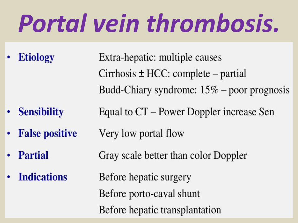
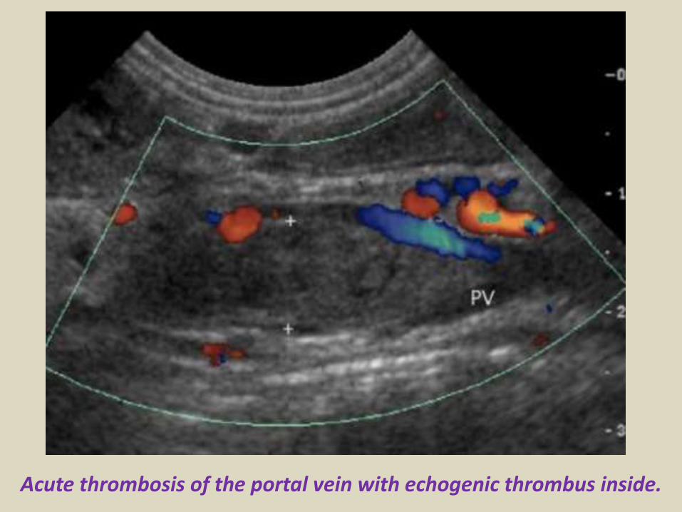
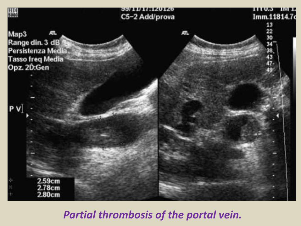
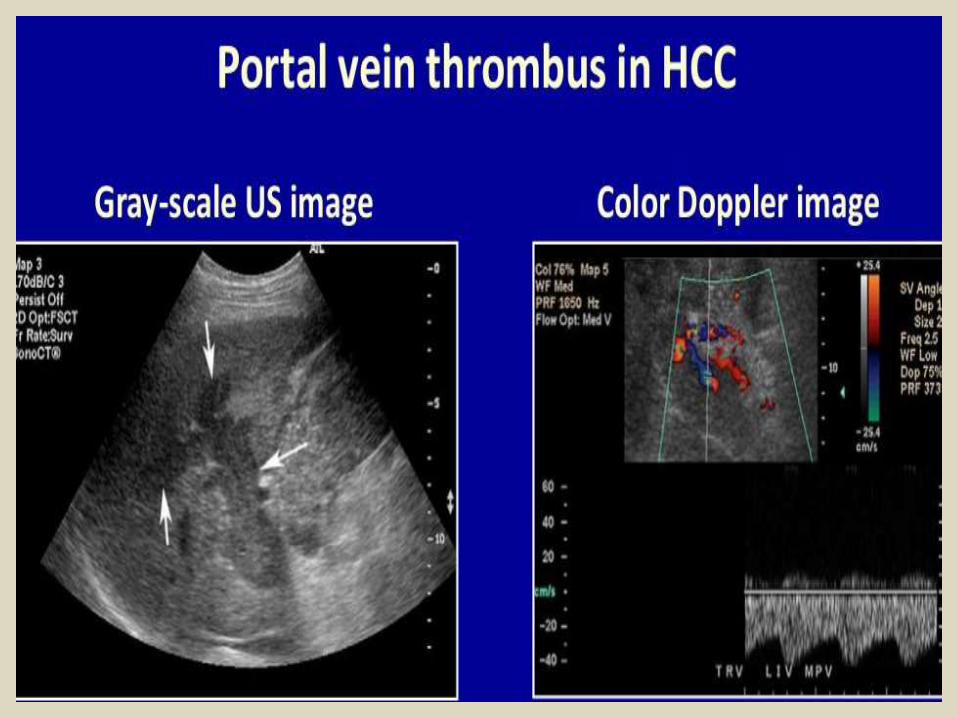
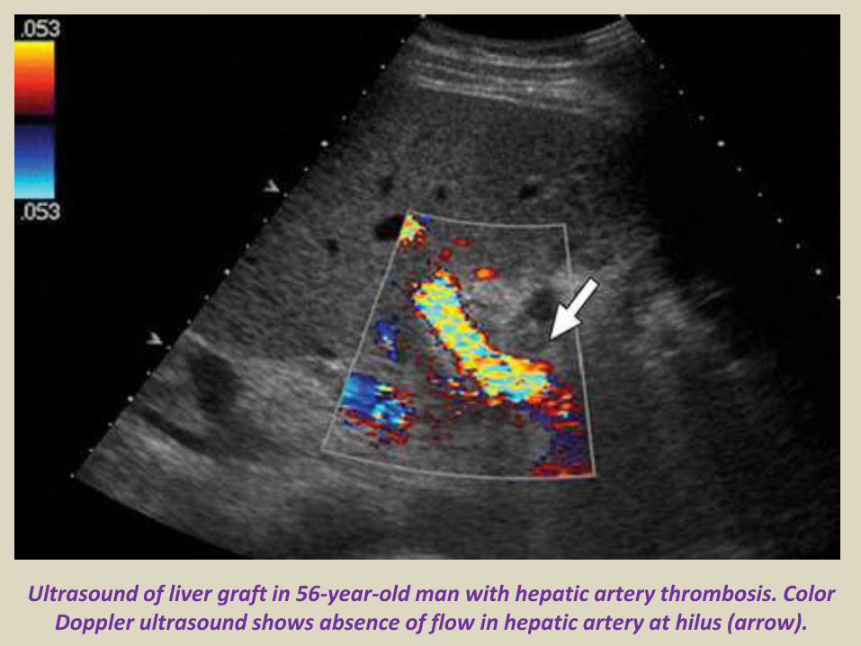
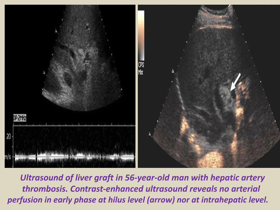
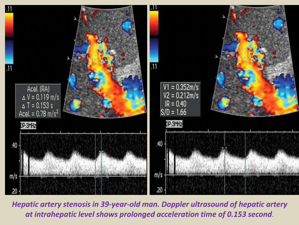
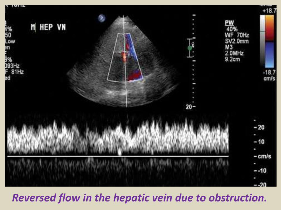
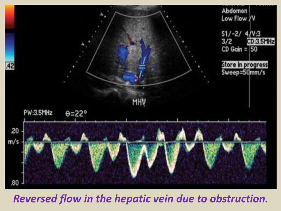
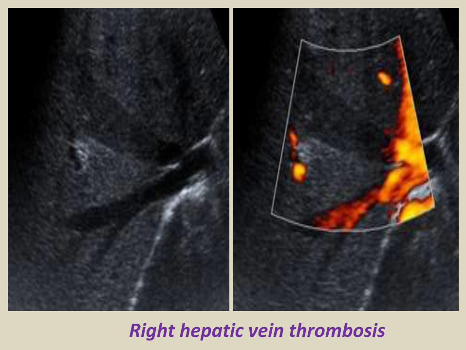
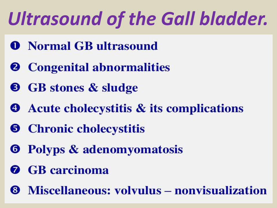
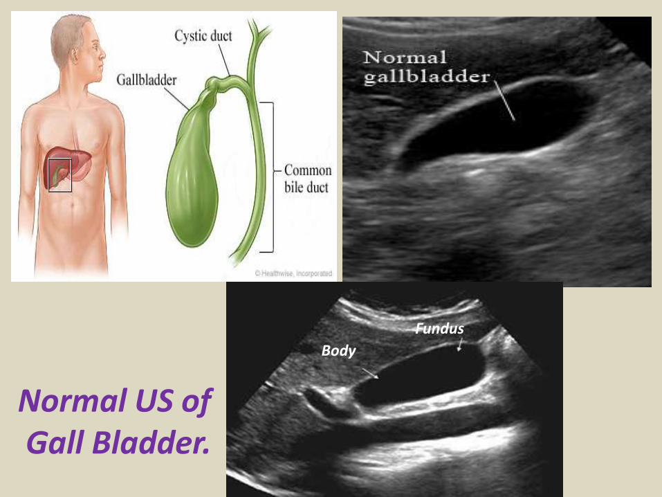
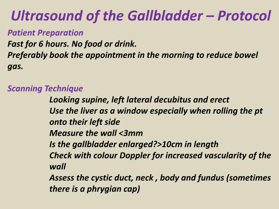
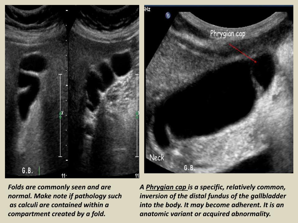
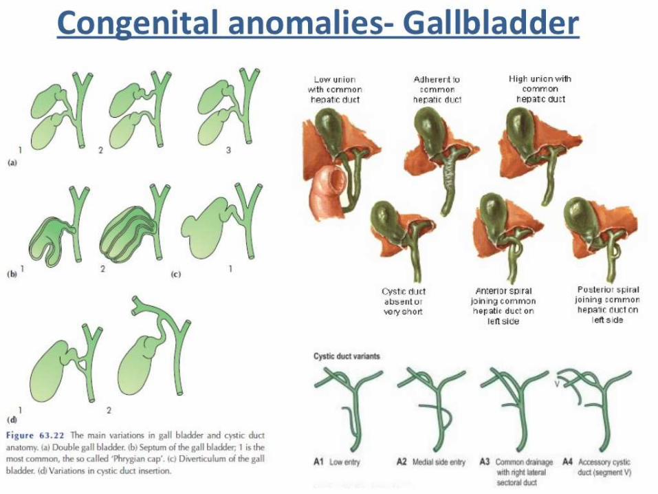
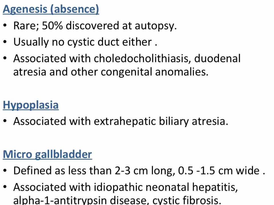
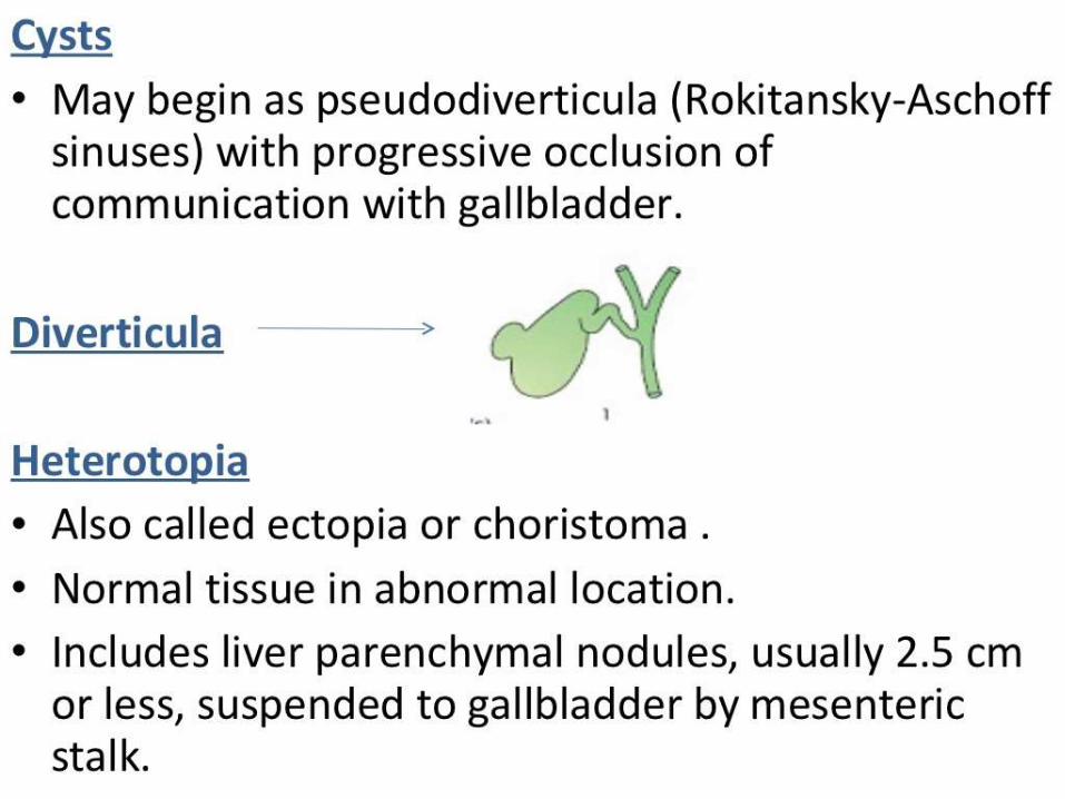
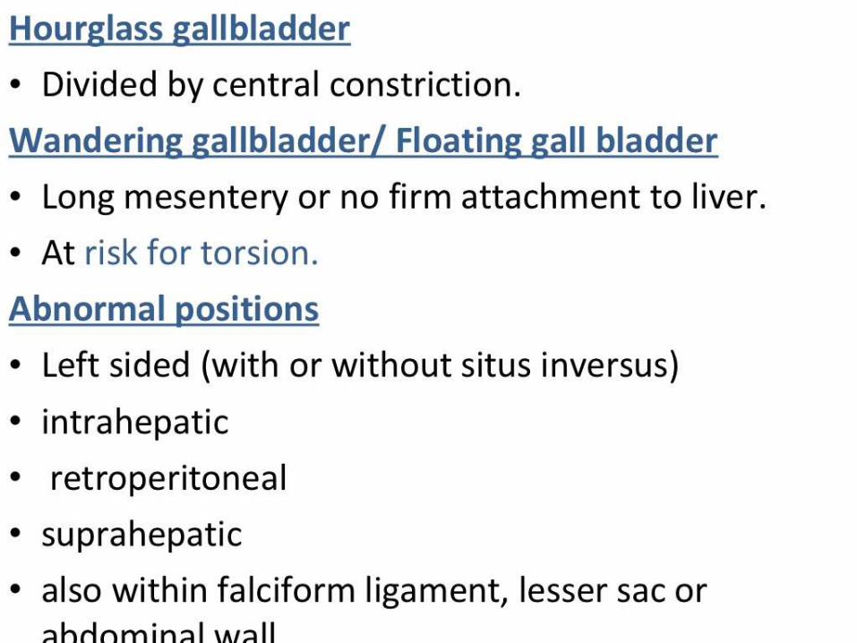
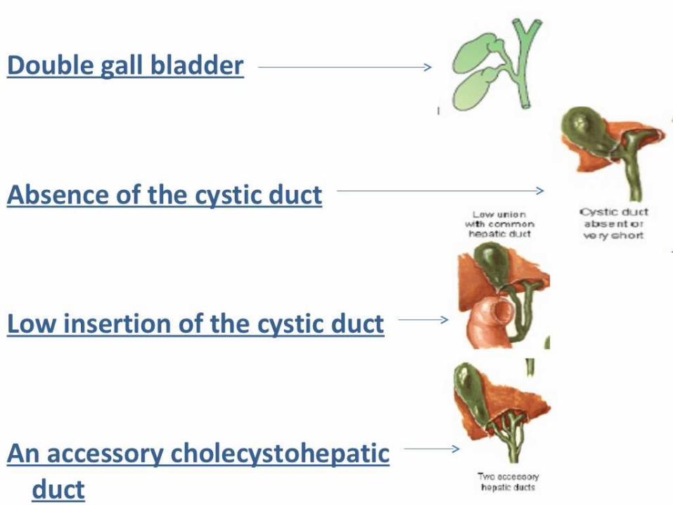
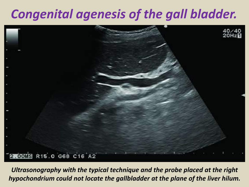
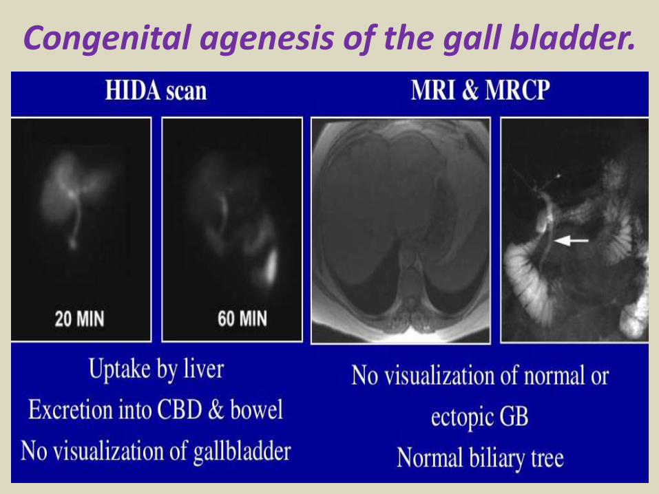
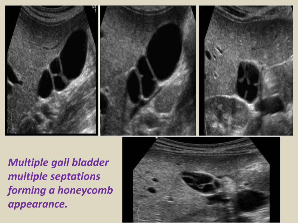
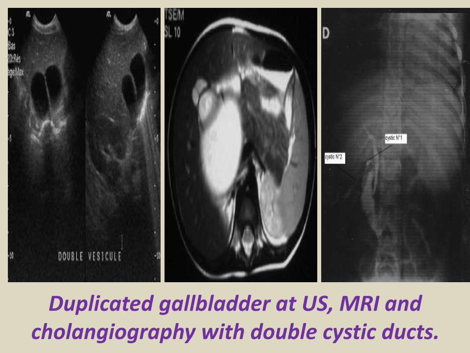
Chronic calcular cholecystitis
Chronic calcular cholecystitis
Distended gall bladder containing echogenic gravity dependent mobile biliary sludge with calculi
Porcelain GBRare due to chronic inflammation mural calcification
Biliary sludge is a mixture of particulate solids that have precipitated from bile Such sediment consists of cholesterol crystals calcium bilirubinate pigment and other calcium salts Sludge is usually detected on transabdominal ultrasonography Microscopy of aspirated bile and endoscopic ultrasonography are far more sensitive Biliary sludge is associated with pregnancy with rapid weight loss particularly in the obese with critical illness involving low or absent oral intake and the use of total parenteral nutrition (TPN) Complications caused by biliary sludge include biliary colic acute cholangitis and acute pancreatitis Asymptomatic patients with sludge or microlithiasis require no therapy
Biliary sludge
Biliary sludge
Gallbladder polyps are relatively frequent seen in up to 5 of the
population Over 90 are benign and the majority are cholesterol polypsCholesterol polyps are most frequently identified in patients between 40-50 years of age and are more common in women (FM 291)
Tiny polyp of the gall bladder wall
Multiple gall bladder polyps
Large gall bladder polyp
Adenomyomatosis of the gallbladder is a hyperplastic
cholestosis of the gallbladder wall It is a relatively common and benign cause of focal gallbladder wall thickening It is most easily seen on ultrasound and MRI Adenomyomatosis is relatively common found in ~5 of all cholecystectomy specimens It is typically seen in patients in their 5th decade The incidence increases with age which may be the result of protracted inflammation (see below) There is a female predilection (MF = 13) It is most often an incidental finding has no intrinsic malignant potential and usually requires no treatment Three morphological types of adenomyomatosis are describedfundal (localized)segmental (annular)generalized (diffuse)
Segmental adenomyomatosis of gall bladder
Diffuse adenomyomatosis of gall bladder
Adenomyomatosis of the gallbladder with a thickened wall Color Dopplershows twinkling artefacts caused by cholesterol crystals in the wall
Adenomyomatosis of the gallbladder with a thickened wall Color Doppler shows twinkling artefacts caused by cholesterol crystals in the wall
Gallbladder adenocarcinomas is the most frequent and are usually asymptomatic until it is no longer curable As such early incidental detection is important if the occasional patient is to be successfully treated Less frequent malignancies is squamous cell carcinoma Small cell carcinoma Carcinosarcoma Lymphoma and metastasisEpidemiologyAlthough overall uncommon gallbladder adenocarcinoma is the most common primary hepatobiliary carcinoma and the 5th most common malignancy of the gastrointestinal tract Predominantly affects older persons with long-standing cholecystolithiasis and as such is most common in elderly women (gt 60 years of age FM ratio = 41) Risk factors include chronic cholecystitis gallstones are seen in 70-90 of casesfamilial adenomatous polyposis syndrome (FAP)inflammatory bowel disease (IBD)porcelain gallbladder
US of Gall bladder carcinoma
Gallbladder adenocarcinoma
US shows echogenic mass adherent to gallbladder fundus (arrows) CT shows enhancing mass within the gallbladder (arrows) with involvement of the liver (arrowhead)
Gallbladder carcinoma
B-cell lymphoma of the gallbladder
50-year-old man with renal-cell carcinoma Intraoperative ultrasound image of polypoid gallbladder lesion Postcontrast axial CT image demonstrates
avidly contrast-enhancing polypoid gallbladder lesion (arrow)
Thank You
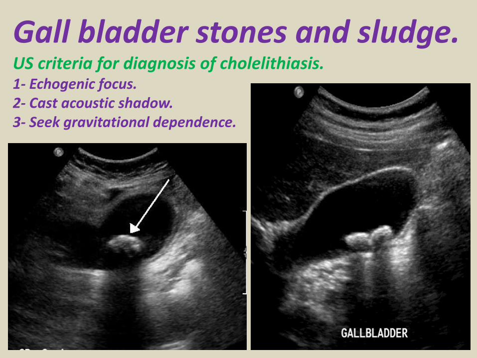
Chronic calcular cholecystitis
Distended gall bladder containing echogenic gravity dependent mobile biliary sludge with calculi
Porcelain GBRare due to chronic inflammation mural calcification
Biliary sludge is a mixture of particulate solids that have precipitated from bile Such sediment consists of cholesterol crystals calcium bilirubinate pigment and other calcium salts Sludge is usually detected on transabdominal ultrasonography Microscopy of aspirated bile and endoscopic ultrasonography are far more sensitive Biliary sludge is associated with pregnancy with rapid weight loss particularly in the obese with critical illness involving low or absent oral intake and the use of total parenteral nutrition (TPN) Complications caused by biliary sludge include biliary colic acute cholangitis and acute pancreatitis Asymptomatic patients with sludge or microlithiasis require no therapy
Biliary sludge
Biliary sludge
Gallbladder polyps are relatively frequent seen in up to 5 of the
population Over 90 are benign and the majority are cholesterol polypsCholesterol polyps are most frequently identified in patients between 40-50 years of age and are more common in women (FM 291)
Tiny polyp of the gall bladder wall
Multiple gall bladder polyps
Large gall bladder polyp
Adenomyomatosis of the gallbladder is a hyperplastic
cholestosis of the gallbladder wall It is a relatively common and benign cause of focal gallbladder wall thickening It is most easily seen on ultrasound and MRI Adenomyomatosis is relatively common found in ~5 of all cholecystectomy specimens It is typically seen in patients in their 5th decade The incidence increases with age which may be the result of protracted inflammation (see below) There is a female predilection (MF = 13) It is most often an incidental finding has no intrinsic malignant potential and usually requires no treatment Three morphological types of adenomyomatosis are describedfundal (localized)segmental (annular)generalized (diffuse)
Segmental adenomyomatosis of gall bladder
Diffuse adenomyomatosis of gall bladder
Adenomyomatosis of the gallbladder with a thickened wall Color Dopplershows twinkling artefacts caused by cholesterol crystals in the wall
Adenomyomatosis of the gallbladder with a thickened wall Color Doppler shows twinkling artefacts caused by cholesterol crystals in the wall
Gallbladder adenocarcinomas is the most frequent and are usually asymptomatic until it is no longer curable As such early incidental detection is important if the occasional patient is to be successfully treated Less frequent malignancies is squamous cell carcinoma Small cell carcinoma Carcinosarcoma Lymphoma and metastasisEpidemiologyAlthough overall uncommon gallbladder adenocarcinoma is the most common primary hepatobiliary carcinoma and the 5th most common malignancy of the gastrointestinal tract Predominantly affects older persons with long-standing cholecystolithiasis and as such is most common in elderly women (gt 60 years of age FM ratio = 41) Risk factors include chronic cholecystitis gallstones are seen in 70-90 of casesfamilial adenomatous polyposis syndrome (FAP)inflammatory bowel disease (IBD)porcelain gallbladder
US of Gall bladder carcinoma
Gallbladder adenocarcinoma
US shows echogenic mass adherent to gallbladder fundus (arrows) CT shows enhancing mass within the gallbladder (arrows) with involvement of the liver (arrowhead)
Gallbladder carcinoma
B-cell lymphoma of the gallbladder
50-year-old man with renal-cell carcinoma Intraoperative ultrasound image of polypoid gallbladder lesion Postcontrast axial CT image demonstrates
avidly contrast-enhancing polypoid gallbladder lesion (arrow)
Thank You
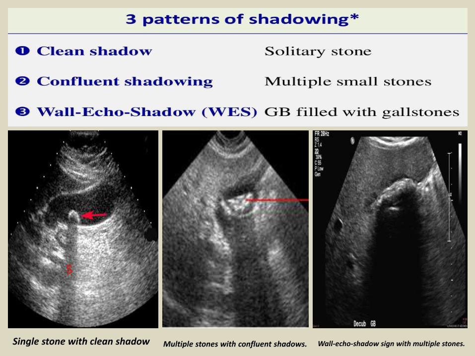
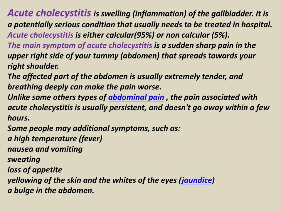
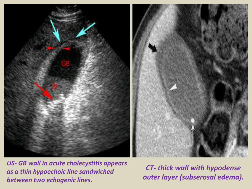
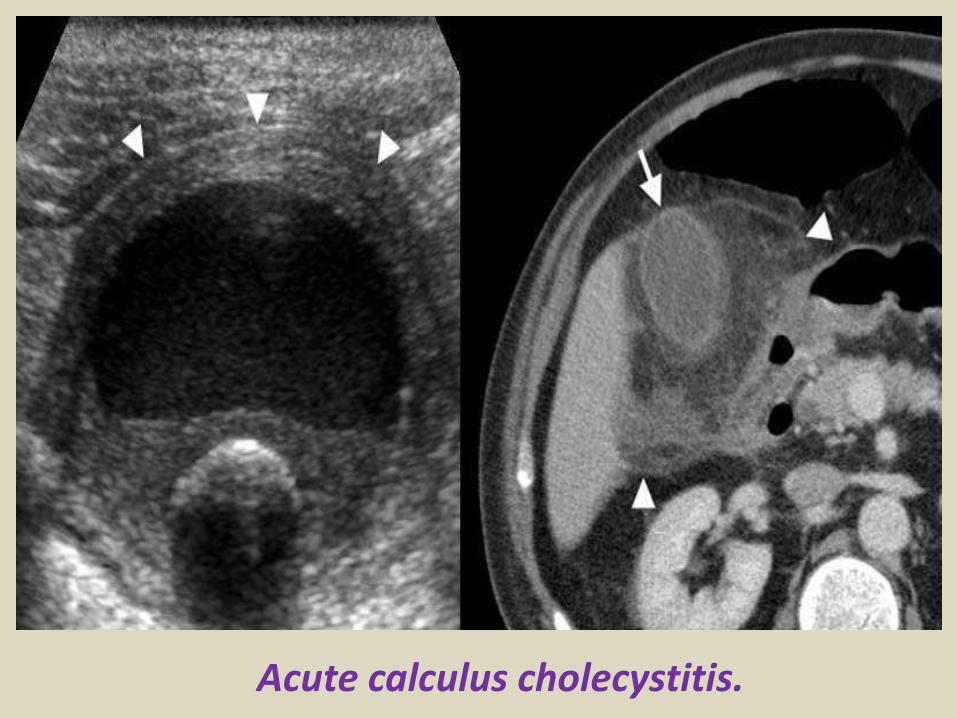
Biliary sludge
Biliary sludge
Gallbladder polyps are relatively frequent seen in up to 5 of the
population Over 90 are benign and the majority are cholesterol polypsCholesterol polyps are most frequently identified in patients between 40-50 years of age and are more common in women (FM 291)
Tiny polyp of the gall bladder wall
Multiple gall bladder polyps
Large gall bladder polyp
Adenomyomatosis of the gallbladder is a hyperplastic
cholestosis of the gallbladder wall It is a relatively common and benign cause of focal gallbladder wall thickening It is most easily seen on ultrasound and MRI Adenomyomatosis is relatively common found in ~5 of all cholecystectomy specimens It is typically seen in patients in their 5th decade The incidence increases with age which may be the result of protracted inflammation (see below) There is a female predilection (MF = 13) It is most often an incidental finding has no intrinsic malignant potential and usually requires no treatment Three morphological types of adenomyomatosis are describedfundal (localized)segmental (annular)generalized (diffuse)
Segmental adenomyomatosis of gall bladder
Diffuse adenomyomatosis of gall bladder
Adenomyomatosis of the gallbladder with a thickened wall Color Dopplershows twinkling artefacts caused by cholesterol crystals in the wall
Adenomyomatosis of the gallbladder with a thickened wall Color Doppler shows twinkling artefacts caused by cholesterol crystals in the wall
Gallbladder adenocarcinomas is the most frequent and are usually asymptomatic until it is no longer curable As such early incidental detection is important if the occasional patient is to be successfully treated Less frequent malignancies is squamous cell carcinoma Small cell carcinoma Carcinosarcoma Lymphoma and metastasisEpidemiologyAlthough overall uncommon gallbladder adenocarcinoma is the most common primary hepatobiliary carcinoma and the 5th most common malignancy of the gastrointestinal tract Predominantly affects older persons with long-standing cholecystolithiasis and as such is most common in elderly women (gt 60 years of age FM ratio = 41) Risk factors include chronic cholecystitis gallstones are seen in 70-90 of casesfamilial adenomatous polyposis syndrome (FAP)inflammatory bowel disease (IBD)porcelain gallbladder
US of Gall bladder carcinoma
Gallbladder adenocarcinoma
US shows echogenic mass adherent to gallbladder fundus (arrows) CT shows enhancing mass within the gallbladder (arrows) with involvement of the liver (arrowhead)
Gallbladder carcinoma
B-cell lymphoma of the gallbladder
50-year-old man with renal-cell carcinoma Intraoperative ultrasound image of polypoid gallbladder lesion Postcontrast axial CT image demonstrates
avidly contrast-enhancing polypoid gallbladder lesion (arrow)
Thank You
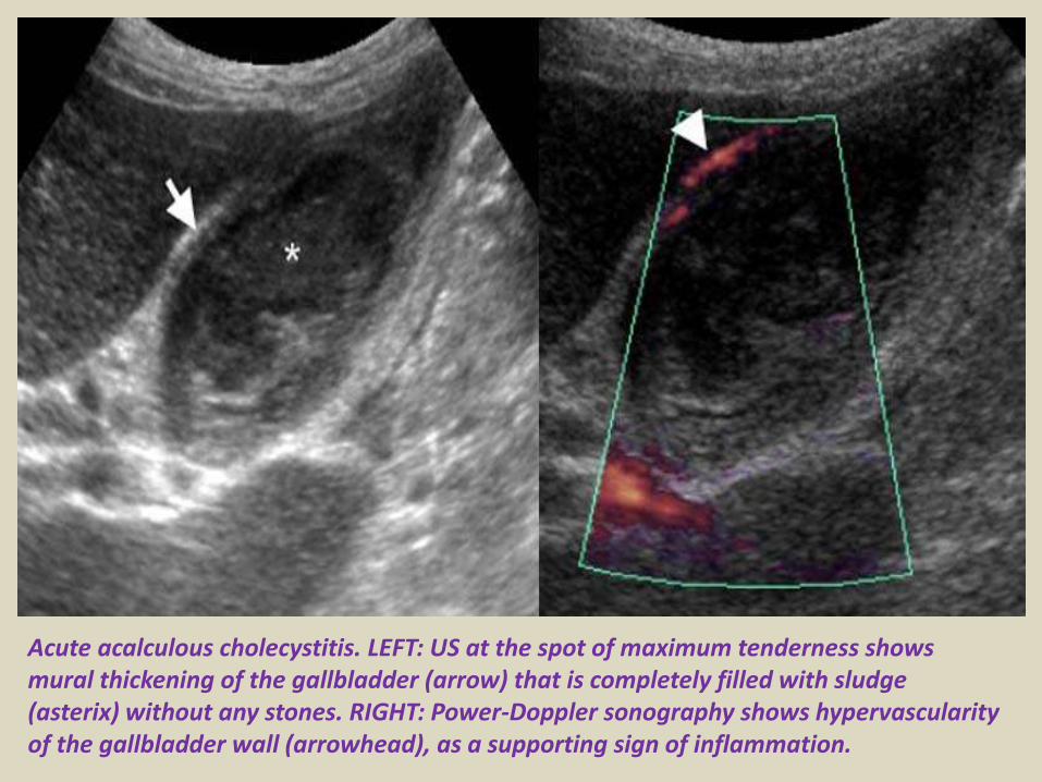
Biliary sludge
Gallbladder polyps are relatively frequent seen in up to 5 of the
population Over 90 are benign and the majority are cholesterol polypsCholesterol polyps are most frequently identified in patients between 40-50 years of age and are more common in women (FM 291)
Tiny polyp of the gall bladder wall
Multiple gall bladder polyps
Large gall bladder polyp
Adenomyomatosis of the gallbladder is a hyperplastic
cholestosis of the gallbladder wall It is a relatively common and benign cause of focal gallbladder wall thickening It is most easily seen on ultrasound and MRI Adenomyomatosis is relatively common found in ~5 of all cholecystectomy specimens It is typically seen in patients in their 5th decade The incidence increases with age which may be the result of protracted inflammation (see below) There is a female predilection (MF = 13) It is most often an incidental finding has no intrinsic malignant potential and usually requires no treatment Three morphological types of adenomyomatosis are describedfundal (localized)segmental (annular)generalized (diffuse)
Segmental adenomyomatosis of gall bladder
Diffuse adenomyomatosis of gall bladder
Adenomyomatosis of the gallbladder with a thickened wall Color Dopplershows twinkling artefacts caused by cholesterol crystals in the wall
Adenomyomatosis of the gallbladder with a thickened wall Color Doppler shows twinkling artefacts caused by cholesterol crystals in the wall
Gallbladder adenocarcinomas is the most frequent and are usually asymptomatic until it is no longer curable As such early incidental detection is important if the occasional patient is to be successfully treated Less frequent malignancies is squamous cell carcinoma Small cell carcinoma Carcinosarcoma Lymphoma and metastasisEpidemiologyAlthough overall uncommon gallbladder adenocarcinoma is the most common primary hepatobiliary carcinoma and the 5th most common malignancy of the gastrointestinal tract Predominantly affects older persons with long-standing cholecystolithiasis and as such is most common in elderly women (gt 60 years of age FM ratio = 41) Risk factors include chronic cholecystitis gallstones are seen in 70-90 of casesfamilial adenomatous polyposis syndrome (FAP)inflammatory bowel disease (IBD)porcelain gallbladder
US of Gall bladder carcinoma
Gallbladder adenocarcinoma
US shows echogenic mass adherent to gallbladder fundus (arrows) CT shows enhancing mass within the gallbladder (arrows) with involvement of the liver (arrowhead)
Gallbladder carcinoma
B-cell lymphoma of the gallbladder
50-year-old man with renal-cell carcinoma Intraoperative ultrasound image of polypoid gallbladder lesion Postcontrast axial CT image demonstrates
avidly contrast-enhancing polypoid gallbladder lesion (arrow)
Thank You
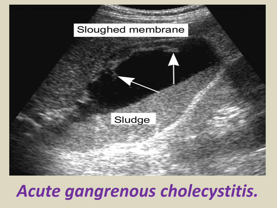
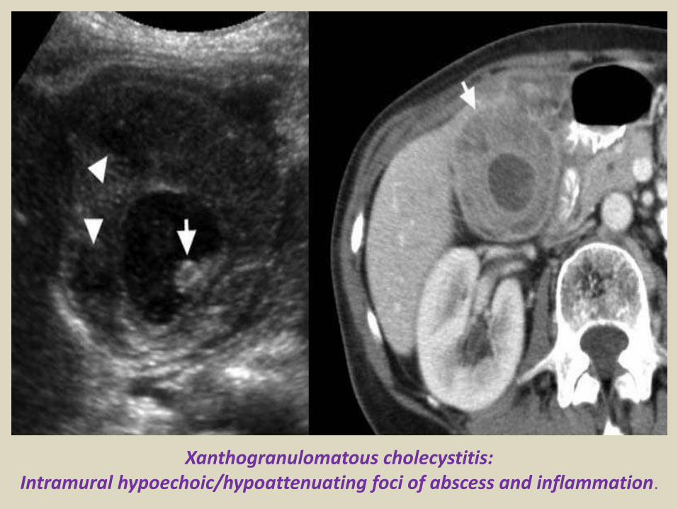
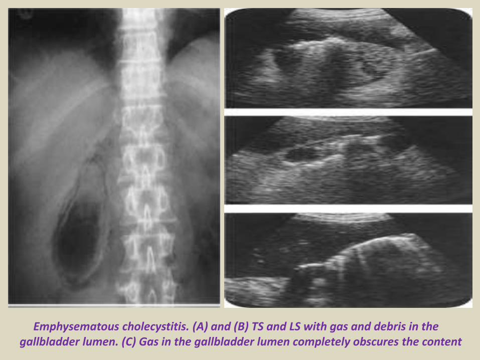
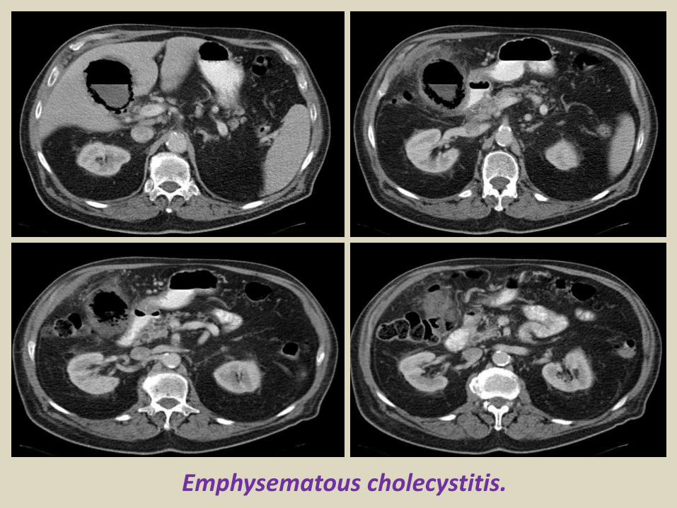
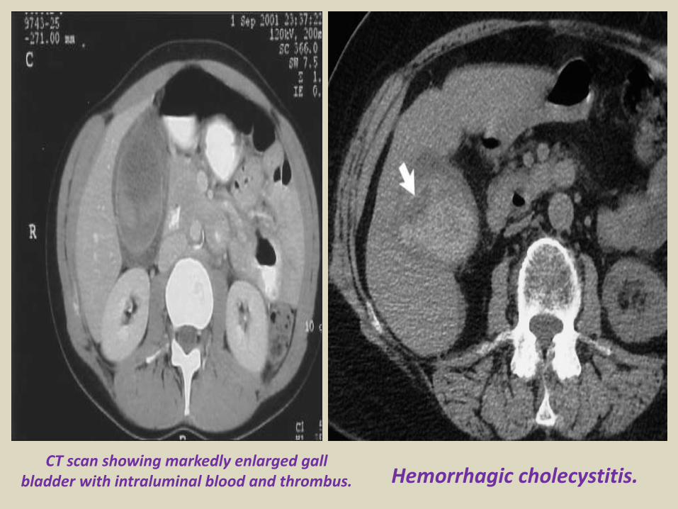
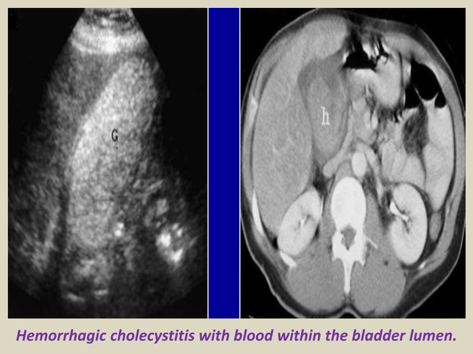
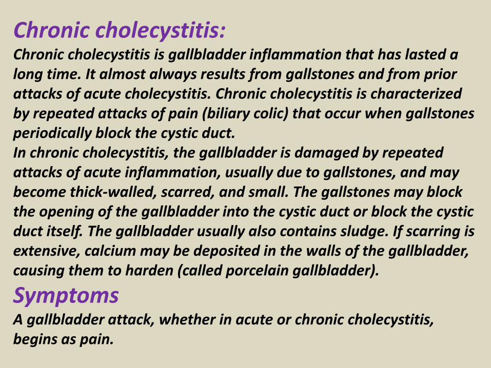
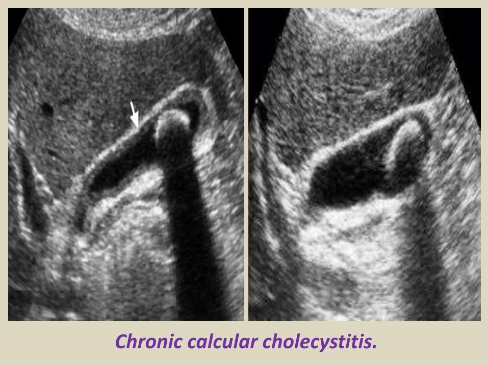
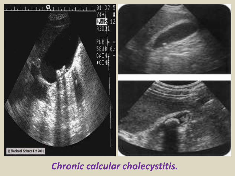
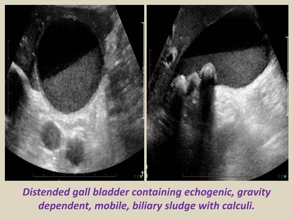
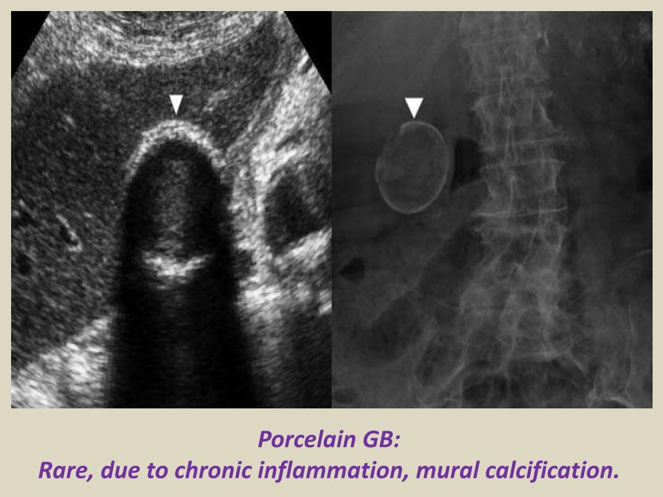
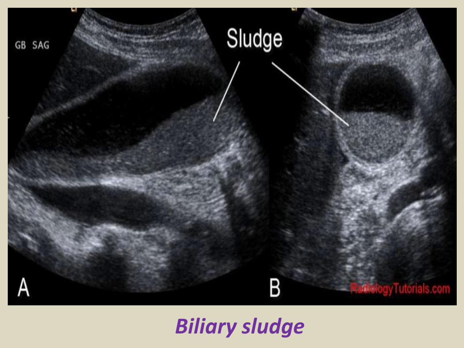
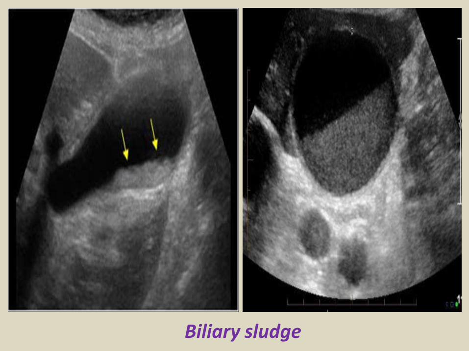
Gallbladder carcinoma
B-cell lymphoma of the gallbladder
50-year-old man with renal-cell carcinoma Intraoperative ultrasound image of polypoid gallbladder lesion Postcontrast axial CT image demonstrates
avidly contrast-enhancing polypoid gallbladder lesion (arrow)
Thank You
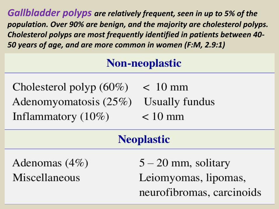
B-cell lymphoma of the gallbladder
50-year-old man with renal-cell carcinoma Intraoperative ultrasound image of polypoid gallbladder lesion Postcontrast axial CT image demonstrates
avidly contrast-enhancing polypoid gallbladder lesion (arrow)
Thank You
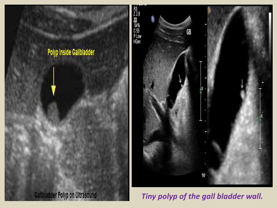
50-year-old man with renal-cell carcinoma Intraoperative ultrasound image of polypoid gallbladder lesion Postcontrast axial CT image demonstrates
avidly contrast-enhancing polypoid gallbladder lesion (arrow)
Thank You

Thank You