preparation of leaf mitochondria and …631106/fulltext01.pdf · and studies on utochondrial...
TRANSCRIPT

f hol
PREPARATION OF LEAF MITOCHONDRIA
AND STUDIES ON
UTOCHONDRIAL PHOTORESPIRATORY REACTIONS
PER GARDESTRÖM
r-* m
Ar
+* 3
UMEÅ 1981


PREPARATION O F L EAF M ITOCHONDRIA
AND STUDIES O N
MITOCHONDRIAL P HOTORESPIRATORY R EACTIONS
DOCTORAL D ISSERTATION
by
PER GARDESTRÖM
From The De partment of Biochemistry
University of Umeå
S-901 87 UMEA
SWEDEN
UMEA 1981
Preparation of Leaf Mito chondria and Studies on Mito chondrial Photo-respiratory Reactions
Abstract
A procedure for the preparation of spinach leaf mitochondria was deve loped. The procedure com bines d ifferential centrifugation, partition in dextran-polyethyleneglycol two-phase syste m and Percoli density gradient centri-fugation. The d ifferent steps separate the material mainly according to size, surface properties and density, respectively. No chlorophyl l was present in the final mitochondrial preparation and the mitochondria were also markedly enriched relative to peroxisomes and microsomes as estimated fro m th e recovery of marker enzy mes. The latency of enzyme a ctivities was use d to study the apparent intactness of the mitochondrial membranes. These mea surements showed th at both th e inner and outer mitochondrial membranes we re m ore than 90 % intact . The mi tochondria were al so functionally intact since the coupling between respiration and o xidative phosphorylation was r etained.
The pur ity of the preparation made i t possible to study cytoc hromes from leaf mitochondria. The c ytochrome co ntent of stalk and leaf mitochondria was m easured in order to compare mitoc hondria from photosynthesizing and non-photosynthesizing tissue. The m easurements were per formed by diff erence spectroscopy both a t room temper ature and a t liquid nitrogen temperature. Qualitatively the cytochrome content in mitochondria from s talks and leaves was i dentical. Quantiatively leaf mitochondria contained,on a protein basis, only half the amount of the different cytochromes as compared to stalk mitochondria. The relative content of the different cytochromes was, however, similar suggesting that the composition of the respiratory chain was the same.
The photorespiratory conversion of glycine to serine takes place in the mitochondria and involves o xidative decarboxylation of glycine. The ab ility to oxidize glycine via the respiratory chain was present in spinach leaf mitochondria, but absent in mitochondria prepared from roo ts, stalks and leaf veins from t he same plants. This confi rmed the specific localization of the glycine oxidizing activity to photosyntheticaliy active tissue, as suggested by st udies with ot her plant material.
The conv ersion of glycine to serine is a complex rea ction depending on the combined a ction of two en zymes: glycine decarboxylase and serine hydroxymethyltransferase. The e ffect of inhibitors on the serine hydroxymethyl transferase activity and the rate of the glycine bicarbonate exch ange reaction associated with glycine decarboxylase w as st udied. These reac tions represent partial steps in the conversion of glycine to serine and the aim was t o investigate the site of inhibition for the different inhibitors, namely, isonicotinyl hydrazide (a pyridoxa!phosphate antagonist), amino-acetonitrile, glycinehydroxamate (glycine analogues) and cyanide. The results showed that these inhibitors had a complex pa ttern of inhibition. The sa me inhibitor affected more than one s ite and often with an a pparently different mechanism. I t was, however, found that aminoacetonitri le at low concentrations specifically inhibited glycine decarboxylase and that cyanide sp ecifically inhibited serine hydroxymethyltransferase.
Key wo rds: leaf mitochondria, photorespiration, aqueous two-phase system, Percoli gradient, cytochromes, glycine decarboyxlase, serine hydroxymethyltransferase, inhitors of photorespiration
ISBN 91-7174-078-3
Number of pages: 29

PREPARATION O F L EAF M ITOCHONDRIA
AND STUDIES O N
MITOCHONDRIAL P HOTORESPIRATORY R EACTIONS
by
PER GARDESTRÖM
AKADEMISK AVHANDLING
som för avläggande av filosofie doktorsexamen vid matematisk-naturvetenskapliga fakulteten vid Universitet i Umeå kommer a tt offentligen försvaras på Kemiska insti tutionen, hörsal B, LU 0, tisdagen den 2 juni 1981 kl. 10.00.

ISBN 91-7174-078-3

Lillemor, Johanna and Johan


Abstract
A procedure for the preparation of spinach leaf mitochondria was devel oped. The procedure combines d ifferential centri fugati on, partition in dextran-polyethyleneglycol two-phase syste m and Percoli density gradient centri-fugation. The d ifferent steps separate the material mainly accord ing to size, surface properties and density, respectively. No chlorop hyll was present in the final mitochondrial preparation and the mitochondria wer e also markedly enriched relative to peroxisomes and microsomes as estimated from the recovery of marker enzymes. The latency of enzyme a ctivities was used to study the apparent intactness of the mitochondrial membranes. These mea surements showed that both the inner and outer mitochondrial membranes we re m ore than 90 % intact. The mitoch ondria were al so functionally intact since the coupling between respiration and oxidative phosphorylation was retained.
The purity of the preparation made i t possible to study cytoc hromes from leaf mitochondria. The cyto chrome content of stalk and leaf mitochondria was m easured in order to compare mitoc hondria from photosynthesizing and non-photosynthesizing tissue. The m easurements we re performed by differe nce spectroscopy both at room tempera ture and a t liquid nitrogen temperature. Qualitatively the cytochrome co ntent in mitochondria from st alks and leaves was identical. Quantiatively leaf mitochondria contained,on a protein basis, only half the amount of the different cytochromes as compared to stalk mitochondria. The relative content of the different cytochromes was, however, similar suggesting that the composition of the respiratory chain was the same.
The photorespiratory conversion of glycine to serine takes place in the mitochondria and involves o xidative decarboxylation of glycine. The a bility to oxidize glycine via the respiratory chain was prese nt in spinach leaf mitochondria, but absent in mitochondria prepared from ro ots, stalks and leaf veins from th e same plants. This confi rmed the specific localization of the glycine oxidizing activity to photosyntheticaliy active tissue, as suggested by st udies with o ther plant material.
The conversion of glycine to serine is a complex re action depending on the combined action of two enz ymes: glycine decarboxylase and se rine hydroxymethyltransferase. The eff ect of inhibitors on the serine hydroxy-methyltransferase activity and the rate of the glycine bicarbonate exch ange reaction associated with glycine decarboxylase w as studied. These rea ctions represent partial steps in the conversion of glycine to serine and the aim was t o investigate the site of inhibit ion for the different inhibitors, namely, isonicotinyl hydrazide (a pyridoxalphosphate antagonist), amino-acetonitrile, glycinehydroxamate (glycine analogues) and cyanide. The results showed that these inhibitors had a complex p attern of inhibition. The same inhibitor affected more than one site and o ften with an appare ntly different mechanism. It was, however, found that aminoacetonitri le at low concentrations specifically inhibited glycine decarboxylase and that cyanide sp ecifically inhibited serine hydroxymethyltransferase.


This summary i s based ori the following a rticles:
I. Gardeström, P., Ericson, I . and Larsson, C. (1978). Preparation of
mitochondria from green leaves of spinach by diff erential centri-
fugation and phase partition. Plant Sci. Lett. 13:231-239.
II . Gardeström, P., Bergman, A. and Ericson, I . (1980). Oxidation of
glycine via the respiratory chain in mitochondria prepared from
different parts of spinach. Plant Physiol. 65:389-391
III. Bergman, A., Gardeström, P. and Ericson, I . (1980). Method to obtain
a chlorophyll-free preparation of intact mitochondria from spinach
leaves. Plant Physiol. 66:442-445.
IV. Gardeström, P., Bergman, A., Ericson, I . and Sahlström, S. (1980).
Cytochromes of spinach mitochondria. Proceedings of the V™ Inter
national Congress on Photosynthesis. In press.
V. Gardeström, P., Bergman, A. and Ericson, I . (1981). Inhibition of the
conversion of glycine to serine in spinach l eaf mitochondria. In
manuscript.
References made t o these art icles in the text will be via the Roman
numerals used above.


TABLE OF CONTENTS
Page 1. INTRODUCTION 1
2. PREPARATION O F L EAF M ITOCHONDRIA 4 2.1 Plant material 5 2.2 Preparation procedure 5
2.2.1 Differential centri fugati on 7 2.2.2 Phase Partition 8 2.2.3 Percoli density gradient centrifugation 9
3. CHARACTERIZATION OF T HE P REPARATION 10 3.1 Purity 10 3.2 Intactness 12 3.3 Cytochrome content 13
4. PHOTORESPIRATION 4.1 The glycol ate cycle 14 4.2 Function and regulation of photorespiration 15 4.3 The mi tochondrial reactions in photorespiration 16
4.3.1 Glycine decarboxylase 17 4.3.2 Serine hydroxymethyltransferase 18 4.3.3 Fate of the products 18
5. STUDIES O N MITOCHONDRIAL P HOTORESPIRATORY R EACTIONS 5.1 Glycine oxidation in mitochondria from di fferent parts of 21
the plant 5.2 Effect of inhibitors on t he conversion of glycine to serine 21 5.3 Absorption peaks arou nd 500 nm associated with the conversion 22
of glycine to serine
6. CONCLUDING REMARKS 24
7. ACKNOWLEDGEMENTS 25
8. REFERENCES 26


- 1 -
1. INTRODUCTION
In the cell, the compartmentation into different eellorganelles is of fundamental importance fo r metabolism an d its regulation. The in teraction between different eellorganelles is very com plex. Studies on i solated eellorganelles can not therefore give a complete picture of cellular metabolism. Despite this, the study of isolated organelles is of great importance fo r the understanding of metabolism as the isolated organelle is a much simpler system to work with than a whole ce ll or an organis m.
Mitochondria a re present in all eucaryotic cells. The mi tochondria from different organisms have si milar composition and basal functions. They are all surrounded by tw o me mbranes, an o uter membrane permeable t o small molecules and an inner membrane with very restricted permeability. They all contain a citric acid cycle and a respiratory chain of similar composition where elec tron transport is coupled to oxidative phosphorylation of ADP to ATP. Apparently, the mechanism for oxidative phosphorylation is identical in all mitochondria. Despite these large overall similarities mitochondria from di fferent sources a re specialized in some way s. The most s triking differences between animal and plant mitochondria a re associated with the respiratory chain (1). Plant mitochondria in general have a NADH dehydrogenase facing outwards from the inner membrane (external NADH dehydrogenase) and the oxidation of external NADH is coupled to phosphorylation of two A DP (2). Such a dehydrogenase is absent from animal mitochondria. Furthermore, most plant mitochondria appear to have two d ifferent terminal oxidases, the cytochrome oxidase and the alternative oxidase, which i s insensitive to KCN inhibition. Electron transport to the alternative oxidase is not coupled to oxidative phosphorylation (3). Malate oxidation is also different as isolated plant mitochondria can oxidize added malate without the addition of other substrates to remove oxaloacetate produced (4).

- 2 -
Mitochondria from leaves have been l ess studied than mitochondria from other plant tissues, depending mai nly on problems in isolating functionally, intact leaf mitochondria. Relatively l itt le is therefore known about special properties of leaf mitochondria. Specializations are, however, l ikely to occur si nce the metabolism of photosynthesizing c ells is, in many re spects special compared to that of other cells. In photosynthetic cells phosphoglycerate derived from C O2 i s the ultimate source f or all carbon meta bolism. Thus, phosphoglycerate is used in the metabolism of the cell, converted to starch as a reserve or converted to sucrose and transported to other parts of the plant. Photosynthesizing cells are also special in that they have tw o s eparate electron transport based systems for ATP synth esis: the chloroplast system an d the mitochondrial system.
Often, mitochondria a re mainly regarded a s ATP-producing orga nelles, and their activity superfluous in light when photophosphorylation in chloro-plasts is active. Many importan t metabolites are, on th e other hand, directly or indirectly derived from th e citric acid cycle in mitochondria. Evidence has also been obtained for mitochondrial activity in light (6). Respiration via the alternative oxidase has been suggest ed to be important in light (7), but l it t le is known about the function of the alternative oxidase in leaf mitochondria. The e lectron flow to the alternative oxidase would have t o be s trictly regulated as plants depend on mitocho ndrial ATP synthesis in the dark. Leaf mitochondria are of importance f or the metabolism in light, but m uch addit ional work rema ins t o establish their role in detail.
In the photorespiration, a light dependent metabolic process by wh ich newly fixed CO2 i s released, both chloroplasts, peroxisomes and mitoc hondria are involved (5). The discovery of this led to an increased interest in leaf mitochondria. In f act, the mitochondrial reactions involved in photorespiration constitute the most studied differences between leaf mitochondria and o ther plant mitochondria.
For more deta iled studies on l eaf mitochondria, i t is important to be able to prepare pure an d functionally intact mitochondria. The mito chondrial fraction obtained by di fferential centrifugation could be considerably purified by phase partition in an aque ous dextran -polyethylene-glycol (PEG) two-phase syste m (paper I). Further purification was possible by Percoli density gradient centrifugation (paper III) . The resulting

- 3 -
preparation was very pure, so that cytochrome m easurements in leaf mitochondria was possible (paper IV). The prepar ations were a lso used to study mitocho ndrial reactions in photorespiration (paper II and V). To compare mitoch ondria from photosynthetic and non-photosynthetic tissue mitochondria were prepared from di fferent parts of the same p lant (paper II and IV).

- 4 -
2. PREPARATION O F L EAF M ITOCHONDRIA
Mitochondria from leaves have been l ess studied than other plant mitochondria. The problems have been to obtain both fun ctionally intact mitochondria and to avoid ch loroplast contamination.
Early attempts to isolate leaf mitochondria resulted in preparations with low ra tes of respiration and bad coupling between electron transport and oxidative phosphorylation (8, 9, 10). Woo (11) prepared mitocho ndria showing respiratory control, but the respiratory rate was re latively low. Douce et al (12) designed a method for preparation of leaf mitochondria that retained respiratory functions better than the preparation methods use d earl ier. The im provement seems to be ma inly th e composition of the preparation medium a nd the use of very s hort grinding times. Although suitable for respiratory measurements the preparation was heavily contam inated with chloroplast material yielding a protein/chlorophyll ratio of about 15. For many stu dies i t is necessary t o have a preparation of higher purity. To obtain this i t is necessary to use a separation method that fractionates the material according to properties other than sedimentation rate.
Sucrose density gradient centrifugation has been used to purify plant mitochondria (13). Recently, Arron et al (14) reported purification of intact mitochondria from leaves of Sedion pr>aeal tvm t a crassulacean acid metabolism p lant. The me thod does not appear to be useful for preparation of intact leaf mitochondria from oth er plants. I t is , however, very useful for studies of enzyme di stribution (15, 16).
Density grad ients based on s il ica sol were used with li t t le success (17) until Percoli, a silica sol coated with polyvinyl pyrrolidone wa s introduced. Jackson et al (18) used Percoli gradients to purify intact spinach leaf mitochondria and obtained a protein/chlorophyll ratio of 123.

- 5 -
Partition in dextran-PEG two -phase system s can be used t o separate substances accordin g to surface properties (19). By co mbining t his method with d ifferential centri fugati on w e coul d prepare intact mitochondria with a protein/chlorophyll ratio of about 100 (paper I). Chlorophyll could be t otally removed fro m t his preparation by furt her purification using Percoli density gradient centri fugati on (paper III):
Another useful approach is to purify organelles from p rotoplasts. This takes advantage of the minimized breakage of chloroplasts that results from rupturing the protoplasts (20). Separation of mitochondria and chloroplast material by free -flow electrophoresis based on charge differences is also possible (21).
2.1 Plant Material
Spinach (Spinacio. Oleracea var. Viking II) was used in all studies. The plants were gr own at about 18 °C und er a rtificial light with a light/dark period of 12 h. The pla nts used in paper I were grown on vermiculite
- 2 - 1 with a l ight intensity of about 80 yE m s . The pla nts used in the subsequent papers we re gr own on a liquid medium accord ing to Siegenthaler and Depéry (22) with doub led Fe concentration, and with a l ight intensity of
-2 -1 -2 -1 80 yE m s or 200 yE m s . The seeds were germ inated on vermiculite. When t he seedlings were a few centimeters high they were tra nsferred to the liquid medium.
Fully expanded lea ves were used for the preparation of mitochondria. The plants were kept in dark two or three days and th e light was turned on half an hour to one hour before harvesting the leaves. The dar k treatment was important to reduce the starch content which otherwise interferred in the phase partition step. The use of liquid medium also made i t possible to prepare mitoch ondria from roots. After harvest, roots were washed several times in destilled water before the preparation.
2.2 Preparation Procedure
Paper I describes a preparation method for spinach leaf mitochondria combining dif ferential centrifugation and phase partition. This preparation can be fu rther purified by Percoli density gradient centrifugation as described in paper III. A summary of the entire preparation procedure is shown in fig. 1.

- 6 -
1 5 0 q l e a v e s
0-
<Ç)~ s u s p e n d i n t o p p h a s e
a d d b o t t o m p h a s e
g r i n d , f i l t e r I
5 0 0 0 g 2 n i n
2 0 0 0 0 g 2 . 5 m i n
m i x i
r e m o v e t o p p h a s e
6 0 0 g
3 m i n *
bå n e w t o p p h a s e 6 0 0 g
n e w t o p , p h a s e 6 0 0 g
3 m i n 3 m i n
(M)-
s u s p e n d
1
d i l u t e w i t h 7 v o l u m e s m e d i u m i
5 0 0 0 g 2 m i n
f S 3
1 2 0 0 0 g 1 0 m i n
KJ
s a m p l e 2 1 2 P e r c o l i 9 0 0 0 9
) 2 7 % "
60 % " 3 0 m i n
t h y 1 a k o i d f r a g m e n t s
M I T O C H O N D R I A w
F i g . 1 . Preparation procedure for spinach leaf mitochondria.
This procedure seems to give the purest preparation of intact leaf mitochondria published so far. The sa me procedure with small modifications could also be use d to prepare pure mitoc hondria from s talks and roots of spinach.
The di fferent steps in the preparation procedure utilize different properties of the material. The m ain separation parameter in the different • s teps and the resulting effect is shown in table I.
The leaf mitochondria oxidized NADH, succinate, malate and glycine with good respiratory control and ADP/0 ra tios. Based on recovery of enzyme activities about 10 % of the mitochondria in the fil trate from le af homo-genate wer e col lected in the final mitochondrial fraction. This c onstitutes a yield of about 5 mg protein from 150 g leaves. As a significant fraction of the mitochondria is damaged during the homogenization, the recovery of intact mitochondria is considerably higher than 10 %.

- 7 -
T a b l e I . Ma i n e f f e e i n o f t h e d i f f e r e n t r s t e p - s i n t h e p r e p a r a t i o n
F r a c t i o n P r e p a r a t i o n p r o c e d u r e
M a i n s e p a r a t i o n p a r a m e t e r
M a i n c o n t a m i - R e c o v e r y o f n a n t s r e m o v e d m i t o c h o n d r i a
P r o t e i n ( m g ) c h l o r o p h y l l ( m g )
L e a f h o m o g e n a t e
C r u d e m i t o c h o r i d r a
D i f f e r e n t i a l c e n t r i f u g a t i o n
s i z e
P h a s e p a r t i t i o n
s u r f a c e p r o p e r t i e s
i n t a c t c h l o r o p l a s t s
p e r o x i s o m e s
m i c r o s o m e s
c h l o r o p l a s t f r a g m e n t s
m i c r o s o m e s
100 %
4 0 % 2 5
P u r i f i e d m i t o c h o n d r i a 1 5 100
P e r c o l i g r a d i e n t c e n t r i f u g a t i o n
d e n s i t y c h l o r o p l a s t f r a g m e n t s
P u r e m i t o c h o n d r i a 1 0 %
2.2.1 Differential centrifugation
Particles sediment in a centrifugal field if their density is higher than that of the surrounding m edium. It is the effective mass and frictional forces that determine the rate of sedimentation. Cellorganelles have about the same den sity and form an d therefore i t is mainly th e differences in size that determines the differences in sedimentation rates.
The d ifferential centrifugation scheme wa s wo rked out with the knowledge that intact chloroplasts were not removed in the phase partition step. Thus th e first centrifugation was designe d especially to sediment intact chloroplasts. This required centrifugation at a higher speed than used before (12) whereupon th e chlorophyll contamination decreased to about half as compared to earlier methods (Jackson, personal communication). The sec ond centrifugation was desi gned with the aim to avoid unnecessary contamination with broken mitoch ondria and light membranous material. The removal of peroxisomes by di fferential centrifugation (table 1) is not due to differences in sedimentation rate, as repeated centri fugations do not further reduce peroxisomal contamination (23), but rather reflects a difference in organelle rupture upon homogenization.

-J
2.2.2 Phase partition
Phase partition in Dextran-PEG two-p hase systems has proven to be very useful for separation of cells and cell organelles (24). In mo st cases phase p artition is combined with d ifferential centrifugati on to give a two-dimensional fractionation of the material. Thus, phase p artition has been use d successfully for the purification of a variety of cell-organelles and membrane fr actions from plant cells, including c hloro-plasts (25); inside-out thylakoid vesicles (26); inside-out submitochondrial particles (27); and plasma membranes (28).
The pa rtition of a given material can be ch anged by chan ging the composition of the phase syst em e. g. molecular weight an d concentration of polymers; type and concentration of buffer, type and conce ntration of salt (29). An empi ric procedure is used to compose a two-phase syste m in which th e particles to be separated partition differently. For t he separation of leaf mitochondria and thylakoid membrane fragment s phase syst ems with several different compositions were tried. A difference was fo und in the response to increasing concentrations of KCl (fig. 2). This d ifference made i t possible to separate mitochondria from thylakoid membranes using a phase system wi th appropriate KCl concentration. It was only broken chi oroplasts (thylakoid membranes) that differed in partition compared with mitochond ria. Intact chloroplasts partitioned similar to mitochondria.
100 .
50
± X JL 0 1 5
m mol KCl / kg
Fig. 2 Phase partition of mitochondria and thylakoid membranes. Phase system: 6.2 % (w/w) Dextrane 500, 6.2 % (w/w) PEG 4000, 0.3 mol/ kg s ucrose, 5 mmol/kg K-phosphate buffer, and KCl as indicated. Malate dehydrogenase activity ( t • ) , A680 (o_ o)<

- 9 -
PEG ha s been reported to interfere with glyci ne oxidation (30) and we too have fou nd that the glycine oxidation is sensitive to storage in top phase wh ere the PEG concentration is relatively high. However, as the mitochondria partition to the dextran rich bottom phas e the PEG concentration in contact with the mitochondria is low (less than 1 %) for most of the time of the partition procedure.
A problem in the use of phase syste ms is that polymers with strictly defined properties are not available. Different batches of both dextran and PEG h ave slightly different molecular weight d istributions and the partition of material in systems using polymers from di fferent batches is not identical. A way t o get around this difficulty is outlined in paper III . In this method the critical point (lowest polymer concentration for phase sep aration), with polymers from dif ferent batches, is determined. Phase syste ms with polymers from diff erent batches were found to give about the same sep aration of mitochondria and thylakoid membranes w hen th e dextran and PEG concentrations were 0.5 % (w/w) higher than the critical point corresponding to the particular pair of polymers used in the system.
2.2.3 Percoli density gradient centri fugati on
Percoli is an insoluble silica sol coated with polyvinylpyrrolidone to protect the biological material from th e silicic acid, which i s harmful. A density gradient can be prepar ed using d ifferent concentrations of Percoli. In c ontrast to sucrose Percoli is osmotically inactive. The very low visc osity of Percoli makes i t convenient to work with.
Mitochondria from 150 g leaves could be pu rified on each stepwise Percoli gradient (7 ml). One milli litre of the mitochondrial fraction recovered after centrifugation contained on an average 5 mg protein. Percoli did not seem to interfere with the mitochondrial functions studied and was therefore not removed.

- 10 -
3. CHARACTERIZATION O F T HE P REPARATION
It is important to characterize leaf mitochondrial preparations with respect to purity, intactness and composition. Apart from increa sing our knowledge about the organelle, the characterization is also important for comparison with results obtained with preparations using oth er preparation methods. Studies on th e composition of leaf mitochondria have been d ifficult due to contamination with non-mitochondrial material. The highly p urified preparation obtained by th e preparation method described in paper III made st udies on the cytochrome content of leaf mitochondria possible. The preparation is also suitable for the study of enzyme localization and membrane composition , such as membrane proteins and membrane l ipids.
3.1 Purity
Several methods can be use d to measure the purity of an o rganelle preparation. In paper I electron microscopy w as use d to compare th e mitochondrial fraction after differential centrifugation and a fter further purification by phase partition. A drastic reduction in the amount of thylakoid membranes af ter the phase partition step was evid ent. Electron microscopy i s not quantitative but gives information about the purity of the preparation, different from t hat obtained by biochemical measurements.
Markers can be use d to estimate the purity of an o rganelle preparation. The am ount of a marker o r the activity of a marker enzyme can be re lated to total protein or to markers for other organelles. A problem i n using marker en zymes i s often the measurements in the crude homogenate. Also, the activity of the marker en zyme is often more or less labile. Therefore the total recovery of activity after the different purification steps must alway s be chec ked. As regards proteins, i t is difficult to determine

- 11 -
them quant itatively due to high amounts of chlorophyll in the crude homogenate an d in the mitochondrial fraction obtained by d ifferential centrifugation. A method to measure mitochon drial protein in the mitochondrial fraction obtained by d ifferential centrifugation has been reported (12). In this method thylakoid protein is subtracted from t otal protein determined according to Lowry et al (31). For the correction, a ratio of 7 between thylakoid protein and chlorophyll was used. Chlorophyll interferes, however, severely with the Lowry m ethod for protein measurement (Åkerlund, personal communication). Therefore, protein has only been deter mined in the mitochondrial fraction after phase p artition and Percoli gradient centrifugation.
The enrich ment of mitochondria relative to a contaminant can be calcu lated as the ratio between the recovery of a mitochondrial marker and a marker for the contaminant studied. The m ethod is quantitative and gives detailed information about removal of the specific contaminant. But as the enrichment is related to the total initial amount of a cell component i t gives, however, no dir ect information about the absolute degree of contamination in the preparation. Thus, in a leaf cell chloroplasts make up a much bigger f raction than mitochon dria and therefore mitochondria must be enrich ed several times relative to chloroplasts before mo re than half of the material in a fraction is mitochondrial.
The enrich ment of mitochondria relative to chloroplast material was 200-400 times a fter the phase partition step (paper I). After the following Percoli gradient centrifugation, the chlorophyll contamination was reduced below th e detection limit, giving an infinite protein to chlorophyll ratio (paper III). In paper III the enrichment of mitochondria relative to peroxisomes and microsomes wa s meas ured. The enrich ment relative to peroxisomes was on the average 12 and to microsomes on the average 33. Peroxisomes a re probably th e most significant contaminant in the preparation. On a protein basis, the contamination with peroxisomes is maximally a few percent as mitochondria probably con tribute to a bigger fraction of the total protein in a leaf cell than peroxisomes do.

- 12 -
3.2 Intactness
The coupling between substrate oxidation and oxidative phosphorylation in the electron transport chain is often used to check the intactness of isolated mitochondria. The parameters measured are respiratory control and ADP/0 ra tios. Functionally intact mitochondria should show respiratory control but the method gives no q uantitative information regarding intactness. The p ossibility of electron transport via alternative pathways, not coupled to oxidative phosphorylation also limits the use of respiratory control as a measure of intactness.
The latency of enzyme a ctivities can be use d to estimate the intactness of the membranes. The mos t commonly use d assay is to measure the ratio of succinate: cytochrome c oxidoreductase activity in isotonic and hypotonic media (2). The m ethod is based on t hat cytochrome c can not penetrate the intact outer membrane. The ratio between the activity in isotonic medium a nd the activity in hypotonic medium estimates the fraction of the outer membranes th at is broken. The cy tochrome c assay is widely used to measure outer membrane intactnes.
No suc h widely accepted method e xists for the measurement of inner membrane integrity. Some c riteria that a method should fullfil can be set up: a) the marker to be used should be s trictly mitochondrial, b) the marker should be m embrane ass ociated in order not to leak out and be w ashed away fr om ruptured mitochondria, c) the activity should depend on the permeability of small molecules and d ) the activity should be stable and easy to measure. No m ethod that full fi Is all these criteria has so far been descr ibed. One me thod i s to compare t he malate dehydrogenase ac tivity measured in isotonic medium w ith the activity that can be separated by centri fugati on from mitoc hondria in hypotonic medium (2, 32). This me thod su ffers from several disadvantages; isoenzymes of malate dehydrogenase e xist in several cell compartments (15); the mitochondrial malate dehydrogenase is a matrix enzyme; the method measures the permeability of protein molecules. An al ternative method is described in paper I . This method is based on th e latency of NAD: isocitrate dehydrogenase, a citric acid cycle enzyme. NAD: isocitrate dehydrogenase seems to be a strictly mitochondrial enzyme (20). NADP linked isocitrate dehydrogenase has been reported in chloroplasts (33) and in peroxisomes (34). This method is based on r estricted permeability for NADH into the matrix. Transport of NADH through the inner membrane a t low r ate has been

- 13 -
reported (35). If transport occurs under t he assay conditions the integrity will be underestimated. The disadvantages with the method is that NAD: isocitrate dehydrogenase is a matrix enzyme (20) and instable in solution. The m ain advanta ge with the NAD: isocitrate dehydrogenase assay compared to the malate dehydrogenase assay is that i t measures th e permeability of small molecules.
The integrity assays give a ratio of activities. From th is ratio apparent % intactness is often calculated. It is not possible to directly compare values obtained with d ifferent methods as the correlation between apparent intactness of membranes an d true intactness is unknown. Moreover, the apparent intactness of membranes need not be corr elated to functional integrity. Despite t hese limitations integrity assays based on la tency of enzyme a ctivities give valuable information about the quality of a preparation.
The apparent intactness of the mitochondrial membranes af ter Percoli gradient centrifugation was 93 % ± 4 % for the outer membrane (based on the succinate; cytochrome c oxidoreductase as say) and 95 % ± 3 % for the inner membrane (based on the NAD: isocitrate dehydrogenase a ssay).
3.3 Cytochrome content
By diffe rence spectroscopy a t 77K (l iquid nitrogen temperature) i t was shown (paper IV) that leaf mitochondria con tain cytochrome a ^, three
b-cytochromes (^557» ^560' ^566^ a n c* ^ w 0 cytochromes (c549> c552^* This is qualitat ively the same cyto chromes a s found in stalk (paper IV) and other plant mitochondria (36). A quantitative difference was, however, evident as leaf mitochondria c ontain only half the amount of cytochromes relative to protein compared to stalk and other plant mitochondria (13). The relative content of the different cytochromes wa s similar for stalk and le af mitochondria, suggesting the same comp osition of their respiratory chains. As the leaf mitochondrial preparations used a re very pure the low values for the cytochrome content in relation to protein are not due to contaminating non-m itochondrial protein. The lo w value can be interpreted as if the mitochondria co ntains fewer respiratory chains per organelle or that the protein content is higher in leaf mitochondria.
- 14 -
4. PHOTORESPIRATION
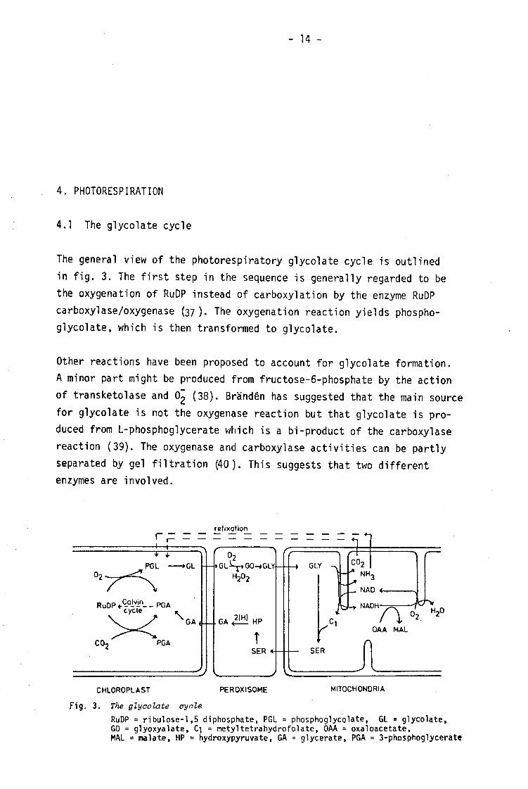
4.1 The glycola te cycle
The gene ral view of the photorespiratory glycolate cycle is outlined in fig. 3. The f irst step in the sequence is generally regarded to be the oxygenation of RuDP instead of carboxylation by th e enzyme RuDP carboxylase/oxygenase (37) . The oxy genation reaction yields phospho-glycolate, which is then transformed to glycolate.
Other reactions have bee n proposed t o account for glycolate formation. A minor part might be produced from fructose-6-phosphate by th e action of transketolase and (38). Brändén has suggeste d that the main source for glycolate is not the oxygenase reaction but that glycolate is produced from L-ph osphoglycerate whi ch i s a bi-product of the carboxylase reaction (39). The ox ygenase an d carboxylase a ctivities can be p artly separated by gel filtration (40). This suggests that two d ifferent enzymes are involved.
refixation
GLY PGL NH.
NAD 4-
R u D P * - - P G A cycle NADH
G A < OAA MAL
CO« PGA SER SER «
CHLOROPLAST PEROXISOME MITOCHONDRIA
Fig. 3. The glycolate cycle RuDP = ribulose-1,5 diphosphate, PGL = phosphoglycolate, GL = glycolate, GO = glyoxyalate, C-j = met yltetrahydrofolate, OAA = oxaloacetate, MAL « malate, HP « hydroxypyruvate, GA = glycerate, PGA = 3-phosphoglycerate

| - 15 -I
Glycolate is transported from the chloroplasts to the peroxisomes and converted to glyoxylate by gly colate oxidase. In this reaction oxygen is consumed an d is produced. The is broken do wn t o and by ca talase present in peroxisome. The glyoxylate is transaminated to glycine and transported into the mitochondria. In the mitochondria glycine is converted to serine, CO2 an d NHg wi th concomitant reduction of NAD to NADH (9). The de tails of the mitochondrial reactions in photo-respiration will be discussed in section 4.3. The decarboxylation of glycine is regareded to be the main site of CO2 rel ease in photorespiration, but ot her reactions may als o contribute. Glyoxylate m ay be oxidized to C02 by H 202 in the peroxisomes (41, 42). I t has also been suggested that glyoxylate can com bine with succinate to yield isocitrate, oxidation of which wo uld y ield CO2 in the citric acid cycle (43).
The s erine is then transported to the peroxisomes, deam inated to hydroxypyruvate and reduced to glycerate. The glycerate can be transported into the chloroplasts and phosphorylated to phosphoglycerate thus compieteing the glycolate cycle. The c yclic nature of the glycolate cycle has been
18 experimentally verified by 0 incorporation studies ( 4 4 ) , but i t has also been suggeste d that the intermediates can be use d in other metabolic reactions.
4.2 Function and regulation of photorespiration
It is difficult to quantify photorespiration accurately (45) but it is generally agreed that a substantial part of assimilated CO2 i s released. For m any C g specie s the rate of photorespiratory release of CO2 i s 50 % or more of CO2 fix ation in the Calvin cycle (46). Many de tails about the photorespiration are not fully understood, especially the metabolic regulation is lit tle studied. I t is important to investigate the details of photorespiration to be able to tell if photorespiration is a negative or an e ssential process in the photosynthesizing c ell .
The re lative amount of O2 an d CO2 in the atmosphere is important for the rate of photorespiration. Generally high CC^ concentrat ions decrease photorespiration and high 02 concentrations increase photorespiration (47) , but the response is complex (48, 49). In plants CO2 i s concentrated in the bundle sheath cells thus decreasing photorespiration. In other cases different O2 to CO2 proportions seem to be of l i t t le physiological significance. Photorespiration increases with temperature mo re than photosynthesis, this might be du e to an indirect effect of temperature

- Ib -
on C02 and 02 solubility (50). Hal 1 dal (51) has suggested that the state of excitation in photosystem I and II might be involved in the regulation of photorespiration.
Many stu dies have bee n c arried out on a rtificial regulation of photorespiration (45) with the aim to reduce photorespiration. Both mutants (52) and inhibitors (53) have been used. So f ar it has not been possible to increase net photosynthesis in long term exp eriments by inhibiting photorespiration.
The function of the apparently wasteful photorespiration is not known, but several hypothesis have been proposed. If the formation of glycol ate in the chloroplasts is inevitable the photorespiratory cycle would shunt 3/4 of the carbon in glycolate back t o the Calcin c ycle (37). As the refixation of both C02 and NH^ i s energy cons uming, photorespiration might be a way fo r plants to get rid of excess light energy to prevent photoinhibition (54).
4.3 The mitocho ndrial reactions in photorespiration
It was shown by Kisaki et al (55) that the conversion of glycine to serine in the glycolate cycle took place in the mitochondria. In isolated mitochondria two molecu les of glycine are consumed to yield one molecule each of CO2, NHg a nd s erine (9), NAD is reduced to NADH which can be reoxidized via the respiratory, chain giving a ratio of 2:1 for C02
evolved to 02 consumed (56). ATP synthe sis is coupled to this reoxi-dation (9, 57).
The reoxidation of NADH can a lso be cou pled to reduction of oxaloacetate to malate by mitochon drial malate dehydrogenase (11). I t has been speculated that this reducing power i s shuttled out from th e mitochondria to the peroxisomes and used for the reduction of hydroxypyruvate t o glycerate.
The ab ility to oxidize glycine via the respiratory chain is specific for leaf mitochondria (paper II) . The induction of the activity is light dependent. However, i t does not follow the increase in the chlorophyll content, but rather the increase in glycolate oxidase a ctivity (58). The convers ion of glycine to serine is a complex re action and involves two enz ymes, glycine decarboxylase and se rine hydroxymethyltransferase. The

- 17 -
reaction can be assaye d in several ways. The m ost common w ays ar e to measure oxy gen con sumption coupled to glycine oxidation, or to
14 14 measure CO2 evolution from 1- C-glycine. The reaction can als o be me asured fotometrically with the artificial electron acceptor DCPIP (2,C-dichlorcphenol indophenol) (59).
4.3.1 Glycinedecarboxylase
Glycinedecarboxylase (E.C. 2.1.2.10) catalyzes the oxidative decarboxylation of glycine:
glycine + tetrahydrofolate + NAD «-• C02 + NH^ + methylene tetrahydrofolate + NADH
The e nzyme al so catalyzes an exch ange betw een the carboxyl carbon of glycine and bicarbonate. The e nzymes fro m ani mal (60, 61) and bacteria (62) have bee n studied in detail. The a nimal and bacterial enzymes are very si milar and both contai n four different proteins. These fro m t he animal enzyme ar e named P-, H-, L- and T-protein. The P-protein is a pyridoxalphosphate con taining enzyme which can cat alyze a very slow decarboxylation of glycine (63). "The H-protein is a small heat stable protein (64). The rate of the decarboxylation catalyzed by the P-protein was increased by a factor 10 if H-protein was prese nt (65). Only t he P- and H-proteins wer e required for the bicarbonate exch ange reaction (66). The L-protein has FAD as prostetic group (60)» a lipoamide deh ydrogenase a ctivity and is involved in the transfer of reducing equ ivalents to NAD. The T-protein is involved in the synthesis of methylene tetrahydrofolate (66). In rat liver mitochondria the glycine decarboxylase i s
(7)-CHO +
OL-COOH I c
NH0
C0o
,0-CH=N-CH2-COOH
<2C < y (T)-CH=N-CH,-Sv-V
S HS^-X
NADH
NAD © 5,10 -CH2-FH4 + NH3 ( ï ï fJL®
^-^SH * FH4
s -CH2-NH2
SH (f> CH0
Fig. 4 . Proposed reaction saheme for glycine decarboxylase P.H.L. and T in the circles represent the respective proteins. FH4 = tetrahydrofolate (according to (62))

- 18 -
loosely bound to the inner membrane (67). The prop osed mechanism for glycine decarboxylase is shown in fig. 4.
The plant glycine decarboxylase is located behind the mitochondrial inner membrane (32, 57). I t has been as sumed that the enzyme is membrane bound (57) in analogy with the rat liver enzyme, but i t has not been possible to study the intramitochondrial localization experimentally. This is because both the decarboxylase a ctivity and the bicarbonate exchange re action are lost when the mitochondrial inner membrane is ruptured. The los s of activity can depend on several factors. One or several components, l ike proteins or cofactors, may leak out from t he mitochondria. Alternatively the enzyme system m ay d emand a membrane potential for activity (11).
4.3.2 Serine hydroxymethyltransferase
Serine hydroxymethyltransferase (E.C.2.1.2.1) catalyzes the reaction:
glycine + methylene tetrahydrofolate +• serine + tetrahydrofolate
The e nzyme has pyridoxalphosphate a s prostetic group (68). In c ontrast to glycine decarboxylase, serine hydroxymethyltransferase is present in both photosynthetic and non-photosynthetic c ells (69). Results obtained with leaf cells have suggested the presence of isoenzymes in different cell compartments (55, 70). In spinach leaves the enzyme w as mainly mito chondrial (71), and was l i berated similar to malate dehydrogenase from th e mitochondria upon rupture of the inner membrane, suggesting that serine hydroxymethyltransferase is mainly a matrix enzyme. The e nzyme isolated from m ung bean see dlings showed al losteric properties (72), but li t t le is known about the function of serine hydroxymethyltransferase in plants besides its function in the conversion of glycine to serine in the photorespiration.
4.3.3 Fate of the products
In isolated mitochondria one molecu le each of serine, CO2 a nd NH^ are formed fo r every two mole cules of glycine consumed. In vivo the products as well as the intermediates in the reaction can be fu rther metabolized inside the mitochondria o r transported to other parts of the cell , thus making the situation more com plex.

- 19 -
The NHg a nd C O2 pro duced i ri the mitochondria can penetrate the membrane and d iffuse to other parts of the cell . Different opinions exist in the literature about the mechanism for the glycine transport through t he mitochondrial membrane. Dench et al . (73) suggested that a specific carrier was involved, but o ther results suggest that glycine can penetrate the membrane without the need f or a carrier (74). Serine transport has been l it tle studied.
Part of the CO2 released in the mitochondria can be refix ed by R uDP carboxylase i n the chloroplast (75), but i t is very d ifficult to quantify this refixation.
The released ammonia mu st be eff ectively reassimilated since chloroplasts are known t o be unc oupled by lo w concentration of ammonia (76). Furthermore, a s the metabolic flux in photorespiration is very high the plant would not survive due to deficiency of organic nitrogen if ammonia w as not refixed. Different opinions exist in the li terature about the mechanism for this refixation. Jackson et al suggested that ammonia can be r efixed in the mitochondria by mitoc hondrial glutamine syn thetase (77). Other groups have reported that glutamine synth etase is absent from mito chondria (16). The latter observation is in agreement with recent work in our laboratory (78). According t o this ammonia can be refix ed by glutamin e synthetase (GS) either in the cytoplasm o r in the chloroplast. The glut amine thus formed is converted to glutamate by chloro plastic glutamine-oxogluta-rateaminotransferase (GOGAT), which is ferridoxin dependent and necessary for the survival of photosynthetic cells with photorespiration (79).
The glutamate formed can then participate in transamination reactions, for example the photorespiratory conversion of glyoxylate to glycine. Thus, in addition to a photorespiratory carbon cy cle a photorespiratory nitrogen cycle can be formu lated (80).
NH3 ATP ADP Glutamate V V / -*G1 utami ne
2-0xoglutarate Glutamate <• GOGAT

- 20 -
If the glycolate pathway i s strictly cyclic then all methylene tetrahydro-folate formed up on glycine cleavage mu st be used f or serine synthesis. Yet, in Euglena the C-j moiety for med from glycine was incorporated in RNA, adenine an d metionine (81) and it has been speculated that the methylene-tetrahydrofolate formed could be important in the C-| metabolism of photo-synthesizing cells (69). Photorespiratory serine has been reported to be an intermediate in sucrose synthesis in leaves (82, 83). It has al so been suggeste d that glycine and serine could participate in protein synthesis (84). Much additio nal work i s needed to establish whether photorespiratory glycine, serine and methylene tetrahydrofolate participate in other metabolic r eactions to a significant degree.

- 21 -
5. STUDIES ON MITOCHONDRIAL P HOTORESPIRATORY R EACTIONS
5.1 Glycine oxidation by mito chondria from diff erent parts of the plant
In paper II i t was sh own that the ability to oxidize glycine via the respiratory chain was sp ecific for mitochondria from photosynthetic tissue. The ab ility was prese nt in spinach leaf mitochondria, but a bsent in mitochondria prepared from r oots, stalks and leaf veins. This is in agreement with earlier studies on mito chondria prepared from photosynt hetic and non-photosynthetic tissue (85). The study in paper II was, however, made on mitoch ondria from di fferent parts of the same plants. This ma kes the interpretation more d irect as i t excludes gen etic differences between different species.
5.2 Effect of inhibitors on th e conversion of glycine to serine
The assays commonly use d to study the conversion of glycine to serine depend on the combined a ction of glycine decarboxylase and se rine hydroxy-methyltransferase as the folic acid pool in the mitochondria is small and must be regenerated during the assay as indicated in fig. 5.
oxaloacetate
malate Y Y respiratory cha
M h CO, NHS NAD NADH
1 î . u glycine
INH AAN GH
glycine decarboxylase
/••s
FH,
/ \ I I I *
^-»C1FH4<
DCPIP DCPIPH2
—1/2 02
KCN
SHMT
INH (AAN)
KCN GH
serine
^—* glycine
Fig. 5. Reactions and sites of inhibition in the conversion of glyoine to serine. FH4; tetrahydrofolate; CVFH4, 5,10-methylenetetrahydrofolate; SHMT, serine hydroxymethyltransferase (from pape r V).

- 22 -
Several inhibitors of the reaction are known, but the assays used a re such that it has not been possible to determine th e site of inhibition in the reaction sequence. In o rder to study this the effect of some of the known inhibitors on th e serine hydroxymethyltransferase ac tivity and the glycine-bicarbonate exchange re action associated with glycine decarboxylase was in vestigated (paper V). The in hibitors used were: isonicotinylhydrazide (a pyridoxalphosphate antagonist), aminoacetoni-trile, glycine hydroxamate (glycine analogues) and KCN.
Isonicotinylhydrazide and glycine hydroxamate both inhibited the two reactions studied. The inhibition of the glycine-bicarbonate exchange was competitive while the inhibition of serine hydroxymethyltransferase was non-competitive. Aminoacetonitrile inhibited the two reactions competitively but the exchange r eaction was inhibited at a much lower concentration than serine hydroxymethyltransferase. KCN inhibited serine hydroxymethyltransferase com petitively at a low concen tration, but the inhibit ion of the exchange r eaction was only partial.
The t otal pattern of inhibition summarized in fig. 5 was thus very com plex, with mo re than one s ite of inhibition for all the inhibitors studied.
5.3 Absorption peaks around 500 nm associated with the conversion of glycine to serine
In di fference spectra between substrate/anaerobic minus oxidized mitochondria two addit ional absorption peaks arou nd 500 nm appeared with glycine as the substrate compared to other respiratory substrates (paper IV). Glycine decarbox ylase and s erine hydroxymethyltransferase a re both pyridoxalphosphate enz ymes an d pyridoxalphosphate-substrate complexes are known t o absorbe light around 500 nm (86). The ob served absorption peaks a re therefore likely to represent enzyme-substrate complexes in the conversion of glycine to serine.
The e ffect of inhibitors of the conversion of glycine to serine on th e absorption peaks was studied (fig. 6). KCN at the concentration used, almost completely reduced the absorption peak a t 493 nm. The sa me concentration inhibited serine hydroxymethyltransferase and had l it tle effect on th e glycine-bicarbonate exchange reaction (paper V). This

- 23 -
0005 A
10 mM AAN
Fig. 6. Effect of inhibitors on absorption peaks in different spectra.
Glycine/aerobic minus oxidized difference spectra at 77 K.
suggests that the peak a t 493 nm is associated with serine hydroxymethyl-transferase. The re sults give no clue to the origin of the 503 nm absorption peak. Additional work i s needed t o establish the origin of this peak an d to determine whet her th e two absorption peaks are useful for the study of the conversion of glycine to serine.
100 pM KCN
100 pM GH
1 C90 500 510

- 24 -
6. CONCLUDING REMARKS
The present work describe s a procedure for preparing the hitherto known purest preparation of functionally intact mitochondria from spinach leaves/The preparation procedure was also useful for the preparation of pure mitoc hondria from othe r parts of the spinach plant, making i t possible to compare mito chondria from photosynthesizing and non-photosynthesizing t issues from th e same plants.
Qualitat ively leaf mitochondria had the same cyto chrome compo sition as other plant mitochondria. On a protein basis, however, the leaf mitochondria contained only half the amount of the different cytochromes as compared to other plant mitochondria.
A study of photorespiratory reactions in mitochondria showed that the mitochondrial oxidation of glycine was sp ecific for photosynthetically active tissue. The s ite of inhibition for different inhibitors of the conversion of glycine to serine was als o studied and some s pecific effects that might be valuable for the further characterization of the reaction were found.
The study of the details of the mitochondrial reactions in relation to photorespiration will certainly lead to a better understanding of the photorespiration and thereby increase the possibility to determine whether photorespiration is negative or essential for a photosynthesizing cell .
The improved preparation procedure w ill also make othe r studies on composition and specialization of leaf mitochondria possible. This would contribute to an increased understanding of the metabolism in photo-synthesizing cells.

- 25 -
7. ACKNOWLEDGEMENTS
I am gratefu l to Professor Per-Åke Albertsson for introducing me to the field of plant biochemistry an d to Dr. Christer Larsson for very valuable support and stimulating discussions throughout the course of this work. The cooperat ion and friendship with Mr. Anders Bergman has been of greatest stimulance and importance f or me. I am also indepted to Dr. Ingemar Ericson for cooperation and co mments on t he manuscripts, to Professor Sven Lin dskog for showing interest in the work, and to Dr. Vithaidas P. Shanbhag fo r revising the English text.
I want t o thank Miss Eleon ore Granström and Miss Katarina Wallgren for skilful technical assistance, and most of all Mrs. Kristina Smeds for her very patient assistance during mos t of this work. The s kilful typing of manuscripts,in spite of my handwriting, by M iss Agneta Ehman, Mrs. Carin Häggström and M rs. Kerstin Montell is also greatly acknowledged.
I also want t o express my gra titude to all others at the Department of Biochemistry f or making the time pleasant and stimulating.
This work w as suppo rted by gr ants from t he Swedish Natural Science Research Cou ncil and the Carl Trygger Foundation.

- 26 -
8 REFERENCES
1 Moore, A.L. and Rich, P.R. (1980) Trends Biochem. Sci. 5, 284-288.
2 Douce, R., Mannella, C.A. and Bonner, W.D. Jr. (1973) Biochim. Biophys. Acta, 292, 105-1 16.
3 Ikuma, H. (1972) Ann. Rev. Plant Physiol. 23, 419-436.
4 Coleman, J.O.D. and Palmer, J.M. (1972) Eur. J . Biochem. 26, 499-509.
5 Tolbert, N.E. (1971) Ann. Rev. Plant Physiol. 22, 45-74.
6 Graham, D. (1980) in: The Bioc hemistry of Plants. Vol 2 (Davies, D.D. ed) pp. 525-579, Academic Pr ess, New York.
7 Palmer, J.M. (1976) Ann. Rev. Plant Physiol. 27, 133-157.
8 Kisaki, T., Yoshida, N. and Imai, A. (1971) Plant Cell Physiol. 12, 275-288.
9 Bird, I .F., Cornelius, M.J., Keys, A.J. and Whittingham, C.P. (1972). Phytochemistry, 11, 1587-1594.
10 Clandinin, M.T. and Cossins, E.A. (1975) Phytochemistry 14, 387-391.
11 Woo, K.C. and Osmond, C.B. (1976) Aust. J . Plant Physiol. 3, 771-785.
12 Douce, R., Moore, A.L. and Neuburger, M. (1977) Plant Physiol. 60, 625-628.
13 Douce, R., Christensen, E.L. and Bonner, W .D. Jr. (1972) Biochim. Biophys. Acta 275, 148-16 0.
14 Arron, G.P., Spalding, M.H. and Edwards, G.E. (1979) Plant Physiol. 64, 182-186.
15 Rocha, V. and Ting, I .P. (1970) Arch. Biochem. Biophys. 140, 398-407.
16 Wallsgrove, R.M., Lea, P.J. and Miflin, B.J. (1979) Plant Physiol. 63, 232-236.
17 Gronebaum-Turk, K. and Willenbrink, J . (1971) Planta 100, 337-346.
18 Jackson, C., Dench, J .E., Hall, D.O. and Moore, A.L. (1979) Plant Physiol. 64, 150-153.
19 Albertsson, P.-Ä. (1974) in: Method. Enzymology Vo l 31A (Fl eischer, S. and Packer, L. eds.), pp 761-769, Academic P ress, New York.
20 Nishimura, M., Graham, D. and Akazawa, T. (1976) Plant Physiol. 58, 309-314.
21 Rustin, P., Julienne, M. and Kader, J.C. (1980) C.R. Acad. Sci. D. 291, 105-1 08.
22 Siegenthaler, P.A. and Depéry, F. (1976) Eur. J . Biochem. 61, 573-580.

- 27 -
23 Jackson, C. and Moore, A.L. (1979) in: Plant Organelles, Methodological surveys (B): Biochemistry, Vol 9 (Reid, E. ed.) pp. 1-12, Ellis Horwood, Chichester.
24 Larsson, C. and Andersson, B. (1979) in: Plant Organelles»Methodological surveys (B): Biochemistry, Vol 9 (Reid, E. ed.) pp. 35-46, Ellis Horwood, Chichester.
25 Larssón, C., Collin, C. and Albertsson, P.-Â. (1971) Biochim. Bio-phys. Acta 245, 425-438.
26 Andersson, B. and Åkerlund, H.-E. (1978) Biochim. Biophys. Acta 503, 462-472.
27 M011er, I .M., Bergman, A., Gardeström, P., Ericson, I. and Palmer, I.M. (1981) FEBS Le tt. 126, 13-17.
28 Widell, S. and Larsson, C. (1981) Physiol. Plant, in press.
29 Karlstam, B. and Albertsson, P.-Â. (1972) Biochim. Biophys. Acta, 255, 539-5 52.
30 Benbadis, A. and Virville.D. de (1980) Physiol. Veget. 18, 571.
31 Lowry, O.H., Rosebrough, N.J., Farr, A.L. and Randall, R.J. (1951) J. Biol. Chem. 193, 265-275.
32 Woo, K.C. and Osmond, C.B. (1977) in: Special Issue of Plant Cell Physiol. Vol 3 (Miyachi, S., Katoh, S., Fuji ta, Y. and Shiba ta, K. eds.) pp. 315-323. Center fo r Academic P ublications, Tokyo.
33 Elias, B.A. and Givan, C.V. (1977) Plant Physiol. 59, 738-740.
34 Yamazaki, R.K. and Tolbert, N.E. (1970) J. Biol. Chem. 245, 5137 -5144.
35 Neuburger, M. and Douce, R. (1978) in: Plant Mitochondria, (Ducet, G. and Lance, C. eds.) pp. 109-116. Elsevier, Amsterdam.
36 Lance, C. and Bonner, W .D. Jr. (1968) Plant Physiol. 43, 756-766.
37 Andrews, T .J. and Lorimer, G.H. (1978) FEBS Le tt. 90, 1-9.
38 Takabe, T., Asami, S. and Akazawa, T. (1980) Biochemistry 19, 3985-3989.
39 Brändén, R., Nilsson, T., Styring, S. and Ångström, J. (1980) Biochem. Biophys. Res. Commun. 92, 1306-1312.
40 Branden, R. (1978) Biochem. Biophys. Res. Commun. 81, 539-546.
41 Grodzinski, B. and Butt, V.S. (1977) Planta 133, 261-266.
42 Oliver, D.J. (1979) Plant Physiol. 64, 1048-1052.
43 Naik, M.S. and Singh, P. (1980) FEBS Le tt. I l l , 277-280.
44 Lorimer, G.H., Woo, K.C., Berry, J.A. and Osmond, C.B. (1978) in: Proceedings of the IVth International Congress on Photosynthesis, Reading (Hall, D.O., Coombs, J . and Goodwin, T.W., eds.) pp. 311-322, The Bio chemical Society, London.
45 Zelitch, I. (1980) in: Method. Enzymology V ol 69C, (Colowick, S.P. and Kaplan, N.O. eds.) pp. 453-464, Academic Pr ess, New York.

46
47
48
49
50
51
52
53
54
55
56
57
58
59
60
61
62
63
64
65
66
67
68
69
70
71
- 28 -
Zelitch, I . (1975) Ann. Rev. Biochem. 44, 123-145.
Servaites, J.C. and Ogren, W.L. (1978) Plant Physiol. 61, 62-67.
Oliver, D.J. (1979) Plant Sci. Lett. 15, 35-40.
Bravdo, B.-A. and Canvin, D. (1979) Plant Physiol. 63, 399-401.
Ku, S.-B. and Edwards, G.E. (1977) Plant Physiol. 59, 991-999.
Ha11da1, P. and Holmen, T. (1979) Physiol. Plant. 47, 195-199.
Berlyn, M.B. (1980) Theor. Appi. Genet. 58, 19-26.
Oliver, D.J. and Zelitch, I. (1977) Science, 196, 1450-1451.
Heber, U. and Krause, G.H. (1980) Trends Biochem. Sci. 5, 32-34.
Kisaki, T., Imai, A. and Tolbert, N.E. (1971) Plant Cell Physiol. 12, 267-273.
Arron, G .P., Spalding, M.H. and Edwards, G.E. (1979) Biochem. J. 184, 457-460.
Moore, A.L., Jackson, C., Halliwell, B., Dench, J.E. and Hall, D.O. (1977) Biochem. Biophys. Res. Commun. 78, 483-491,
Arron, G.P. and Edwards, G.E. (1980) Plant Sci. Lett. 18, 229-235.
Moore, A.L., Dench, J.E., Jackson, C. and Hall, D.O. (1980) FEBS Lett. 115, 54-58.
Motokawa, Y. and Kikuchi, G. (1972) J. Biochem. 72, 1281-1284
Hiraga, K., Kochi, H., Motokawa, Y. and Kikuchi, G. (1972) J. Biochem. 72, 1285-1289.
Kochi, H. and Kikuchi, G. (1974) J. Biochem. 75, 1113-1127.
Hiraga, K. and Kikuchi, G. (1980) J. Biol. Chem. 255, 116 64-11670.
Motokawa, Y. and Kikuchi, G. (1969) Arch. Biochem. Biophys. 135, 402-409.
Hiraga, K. and Kikuchi, G. (1980) J . Biol. Chem. 255, 116 71-11676.
Motokawa, Y. and Kikuchi, G. (1974) Arch. Biochem. Biophys. 164, 624-633.
Motokawa, Y. and Kikuchi, G. (1971) Arch. Biochem. Biophys. 146, 461-466.
Rader, I .J. and Hue nnekens, F.M. (1973) The En zymes, third edition, Vol 9 (Boyer, P.D. ed.) pp. 215-221. Academic P ress, New Y ork.
Cossins, E.A. (1980) in: The Biochemistry of Plants Vol 2 (Davies, D.D. ed.) pp. 365-418. Academic P ress, New York.
Shah, S.P.J, and Cossins, E.A. (1970) Phytochemistry 9, 1545-1551.
Woo, K.C. (1979) Plant Physiol. 63, 783-787.

- 29 -
72 Rao, N.D. and Rao, A.N. (1980) Biochem. Biophys. Res. Commun. 92, 1166-1171.
73 Dench, J.E., Briand, Y., Jackson, C., Hall , D.O. and Moore, A.L. (1978) in: Plant Mitochondria (Ducet, G. and Lance, C. eds.) pp. 133-140. Elsevier, Amsterdam.
74 Day, D.A. and Wiskich, J.T. (1980) FEBS Le tt. 112, 191-194.
75 Schaefer, J. , Kier, L.D. and S tejskal, E.O. (1980) P lant Physiol. 65, 254-259.
76 Krogmann, D.W., Jagendorf, A.T. and Avron, M. (1959) Plant Physiol. 34, 272-277.
77 Jackson, C., Dench, J .E., Morris, P. Lui, S.C., Hall, D.O. and Moore, A.L. (1979) Biochem. Soc. Trans. 7, 1122-1124.
78 Bergman, A., Gardeström, P. and Ericson, I . (1981) Submitted for publication to Physiol. Plant.
79 Somerville, C.R. and Ogren, W.L. (1980) Nature 286, 257-259.
80 Keys, A.J., Bird, I .F., Cornelius, M.J., Lea, P.J. , Wallsgrove, R.M. and Mi f l in, B.J. (1978) Nature, 275, 741-743.
81 Foo,S.S.K. and Cossins, E.A. (1978) Phytochemistry, 17, 1711-1715.
82 Waidyanatha, V.P. de S ., Keys, A.J. and Whi ttingham, C.P. (1975) J. Exp. Bot. 26, 15-26.
83 Champigny, M. -L. (1977) in: Proceedings of the IV International Congress on Photosynthesis, Reading (Hall, D.O., Coombs, J . and Goodwin, T.W. eds.) pp. 479-488. The Biochemical Society, London.
84 Tolbert, N.E. (1980) in: The Bioc hemistry of Plants Vol 2 (Davies, D.D. ed.) pp. 487-523. Academic P ress, New York.
85 Neuburger, M. and Do uce, R. (1977) C.R. Acad. Sci. D. 285, 881-8 84.
86 Matsumoto, S. and Matsushima, Y. (1964) J. Am. Chem. Soc. 94, 7211-7213.
