preliminarycommunication ......nipt can be offered to pregnant women at high risk for hav-ing a...
TRANSCRIPT

Confidential. Do not distribute. Pre-embargo material.
Noninvasive Prenatal Testing and Incidental Detectionof Occult Maternal MalignanciesDiana W. Bianchi, MD; Darya Chudova, PhD; Amy J. Sehnert, MD; Sucheta Bhatt, MD; Kathryn Murray, MS;Tracy L. Prosen, MD; Judy E. Garber, MD; Louise Wilkins-Haug, MD, PhD; Neeta L. Vora, MD;Stephen Warsof, MD; James Goldberg, MD; Tina Ziainia, MD; Meredith Halks-Miller, MD
IMPORTANCE Understanding the relationship between aneuploidy detection on noninvasiveprenatal testing (NIPT) and occult maternal malignancies may explain results that arediscordant with the fetal karyotype and improve maternal clinical care.
OBJECTIVE To evaluate massively parallel sequencing data for patterns of copy-numbervariations that might prospectively identify occult maternal malignancies.
DESIGN, SETTING, AND PARTICIPANTS Case series identified from 125 426 samples submittedbetween February 15, 2012, and September 30, 2014, from asymptomatic pregnant womenwho underwent plasma cell-free DNA sequencing for clinical prenatal aneuploidy screening.Analyses were conducted in a clinical laboratory that performs DNA sequencing. Among theclinical samples, abnormal results were detected in 3757 (3%); these were reported to theordering physician with recommendations for further evaluation.
EXPOSURES NIPT for fetal aneuploidy screening (chromosomes 13, 18, 21, X, and Y).
MAIN OUTCOMES AND MEASURES Detailed genome-wide bioinformatics analysis wasperformed on available sequencing data from 8 of 10 women with known cancers.Genome-wide copy-number changes in the original NIPT samples and in subsequent serialsamples from individual patients when available are reported. Copy-number changesdetected in NIPT sequencing data in the known cancer cases were compared with the typesof aneuploidies detected in the overall cohort.
RESULTS From a cohort of 125 426 NIPT results, 3757 (3%) were positive for 1 or moreaneuploidies involving chromosomes 13, 18, 21, X, or Y. From this set of 3757 samples, 10cases of maternal cancer were identified. Detailed clinical and sequencing data were obtainedin 8. Maternal cancers most frequently occurred with the rare NIPT finding of more than 1aneuploidy detected (7 known cancers among 39 cases of multiple aneuploidies by NIPT, 18%[95% CI, 7.5%-33.5%]). All 8 cases that underwent further bioinformatics analysis showedunique patterns of nonspecific copy-number gains and losses across multiple chromosomes.In 1 case, blood was sampled after completion of treatment for colorectal cancer and theabnormal pattern was no longer evident.
CONCLUSIONS AND RELEVANCE In this preliminary study, a small number of cases of occultmalignancy were subsequently diagnosed among pregnant women whose noninvasiveprenatal testing results showed discordance with the fetal karyotype. The clinical importanceof these findings will require further research.
JAMA. 2015;314(2):162-169. doi:10.1001/jama.2015.7120Published online July 13, 2015.
Editorial page 131
JAMA Report Video andAuthor Video Interview atjama.com
JAMA Patient Page page 198
Author Affiliations: Authoraffiliations are listed at the end of thisarticle.
Corresponding Author: Diana W.Bianchi, MD, Mother Infant ResearchInstitute, Tufts Medical Center,800 Washington St, Box 394,Boston, MA 02111 ([email protected]).
Research
Preliminary Communication
162 (Reprinted) jama.com
Confidential. Do not distribute. Pre-embargo material.
N oninvasive prenatal testing (NIPT) using massivelyparallel sequencing of cell-free DNA (cfDNA) inmaternal plasma has recently changed the clin-
ical paradigm of prenatal screening for the common fetalautosomal aneuploidies (abnormal numbers of wholechromosomes).1,2 Using this technology, the sensitivities forthe detection of fetal trisomies 21 and 18 are, on average,99% and 96%, respectively, with specificities of 99% to100%.3 Many professional societies have recommended thatNIPT can be offered to pregnant women at high risk for hav-ing a fetus with autosomal aneuploidy, with follow-up diag-nostic testing recommended to confirm a positive testresult.1,4-6
Although NIPT performs well, it is an advanced screen,not a diagnostic test. The reason for this distinction is thatthe cfDNA in the plasma of pregnant women is a mixture ofplacental (used as a proxy for the fetus) and maternal DNA.Follow-up studies have shown that some cfDNA results arediscordant with the direct fetal karyotype.7,8 Potentialbiological explanations for discordance include confinedplacental mosaicism,9 co-twin demise,10 maternal chromo-somal mosaicism11 and DNA copy-number variants,12 mater-nal organ transplant from a male donor,11 and maternalmalignancy.13,14
The diagnosis of cancer during pregnancy is relatively un-common, with an incidence of about 1 in 1000 gestations.15 Themost common malignancies observed in pregnant women arebreast and cervical cancers, Hodgkin and non-Hodgkin lym-phomas, malignant melanoma, leukemia, ovarian cancer, andcolorectal cancer.15
The purpose of this study was to retrospectively exam-ine DNA sequencing data in a series of pregnant womenwith abnormal NIPT results involving aneuploidies of chro-mosomes 13, 18, 21, X, or Y, who were diagnosed with cancerafter prenatal testing occurred. In addition, to better under-stand the frequency with which maternal cancer might pro-vide an explanation for abnormal NIPT results that are dis-cordant with the fetal karyotype, all abnormal test results inthe clinical laboratory and available clinical outcomes werereviewed.
MethodsThe current case series was identified from a population of125 426 pregnant women undergoing plasma cfDNA sequenc-ing in the Illumina clinical laboratory (Redwood City, Califor-nia) between February 15, 2012, and September 30, 2014.Patients were included if their clinician voluntarily informedthe laboratory at any time prior to November 15, 2014, thatmaternal cancer had been diagnosed after NIPT. As part of astandard laboratory follow-up process, the laboratory con-tacts the referring clinicians to discuss all positive NIPTresults and to recommend a diagnostic procedure to obtain aconfirmatory fetal karyotype. When NIPT results and thekaryotype are discordant, the medical director (S.B.) and thecertified genetic counselors who work with her review pos-sible explanations for the discordant results with the refer-
ring clinician. Maternal cancer had not been reported as areason for NIPT discordance until publication of a single casereport in 2013,13 so maternal cancer was only included in thedifferential diagnosis after that time.
Evolving knowledge and experience have resulted inchanges in the bioinformatics analytic algorithms used inthe clinical laboratory during the time frame of this study.The ability to analyze and visualize whole-genome sequenc-ing results was not technically possible until October 2013.After October 2013, if referring clinicians requested theexpanded bioinformatics results, these were communicateddirectly to the physician. In all cases, the patient’s clinicianwas responsible for determining the follow-up clinical man-agement.
For each patient reported in detail in this article, inaddition to the consent obtained for the original, clinicallyindicated noninvasive prenatal test, a separate individualwritten consent for medical records review, further genomicanalysis, and possible publication of findings was obtainedafter the abnormal NIPT results were reported to thepatient’s physician. The Tufts Medical Center institutionalreview board waived review of this study. Informationregarding the patient’s pregnancy, cancer diagnosis, andmedical history was obtained from her clinicians and medi-cal records, by direct discussion with the first author(D.W.B.), or both. In some of the cases, additional bloodsamples (including postpartum) were obtained and ana-lyzed. For these samples, the clinical laboratory team per-forming the sequencing was blinded to the fact that thesewomen were no longer pregnant.
Using whole blood samples, the verifi Prenatal Test(Illumina) screens for the presence of whole chromosomeaneuploidy for chromosomes 13, 18, and 21. Testing for sexchromosome aneuploidy by analyzing sequencing countsfor chromosomes X and Y is optional.11 The method usesmassively parallel sequencing of cfDNA isolated frommaternal plasma.16-20 To identify on which chromosome thesequenced DNA fragment mapped, a software programknown as bowtie21 was used to align the short (25 base-pair)sequence reads to the 19th reference version of the humangenome sequence map (hg19). The data were filtered toremove nonunique alignments and genomic regions associ-ated with high variation. They were then normalized basedon the percentage of guanine (G) cytosine (C) representationin the sequence of each chromosome and corrected toremove other assay and sample-specific biases.
Overrepresentation or underrepresentation of the targetchromosomes (13, 18, 21, X, and Y) was evaluated by con-structing a ratio between the normalized coverage on eachchromosome of interest and the sum of normalized cover-age on a respective set of reference chromosomes.16 Typi-cally, there were between 2 and 6 reference chromosomesper target chromosome (eg, 13, 18, 21, X, and Y). Specific ref-erence chromosomes have changed with evolution of theclinical bioinformatics algorithms. Upper and lower normallimits were then applied to the test results to generate ananeuploidy classification status for chromosomes 13, 18,and 21 (aneuploidy detected, aneuploidy suspected, or no
Maternal Malignancy and Noninvasive Prenatal Testing Preliminary Communication Research
jama.com (Reprinted) JAMA July 14, 2015 Volume 314, Number 2 163

Confidential. Do not distribute. Pre-embargo material.aneuploidy detected)17,19 and for sex chromosomes (sexchromosome aneuploidy detected or no sex chromosomeaneuploidy detected).11 If no sex chromosome aneuploidywas detected, a sex chromosome result of XX or XY wasprovided.11
All whole blood samples received within 5 days ofsampling with a complete test requisition form authorizedby an ordering physician were entered into the laboratorymanagement system. Maternal age, gestational age, andindication for testing (if included) on the test requisitionform were recorded. All test results with an aneuploidy“detected” or “suspected” were telephoned to the orderingphysician by a certified genetic counselor employed by theclinical laboratory. If a diagnostic procedure for fetalkaryotyping was performed, clinicians were requested on 2separate occasions to inform the laboratory whether theNIPT results were concordant or discordant with the fetalkaryotype. Whenever the laboratory was notified of discor-dant results, pertinent history was obtained from thepatients’ physicians and genetic counselors, and possiblebiological mechanisms for abnormal results were discussedas stated earlier in the Methods. An internal quality assur-ance process was also followed to evaluate any potentialtechnical explanations for the discordant result.
When detailed bioinformatics analysis of the previouslysequenced DNA sample was performed, mapped sections ofthe human genome were analyzed using circular binarysegmentation,22 in order to identify copy-number variants(CNVs). Copy-number variants are genomic regions associ-ated with significant deviation from the expected 2 copiesacross a contiguous span of the human genome. For a dip-loid genome, normalized coverage is expected to be 1.0.If there is a gain of a single copy, the expected result is 1.5(a 50% gain in amplitude). Similarly, for the loss of a singlecopy, the expected result is 0.5 (a 50% loss in amplitude).Using this scale, a maternal plasma sample from a womancarrying a fetus with trisomy 21 that contains 10% circulat-ing fetal DNA will have a 5% gain in coverage across thelength of chromosome 21 (0.1 × 0.50 = 0.05). In this study,identified CNVs were counted as gains or losses if theyexceeded either 10 megabase pairs (Mb) in length and 2.5%in deviation from the expected diploid coverage, or 40 Mbin length and 1% in deviation from the expected diploid cov-erage. These parameters were only used for visual interpre-tation of the data and were not intended to identify cancersignatures.
To evaluate the frequency of reported maternal malig-nancies in relation to the overall frequency of aneuploidypositive results, all clinical laboratory reports, as well as alltests that were cancelled due to abnormal underlying chro-mosomal patterns generated within the study time frame,were reviewed and the findings were grouped into 1 of 5 cat-egories: single trisomy, single monosomy, single sex chro-mosome aneuploidy, single sex chromosome aneuploidyplus single trisomy, or multiple aneuploidies.
Statistical analysis of the reported proportions was per-formed using Clopper-Pearson exact binomial 2-sided confi-dence intervals at the 95% level (using R version 3.1.2).
Results
Review of Clinical CasesFrom a cohort of 125 426 NIPT tests, 3757 (3.0%) were posi-tive for 1 or more aneuploidies involving chromosome 13, 18,21, X, or Y. In 10 of these “aneuploidy-detected” cases, thereferring clinician voluntarily reported to the clinical labora-tory within weeks to months after the initial discussionregarding the clinical significance of the positive NIPT resultsthat the patient had been diagnosed with a malignancy. The10 cancer cases were clinically diverse and included 3 cases ofB-cell lymphoma and 1 case each of T-cell leukemia; Hodgkinlymphoma; unspecified adenocarcinoma; leiomyosarcoma;and neuroendocrine, colorectal, and anal carcinomas. In 2cases (leiomyosarcoma and unspecified adenocarcinoma),the referring physicians reported that the women were criti-cally ill, and they declined to approach them for consent toparticipate in this study.
Table 1 shows demographic factors, NIPT results, fetal sta-tus, and cancer stage for the remaining 8 cases, in which per-mission was granted for further analysis. At the time of initialNIPT, the mean maternal age was 35 years (range, 23-39 years),and the mean gestational age was 13.9 weeks (range, 10-20weeks). Cancer was subsequently diagnosed (during preg-nancy or postpartum) in these women at a mean of 16 weeks(range, 3-39 weeks) after the initial NIPT. The clinical presen-tations ranged from early-stage to metastatic disease. In 3 pa-tients (cases 4, 5, and 8), the discordant NIPT results prompteda further medical workup that led to the diagnosis of cancer.The 3 patients with B-cell lymphoma (cases 2, 6, and 7) pre-sented with a palpable mass. In 2 cases of maternal malig-nancy (cases 1 and 3), the patients presented with advancedsymptoms: pain due to bone metastases and colon obstruc-tion, respectively.
In 7 of the 8 cases, diagnostic fetal karyotyping was per-formed and showed a euploid result (46,XY or 46,XX). Of 3 pre-term deliveries, 1 was at 29 weeks due to maternal preeclamp-sia, 1 at 35 weeks due to spontaneous labor, and 1 at 32 weeksto facilitate maternal treatment (cases 5, 7, and 8, respec-tively, Table 1).
Bioinformatics AnalysisDetailed genome-wide analysis of the original sequencingdata obtained from cfDNA of the 8 study participantsrevealed CNVs that affected multiple chromosomes andspanned between 4% and 44% (median, 29%) of thegenome (Figure 1). Cases with trisomies detected by NIPTcould be explained by whole or large partial copy-numbergains on the test chromosomes or losses on any of the refer-ence chromosomes. Conversely, cases with monosomiesdetected could be explained by either losses on the targetchromosomes or gains on the reference chromosomes. For 2cases (3 and 5), in which replicate testing of the same initialblood sample was performed, the CNV detection resultswere highly consistent, resulting in identical NIPT calls and91% to 99% identical gain or loss profiles across the entiregenome (Figure 1, lines 3B and 3B’ and 5A and 5A’).
Research Preliminary Communication Maternal Malignancy and Noninvasive Prenatal Testing
164 JAMA July 14, 2015 Volume 314, Number 2 (Reprinted) jama.com

Confidential. Do not distribute. Pre-embargo material.
Additional whole blood samples were collected from 3 ofthe participants at time points subsequent to the initial NIPT:for case 1, 6 weeks after initial NIPT (but still pregnant); forcase 3, 5 months after delivery, immediately prior to surgicalresection of the colorectal tumor, and 14 months after deliv-ery following completion of radiation and chemotherapy;and for case 4, 8 months after delivery. Detected CNVs werehighly consistent prior to treatment in samples obtained upto 11 months apart. Areas of CNV detection overlapped by76%, 79%, and 93%, respectively, for cases 1, 3, and 4; the dif-ferences were mostly due to increased amplitude of signalwith time and additional detectable gains/losses in latersamples (Figure 1).
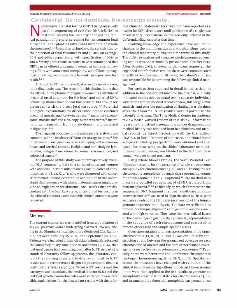
Blood samples were obtained at 3 different clinically sig-nificant time points for case 3. The sequencing data for chro-mosome 13 (a test chromosome) and 8 (a reference chromo-some for chromosome 18) are shown in Figure 2. An increasein normalized coverage for chromosome 13 is evident in thepretreatment samples, consistent with the original NIPT re-sult of trisomy 13 detected. Confined placental mosaicism wasruled out using microarrays in another laboratory. The mag-nitude of the CNV in the maternal blood above baseline in-creased over time (from 1%-3% deviation from the expecteddiploid genome) in the postpartum presurgical resectionsample (Figure 1 and Figure 2). Similarly, chromosome 8 (a ref-
erence chromosome for chromosome 18) displayed partial am-plifications and losses in the original NIPT sample. In the sec-ond sample, the chromosome 8 signal gained sufficientamplitude to affect the calculations for chromosome 18, caus-ing a monosomy 18–detected test result. The patient’s thirdsample, obtained after completion of all treatment, showed noabnormal deviations from baseline.
Aneuploidy Patterns in Maternal CancerFollow-up of the 3757 abnormal NIPT results was incom-plete. Seven of the 10 cases of maternal cancer reported tothe clinical laboratory had multiple aneuploidies (Table 2).Of the 39 cases of multiple aneuploidy, 7 cases (18% [95% CI,7.5%-33.5%]) were in women with an occult cancer. Of the39 cases of multiple aneuploidies detected, 4 were concor-dant or partially concordant, meaning that at least 1 of theaneuploidies detected by NIPT was confirmed by fetal diag-nostic testing. Sixteen of the remaining 35 NIPT results wereconfirmed to be discordant with results from follow-upinvasive diagnostic testing. In the other 19 cases, the out-come was unknown because of fetal loss without karyotypeinformation or a lack of clinical information from the refer-ring physician.
Although the patient follow-up was incomplete, we esti-mate that the risk of maternal cancer in the small subset of preg-
Table 1. Clinical Details on the 8 Cases of Maternal Cancer That Underwent Genome-wide Analysis
Case 1a Case 2 Case 3 Case 4 Case 5 Case 6 Case 7 Case 8Maternaldemographics
Age, y 37 36 33 36 23 37 39 39
GA, wk 13 12 13 20 20 12 11 10
Aneuploidy detectionby NIPT
Chromosome 21 Not detected Not detected Not detected Monosomy Trisomy Not detected Not detected Trisomy
Chromosome 18 Monosomy Monosomy Not detected Monosomy Monosomy Trisomy Monosomy Trisomy
Chromosome 13 Trisomy Not detected Trisomy Monosomy Trisomy Not detected Not detected Trisomy
Sex chromosomes Not done Not done Not done Not done XY XX XXY Monosomy X
No. of NIPTaneuploidies
2 1 1 3 3 1 2 4
Fetal/newborn status
Fetal karyotype 46,XY Not done 46,XY 46,XY 46,XY 46,XX 46,XY 46,XX
Pregnancy outcome Term male Term female Term male Term male Preterm male,preeclampsia, 29 wk
Term female Pretermmale, 35 wk
Preterm female,32 wk
Cancer characteristics
Cancer type Neuro-endocrine(unknownorigin)
Non-Hodgkin(B-cell)lymphoma
Colorectal Hodgkinlymphoma
Acute T-celllymphoblasticleukemia
Non-Hodgkin(B-cell)lymphoma
Non-Hodgkin(B-cell)lymphoma
Anal
Stage at diagnosis IV, metastatic IVB IIIC IIA NA IV II IIIB
Time elapsed fromNIPT to diagnosis
28 wk 13 wk 39 wk 3 wk to MRI, 29 wkto biopsy
3 wk ≈20 wk ≈10 wk 8 wk
Timing of cancerdiagnosis
Postnatal Prenatal Postnatal Postnatal Prenatal Prenatal Prenatal Prenatal
Postnatal DNAsequencing results
Not done Not done Trisomy 13,monosomy18
Monosomy 13,monosomy 18,monosomy 21,monosomy X
Not done Not done Not done Not done
Abbreviations: GA, gestational age at time of NIPT blood draw as obtained from test request form; MRI, magnetic resonance imaging; NA, not applicable;NIPT, noninvasive prenatal testing.a Case previously published.13
Maternal Malignancy and Noninvasive Prenatal Testing Preliminary Communication Research
jama.com (Reprinted) JAMA July 14, 2015 Volume 314, Number 2 165

Confidential. Do not distribute. Pre-embargo material.
nant women with abnormal discordant NIPT results due tomultiple aneuploidies detected and a normal fetal karyotypeis as follows. If all 19 cases of multiple aneuploidies in whichfollow-up information was unavailable were concordant withthe fetal karyotype, the risk of maternal cancer as an expla-nation for the discordant results would be 7 of 16 cases (44%).If, however, all of the 19 cases were discordant with the fetalkaryotype (eg, the fetal karyotype is normal), then the riskwould be 7 of 35 cases (20%).
DiscussionIn this case series of 125 426 NIPT results, 3757 were positivefor 1 or more aneuploidies involving chromosomes 13, 18,21, X, or Y. Some of the abnormal NIPT results were discor-
dant with the diagnostic fetal karyotypes obtained byamniocentesis or chorionic villus sampling. Here we haveshown that occult maternal malignancies may provide abiological explanation for some discordant NIPT results.This is presumably due to the cfDNA that is released intomaternal circulation from apoptotic malignant cells. Thetypes of cancers diagnosed were among those most fre-quently reported in women of childbearing age,15 althoughthere were more hematologic malignancies than would beexpected and no cases of malignant melanoma or cervicalcancer. The expected cancer rate in pregnant women isabout 0.1%.15 This series of cancer cases, reported volun-tarily, represents 0.008% (10/125 426) of the laboratory casevolume, a cancer frequency that is 10-fold lower than whatmight be expected. However, this patient series is inher-ently incomplete; maternal cancers diagnosed after delivery
Figure 1. Whole-Genome View of Copy-Number Changes in 8 Cases of Maternal Cancer
Chromosomes
6.3NIPT8 A
15.7NIPT7 A
12.5NIPT6 A
20.4NIPT
5 A
16.5NIPT4 A
4.6NIPT3 A
36.4NIPT2 A
Case Sample Run Whole-genome copy-number representation (normalized coverage)Maximum deviation, %
Trisomy
Monosomy
22.2A’
14.9BPretreatment
After delivery
6.3NIPT1 A
9.2BPretreatment
During pregnancy
6.6B
PretreatmentAfter delivery
6.7B’
0.0CPosttreatment
1 2 3 4 5 6 7 8 9 10 11 12 13 14 15 17 19 21
1.00.9
1.1
1.00.9
1.1
1.00.9
1.1
1.00.9
1.1
1.00.9
1.1
1.00.9
1.1
1.00.9
1.1
1.00.9
1.1
1.00.9
1.1
1.00.9
1.1
1.00.9
1.1
1.00.9
1.1
1.00.9
1.1
1.00.9
1.1
Whole-genome view of copy-number gains and losses in plasma samples fromwomen with known cancer. Smoothed normalized coverage (in black) is plottedalong the genomic coordinates (x-axis), sorted by chromosome number andgenomic location within the chromosomes. The data for all samples are shownas normalized coverage on the same scale, on the left side of the y-axis (0.9-1.1).The scale chosen for this figure is less than 0.5-1.5 because of fractionalrepresentation of the tumor DNA in the mixed sample. For some samples, theamplitude of the copy-number variants exceeded the scale; the maximum
deviation from expected diploid representation is shown as a percentage on theright side of the y-axis. Copy-number gains or losses relative to the diploidreference genome are shown as blue or red, respectively. If a trisomy wasreported, the relevant chromosome is shown by a light blue bar. If a monosomywas reported, the relevant chromosome is shown by a light red bar. Cases 3 and5 include replicates of the same blood sample, identified by an apostrophe (‘).Cases 1, 3, and 4 had longitudinal samples obtained (see text for details).
Research Preliminary Communication Maternal Malignancy and Noninvasive Prenatal Testing
166 JAMA July 14, 2015 Volume 314, Number 2 (Reprinted) jama.com
Confidential. Do not distribute. Pre-embargo material.
might not routinely be reported to the NIPT laboratory.Even cancers diagnosed during pregnancy would not neces-sarily trigger notification of the laboratory, especially if noaneuploidies had been detected by NIPT. The lower ratemay also reflect that the chromosomal aneuploidies and theamount of apoptotic tumor cfDNA released into the mater-nal circulation could be below the detection limit at thetime NIPT is performed. A recent study using sequencing toanalyze plasma cfDNA in patients with known cancers23
found evidence of abnormal cfDNA patterns in more than80% of metastatic solid tumor cases and 50% of localizedcancers. The rates of detection varied widely by tumor type.
Genome-wide bioinformatics analysis for the 8 reportedcases revealed extensive copy-number changes involving sev-eral chromosomes and ranged from numerous focal amplifi-cations or deletions to multiple whole chromosomes. Thesetypes of changes are more likely to be visible using a whole ge-nome rather than a targeted sequencing approach. In addi-tion, the visualized changes were reproducible in replicates ob-tained from the same blood sample, and the overall pattern ofchromosomal changes was stable in samples taken manymonths apart.
Autosomal monosomies, and especially multiple-aneuploidy test results, are rarely identified in NIPT samples.
Figure 2. Longitudinal Evolution of Chromosomal Profiles for Maternal Cancer Case 3
Posttreatment postdelivery sampleC
B Pretreatment postdelivery sample
NIPT sample (13 weeks’ gestation)A
0.8
0.9
1.0
1.1
1.2
0.8
0.9
1.0
1.1
1.2
0.8
0.9
1.0
1.1
1.213 12
11.2
11.1
1112
.11
12.1
212
.13
12.2
12.3
13.1
13.2
13.3
14.1
114
.12
14.1
314
.214
.321
.121
.221
.31
21.3
221
.33
22.1
22.2
22.3
31.1
31.2
31.3
32.1
32.2
32.3
33.1
33.2
33.3
34
0.8
0.9
1.0
1.1
1.2
0.8
0.9
1.0
1.1
1.2
0.8
0.9
1.0
1.1
1.2
23.3
23.2
23.1 22
21.3
21.2
21.1
1211
.23
11.2
211
.21
11.1
11.1
11.2
111
.22
11.2
312
.112
.212
.313
.113
.213
.321
.11
21.1
221
.13
21.2
21.3
22.1
22.2
22.3
23.1
23.2
23.3
24.1
124
.12
24.1
324
.21
24.2
224
.23
24.3
Nor
mal
ized
cov
erag
eN
orm
aliz
ed c
over
age
Nor
mal
ized
cov
erag
e
Chromosome 13 Chromosome 8
p qp q
Individual chromosome views of data shown in Figure 1. Chromosomal coverageprofiles in samples from case 3 taken at different intervals of time. The gray dotsshow the normalized coverage, and the solid colored lines show smoothedprofiles (obtained from the median values across 31 genomic 100-kilobasebins). The upper panel is from the sample taken during pregnancy at 13 weeksof gestation. The middle panel is after delivery, immediately before surgicalresection of an obstructing colorectal tumor. The lower panel is after delivery,following completion of chemotherapy and radiation. The x-axis shows thephysical location of the increased counts as mapped against an ideogram for
chromosomes 13 or 8 (SNPchip package, R version 3.1.2; resolution = 1megabase pair). Chromosome 8 is included because it served as one of thereference chromosomes and contributed to the monosomy 18 classification inthe postdelivery sample (see Table 1). The y-axis shows the percentage of signalabove or below baseline corresponding to a diploid genome (y = 1.0). As anexample, in the middle-right panel (chromosome 8 after delivery), the data atthe highest peak (indicated by the arrow) show that there is approximately 12%excess representation of this part of the genome compared with the reference.NIPT indicates noninvasive prenatal testing.
Maternal Malignancy and Noninvasive Prenatal Testing Preliminary Communication Research
jama.com (Reprinted) JAMA July 14, 2015 Volume 314, Number 2 167

Confidential. Do not distribute. Pre-embargo material.
For this reason, NIPT results demonstrating a single autoso-mal monosomy or multiple aneuploidies may warrant a moredetailed analysis of the whole genome using an advanced bio-informatics review process to determine if a pattern sugges-tive of malignancy is present.
To date, there have been 3 individual reports of pregnantwomen with abnormal NIPT results and chromosomally nor-mal fetuses in which the discordant results were explainedby the presence of maternal malignancies (metastatic smallcell neuroendocrine c arcinoma of vaginal origin,1 3
lymphomas,14,24 and ovarian carcinoma24) in which tumorDNA was presumably shed into the maternal circulation anddetected at the time of noninvasive prenatal testing. Thedata presented here underscore the necessity of performinga diagnostic procedure to determine the true fetal karyotypewhenever NIPT results reveal chromosomal abnormalities.
Many genetic counselors and obstetricians are concernedthat an NIPT result of multiple aneuploidies or autosomalmonosomy may be suggestive of maternal cancer. Whenthere is discordance between the fetal karyotype and NIPTresult, occult maternal malignancy, although very uncom-mon, may be an explanation for the findings. Based on theresults of the study, we estimate there is between a 20% and44% risk of maternal cancer if multiple aneuploidies aredetected. However, until further studies are done to assessthe clinical implications of discordant NIPT and fetal karyo-type results, it is not clear what, if any, follow-up clinicalevaluation is appropriate.24
All 8 women in this case series were asymptomatic at thetime of their NIPT test. In 3 cases, the NIPT results promptedthe diagnosis of malignancy. Whether earlier detection ofdisease would have made a difference in the course of theirillnesses cannot be determined. Cases 1 and 3 presented withadvanced symptoms, and their clinicians stated that forthem, earlier diagnosis would have had a positive effect ontheir care.
Limitations of this study include its small size and retro-spective design, incomplete clinical follow-up information, po-tential for bias of ascertainment in the way that the cancer di-agnoses were reported back to the clinical laboratory, and theevolving nature of the technical parameters, especially in thebioinformatics analyses over 2.5 years.
ConclusionsIn this preliminary study, a small number of occult malignan-cies were subsequently diagnosed among pregnant womenwhose noninvasive prenatal testing results showed discor-dance with the fetal karyotype. The clinical importance of thesefindings will require further research to determine appropri-ate follow-up for the mother and her infant.
ARTICLE INFORMATION
Published Online: July 13, 2015.doi:10.1001/jama.2015.7120.
Author Affiliations: Mother Infant ResearchInstitute, Tufts Medical Center, Boston,Massachusetts (Bianchi); Illumina, Redwood City,California (Chudova, Sehnert, Bhatt, Halks-Miller);Center for Genetics and Maternal Fetal Medicine,Springfield, Oregon (Murray); Division of Maternal-Fetal Medicine, Department of Obstetrics andGynecology, University of Minnesota, Minneapolis(Prosen); Dana-Farber Cancer Institute, Boston,Massachusetts (Garber); Division of Maternal FetalMedicine and Reproductive Genetics, Departmentof Obstetrics and Gynecology, Brigham andWomen’s Hospital, Boston, Massachusetts(Wilkins-Haug); University of North Carolina,Chapel Hill (Vora); Division of Maternal-FetalMedicine, Department of Obstetrics andGynecology, Eastern Virginia Medical School,Norfolk (Warsof); San Francisco PerinatalAssociates, San Francisco, California (Goldberg);Sharp Rees-Stealy Medical Group, San Diego,California (Ziainia).
Author Contributions: Dr Bianchi had full access toall of the data in the study and takes responsibilityfor the integrity of the data and the accuracy of thedata analysis.Study concept and design: Bianchi, Chudova,Sehnert, Murray, Halks-Miller.Acquisition, analysis, or interpretation of data: Allauthors.Drafting of the manuscript: Bianchi, Chudova,Sehnert, Murray, Goldberg, Halks-Miller.Critical revision of the manuscript for importantintellectual content: All authors.Statistical analysis: Chudova, Sehnert, Bhatt.Administrative, technical, or material support:Bianchi, Murray, Prosen, Garber, Wilkins-Haug,Vora, Warsof, Goldberg, Ziainia, Halks-Miller.Study supervision: Bianchi, Chudova, Sehnert,Bhatt, Halks-Miller.
Conflict of Interest Disclosures: All authors havecompleted and submitted the ICMJE Form forDisclosure of Potential Conflicts of Interest. DrBianchi reported being a member of theReproductive and Genetic Health Expert AdvisoryPanel of Illumina, for which she receives anhonorarium, and having received sponsored
research funding from Illumina that is administeredthrough Tufts Medical Center. Drs Chudova,Sehnert, Bhatt, and Halks-Miller reported beingfull-time employees of Illumina. Ms Murrayreported having served on the speakers’ bureau forMyriad Genetics. Dr Prosen reported being amember of the Illumina speakers’ bureau. Dr Garberreported having received sponsored researchfunding from Myriad Genetics and Novartis andserving as a consultant for Pfizer and Sequenom. DrWilkins-Haug reported having received sponsoredresearch support from Ariosa and Sequenom. Noother disclosures were reported.
Funding/Support: Funding for the study wasprovided by Illumina. Sponsored research fundingfrom Illumina that is administered through TuftsMedical Center paid for the time that Dr Bianchispent working with the full-time Illuminaemployees to design the study, analyze the data,and prepare the manuscript.
Role of the Funder/Sponsor: Illumina reviewedand approved submission of the manuscript forpublication (as per their internal policies), but theydid not make any changes to the manuscript.Illumina had no role in the design and conduct of
Table 2. Association of Maternal Cancers With Different Typesof Aneuploidies Detected at Noninvasive Prenatal Testing
Type of AneuploidyDetected by NIPT
Total No.of Samples
No. of Known MaternalCancers (%) [95% CI]
Single trisomya 2650 2 (0.08) [0-0.27]
Single SCAb 950 0 (0) [0-0.39]
Single trisomy + SCA 30 0 (0) [0-11.5]
Single monosomy 88 1 (1.14) [0-6.1]
Multiple aneuploidyc 39 7 (17.9) [7.5-33.5]
Total abnormal NIPT 3757 10 (0.26) [0.12-0.48]
Abbreviations: NIPT, noninvasive prenatal testing; SCA, sex chromosomeabnormality.a Single trisomy refers specifically to trisomy of chromosomes 13, 18, or 21.b Single SCA refers to the presence of 1 of the sex chromosome aneuploidies:
Turner syndrome (monosomy X), Klinefelter syndrome (XXY), XYY syndrome,or trisomy X (XXX).
c The multiple aneuploidy category includes every other combination ofautosomal and/or sex chromosome aneuploidy except single trisomy and SCAas noted in the Table.
Research Preliminary Communication Maternal Malignancy and Noninvasive Prenatal Testing
168 JAMA July 14, 2015 Volume 314, Number 2 (Reprinted) jama.com
Confidential. Do not distribute. Pre-embargo material.the study; collection, management, analysis, andinterpretation of the data; or preparation of themanuscript.
Additional Contributions: We thank the casepatients whose data are reported herein for givingpermission to publish their information. They weregracious and generous with their time andfollow-up information with the hope of helpingothers. We also thank Shannon X. Jeddi, CGC, forher help in obtaining medical record information.She received no financial compensation. We alsothank Richard Rava, PhD, for his scientific advice;he was not compensated for his contributionbesides salary.
REFERENCES
1. American College of Obstetricians andGynecologists Committee on Genetics, Society forMaternal-Fetal Medicine. Committee opinion No.640: cell-free DNA screening for fetal aneuploidy[published online June 26, 2015]. Obstet Gynecol.doi:10.1097/AOG.0000000000001007.
2. Warsof SL, Larion S, Abuhamad AZ. Overview ofthe impact of noninvasive prenatal testing ondiagnostic procedures [published online May 21,2015]. Prenat Diagn. doi:10.1002/pd.4601.
3. Gil MM, Quezada MS, Revello R, Akolekar R,Nicolaides KH. Analysis of cell-free DNA in maternalblood in screening for fetal aneuploidies: updatedmeta-analysis. Ultrasound Obstet Gynecol. 2015;45(3):249-266.
4. Benn P, Borrell A, Cuckle H, et al. Prenataldetection of Down syndrome using massivelyparallel sequencing (MPS): a rapid responsestatement from a committee on behalf of the Boardof the International Society for Prenatal Diagnosis,24 October 2011. Prenat Diagn. 2012;32(1):1-2.
5. Devers PL, Cronister A, Ormond KE, Facio F,Brasington CK, Flodman P. Noninvasive prenataltesting/noninvasive prenatal diagnosis: the positionof the National Society of Genetic Counselors.J Genet Couns. 2013;22(3):291-295.
6. Gregg AR, Gross SJ, Best RG, et al.ACMG statement on noninvasive prenatalscreening for fetal aneuploidy. Genet Med. 2013;15(5):395-398.
7. Wang JC, Sahoo T, Schonberg S, et al. Discordantnoninvasive prenatal testing and cytogeneticresults: a study of 109 consecutive cases. Genet Med.2015;17(3):234-236.
8. Bianchi DW, Wilkins-Haug L. Integration ofnoninvasive DNA testing for aneuploidy intoprenatal care: what has happened since the rubbermet the road? Clin Chem. 2014;60(1):78-87.
9. Lau TK, Jiang FM, Stevenson RJ, et al. Secondaryfindings from non-invasive prenatal testing forcommon fetal aneuploidies by whole genomesequencing as a clinical service. Prenat Diagn. 2013;33(6):602-608.
10. Curnow KJ, Wilkins-Haug L, Ryan A, et al.Detection of triploid, molar, and vanishing twinpregnancies by a single-nucleotidepolymorphism-based noninvasive prenatal test. AmJ Obstet Gynecol. 2015;212(1):79.e1-79.e9.
11. Bianchi DW, Parsa S, Bhatt S, et al. Fetal sexchromosome testing by maternal plasma DNAsequencing: clinical laboratory experience andbiology. Obstet Gynecol. 2015;125(2):375-382.
12. Snyder MW, Simmons LE, Kitzman JO, et al.Copy-number variation and false positive prenatalaneuploidy screening results. N Engl J Med. 2015;372(17):1639-1645.
13. Osborne CM, Hardisty E, Devers P, et al.Discordant noninvasive prenatal testing results in apatient subsequently diagnosed with metastaticdisease. Prenat Diagn. 2013;33(6):609-611.
14. Vandenberghe P, Wlodarska I, Tousseyn T, et al.Non-invasive detection of genomic imbalances inHodgkin/Reed-Sternberg cells in early andadvanced stage Hodgkin’s lymphoma bysequencing of circulating cell-free DNA: a technicalproof-of-principle study. Lancet Haematol. 2015;2(2):e55-e65.
15. Pavlidis NA. Coexistence of pregnancy andmalignancy. Oncologist. 2002;7(4):279-287.
16. Sehnert AJ, Rhees B, Comstock D, et al. Optimaldetection of fetal chromosomal abnormalities bymassively parallel DNA sequencing of cell-free fetalDNA from maternal blood. Clin Chem. 2011;57(7):1042-1049.
17. Bianchi DW, Platt LD, Goldberg JD, AbuhamadAZ, Sehnert AJ, Rava RP; Maternal Blood Is Sourceto Accurately diagnose fetal aneuploidy (MELISSA)Study Group. Genome-wide fetal aneuploidydetection by maternal plasma DNA sequencing.Obstet Gynecol. 2012;119(5):890-901.
18. Rava RP, Srinivasan A, Sehnert AJ, Bianchi DW.Circulating fetal cell-free DNA fractions differ inautosomal aneuploidies and monosomy X. Clin Chem.2014;60(1):243-250.
19. Futch T, Spinosa J, Bhatt S, de Feo E, Rava RP,Sehnert AJ. Initial clinical laboratory experience innoninvasive prenatal testing for fetal aneuploidyfrom maternal plasma DNA samples. Prenat Diagn.2013;33(6):569-574.
20. Bianchi DW, Parker RL, Wentworth J, et al;CARE Study Group. DNA sequencing versusstandard prenatal aneuploidy screening. N Engl J Med.2014;370(9):799-808.
21. Langmead B, Trapnell C, Pop M, Salzberg SL.Ultrafast and memory-efficient alignment of shortDNA sequences to the human genome. Genome Biol.2009;10(3):R25.
22. Olshen AB, Venkatraman ES, Lucito R, WiglerM. Circular binary segmentation for the analysis ofarray-based DNA copy number data. Biostatistics.2004;5(4):557-572.
23. Bettegowda C, Sausen M, Leary RJ, et al.Detection of circulating tumor DNA in early- andlate-stage human malignancies. Sci Transl Med.2014;6(224):224ra24.
24. Amant F, Verheecke M, Wlodarska I, et al.Presymptomatic identification of cancers inpregnant women during noninvasive prenataltesting [published online June 5, 2015]. JAMA Oncol.doi:10.1001/jamaoncol.2015.1883.
Maternal Malignancy and Noninvasive Prenatal Testing Preliminary Communication Research
jama.com (Reprinted) JAMA July 14, 2015 Volume 314, Number 2 169