practitioner’s fitting guide - abba opticalabbaoptical.com/rosek_files/rosekfittingguide.pdf ·...
TRANSCRIPT
The Rose K "System" is designed off a complex computer modelyielding the ultimate base curve geometry with appropriateperipheral curve structures. The back surface of the lens ismanufactured on computerized lathes in a series of spherical cuts. A special software program was written for the lathes to not only cutthe multiple curves but to blend them as well. Only a light polishneeds to be performed to finish the base of the lens by the lab. This means total reproducibility assurance for the fitter, lens after lens.The back surface is a series of spherical cuts that are well blended.Looking at the lens in a light source, it may appear to be aspheric but it is not.
There are three standard peripheral systems offered in the Rose K lensdesign - standard, increased or decreased. You may require a changein the peripheral curve from standard to increased lift or decreased lift.Unfortunately, if you modify a Rose K lens, you no longer have aRose K lens. Further explanation is provided. The most frequentperipheral change is to increased lift where the peripheral curves areapproximately 1.0mm flatter than the standard lift. Flatter peripheralsystems can be generated 0.5mm, 1.0mm (standard increased lift),1.5mm, 2.0mm, 2.5mm or 3.0mm flatter than the standard lift. Althoughnot usually necessary, they are available. These curve systems need tobe cut on the computerized lathes by the lab. Steeper peripheralsystems are available as well.
The basic steep peripheral lift is approximately 0.5mm steeper thanthe standard lift. A steeper peripheral-system beyond this of 1.0mmcan also be generated. Again, these curves must be lathe cut by thelab. Patients exhibiting early keratoconus usually need flatter base curves, often steeper peripheral systems and larger diameters.
Changing peripheral systems other than the standard increased and standard decreased lifts are for rare cases. The basic standardthree options for peripheral curve lifts should fit over 90% of yourkeratoconus patients.
9. Sagital ChangesChanging the peripheral system to steep or flat will change the sagital height of the lens and consequently the central fit. The lab willtherefore calculate a new base curve to keep the sag of the lens thesame, so that the fitting relationship is identical to the diagnostic orthe previous prescription lens. The power will alter with the base curvechange as well to compensate for this change. Changing fromstandard peripheral lift to the standard increased lift, the lab willcompensate the base curve by steepening it 0.05mm and by adding - 0.50D of power. Changing from a standard peripheral lift tothe standard decreased lift, the lab will compensate the base curveby flattening it 0.03mm and by adding +0.25 D power.
10. Residual Astigmatism (R.A.)A. It is usual to leave low degrees of R.A. uncorrected. Suggested table below for the spherical equivalent:
R.A. - 0.25 to - 0.50D add -0.25D
R.A. - 0.75 to - 1.00D add -0.50D
R.A. -1.25D to -1.50D add -0.75D
It is rare to see R.A. over this, when it is, toric lensesare usually needed anyway
B. To assess if the R.A. needs to be considered put the uncorrectedcylinder in spectacles and assess whether it makes a significantdifference to the visual acuity. These patients often have some flare orglare especially for night driving. If prescribing the R.A. in spectaclesadd an A/R coating to the lenses to help eliminate this. Given thechoice between a front cylinder toric lens and a combination of aspherical contact lenses and a spectacle over correction, most patientsselect the spectacles. Each patient needs must be assessed for thebest recommendation.
C. A front surface toric lens can be made if the fluoroscein patternlooks good with the spherical diagnostic lens. Once base curve andperipheral system is determined, the lab can design the front surfacetoric lens. An 0.3mm truncation is used, so the toric lens will be made0.3mm larger than the diagnostic lens and then a lower truncation isadded. Normally 1 1/4 prism ballast is used to start. Steeper bases andoblique cylinders tend to rotate more and are often more difficult toposition.
D. When R.A is present and the fluoroscein pattern indicates a toricbase is needed, a base toric or bitoric can be made. From the bestspherical base curve, add and subtract 0.4mm to get the proper baseseg: 6.50 diagnostic lens, need 6.90/6.10 base toric. Next select aspherical lens equal to the flattest base toric and over refract ie: a 6.90in the example here. Adding this to the power of the diagnostic lenswill determine the required spherical power.
11. AstigmatismA. TSP (Toric Periphery Only) - lens can also be made where the OZ isspherical but the last 1.0mm of the lens is toric. The tight areas, usuallyat 3 & 9 o'clock, will be eliminated with a TSP design.
B. Full Back Surface Toric - The Rose K lens can be made in either fullback surface toric or bitoric form.
C. Front Surface Toric - The Rose K lens can be supplied as a frontsurface toric with a spherical base to correct residual astigmatism.
12. Dimple VeilingOccasionally a patient will present with small bubbles trapped underthe lens. A combination of increasing the edge lift, flattening the basecurve and/or reducing the diameter should be tried first. If thebubbles persist, the lens can be fenestrated to relieve the problem.The fenestration will be at the juncture of the optic zone and thesecondary curve. Bubbling will not damage the cornea but willdecrease the visual acuity.
13. Pooling at the Cone BaseWith excessive pooling at the cone base, reduce the diameter as thisreduces the O.Z and the cone is fitted closer and the pooling isreduced. Flattening the Base Curve and or increasing the lift can alsohelp. Caution: don't go so small that vision is compromised with anoptic zone that is too small.
The Boston Logo is a registered trademark of polymer technology,a Bausch & Lomb company.
Practitioner’sFitting Guide
Prin
ted
in C
anad
a •
BC
L220
404A
C O N T A C T L E N S I N C .
1-800-367-4009
Complex lens geometry, combined with the enhanced benefits
of Boston materials, makes the Rose K™ lens the better fit for superior
patient comfort and visual acuity.
Multiple parameters make fitting the Rose K lens easy,for reduced chair time and increased practice efficiency.
The Rose K Lens Features:
Flexible peripheral systems Base curves: 4.75mm - 8.40mmDiameters: 7.9mm - 10.2mm Power : any
The Rose K Design Benefits:
— Fitting ease - our 26-lens diagnostic fitting set makes it easyto find just the right fit for your patient.
— Better visual acuity - clinical results have consistently shownan improvement in patient vision.
— Increased patient comfort - patients who have worn other Keratoconus RGP designschoose Rose K. It's simply more comfortable.
1
2
3
4
5
6
7
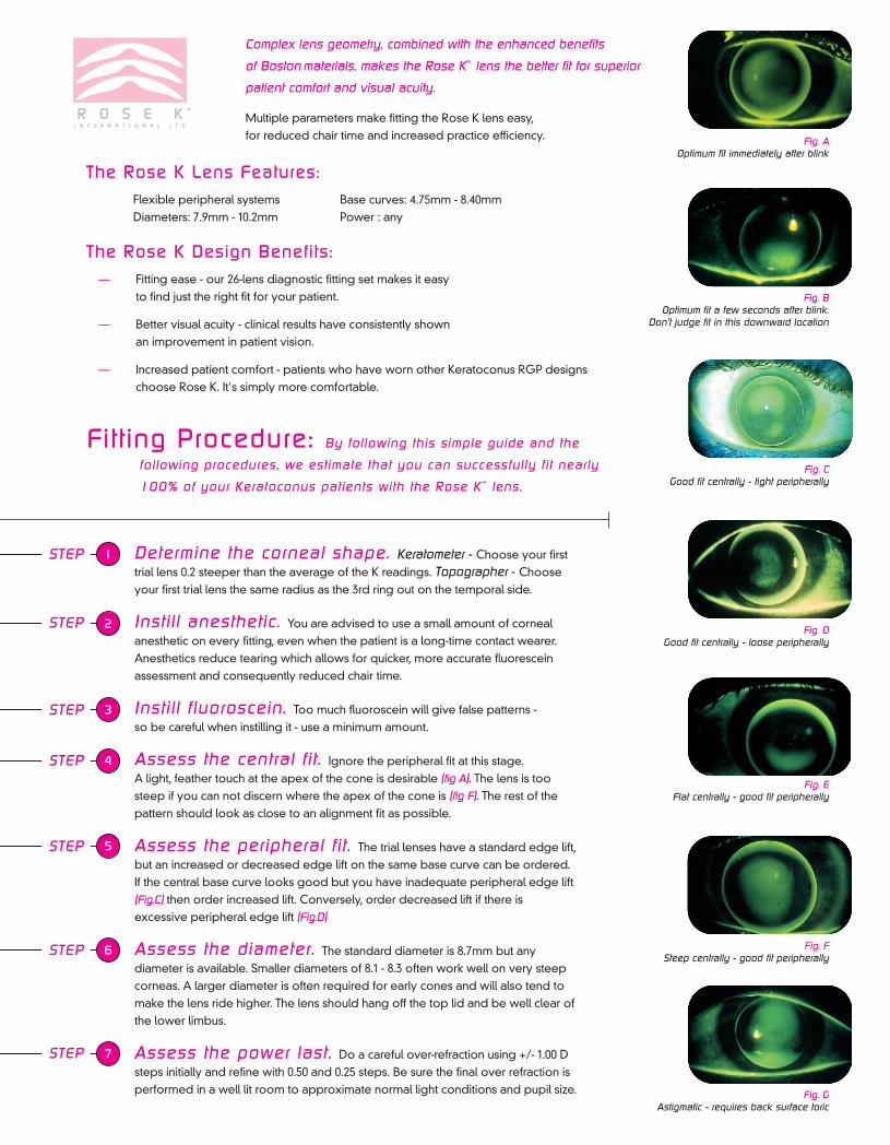
Fig. AOptimum fit immediately after blink
Fig. BOptimum fit a few seconds after blink.
Don’t judge fit in this downward location
Fig. CGood fit centrally - tight peripherally
Fig. DGood fit centrally - loose peripherally
Fig. EFlat centrally - good fit peripherally
Fig. FSteep centrally - good fit peripherally
Fig. GAstigmatic - requires back surface toric
1. Diagnostic Fitting Procedures and Tips
Placing a series of diagnostic lenses on the eye can be arduous and uncomfortable for the patient. We suggest using a minimalamount of anaesthetic for maximum patient comfort and to minimizetear flow. We recommend this for all patients not just new contact lenswearers. Increased tear flow will cause the lens to sit low and giveabnormal fluorescein patterns. Also instilling large amount offluoroscein can give false patterns, so be cautious about the amountplaced on the eye.
2. Fluoroscein Evaluation
Critical to the success of a Rose K lens fit is careful fluoroscein patternevaluation. Look to achieve a light feather touch on the cone apex, asclose to alignment as possible across the cornea, with lift off over thelast 0.6 to 0.8mm. It is important that the lens is locating centrally tojudge accurately the fluoroscein pattern. If the lens lags down useupward the pressure on the lower lid to centralise the lens to judgethe fit. Some initial low riding lenses will position better after severaldays wear.
3. Apical Staining
Apical staining should not be permitted with the Rose K lens. If at anyafter care visit apical staining is obvious, refit the lens with a steeperbase curve.
4. Diameter
The Rose K lens design is based on a complex mathematicalcomputer model that yields thousands of peripheral curvecombinations. These vary by base curve, edge lift, power, anddiameter. One common request is to take a larger lens and cut it downwhen the larger diameter does not function optimally. This can not bedone because the peripheral system on an 8.3mm lens is drasticallydifferent from the peripheral system on an 8.7mm lens. The smallerlens must be remade.
Reasons for Considering Diameter Changes:
A. Moderate amounts of astigmatism outside the cone can often befitted adequately with a spherical Rose K lens. If the lens is tighthorizontally and lifts vertically, especially at the base, reducing thediameter will minimize these problems.
B. 3 and 9 o'clock staining can often be reduced by reducing thediameter by 0.3mm or more and/or increasing the edge lift.
C. Excessively low riding lenses may need to be made larger by atleast 0.3mm The upper edge of the lens should touch the tarsal plateof the upper lid. Measure from the top of the lens to the bottom of theupper lid, and add half that distance to the overall diameter of the lenson the eye.
D. If you see superior limbal staining, reduce the diameter by 0.3mmand/or increase edge lift.
E. If the lens exhibits excessive edge stand off usually in the verticalmeridian, a reduced diameter and/or steepened peripheral systemmay be necessary. Be sure that the smaller overall diameter andtherefore smaller resultant O.Z. does not cause the lens to rideinferiorly resulting in poor vision.
F. Early cones often require a larger diameter. Advanced cones oftenrequire a smaller diameter.
G. With larger eyes, start with a 9.0mm lens.
5. Visual Assessment A. For patients that complain of ghosting or cloudy vision, increase thediameter if possible. This will increase the optic zone and often giveclearer vision. In many instances, the problem can also be improvedby having the patient wear spectacles with an anti-reflective coating.
B. If visual acuity is not as good as expected try flattening the basecurve by 0.1 to 0.2m.m
6. Positioning LensesRemember that the Rose K lens will position over the apex of thecone. Getting a low riding lens to position may require an increaseddiameter, flattened base curve, increased edge lift or a combinationof these. A high riding lens can be positioned by reducing thediameter, steepening the base curve or reducing the edge lift. Neverjudge the fit of a lens in a low riding position. Manually manipulate thelens to a central position and then evaluate the fluroscein pattern.
An important rule of thumb is to fit a flatter base curve rather than asteeper one, if a choice is available.
7. Problem Solving Order of PreferenceStart with a 8.7mm diameter with a standard edge lift. Fit the centralbase curve first followed by the peripheral fit.
If required, improve the peripheral fit by flattening or steepening theperipheral system.
If excessive pooling results around the cone base, or the fluorosceinpattern shows high astigmatism, reduce the lens diameter. This optionis not available with large corneas as they need larger lenses forproper positioning and good vision.
For larger corneas and/or large cones select a larger diameter(9.0mm) to start. Lens fits which exhibit significant toric "dumb bell"patterns, which can result in poor location, poor comfort, 3 and 9o'clock staining, or excessive lift off inferiorily, may require a Rose Kback surface toric lens.
8. The Peripheral SystemThe Rose K lens design is a departure from other lenses for thetreatment of KC. The present approach is to order a diameter andbase curve and then try to figure out a peripheral curve structure thatwill work for the patient. This can be very time consuming, many timesunsuccessful, and in-office modifications can be hard to reproducelens after lens.
Important information for successful fitting of Rose K lenses
Determine the corneal shape. Keratometer - Choose your firsttrial lens 0.2 steeper than the average of the K readings. Topographer - Chooseyour first trial lens the same radius as the 3rd ring out on the temporal side.
Instill anesthetic. You are advised to use a small amount of cornealanesthetic on every fitting, even when the patient is a long-time contact wearer.Anesthetics reduce tearing which allows for quicker, more accurate fluoresceinassessment and consequently reduced chair time.
Instill fluoroscein. Too much fluoroscein will give false patterns - so be careful when instilling it - use a minimum amount.
Assess the central fit . Ignore the peripheral fit at this stage. A light, feather touch at the apex of the cone is desirable (fig A). The lens is toosteep if you can not discern where the apex of the cone is (fig F). The rest of thepattern should look as close to an alignment fit as possible.
Assess the peripheral fit . The trial lenses have a standard edge lift,but an increased or decreased edge lift on the same base curve can be ordered.If the central base curve looks good but you have inadequate peripheral edge lift(Fig.C) then order increased lift. Conversely, order decreased lift if there isexcessive peripheral edge lift (Fig.D)
Assess the diameter. The standard diameter is 8.7mm but anydiameter is available. Smaller diameters of 8.1 - 8.3 often work well on very steepcorneas. A larger diameter is often required for early cones and will also tend tomake the lens ride higher. The lens should hang off the top lid and be well clear ofthe lower limbus.
Assess the power last. Do a careful over-refraction using +/- 1.00 Dsteps initially and refine with 0.50 and 0.25 steps. Be sure the final over refraction isperformed in a well lit room to approximate normal light conditions and pupil size.
Fitting Procedure: By following this simple guide and the
following procedures, we estimate that you can successfully fit nearly
100% of your Keratoconus patients with the Rose K™ lens.
STEP
STEP
STEP
STEP
STEP
STEP
STEP
Complex lens geometry, combined with the enhanced benefits
of Boston materials, makes the Rose K™ lens the better fit for superior
patient comfort and visual acuity.
Multiple parameters make fitting the Rose K lens easy,for reduced chair time and increased practice efficiency.
The Rose K Lens Features:
Flexible peripheral systems Base curves: 4.75mm - 8.40mmDiameters: 7.9mm - 10.2mm Power : any
The Rose K Design Benefits:
— Fitting ease - our 26-lens diagnostic fitting set makes it easyto find just the right fit for your patient.
— Better visual acuity - clinical results have consistently shownan improvement in patient vision.
— Increased patient comfort - patients who have worn other Keratoconus RGP designschoose Rose K. It's simply more comfortable.
1
2
3
4
5
6
7
Fig. AOptimum fit immediately after blink
Fig. BOptimum fit a few seconds after blink.
Don’t judge fit in this downward location
Fig. CGood fit centrally - tight peripherally
Fig. DGood fit centrally - loose peripherally
Fig. EFlat centrally - good fit peripherally
Fig. FSteep centrally - good fit peripherally
Fig. GAstigmatic - requires back surface toric
1. Diagnostic Fitting Procedures and Tips
Placing a series of diagnostic lenses on the eye can be arduous and uncomfortable for the patient. We suggest using a minimalamount of anaesthetic for maximum patient comfort and to minimizetear flow. We recommend this for all patients not just new contact lenswearers. Increased tear flow will cause the lens to sit low and giveabnormal fluorescein patterns. Also instilling large amount offluoroscein can give false patterns, so be cautious about the amountplaced on the eye.
2. Fluoroscein Evaluation
Critical to the success of a Rose K lens fit is careful fluoroscein patternevaluation. Look to achieve a light feather touch on the cone apex, asclose to alignment as possible across the cornea, with lift off over thelast 0.6 to 0.8mm. It is important that the lens is locating centrally tojudge accurately the fluoroscein pattern. If the lens lags down useupward the pressure on the lower lid to centralise the lens to judgethe fit. Some initial low riding lenses will position better after severaldays wear.
3. Apical Staining
Apical staining should not be permitted with the Rose K lens. If at anyafter care visit apical staining is obvious, refit the lens with a steeperbase curve.
4. Diameter
The Rose K lens design is based on a complex mathematicalcomputer model that yields thousands of peripheral curvecombinations. These vary by base curve, edge lift, power, anddiameter. One common request is to take a larger lens and cut it downwhen the larger diameter does not function optimally. This can not bedone because the peripheral system on an 8.3mm lens is drasticallydifferent from the peripheral system on an 8.7mm lens. The smallerlens must be remade.
Reasons for Considering Diameter Changes:
A. Moderate amounts of astigmatism outside the cone can often befitted adequately with a spherical Rose K lens. If the lens is tighthorizontally and lifts vertically, especially at the base, reducing thediameter will minimize these problems.
B. 3 and 9 o'clock staining can often be reduced by reducing thediameter by 0.3mm or more and/or increasing the edge lift.
C. Excessively low riding lenses may need to be made larger by atleast 0.3mm The upper edge of the lens should touch the tarsal plateof the upper lid. Measure from the top of the lens to the bottom of theupper lid, and add half that distance to the overall diameter of the lenson the eye.
D. If you see superior limbal staining, reduce the diameter by 0.3mmand/or increase edge lift.
E. If the lens exhibits excessive edge stand off usually in the verticalmeridian, a reduced diameter and/or steepened peripheral systemmay be necessary. Be sure that the smaller overall diameter andtherefore smaller resultant O.Z. does not cause the lens to rideinferiorly resulting in poor vision.
F. Early cones often require a larger diameter. Advanced cones oftenrequire a smaller diameter.
G. With larger eyes, start with a 9.0mm lens.
5. Visual Assessment A. For patients that complain of ghosting or cloudy vision, increase thediameter if possible. This will increase the optic zone and often giveclearer vision. In many instances, the problem can also be improvedby having the patient wear spectacles with an anti-reflective coating.
B. If visual acuity is not as good as expected try flattening the basecurve by 0.1 to 0.2m.m
6. Positioning LensesRemember that the Rose K lens will position over the apex of thecone. Getting a low riding lens to position may require an increaseddiameter, flattened base curve, increased edge lift or a combinationof these. A high riding lens can be positioned by reducing thediameter, steepening the base curve or reducing the edge lift. Neverjudge the fit of a lens in a low riding position. Manually manipulate thelens to a central position and then evaluate the fluroscein pattern.
An important rule of thumb is to fit a flatter base curve rather than asteeper one, if a choice is available.
7. Problem Solving Order of PreferenceStart with a 8.7mm diameter with a standard edge lift. Fit the centralbase curve first followed by the peripheral fit.
If required, improve the peripheral fit by flattening or steepening theperipheral system.
If excessive pooling results around the cone base, or the fluorosceinpattern shows high astigmatism, reduce the lens diameter. This optionis not available with large corneas as they need larger lenses forproper positioning and good vision.
For larger corneas and/or large cones select a larger diameter(9.0mm) to start. Lens fits which exhibit significant toric "dumb bell"patterns, which can result in poor location, poor comfort, 3 and 9o'clock staining, or excessive lift off inferiorily, may require a Rose Kback surface toric lens.
8. The Peripheral SystemThe Rose K lens design is a departure from other lenses for thetreatment of KC. The present approach is to order a diameter andbase curve and then try to figure out a peripheral curve structure thatwill work for the patient. This can be very time consuming, many timesunsuccessful, and in-office modifications can be hard to reproducelens after lens.
Important information for successful fitting of Rose K lenses
Determine the corneal shape. Keratometer - Choose your firsttrial lens 0.2 steeper than the average of the K readings. Topographer - Chooseyour first trial lens the same radius as the 3rd ring out on the temporal side.
Instill anesthetic. You are advised to use a small amount of cornealanesthetic on every fitting, even when the patient is a long-time contact wearer.Anesthetics reduce tearing which allows for quicker, more accurate fluoresceinassessment and consequently reduced chair time.
Instill fluoroscein. Too much fluoroscein will give false patterns - so be careful when instilling it - use a minimum amount.
Assess the central fit . Ignore the peripheral fit at this stage. A light, feather touch at the apex of the cone is desirable (fig A). The lens is toosteep if you can not discern where the apex of the cone is (fig F). The rest of thepattern should look as close to an alignment fit as possible.
Assess the peripheral fit . The trial lenses have a standard edge lift,but an increased or decreased edge lift on the same base curve can be ordered.If the central base curve looks good but you have inadequate peripheral edge lift(Fig.C) then order increased lift. Conversely, order decreased lift if there isexcessive peripheral edge lift (Fig.D)
Assess the diameter. The standard diameter is 8.7mm but anydiameter is available. Smaller diameters of 8.1 - 8.3 often work well on very steepcorneas. A larger diameter is often required for early cones and will also tend tomake the lens ride higher. The lens should hang off the top lid and be well clear ofthe lower limbus.
Assess the power last. Do a careful over-refraction using +/- 1.00 Dsteps initially and refine with 0.50 and 0.25 steps. Be sure the final over refraction isperformed in a well lit room to approximate normal light conditions and pupil size.
Fitting Procedure: By following this simple guide and the
following procedures, we estimate that you can successfully fit nearly
100% of your Keratoconus patients with the Rose K™ lens.
STEP
STEP
STEP
STEP
STEP
STEP
STEP

The Rose K "System" is designed off a complex computer modelyielding the ultimate base curve geometry with appropriateperipheral curve structures. The back surface of the lens ismanufactured on computerized lathes in a series of spherical cuts. A special software program was written for the lathes to not only cutthe multiple curves but to blend them as well. Only a light polishneeds to be performed to finish the base of the lens by the lab. This means total reproducibility assurance for the fitter, lens after lens.The back surface is a series of spherical cuts that are well blended.Looking at the lens in a light source, it may appear to be aspheric but it is not.
There are three standard peripheral systems offered in the Rose K lensdesign - standard, increased or decreased. You may require a changein the peripheral curve from standard to increased lift or decreased lift.Unfortunately, if you modify a Rose K lens, you no longer have aRose K lens. Further explanation is provided. The most frequentperipheral change is to increased lift where the peripheral curves areapproximately 1.0mm flatter than the standard lift. Flatter peripheralsystems can be generated 0.5mm, 1.0mm (standard increased lift),1.5mm, 2.0mm, 2.5mm or 3.0mm flatter than the standard lift. Althoughnot usually necessary, they are available. These curve systems need tobe cut on the computerized lathes by the lab. Steeper peripheralsystems are available as well.
The basic steep peripheral lift is approximately 0.5mm steeper thanthe standard lift. A steeper peripheral-system beyond this of 1.0mmcan also be generated. Again, these curves must be lathe cut by thelab. Patients exhibiting early keratoconus usually need flatter base curves, often steeper peripheral systems and larger diameters.
Changing peripheral systems other than the standard increased and standard decreased lifts are for rare cases. The basic standardthree options for peripheral curve lifts should fit over 90% of yourkeratoconus patients.
9. Sagital ChangesChanging the peripheral system to steep or flat will change the sagital height of the lens and consequently the central fit. The lab willtherefore calculate a new base curve to keep the sag of the lens thesame, so that the fitting relationship is identical to the diagnostic orthe previous prescription lens. The power will alter with the base curvechange as well to compensate for this change. Changing fromstandard peripheral lift to the standard increased lift, the lab willcompensate the base curve by steepening it 0.05mm and by adding - 0.50D of power. Changing from a standard peripheral lift tothe standard decreased lift, the lab will compensate the base curveby flattening it 0.03mm and by adding +0.25 D power.
10. Residual Astigmatism (R.A.)A. It is usual to leave low degrees of R.A. uncorrected. Suggested table below for the spherical equivalent:
R.A. - 0.25 to - 0.50D add -0.25D
R.A. - 0.75 to - 1.00D add -0.50D
R.A. -1.25D to -1.50D add -0.75D
It is rare to see R.A. over this, when it is, toric lensesare usually needed anyway
B. To assess if the R.A. needs to be considered put the uncorrectedcylinder in spectacles and assess whether it makes a significantdifference to the visual acuity. These patients often have some flare orglare especially for night driving. If prescribing the R.A. in spectaclesadd an A/R coating to the lenses to help eliminate this. Given thechoice between a front cylinder toric lens and a combination of aspherical contact lenses and a spectacle over correction, most patientsselect the spectacles. Each patient needs must be assessed for thebest recommendation.
C. A front surface toric lens can be made if the fluoroscein patternlooks good with the spherical diagnostic lens. Once base curve andperipheral system is determined, the lab can design the front surfacetoric lens. An 0.3mm truncation is used, so the toric lens will be made0.3mm larger than the diagnostic lens and then a lower truncation isadded. Normally 1 1/4 prism ballast is used to start. Steeper bases andoblique cylinders tend to rotate more and are often more difficult toposition.
D. When R.A is present and the fluoroscein pattern indicates a toricbase is needed, a base toric or bitoric can be made. From the bestspherical base curve, add and subtract 0.4mm to get the proper baseseg: 6.50 diagnostic lens, need 6.90/6.10 base toric. Next select aspherical lens equal to the flattest base toric and over refract ie: a 6.90in the example here. Adding this to the power of the diagnostic lenswill determine the required spherical power.
11. AstigmatismA. TSP (Toric Periphery Only) - lens can also be made where the OZ isspherical but the last 1.0mm of the lens is toric. The tight areas, usuallyat 3 & 9 o'clock, will be eliminated with a TSP design.
B. Full Back Surface Toric - The Rose K lens can be made in either fullback surface toric or bitoric form.
C. Front Surface Toric - The Rose K lens can be supplied as a frontsurface toric with a spherical base to correct residual astigmatism.
12. Dimple VeilingOccasionally a patient will present with small bubbles trapped underthe lens. A combination of increasing the edge lift, flattening the basecurve and/or reducing the diameter should be tried first. If thebubbles persist, the lens can be fenestrated to relieve the problem.The fenestration will be at the juncture of the optic zone and thesecondary curve. Bubbling will not damage the cornea but willdecrease the visual acuity.
13. Pooling at the Cone BaseWith excessive pooling at the cone base, reduce the diameter as thisreduces the O.Z and the cone is fitted closer and the pooling isreduced. Flattening the Base Curve and or increasing the lift can alsohelp. Caution: don't go so small that vision is compromised with anoptic zone that is too small.
The Boston Logo is a registered trademark of polymer technology,a Bausch & Lomb company.
Practitioner’sFitting Guide
Prin
ted
in C
anad
a •
BC
L220
404A
C O N T A C T L E N S I N C .
1-800-367-4009