potential application in bioremediation of arsenic contaminated...
TRANSCRIPT
Arsenic oxidation of Cenibacterium arsenoxidans :
Potential application in bioremediation of arsenic
contaminated water
Soutenue en public le 22 Octobre 2004
JuryMme Marie-Claire Lett Professeur à l’Université Louis Pasteur, Strasbourg Directrice de Thèse
Mme Veneta Groudeva Professeur à l’université de Sofia « St. Kliment Ohridsky » Directrice de Thèse
M. Stéphane Vuilleumier Professeur à l’Université Louis Pasteur, Strasbourg Président du jury
Mme Agnès Hagège Chargée de Recherche, CNRS, Strasbourg Rapporteur interne
Mme Anna Kujumdjieva Professeur à l’université de Sofia « St. Kliment Ohridsky » Rapporteur externe
M. Thierry Lebeau Professeur à l’Université de Haute Alsace, Colmar Rapporteur externe
Thèse présentée pour obtenir le grade de Docteur
de l’Université Louis Pasteur, Strasbourg I
Et
de l’Université de Sofia “St. Kliment Ohridsky”
Discipline : Sciences du vivantAspects moléculaires et cellulaires de la
biologie
par
Diliana D.Simeonova

Acknowledgements

Firstly I have to thank my family for giving me the best of their life and supporting my
education with all means, and especially to my mother Temenouga Nenova. Thanks Mum!
I do appreciate the irreplaceable support of Ilian Pashov, who has provided me with an
invaluable help for my education, my work and my personal development. Thank you Ilian!
I appreciate the support of my supervisor Professor Marie-Claire Lett from the University
Louis Pasteur, Strasbourg I, France for providing the constructive and pleasant working
atmosphere at the laboratory, for guiding my research and giving me precious and wise
advice.
I am thankful to my supervisor Associate Professor Veneta Groudeva from Sofia University
“St. Kliment Ohridsky”, Bulgaria for her wise advise and encouragement.
I am thankful to Mme Agnès Hagège CR, CNRS, Strasbourg, Associate Professor Anna
Kujumdjieva from Sofia University ”St. Kliment Ohridsky”, Professor Thierry Lebeau from
University of Haute-Alsace, Colmar and Professor Stéphane Vuilleumier from University
Louis Pasteur, Strasbourg, for having agreed to read this thesis and for their remarks.
I am thankful to Didier Lièvremont and Marie-France Demouveau from the Laboratoire de
Microbiologie et de Génétique in Strasbourg for their help and support during my work.
I would like to give my special regards to all my colleagues and friends for their constructive
critique, which has helped my ideas to elaborate and to become more valuable.

Special thanks to my colleague Niamh Gilmartin for her assistance concerning the quality of
the English language used in this thesis.
This work was held in the context of a project between the University Louis Pasteur,
Strasbourg and SU “St. Kliment Ohridsky”, Sofia. The work would not have been possible
without the support of the European Doctoral College, the SU “St. Kliment Ohridsky”, the
Region Alsace and Eramus/Socrates program.
Summary
Arsenic is a naturally occurring metalloid present in many organic and inorganic compounds. The most abundant arsenic species are the inorganic As[III] and As[V]. The prolonged exposure (occupational or natural) of humans to nonlethal arsenic doses causes chronic health effects, but in long time period usually causes death. Therefore, different chemical technologies were developed for arsenic decontamination of water. Most of them have two stages – the oxidation of As[III] into As[V] and the subsequent immobilization of As[V]. The main disadvantage of these technologies is the use of strong chemical oxidants, which causes a secondary pollution of the environment. The replacement of the chemical oxidation step by a biological one has potential for development, mainly due to the lack of secondary pollution and the low impact on the environment.
We focused our interest on the studies of an arsenic-oxidizing β-Proteobacterium, recently named Cenibacterium arsenoxidans, which possess high arsenic –oxidation capacity. These studies are the preliminary step in order to develop a microbial oxidation step for an arsenic contaminated water cleanup technology. We investigated the optimal growth conditions of the strain, and new nutrient media were tested and developed. In addition to the studies of the As[III] oxidation from free cells, the As[III] oxidation from immobilized C. arsenoxidans cells were studied. Thereafter, a tracking of the growth of C. arsenoxidans gfp-tagged cells in an “open” system was performed, which aimed to clarify the colonization and survival ability of the strain in such system, where randomly introduced microorganisms were presented. Also a method for rapid screening of arsenic-transforming bacteria was developed. Titre en Français :
Oxydation de l’arsenic chez Cenibacterium arsenoxidans : Applications potentielles dans la
bioremédiation des eaux contaminées par de l’arsenic
Résumé
L’arsenic est un métalloïde naturellement présent dans différents environnements. Les formes inorganiques, l’arsénite (As[III]) et l’arséniate (As[V]) sont les plus abondantes. Ce sont aussi les formes les plus toxiques. L’ingestion d’arsenic, en particulier via l’absorption d’eau contaminée, est à l’origine de graves problèmes de santé publique dans des nombreuses parties du monde. C’est pourquoi, différentes méthodes de bio-réhabilitation ont été mises au point. La plupart de ces méthodes utilisent deux étapes : une oxydation chimique de As[III] en As[V], suivie de l’immobilisation de l’As[V]. L’utilisation d’oxydants puissants est à l’origine de pollutions secondaires.
L’oxydation par voie microbiologique de l’As[III] permet de proposer une méthode alternative intéressante puisque non polluante. Notre travail s’est focalisé sur l’analyse d’une β-protéobactérie, Cenibacterium arsenoxidans, capable d’oxyder efficacement l’As[III] en As[V]. Nos études constituent des étapes préliminaires pour le développement de méthodologies destinées au traitement d’eaux contaminées par l’arsenic. Nous avons établi les conditions d’obtention de la biomasse d’intérêt en testant de nouveaux supports de culture, basé sur la valorisation de déchets d’industries agroalimentaires. L’oxydation d’As[III] par C. arsenoxidans a été testée avec des cellules en suspension ainsi qu’avec des cellules immobilisées dans des billes d’alginate. En utilisant des cellules marquées avec la protéine GFP, nous avons étudié la survie et l’implantation de C. arsenoxidans en milieu non stérile. Enfin, dans le but d’isoler d’autres bactéries utilisables dans les processus de traitements de milieux contaminés par l’arsenic, nous avons développé une méthode simple et rapide pour le criblage de bactéries capables de réaliser l’oxydation d’As[III].
DISCIPLINE : Aspects moléculaires et cellulaire de la Biologie MOTS-CLES : arsenite oxidation , arsenate reduction, arsenic bioremediation, INTITULE ET ADRESSE DU LABORATOIRE Laboratoire de Microbiologie et de Génétique Dynamique-Evolution-Expression des Génomes de Microorganismes FRE 2326 ULP/CNRS 28, rue Goethe 67083 Strasbourg Cedex-France Tel. (33) 3 90 24 18 18
1
CONTENTS
INTRODUCTION................................................................................................................................................. 4
I. BIOGEOCHEMICAL CYCLE OF ARSENIC .............................................................................................. 5
1. MAIN FORMS AND TRANSFORMATIONS OF ARSENIC IN NATURE ...................................................................... 7
1.1. Inorganic arsenic species ........................................................................................................................ 7
1.2. Organic arsenic species .......................................................................................................................... 8
2. BIOAVAILABILITY AND TOXICITY OF ARSENIC SPECIES ................................................................................. 10
2.1. Bioavailability ...................................................................................................................................... 10
2.2. Toxicity ................................................................................................................................................ 11
Inorganic forms ...................................................................................................................................... 11 Organic forms......................................................................................................................................... 14
3. MICROORGANISMS INVOLVED IN THE ARSENIC CYCLE .................................................................................. 17
3.1. Oxidation of inorganic As[III].............................................................................................................. 17
3.2. Reduction of arsenic species................................................................................................................. 19
3.2.1. Mechanism of arsenic detoxification (periplasmic As reduction)................................................. 19 3.2.2. Arsenate respiration (a second type of arsenic reduction) ........................................................... 22
II. METHODS FOR ARSENIC DECONTAMINATION............................................................................... 24
1. CHEMICAL METHODS AND TECHNOLOGIES FOR REMEDIATION OF ARSENIC-CONTAINING WATER.............. 24
1.1. Chemical arsenite-oxidation. ................................................................................................................ 24
1.2. Coagulation and co-precipitation techniques........................................................................................ 25
1.3. Filtration ............................................................................................................................................... 27
1.4. Adsorption ............................................................................................................................................ 28
1.5. Ion exchange......................................................................................................................................... 29
1.6. Membrane / Reverse osmosis ............................................................................................................... 30
1.7. Sedimentation of arsenic containing particles ...................................................................................... 30
2. BIOLOGICAL METHODS AND TECHNOLOGIES FOR REMEDIATION OF ARSENIC-CONTAINING WATER AND
SOILS ..................................................................................................................................................................... 31
2.1. Properties and application of plants in arsenic remediation ................................................................. 31
2.1.1. Studies of plants for phytoremediation of arsenic contaminated water and wetlands .................. 32 2.1.2. Studies of plants suitable for phytoremediation of arsenic contaminated soils ............................ 33
2.2. Properties and application of microorganisms in arsenic remediation ................................................. 34
3. IMMOBILIZATION TECHNIQUES....................................................................................................................... 36
3.1. Attachment or adsorption on solid carrier surfaces .............................................................................. 36
3.2. Entrapment within a porous matrix ...................................................................................................... 37
3.3. Selfaggregation by flocculation (natural) or with cross-linking agents (artificially induced) .............. 38
3.4. Cell containment behind barriers.......................................................................................................... 39
3.5. Alginates............................................................................................................................................... 40
3.5.1. Structural units and molecular structure ...................................................................................... 40
2
3.5.2. Mechanical properties of alginates............................................................................................... 42 3.5.3. Application of alginates ................................................................................................................ 43
AIMS OF THE PROJECT................................................................................................................................. 45
RESULTS AND DISCUSSIONS ....................................................................................................................... 47
PART I: DEVELOPMENT OF NUTRIENT MEDIA..................................................................................... 48
1. INTRODUCTION................................................................................................................................................. 48
2. MANUSCRIPT OF ARTICLE I ............................................................................................................................. 50
3. ADDITIONAL RESULTS ...................................................................................................................................... 69
3.1. Chemically defined carbon sources ...................................................................................................... 69
3.2. Liquid organic by-products .................................................................................................................. 69
3.2.1. Sauerkraut brine medium and Whey medium ............................................................................... 69 3.2.2 Molasses based medium................................................................................................................. 72
3. CONCLUSION..................................................................................................................................................... 75
PART II: IMMOBILIZATION OF C. ARSENOXIDANS IN CA-ALGINATE BEADS ............................. 77
1. INTRODUCTION................................................................................................................................................. 77
2. MANUSCRIPT OF ARTICLE II ........................................................................................................................... 78
3. ADDITIONAL RESULTS ...................................................................................................................................... 95
4. CONCLUSION..................................................................................................................................................... 95
PART III: STUDIES ON THE DEVELOPMENT OF C. ARSENOXIDANS IN A SIMULATED “OPEN” REMEDIATION SYSTEM................................................................................................................................ 97
1. INTRODUCTION................................................................................................................................................. 97
A. CONSTRUCTION AND SELECTION OF GFP MUTANTS OF C. ARSENOXIDANS .................................................... 98
B. FLUORESCENCE MICROSCOPY...................................................................................................................... 101
C. MICROBIAL DIVERSITY IN SAUERKRAUT BRINE ........................................................................................... 106
2. CONCLUSION................................................................................................................................................... 111
PART IV: METHOD FOR SCREENING OF ARSENIC-TRANSFORMING BACTERIA .................... 113
1. INTRODUCTION............................................................................................................................................... 113
2. ARTICLE III .................................................................................................................................................... 114
3. ADDITIONAL RESULTS .................................................................................................................................... 115
3.1. Definition of color formation.............................................................................................................. 115
3.2. Chromameter measurements of the colors.......................................................................................... 116
3.3. Application of the screening assay in Petri dishes.............................................................................. 117
4. CONCLUSION................................................................................................................................................... 118
MATERIALS AND METHODS ..................................................................................................................... 119
1. BACTERIAL STRAINS............................................................................................................................... 120
2. PLASMIDS.................................................................................................................................................... 123
3. NUTRIENT MEDIA..................................................................................................................................... 123
3.1. Chemically defined medium (CDM) .................................................................................................. 123
3
3.2. Solid CDM ......................................................................................................................................... 124
3.3. Sauerkraut brine medium (SBM)........................................................................................................ 124
3.4. Whey medium (WM).......................................................................................................................... 125
3.5. Molasses medium ............................................................................................................................... 125
3.6. Nutrient Medium “DV”...................................................................................................................... 125
3.7.Nutrient Medium “Dc&Ds”................................................................................................................. 126
3.8. Nutrient Medium “Dtm”..................................................................................................................... 126
3.9. Nutrient Medium “DvB” .................................................................................................................... 126
3.10. Nutrient Medium “BDtm” ................................................................................................................ 127
4. MOLECULAR BIOLOGY TECHNIQUES............................................................................................... 128
4.1. Extraction of total DNA ..................................................................................................................... 128
4.2.Amplification of DNA fragments through PCR .................................................................................. 129
4.3. Purification of DNA fragments for sequencing .................................................................................. 131
4.4. Temperature gradient gel electrophoresis (TGGE)............................................................................. 131
5. BIOCHEMICAL TECHNIQUES ............................................................................................................... 132
6. MICROSCOPY TECHNIQUES ................................................................................................................. 132
7. IMMOBILIZATION OF BACTERIA........................................................................................................ 134
8. CHEMICAL ANALYSIS (HPLC-ICP-AES)...................................................................................................... 135
9. PHYSICAL AND CHEMICAL PARAMETERS OF NUTRIENT MEDIA ........................................... 135
REFERENCES.................................................................................................................................................. 136
APPENDIX........................................................................................................................................................ 147

4
CHAPTER I
INTRODUCTION
5
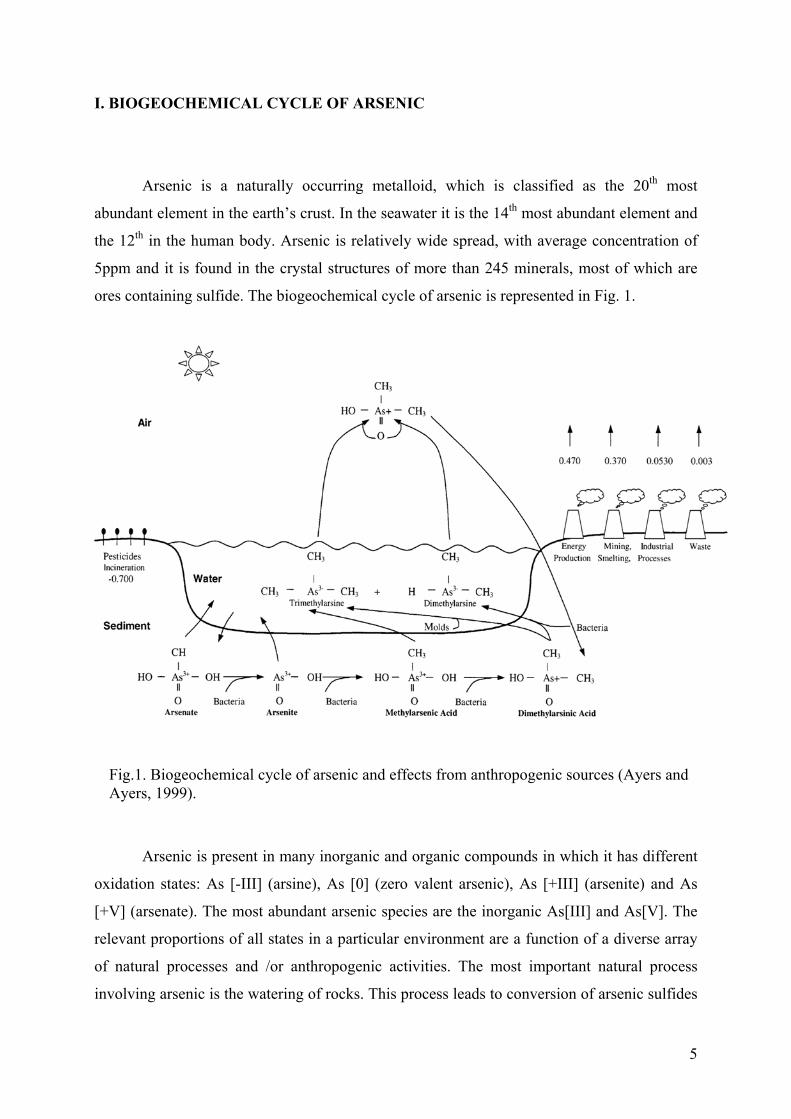
I. BIOGEOCHEMICAL CYCLE OF ARSENIC
Arsenic is a naturally occurring metalloid, which is classified as the 20th most
abundant element in the earth’s crust. In the seawater it is the 14th most abundant element and
the 12th in the human body. Arsenic is relatively wide spread, with average concentration of
5ppm and it is found in the crystal structures of more than 245 minerals, most of which are
ores containing sulfide. The biogeochemical cycle of arsenic is represented in Fig. 1.
Arsenic is present in many inorganic and organic compounds in which it has different
oxidation states: As [-III] (arsine), As [0] (zero valent arsenic), As [+III] (arsenite) and As
[+V] (arsenate). The most abundant arsenic species are the inorganic As[III] and As[V]. The
relevant proportions of all states in a particular environment are a function of a diverse array
of natural processes and /or anthropogenic activities. The most important natural process
involving arsenic is the watering of rocks. This process leads to conversion of arsenic sulfides
Fig.1. Biogeochemical cycle of arsenic and effects from anthropogenic sources (Ayers and Ayers, 1999).
6
to arsenic trioxide, which enters the arsenic cycle as dust or by dissolution in rain, river and
groundwater. Another natural mobilization of arsenic is due to bacterial biotransformation of
arsenic compounds, which includes reduction (including dissimilatory reduction), oxidation
and methylation. Other natural ways for distribution of arsenic are the volcanoes and forest
fires. From the natural sources arsenic is released into air, water and soil.
The main sources for anthropogenic release are: i) ores - mining and smelting
industries; ii) metallurgic industry (additive for special lead and copper alloys); iii)
agriculture-pesticides (mainly sodium arsenite) and insecticides such as lead arsenate,
Ca3AsO4, copper acetoarsenite, monosodium methanearsonate (MSMA), disodium
methanearsonate (DSMA); iv) wood preserving industry, mainly sodium arsenite and Fluor-
Chrome-Arsenic-Phenol (FCAP), Chromated Copper Arsenate (CCA), Ammonical Copper
Arsenate (ACA), etc. (Mandal and Suzuki, 2002); v) feed additivities, pharmaceutical
industry, glass production, semiconductors production, electrical industry (combustion of
wood and low quality coils) etc. The anthropogenic sources of arsenic exceed the natural
sources in the environment.
7
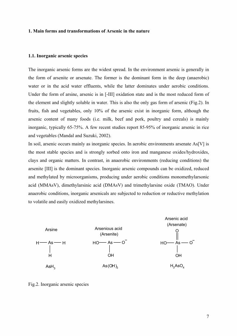
1. Main forms and transformations of Arsenic in the nature
1.1. Inorganic arsenic species
The inorganic arsenic forms are the widest spread. In the environment arsenic is generally in
the form of arsenite or arsenate. The former is the dominant form in the deep (anaerobic)
water or in the acid water effluents, while the latter dominates under aerobic conditions.
Under the form of arsine, arsenic is in [-III] oxidation state and is the most reduced form of
the element and slightly soluble in water. This is also the only gas form of arsenic (Fig.2). In
fruits, fish and vegetables, only 10% of the arsenic exist in inorganic form, although the
arsenic content of many foods (i.e. milk, beef and pork, poultry and cereals) is mainly
inorganic, typically 65-75%. A few recent studies report 85-95% of inorganic arsenic in rice
and vegetables (Mandal and Suzuki, 2002).
In soil, arsenic occurs mainly as inorganic species. In aerobic environments arsenate As[V] is
the most stable species and is strongly sorbed onto iron and manganese oxides/hydroxides,
clays and organic matters. In contrast, in anaerobic environments (reducing conditions) the
arsenite [III] is the dominant species. Inorganic arsenic compounds can be oxidized, reduced
and methylated by microorganisms, producing under aerobic conditions monomethylarsonic
acid (MMAsV), dimethylarsinic acid (DMAsV) and trimethylarsine oxide (TMAO). Under
anaerobic conditions, inorganic arsenicals are subjected to reduction or reductive methylation
to volatile and easily oxidized methylarsines.
Fig.2. Inorganic arsenic species
AsHO O
OH
As(OH)3
Arsenious acid(Arsenite)
AsHO O
OH
O
H3AsO4
(Arsenate)Arsenic acid
AsH
H
H
AsH3
Arsine
8
In natural water arsenic is found in low concentrations. The major chemical form in which
arsenic appears to be thermodynamically stable is the arsenate ion. The concentrations in
unpolluted fresh waters range from 1–10µg l-1 rising to 100–5000µg l-1 (Smedley et al., 1996).
At moderate or high redox potentials arsenic is stabilized under series of pentavalent
(arsenate) oxyanions. However under acid and mildly alkaline conditions and lower redox
potential, the trivalent arsenite species predominate. In aquatic environments the As[0] and
As[-III] oxidation states are very rare.
In air, arsenic exists predominantly absorbed on particle matters or under the form of arsine
and it has being of negligible importance except in the areas of arsenic pesticides application
or biotic activity.
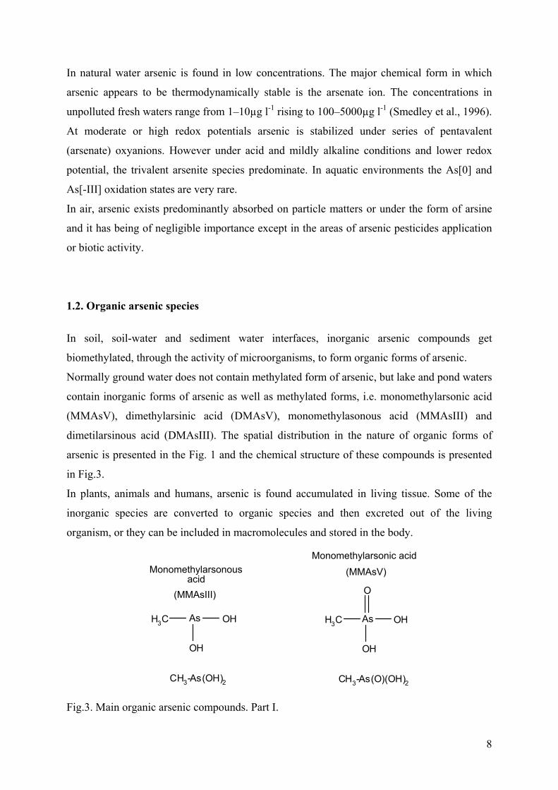
1.2. Organic arsenic species
In soil, soil-water and sediment water interfaces, inorganic arsenic compounds get
biomethylated, through the activity of microorganisms, to form organic forms of arsenic.
Normally ground water does not contain methylated form of arsenic, but lake and pond waters
contain inorganic forms of arsenic as well as methylated forms, i.e. monomethylarsonic acid
(MMAsV), dimethylarsinic acid (DMAsV), monomethylasonous acid (MMAsIII) and
dimetilarsinous acid (DMAsIII). The spatial distribution in the nature of organic forms of
arsenic is presented in the Fig. 1 and the chemical structure of these compounds is presented
in Fig.3.
In plants, animals and humans, arsenic is found accumulated in living tissue. Some of the
inorganic species are converted to organic species and then excreted out of the living
organism, or they can be included in macromolecules and stored in the body.
Fig.3. Main organic arsenic compounds. Part I.
AsH3C
OH
OH
CH3-As(OH)2
(MMAsIII)
Monomethylarsonous acid
AsH3C
OH
O
OH
CH3-As(O)(OH)2
Monomethylarsonic acid
(MMAsV)
9
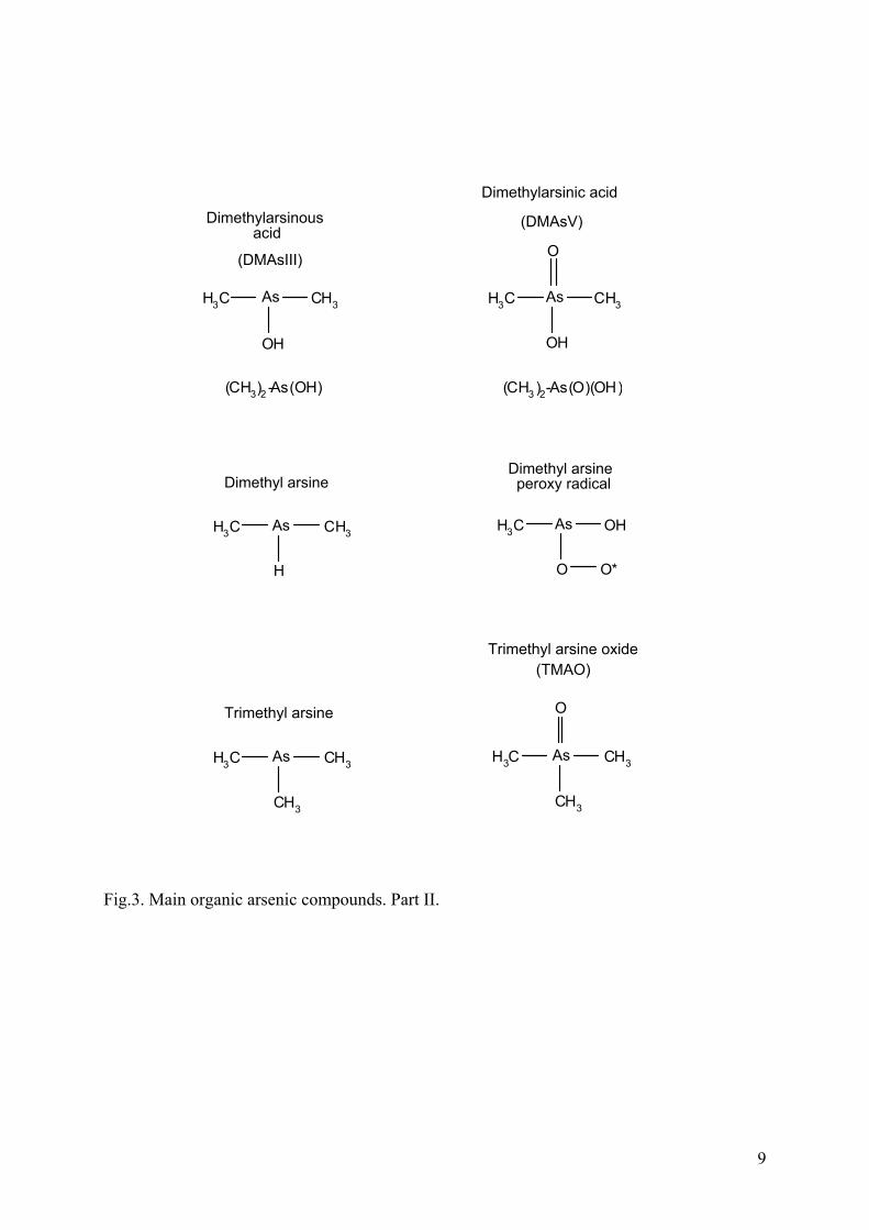
Fig.3. Main organic arsenic compounds. Part II.
AsH3C
OH
CH3
(CH3)2 -As(OH)
(DMAsIII)
Dimethylarsinous acid
AsH3C
OH
O
(CH3 )2-As(O)(OH)
CH3
(DMAsV)
Dimethylarsinic acid
AsH3C
H
CH3
Dimethyl arsine
AsH3C OH
O O*
Dimethyl arsine peroxy radical
AsH3C
CH3
CH3
Trimethyl arsine
AsH3C
CH3
CH3
O
Trimethyl arsine oxide(TMAO)
10
2. Bioavailability and toxicity of Arsenic species
2.1. Bioavailability
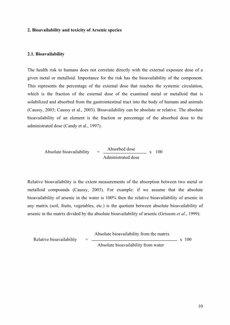
The health risk to humans does not correlate directly with the external exposure dose of a
given metal or metalloid. Importance for the risk has the bioavailability of the component.
This represents the percentage of the external dose that reaches the systemic circulation,
which is the fraction of the external dose of the examined metal or metalloid that is
solubilized and absorbed from the gastrointestinal tract into the body of humans and animals
(Caussy, 2003; Caussy et al., 2003). Bioavailability can be absolute or relative. The absolute
bioavailability of an element is the fraction or percentage of the absorbed dose to the
administrated dose (Candy et al., 1997).
Relative bioavailability is the extent measurements of the absorption between two metal or
metalloid compounds (Caussy, 2003). For example: if we assume that the absolute
bioavailability of arsenic in the water is 100% then the relative bioavailability of arsenic in
any matrix (soil, fruits, vegetables, etc.) is the quotient between absolute bioavailability of
arsenic in the matrix divided by the absolute bioavailability of arsenic (Grissom et al., 1999):
Absolute bioavailability Absorbed dose
Administrated dose = x 100
Absolute bioavailability from the matrix
Absolute bioavailability from water = Relative bioavailability x 100
11
2.2. Toxicity
The consequences for the human health from an arsenic exposure depend on the type of
arsenic species, its dose, modality and the duration of exposure. According to the Word
Health Organization (WHO), there are two types of exposure – acute, short exposure and
prolonged (natural or occupational) exposure to nonlethal doses (WHO, 2001a). The First
type of exposure to arsenic results in acute effects such as vomiting, abdominal colics and
diarrhea. The second type of exposure to nonlethal doses causes chronic health effects which
can be respiratory, pulmonary, cardiovascular, gastrointestinal, hematological, hepatic, renal,
dermal, neurological, developmental, reproductive, immunologic, mutagenic and
carcinogenic. The toxicity of arsenicals decrease in the order: arsines > inorganic As[III] >
arsenoxides (org As[III]-containing compounds) > inorganic As[V] > arsonium compounds >
As (Hindmarsh, 2000). Lately another toxicity order of arsenicals has been reported:
inorganic As[III] > monomethylarsine oxide (MMAO[III]) > dimethylarsinous acid
(DMAsIII) > dimethylarsinic acid (DMAsV) > monomethylarconic acid (MMAsV) >
inorganic As[V] (Vega et al., 2001).
Inorganic forms
Inorganic forms of arsenic are more toxic than organic forms. The As[V] is less mobile and
potentially less bioavailable than As[III] due to its stronger adsorption to mineral surfaces
(Korte and Fernando, 1991). The uptake of arsenite in the cell is considered to be much faster
than the uptake of arsenate, since the As[III] is uncharged at physiological pH. However, now
it is believed that both forms of inorganic arsenic are actively transported into the cell.
Arsenite is transported by aquaglyceroporins, which transport water and glycerol (2004) and
arsenate is transported by the phosphate transporter. As[III] reacts via three mechanisms: the
first mode of action is related to binding (or complexing) with electron – rich groups, -SH or -
OH. In this type of action the reaction is a metal and the biological targets are the proteins.
The second type of action mode is the oxidation-reduction reactions in which the chemical
reaction type are metal and non-metal, and as target are used molecules that can transfer
electrons. The third mode of action is the substitution of C and N in which the biological
targets are choline and lipids and the chemical type of reaction is nonmetal.
12
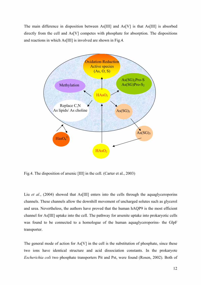
The main difference in disposition between As[III] and As[V] is that As[III] is absorbed
directly from the cell and As[V] competes with phosphate for absorption. The dispositions
and reactions in which As[III] is involved are shown in Fig.4.
Fig.4. The disposition of arsenic [III] in the cell. (Carter et al., 2003)
Liu et al., (2004) showed that As[III] enters into the cells through the aquaglyceroporins
channels. These channels allow the downhill movement of uncharged solutes such as glycerol
and urea. Nevertheless, the authors have proved that the human hAQP9 is the most efficient
channel for As[III] uptake into the cell. The pathway for arsenite uptake into prokaryotic cells
was found to be connected to a homologue of the human aquaglyceroporins- the GlpF
transporter.
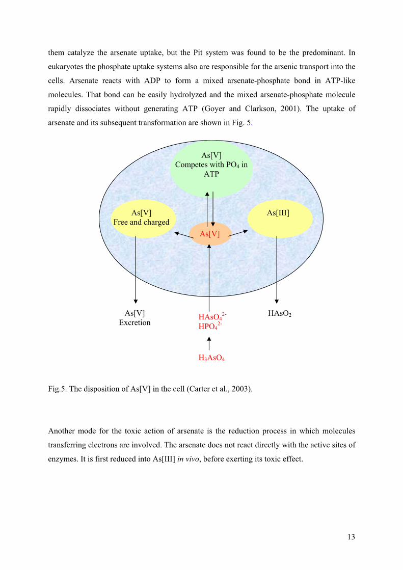
The general mode of action for As[V] in the cell is the substitution of phosphate, since these
two ions have identical structure and acid dissociation constants. In the prokaryote
Escherichia coli two phosphate transporters Pit and Pst, were found (Rosen, 2002). Both of
HasO42-
HAsO2
As(SG)3
HAsO2
As(SG)3
Oxidation-Reduction Active species
(As, O, S)
Methylation As(SG)2Pro-S As(SG)Pro-S2
Replace C,N As lipids/ As choline
13
them catalyze the arsenate uptake, but the Pit system was found to be the predominant. In
eukaryotes the phosphate uptake systems also are responsible for the arsenic transport into the
cells. Arsenate reacts with ADP to form a mixed arsenate-phosphate bond in ATP-like
molecules. That bond can be easily hydrolyzed and the mixed arsenate-phosphate molecule
rapidly dissociates without generating ATP (Goyer and Clarkson, 2001). The uptake of
arsenate and its subsequent transformation are shown in Fig. 5.
Fig.5. The disposition of As[V] in the cell (Carter et al., 2003).
Another mode for the toxic action of arsenate is the reduction process in which molecules
transferring electrons are involved. The arsenate does not react directly with the active sites of
enzymes. It is first reduced into As[III] in vivo, before exerting its toxic effect.
HAsO2 As[V] Excretion
H3AsO4
HAsO42-
HPO42-
As[V] Free and charged
As[V] Competes with PO4 in
ATP
As[V]
As[III]
14
Organic forms
The term "Biomethylation" describes the formation of nonvolatile and volatile methylated
compounds. The major volatile compounds of arsenic, formed by methylation have the
structure (CH3)nAsH3-n, where n = 1, 2 or 3 methyl radicals. The major nonvolatile
compounds are the methylarsonate and dimethylarsinate (Bentley and Chasteen, 2002).
Organic arsenic species important in toxicity are the monomethylarsonous acid (MMAsIII)
and dimethylarsinous acid (DMAsIII) for arsenic in oxidation state +III. For As[V] the
organic forms are monomethylarconic acid (MMAsV) and dimethylarsinic acid (DMAsV).
MMAsV is a methylated metabolite of As[III], consequently metabolized to DMAsV. The
MMAsIII is a reduced metabolite of MMAsV and the DMAsIII is a reduced metabolite of
DMAsV. The DMAsV is a dimethylated metabolite of As[III] and methylated metabolite of
MMAsIII. Recent comparative investigations on the toxicity of methylated and dimethylated
arsenicals showed that those containing As[III] are more cytotoxic, more genotoxic and more
potent inhibitors of the activities of some enzymes even than inorganic As[III] (Rossman,
2003).
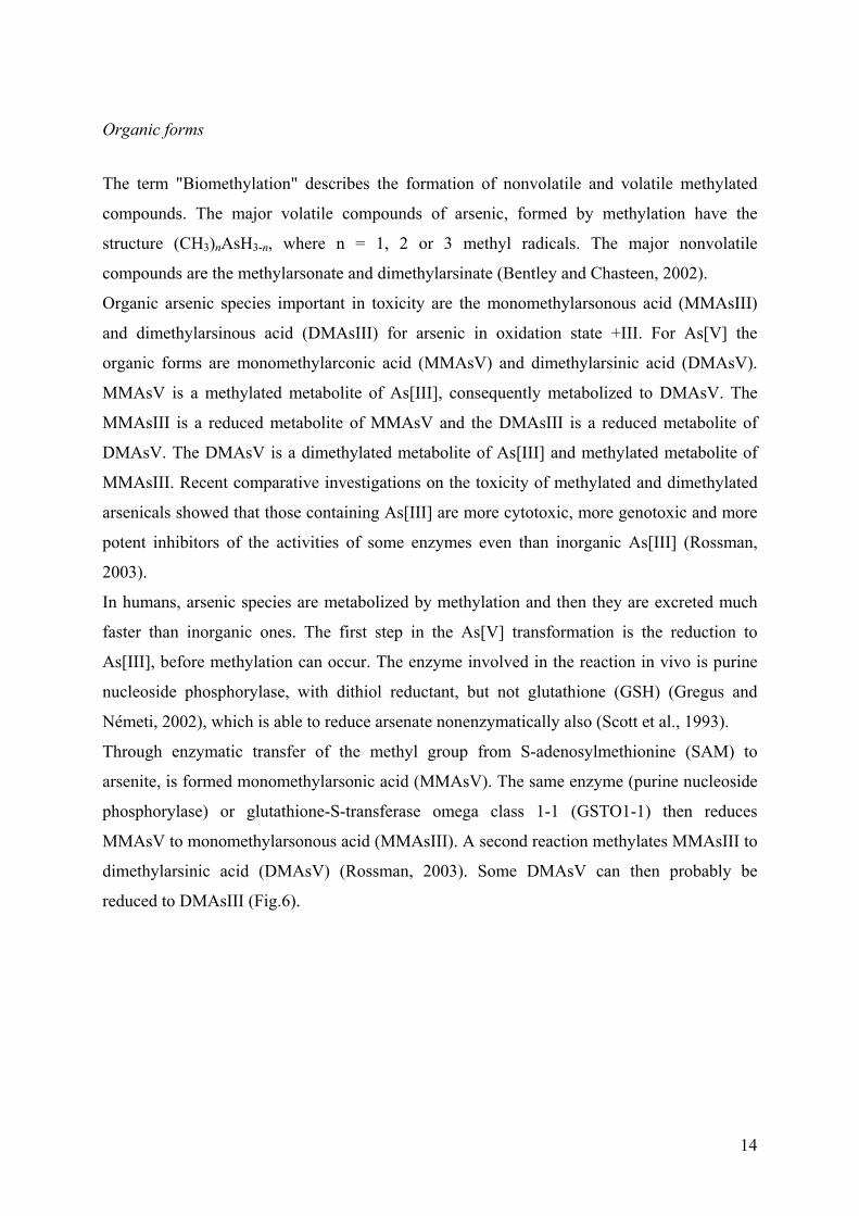
In humans, arsenic species are metabolized by methylation and then they are excreted much
faster than inorganic ones. The first step in the As[V] transformation is the reduction to
As[III], before methylation can occur. The enzyme involved in the reaction in vivo is purine
nucleoside phosphorylase, with dithiol reductant, but not glutathione (GSH) (Gregus and
Németi, 2002), which is able to reduce arsenate nonenzymatically also (Scott et al., 1993).
Through enzymatic transfer of the methyl group from S-adenosylmethionine (SAM) to
arsenite, is formed monomethylarsonic acid (MMAsV). The same enzyme (purine nucleoside
phosphorylase) or glutathione-S-transferase omega class 1-1 (GSTO1-1) then reduces
MMAsV to monomethylarsonous acid (MMAsIII). A second reaction methylates MMAsIII to
dimethylarsinic acid (DMAsV) (Rossman, 2003). Some DMAsV can then probably be
reduced to DMAsIII (Fig.6).
15
Fig.6. Metabolism of arsenic compounds in eukaryotes (mammals).
In the case of microorganisms (fungi, yeasts and bacteria) the use of arsenic as methyl group
acceptor has a major emphasis on the production of volatile compounds. For example
Alcaligenes sp. and Pseudomonas sp. have been reported to be able to reduce arsenate and
arsenite anaerobically to arsine (Cheng and Focht, 1979). A strict anaerobe belonging to
Archae Methanobacterium M.o.H. (now M. bryantii), has been shown to produce
dimethylarsine from arsenate with H2 (McBride and Wolfe, 1971). The fungi from genera
Aspergillus, Mucor, Fusarium, Paecilomyces as well as Candida humicola and
Scopulariopsis brevicaulis are able to convert arsenite to trimethylarsine (Alexander, 1977;
Cullen and Reimer, 1989; Pickett et al., 1981). In the case of algae and phytoplankton, is
proved that As[V] is the most toxic species for these organisms and they reduce it to As[III],
which is consistent with conventional view that this serves as a detoxification mechanism for
As[V] (Knauer et al., 1999).
To proceed from arsenate to trimethylarsine for the fungi and yeast, four two electron
reductions are required. They are likely to be enzyme catalyzed reactions with the reductants
providing the necessary 2e-. The reductants are predicted to be thiols, and a particular
attention was given to glutathione (GSH) and lipoic acid (6,8-dithiooctanoic acid).
Nevertheless, there is still little experimental evidence from microbial systems. Thus, it is
assumed the reaction resembles those for formation of methylarsonate and dimethylarsinate of
mammalians (Thompson, 1993).
Kuroda et al. (2001), showed that in E. coli cysteine, but not GSH was required for
dimethylarsinate metabolism. Thus, there may be different arsenic metabolic pathways in
+ thiol DMAsIII
As[III] SAM MMAsV+
+ MMAsIIIthiol
+ DMAsV SAM
As[V] + 2e

16
bacteria. Moreover there is no general consensus as to a mechanism for anaerobic
dimethylarsine formation.
The three oxidation states of this element and the small amounts of arsenic compounds
present in biological specimens complicate the enzymology of arsenic biomethylation. The
chemical intermediates and reactions in the arsenate metabolism are generally assumed to be
the same in microorganisms and animals. However, there is a difference: in microorganisms
the reactions tend to proceed to volatile species (methylarsines), whereas in mammalians turn
to nonvolatile species, which are excreted via urine. The major urinary metabolite is the
dimethylarsinate (Bentley and Chasteen, 2002).
17
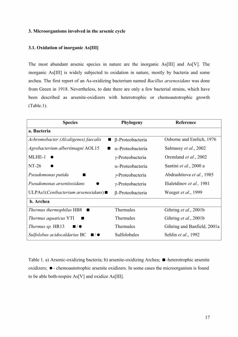
3. Microorganisms involved in the arsenic cycle
3.1. Oxidation of inorganic As[III]
The most abundant arsenic species in nature are the inorganic As[III] and As[V]. The
inorganic As[III] is widely subjected to oxidation in nature, mostly by bacteria and some
archea. The first report of an As-oxidizing bacterium named Bacillus arsenoxidans was done
from Green in 1918. Nevertheless, to date there are only a few bacterial strains, which have
been described as arsenite-oxidizers with heterotrophic or chemoautotrophic growth
(Table.1).
Species Phylogeny Reference
a. Bacteria
Achromobacter (Alcaligenes) faecalis β-Proteobacteria Osborne and Enrlich, 1976
Agrobacterium albertimagni AOL15 α-Proteobacteria Salmassy et al., 2002
MLHE-1 γ-Proteobacteria Oremland et al., 2002
NT-26 α-Proteobacteria Santini et al., 2000 a
Pseudomonas putida γ-Proteobacteria Abdrashitova et al., 1985
Pseudomonas arsenitoxidans γ-Proteobacteria Ilialetdinov et al., 1981
ULPAs1(Cenibacterium arsenoxidans) β-Proteobacteria Weeger et al., 1999
b. Archea
Thermus thermophilus HB8 Thermales Gihring et al., 2001b
Thermus aquaticus YTI Thermales Gihring et al., 2001b
Thermus sp. HR13 / Thermales Gihring and Banfield, 2001a
Sulfolobus acidocaldarius BC / Sulfolobales Sehlin et al., 1992
Table 1. a) Arsenic-oxidizing bacteria; b) arsenite-oxidizing Archea; -heterotrophic arsenite
oxidizers; - chemoautotrophic arsenite oxidizers. In some cases the microorganism is found
to be able both-respire As[V] and oxidize As[III].
18
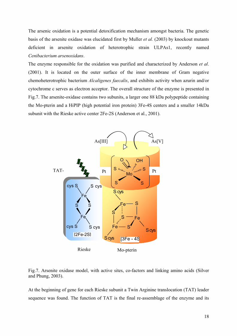
The arsenic oxidation is a potential detoxification mechanism amongst bacteria. The genetic
basis of the arsenite oxidase was elucidated first by Muller et al. (2003) by knockout mutants
deficient in arsenite oxidation of heterotrophic strain ULPAs1, recently named
Cenibacterium arsenoxidans.
The enzyme responsible for the oxidation was purified and characterized by Anderson et al.
(2001). It is located on the outer surface of the inner membrane of Gram negative
chemoheterotrophic bacterium Alcaligenes faecalis, and exhibits activity when azurin and/or
cytochrome c serves as electron acceptor. The overall structure of the enzyme is presented in
Fig.7. The arsenite-oxidase contains two subunits, a larger one 88 kDa polypeptide containing
the Mo-pterin and a HiPIP (high potential iron protein) 3Fe-4S centers and a smaller 14kDa
subunit with the Rieske active center 2Fe-2S (Anderson et al., 2001).
Fig.7. Arsenite oxidase model, with active sites, co-factors and linking amino acids (Silver and Phung, 2003).
At the beginning of gene for each Rieske subunit a Twin Arginine translocation (TAT) leader
sequence was found. The function of TAT is the final re-assemblage of the enzyme and its
TAT-
Fe
Fe
SS
S cys
S cyscys S
cys S
[2Fe-2S]
Rieske Mo-pterin
SFe
S Fe
Fe S
S
S cys
S cys
S cys [3Fe - 4S
OH
SMo
O
SS
S Pt Pt
As[V] As[III]
19
translocation from the cytoplasm to the periplasm of the cell. The arsenite oxidase enzyme is
in the large super-family of dimethyl sulfoxide (DMSO) reductase proteins. This is a diverse
family, in which enzymes vary in substrates, midpoint electric potential and direction. That is
why, some enzymes of this family function physiologically as oxidases as is the case with
arsenite oxidase and others function as reductases (Mukhopadhyay et al., 2002; Silver and
Phung, 2003).
Santini and vanden Hoven (2003) recently described the purification and preliminary
characterization of the arsenic oxidase of chemolitoautotroph strain NT-26 a member of α-
Proteobacteria together with the cloning, sequencing and molecular analysis of the gene
structure. The enzyme consist of two heterologous subunits, named AroA (98 kDa) and AroB
(14 kDa), and has two molybdenium and 9 or 10 iron atoms per unit. The suggested
configuration of the protein is α2β2.
3.2. Reduction of arsenic species
To date two different mechanisms of arsenate reduction have been found in bacteria, fungi
and algae. One of them is common for all microorganisms and is considered as a means of
detoxification. The second one is specific only for Bacteria and some Archea. It represent the
capability of microorganisms to use the arsenic as final electron acceptor in their respiratory
chains, in autotrophic or heterotrophic growth.
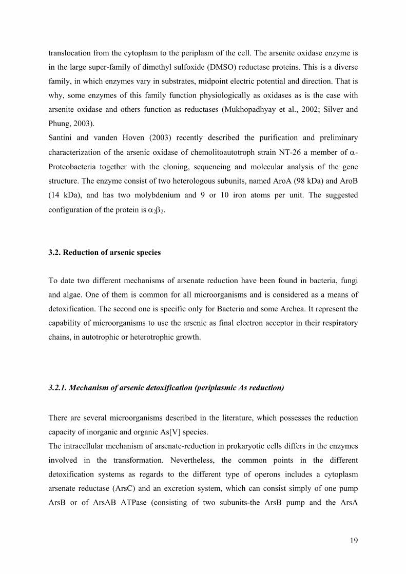
3.2.1. Mechanism of arsenic detoxification (periplasmic As reduction)
There are several microorganisms described in the literature, which possesses the reduction
capacity of inorganic and organic As[V] species.
The intracellular mechanism of arsenate-reduction in prokaryotic cells differs in the enzymes
involved in the transformation. Nevertheless, the common points in the different
detoxification systems as regards to the different type of operons includes a cytoplasm
arsenate reductase (ArsC) and an excretion system, which can consist simply of one pump
ArsB or of ArsAB ATPase (consisting of two subunits-the ArsB pump and the ArsA
20
ATPase). The differences in the type of the enzymes involved in the intracellular arsenic
reduction and the way of their action are presented in Fig 8A.
Fig. 8A. General detoxification mechanism in prokaryotes.
The arsenate reductase ArsC is coded by gene, which is widely found in Bacteria and in some
Archea, in ars operons. This is the enzyme responsible for the reduction of As[V] to As[III].
Currently, three unrelated clades of ArsC sequences are recognized, with a common basic
function, but without evolutionary relationship (Mukhopadhyay et al., 2002). All of them are
glutaredoxin/glutathione (Grx/GSH) coupled enzymes and it is now believed that they are
associated with the arsenite oxidase of Alcaligenes sp. and the respiratory arsenate reductase
of Shewanella. The ars operon and ArsC reductase genes in Gram negative bacteria are found
on plasmids and in the chromosomes. The ArsC from glutaredoxin/glutathione clade is about
135 amino acid residues protein, with 3 essential cysteine residues.
The thioredoxin-coupled ArsC (Trx ArsC) was firstly found in a Gram positive
Staphylococcus (Ji and Silver, 1992). This enzyme also utilizes 3 cysteines all of them in the
primary sequence of the polypeptide. The clade of thioredoxin-coupled ArsC enzymes is
widespread on the plasmids and genomes of Gram positive bacteria, but it is also found in
some Gram negative bacteria. For example the genome of P. aeruginosa possesses genes for
both ArsC reductase families. (Mukhopadhyay et al., 2002).
The mechanism of arsenic detoxification in an eukaryotic type of cell is connected to the third
clade of cytoplasmic arsenate reductases, represented by the Acr2p Saccharomyces cerevisiae
arsenate reductase. Its overall function in the cell is illustrated in Fig. 8B. This Acr2p S.
cerevisiae arsenate reductase is the only currently identified enzyme in the eucaryotic
organisms, known to convert arsenate to arsenite. This is a homodimer of 130 monomers and
it has common substrates of GSH and Grx with those for Grx/GSH ArsC reductase in
As[V] As[III]
ArsB
ArsA
ArsBArsC
ATP
ADP
21
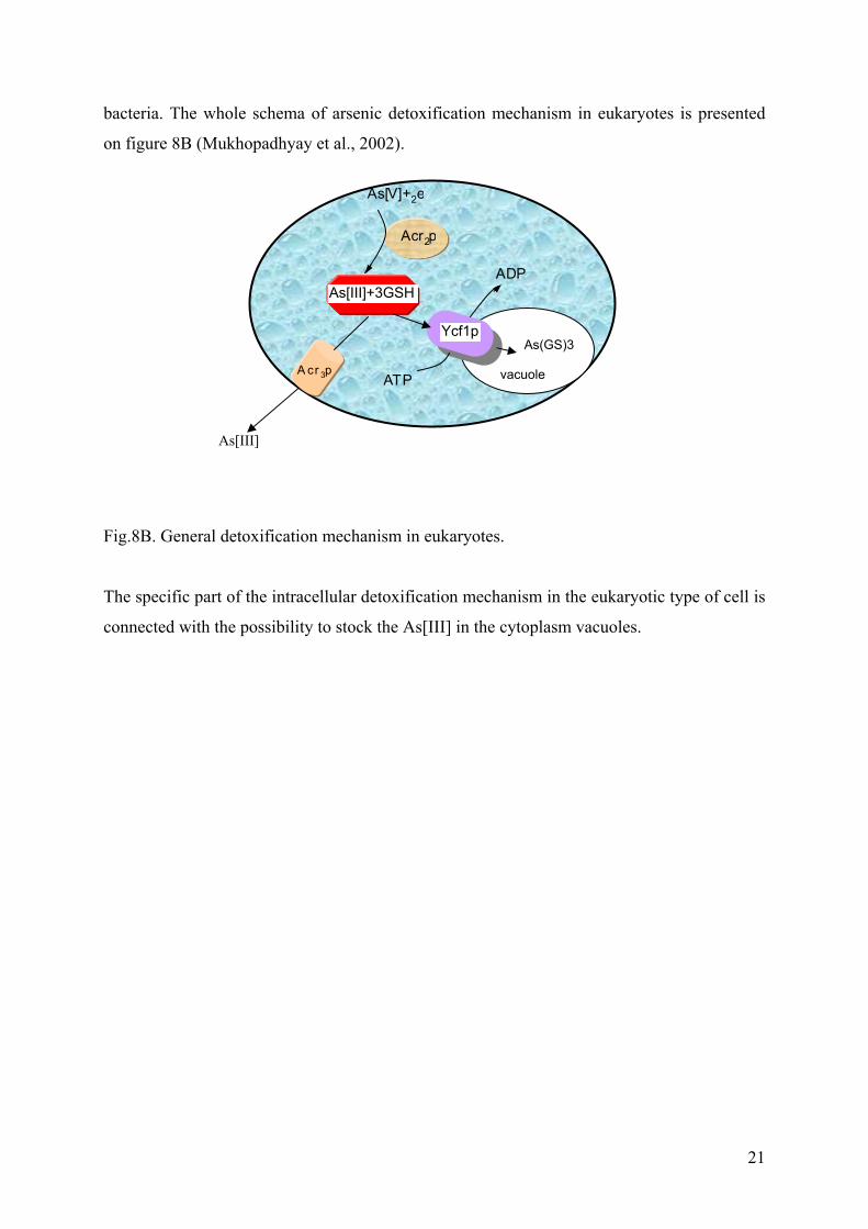
bacteria. The whole schema of arsenic detoxification mechanism in eukaryotes is presented
on figure 8B (Mukhopadhyay et al., 2002).
Fig.8B. General detoxification mechanism in eukaryotes.
The specific part of the intracellular detoxification mechanism in the eukaryotic type of cell is
connected with the possibility to stock the As[III] in the cytoplasm vacuoles.
As[III]
As[V]+2e
vacuole
As(GS)3Ycf1p
A cr 3p
As[III]+3GSH
Acr2p
ATP
ADP
22
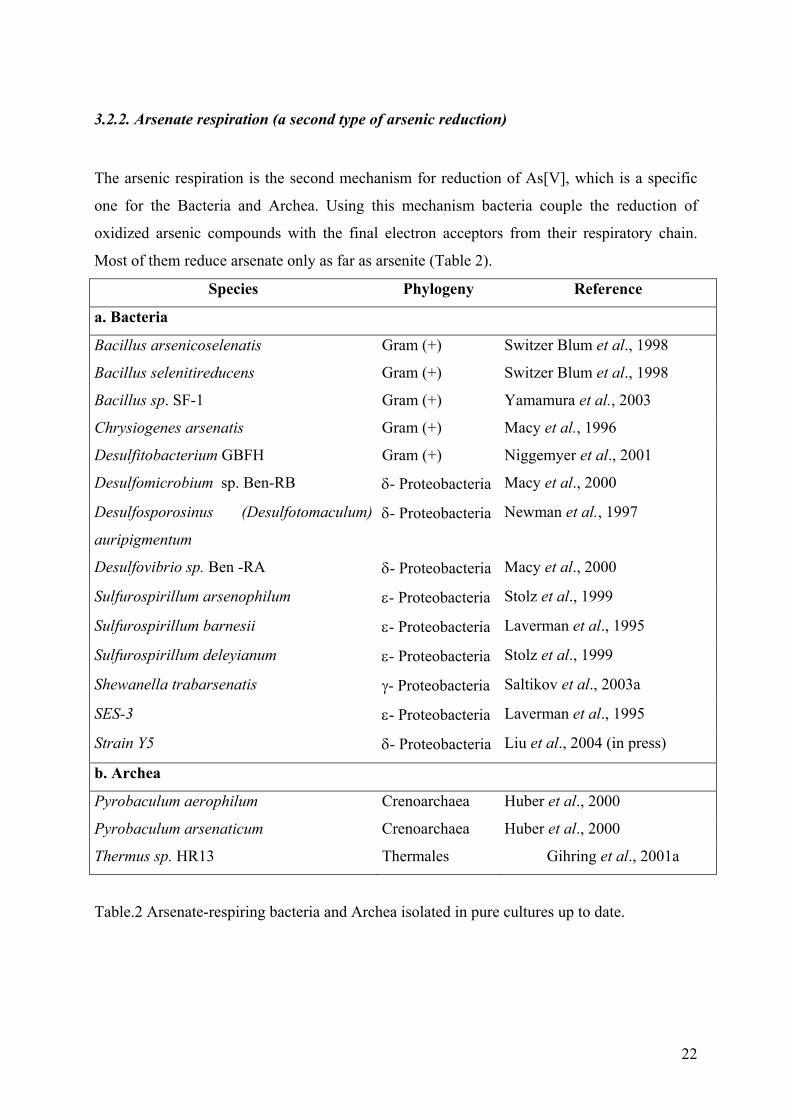
3.2.2. Arsenate respiration (a second type of arsenic reduction)
The arsenic respiration is the second mechanism for reduction of As[V], which is a specific
one for the Bacteria and Archea. Using this mechanism bacteria couple the reduction of
oxidized arsenic compounds with the final electron acceptors from their respiratory chain.
Most of them reduce arsenate only as far as arsenite (Table 2).
Species Phylogeny Reference
a. Bacteria
Bacillus arsenicoselenatis Gram (+) Switzer Blum et al., 1998
Bacillus selenitireducens Gram (+) Switzer Blum et al., 1998
Bacillus sp. SF-1 Gram (+) Yamamura et al., 2003
Chrysiogenes arsenatis Gram (+) Macy et al., 1996
Desulfitobacterium GBFH Gram (+) Niggemyer et al., 2001
Desulfomicrobium sp. Ben-RB δ- Proteobacteria Macy et al., 2000
Desulfosporosinus (Desulfotomaculum)
auripigmentum
δ- Proteobacteria Newman et al., 1997
Desulfovibrio sp. Ben -RA δ- Proteobacteria Macy et al., 2000
Sulfurospirillum arsenophilum ε- Proteobacteria Stolz et al., 1999
Sulfurospirillum barnesii ε- Proteobacteria Laverman et al., 1995
Sulfurospirillum deleyianum ε- Proteobacteria Stolz et al., 1999
Shewanella trabarsenatis γ- Proteobacteria Saltikov et al., 2003a
SES-3 ε- Proteobacteria Laverman et al., 1995
Strain Y5 δ- Proteobacteria Liu et al., 2004 (in press)
b. Archea
Pyrobaculum aerophilum Crenoarchaea Huber et al., 2000
Pyrobaculum arsenaticum Crenoarchaea Huber et al., 2000
Thermus sp. HR13 Thermales Gihring et al., 2001a
Table.2 Arsenate-respiring bacteria and Archea isolated in pure cultures up to date.
23
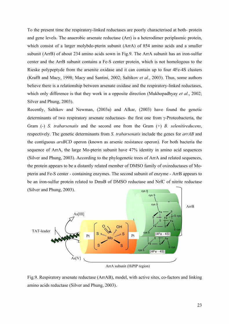
To the present time the respiratory-linked reductases are poorly characterised at both- protein
and gene levels. The anaerobic arsenate reductase (Arr) is a heterodimer periplasmic protein,
which consist of a larger molybdo-pterin subunit (ArrA) of 854 amino acids and a smaller
subunit (ArrB) of about 234 amino acids sown in Fig.9. The ArrA subunit has an iron-sulfur
center and the ArrB subunit contains a Fe-S center protein, which is not homologous to the
Rieske polypeptyde from the arsenite oxidase and it can contain up to four 4Fe-4S clusters
(Krafft and Macy, 1998; Macy and Santini, 2002; Saltikov et al., 2003). Thus, some authors
believe there is a relationship between arsenate oxidase and the respiratory-linked reductases,
which only difference is that they work in a opposite direction (Mukhopadhyay et al., 2002;
Silver and Phung, 2003).
Recently, Saltikov and Newman, (2003a) and Afkar, (2003) have found the genetic
determinants of two respiratory arsenate reductases- the first one from γ-Proteobacteria, the
Gram (-) S. trabarsenatis and the second one from the Gram (+) B. selenitireducens,
respectively. The genetic determinants from S. trabarsenatis include the genes for arrAB and
the contiguous arsBCD operon (known as arsenic resistance operon). For both bacteria the
sequence of ArrA, the large Mo-pterin subunit have 47% identity in amino acid sequences
(Silver and Phung, 2003). According to the phylogenetic trees of ArrA and related sequences,
the protein appears to be a distantly related member of DMSO family of oxireductases of Mo-
pterin and Fe-S center - containing enzymes. The second subunit of enzyme - ArrB appears to
be an iron-sulfur protein related to DmsB of DMSO reductase and NrfC of nitrite reductase
(Silver and Phung, 2003).
Fig.9. Respiratory arsenate reductase (ArrAB), model, with active sites, co-factors and linking
amino acids reductase (Silver and Phung, 2003).
As[V]
As[III]
TAT-leader SFe
S Fe
Fe S
S
S cys
FeS cys
cys S
cys S [4Fe - 4S]
SFe
S Fe
Fe S
S
S cys
FeS cys
cys S
cys S [4Fe - 4S]
SFe
S Fe
Fe S
S
S cys
FeS cys
cys S
cys S [4Fe - 4S]
SFe
S Fe
Fe S
S
S cys
FeS cys
cys S
cys S [4Fe - 4S]
OH
SMo
O
SS
S PtPt
ArrB
ArrA subunit (HiPIP region)
24
II. METHODS FOR ARSENIC DECONTAMINATION
1. Chemical methods and technologies for remediation of arsenic-containing water
To date most of the technologies for arsenic decontamination of polluted water are based on
chemical methods and treatment techniques. Predominantly, they consist of two steps. The
first step is generally the oxidation of As[III] into As[V]. The As[V] is easily removed from
water by precipitation and sorption due to its high reaction activity. This form of arsenic has
low mobility and less toxicity (Arai et al., 2001; Meng et al., 2001; Goldberg and Jonhston,
2000). Ionic strength effects more the As[III]sorption. It is generally held that As[V] is more
strongly bound and forms predominantly inner-sphere complexes regardless of pH and ion
strength (I). Therefore, most of the chemical treatment technologies in a given effluent are
based on the preliminary oxidation of total arsenic to As[V].
1.1. Chemical arsenite-oxidation
Generally chemical methods for arsenite oxidation are based on the use of strong oxidizing
agents such as hypochlorite, permanganate and hydrogen peroxide or Fenton’s reagent (FR).
Fenton’s reagent is a widely applied oxidative technology, which utilizes the reaction of
hydrogen peroxide with ferrous iron (generally added as FeCl2 or FeSO4) to produce free
radicals capable to oxidize aqueous phase contaminants. The addition of ferrous salts
containing chloride or sulfur or the use of strong oxidants is undesirable, because of the
generation of organochloride byproducts, which cause the additional destruction and impact
on the environment. Recently Arienzo et al. (2002), proposed an electrochemical peroxidation
(ECP) technique for the oxidation of As[III], which is based on the application of direct
electric current to steel electrode, complemented with a small addition of H2O2.
Zaw and Emett (2002), proposed an advanced oxidation method for arsenic removal, since the
oxidation rate of dissolved As[III] by oxygen is extremely low. The advance oxidation
method is based on the use of ultraviolet light and a photo absorber. The maintenance of
dissolved oxygen was achieved by air sparging. This process needed a pH adjustment of
treated water, which was made through the addition of HCl. The targets of the photolytic
reaction were the Fe[III] ions, involved in the transfer of one electron from the complexed
ligand, such as organic, hydroxide or chloride species, to a Fe[III] -centered orbital, forming
25
Fe[II] and a free radical (Zafiriou et al., 1984). The generated free (hydroxyl) radicals was
then used to oxidize As[III].
1.2. Coagulation and co-precipitation techniques
Coagulation encompasses all reactions, mechanisms and results in the overall process of
particle growth and particle aggregation within water. Coagulation involves the removal of
colloidal and settleable particles, but the term commonly refers to the removal of dissolved
ions, which is actually precipitation. The precipitation is the process in which dissolved ions
in solution form an insoluble solid as a result of a chemical reaction. A co-precipitation
indicates the cases in which an inorganic contaminant forms an insoluble complex with a
coagulant. The valence of the inorganic contaminant and the pH of the solution are important
for the removal by co-precipitation. There are four types of co-precipitation:
• inclusion – mechanical entrapment of a portion of the solution surrounding the growing
particle (significant for the large particles);
• adsorption – the attachment of an impurity onto the surface of a particle or precipitate
(significant for the small particles);
• occlusion – entrapment of a contaminant in the interior of a particle of precipitate, which
continues to grow;
• solid-solution formation – a type of occlusion in which a particle of precipitate becomes
contaminated with a different type of particle that precipitates under similar conditions
and is formed from ions whose sizes are nearly equal to those of the original precipitate.
Throughout coagulation dissolved arsenic is converted into insoluble reaction products, which
can be further eliminated by sedimentation and/or filtration. This arsenic removal involves
three mechanisms:
• Precipitation: the formation of insoluble or less soluble compounds of Al(AsO4) or
Fe(AsO4)
• Co-precipitation: the incorporation of soluble arsenic species into the metal hydroxide
flock
26
• Adsorption: based on the electrostatic binding of soluble arsenic to the external surfaces
of the insoluble metal hydroxides.
The direct precipitation plays the least important role in arsenic removal, therefore is the least
abundant technique.
The co-precipitation is much more active arsenic removal mechanism, and different
techniques still are under amelioration and development. The advantages of these techniques
are the high adsorption capacity for arsenate (As[V]) of iron hydroxides formed from ferric
salts and the low leachability of arsenic in the sludge, when compared to the As-precipitates
formed with soil minerals such as clays (Sadiq, 1997). The disadvantages are the necessity of
As[III] oxidation (usually in this cases are used oxidizing agents as hypochlorite,
permanganate and hydrogen peroxide), low level of phosphates and silicates in treated water
(they decrease the effectiveness of the process and phosphates enhances the mobility of
As[V]), requirement of high Fe/As ratio (in average 40 to reach about 85% of arsenic
removal). This leads to the addition of Fe-salts in treated water (Meng et al., 2000).
Nishimura and Umetsu, (2001) have developed a co-precipitation technique, based on the use
of ozone for As[III] and Mn[II]oxidation and a consequent co-precipitation of As[V] with Mn
[III]. This method is useful for removal of arsenic from dilute acidic sulfate solutions in the
pH range of 1.0-3.0, in which the co-precipitation of arsenic with ferric hydroxide does not
take a place. Nevertheless, it also requires a mole ratio of Mn/As = 10-100.
Lackovic and co-workers (2000) have proposed an arsenic removal system by zero-valent
iron. Recently Nikolaidis and co-workers (2003) improved this technique. They state that both
As[III] and As[V] are removed efficiently and that the efficiency is related to the surface area
or the type of iron used. However, ferrous iron was leached from the columns, with average
concentration of 50mg/L and finally 72% of the iron was exported from the system. The
major advantage of this method is the direct removal of arsenite over the two-step systems.
In general the adsorption of arsenic is based on its chemical specificity and that at natural pH
values the As[III] exists only as H3AsO3 and H2AsO3- with high pKa values of 9.2 and 12.7,
respectively. The As[V] can exist in solution as H3AsO4, H2AsO4-, HAsO4
2- and AsO43-, with
pK1a 2.3, pK2
a 6.8 and pK3a 11.6. The mechanisms of arsenic adsorption were investigated
and it was found that the As[V] forms inner- sphere surface complexes on Al and Fe oxide
surfaces while As[III] forms inner- and outer-sphere surface complexes on amorphous Fe
oxides and outer- sphere complexes on Al oxides. It was also found that the position of the
As-O stretching bands, for both As[III] and As[V], are strongly pH and ionic strength (I)
27
dependent (Goldberg and Johnston, (2001). The inner-sphere complexes from a ligand
exchange reaction with a surface functional group, results in lost of water molecules between
surface functional groups and the adsorbate ions. Outer-sphere complexes mainly due to
electrostatic interactions contain more than one water molecule between the adsorbate and the
adsorbent functional group (Arai, 2001). In general, As[V] sorption on Al and Fe-oxides is
characterized with a maximum at a pH value of 4 and As[III] has a maximum sorption in the
pH range of 7 to 8.5.
1.3. Filtration
The conventional filtration process represents the separation of solid particles from water
(liquid effluents) by passing the solution through a medium. The solid particles are removed
during this process as a result of any one or combination of mechanisms: mechanical
straining, flocculation, sedimentation, adsorption and/or biological metabolism. The filter
medium can consist of different materials such as: sand, activated carbon or paper, that retains
the solid on its surface and allows the water to pass through. Common particles removed by
filtration are silt, clay, colloidal and precipitated natural organic matter, naturally-occurring
iron and manganese precipitates, precipitates from different metal salts or polymer
coagulation, microorganisms and/or their metabolic products. Filters can be classified in
various ways, according to the hydraulic system (gravity, up-flow, etc.), rate of filtration, the
type of granular medium used and/or by the location of particle accumulation (e.g. cake
filtration, depth filtration, etc).
The technology for treatment of arsenic contaminated water, proposed by Meng et al. (2000)
is based on the conjugation of two chemical methods – the co-precipitation of arsenic with
iron hydroxides, combined with filtration of the treated water through a bucket sand filter.
Nikolaidis and co-workers (2003), developed an iron filing filter system for arsenic
remediation, in which a part of the As[III] is oxidized into As[V] and both are simultaneously
removed due to precipitation on the Fe0 surface during the filtration of the treated water.
Wang and Reardon (2001), developed a two-column reactor, to remove the dissolved As. In
this system the treated water is filtered through two columns, fulfilled with crushed siderite
and limestone, respectively. Although the arsenic is removed by the sorption mechanism, not
by the filtration, because the Fe[III] hydroxide was formed from the siderite and it is a sorbent
with high affinity towards As[V] ions.
28
1.4. Adsorption
The process of adsorption is the accumulation of material of an interface or the liquid/solid
boundary layer. It is a mass transfer process where a substance is transferred from the liquid
phase to the surface of a solid and is bound through chemical or physical forces. The
adsorption takes place also within suspended particles, as a part of the process coagulation/co-
precipitation or on fixed medium. Therefore, adsorption is a surface phenomenon and the
greater the surface, the greater is its capacity to accumulate material. Each adsorbent medium
possesses different associated properties, performances and costs. Arsenic is adsorbed onto
the surface of various granular, usually activated clay, metal oxides or hydroxides of Fe, Al or
Mn, organic adsorbents based on cellulose, such as:
1. Oxides – hydrated ferric oxide, aluminum oxide, titanium oxide, silicium oxide, etc.;
2. Sand – Fe-oxide or MnO2-coated;
3. Minerals containing Fe, Mn and Al – bauxite, hematite, feldshpar, trolite, etc.;
4. Clay minerals – kaolinite, bentonite, Bijoypur clay, zeolite, kutnahorite, khabazite etc.;
5. Synthetic anion exchange resins;
6. Chitin and chitosan;
7. Cellulose materials – sawdust, newspaper pulp.
The most abundant technologies and applications of adsorption of arsenic are connected with
the use of oxides, Fe- or MnO2-coated sands, minerrals containing Fe, Mn and Al and clay
minerals. Lièvremont and co-workers (2003) proposed the use of kutnahorite (Mn, Ca-
carbonate (CaMn(CO3)2)) and chabazite (microporous aluminosilicate zeolite
(CaAl2Si4O12.6H2O)) as a support and at the same time as the sorption materials for As[V].
Wang and Reardon (2001) developed a reactor for dissolved arsenic decontamination of
water. The system functions by equilibrating the targeted water with CO2 and directing it via
saturated flow through crushed siderite. The result was a siderite dissolution and an increase
of dissolved Fe[II]. The second step of the system includes aeration in the input of the treated
water, before its passage through limestone. The aeration in the second step of the system, is
supposed to increase the oxidation of the Fe[II] into Fe[III] and to help to remove the CO2
from the feed water, resulting in precipitates of Fe[III]-oxyhydroxides, which are an effective
sorbents of AsO43-.
29
In anoxic environments, mobility and availability of arsenic are largely controlled by sorption
on sulfide minerals. Bostick and Fendorf, (2003) have investigated the reactions of As[III],
since this is the dominant form under these conditions, and more precisely its sorption on
troilite (FeS) and pyrite (FeS2). They found that As[III] sorbed most strongly at pH (>5 to 6),
and it was coordinated to both sulfur and iron, which is a characteristic of As coordination in
arsenopyrite. In parallel the arsenic reduction was accompanied by oxidation of surface S and
Fe[II]. Although the formation of arsenic sulfides, such as orpiment (As2S3), realgar (AsS), or
arsenopyrite (FeAsS) is the way for explaining the arsenic uptake in anoxic environments,
arsenic solubility seldom conforms to that of pure mineral sulfides (Sadiq, 1990; Sadiq,
1997). Therefore, the specific mechanism of As retention in anoxic systems remains to be
elucidated, despite the relationship between the sulfide minerals and arsenic solubility.
1.5. Ion exchange
Ion exchange is the reversible interchange of ions between the solid and the liquid phase and
there is no permanent change in the structure of the solid. All applications based on this
method are developed for large-scale usage. Synthetic ion exchange resins are based on a
cross-linked polymer matrix usually composed of polystyrene cross-linked with vinylbenzene.
Charged functional groups are attached to the matrix through covalent bonding and fall into
four groups: strongly acidic, weakly acidic, strongly basic and weakly basic. Various strong
base anion exchange resins are available. They can effectively remove arsenic from water,
producing effluents with less than 1µl/L. Arsenic, being uncharged, can not be removed from
the water, unless an oxidation step to convert As[III] into As[V] is included in the process.
The sulfate-selective resins, which are conventional, are suited also for arsenic removal. The
nitrate-selective resins also remove arsenic, but arsenic breakthrough occurs earlier. Ion
exchangers are typically down-flow, packed bed columns with ion resins beads pre-saturated
with an exchangeable ion. The water is passed through the packed bed until the appearance of
unwanted contaminant in the effluent. At this stage a reactivation of the ion exchange media
with a regenerating solution is necessary and it should be rinsed with water in preparation for
another treatment cycle. The redox potential and the pH are important factors with regard to
arsenic removal by this technique.
30
1.6. Membrane / Reverse osmosis
Membrane separation is based on the use of semi-permeable membranes. Their permeability
is selective for water and certain solutes to separate impurities from water. Membranes are
able to remove dissolved solids, including arsenic. However they are usually expensive and
therefore are typically considered in applications such as brackish water conversion and for
removal of specific ions, such as arsenic, that are difficult to remove by other means. There
are many different membrane alternatives including microfiltration, reverse osmosis,
electrodialysis, ultrafiltration and nanofiltration. Membrane process treatment performance
depends on the quality of the treated water and the desired quality of the product water.
Generally the main problems are the scaling, fouling caused by particle matter and the
biofouling of the membranes.
1.7. Sedimentation of arsenic containing particles
The sedimentation is the gravity separation of solid particles from liquid by settling.
Generally it is used in conjunction with coagulation or precipitation.
31
2. Biological methods and technologies for remediation of arsenic-containing water and
soils
The biological remediation of arsenic from water and soils has enormous advantages, most of
them are still to be elucidated. The biological remediation techniques are based on the use of
natural utilities such as the plants and bacteria. Moreover these techniques can prevent and
preserve the treated environment from secondary pollution. The mechanisms of biological
remediation can be classified by the type of organisms involved in the processes:
• plants
• microorganisms
Or by the type of the precise reaction of the organism:
• biological oxidation
• biological reduction
• biosorption
• bioprecipitation / biomineralization
• rhizofiltration
• phytoextraction
The main part of bioremediation methods is attributed to microorganisms, and especially
prokaryotic microorganisms (Bacteria and Archea), due to the high resistance and tolerance of
microorganisms toward the inorganic forms of the element. Lately the interests of plant
application for remediation of arsenic increase. Several studies for the arsenic uptake from
wild and some genetically modified plants have been done.
2.1. Properties and application of plants in arsenic remediation
Plants can be used in biosorption, bioaccumulation, rhizofiltration and phytoextraction. These
techniques are named after the processes which represents the assimilation of an element from
the environment and its retention and accumulation in the plant tissues.
32
The phytoremediation is a promising method, based on the use of green plants to assimilate or
detoxify metals and organic chemicals. The term dates from 1991 and describes the
accumulation of metals from the plants in soil and ground water (Liht et al., 1995;
Visoottiviseth et al., 2002).
The plants, which accumulate high concentrations of metals are referred as
”hyperaccumulators”, a term first used by Brooks et al. (1977) to describe those plants that
take up more than 1000 µg metal g-1 dry mass.
To date the main area of studies in the domain of phytoremediation are represented by the
research of wild types hyperaccumulators (soil and water plants) and a few recent studies of
genetically modified plants Brassica napa and Arabidopsis thaliana. The biochemistry of
arsenic uptake and the subsequent transformations in the plants are yet to be elucidated.
2.1.1. Studies of plants for phytoremediation of arsenic contaminated water and wetlands
The research of water hyperaccumulators plant of arsenic, still do not encompass the wild
type vegetation and all the studies are based predominantly on the investigation of the more
potent arsenic accumulator between the plants in a given water ecosystem. Lee et al. (1991),
have observed that aquatic plants accumulate arsenic and another metals to concentrations far
excess of environmental levels. One of the hypothesis suggested that arsenic (arsenate)
bioaccumulation and uptake are intimately associated with phosphate uptake, because of the
strong similarity between these two elements.
Dushenko et al. (1995), have studied the bioaccumulation in aquatic macrophytes in regard to
the relationship with environmental partitioning, specific metal uptake and nutrients. Rooted
aquatic macrophytes play an important role in the cycling and toxicity of arsenic, because
they are in the base of the aquatic food chain and are closely associated with sediments. The
following emergent aquatic plant species were studied cattail (Typha latifolia L.), water
horsetail (Equisetum fluviatile L.) and arrow-grass (Triglochin palustre L.) and as submergent
– pondweed (Potamogeton pectinatus L.), water milfoil (Myriophyllum exalbescens) and bur-
reed (Sparganium sp.). The authors found that macrophytes tended to bioconcentrate arsenic
relative to sediment concentrations, up to a factor of ten, and that the submerged species
contained much higher levels of arsenic than the emerged. Nevertheless, this study did not
revealed the main reason for that phenomenon - the specific potential of plants or the
33
importance of environmental conditions and their influence on the plants development and
arsenic uptake.
Del Rio et al (2002) studied 99 different wild plant species, belonging to 89 genera and 29
families from the Donana National Park in Spain in order to identify most suitable metal
accumulator species for a phytoremediation. This is the largest wetland area in Europe, which
was polluted with arsenic from spilling of pyrite sludge. Among the 99 different plant species
studied, above all Amaranthus blitoides was highlited as the most promising to be used in the
remediation of the affected area.
Robinson et al. (2002) studied the watercress (Lepidum satvium) an aquatic macrophyte and
consumed as a vegetable in New Zealand. They found that this plant can uptake arsenic from
substrates containing relatively low concentration of the element (reduction of arsenic
concentration in solutions by 7.3% in average), which indicate that the plant may have
potential for phytoremediation by extracting arsenic from contaminated soils and water. Thus,
this plant should not be used as a diary food, especially from arsenic containing environments.
2.1.2. Studies of plants suitable for phytoremediation of arsenic contaminated soils
The phytoremediation of soils contaminated with arsenic is focused on the research of wild
type plants hyperaccumulators as well as on the use of genetically modified plants.
Visoottiviseth et al. (2002) and Francesconi et al. (2001) nominated the fern species
Pityrogramma calomelanos as the most proficient plant in arsenic accumulation from soil,
attaining concentrations of 8350 µg g-1 (dry mass) in the frond. The authors estimated the
potential for phytoremediation of that plant at 2% of the soil arsenic load per year.
Visoottiviseth et al. (2001) studied 36 plant species amongst which only four species seemed
suitable for phytoremediation - two fern species Pityrogramma calomelanos and Pteris
vittata, a herb Mitosa pudica and a shrub Melastoma malabrathricum.
Recently, three other ferns Pteris cretica, Pteris longifolia and Pteris umbrosa were also
identified as arsenic hyperaccumulators. Zhang et al. (2004) investigated the total thiols and
acid-soluble thiols in the Chinese brake fern Pteris vittata, when grown under arsenic
exposure. This study objective were to elucidate whether thiols are involved in the arsenic
detoxification in the plant and what is the possible relationship of arsenic and the
34
phytochelatin production, which represents a new research domain in the arsenic – plants
interactions.
The hyperaccumulation of Pteris species seems to be constitutive in nature. Moreover, all of
them have shown similar capacity to accumulate arsenic.
The interest of use of genetically modified plants in the arsenic bioremediation arose lately
and it could be a good possibility to get inside in the structural, signal and overall regulation
in arsenic uptake process in the plants. Nie et al. (2002) have shown the first clear evidence of
arsenic phytoremediation using transgenic plants Brassica napa. These plants express the
Enterobacter cloacae UW4 1-aminocyclopropane-1-carboxylate deaminase (ACC),
responsible to lower stress levels induced by ethylene in the plant. As a consequence, these
plants can tolerate several environmental stresses. The most advantageous characteristic,
however, was their ability to uptake up to four times more arsenate compared with non-
modified canola plants.
Assays with genetically modified Arabidopsis thaliana, transformed with bacterial genes
which led to accumulation of arsenic in the plant leaves of quantities otherwise lethal, also
were done (Chaffey, 2002).
2.2. Properties and application of microorganisms in arsenic remediation
Microorganisms have been found to have the capacity to concentrate or remediate the metals
into forms that are further precipitated or volatilized from solution and hence less toxic or
easily disposable. Thus, the microorganisms can only alter the speciation of the metal
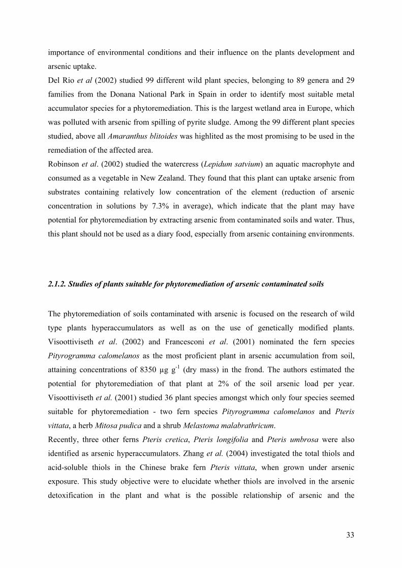
contaminants and convert them into less toxic form (Singh and Cameotra, 2004). The main
two processes of metal form alterations, in which the microorganisms are involved, are the
extracellular transformation and the metal uptake (Fig.10). The oxidation of arsenite had a
special place in the extracellular transformation of this element.
The microbial influence on metal mobility can be applied for bioremediation purposes. The
microorganisms can mobilize metals through autotrophic and heterotrophic leaching,
chelation by microbial metabolites and siderophores, methylation (which can result in
volatilization), (Gadd, 2004). For arsenic remediation, most important processes are the
oxidation, biomethylation, biosorption, dissimilative arsenate-reduction or sulfate-reduction.
Biosorption can be defined as a microbial uptake of organic and inorganic metal species,
soluble and insoluble on the cell surface or intracellular.
Fig. 10. Microbial processes for metal uptake and sequestration (Singh et Cameotra, 2004)
METAL UPTAKE
DIRECT INVOLVEMENT
ACCUMULATION Surface binding to the
microbial cell wall or extracellular materials
UPTAKE For use in
metabolic processes as nutrients
INDIRECT INVOLVEMENT
METABOLITES EXCRETION
H2S production, precipitation of
insoluble metal sulfides from solution
OXYGEN CONSUMPTION
Limitation of biooxidation
COMPLEXATIONBy cellular organic
compounds
CHELATION By cellular membrane
components
PRECIPITATIONand ion exchange
MINERALISATIONFrom interactions with
extracellular byproducts of cell
metabolism
35
Biosorption has been demonstrated to be a useful alternative to conventional treatment
systems for the removal of metals from the dilute aqueous solution. Nevertheless, there are a
few authors who have worked on the application of fungi in the arsenic treatment
technologies. Loukidou et al. (2003) proposed employment of fungal, non- living biomass of
P. chrysogenum as As[V] biosorptive agent.
In the domain of bacteria some work being carried out and also a few patents for their
possible application in arsenic remediation were published. Most of works for the biological
removal of arsenic are based on the filtration of water trough sands or another support on
which is possible to form a biofilm from arsenic oxidizing bacteria. Another type of treatment
is based on the production of H2S from sulfate-reducing bacteria, which consequently leads to
the precipitation of arsenic under arsenic sulfides (Groudeva et al., 2001).
Katsoyanidis and Zouboulis (2004) proposed the use of biological iron oxidation for the
removal of arsenic. The main incertitude in this processes comes from the usage of consortia
of bacteria, which are often natural formed, but the precise function of different types of
microorganisms are not investigated in depth.
Lièvremont et al. (2003) demonstrated a new insight to the arsenic removal process, based on
the use of arsenic-oxidizing bacteria for the conversion of As[III] into As[V] followed of a
chemical sorption of the arsenate in chabazite or kutnahorite.
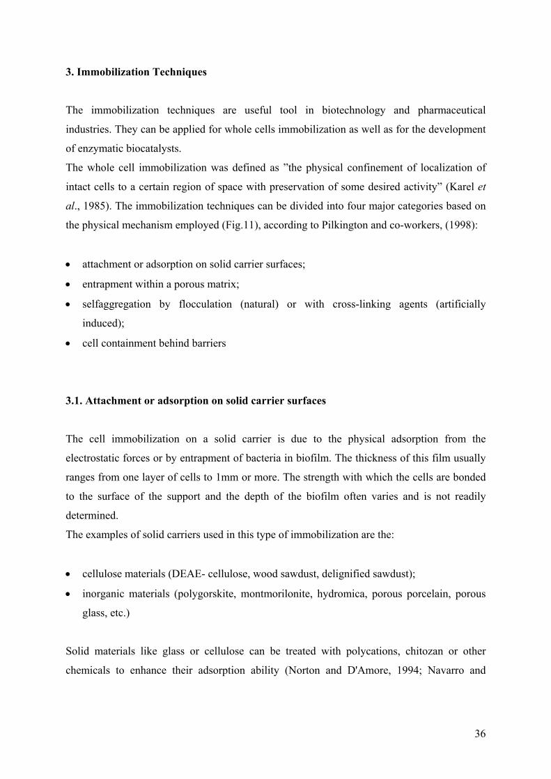
36
3. Immobilization Techniques
The immobilization techniques are useful tool in biotechnology and pharmaceutical
industries. They can be applied for whole cells immobilization as well as for the development
of enzymatic biocatalysts.
The whole cell immobilization was defined as ”the physical confinement of localization of
intact cells to a certain region of space with preservation of some desired activity” (Karel et
al., 1985). The immobilization techniques can be divided into four major categories based on
the physical mechanism employed (Fig.11), according to Pilkington and co-workers, (1998):
• attachment or adsorption on solid carrier surfaces;
• entrapment within a porous matrix;
• selfaggregation by flocculation (natural) or with cross-linking agents (artificially
induced);
• cell containment behind barriers
3.1. Attachment or adsorption on solid carrier surfaces
The cell immobilization on a solid carrier is due to the physical adsorption from the
electrostatic forces or by entrapment of bacteria in biofilm. The thickness of this film usually
ranges from one layer of cells to 1mm or more. The strength with which the cells are bonded
to the surface of the support and the depth of the biofilm often varies and is not readily
determined.
The examples of solid carriers used in this type of immobilization are the:
• cellulose materials (DEAE- cellulose, wood sawdust, delignified sawdust);
• inorganic materials (polygorskite, montmorilonite, hydromica, porous porcelain, porous
glass, etc.)
Solid materials like glass or cellulose can be treated with polycations, chitozan or other
chemicals to enhance their adsorption ability (Norton and D'Amore, 1994; Navarro and
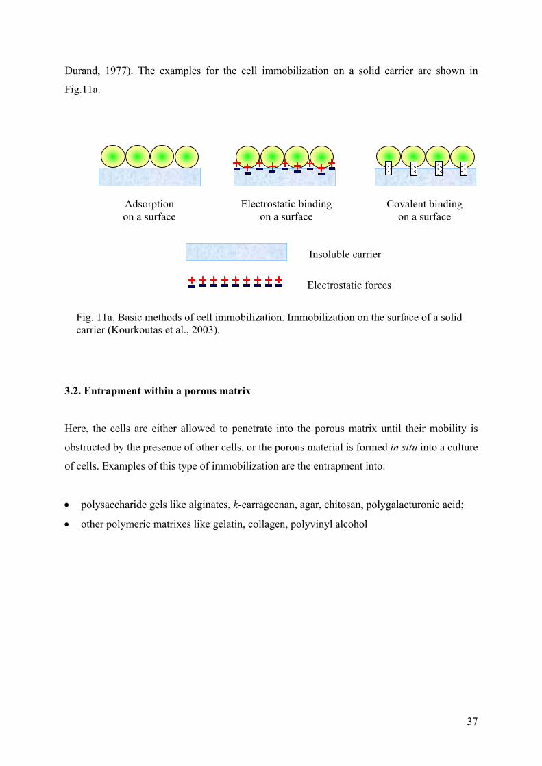
37
Durand, 1977). The examples for the cell immobilization on a solid carrier are shown in
Fig.11a.
3.2. Entrapment within a porous matrix
Here, the cells are either allowed to penetrate into the porous matrix until their mobility is
obstructed by the presence of other cells, or the porous material is formed in situ into a culture
of cells. Examples of this type of immobilization are the entrapment into:
• polysaccharide gels like alginates, k-carrageenan, agar, chitosan, polygalacturonic acid;
• other polymeric matrixes like gelatin, collagen, polyvinyl alcohol
Fig. 11a. Basic methods of cell immobilization. Immobilization on the surface of a solid carrier (Kourkoutas et al., 2003).
Adsorption on a surface
Electrostatic binding on a surface
Covalent bindingon a surface
Insoluble carrier
Electrostatic forces
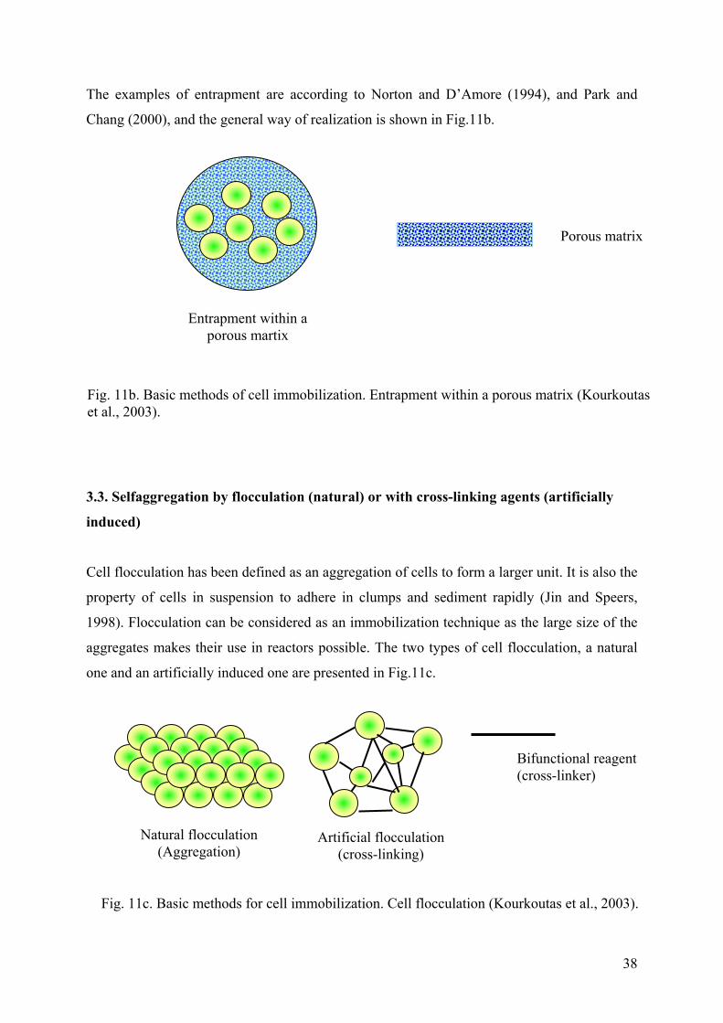
38
The examples of entrapment are according to Norton and D’Amore (1994), and Park and
Chang (2000), and the general way of realization is shown in Fig.11b.
3.3. Selfaggregation by flocculation (natural) or with cross-linking agents (artificially
induced)
Cell flocculation has been defined as an aggregation of cells to form a larger unit. It is also the
property of cells in suspension to adhere in clumps and sediment rapidly (Jin and Speers,
1998). Flocculation can be considered as an immobilization technique as the large size of the
aggregates makes their use in reactors possible. The two types of cell flocculation, a natural
one and an artificially induced one are presented in Fig.11c.
Porous matrix
Entrapment within a porous martix
Fig. 11b. Basic methods of cell immobilization. Entrapment within a porous matrix (Kourkoutas et al., 2003).
Natural flocculation (Aggregation)
Artificial flocculation (cross-linking)
Fig. 11c. Basic methods for cell immobilization. Cell flocculation (Kourkoutas et al., 2003).
Bifunctional reagent (cross-linker)
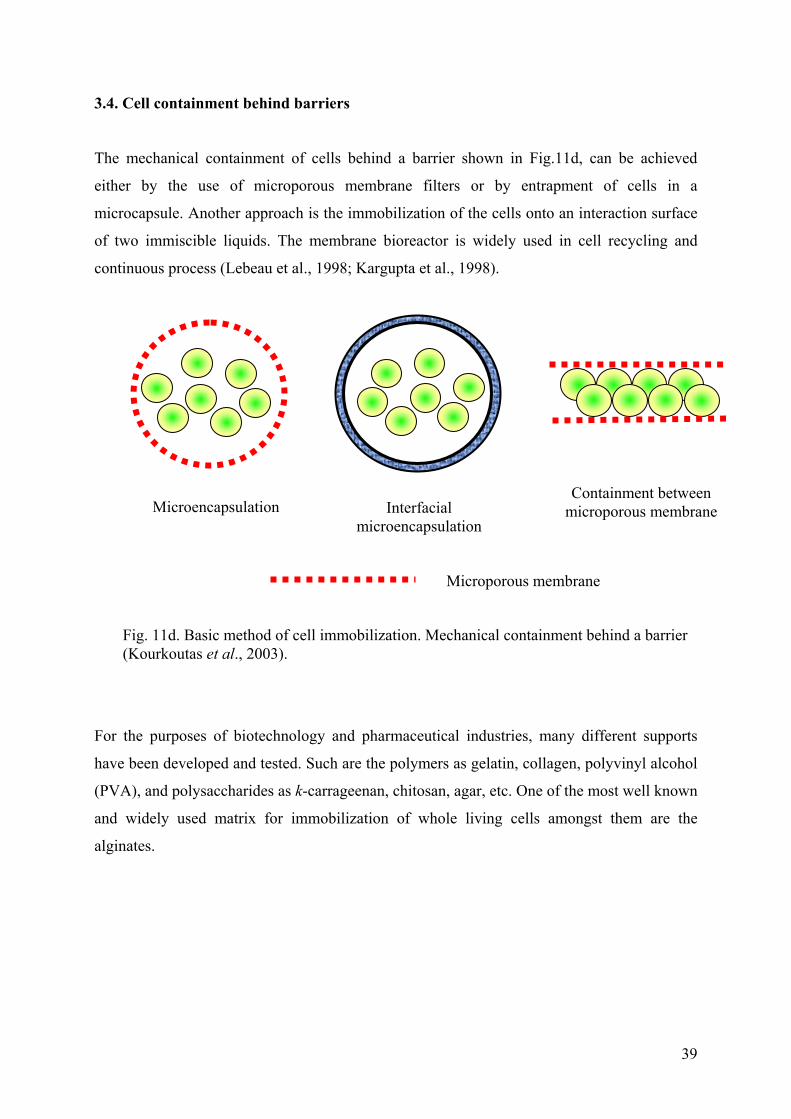
39
3.4. Cell containment behind barriers
The mechanical containment of cells behind a barrier shown in Fig.11d, can be achieved
either by the use of microporous membrane filters or by entrapment of cells in a
microcapsule. Another approach is the immobilization of the cells onto an interaction surface
of two immiscible liquids. The membrane bioreactor is widely used in cell recycling and
continuous process (Lebeau et al., 1998; Kargupta et al., 1998).
For the purposes of biotechnology and pharmaceutical industries, many different supports
have been developed and tested. Such are the polymers as gelatin, collagen, polyvinyl alcohol
(PVA), and polysaccharides as k-carrageenan, chitosan, agar, etc. One of the most well known
and widely used matrix for immobilization of whole living cells amongst them are the
alginates.
Interfacial microencapsulation
Microencapsulation Containment between
microporous membrane
Fig. 11d. Basic method of cell immobilization. Mechanical containment behind a barrier (Kourkoutas et al., 2003).
Microporous membrane
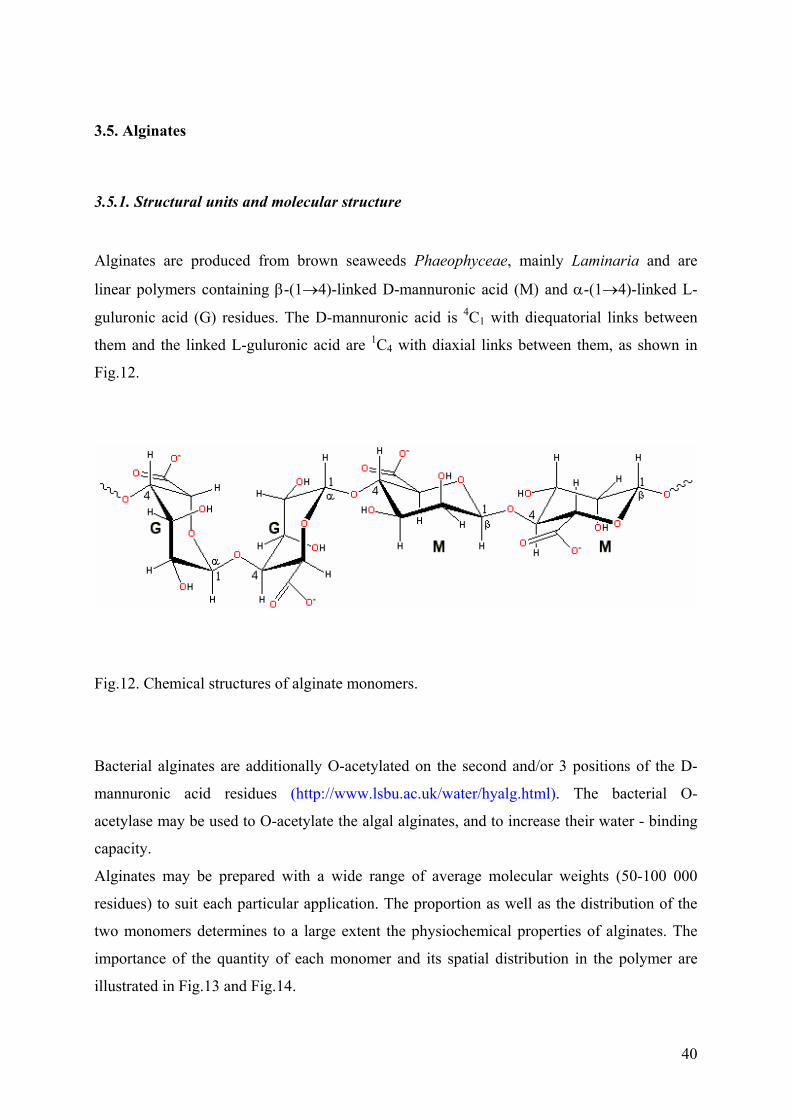
40
3.5. Alginates
3.5.1. Structural units and molecular structure
Alginates are produced from brown seaweeds Phaeophyceae, mainly Laminaria and are
linear polymers containing β-(1→4)-linked D-mannuronic acid (M) and α-(1→4)-linked L-
guluronic acid (G) residues. The D-mannuronic acid is 4C1 with diequatorial links between
them and the linked L-guluronic acid are 1C4 with diaxial links between them, as shown in
Fig.12.
Fig.12. Chemical structures of alginate monomers.
Bacterial alginates are additionally O-acetylated on the second and/or 3 positions of the D-
mannuronic acid residues (http://www.lsbu.ac.uk/water/hyalg.html). The bacterial O-
acetylase may be used to O-acetylate the algal alginates, and to increase their water - binding
capacity.
Alginates may be prepared with a wide range of average molecular weights (50-100 000
residues) to suit each particular application. The proportion as well as the distribution of the
two monomers determines to a large extent the physiochemical properties of alginates. The
importance of the quantity of each monomer and its spatial distribution in the polymer are
illustrated in Fig.13 and Fig.14.
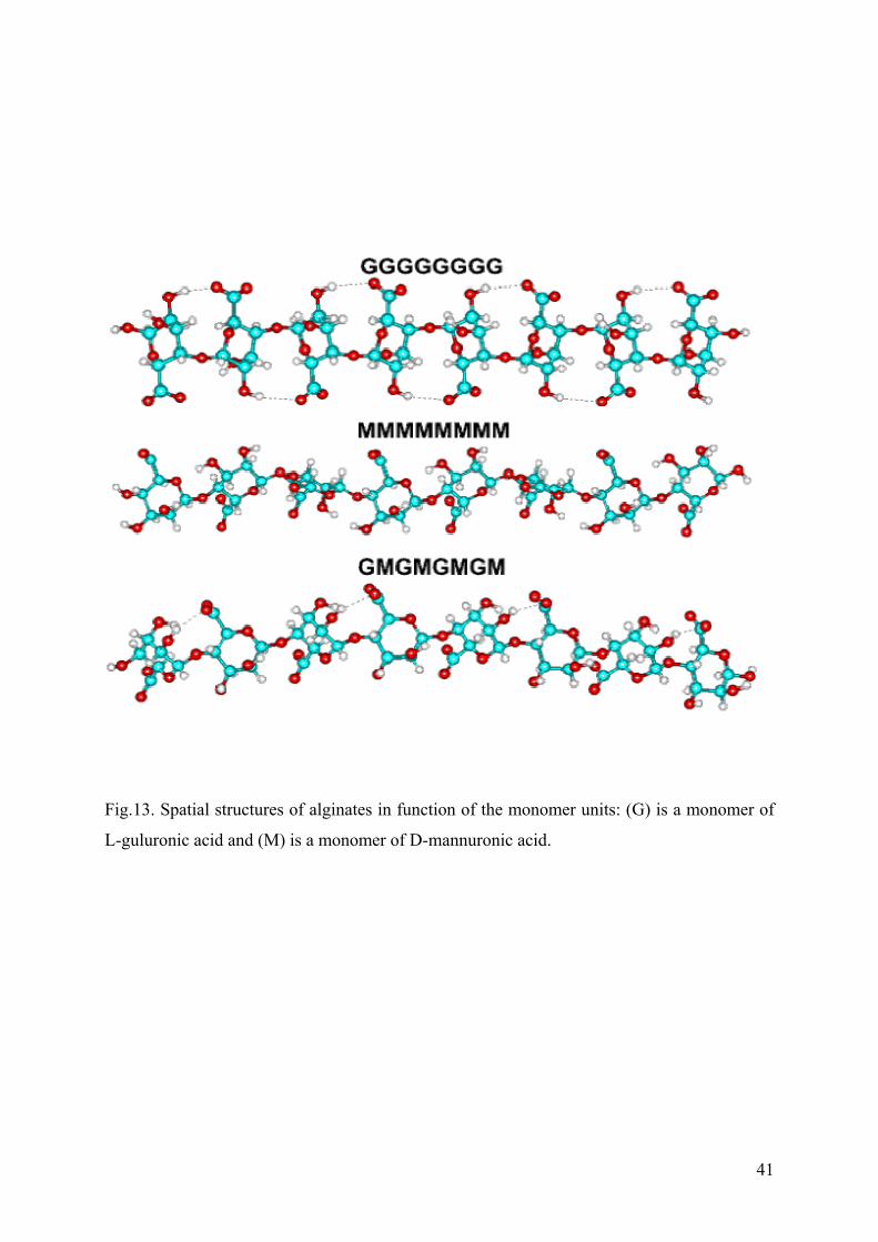
41
Fig.13. Spatial structures of alginates in function of the monomer units: (G) is a monomer of
L-guluronic acid and (M) is a monomer of D-mannuronic acid.
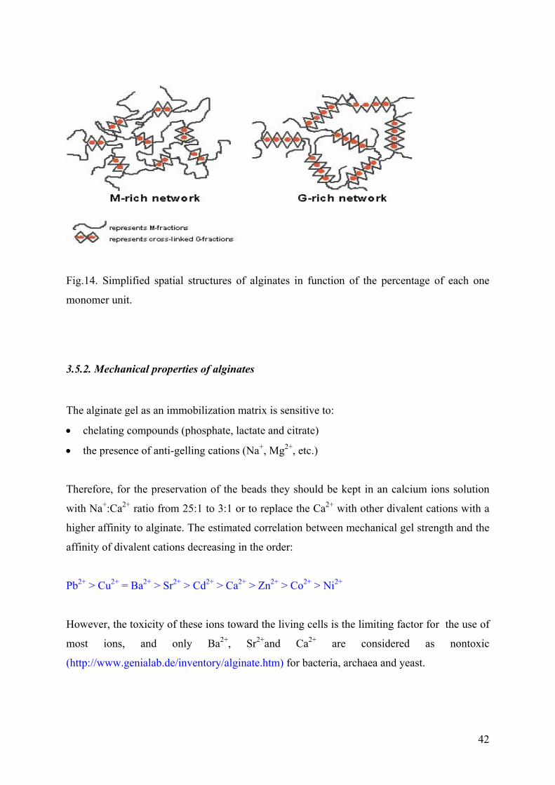
42
Fig.14. Simplified spatial structures of alginates in function of the percentage of each one
monomer unit.
3.5.2. Mechanical properties of alginates
The alginate gel as an immobilization matrix is sensitive to:
• chelating compounds (phosphate, lactate and citrate)
• the presence of anti-gelling cations (Na+, Mg2+, etc.)
Therefore, for the preservation of the beads they should be kept in an calcium ions solution
with Na+:Ca2+ ratio from 25:1 to 3:1 or to replace the Ca2+ with other divalent cations with a
higher affinity to alginate. The estimated correlation between mechanical gel strength and the
affinity of divalent cations decreasing in the order:
Pb2+ > Cu2+ = Ba2+ > Sr2+ > Cd2+ > Ca2+ > Zn2+ > Co2+ > Ni2+
However, the toxicity of these ions toward the living cells is the limiting factor for the use of
most ions, and only Ba2+, Sr2+and Ca2+ are considered as nontoxic
(http://www.genialab.de/inventory/alginate.htm) for bacteria, archaea and yeast.
43
3.5.3. Application of alginates
Entrapment within beads of Ca-alginate gel has become the most widely used technique for
immobilization of living cells (algae, animal cells, bacteria, cyanobacteria, fungi, plant cells
and protoplasts, and yeast). The applications of alginates, produced commercially have a wide
range (Rehm and Valla, 1997):
• Textile printing;
• Paper and board treatments;
• Welding-rot production;
• Water treatment;
• Can sealing;
• Creaming of latex;
• Production of ceramics; food for human and pets;
• Pharmaceutical industry
In textile printing alginates are used for fixation, color yield and brightness, and even ensuring
the printing. In paper treatment the application of alginates improve the surface uniformity.
This is important prior to printing, to enable de deposition and adherence of dyes and inks
(Sutherland and Elwood, 1979) In the welding-rot production, alginates are used as
component of covering materials, and also as component improving bendability. The alginate
application in water treatment technologies is connected mainly with increase of aggregate
size in artificial flocculation.
Commercially produced alginate is used mainly in the food industry, for example: in ice-
creams, cream and cake mixtures, in beer production (to enhancing the foam), fruit drinks (to
assist the suspension of fruit pulp).
In pharmaceutical industry the alginates are used in dermatology, and wound healing, dental
impression materials, treatment of oesophageal reflux, immunostimulants or in medical
transplantation technologies (Sabra et al., 2001).
44
In conclusion:
Arsenic is a wide spread toxic element, which causes several problems. The main
problem in regard to human health is the contamination of drinking water and agricultural
soils.
The biological methods for arsenic remediation described here have several
advantages, when compared to the chemical ones, mainly due to the lack of secondary
pollution and the low impact on the environment.
However the biological methods for arsenic remediation are a relatively new area of
study. Phytoremediation has been widely studied in arsenic remediation, but very little
characterization of the plant species involved in this process has been carried out to date. In
microbial remediation there is still only small amount of information known about the role of
different microorganisms involved in the overall regulation of the transformation processes.
It is known already that the main processes of inorganic arsenic species transformation are the
oxidation of As[III] or the reduction of As[V], but only few bacterial and yeast species are
reported as arsenite-oxidizers or arsenic-reducers. There is also a lack of information
concerning the microorganisms involved in the biomethylation of inorganic arsenic.
Similarly, there is little information to date, regarding the use of yeast in arsenic
bioremediation.
Therefore, the investigation of microorganisms (bacteria and yeast), possessing arsenic
transformation abilities in regard with their further application in bioremediation processes is
an open and wide area, which is worth of being explored.

45
CHAPTER II
AIMS OF THE PROJECT
46
The main aim of this project was to study the potential and suitability of an arsenite–oxidizing
strain, recently named Cenibacterium arsenoxidans, in arsenic remediation of water in regard
of further technology development. This study was done with the view of the replacement of
the chemical oxidation in the treatment technologies of arsenic polluted water.
The strain, Cenibacterium arsenoxidans was previously isolated (Weeger et al., 1999)
and was chosen because of its high oxidation capacity (a maximum of As[+III] – 0.4 mM.h-1
from free cells, with concentration of 5x10⁸ CFU/ml). This organism oxidizes the inorganic
arsenic using the enzyme arsenite – oxidase. The gene structure and operon arrangement were
recently identified and described (Muller et al., 2003).
Preliminary laboratory-scale studies with Cenibacterium arsenoxidans in a batch
reactor system using Na-lactate as a sole carbon-source showed complete oxidation of As[III]
to As[V] from the strain (Weeger et al., 1999). However, the estimated cost of chemically
defined medium is relatively high.
Therefore, two new nutrient media, based on the use of alternative, inexpensive
industrial waste products as a carbon source, were developed and tested. In this way it is
possible to join the arsenic remediation with the treatment of effluents with high organic
charges, which usually causes problems when treated in industrial water purification plants.
From another point of view, such media could be used only as a cheap and efficient
alternative for biomass production of C. arsenoxidans.
The development of the strain was also studied in sterile and non-sterile conditions as
a simulation of a real time process. It was achieved through a gfp-tagged clone of C.
arsenoxidans.
As an alternative to the free cells culture application in the cleanup process, the
remediation potential of entrapped C. arsenoxidans cells in Ca2+ alginate beads was studied.
Part of the research work was devoted to the development of a new screening method
for arsenic-transforming bacteria. This assay was developed in order to enhance the rapid
screening of bacterial strains possessing arsenite-oxidizing ability, which were of interest in
this work as well as for future work.
Thus, this research work focused on the development of a microbiological method for
remediation of arsenic in which the chemical oxidation is replaced by a bacterial one.

47
CHAPTER III
RESULTS AND DISCUSSION
48
In this Chapter the results obtained throughout the different stages of the study are presented
and discussions included. They are arranged in four parts, according to the main aim of each
set of experiments performed. The overall presentation of results in each part follows the
pattern:
1. Introduction, consisting of short description of aims of the part and the experiments
done.
2. Presentation of a manuscript or a paper, concerning the results.
3. Presentation of additional results, obtained during the assays, which were not included
into the manuscript or the paper.
4. Conclusion and Perspectives, based on the obtained results in each set of experiments.
Part III does not follow the above pattern. In this part the results are introduced, and
presented, but there is not a manuscript or a paper added.
Part I: Development of nutrient media
1. Introduction
One of the research objectives of my work was the development of a nutrient medium to
increase the biomass production of arsenite-oxidizing strain. Several carbon sources were
tested in order to define the growth characteristics of the strain C. arsenoxidans. We focused
on the tests of some additional carbon sources, which were not previously tested. The growth
of the strain was firstly tested on carbon sources, such as ethanol, methanol and sucrose. In
parallel three different liquid organic wastes from food industry were also tested as possible
organic sources for the growth of C. arsenoxidans. The tested by-products were molasses,
sauerkraut brine and whey. An additional objective was the achievement of an optimal ratio
of cost of the medium/quantity of biomass produced. Thus the importance of each component
from the chemically defined medium (CDM) was assayed for its importance in the overall
growth of the strain.
In this part the results obtained from the three chemically pure carbon-sources assayed and the
three by-products tested as a carbon-source are presented.
49
• The results for the sauerkraut based media and whey media are presented in the
manuscript of the paper, named: Sauerkraut brine and whey-inexpensive by-products
for biomass production of an arsenite-oxidizing bacterium C. arsenoxidans.
• Additional results for the development of the strain in the sauerkraut broth media and
whey based media are presented after the paper as additional results.
• The results obtained for the growth of the strain on molasses medium and its
modifications as a carbon-source are presented as additional results after the
manuscript of the paper.
• The results of the growth of the strain on ethanol, methanol and sucrose are presented
as additional results.
The conclusions and perspectives concerning this part of results are presented at the end of
the part.
50
2. Manuscript of Article I
Simeonova, D. D., Lièvremont, D., Groudeva, V. I. and Lett M-C.
Sauerkraut brine and whey – inexpensive by-products for biomass production of
an arsenite-oxidizing bacterium Cenibacterium arsenoxidans
Manuscript submitted to Applied Microbiology and Biotechnology
51
SAUERKRAUT BRINE AND WHEY – INEXPENSIVE BY-PRODUCTS FOR 1
BIOMASS PRODUCTION OF AN ARSENITE-OXIDIZING BACTERIUM 2
CENIBACTERIUM ARSENOXIDANS 3
4
Diliana D. Simeonova a, b, Didier Lièvremont a, Veneta I. Groudeva b, Marie-Claire Lett a* 5
6
7
a Laboratoire de Dynamique, Evolution et Expression de Génomes de Microorganismes, FRE 8
2326 CNRS – Université Louis - Pasteur, 28 rue Gœthe, 67083 Strasbourg Cédex, France 9
b Department of General and Industrial Microbiology, Sofia University "St. Kliment 10
Ohridski", 8,"Dragan Tzankov" blvd., 1421 Sofia, Bulgaria 11
12
13
14
15
Keywords: arsenic bioremediation, sauerkraut brine, whey 16
*Corresponding Author. Mailing address: Laboratoire de Dynamique, Evolution et 17
Expression de Génomes de Microorganismes, FRE 2326 CNRS – Université Louis - Pasteur, 18
28 rue Gœthe, 67083 Strasbourg Cédex, France. 19
E-mail: [email protected] 20
Phone: (+33) 390 24 19 97 21
Fax: (+33) 390 24 19 26 22
52
Abstract 23
24
An inexpensive nutrient medim was required for the optimization of the biomass production 25
of Cenibacterium arsenoxidans, an arsenite-oxidizing bacterium which can be used in arsenic 26
remediation process. Since this strain was initially isolated on a medium containing sodium 27
lactate, different liquid organic waste containing lactate, such as whey and sauerkraut brine, 28
were tested. The growth of the strain, expressed by the optical density of the media containing 29
20% (vol./vol.) water solutions of whey or sauerkraut brine was much higher, when compared 30
to the growth in a medium with Na-lactate (OD 0.4-0.5 ± 0.01-0.03 at 600 nm) as a unique 31
carbon-source, which corresponded to 20 x 108 CFU/ml. In whey medium the OD was in the 32
range of 0.99 ± 0.07 at 600 nm, which corresponded to 100 x 108 CFU/ml and in the 33
sauerkraut brine medium the OD was in the range of 0.95 ± 0.03, which corresponded to 140 34
x 108 CFU/ml. 35
The determination of inorganic arsenic species and their quantification (As[III] and As[V]) 36
were obtained by high-performance liquid chromatography coupled with inductively coupled 37
plasma atomic emission spectrometry (HPLC-ICP-AES). Both new nutrient media had no 38
influence on the oxidation of arsenite (As[III]) by the strain. 39
53
Introduction 40
41
Arsenic is a ubiquitous element in the earth’s crust and naturally occurs in many inorganic 42
and organic compounds in different oxidation states as a result of natural processes 43
(weathering reactions, biological activity, volcanic emissions, etc.) or anthropogenic activities 44
(mining, smelting, glass manufacturing, pharmaceutical industry, electronics, pesticides, 45
chemical weapons). The inorganic arsenite (As [III]) and arsenate (As [V]) are the most stable 46
forms of the element. 47
To date, there are different technologies for arsenic decontamination of waters, which are 48
predominantly based on chemical treatments. In these technologies the first step is the 49
oxidation of arsenite As[III] into arsenate As[V], the second step in these is then the 50
precipitation of As[V]. The As[V] is favored for precipitation due to its high reaction activity, 51
low mobility and less toxicity (Arai et al., 2001; Meng et al., 2001). As a consequence of the 52
powerful oxidants (H2O2, KMnO4, chlorine, hypochlorite, Fe3+) used in the oxidation step, the 53
chemical oxidation of arsenic causes additional destruction and pollution of the environment. 54
Other methods used are the ion exchange or reverse osmosis remediation techniques, although 55
these have high removal efficiency, they require high investments, running and maintenance 56
costs. 57
It is well known that microbial activity strongly influences arsenic speciation and its 58
transport in the environment. Bacterial transformation systems of both As[III] and As[V] 59
forms are likely to be ancient (Gihring et al., 2003; Lebrun et al., 2003) and are found in 60
numerous bacterial species encoded on chromosomes or mobile plasmids (Silver, 1996). 61
Several bacteria are known to transform inorganic arsenic through oxidation (Coram and 62
Rawlings, 2002; Duquesne et al., 2003; Ehrlich, 2001; Macy et al., 2000; Weeger et al., 63
54
1999). Such biological processes can be applied to bioremediation since they generate less 64
toxic or less mobile forms of arsenic as well as being environmental friendly. 65
A bacterial strain, recently named Cenibacterium arsenoxidans (Weeger et al., 1999) 66
was chosen because of its high oxidation capacity (a maximum of 0.4 mM (As[III])/h from 67
free cells, with concentration of 5 x 10⁸ CFU/ml). This organism oxidizes the inorganic 68
arsenic using the enzyme arsenite – oxidase. The gene structure and operon arrangement were 69
recently identified and described (Muller et al., 2003). 70
Preliminary laboratory-scale studies with C. arsenoxidans in a batch reactor system 71
using Na-lactate as a sole carbon-source showed complete oxidation of 100 mg of As[III] to 72
As[V] from the strain. However, the cost of chemically defined medium is high. For those 73
purpose alternatives nutrient media, based on the use of inexpensive industrial waste product 74
as a carbon source, such as sauerkraut brine or whey, were developed. These waste products 75
are abundantly available since sauerkraut brine is the by-product from sauerkraut production 76
and whey is the major by-product of cheese manufacturing and casein production. Both have 77
a high organic charge and acidic pH and usually causes problems when treated in industrial 78
water purification plants. 79
Thus, this research work focused on the development of a microbiological method for 80
remediation of arsenic in which: 1) the chemical oxidation is replaced by a bacterial one and 81
2) the oxidation of arsenite can be directly connected to the conversion of liquid organic 82
waste. 83
55
Materials and Methods 84
85
Bacterial strain 86
87
A heterotrophic arsenite-oxidizing bacterial strain ULPAs1, recently renamed Cenibacterium 88
arsenoxidans, was previously isolated from an aquatic environment, contaminated with 89
arsenic (Weeger et al., 1999). The strain had a doubling time of 1.5 h and showed a minimum 90
inhibitory concentration (MIC) of 6.65 mM (498 mg/l) for As[III]. 91
92
Inoculation and growth conditions 93
94
A preculture from a single colony of the strain, was prepared in 20 ml of liquid chemically 95
defined medium (CDM) and incubated for 72 hours at 220 rpm (Certomat, U) at 25°C in the 96
dark. The inoculum was not induced with arsenite. The preculture was centrifuged for 15min 97
at 5000 rpm (Jouan, GR 412), washed twice with double deionised, sterile water (Milli-Q 98
system, Millipore) and finally resuspended in 10 fold smaller volume of sterile ddH2O. The 99
oxidation of As[III] in the tested media was carried out in sterile tubes, with final volume of 100
20 ml. The tubes were inoculated with 20 µl from the preculture of C. arsenoxidans, (initial 101
concentration of 1 x 10⁵ CFU/ml) and were incubated at 25°C, on a rotary shaker (220 rpm) in 102
the dark. Samples for viability counts and optical density at 600 nm were taken three times a 103
day every 4 hours during the experiments. The dilutions of the samples for CFU counting, 104
were made in sterile ddH2O and were plated on CDM agar in duplicate. All experiments were 105
performed at these standard conditions and each set of experiments was carried out three 106
separate times with samples prepared in triplicate (n=9). 107
108
56
Nutrition media 109
110
Chemically defined medium (CDM). The compounds of the CDM and its preparation, were 111
previously described in Weeger et al. (1999). The CDM had a final pH of 7.2. 112
Sauerkraut brine medium. The sauerkraut brine was kindly supplied by the producer 113
Coopchou, (Vendenheim, France). The natural pH of the sauerkraut brine was 3.5 - 3.7. The 114
sauerkraut brine was pretreated by filtration through a paper filter, centrifugation at 6000 rpm 115
for 20 min, heating twice at 90°C in a water bath - for 15 min and allowed to settle for 24 h at 116
4°C. A second filtration through a paper filter, dilution 1:5 (vol./vol.) with sterile ddH2O and 117
final filtration through 0.20 µm pore-size membrane (Nalgene, France) were then carried out. 118
The sauerkraut brine medium was prepared in two modifications- sauerkraut brine medium 119
without salt solutions from CDM, named SBM and with addition of salts solutions named 120
SBM-A*BC. The SBM-A*BC consisted of salts solutions A*, B and C in the same quantity 121
as in the CDM, except that solution A* did not contain Na - lactate. The final pH of SBM and 122
SBM-A*BC were adjusted to 7.2, with 1 M NaOH. 123
Whey medium. For the whey based medium commercially available products from the french 124
cheese industry (fromage blanc, Auchan and Rians) were used. The initial whey pH was 4.0–125
4.3 and was pretreated for the preparation of nutrition medium by filtration through paper 126
filter, dilution with double deionised water 1:1.5 (vol./vol.), neutralization with 1 M NaOH up 127
to pH 7.2, a second filtration through paper filter, followed by sterile filtration through 128
0.20µm pore-size membrane. 129
The whey medium was also prepared and tested in two modifications- without salt solutions 130
from CDM, named WM and with addition of salt solutions WM-A*BC. The WM-A*BC 131
consisted of solutions A*, B, and C from the CDM in the same quantities, except that solution 132
A* again did not contain Na -lactate. 133
57
D-, L- lactate assay. The measurements of initial and final quantity of D- and L-lactate as a 134
unique carbon source in the all tested media were done as described by Gutmann and 135
Wahlefeld (1974). 136
137
Physico-chemical parameters of the tested media 138
139
The analysis of the basic physico-chemical parameters of SBM and WM were made in the 140
Centre d’Analyse et de Recherche (CAR, Strasbourg, France). The methods used were carried 141
out as the Norme Française Technique (NFT). The parameters tested were: chemical oxygen 142
demand (COD) (NFT 90 - 101), biological oxygen demand in 5 days (BOD5) (NFT 90 - 103), 143
ammonium concentration (NFT 90 - 015), Kjeldahl NTK (NFT 90 - 110), Nitrates (NFT 90 - 144
012) and Nitrites (NFT 90 - 013) concentrations. 145
146
Determination of arsenic transformations 147
148
The arsenite-oxidation from C. arsenoxidans during growth was investigated by taking 149
samples of the cultures supernatants at 24 h. In parallel, as a control, uninoculated SBM and 150
WM were assayed for arsenate-oxidation capacity. In tested media 10 mg/l of As[III] was 151
added. The supernatants samples from the different media were filtrated through a 0.22 µm 152
pore - size membrane (Durapore, Millipore-GV), and stored at 4°C, in the dark for no more 153
than 1 week before measurement. 154
For the arsenic speciation and quantification a technique based on the coupling of HPLC and 155
ICP-AES (Amran et al., 1997; Moll et al., 1996) was used. Arsenic was eluted with a 156
phosphate buffer, degassed and filtered through a cellulose acetate membrane (0.65 µm pore - 157
size, Millipore). The detection of arsenic compounds was carried out at a fixed wavelength as 158
58
described in Weeger et al. (1999). Detection limits were 0.12 mg/l for As[III] and 0.14 mg/l 159
for As[V], and quantification limits were 0.43 mg/l and 0.36 mg/l for As[III] and As[V], 160
respectively. All data obtained in this study were from at least two independent experiments, 161
on duplicate samples (n=4). 162
163
59
Results 164
165
Sauerkraut Brine Medium 166
167
Preliminary studies for arsenate reduction, arsenite oxidation or uptake of inorganic arsenic by 168
the sauerkraut brine were done using the HPLC-ICP-AES method. The results showed 100% 169
recovery of As[III] from an initial As[III] concentration of 10 mg/l. Thus, the sauerkraut brine 170
did not oxidize As[III] or reduce As[V]. 171
Growth experiments were performed in two concentrations of sauerkraut brine (10 and 172
20 % (vol./vol.)) as a sole carbon- source. The growth and the arsenate-oxidation ability of the 173
strain were studied in SBM with its natural pH 3.6 - 4.0 and in a neutral pH 7.2 in the 174
presence and absence of 10 mg/l of As[III]. The influence on the growth of the strain of salt 175
solutions A*, B and C supplied to the SBM-A*BC were also tested. The control used was 176
CDM, supplemented with 10 mg/l of As[III]. All experiments were carried out at 25°C. The 177
growth of the strain in SBM-A*BC and SBM are presented in Fig. 1a and 1b, respectively. 178
No growth of the strain was observed in the medium with a natural pH for the sauerkraut 179
brine (data not shown). When sauerkraut brine was neutralized, the strain grew equally well in 180
presence and in absence of salts solutions and in the presence and in absence of As[III]. 181
The concentrations of lactate in 20% (vol./vol.) solution of sterile sauerkraut brine, without 182
salts solutions, at the natural pH 3.6 - 3.9, was 1.39 x 10⁻² M in the D-form and 1.11 x 10⁻² M 183
in the L-form. After neutralization of SBM up to pH 7.2 - 7.5, the concentrations of D-lactate 184
was 1.53 x 10⁻² M and no change in the L-lactate concentration, was observed. 185
In 20% solution of sauerkraut brine, without salts solutions (SBM) at neutral pH after 186
96 h of growth of C. arsenoxidans, the quantity of both forms of lactate decreased 187
significantly to 3.8 x 10⁻⁵ M of each. When salts solutions A*, B and C were added, into the 188
60
sauerkraut brine (SBM-A*BC), the level of lactate decreased to 3.1 x 10⁻⁵ M (D-form) and 189
2.8 x 10⁻⁵ M (L-form). Therefore the strain grows better in SBM supplemented with salts 190
solutions. 191
The common physico-chemical parameters TOC, COD, BOD and N were determined 192
for SBM-A*BC. These analyses were assayed in triplicate. The results obtained for all of the 193
parameters in sterile SBM-A*BC (control) and after the growth of strain for five days 194
(sample) are presented in Fig. 2. The decrease in COD, BOD and TOC are very significant 195
after 5 days of incubation of the strain. The amount of the total carbon in SBM decreased 196
from 5415 mg/l to 2912 mg/l, which represent an assimilation of 2503 mg/l from the strain in 197
5 days. The COD decrease with 5480mg/l and the BOD with 3975mg/l, for the same time 198
period, which represent about 40% of organic and carbon discharge of the sauerkraut brine. 199
The initial concentration of nitrate was less than 1.2 mg/l and no changes in its concentration 200
were observed after 5 days in presence of the strain. The initial nitrite concentration was 201
1.3 mg/l and at the end of assay was below 1.2 mg/l. 202
203
Whey medium 204
205
The preliminary tests for oxidation, reduction and uptake of arsenic ions from the whey were 206
also performed using the HPLC - ICP - AES method. Results showed no transformation or 207
uptake of arsenic. 208
The growth of C. arsenoxidans in this medium was also assayed with or without 209
supplementation of salts solution A*, B and C. The growth at two levels of pH 4.0 - 4.3 and 210
7.2, and the oxidation of As[III] to As[V], were tested. All tests were assayed at two 211
concentrations (10 and 20% (vol./vol.)) of whey as a sole carbon source in the medium. 212
Results obtained are shown in Fig. 3a and 3b. In WM and WM-A*BC at the natural pH of the 213
61
whey, the strain did not grow (data not shown). At neutral pH of the WM, the presence of 214
salts solutions A*, B, and C improved the growth of the strain WM-A*BC. The presence or 215
the absence of arsenic in the medium had no influence on the strain growth in this medium. 216
The initial concentrations of D- and L-lactate in a 20% (vol./vol.) WM at natural 217
pH 4.0 - 4.3 were 0.075 x 10⁻² M and 3.05 x 10⁻² M, respectively. After neutralization of the 218
whey up to pH 7.2 - 7.5, they were 0.06 x 10⁻² M (D-lactate) and 2.96 x 10⁻² M (L-lactate). 219
The decrease in the concentrations of both forms of lactate after 96 h of incubation of the 220
strain, without salts and arsenic supplementation, and at neutral pH were in the range of 221
0.6 x 10⁻⁵ M for D-lactate and 1.7 x 10⁻⁵ M for L-lactate, showing a significant decrease. 222
When salts solutions were introduced into the whey, in contrast to the SBM, the use of lactate 223
was slightly lower and the levels of both forms slightly increased to 0.9 x 10⁻⁵ M (D-form) 224
and 2.9 x 10⁻⁵ M (L-form). 225
The main physico-chemical parameters of WM-A*BC (COD, BOD, TOC) were 226
determined in triplicate. The final concentration of whey in the medium used for these 227
experiments was 20%. The results obtained showed a significant level of degradation of 228
organic mater in presence of the strain after incubation for 5 days period when compared to 229
the control (the same medium, uninoculated with the strain). The results are presented in 230
Fig. 2. The decrease of total carbon was of 1205 mg/l. The necessity of oxygen for the 231
chemical oxidation decreased with 3270 mg/l and for the biological oxidation with 2975 mg/l, 232
after the 5 days period of incubation with the strain. This represented decrease of above 35%, 233
when compared to the control. The initial concentrations of NO2- and NO3
- were below 234
1.2 mg/l and no changes were observed in their concentrations after 5 days growth of the 235
strain. 236
62
Discussion 237
238
The development and the application of a microbiological method for arsenic remediation is 239
necessary to prevent secondary pollution of the environment due to the use of chemical 240
products in the oxidation step of As[III] to As[V]. In previous studies it was shown that the 241
strain C. arsenoxidans possesses a high capacity for oxidation of As[III] (Weeger et al., 242
1999). The high cost of the use of Na-lactate as a sole carbon-source for this strain led to the 243
search for an alternative, cost effective medium, which would easily replace CDM in a large –244
scale system. 245
Whey and sauerkraut brine are waste products and their disposal is problematic, 246
because of their high organic charges and acidic pH. These waste products contain a high 247
quantity of lactate, which supports growth of the strain. Both by-products provided better 248
nutrition for the strain when compared to the medium of reference CDM. The complete media 249
based on the whey proved better for obtaining biomass than SBM (Fig. 1a and Fig. 3a). 250
Nevertheless, in absence of supplemented salt solutions (Fig. 1b and Fig. 3b) the strain 251
showed better growth in SBM (Fig. 1b). Organic charge was higher for SBM-A*BC, with 252
COD changes decreasing in the presence of the strain from 14160 mg/l to 8680 mg/l. In 253
contrast the organic charge for WM-A*BC showed a smaller decrease from 9750 mg/l to 254
6480 mg/l. Therefore biodegradation was higher in the SBM-A*BC medium as shown in 255
Fig.2. Based on these results, the elaborated medium on the use of sauerkraut brine is more 256
efficient when compared to the WM-A*BC and WM, although both media showed better 257
growth of the strain then CDM, which is presently used for biomass production of the strain. 258
Thus, successful optimization of biomass production for the arsenic-oxidizing bacterium 259
C.arsenoxidans, coupled with a low-cost and highly effective media was achieved. 260

63
Acknowledgements 261
262
This research was supported by a grant from the Région Alsace, via the European Doctoral 263
College (EDC), Strasbourg, France. We thank Coopchou for supplying the sauerkraut brine. 264
We thank Niamh Gilmartin for the comments on the manuscript. 265
266
267
64
References: 268
269
Amran MB, Lagarde F and Leroy MJF (1997) Determination of arsenic species in marine 270
organisms by HPLC-ICP-AES and HPLC-HG-QFAAS. Mikrochimica Acta 127: 195–271
202 272
Arai Y, Elmzinga EJ and Sparks DL (2001) X-ray Absorption Spectroscopic Investigation of 273
Arsenite and Arsenate Adsorption at the Aluminium Oxide-Water Interface. J Colloid 274
Interface Sci 235: 80-88 275
Coram NG and Rawlings DE (2002) Molecular relationship between two groups of the genus 276
Leptospirillum and the finding that Leptospirillum ferriphilum sp.nov. dominates 277
South Africain commercial biooxidation tanks that operate at 40°C. Appl Environ 278
Microbiol 68: 838-845 279
Duquesne K, Lebrun S, Casiot C, Bruneel O, Personné J-C, Leblanc M, Elbaz-Poulichet F, 280
Morin G, Bonnefoy V (2003) Immobilization of Arsenite and Ferric Iron, By 281
Acidithiobacillus ferrooxidans and Its Relevance to Acid Mine Drainage. Appl 282
Environ Microbiol 69: 6165-6173 283
Ehrlich HL (2001) Bacterial oxidation of As(III) compounds. In: Environmental Chemistry 284
of Arsenic. Marcel Dekker, New York, pp 313-328 285
Gihring TM, Bond PL, Peters SC and Banfield JF (2003) Arsenic resistance in the archaeon 286
"Ferroplasma acidarmanus": new insights into the structure and evolution of the 287
arsgenes. Extremophiles 7: 123-30 288
Gutmann ABC and Wahlefeld AW (1974) L-Lactat Bestimmung mit Lactat-Dehydrogenase 289
und NAD. Methoden der enzymatischen Analyse, 3-rd edition, Weinheim 1510 - 1514 290
Lebrun E, Brugna M, Baymann F, Muller D, Lièvremont D, Lett M-C, Nitschke W (2003) 291
Arsenite oxidase, an ancient bioenergetic enzyme. Mol Biol Evol 20: 686-693 292
65
Macy JM, Santini JM, Pauling BV, O'Neill AH and Sly L (2000) Two new arsenate/sulfate-293
reducing bacteria: mechanisms of arsenate reduction. Arch Microbiol 173: 49-57 294
Meng X, Korfiatis GP, Christodoulatos C and Bang S (2001) Treatment of arsenic in 295
Bangladesh well water using a household co-precipitation and filtration system. Water 296
Res 35: 2805-2810 297
Moll AE, Heimburger R, Lagarde F, Leroy MJ and Maier E (1996) Arsenic speciation in 298
marine organisms: from the analytical methodology to the constitution of reference 299
materials. Anal Bioanal Chem 354: 550-556 300
Muller D, Lièvremont D, Simeonova DD, Hubert J-C and Lett M-C (2003) Arsenite oxidase 301
aox genes from a metal-resistant ß-Proteobacterium. J Bacteriol 185: 135-141 302
Silver S (1996) Bacterial resistances to toxic metal ions — a review. Gene 179: 9-19 303
Weeger W, Lièvremont D, Perret M, Lagarde F, Hubert J-C, Leroy M and Lett M-C (1999) 304
Oxidation of arsenite to arsenate by a bacterium isolated from an aquatic environment. 305
Biometals 12: 141-149 306
307
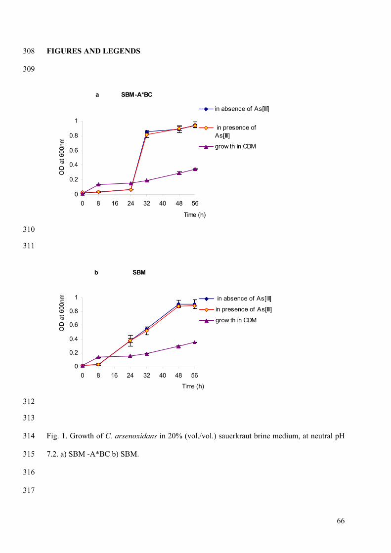
66
FIGURES AND LEGENDS 308
309
a SBM-A*BC
0
0.2
0.4
0.6
0.8
1
0 8 16 24 32 40 48 56
Time (h)
OD
at 6
00nm
in absence of As[III]
in presence ofAs[III]
grow th in CDM
310
311
b SBM
0
0.2
0.4
0.6
0.8
1
0 8 16 24 32 40 48 56
Time (h)
OD
at 6
00nm in absence of As[III]
in presence of As[III]
grow th in CDM
312
313
Fig. 1. Growth of C. arsenoxidans in 20% (vol./vol.) sauerkraut brine medium, at neutral pH 314
7.2. a) SBM -A*BC b) SBM. 315
316
317
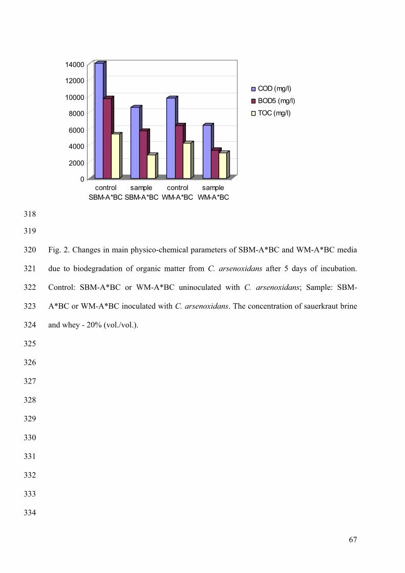
67
0
2000
4000
6000
8000
10000
12000
14000
controlSBM-A*BC
sampleSBM-A*BC
controlWM-A*BC
sampleWM-A*BC
COD (mg/l)
BOD5 (mg/l)
TOC (mg/l)
318
319
Fig. 2. Changes in main physico-chemical parameters of SBM-A*BC and WM-A*BC media 320
due to biodegradation of organic matter from C. arsenoxidans after 5 days of incubation. 321
Control: SBM-A*BC or WM-A*BC uninoculated with C. arsenoxidans; Sample: SBM-322
A*BC or WM-A*BC inoculated with C. arsenoxidans. The concentration of sauerkraut brine 323
and whey - 20% (vol./vol.). 324
325
326
327
328
329
330
331
332
333
334
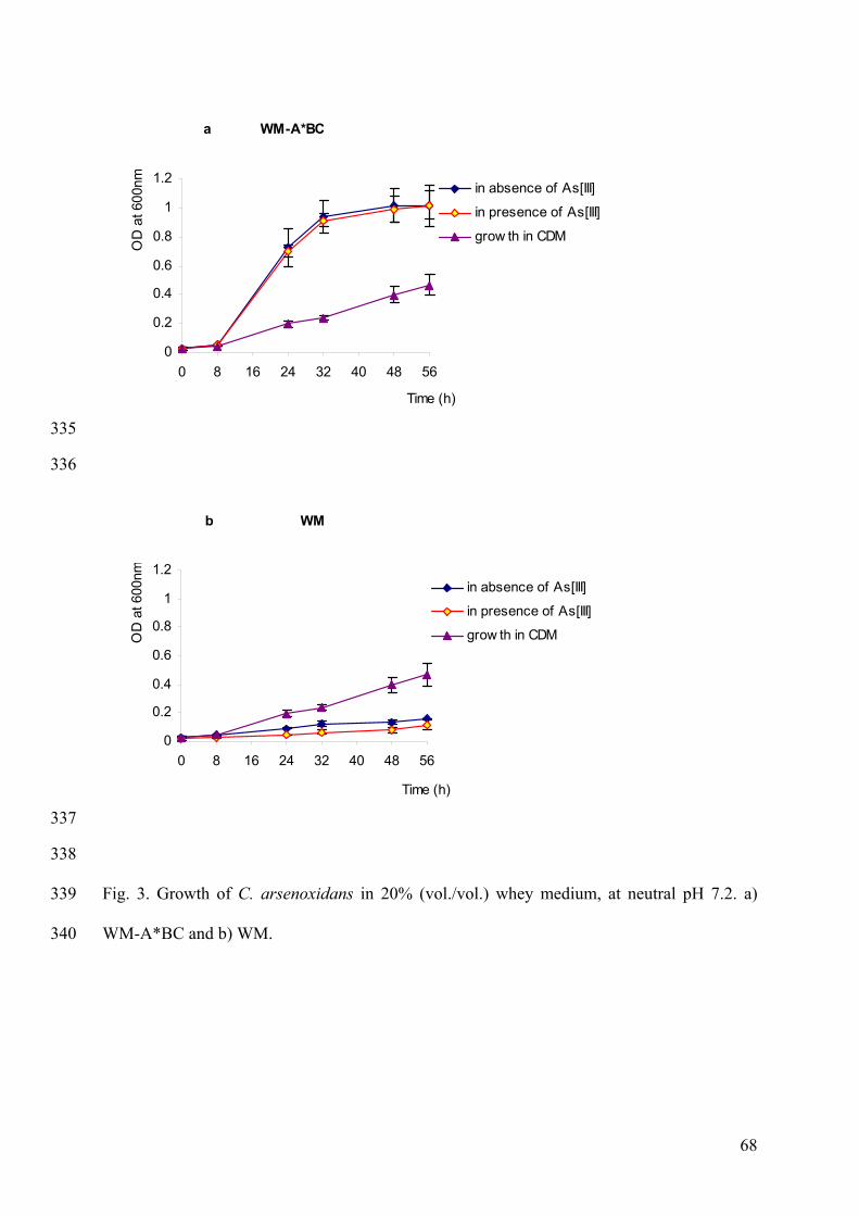
68
a WM-A*BC
0
0.2
0.4
0.6
0.8
1
1.2
0 8 16 24 32 40 48 56
Time (h)
OD
at 6
00nm in absence of As[III]
in presence of As[III]
grow th in CDM
335
336
b WM
0
0.2
0.4
0.6
0.8
1
1.2
0 8 16 24 32 40 48 56
Time (h)
OD
at 6
00nm
in absence of As[III]
in presence of As[III]
grow th in CDM
337
338
Fig. 3. Growth of C. arsenoxidans in 20% (vol./vol.) whey medium, at neutral pH 7.2. a) 339
WM-A*BC and b) WM. 340
69
3. Additional results
3.1. Chemically defined carbon sources
The chemically grade pure and defined carbon sources ethanol, methanol and sucrose were
assayed in three different concentrations as follows: 10 mM, 50 mM and 100 mM at two
experiments in triplicate. The results obtained from the growth assays using ethanol, methanol
and sucrose showed that these carbon-sources did not support the growth of the strain.
3.2. Liquid organic by-products
The by-product carbon-sources were assayed after the development of preliminary treatment
protocols for each by-product used. The protocols concerning the sauerkraut brine based
medium and the whey based medium were described in the paper. The protocol for the
preliminary treatment of molasses based medium is described below.
3.2.1. Sauerkraut brine medium and Whey medium
The preliminary studies of sauerkraut brine as a carbon-source for the growth of strain C.
arsenoxidans were assayed at two concentrations. The concentrations assayed were 10 and
20 %. The influence of the three salt solutions from the CDM on the growth of the strain were
also tested in the presence as well as in absence of As[III] with concentration of 10 mg/l. The
cell cultures were also plated on the agar plates of the tested medium in order to establish the
correlation between the optical density of the culture and the CFU/ml. Thus the maximal
specific growth of the strain (µmax) and the doubling time (dt) were determined, according to
the following equations:
Determination of the specific growth (µ) of the strain, according to the growth media:
12
12 )ln()ln(tt
NtNt−−
=µ , where
Nt2- is the number of cell colonies from the exponential phase of growth counted at time t2
70
Nt1- is the number of cell colonies from the exponential phase of growth counted at time t1
Or
dt2ln
=µ , where
dt – is the doubling time or the time for 1 cell generation.
Therefore, the calculation of doubling time of the strain (td), for each media used, was done
according to the equation:
µ2ln
=td , where:
12
12 lnln2ln
ttNtNt
dt
−−
= , therefore
12
12
lnln).(2ln
NtNtttdt −
−=
The characteristics for the growth of the strain in the different media assayed are shown in
Table 1.
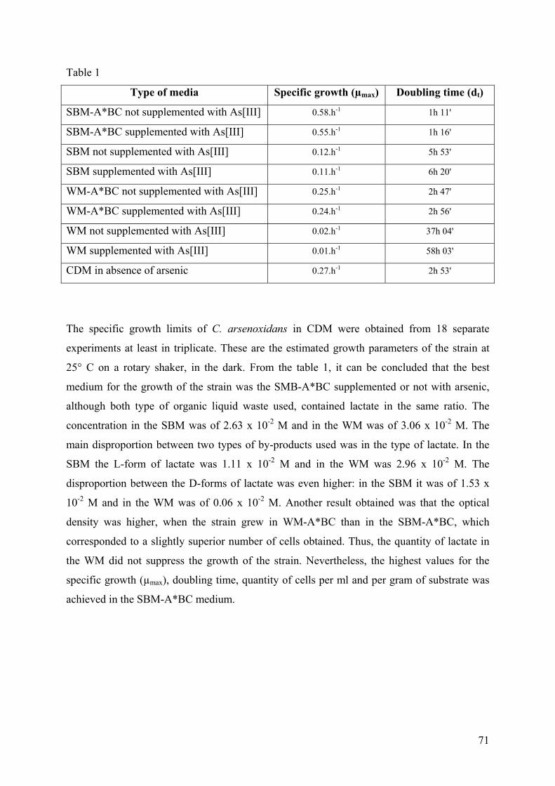
71
Table 1
Type of media Specific growth (µmax) Doubling time (dt)
SBM-A*BC not supplemented with As[III] 0.58.h-1 1h 11'
SBM-A*BC supplemented with As[III] 0.55.h-1 1h 16'
SBM not supplemented with As[III] 0.12.h-1 5h 53'
SBM supplemented with As[III] 0.11.h-1 6h 20'
WM-A*BC not supplemented with As[III] 0.25.h-1 2h 47'
WM-A*BC supplemented with As[III] 0.24.h-1 2h 56'
WM not supplemented with As[III] 0.02.h-1 37h 04'
WM supplemented with As[III] 0.01.h-1 58h 03'
CDM in absence of arsenic 0.27.h-1 2h 53'
The specific growth limits of C. arsenoxidans in CDM were obtained from 18 separate
experiments at least in triplicate. These are the estimated growth parameters of the strain at
25° C on a rotary shaker, in the dark. From the table 1, it can be concluded that the best
medium for the growth of the strain was the SMB-A*BC supplemented or not with arsenic,
although both type of organic liquid waste used, contained lactate in the same ratio. The
concentration in the SBM was of 2.63 x 10⁻² M and in the WM was of 3.06 x 10⁻² M. The
main disproportion between two types of by-products used was in the type of lactate. In the
SBM the L-form of lactate was 1.11 x 10⁻² M and in the WM was 2.96 x 10⁻² M. The
disproportion between the D-forms of lactate was even higher: in the SBM it was of 1.53 x
10-2 M and in the WM was of 0.06 x 10⁻² M. Another result obtained was that the optical
density was higher, when the strain grew in WM-A*BC than in the SBM-A*BC, which
corresponded to a slightly superior number of cells obtained. Thus, the quantity of lactate in
the WM did not suppress the growth of the strain. Nevertheless, the highest values for the
specific growth (µmax), doubling time, quantity of cells per ml and per gram of substrate was
achieved in the SBM-A*BC medium.
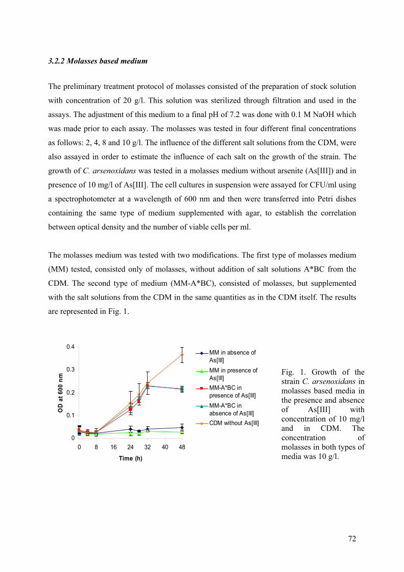
72
3.2.2 Molasses based medium
The preliminary treatment protocol of molasses consisted of the preparation of stock solution
with concentration of 20 g/l. This solution was sterilized through filtration and used in the
assays. The adjustment of this medium to a final pH of 7.2 was done with 0.1 M NaOH which
was made prior to each assay. The molasses was tested in four different final concentrations
as follows: 2, 4, 8 and 10 g/l. The influence of the different salt solutions from the CDM, were
also assayed in order to estimate the influence of each salt on the growth of the strain. The
growth of C. arsenoxidans was tested in a molasses medium without arsenite (As[III]) and in
presence of 10 mg/l of As[III]. The cell cultures in suspension were assayed for CFU/ml using
a spectrophotometer at a wavelength of 600 nm and then were transferred into Petri dishes
containing the same type of medium supplemented with agar, to establish the correlation
between optical density and the number of viable cells per ml.
The molasses medium was tested with two modifications. The first type of molasses medium
(MM) tested, consisted only of molasses, without addition of salt solutions A*BC from the
CDM. The second type of medium (MM-A*BC), consisted of molasses, but supplemented
with the salt solutions from the CDM in the same quantities as in the CDM itself. The results
are represented in Fig. 1.
0
0.1
0.2
0.3
0.4
0 8 16 24 32 40 48
Time (h)
OD
at 6
00 n
m
MM in absence ofAs[III]MM in presence ofAs[III]MM-A*BC inpresence of As[III]MM-A*BC inabsence of As[III]CDM without As[III]
Fig. 1. Growth of the strain C. arsenoxidans in molasses based media in the presence and absence of As[III] with concentration of 10 mg/land in CDM. The concentration of molasses in both types of media was 10 g/l.
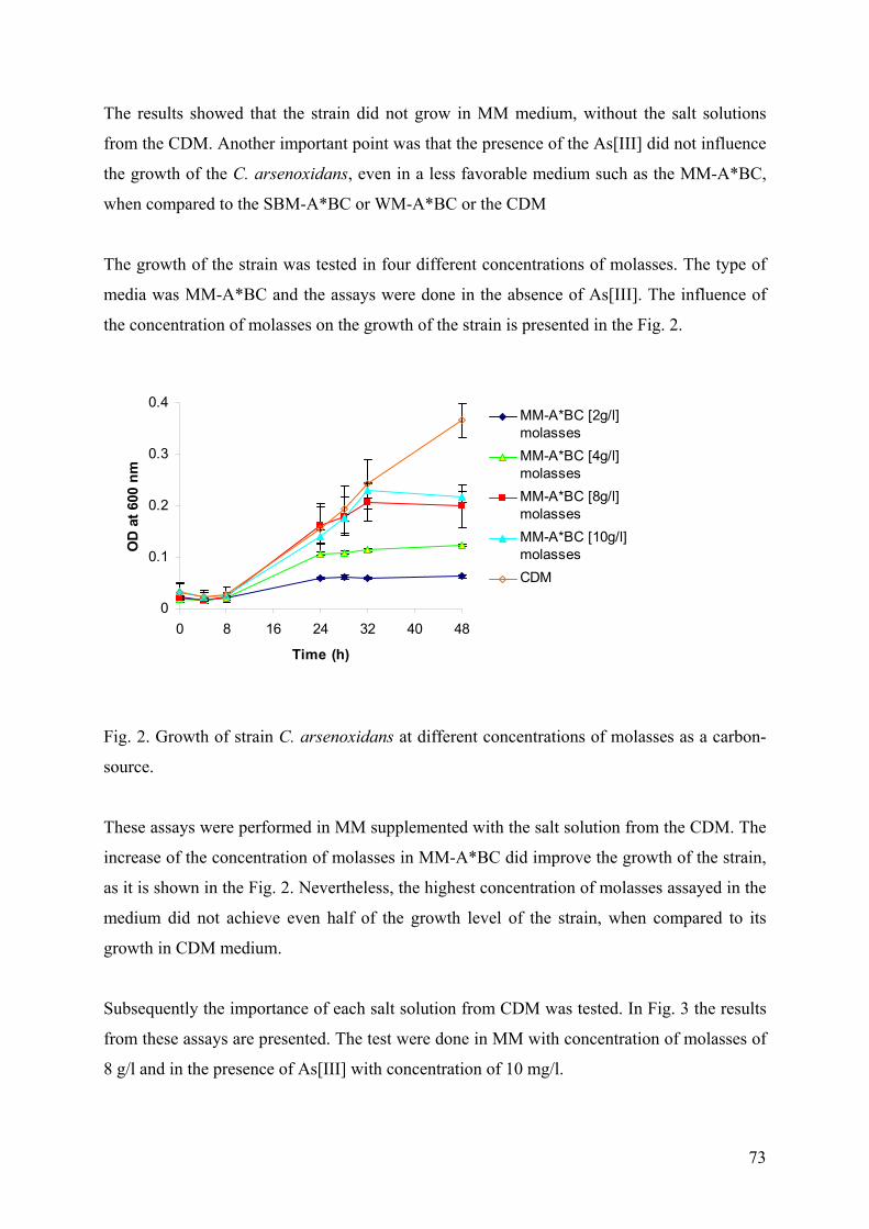
73
The results showed that the strain did not grow in MM medium, without the salt solutions
from the CDM. Another important point was that the presence of the As[III] did not influence
the growth of the C. arsenoxidans, even in a less favorable medium such as the MM-A*BC,
when compared to the SBM-A*BC or WM-A*BC or the CDM
The growth of the strain was tested in four different concentrations of molasses. The type of
media was MM-A*BC and the assays were done in the absence of As[III]. The influence of
the concentration of molasses on the growth of the strain is presented in the Fig. 2.
0
0.1
0.2
0.3
0.4
0 8 16 24 32 40 48
Time (h)
OD
at 6
00 n
m
MM-A*BC [2g/l]molassesMM-A*BC [4g/l]molassesMM-A*BC [8g/l]molassesMM-A*BC [10g/l]molassesCDM
Fig. 2. Growth of strain C. arsenoxidans at different concentrations of molasses as a carbon-
source.
These assays were performed in MM supplemented with the salt solution from the CDM. The
increase of the concentration of molasses in MM-A*BC did improve the growth of the strain,
as it is shown in the Fig. 2. Nevertheless, the highest concentration of molasses assayed in the
medium did not achieve even half of the growth level of the strain, when compared to its
growth in CDM medium.
Subsequently the importance of each salt solution from CDM was tested. In Fig. 3 the results
from these assays are presented. The test were done in MM with concentration of molasses of
8 g/l and in the presence of As[III] with concentration of 10 mg/l.
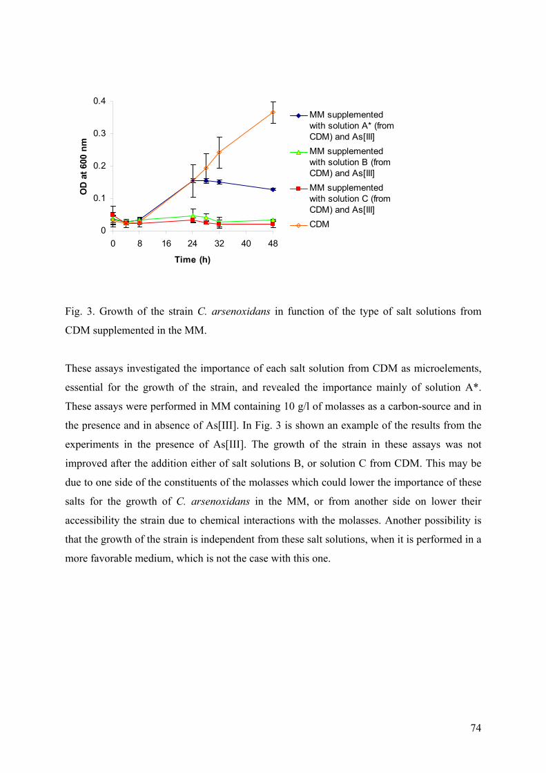
74
0
0.1
0.2
0.3
0.4
0 8 16 24 32 40 48
Time (h)
OD
at 6
00 n
m
MM supplementedwith solution A* (fromCDM) and As[III]MM supplementedwith solution B (fromCDM) and As[III]MM supplementedwith solution C (fromCDM) and As[III]CDM
Fig. 3. Growth of the strain C. arsenoxidans in function of the type of salt solutions from
CDM supplemented in the MM.
These assays investigated the importance of each salt solution from CDM as microelements,
essential for the growth of the strain, and revealed the importance mainly of solution A*.
These assays were performed in MM containing 10 g/l of molasses as a carbon-source and in
the presence and in absence of As[III]. In Fig. 3 is shown an example of the results from the
experiments in the presence of As[III]. The growth of the strain in these assays was not
improved after the addition either of salt solutions B, or solution C from CDM. This may be
due to one side of the constituents of the molasses which could lower the importance of these
salts for the growth of C. arsenoxidans in the MM, or from another side on lower their
accessibility the strain due to chemical interactions with the molasses. Another possibility is
that the growth of the strain is independent from these salt solutions, when it is performed in a
more favorable medium, which is not the case with this one.
75
3. Conclusion
From the preliminary assays of the growth of the arsenite-oxidizing strain C. arsenoxidans in
different nutrition media, only those based on organic by-products, containing lactate -
sauerkraut brine and whey, proved to be suitable for further studies. An important point for
the use of organic by-products as a carbon-source for the growth of C. arsenoxidans was their
preliminary treatment for the preparation of the media. The neutralization of whey and
sauerkraut brine was very important as the C. arsenoxidans is a neutrophilic bacterium.
Molasses as a carbon-source did not support the growth of the strain to a higher level, when
compared to the growth of the strain in CDM, and therefore was not suitable for biomass
production. Nevertheless, the assays performed in this medium proved the importance of salt
solution A* supplementation in all media based on organic waste from food industry.
Moreover, results obtained for the growth of the strain in whey based media showed that the
addition of salts solutions from CDM resulted in better biomass production and enhanced
growth. The use of the whey as a diary by-product, which can be used for the biomass
production of different microorganisms is already studied (Lee et al., 2003; Cristiani-Urbina
et al., 2000; Bledsoe et al., 1999) and proposed for use as an inexpensive carbon-source.
The use of sauerkraut brine as a carbon-source supporting the growth of microorganisms has
not yet been proposed. An important differentiation between these two products, when used
as carbon-sources for C. arsenoxidans, is that only in media based on the sauerkraut brine the
three salt solutions from CDM were not observed to be important for the growth of the strain.
One possible explanation of these results and observations may be the differentiation in the
chemical compounds of the sauerkraut brine and the whey.
Subsequently, the sauerkraut brine based media and the whey based media, which have
shown the best ratio of biomass production were also assayed for the possible interactions
with the As[III]. These measurements were done by HPLC-ICP-AES. The obtained results
showed that neither sauerkraut brine based media, nor the whey based media underwent
interactions with arsenite or arsenate and did not oxidize or reduce it in absence of the
bacterial strain. These media also did not uptake or bind the arsenic species. Therefore, these
media were suitable for the simultaneously conversion and production of biomass from C.
arsenoxidans in order to be used for arsenic remediation purposes in wastewater treatment
technology.
The fact that the addition of salts solution to sauerkraut based media was not necessary to give
a high biomass and the media indifference toward the As[III], established these media as the
76
most favorable for the overall process. Therefore, all further assays were performed in these
media and were compared with the results obtained in CDM.
The first essential contribution of this study is the proposition for the use of neutralized
sauerkraut brine as a carbon-source supporting the growth of a bacterial strain, which was not
assayed prior to our knowledge. The second essential contribution of these results is the
possibility of simultaneous conversion of organic by-products and the oxidation of arsenic in
water effluents, through the use of bacterial strain C. arsenoxidans. The importance of the
simultaneous conversion of liquid organic waste products and the oxidation of As[III] is
based on the As[V] formation, which is less toxic then the As[III], as well as the degradation
of organic waste. These achievements can be used in further studies for the development of a
biological or biochemical technology for treatment of arsenic polluted water.
77
Part II: Immobilization of C. arsenoxidans in Ca-alginate beads
1. Introduction
The oxidation of As[III] into As[V] by C. aresenoxidans was studied in shaking flasks using
free cells as it was described in the Part I and using calcium –alginate-entrapped cells. The
immobilization of microorganisms makes them easier to handle as biocatalysts, which is of
great advantage in many industrial processes and for biodegradation. The entrapment of
whole cells of C. aresenoxidans in Ca-alginate beads was carried out in order to establish
whether the strain could be used when the cells are immobilized. This possibility for the
application of the strain in water treatment is connected with the increase of the volume of
water treated as well as decrease of possible release of the cells into the environment.
The influence of the main parameters and experimental systems on the oxidation of As[III]
from immobilized C. arsenoxidans, was examined. For this purpose an experimental design
technique was set up. The factorial experimental design methodology involves changing all
variables from one experiment to the next. The reason for this is that the variables can
influence each other, and the ideal value for one of them can depend on the values of the
others.
The objective of this part of the work was to establish how the main studied parameters: the
temperature, pH, the concentration of carbon-source in the medium and the concentration of
As[III] interacted and affected the arsenic oxidation efficiency of C. arsenoxidans. Therefore,
a full factorial design 24 scheme was developed and used to study the oxidation of As[III] in
aqueous solutions. All the experiments were performed in SBM and SBM-A*BC media
containing 10% (vol/vol) sauerkraut brine, with the exception of the experiments investigating
the influence of carbon-source concentration as a variable. The results were analyzed
statistically using the Student´s t-test.
78
2. The manuscript of Article II
Diliana D. Simeonova, Kalina Micheva, Daniel A. E. Muller, Florence Lagarde,
Marie-Claire Lett, Veneta I. Groudeva and Didier Lièvremont
Arsenite oxidation in batch reactors with alginate-immobilized
Cenibacterium arsenoxidans
Manuscript submitted to Biotechnology and Bioengineering
79
Arsenite oxidation in batch reactors with alginate-immobilized Cenibacterium 1
arsenoxidans 2
3
Diliana D. Simeonovaa, c, Kalina Michevaa, c, Daniel A. E. Mullera, Florence Lagardeb, 1, 4
Marie-Claire Letta, Veneta I. Groudevac and Didier Lièvremonta* 5
6 a Laboratoire de Dynamique, Evolution et Expression de Génomes de Microorganismes, 7 FRE 2326 Université Louis Pasteur—CNRS, 28 rue Gœthe, 67083 Strasbourg Cédex, France 8 b Laboratoire de Chimie Analytique et Minérale, UMR 7512 Université Louis Pasteur—9 CNRS, Ecole Européenne de Chimie, Polymères et Matériaux, 25 rue Becquerel, 67087 10 Strasbourg Cédex 02, France 11 c Department of General and Industrial Microbiology, Sofia University "St. Kliment 12 Ohridski", 8,"Dragan Tzankov" blvd., 1421 Sofia, Bulgaria 13
*Corresponding author. Tel.: +33-3-90241811; fax: +33-3-90242028. 14 E-mail address: [email protected] (D. Lièvremont). 15 1Present address: Laboratoire des Sciences Analytiques, UMR 5180 Université Claude 16 Bernard—CNRS, CPE, 43 boulevard du 11 novembre 1918, 69622 Villeurbanne cédex 17
18
Abstract: 19
Arsenic is one of the major groundwater contaminants world-wide. It was previously 20
demonstrated that the ß-proteobacterium Cenibacterium arsenoxidans has an efficient As[III] 21
oxidation ability. The present study was conducted to evaluate the performance of alginate-22
encapsulated C. arsenoxidans in the oxidation of As[III] to As[V] in batch reactors. A two-23
level full factorial experimental design was applied to investigate the influence of main 24
parameters involved in the oxidation process i.e. pH (7–8), temperature (4°C–25°C), kind of 25
nutrient media (2%–20% sauerkraut brine), and arsenic concentration (10–100 mg L-1). 26
100 mg L-1 As[III] was fully oxidized by calcium alginate immobilized cells in 1 h. It was 27
found that the temperature as well as the kind of nutrient media used were significant 28
parameters at a 95% confidence interval whereas only temperature was a significant 29
parameter at a 99% confidence interval. The immobilization of the As[III] oxidizing strain in 30
alginate beads offers a promising way to implement new treatment processes in the 31
remediation of arsenic contaminated waters. 32
33
80
Keywords: Calcium alginate; immobilized bacteria; Cenibacterium arsenoxidans; arsenic; 34
arsenite 35
36
INTRODUCTION 37
Arsenic (As) is a metalloid constitutive of more than 300 natural minerals. It is often found in 38
association with sulphur as for example in arsenopyrite (FeAsS), realgar (AsS) and orpiment 39
(As2S3). The environmental mobilization of arsenic, through the dissolution of these minerals 40
during erosion processes, as well as through anthropogenic activities (mining, smelting, glass 41
manufacturing, pesticides, chemical weapons…), contribute to groundwaters enrichment. 42
Literature reports a very large range of arsenic concentrations (0.5-10000 µg L-1) in numerous 43
aquifers distributed all over the world (Smedley and Kinniburgh, 2002). High concentrations 44
are primarily related to natural sources i.e. geogenic arsenic of sedimentary aquifers. As a 45
consequence, more than 100,000,000 people world wide are exposed to excessive amounts of 46
inorganic arsenic (Chowdhury et al., 2000) whereas 13,000,000 people drink water with more 47
than 10 µg L-1 in the United States (O'Connor, 2002), which corresponds in many countries to 48
the maximal level authorized (WHO, 1996; EC, 1998; USEPA, 2001). Unquestionably, 49
inorganic arsenic induces apoptosis, but paradoxically, arsenic appears to be a valuable 50
chemotherapeutic agent for treating some types of cancers (Slack and Rusiniak, 2000; Bode 51
and Dong, 2002). The toxicity of arsenic depends not only on its oxidation state but also on its 52
chemical form i.e. inorganic forms of arsenic are the most hazardous forms whereas organic 53
forms are generally considered to be less or even non toxic. It is known that microorganisms 54
play an important role in arsenic transformations i.e. changes in valence or methylation which 55
partly control arsenic speciation in the environment. The oxidation of arsenite (As[III]) to 56
arsenate (As[V]) by heterotrophic bacterial species is considered as a detoxification 57
mechanism since trivalent arsenic compounds are more toxic than pentavalent compounds. 58
Different water treatment technologies have been developed for arsenic removal. They are 59
based on diverse processes such as coagulation, filtration, adsorption, reverse osmosis… 60
(Bissen and Frimmel, 2003). Nevertheless, most of these technologies target As[V] and 61
require a pre-oxidation step to transform As[III] to As[V]. This step is usually performed by 62
using powerful chemical oxidants (ozone, chlorine, or H2O2) but microbial As[III] oxidation 63
might constitute an attractive alternative strategy. C. arsenoxidans is a ß-proteobacterium 64
which has already demonstrated its ability to oxidize As[III] in free suspension (Weeger et al., 65
1999). In this work, we studied the performances of the strain after its immobilization in a Ca-66
81
alginate gel. Immobilization offers the advantage to stabilize the cells and may also improve 67
their productivity by increasing their local concentration (Gikas and Livingston, 1993). 68
Moreover, an inexpensive sauerkraut-based nutrient media was used as carbon source. 69
Optimal conditions of oxidation in laboratory batch reactors were determined by 70
implementing an experimental design. 71
72
MATERIALS AND METHODS 73
Chemicals 74
All chemicals used in this study exceeded 98% purity and were used without further 75
purification. All buffers and solutions were prepared with doubly deionized water (Milli-Q, 76
18 MΩ cm-1, Millipore) and sterilized by membrane filtration (0.22 µm pore size) or by 77
autoclaving at 120°C for 20 min. As[III] and As[V] solutions were prepared from 78
10,000 mg L-1 (0.13 M) stock solutions of sodium arsenite (NaAsO2) and sodium arsenate 79
(Na2HAsO4.7H2O), respectively. Stock solutions were stored at 4°C in the dark. 80
Bacterial strain and growth medium 81
The arsenite oxidizing strain C. arsenoxidans was isolated from an arsenic contaminated 82
aquatic environment and further preserved in a 20 % glycerol aqueous solution at –80°C. The 83
strain's characteristics and growth conditions (Weeger et al., 1999) as well as its arsenite 84
oxidase genes (Muller et al., 2003) have been previously described. 85
The synthetic growth medium (SB) was prepared by mixing the following three solutions : 86
Solution A : 0.0812 M of MgSO4,7H2O (Sigma), 0.187 M of NH4Cl (Merck, 99.8%), 0.07 M 87
of Na2SO4 (Prolabo, 99%), 0.574 mM of K2HPO4 (Prolabo, 97%), 4.57 mM of CaCl2,2H2O 88
(Merck, 99.5%). Solution B : 4.8 mM Fe2SO4,7H2O (Prolabo, 99%). Solution C : 0.95 M 89
NaHCO3 (Prolabo, 99.5%). 100 mL of solution A, 2.5 mL of solution B, 10 mL of solution C 90
were mixed and made up to 1 L with sauerkraut brine 10% (Coopchou, Vendenheim, France). 91
Sauerkraut brine solution (10%) was used as the organic carbon source (Simeonova et al., 92
2004). The final pH of this complete medium was 7.2. All the solutions were prepared with 93
doubly deionized water (Milli-Q system, Millipore) previously sterilized by autoclaving 94
(120°C, 20 min). Solution A was sterilized by autoclaving (120°C, 20 min), and solutions B 95
and C by filtration through a 0.45 µm pore size filter (Millipore). 96
Bead preparation and measurement of bead size 97
The calcium alginate (CA) beads were prepared as follows: sterile 0.2 M solutions of CaCl2 98
and 3 % (w/v) calcium alginate in Milli-Q water were prepared. Calcium alginate solution 99
82
was added drop by drop through pipette tips (d=0.3 mm) into stirred CaCl2 solution. The 100
mean diameter size of beads was 2.2 ± 0.2 mm (n=1000). 101
Encapsulation and cell loading 102
C. arsenoxidans was maintained on SB agar medium at 4°C. One colony was transferred to 103
40 mL of chemically defined growth medium CDM (Weeger et al., 1999) supplemented with 104
10 mg L-1 of As[III] to obtain arsenic-induced cells. They were incubated for 72 h at 25°C in 105
dark on a rotary shaker (250 rpm). C. arsenoxidans (OD 0.3-0.4) were harvested by 106
centrifugation (5,000 rpm, 15 min), washed twice with Milli-Q water and resuspended in 107
Milli-Q water. Three 200 µL aliquots were dried under vacuum until constant weight to 108
calculate the cell mass. Adequate concentration of cells were further mixed with an aqueous 109
solution of alginate (final concentration 3 % w/v) to yield an appropriate final cell 110
concentration of 0.5 to 4 mg of cells (dry weight) per g of alginate bead (wet weight). Cells-111
alginate suspension was further used to produce beads as previously described. The cell-gel 112
beads were maintained in the CaCl2 solution for 2 h, washed with sterile distilled water and 113
used immediately. Cell concentration was determined by absorbance at 600 nm and correlated 114
to the cell dry weight through a calibration curve. 115
Cell release 116
Cells release from beads was investigated as follows: 2.5 mg of beads were introduced into 117
sterile 40 mL polystyrene flasks containing 8 mL of SB broth (pH 7.2). Two different cell 118
mass loadings (1 and 2 mgcells g-1 beads) and two Ca-alginate concentrations (3 and 5 %) were 119
used. After 1 h and 24 h, aliquots of liquid phase were sampled and viable cell counts 120
(CFU ml-1) were estimated by plating serial dilutions on CDM agar (Weeger et al., 1999). 121
Plates were incubated aerobically at 25°C for 48 h. After 24 h, beads were filtered and 122
transferred to fresh media for another 24 h period and sampling was carried out under the 123
same conditions. The experiment was performed for 96 h. 124
Sorption of As[III] and As[V] onto beads 125
Sorption of both As[III] and As[V] by alginate beads (2.5 mg) was investigated as a function 126
of time (contact time 1 h and 24 h) in the SB broth (pH 7.2; volume 8 mL) supplemented with 127
different arsenic concentrations (0, 5, 10, 15, 25, 50, 75 and 100 mg L-1) at 25°C. Adsorption 128
experiments (n=4) were performed in sterile 40 mL polystyrene flasks. The concentrations of 129
arsenic species in liquid phase were determined by HPLC–ICP–AES. The amount of arsenic 130
sorbed by the gel was determined as the difference between the final and initial concentration. 131
Controls with arsenic without beads were run simultaneously. 132
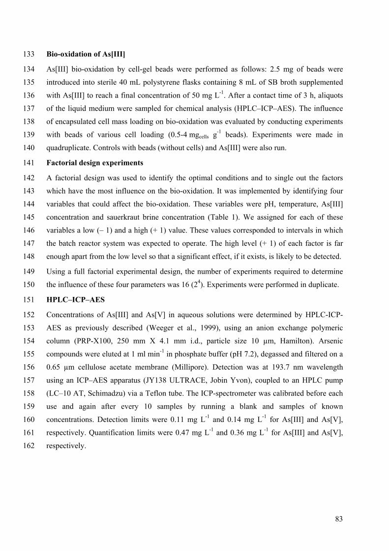
83
Bio-oxidation of As[III] 133
As[III] bio-oxidation by cell-gel beads were performed as follows: 2.5 mg of beads were 134
introduced into sterile 40 mL polystyrene flasks containing 8 mL of SB broth supplemented 135
with As[III] to reach a final concentration of 50 mg L-1. After a contact time of 3 h, aliquots 136
of the liquid medium were sampled for chemical analysis (HPLC–ICP–AES). The influence 137
of encapsulated cell mass loading on bio-oxidation was evaluated by conducting experiments 138
with beads of various cell loading (0.5-4 mgcells g-1 beads). Experiments were made in 139
quadruplicate. Controls with beads (without cells) and As[III] were also run. 140
Factorial design experiments 141
A factorial design was used to identify the optimal conditions and to single out the factors 142
which have the most influence on the bio-oxidation. It was implemented by identifying four 143
variables that could affect the bio-oxidation. These variables were pH, temperature, As[III] 144
concentration and sauerkraut brine concentration (Table 1). We assigned for each of these 145
variables a low (– 1) and a high (+ 1) value. These values corresponded to intervals in which 146
the batch reactor system was expected to operate. The high level (+ 1) of each factor is far 147
enough apart from the low level so that a significant effect, if it exists, is likely to be detected. 148
Using a full factorial experimental design, the number of experiments required to determine 149
the influence of these four parameters was 16 (24). Experiments were performed in duplicate. 150
HPLC–ICP–AES 151
Concentrations of As[III] and As[V] in aqueous solutions were determined by HPLC-ICP-152
AES as previously described (Weeger et al., 1999), using an anion exchange polymeric 153
column (PRP-X100, 250 mm X 4.1 mm i.d., particle size 10 µm, Hamilton). Arsenic 154
compounds were eluted at 1 ml min-1 in phosphate buffer (pH 7.2), degassed and filtered on a 155
0.65 µm cellulose acetate membrane (Millipore). Detection was at 193.7 nm wavelength 156
using an ICP–AES apparatus (JY138 ULTRACE, Jobin Yvon), coupled to an HPLC pump 157
(LC–10 AT, Schimadzu) via a Teflon tube. The ICP-spectrometer was calibrated before each 158
use and again after every 10 samples by running a blank and samples of known 159
concentrations. Detection limits were 0.11 mg L-1 and 0.14 mg L-1 for As[III] and As[V], 160
respectively. Quantification limits were 0.47 mg L-1 and 0.36 mg L-1 for As[III] and As[V], 161
respectively. 162
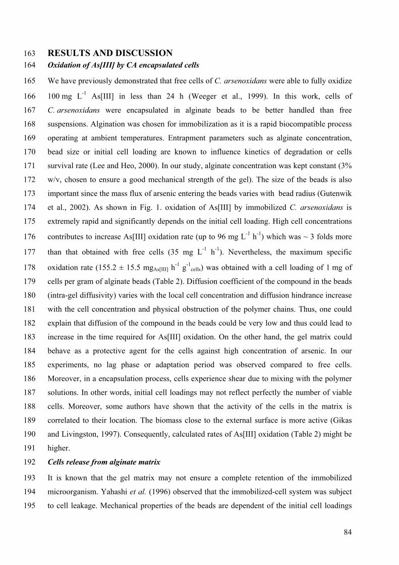
84
RESULTS AND DISCUSSION 163 Oxidation of As[III] by CA encapsulated cells 164
We have previously demonstrated that free cells of C. arsenoxidans were able to fully oxidize 165
100 mg L-1 As[III] in less than 24 h (Weeger et al., 1999). In this work, cells of 166
C. arsenoxidans were encapsulated in alginate beads to be better handled than free 167
suspensions. Algination was chosen for immobilization as it is a rapid biocompatible process 168
operating at ambient temperatures. Entrapment parameters such as alginate concentration, 169
bead size or initial cell loading are known to influence kinetics of degradation or cells 170
survival rate (Lee and Heo, 2000). In our study, alginate concentration was kept constant (3% 171
w/v, chosen to ensure a good mechanical strength of the gel). The size of the beads is also 172
important since the mass flux of arsenic entering the beads varies with bead radius (Gutenwik 173
et al., 2002). As shown in Fig. 1. oxidation of As[III] by immobilized C. arsenoxidans is 174
extremely rapid and significantly depends on the initial cell loading. High cell concentrations 175
contributes to increase As[III] oxidation rate (up to 96 mg L-1 h-1) which was ~ 3 folds more 176
than that obtained with free cells (35 mg L-1 h-1). Nevertheless, the maximum specific 177
oxidation rate (155.2 ± 15.5 mgAs[III] h-1 g-1cells) was obtained with a cell loading of 1 mg of 178
cells per gram of alginate beads (Table 2). Diffusion coefficient of the compound in the beads 179
(intra-gel diffusivity) varies with the local cell concentration and diffusion hindrance increase 180
with the cell concentration and physical obstruction of the polymer chains. Thus, one could 181
explain that diffusion of the compound in the beads could be very low and thus could lead to 182
increase in the time required for As[III] oxidation. On the other hand, the gel matrix could 183
behave as a protective agent for the cells against high concentration of arsenic. In our 184
experiments, no lag phase or adaptation period was observed compared to free cells. 185
Moreover, in a encapsulation process, cells experience shear due to mixing with the polymer 186
solutions. In other words, initial cell loadings may not reflect perfectly the number of viable 187
cells. Moreover, some authors have shown that the activity of the cells in the matrix is 188
correlated to their location. The biomass close to the external surface is more active (Gikas 189
and Livingston, 1997). Consequently, calculated rates of As[III] oxidation (Table 2) might be 190
higher. 191
Cells release from alginate matrix 192
It is known that the gel matrix may not ensure a complete retention of the immobilized 193
microorganism. Yahashi et al. (1996) observed that the immobilized-cell system was subject 194
to cell leakage. Mechanical properties of the beads are dependent of the initial cell loadings 195
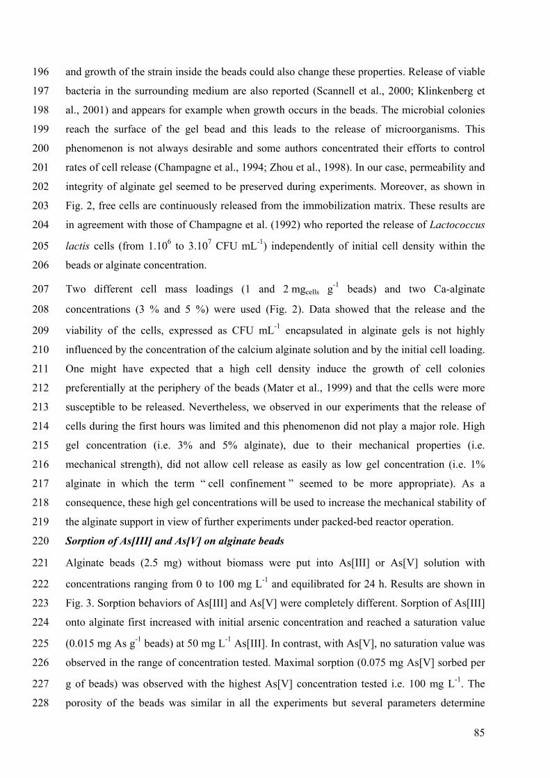
85
and growth of the strain inside the beads could also change these properties. Release of viable 196
bacteria in the surrounding medium are also reported (Scannell et al., 2000; Klinkenberg et 197
al., 2001) and appears for example when growth occurs in the beads. The microbial colonies 198
reach the surface of the gel bead and this leads to the release of microorganisms. This 199
phenomenon is not always desirable and some authors concentrated their efforts to control 200
rates of cell release (Champagne et al., 1994; Zhou et al., 1998). In our case, permeability and 201
integrity of alginate gel seemed to be preserved during experiments. Moreover, as shown in 202
Fig. 2, free cells are continuously released from the immobilization matrix. These results are 203
in agreement with those of Champagne et al. (1992) who reported the release of Lactococcus 204
lactis cells (from 1.106 to 3.107 CFU mL-1) independently of initial cell density within the 205
beads or alginate concentration. 206
Two different cell mass loadings (1 and 2 mgcells g-1 beads) and two Ca-alginate 207
concentrations (3 % and 5 %) were used (Fig. 2). Data showed that the release and the 208
viability of the cells, expressed as CFU mL-1 encapsulated in alginate gels is not highly 209
influenced by the concentration of the calcium alginate solution and by the initial cell loading. 210
One might have expected that a high cell density induce the growth of cell colonies 211
preferentially at the periphery of the beads (Mater et al., 1999) and that the cells were more 212
susceptible to be released. Nevertheless, we observed in our experiments that the release of 213
cells during the first hours was limited and this phenomenon did not play a major role. High 214
gel concentration (i.e. 3% and 5% alginate), due to their mechanical properties (i.e. 215
mechanical strength), did not allow cell release as easily as low gel concentration (i.e. 1% 216
alginate in which the term “ cell confinement ” seemed to be more appropriate). As a 217
consequence, these high gel concentrations will be used to increase the mechanical stability of 218
the alginate support in view of further experiments under packed-bed reactor operation. 219
Sorption of As[III] and As[V] on alginate beads 220
Alginate beads (2.5 mg) without biomass were put into As[III] or As[V] solution with 221
concentrations ranging from 0 to 100 mg L-1 and equilibrated for 24 h. Results are shown in 222
Fig. 3. Sorption behaviors of As[III] and As[V] were completely different. Sorption of As[III] 223
onto alginate first increased with initial arsenic concentration and reached a saturation value 224
(0.015 mg As g-1 beads) at 50 mg L-1 As[III]. In contrast, with As[V], no saturation value was 225
observed in the range of concentration tested. Maximal sorption (0.075 mg As[V] sorbed per 226
g of beads) was observed with the highest As[V] concentration tested i.e. 100 mg L-1. The 227
porosity of the beads was similar in all the experiments but several parameters determine 228
86
biosorption values viz. structural properties of the beads, initial concentration of metalloid 229
ions, existence of other competing ions (phosphate for example). As other biopolymers, 230
alginate which is an anionic sorbent has been shown to be effective at removing cationic 231
metals i.e. Pb2+, Zn2+, Cd2+, Ni2+, Cu2+ from solution. As[V] occurs at neutral pH (about 7) as 232
H2AsO4- and HAsO42- whereas As[III] occurs within the same pH range as the neutral species 233
H3AsO4. Calcium alginate beads showed no significant As[III] sorption in the whole range of 234
concentrations tested. This low As[III] accumulation is the result of passive diffusion of the 235
neutral species in the beads. The negatively charged As[V] species were better removed from 236
the solution but the extent of the sorption remained low (Fig. 3). Min and Hering (1998) 237
reported low As[V] sorption on Fe[III] doped alginate beads. 238
Evaluation of factors affecting As[III] oxidation 239
A 24 full factorial design (16 factor/level treatment combinations) was performed with the 240
values of the major variables shown in Table 1. Experimental conditions (column 2 to column 241
5) and results of the experiments (column 6) are shown in Table 3. The results are expressed 242
as the quantity of As[III] oxidized per gram of biomass (dry weight) per hour. Experimental 243
runs were randomly performed to eliminate any kind of extraneous effects tied with the 244
method used. 245
As could be determined from the results (Table 3), the ability of the strain to oxidize As[III] 246
showed wide variation. As[III] oxidation varied from 25 to 145 mg As[III] g-1 h-1. All of these 247
As[III] oxidation rates were very high. Nevertheless, factors affect the oxidation ability of the 248
strain in different ways. The optimal As[III] oxidation was found with the low values of pH 249
(7) and arsenic concentration (10 mg L-1) and the high values of temperature (25°C) and 250
sauerkraut brine (20%) added in the medium (Table 4). Nevertheless, data showed that 251
C. arsenoxidans had the ability to oxidize As[III] efficiently with each and every set of 252
parameters. In applied wastewater treatment systems, these characteristics are highly desirable 253
since conditions are continuously changing. Two variables were found to be statistically 254
significant at α = 0.05 : temperature (T) and the concentration of sauerkraut brine (Cs) in the 255
medium whereas at α = 0.01, only temperature (T) was statistically significant. A new matrix 256
was constructed with the two significant factors at α = 0.05 and results showed that the 257
highest As[III] oxidation rate was obtained when experiments were performed with a 258
temperature of 25°C and a concentration of 20% of sauerkraut brine in the culture medium 259
(Table 5). 260
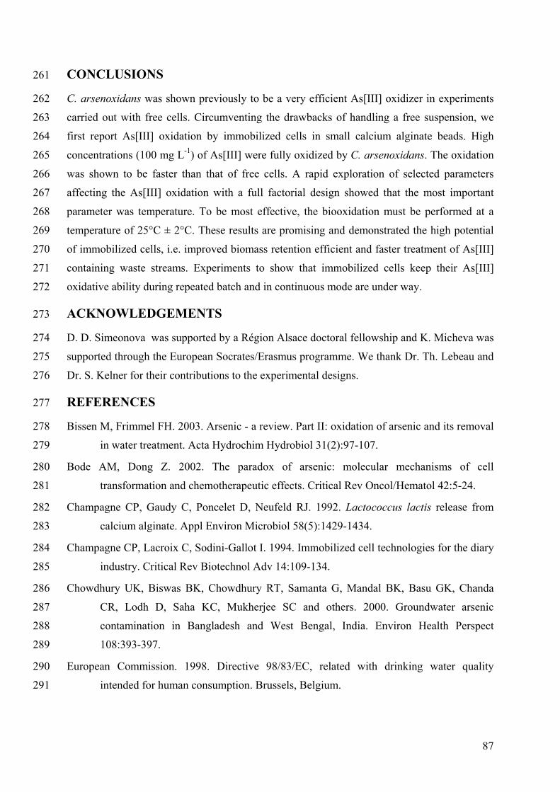
87
CONCLUSIONS 261
C. arsenoxidans was shown previously to be a very efficient As[III] oxidizer in experiments 262
carried out with free cells. Circumventing the drawbacks of handling a free suspension, we 263
first report As[III] oxidation by immobilized cells in small calcium alginate beads. High 264
concentrations (100 mg L-1) of As[III] were fully oxidized by C. arsenoxidans. The oxidation 265
was shown to be faster than that of free cells. A rapid exploration of selected parameters 266
affecting the As[III] oxidation with a full factorial design showed that the most important 267
parameter was temperature. To be most effective, the biooxidation must be performed at a 268
temperature of 25°C ± 2°C. These results are promising and demonstrated the high potential 269
of immobilized cells, i.e. improved biomass retention efficient and faster treatment of As[III] 270
containing waste streams. Experiments to show that immobilized cells keep their As[III] 271
oxidative ability during repeated batch and in continuous mode are under way. 272
ACKNOWLEDGEMENTS 273
D. D. Simeonova was supported by a Région Alsace doctoral fellowship and K. Micheva was 274
supported through the European Socrates/Erasmus programme. We thank Dr. Th. Lebeau and 275
Dr. S. Kelner for their contributions to the experimental designs. 276
REFERENCES 277
Bissen M, Frimmel FH. 2003. Arsenic - a review. Part II: oxidation of arsenic and its removal 278
in water treatment. Acta Hydrochim Hydrobiol 31(2):97-107. 279
Bode AM, Dong Z. 2002. The paradox of arsenic: molecular mechanisms of cell 280
transformation and chemotherapeutic effects. Critical Rev Oncol/Hematol 42:5-24. 281
Champagne CP, Gaudy C, Poncelet D, Neufeld RJ. 1992. Lactococcus lactis release from 282
calcium alginate. Appl Environ Microbiol 58(5):1429-1434. 283
Champagne CP, Lacroix C, Sodini-Gallot I. 1994. Immobilized cell technologies for the diary 284
industry. Critical Rev Biotechnol Adv 14:109-134. 285
Chowdhury UK, Biswas BK, Chowdhury RT, Samanta G, Mandal BK, Basu GK, Chanda 286
CR, Lodh D, Saha KC, Mukherjee SC and others. 2000. Groundwater arsenic 287
contamination in Bangladesh and West Bengal, India. Environ Health Perspect 288
108:393-397. 289
European Commission. 1998. Directive 98/83/EC, related with drinking water quality 290
intended for human consumption. Brussels, Belgium. 291
88
Gikas P, Livingston AG. 1993. Use of ATP to characterize biomass viability in freely 292
suspended and immobilized biomass bioreactors. Biotechnol Bioeng 42:1337-1351. 293
Gikas P, Livingston AG. 1997. Specific ATP and specific oxygen uptake rate in immobilized 294
cell aggregates: experimental results and theoretical analysis using a structured model 295
of immobilized cell growth. Biotechnol Bioeng 55:660-673. 296
Gutenwik J, Nilsson B, Axelsson A. 2002. Mass transfer effects on the reaction rate for 297
heterogeneously distributed immobilized yeast cells. Biotechnol Bioeng 79:664-673. 298
Klinkenberg G, Lystad KQ, Levine DW, Dyrset N. 2001. pH-controlled cell release and 299
biomass distribution of alginate-immobilized Lactococcus lactis subsp. lactis. J Appl 300
Microbiol 91:705-714. 301
Lee K-Y, Heo T-R. 2000. Survival of Bifidobacterium longum immobilized in calcium 302
alginate beads in simulated gastric juices and bile salt solution. Appl Environ 303
Microbiol 66(2):869-873. 304
Mater DDG, Saucedo JEN, Truffaut N, Barbotin JN, Thomas D. 1999. Conjugative plasmid 305
transfer between Pseudomonas strains within alginate bead microcosms: effect of the 306
Internal gel structure. Biotechnol Bioeng 65:34-43. 307
Min JH, Hering JG. 1998. Arsenate sorption by Fe(III)-doped alginate gels. Wat Res 308
32(5):1544-1552. 309
Muller D, Lièvremont D., Simeonova D. D., Hubert J.-C., Lett M-C. 2003. Arsenite oxidase 310
aox genes from a metal-resistant ß-Proteobacterium. J bacteriol 185:135-141. 311
O'Connor JT. 2002. Arsenic in drinking water. Part 3: occurrence of arsenic in U.S. waters. 312
Water Eng manage 5:45-48. 313
Scannell AGM, Hill C, Ross RP, Marx S, Hartmeier W, Arendt AK. 2000. Continuous 314
production of lacticin 3147 and nisin using cells immobilized in calcium alginate. J 315
Appl Microbiol 89:573-579. 316
Simeonova D. D, Lièvremont D., Groudeva V. I., Lett M-C. 2004. Sauerkraut brine and 317
whey – inexpensive by-products for biomass production of an arsenite-oxidizing 318
bacterium Cenibacterium arsenoxidans. Appl. Microbiol. Biotechnol. Submitted. 319
Slack JL, Rusiniak ME. 2000. Current issues in the management of acute promyelocytic 320
leukemia. Ann Hematol 79:227-238. 321
Smedley PL, Kinniburgh DG. 2002. A review of the source, behaviour and distribution of 322
arsenic in natural waters. Appl Geochem 17:517-568. 323

89
USEPA. 2001. National Primary Drinking Water Regulations; Arsenic and Clarifications to 324
Compliance and New Source Contaminants Monitoring; Final Rule. Federal Register 325
66(14):6976-7066. 326
Weeger W, Lièvremont D, Perret M, Lagarde F, Hubert J-C, Leroy M, Lett M-C. 1999. 327
Oxidation of arsenite to arsenate by a bacterium isolated from an aquatic environment. 328
Biometals 12:141-149. 329
WHO. 1996. Guidelines for drinking water quality. Health criteria and other supporting 330
information, 2nd ed., vol. 2. Geneva, Switzerland: World Health Organization. p 940-331
949. 332
Yahashi Y, Hatsu M, Horitsu H, Kawai K, Suzuki T, Takamizawa K. 1996. D-glucose 333
feeding for improvement of xylitol productivity from D-xylose using Candida 334
tropicalis immobilized on a non woven fabric. Biotechnol Lett 18:1395-1400. 335
Zhou Y, Martins E, Groboillot A, Champagne CP, Neufeld RJ. 1998. Spectrophotometric 336
quantification and control of cell release with chitosan coating. J Appl Microbiol 337
84:342-348. 338
90
FIGURES AND TABLES 339
340
341 Figure 1. Disappearance of As[III] vs time (open symbols) as a function of initial cell 342
loadings expressed as mg of cells (Cenibacterium arsenoxidans) per g of Ca-alginate beads. 343
Closed symbol represents the concentration of As[V] appearing in the medium for an initial 344
cell loading of 4 mgcells g-1. The As[V] curves corresponding to other cell loadings are not 345
shown. Data are mean of two independent experiments made in duplicate and errors bars 346
show the maximum deviation from the mean value (5 %). Initial As[III] concentration: 50 mg 347
L-1; volume of SB broth: 8 mL; beads mass loading: 2.5 mg; pH: 7.2; temperature: 25°C. 348
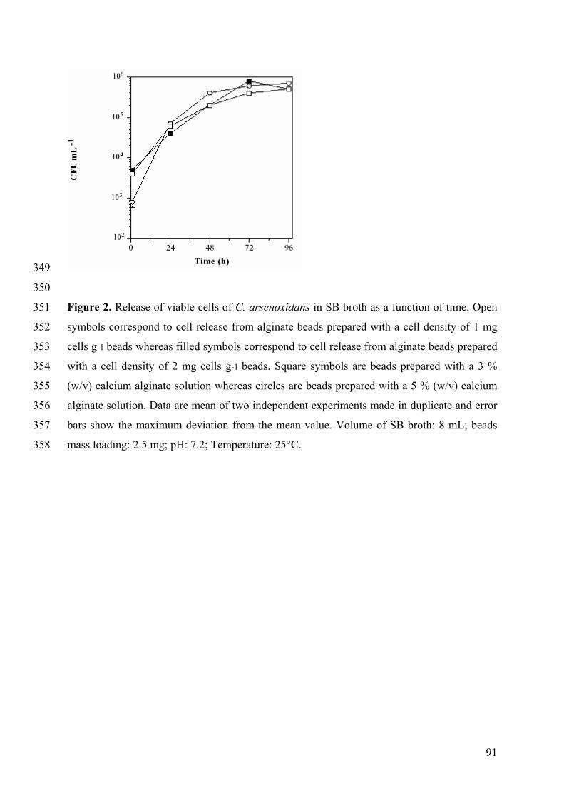
91
349
350
Figure 2. Release of viable cells of C. arsenoxidans in SB broth as a function of time. Open 351
symbols correspond to cell release from alginate beads prepared with a cell density of 1 mg 352
cells g-1 beads whereas filled symbols correspond to cell release from alginate beads prepared 353
with a cell density of 2 mg cells g-1 beads. Square symbols are beads prepared with a 3 % 354
(w/v) calcium alginate solution whereas circles are beads prepared with a 5 % (w/v) calcium 355
alginate solution. Data are mean of two independent experiments made in duplicate and error 356
bars show the maximum deviation from the mean value. Volume of SB broth: 8 mL; beads 357
mass loading: 2.5 mg; pH: 7.2; Temperature: 25°C. 358
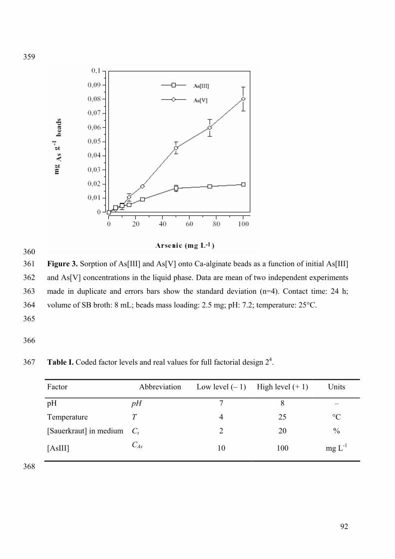
92
359
360 Figure 3. Sorption of As[III] and As[V] onto Ca-alginate beads as a function of initial As[III] 361
and As[V] concentrations in the liquid phase. Data are mean of two independent experiments 362
made in duplicate and errors bars show the standard deviation (n=4). Contact time: 24 h; 363
volume of SB broth: 8 mL; beads mass loading: 2.5 mg; pH: 7.2; temperature: 25°C. 364
365
366
Table I. Coded factor levels and real values for full factorial design 24. 367
Factor Abbreviation Low level (– 1) High level (+ 1) Units
pH pH 7 8 –
Temperature T 4 25 °C
[Sauerkraut] in medium Cs 2 20 %
[AsIII] CAs 10 100 mg L-1
368
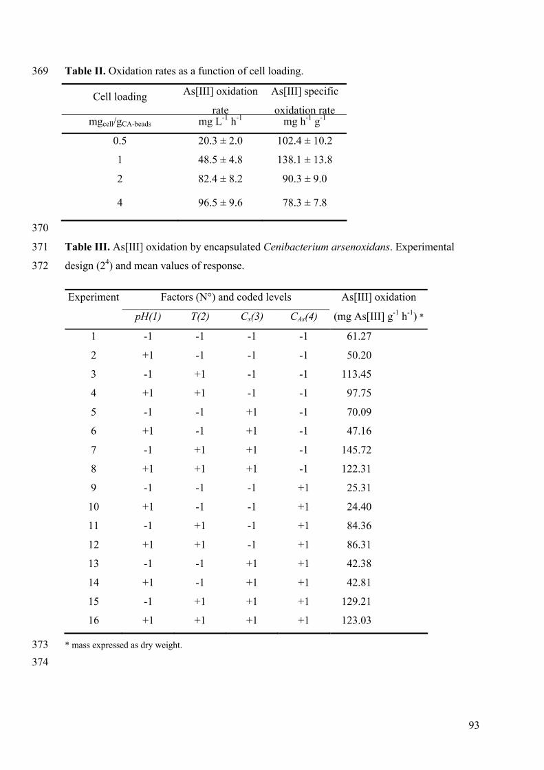
93
Table II. Oxidation rates as a function of cell loading. 369
Cell loading As[III] oxidation
rate
As[III] specific
oxidation ratemgcell/gCA-beads mg L-1 h-1 mg h-1 g-1
0.5 20.3 ± 2.0 102.4 ± 10.2
1 48.5 ± 4.8 138.1 ± 13.8
2 82.4 ± 8.2 90.3 ± 9.0
4 96.5 ± 9.6 78.3 ± 7.8
370
Table III. As[III] oxidation by encapsulated Cenibacterium arsenoxidans. Experimental 371
design (24) and mean values of response. 372
Experiment Factors (N°) and coded levels As[III] oxidation
pH(1) T(2) Cs(3) CAs(4) (mg As[III] g-1 h-1) *
1 -1 -1 -1 -1 61.27
2 +1 -1 -1 -1 50.20
3 -1 +1 -1 -1 113.45
4 +1 +1 -1 -1 97.75
5 -1 -1 +1 -1 70.09
6 +1 -1 +1 -1 47.16
7 -1 +1 +1 -1 145.72
8 +1 +1 +1 -1 122.31
9 -1 -1 -1 +1 25.31
10 +1 -1 -1 +1 24.40
11 -1 +1 -1 +1 84.36
12 +1 +1 -1 +1 86.31
13 -1 -1 +1 +1 42.38
14 +1 -1 +1 +1 42.81
15 -1 +1 +1 +1 129.21
16 +1 +1 +1 +1 123.03
* mass expressed as dry weight. 373 374
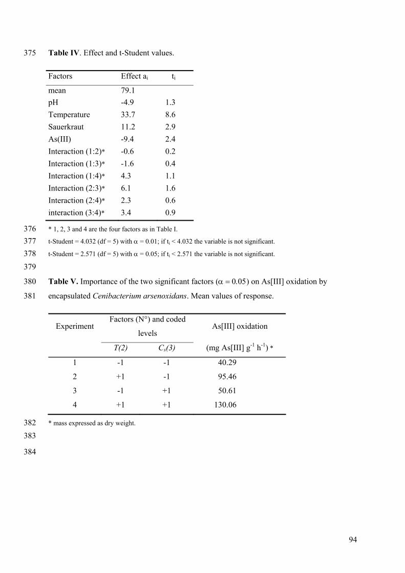
94
Table IV. Effect and t-Student values. 375
Factors Effect ai ti
mean 79.1 pH -4.9 1.3 Temperature 33.7 8.6 Sauerkraut 11.2 2.9 As(III) -9.4 2.4 Interaction (1:2)* -0.6 0.2 Interaction (1:3)* -1.6 0.4 Interaction (1:4)* 4.3 1.1 Interaction (2:3)* 6.1 1.6 Interaction (2:4)* 2.3 0.6 interaction (3:4)* 3.4 0.9
* 1, 2, 3 and 4 are the four factors as in Table I. 376 t-Student = 4.032 (df = 5) with α = 0.01; if ti < 4.032 the variable is not significant. 377 t-Student = 2.571 (df = 5) with α = 0.05; if ti < 2.571 the variable is not significant. 378 379
Table V. Importance of the two significant factors (α = 0.05) on As[III] oxidation by 380
encapsulated Cenibacterium arsenoxidans. Mean values of response. 381
Experiment Factors (N°) and coded
levels As[III] oxidation
T(2) Cs(3) (mg As[III] g-1 h-1) *
1 -1 -1 40.29
2 +1 -1 95.46
3 -1 +1 50.61
4 +1 +1 130.06
* mass expressed as dry weight. 382 383
384
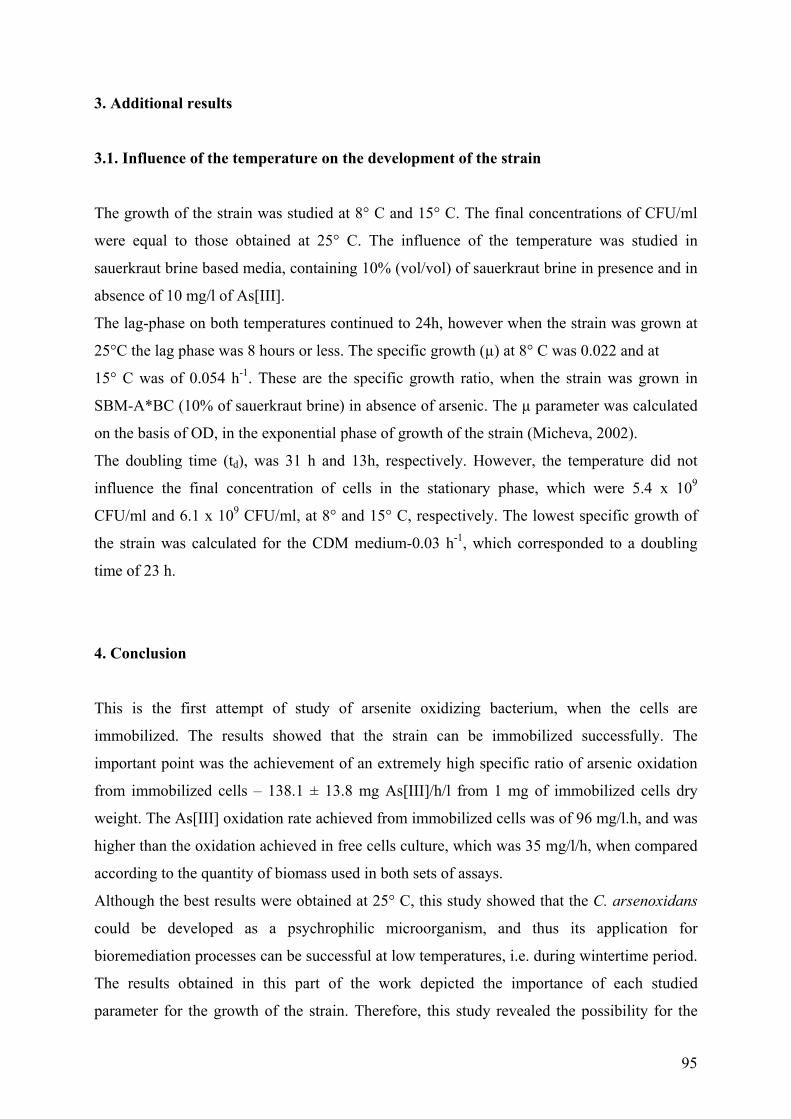
95
3. Additional results
3.1. Influence of the temperature on the development of the strain
The growth of the strain was studied at 8° C and 15° C. The final concentrations of CFU/ml
were equal to those obtained at 25° C. The influence of the temperature was studied in
sauerkraut brine based media, containing 10% (vol/vol) of sauerkraut brine in presence and in
absence of 10 mg/l of As[III].
The lag-phase on both temperatures continued to 24h, however when the strain was grown at
25°C the lag phase was 8 hours or less. The specific growth (µ) at 8° C was 0.022 and at
15° C was of 0.054 h-1. These are the specific growth ratio, when the strain was grown in
SBM-A*BC (10% of sauerkraut brine) in absence of arsenic. The µ parameter was calculated
on the basis of OD, in the exponential phase of growth of the strain (Micheva, 2002).
The doubling time (td), was 31 h and 13h, respectively. However, the temperature did not
influence the final concentration of cells in the stationary phase, which were 5.4 x 10⁹
CFU/ml and 6.1 x 10⁹ CFU/ml, at 8° and 15° C, respectively. The lowest specific growth of
the strain was calculated for the CDM medium-0.03 h-1, which corresponded to a doubling
time of 23 h.
4. Conclusion
This is the first attempt of study of arsenite oxidizing bacterium, when the cells are
immobilized. The results showed that the strain can be immobilized successfully. The
important point was the achievement of an extremely high specific ratio of arsenic oxidation
from immobilized cells – 138.1 ± 13.8 mg As[III]/h/l from 1 mg of immobilized cells dry
weight. The As[III] oxidation rate achieved from immobilized cells was of 96 mg/l.h, and was
higher than the oxidation achieved in free cells culture, which was 35 mg/l/h, when compared
according to the quantity of biomass used in both sets of assays.
Although the best results were obtained at 25° C, this study showed that the C. arsenoxidans
could be developed as a psychrophilic microorganism, and thus its application for
bioremediation processes can be successful at low temperatures, i.e. during wintertime period.
The results obtained in this part of the work depicted the importance of each studied
parameter for the growth of the strain. Therefore, this study revealed the possibility for the

96
application of the strain in a technology for preliminary oxidation of arsenic in water
effluents. It also increased the possibilities for the application of the strain using immobilized
cells.
97
Part III: Studies on the development of C. arsenoxidans in a simulated “open”
remediation system
1. Introduction
This part of the assays was devoted to the understanding of the development of the strain in
nonsterile sauerkraut brine medium. The aim was to establish whether the strain was capable
of growth in a complex system, including other randomly introduced microorganisms. In the
system bacteria and yeast were introduced into it from the air or water. The nutrient media
used in these assays were prepared as described in Materials and Methods Chapter.
The necessity of proving the development of the C. arsenoxidans in an “open” system arose.
Therefore, the strain was marked with GFP by using Mini-Tn5 delivery plasmid derivatives,
which are described in Materials and Methods Chapter. The GFP-tagged clones of C.
arsenoxidans, were subjected to two selections, according to the type of plasmid used and the
level of luminescence emitted. Thus, the expression of the GFP in the C. arsenoxidans clones
was defined. The clone with highest expression of gfp was chosen and this strain was used to
perform the further assays. These assays tracked the development of C. arsenoxidans during
the simulation of an “open” system. For these assays a SBM-A*BC medium supplemented
with arsenic, was used because this was the most favorable medium for the growth of the
strain.
The final set of tests differentiated the microorganisms originated from the sauerkraut brine
using the TTGE technique. This technique allowed the separation of bacteria from yeast as
well as distinguishing the approximate number of different species presented in the sauerkraut
brine. A combined protocol for total DNA extraction from bacterial and yeast cells was used.
The aim was the simultaneous DNA extraction from both types, when they are presented in
the same sample.
98
A. Construction and selection of gfp mutants of C. arsenoxidans
C. arsenoxidans strains were conjugated with three strains of E. coli each of them containing
a suicide plasmid of type R6K. Each of these plasmids contained a gfp-tagged transposon.
The gfp-tagged strains were then used to track the development of C. arsenoxidans in an
“open” system. The system, in which the arsenite-oxidizer C. arsenoxidans grew in the
presence of random microorganisms introduced into the nutrient medium from the air or the
water used, was considered as an “open” system.
The C. arsenoxidans strains were conjugated with E. coli strains, each one containing one of
the following transposons: mTn5gusA-pgfp21, mTn5gfp-pgusA, mTn5gfp-km. The
construction of gfp mutants of C. arsenoxidans was done by D. Muller and F. Den Hartoog.
For each type of transposon 192 clones were isolated. The clones were then subjected to two
selections in order to establish the clones with the highest level of gfp protein expression. The
first selection found clones emitting the most light. This selection was carried out after 48 h
growth of the clones in microplates containing 150 µl of liquid CDM. The preliminary
selection was done in duplicate for all strains. From the 192 clones containing the mTn5gfp-
pgusA only 3 colonies were selected for further study. From the clones containing mTn5gfp-
km 6 clones were selected for further studies and from the mutants containing the mTn5gusA-
pgfp21 5 clones were chosen with the highest fluorescence expression.
The second selection found the clone with the highest expression level and most stable
parameters of expression- both in liquid medium and on solid medium. Results from the
comparative selection of gfp clones in liquid CDM are presented in Fig. 1.
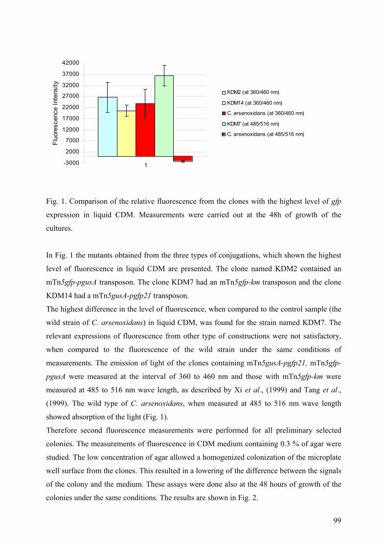
99
-3000
2000
7000
12000
17000
22000
27000
32000
37000
42000
1
Fluo
resc
ence
Inte
nsity
KDM2 (at 360/460 nm)
KDM14 (at 360/460 nm)
C. arsenoxidans (at 360/460 nm)
KDM7 (at 485/516 nm)
C. arsenoxidans (at 485/516 nm)
Fig. 1. Comparison of the relative fluorescence from the clones with the highest level of gfp
expression in liquid CDM. Measurements were carried out at the 48h of growth of the
cultures.
In Fig. 1 the mutants obtained from the three types of conjugations, which shown the highest
level of fluorescence in liquid CDM are presented. The clone named KDM2 contained an
mTn5gfp-pgusA transposon. The clone KDM7 had an mTn5gfp-km transposon and the clone
KDM14 had a mTn5gusA-pgfp21 transposon.
The highest difference in the level of fluorescence, when compared to the control sample (the
wild strain of C. arsenoxidans) in liquid CDM, was found for the strain named KDM7. The
relevant expressions of fluorescence from other type of constructions were not satisfactory,
when compared to the fluorescence of the wild strain under the same conditions of
measurements. The emission of light of the clones containing mTn5gusA-pgfp21, mTn5gfp-
pgusA were measured at the interval of 360 to 460 nm and those with mTn5gfp-km were
measured at 485 to 516 nm wave length, as described by Xi et al., (1999) and Tang et al.,
(1999). The wild type of C. arsenoxidans, when measured at 485 to 516 nm wave length
showed absorption of the light (Fig. 1).
Therefore second fluorescence measurements were performed for all preliminary selected
colonies. The measurements of fluorescence in CDM medium containing 0.3 % of agar were
studied. The low concentration of agar allowed a homogenized colonization of the microplate
well surface from the clones. This resulted in a lowering of the difference between the signals
of the colony and the medium. These assays were done also at the 48 hours of growth of the
colonies under the same conditions. The results are shown in Fig. 2.
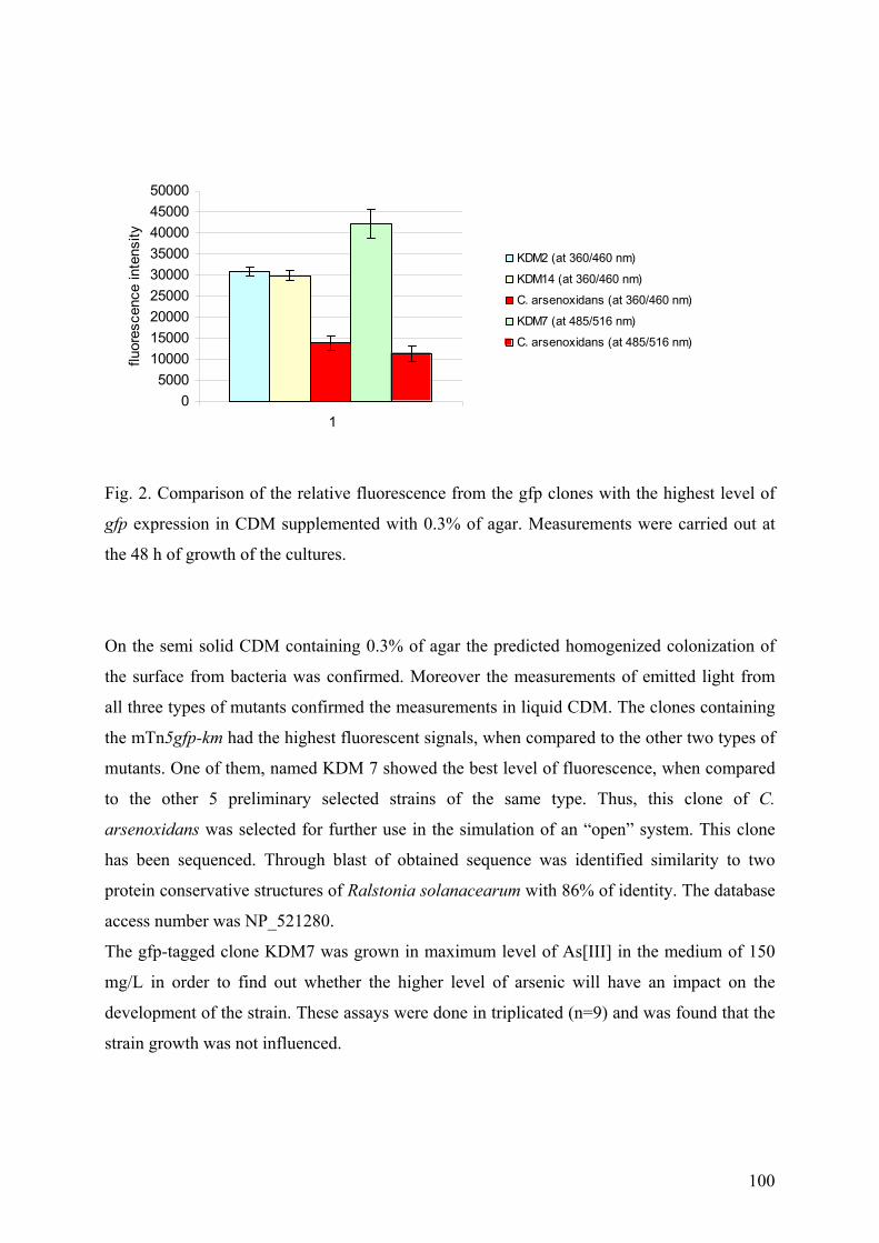
100
05000
100001500020000250003000035000400004500050000
1
fluor
esce
nce
inte
nsity
KDM2 (at 360/460 nm)
KDM14 (at 360/460 nm)
C. arsenoxidans (at 360/460 nm)
KDM7 (at 485/516 nm)
C. arsenoxidans (at 485/516 nm)
Fig. 2. Comparison of the relative fluorescence from the gfp clones with the highest level of
gfp expression in CDM supplemented with 0.3% of agar. Measurements were carried out at
the 48 h of growth of the cultures.
On the semi solid CDM containing 0.3% of agar the predicted homogenized colonization of
the surface from bacteria was confirmed. Moreover the measurements of emitted light from
all three types of mutants confirmed the measurements in liquid CDM. The clones containing
the mTn5gfp-km had the highest fluorescent signals, when compared to the other two types of
mutants. One of them, named KDM 7 showed the best level of fluorescence, when compared
to the other 5 preliminary selected strains of the same type. Thus, this clone of C.
arsenoxidans was selected for further use in the simulation of an “open” system. This clone
has been sequenced. Through blast of obtained sequence was identified similarity to two
protein conservative structures of Ralstonia solanacearum with 86% of identity. The database
access number was NP_521280.
The gfp-tagged clone KDM7 was grown in maximum level of As[III] in the medium of 150
mg/L in order to find out whether the higher level of arsenic will have an impact on the
development of the strain. These assays were done in triplicated (n=9) and was found that the
strain growth was not influenced.
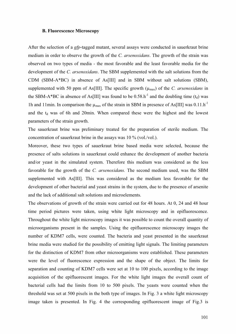
101
B. Fluorescence Microscopy
After the selection of a gfp-tagged mutant, several assays were conducted in sauerkraut brine
medium in order to observe the growth of the C. arsenoxidans. The growth of the strain was
observed on two types of media - the most favorable and the least favorable media for the
development of the C. arsenoxidans. The SBM supplemented with the salt solutions from the
CDM (SBM-A*BC) in absence of As[III] and in SBM without salt solutions (SBM),
supplemented with 50 ppm of As[III]. The specific growth (µmax) of the C. arsenoxidans in
the SBM-A*BC in absence of As[III] was found to be 0.58.h-1 and the doubling time (td) was
1h and 11min. In comparison the µmax of the strain in SBM in presence of As[III] was 0.11.h-1
and the td was of 6h and 20min. When compared these were the highest and the lowest
parameters of the strain growth.
The sauerkraut brine was preliminary treated for the preparation of sterile medium. The
concentration of sauerkraut brine in the assays was 10 % (vol./vol.).
Moreover, these two types of sauerkraut brine based media were selected, because the
presence of salts solutions in sauerkraut could enhance the development of another bacteria
and/or yeast in the simulated system. Therefore this medium was considered as the less
favorable for the growth of the C. arsenoxidans. The second medium used, was the SBM
supplemented with As[III]. This was considered as the medium less favorable for the
development of other bacterial and yeast strains in the system, due to the presence of arsenite
and the lack of additional salt solutions and microelements.
The observations of growth of the strain were carried out for 48 hours. At 0, 24 and 48 hour
time period pictures were taken, using white light microscopy and in epifluorescence.
Throughout the white light microscopy images it was possible to count the overall quantity of
microorganisms present in the samples. Using the epifluorescence microscopy images the
number of KDM7 cells, were counted. The bacteria and yeast presented in the sauerkraut
brine media were studied for the possibility of emitting light signals. The limiting parameters
for the distinction of KDM7 from other microorganisms were established. These parameters
were the level of fluorescence expression and the shape of the object. The limits for
separation and counting of KDM7 cells were set at 10 to 100 pixels, according to the image
acquisition of the epifluorescent images. For the white light images the overall count of
bacterial cells had the limits from 10 to 500 pixels. The yeasts were counted when the
threshold was set at 500 pixels in the both type of images. In Fig. 3 a white light microscopy
image taken is presented. In Fig. 4 the corresponding epifluorescent image of Fig.3 is
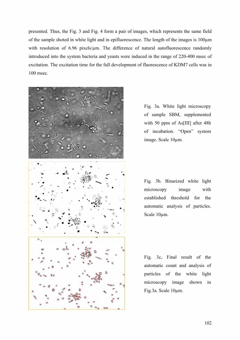
102
presented. Thus, the Fig. 3 and Fig. 4 form a pair of images, which represents the same field
of the sample shoted in white light and in epifluorescence. The length of the images is 100µm
with resolution of 6.96 pixels/µm. The difference of natural autofluorescence randomly
introduced into the system bacteria and yeasts were induced in the range of 220-400 msec of
excitation. The excitation time for the full development of fluorescence of KDM7 cells was in
100 msec.
Fig. 3a. White light microscopy
of sample SBM, supplemented
with 50 ppm of As[III] after 48h
of incubation. “Open” system
image. Scale 10µm.
Fig. 3b. Binarized white light
microscopy image with
established threshold for the
automatic analysis of particles.
Scale 10µm.
Fig. 3c. Final result of the
automatic count and analysis of
particles of the white light
microscopy image shown in
Fig.3a. Scale 10µm.
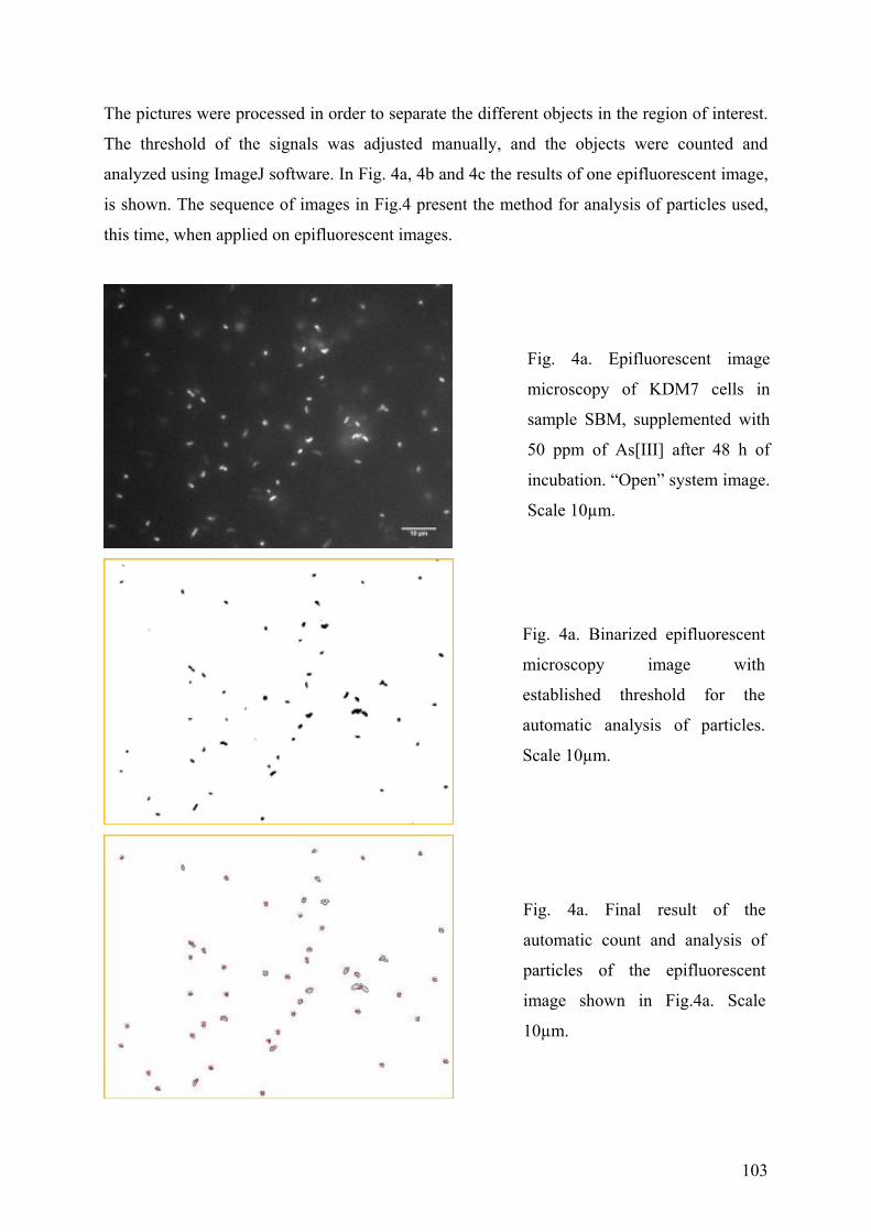
103
The pictures were processed in order to separate the different objects in the region of interest.
The threshold of the signals was adjusted manually, and the objects were counted and
analyzed using ImageJ software. In Fig. 4a, 4b and 4c the results of one epifluorescent image,
is shown. The sequence of images in Fig.4 present the method for analysis of particles used,
this time, when applied on epifluorescent images.
Fig. 4a. Epifluorescent image
microscopy of KDM7 cells in
sample SBM, supplemented with
50 ppm of As[III] after 48 h of
incubation. “Open” system image.
Scale 10µm.
Fig. 4a. Binarized epifluorescent
microscopy image with
established threshold for the
automatic analysis of particles.
Scale 10µm.
Fig. 4a. Final result of the
automatic count and analysis of
particles of the epifluorescent
image shown in Fig.4a. Scale
10µm.
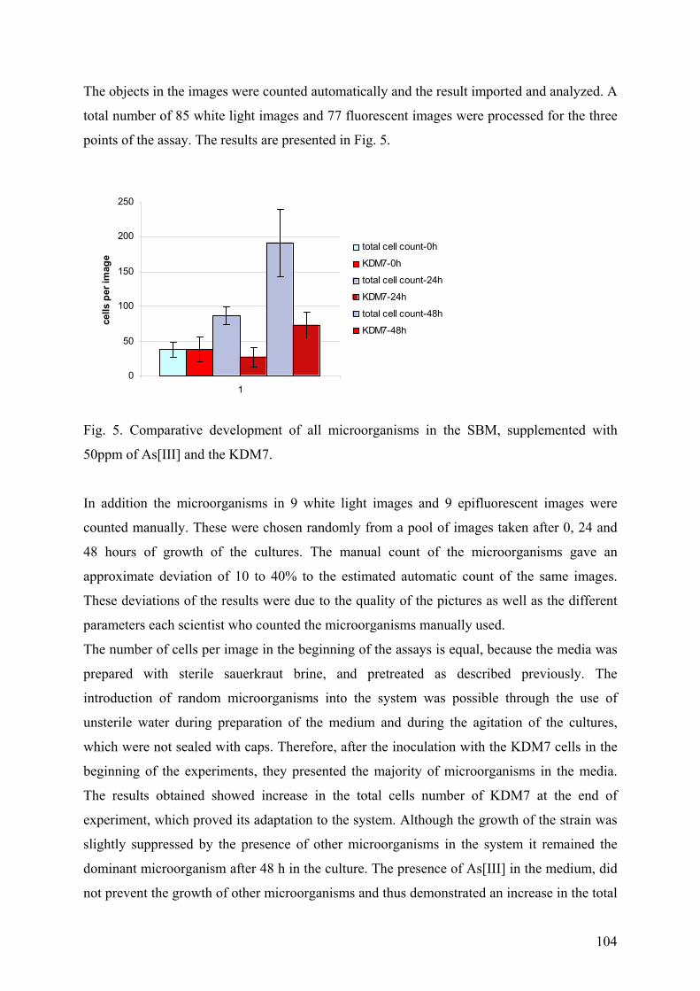
104
The objects in the images were counted automatically and the result imported and analyzed. A
total number of 85 white light images and 77 fluorescent images were processed for the three
points of the assay. The results are presented in Fig. 5.
0
50
100
150
200
250
1
cel
ls p
er im
age
total cell count-0h
KDM7-0h
total cell count-24h
KDM7-24h
total cell count-48h
KDM7-48h
Fig. 5. Comparative development of all microorganisms in the SBM, supplemented with
50ppm of As[III] and the KDM7.
In addition the microorganisms in 9 white light images and 9 epifluorescent images were
counted manually. These were chosen randomly from a pool of images taken after 0, 24 and
48 hours of growth of the cultures. The manual count of the microorganisms gave an
approximate deviation of 10 to 40% to the estimated automatic count of the same images.
These deviations of the results were due to the quality of the pictures as well as the different
parameters each scientist who counted the microorganisms manually used.
The number of cells per image in the beginning of the assays is equal, because the media was
prepared with sterile sauerkraut brine, and pretreated as described previously. The
introduction of random microorganisms into the system was possible through the use of
unsterile water during preparation of the medium and during the agitation of the cultures,
which were not sealed with caps. Therefore, after the inoculation with the KDM7 cells in the
beginning of the experiments, they presented the majority of microorganisms in the media.
The results obtained showed increase in the total cells number of KDM7 at the end of
experiment, which proved its adaptation to the system. Although the growth of the strain was
slightly suppressed by the presence of other microorganisms in the system it remained the
dominant microorganism after 48 h in the culture. The presence of As[III] in the medium, did
not prevent the growth of other microorganisms and thus demonstrated an increase in the total
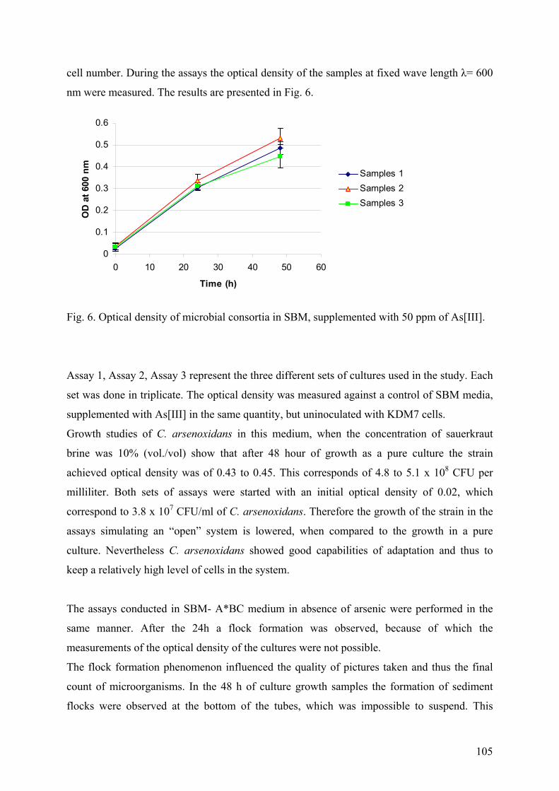
105
cell number. During the assays the optical density of the samples at fixed wave length λ= 600
nm were measured. The results are presented in Fig. 6.
0
0.1
0.2
0.3
0.4
0.5
0.6
0 10 20 30 40 50 60
Time (h)
OD
at 6
00 n
m
Samples 1Samples 2Samples 3
Fig. 6. Optical density of microbial consortia in SBM, supplemented with 50 ppm of As[III].
Assay 1, Assay 2, Assay 3 represent the three different sets of cultures used in the study. Each
set was done in triplicate. The optical density was measured against a control of SBM media,
supplemented with As[III] in the same quantity, but uninoculated with KDM7 cells.
Growth studies of C. arsenoxidans in this medium, when the concentration of sauerkraut
brine was 10% (vol./vol) show that after 48 hour of growth as a pure culture the strain
achieved optical density was of 0.43 to 0.45. This corresponds of 4.8 to 5.1 x 10⁸ CFU per
milliliter. Both sets of assays were started with an initial optical density of 0.02, which
correspond to 3.8 x 10⁷ CFU/ml of C. arsenoxidans. Therefore the growth of the strain in the
assays simulating an “open” system is lowered, when compared to the growth in a pure
culture. Nevertheless C. arsenoxidans showed good capabilities of adaptation and thus to
keep a relatively high level of cells in the system.
The assays conducted in SBM- A*BC medium in absence of arsenic were performed in the
same manner. After the 24h a flock formation was observed, because of which the
measurements of the optical density of the cultures were not possible.
The flock formation phenomenon influenced the quality of pictures taken and thus the final
count of microorganisms. In the 48 h of culture growth samples the formation of sediment
flocks were observed at the bottom of the tubes, which was impossible to suspend. This
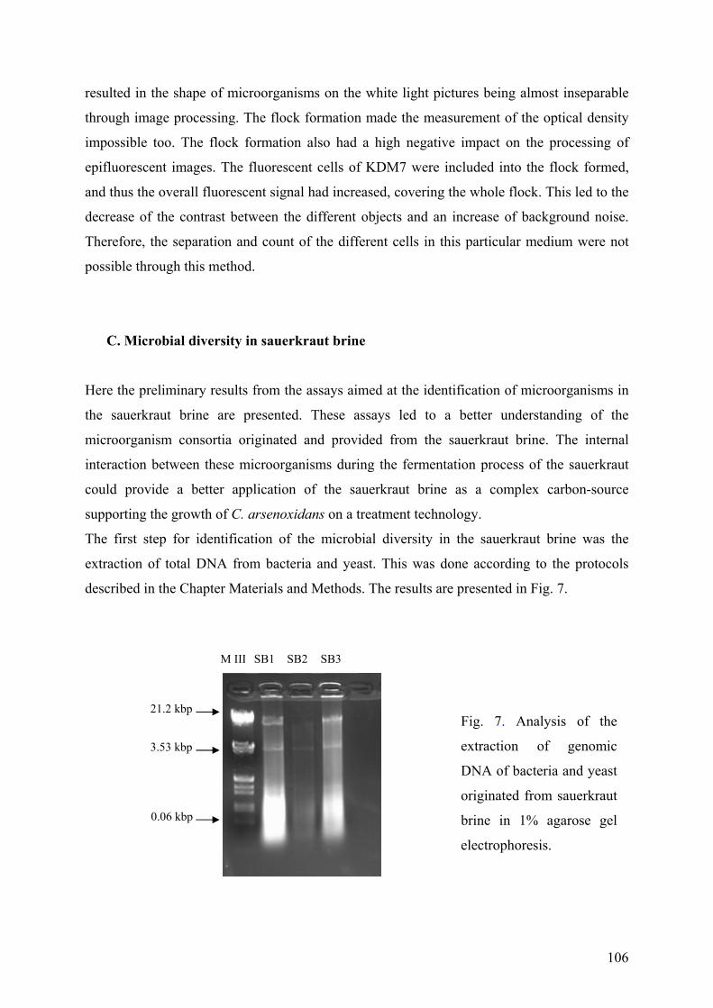
106
resulted in the shape of microorganisms on the white light pictures being almost inseparable
through image processing. The flock formation made the measurement of the optical density
impossible too. The flock formation also had a high negative impact on the processing of
epifluorescent images. The fluorescent cells of KDM7 were included into the flock formed,
and thus the overall fluorescent signal had increased, covering the whole flock. This led to the
decrease of the contrast between the different objects and an increase of background noise.
Therefore, the separation and count of the different cells in this particular medium were not
possible through this method.
C. Microbial diversity in sauerkraut brine
Here the preliminary results from the assays aimed at the identification of microorganisms in
the sauerkraut brine are presented. These assays led to a better understanding of the
microorganism consortia originated and provided from the sauerkraut brine. The internal
interaction between these microorganisms during the fermentation process of the sauerkraut
could provide a better application of the sauerkraut brine as a complex carbon-source
supporting the growth of C. arsenoxidans on a treatment technology.
The first step for identification of the microbial diversity in the sauerkraut brine was the
extraction of total DNA from bacteria and yeast. This was done according to the protocols
described in the Chapter Materials and Methods. The results are presented in Fig. 7.
M III SB1 SB2 SB3
3.53 kbp
0.06 kbp
21.2 kbp
Fig. 7. Analysis of the
extraction of genomic
DNA of bacteria and yeast
originated from sauerkraut
brine in 1% agarose gel
electrophoresis.
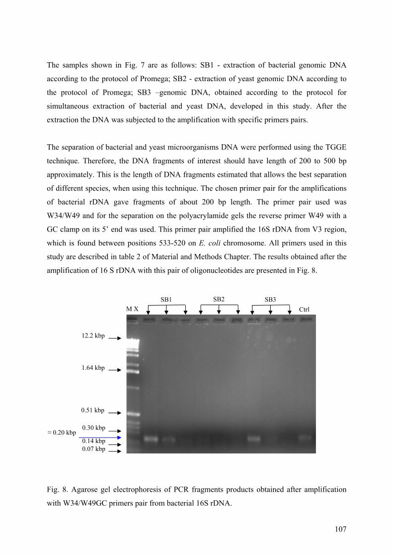
107
The samples shown in Fig. 7 are as follows: SB1 - extraction of bacterial genomic DNA
according to the protocol of Promega; SB2 - extraction of yeast genomic DNA according to
the protocol of Promega; SB3 –genomic DNA, obtained according to the protocol for
simultaneous extraction of bacterial and yeast DNA, developed in this study. After the
extraction the DNA was subjected to the amplification with specific primers pairs.
The separation of bacterial and yeast microorganisms DNA were performed using the TGGE
technique. Therefore, the DNA fragments of interest should have length of 200 to 500 bp
approximately. This is the length of DNA fragments estimated that allows the best separation
of different species, when using this technique. The chosen primer pair for the amplifications
of bacterial rDNA gave fragments of about 200 bp length. The primer pair used was
W34/W49 and for the separation on the polyacrylamide gels the reverse primer W49 with a
GC clamp on its 5’ end was used. This primer pair amplified the 16S rDNA from V3 region,
which is found between positions 533-520 on E. coli chromosome. All primers used in this
study are described in table 2 of Material and Methods Chapter. The results obtained after the
amplification of 16 S rDNA with this pair of oligonucleotides are presented in Fig. 8.
Fig. 8. Agarose gel electrophoresis of PCR fragments products obtained after amplification
with W34/W49GC primers pair from bacterial 16S rDNA.
M X Ctrl SB1 SB2 SB3
12.2 kbp
1.64 kbp
0.51 kbp
0.14 kbp 0.07 kbp
0.30 kbp ≈ 0.20 kbp
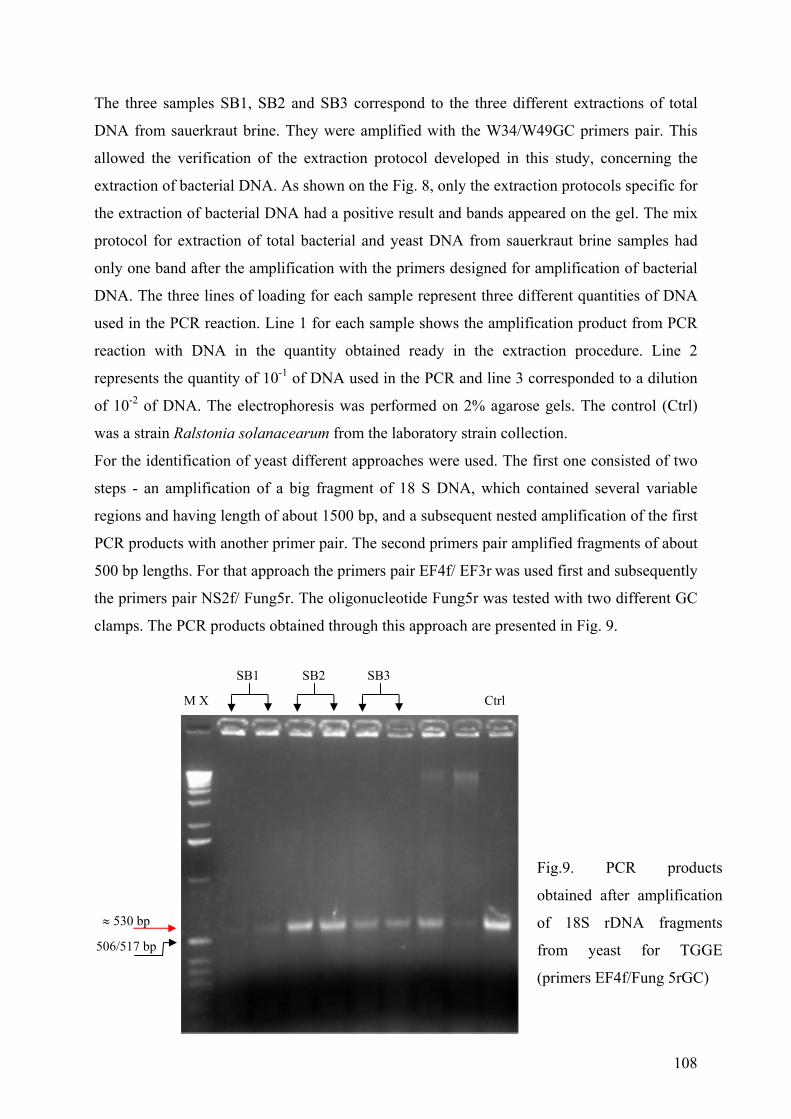
108
The three samples SB1, SB2 and SB3 correspond to the three different extractions of total
DNA from sauerkraut brine. They were amplified with the W34/W49GC primers pair. This
allowed the verification of the extraction protocol developed in this study, concerning the
extraction of bacterial DNA. As shown on the Fig. 8, only the extraction protocols specific for
the extraction of bacterial DNA had a positive result and bands appeared on the gel. The mix
protocol for extraction of total bacterial and yeast DNA from sauerkraut brine samples had
only one band after the amplification with the primers designed for amplification of bacterial
DNA. The three lines of loading for each sample represent three different quantities of DNA
used in the PCR reaction. Line 1 for each sample shows the amplification product from PCR
reaction with DNA in the quantity obtained ready in the extraction procedure. Line 2
represents the quantity of 10-1 of DNA used in the PCR and line 3 corresponded to a dilution
of 10-2 of DNA. The electrophoresis was performed on 2% agarose gels. The control (Ctrl)
was a strain Ralstonia solanacearum from the laboratory strain collection.
For the identification of yeast different approaches were used. The first one consisted of two
steps - an amplification of a big fragment of 18 S DNA, which contained several variable
regions and having length of about 1500 bp, and a subsequent nested amplification of the first
PCR products with another primer pair. The second primers pair amplified fragments of about
500 bp lengths. For that approach the primers pair EF4f/ EF3r was used first and subsequently
the primers pair NS2f/ Fung5r. The oligonucleotide Fung5r was tested with two different GC
clamps. The PCR products obtained through this approach are presented in Fig. 9.
M X
SB1 SB2 SB3
Ctrl
≈ 530 bp
506/517 bp
Fig.9. PCR products
obtained after amplification
of 18S rDNA fragments
from yeast for TGGE
(primers EF4f/Fung 5rGC)
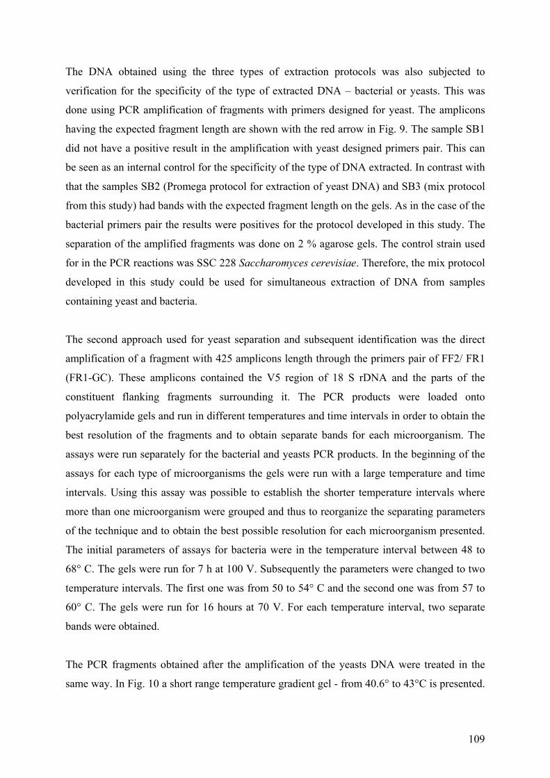
109
The DNA obtained using the three types of extraction protocols was also subjected to
verification for the specificity of the type of extracted DNA – bacterial or yeasts. This was
done using PCR amplification of fragments with primers designed for yeast. The amplicons
having the expected fragment length are shown with the red arrow in Fig. 9. The sample SB1
did not have a positive result in the amplification with yeast designed primers pair. This can
be seen as an internal control for the specificity of the type of DNA extracted. In contrast with
that the samples SB2 (Promega protocol for extraction of yeast DNA) and SB3 (mix protocol
from this study) had bands with the expected fragment length on the gels. As in the case of the
bacterial primers pair the results were positives for the protocol developed in this study. The
separation of the amplified fragments was done on 2 % agarose gels. The control strain used
for in the PCR reactions was SSC 228 Saccharomyces cerevisiae. Therefore, the mix protocol
developed in this study could be used for simultaneous extraction of DNA from samples
containing yeast and bacteria.
The second approach used for yeast separation and subsequent identification was the direct
amplification of a fragment with 425 amplicons length through the primers pair of FF2/ FR1
(FR1-GC). These amplicons contained the V5 region of 18 S rDNA and the parts of the
constituent flanking fragments surrounding it. The PCR products were loaded onto
polyacrylamide gels and run in different temperatures and time intervals in order to obtain the
best resolution of the fragments and to obtain separate bands for each microorganism. The
assays were run separately for the bacterial and yeasts PCR products. In the beginning of the
assays for each type of microorganisms the gels were run with a large temperature and time
intervals. Using this assay was possible to establish the shorter temperature intervals where
more than one microorganism were grouped and thus to reorganize the separating parameters
of the technique and to obtain the best possible resolution for each microorganism presented.
The initial parameters of assays for bacteria were in the temperature interval between 48 to
68° C. The gels were run for 7 h at 100 V. Subsequently the parameters were changed to two
temperature intervals. The first one was from 50 to 54° C and the second one was from 57 to
60° C. The gels were run for 16 hours at 70 V. For each temperature interval, two separate
bands were obtained.
The PCR fragments obtained after the amplification of the yeasts DNA were treated in the
same way. In Fig. 10 a short range temperature gradient gel - from 40.6° to 43°C is presented.
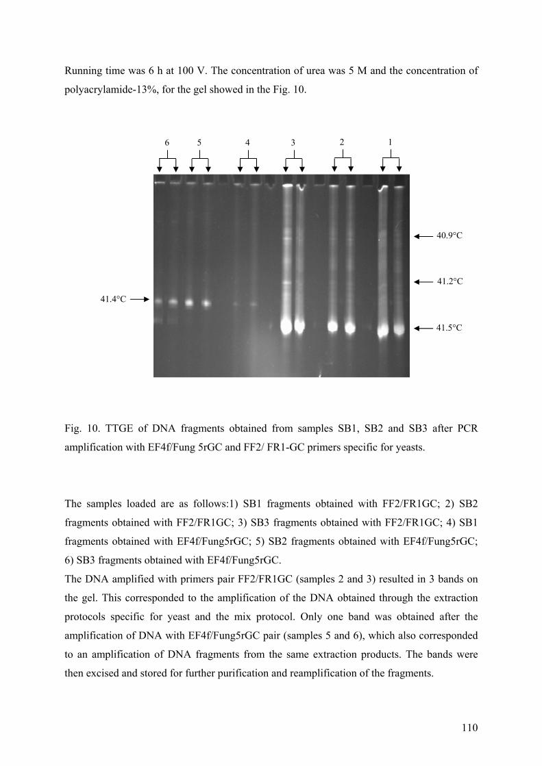
110
Running time was 6 h at 100 V. The concentration of urea was 5 M and the concentration of
polyacrylamide-13%, for the gel showed in the Fig. 10.
Fig. 10. TTGE of DNA fragments obtained from samples SB1, SB2 and SB3 after PCR
amplification with EF4f/Fung 5rGC and FF2/ FR1-GC primers specific for yeasts.
The samples loaded are as follows:1) SB1 fragments obtained with FF2/FR1GC; 2) SB2
fragments obtained with FF2/FR1GC; 3) SB3 fragments obtained with FF2/FR1GC; 4) SB1
fragments obtained with EF4f/Fung5rGC; 5) SB2 fragments obtained with EF4f/Fung5rGC;
6) SB3 fragments obtained with EF4f/Fung5rGC.
The DNA amplified with primers pair FF2/FR1GC (samples 2 and 3) resulted in 3 bands on
the gel. This corresponded to the amplification of the DNA obtained through the extraction
protocols specific for yeast and the mix protocol. Only one band was obtained after the
amplification of DNA with EF4f/Fung5rGC pair (samples 5 and 6), which also corresponded
to an amplification of DNA fragments from the same extraction products. The bands were
then excised and stored for further purification and reamplification of the fragments.
6 5 4 3 2 1
41.4°C
40.9°C
41.2°C
41.5°C
111
2. Conclusion
This part of the work led toward the definition of the possible interaction and development of
strain C. arsenoxidans in an “open” system. The first part of the studies simulated a real
“open” system, in which the development of gfp-tagged C. arsenoxidans strain was observed
through epifluorescent images, while the overall number of microorganisms in the same
system was evidenced through white light image microscopy. The main point of these
experiments was the tracking of C. arsenoxidans development, when other microorganisms
were presented in the system. Through this assays it was possible to establish the average
representation of C. arsenoxidans cells, when compared to the global development of random
microorganisms.
Two types of sauerkraut brine based medium were selected, because the presence of salts
solutions in sauerkraut did not improve the growth of C. arsenoxidans, but it may enhance the
development of other microorganisms in the system. The SBM-A*BC medium in absence of
arsenic was considered less favorable for the growth of the C. arsenoxidans, because of the
lack of arsenic, which can play the role of suppressor for the development of other
microorganisms in the medium. The second type medium-the SBM supplemented with
arsenic, was considered as the medium less favorable for the development of other bacterial
and yeast strains in the system.
The observations over the development of the random microorganisms and KDM7 cells in the
two media used resulted in the finding of flock formation in the SBM-A*BC medium and in
the absence of arsenic. This process was not observed in SBM supplemented with 50 mg/l of
As[III].
The interest of these assays is the possibility of distinction of an aimed microorganism having
special properties as a GFP, or stained with fluorescent dyes, from consortia of
microorganisms. Such results cannot be obtained through classical methods in microbiology.
The method of image cell counts can be influenced through the quality of the images.
Therefore, the most important parameter for the image processing is the quality of the images
captured.
The second part of assays aimed at the identification of the microorganisms originated from
the sauerkraut brine. These were the preliminary studies, which led toward to the
identification of the microorganisms originated from the sauerkraut brine. This information
112
could help the better application of the sauerkraut brine, after a simple neutralization as a
carbon-source supporting the growth of the arsenite-oxidizer C. arsenoxidans. During these
assays a new protocol for simultaneous extraction of rDNA from bacteria and yeast was
tested. This protocol had positive results when specific primers for amplification of bacterial
16 S or yeasts 18 S rDNA fragments, respectively were used. The number of fragments
obtained after amplification of DNA with primers having GC-clamp obtained through the
protocols described from Promega and the newly developed protocol were equal, when were
separated on polyacrylamide gels. The TGGE technique used allowed the separation of
bacteria and yeasts and resulted in the finding of 3 bacterial species and 4 yeast species. The
final identification of these microorganisms is still to be done.
The overall results showed that the sauerkraut brine can be used successfully as a nutrient
medium for the development of C. arsenoxidans, only after neutralization even when the
system is “open” for randomly introduced microorganisms from the environment. Thus, these
preliminary studies resulted in a high level growth performance of the C. arsenoxidans, when
the strain had to grow in presence of other microorganisms.
113
Part IV: Method for screening of arsenic-transforming bacteria
1. Introduction
The microplate screening assay was developed as a tool, to facilitate the screening of arsenic
transforming bacteria. This method provides easy evaluation of abilities of different bacterial
strains possessing oxidation or reduction ability toward arsenic species, prior to the complete
chemical analysis of each strain of interest.
All strains used in the development of this method were also subjected to chemical analysis.
Bacterial oxidation and reduction of arsenic As[III] and As[V] respectively in the culture
supernatants were tested using the HPLC-ICP-AES technique. An alternative application of
this method was developed for bacterial colonies plated in Petri dishes. The method was
successfully used when As[III] was added into CDM and the oxidation of the arsenic from the
arsenate-oxidizing strains were evidenced through a brown-red color formation.
Therefore the limits of the color formation were established using the chemical reactions
between either As[III] or As[V] and AgNO3 solution in Tris-HCl or Tris-maleate. The
intensity of the color was also established as a function of the As[V] and As[III] content. The
final stage of the assays was to standardize the colors of precipitates formed according to the
CIE system. Color measurements of the precipitates were made according to the tristimulus
coordinate system CIE L*a*b* scale (Commission Internationale de l'Eclairage, L*a*b* color
system) which expresses any color as three parameters: L* (lightness), a* and b*
(chromaticity coordinates). The L* reference value varies from 0 (black) to 100 (white), a*
values from –60 (green) to +60 (red), b* values from –60 (blue) to +60 (yellow). The decision
for the choice of the CIE system was evaluated after screening for different color systems.
The CIE system is the most abundant and well known color system and also serves as the
basis for some recently developed systems for color definition (http://hyperphysics.phy-
astr.gsu.edu/hbase/vision/cie.html).
114
2. The Article III
Diliana D. Simeonova, Didier Lièvremont, Florence Lagarde, Daniel A. E.
Muller, Veneta I. Groudeva and Marie-Claire Lett
Microplate screening assay for the detection of arsenite-oxidizing and arsenate-reducing bacteria
FEMS Microbiology Letters 237 (2004) 249–253
[signalement bibliographique ajouté par : ULP – SCD – Service des thèses électroniques]
Diliana D. Simeonova, Didier Lièvremont, Florence Lagarde, Daniel A. E. Muller,
Veneta I. Groudeva and Marie-Claire Lett
Microplate screening assay for the detection of arsenite-oxidizing and arsenate-
reducing bacteria.
FEMS Microbiology Letters 237 (2004) 249-253
Pages 249-253 :
La publication présentée ici dans la thèse est soumise à des droits détenus par un éditeur
commercial.
Pour les utilisateurs ULP, il est possible de consulter cette publication sur le site de
l'éditeur
http://dx.doi.org/10.1016/j.femsle.2004.06.040
Il est également possible de consulter la thèse sous sa forme papier ou d'en faire une
demande via le service de prêt entre bibliothèques (PEB), auprès du Service Commun
de Documentation de l'ULP: [email protected].
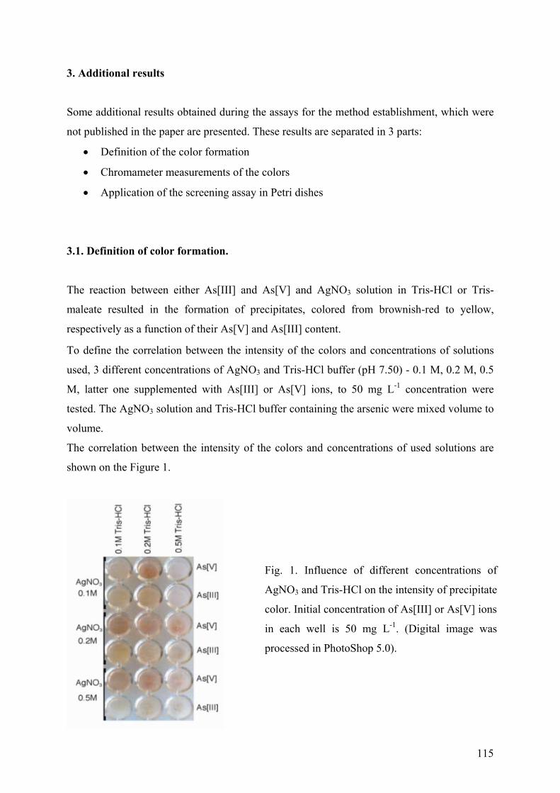
115
3. Additional results
Some additional results obtained during the assays for the method establishment, which were
not published in the paper are presented. These results are separated in 3 parts:
• Definition of the color formation
• Chromameter measurements of the colors
• Application of the screening assay in Petri dishes
3.1. Definition of color formation.
The reaction between either As[III] and As[V] and AgNO3 solution in Tris-HCl or Tris-
maleate resulted in the formation of precipitates, colored from brownish-red to yellow,
respectively as a function of their As[V] and As[III] content.
To define the correlation between the intensity of the colors and concentrations of solutions
used, 3 different concentrations of AgNO3 and Tris-HCl buffer (pH 7.50) - 0.1 M, 0.2 M, 0.5
M, latter one supplemented with As[III] or As[V] ions, to 50 mg L-1 concentration were
tested. The AgNO3 solution and Tris-HCl buffer containing the arsenic were mixed volume to
volume.
The correlation between the intensity of the colors and concentrations of used solutions are
shown on the Figure 1.
Fig. 1. Influence of different concentrations of
AgNO3 and Tris-HCl on the intensity of precipitate
color. Initial concentration of As[III] or As[V] ions
in each well is 50 mg L-1. (Digital image was
processed in PhotoShop 5.0).
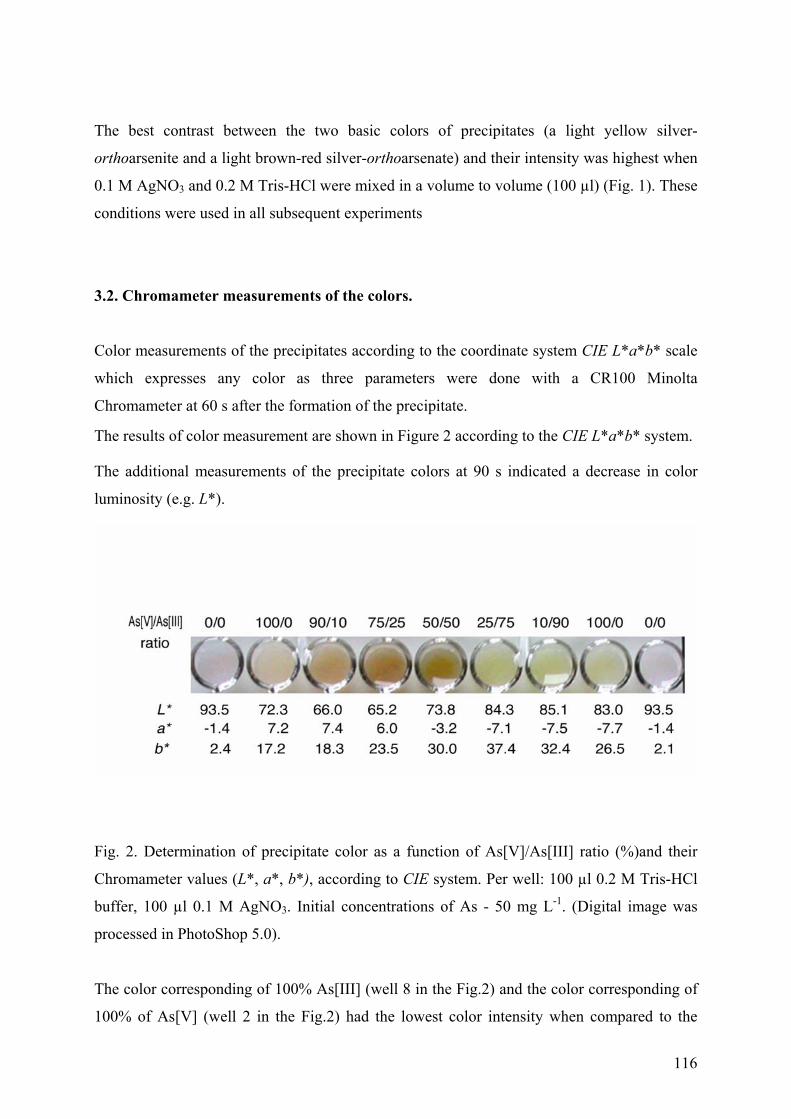
116
The best contrast between the two basic colors of precipitates (a light yellow silver-
orthoarsenite and a light brown-red silver-orthoarsenate) and their intensity was highest when
0.1 M AgNO3 and 0.2 M Tris-HCl were mixed in a volume to volume (100 µl) (Fig. 1). These
conditions were used in all subsequent experiments
3.2. Chromameter measurements of the colors.
Color measurements of the precipitates according to the coordinate system CIE L*a*b* scale
which expresses any color as three parameters were done with a CR100 Minolta
Chromameter at 60 s after the formation of the precipitate.
The results of color measurement are shown in Figure 2 according to the CIE L*a*b* system.
The additional measurements of the precipitate colors at 90 s indicated a decrease in color
luminosity (e.g. L*).
Fig. 2. Determination of precipitate color as a function of As[V]/As[III] ratio (%)and their
Chromameter values (L*, a*, b*), according to CIE system. Per well: 100 µl 0.2 M Tris-HCl
buffer, 100 µl 0.1 M AgNO3. Initial concentrations of As - 50 mg L-1. (Digital image was
processed in PhotoShop 5.0).
The color corresponding of 100% As[III] (well 8 in the Fig.2) and the color corresponding of
100% of As[V] (well 2 in the Fig.2) had the lowest color intensity when compared to the
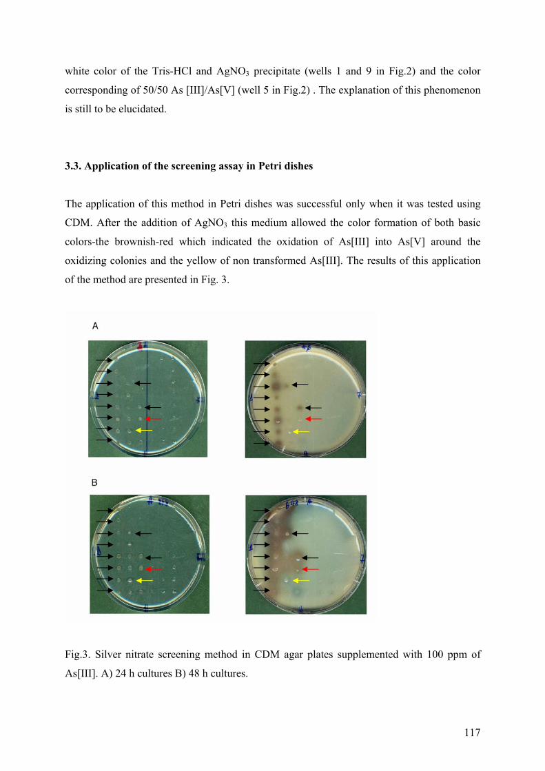
117
white color of the Tris-HCl and AgNO3 precipitate (wells 1 and 9 in Fig.2) and the color
corresponding of 50/50 As [III]/As[V] (well 5 in Fig.2) . The explanation of this phenomenon
is still to be elucidated.
3.3. Application of the screening assay in Petri dishes
The application of this method in Petri dishes was successful only when it was tested using
CDM. After the addition of AgNO3 this medium allowed the color formation of both basic
colors-the brownish-red which indicated the oxidation of As[III] into As[V] around the
oxidizing colonies and the yellow of non transformed As[III]. The results of this application
of the method are presented in Fig. 3.
Fig.3. Silver nitrate screening method in CDM agar plates supplemented with 100 ppm of
As[III]. A) 24 h cultures B) 48 h cultures.
118
The bacterial strains shown in Fig. 3 are from the culture collection of arsenite-oxidizing and
arsenate-reducing bacteria in the laboratory. On Figure 3 two Petri dishes containing the same
strains are presented. The Petri dishes in Fig. 3A were cultivated 24h prior the addition of
AgNO3 solution. The Petri dishes on Fig. 3B were cultivated for 48h prior the addition of
AgNO3 solution. The black arrows point out the strains which had arsenite – oxidizing ability.
As can be seen in Fig. 3 the colonies had developed a brown color after supplementation of
AgNO3 solution. These strains showed arsenite – oxidizing ability after 24 h. The red arrow
points out a colony, which showed an arsenite – oxidizing ability after 48 h of growth and the
arrow in yellow points out a strain, which have no arsenite – oxidation ability, but high
resistance toward the As[III]. The CDM was supplemented with 100 ppm of As[III].
4. Conclusion
Best color formation was observed when the following concentrations of silver nitrate and
Tris-HCl were used: 1) 0.1 M Tris-HCl and 0.2 M silver nitrate solution, and 2) 0.2 M Tris-
HCl and 0.1 M silver nitrate. The lower limits of color formation as a function of pH of the
buffer were: pH 5.8 for As[V] and pH 6.0 for As[III], in Tris-maleate buffer. The highest
level of color formation tested was at pH 8.4 for both types of arsenic species.
To extend the possibilities of the application of this assay, further studies of color formation
in acid environment could be developed. Duquesne, (2004) successfully used the color
formation between the silver nitrate and As[III] and As[V] in an acid pH range. This was
carried out in the specific nutrient medium, used for the growth of the bacterial strains
studied. The reported colors were: gray for the As[III] species and yellow-orange for As[V]
species, but still the method has not been standardized in acid pH levels.
Chromameter measurements were done in order to standardize the colors and therefore to
diminishing the personal appreciation of the precipitate colors as much as possible. Further
development of the assay toward automatic measurements of colors is also possible. In this
way the possible misreading of the colors formed, will have less influence upon the final
results. Thus, such kind of standardization or automatic measurements of the colors, can be
developed in a future studies. Nevertheless, the assay in its present form has the advantages of
rapid and easily performed screening for arsenic-transforming bacterial strains.
119
CHAPTER IV
MATERIALS AND METHODS
120
1. Bacterial strains
The strains from the bacterial strain collection of the Laboratoire de Microbiologie et de
Génétique (group of Professor Lett) and the newly isolated strains of the laboratory of
General and Industrial Microbiology (group of Assoc. Professor Groudeva), which were used
in this study are presented in Table 1.
Table. 1.
Strain Sample Reference Identification (16S rDNA)
S1, S8,
S11-S16
Soil sample 1 (sewage sludge),
Salsigne, France
J.-B. Bucher,
(1999)
Unidentified strains
S2, S3 idem Same work Pseudomonas sp. Q37-87
S4-S6,
S9, S10
idem Same work P. stutzeri
S7 idem Same work Acinetobacter calcoaceticus
S73,S83 idem Delbecque Unidentified strains
S82 idem Same work P. stutzeri
S84 idem Same work Pseudomonas sp. 5A
S17-S19 Soil sample 2 (gypsum,
containing arsenic), Salsigne,
France
J.-B. Bucher,
1999
P. stutzeri
S20 idem Same work Unidentified strain
S21 Soil sample 3 (Calcium oxide),
Salsigne, France
Same work Unidentified strain
S22 idem Same work R. solanacearum
S76 idem A. Delbecque Unidentified strain
S24 Soil sample 4 (Champ Magné),
Salsigne, France
J.-B. Bucher,
1999
P. fluorescens
S25 idem Same work P. corrugata
121
Strain Sample Reference Identification (16S rDNA)
S26 idem Same work Pseudomonas sp.
S27 idem Same work P. stutzeri
S28, S33 idem Same work Acinetobacter calcoaceticus
S31 idem Same work Nocardioides simplex
S77-S79,
S87
idem A. Delbecque P. stutzeri
S88 idem Same work Unidentified strain
S34 Soil sample 5 (concentrated
agglomerations), Salsigne,
France
J.-B. Bucher,
1999
P. tolaasii
S35 idem Same work Pseudomonas sp
S36 Soil sample 6 (soil under
monitoring), Salsigne, France
Same work P. stutzeri
S38 idem Same work Pseudomonas sp.
S39 idem Same work P. corrugata
S40-S43 idem Same work Unidentified strains
S44 idem Same work Pseudomonas sp. Q37-87
S80 idem A. Delbecque P. fluorescens
S81, S89 idem Same work Unidentified strain
S47, S48 Soil sample 7 (pile of wastes,
left from cyanic treatment),
Salsigne, France
J.-B. Bucher,
1999
Unidentified strains
S55, S56
S58-S60
Soil sample 8 (soil from
Rouyre), Salsigne, France
Same work Unidentified strains
S57 idem Same work Acinetobacter calcoaceticus
S64 Soil sample 9 (Château le
Caunette soil)
Same work Pseudomonas sp. Q37-87
122
Strain Sample Reference Identification (16S rDNA)
SM1
SM2
Soil sample As75 from mining
site Astano, Swiss.
This study Ralstonia eutropha kT1
SM3,
SM4
idem This study Ralstonia eutropha
SM5 Soil sample As81, from mining
site Astano, Swiss
This study P. putida
SM7 idem This study P. orysihabitans
SM9 idem This study Comamonas testosteroni
SM11 idem This study P. stutzeri
SM12
SM23
idem This study Alcaligenes sp.
SM22 Soil sample (place ”C”) from a
desolated agricultural site, next
to the smelt factory KZM
Plovdiv area, Bulgaria
This study Stenotrophomonas maltophilia
SM26 Soil sample (place “A”) at
10cm distance from the iron
fence of KZM Plovdiv factory,
Bulgaria
This study P. putida
SM27 Soil sample (place “B”), next to
the canal for evacuation of
wastewater out off KZM
Plovdiv, Bulgaria
This study P. fluorescens
ULPAs1 Sample of water purification
plant in Germany
Weeger, 1999 Cenibacterium arsenoxidans
Table.1. Name and origin of bacterial strains used.
123
2. Plasmids
All the three plasmids used for the transformation of ULPAs1 were from the type R6K
(suicidal).These plasmids were gfp-tagged ones.
mTn5gusA-pgfp21 - from Faculty of Agricultural and Applied Biological Sciences,
Katholieke Universitait Leuven, Belgium (Xi et al., 1999)
mTn5gfp-pgusA – kindly supplied by the author from Faculty of Agricultural and Applied
Biological Sciences, Katholieke Universitait Leuven, Belgium (Xi et al., 1999)
mTn5gfp-km- kindly supplied by the author (Tang et al., 1999)
3. Nutrient Media
3.1. Chemically defined medium (CDM)
Stock solutions
Solution A : Solution B : Solution C :
Na-lactate: 50 g FeSO4 : 160 mg NaHCO3 : 4 g
MgSO4. 7H2O : 20 g 100 mL deionazed H2O 50 mL deionazed H2O.
NH4Cl : 10 g
Na2SO4 :10 g
K2HPO4 : 100 mg
CaCl2. 2H2O : 672 mg
Solution A - sterilized by autoclave for 15 min at 120°C;
Solutions B and C - sterilized by filtration through membrane filter of 0,22 µm pore-size,
Nalgène.
Preparation of the medium: 1 liter of the medium was prepared from
100 mL of Solution A;
2,5 mL of Solution B;
10 mL of Solution C
Made up to 1L with sterile double deionazed H2O. Final pH of this medium - 7.2
124
3.2. Solid CDM
The preparation of the solid CDM was the same, as CDM broth except that the necessary
quantity of agar to obtain a final concentration of 20 g.L-1, was added to Solution 1 before
autoclaving.
3.3. Sauerkraut brine medium (SBM)
The sauerkraut brine was kindly supplied by the producer Coopchou, Vendenheim (France)
and had a natural pH of 3.5 - 3.7.
The preliminary treatment of sauerkraut brine for the preparation of nutrient media was, as
follows:
filtration through a paper filter, centrifugation at 6000 rpm for 20 min, heating twice at 90°C
in a water bath - for 15 min and allowed to settle for 24h at 4°C. A second filtration through a
paper filter, dilution 1:5 (vol./vol.) with sterile ddH2O and final filtration through 0.20µm
pore-size membrane (Nalgene, France), were performed.
The Sauerkraut brine medium was prepared in two variants:
• with salts solutions from CDM, except Na-lactate.
• without salts solutions from CDM
The final pH of SBM was adjusted to 7.2, with 1 M NaOH.
125
3.4. Whey medium (WM)
The whey based medium used commercially available products from the french cheese
industry (fromage blanc Rians and Auchan). The initial whey pH was 4.0 - 4.3.
The preliminary treatment of the whey for the preparation of nutrition medium followed the
protocol described below:
Filtration through paper filter, dilution with double deionised water 1:1,5 (vol./vol.),
neutralization with 1M NaOH up to pH 7.2, a second filtration through paper filter, followed
by sterile filtration through 0.20µm pore-size membrane.
The whey medium was prepared in two variants:
• with salts solutions from CDM, except Na-lactate.
• without salts solutions from CDM
3.5. Molasses medium
The molasses concentrate was supplied by Les Sucreries et Raffineries d'Erstein, France
(http://www.sucre-erstein.com/cadres/presentation.html). The used medium contained the
same salts solutions as CDM (in the same concentrations), except that molasses replaced the
Na-lactate as a sole carbon source.
A stock solution of molasses with 20g.L-1 initial concentration was prepared and sterilized
through filtration. The final medium was prepared by the addition of the molasses into the salt
solution.
3.6. Nutrient Medium “DV”
This medium is the Desulfovibrio medium described in Handbook of Microbilogical Media
by Ronald M. Atlas (1997).
126
3.7.Nutrient Medium “Dc&Ds”
This medium is based on the DSMZ media 197 (http:// dsmz.de/media/med197.htm) and 198
(http:// dsmz.de/media/med198.htm). The preparation and the use of the medium was under
aerobic conditions.
3.8. Nutrient Medium “Dtm”
This medium is based on the DSMZ medium 682, which consisted of DSMZ medium 197,
with lowered concentration of sodium sulfate to 0,7 g/l and “seven vitamins solutions” of 503
DSMZ medium. In “Dtm” nutrient medium the 3,4,5-trimetoxabenzoate as a carbon source in
medium 682, was replaced with Na-lactate (2 g/l). The preparation of the nutrient medium
was under aerobic conditions (http:// dsmz.de/media/med682.htm).
3.9. Nutrient Medium “DvB”
Solutions
Solution A: Solution B: Solution D :
Na-lactate: 3.5 g Na-thioglicolate: 0.1 g FeSO4.7H2O
MgSO4, 7H2O: 2.0 g 10 ml ddH2O 10 ml ddH2O
NH4Cl: 1.0 g
KH2PO4: 0.5 g Solution C:
CaSO4: 1.0 g Ascorbic acid: 0.1 g
Yeast extract: 1.0 g 10 ml ddH2O
970 ml ddH2O
127
The medium is based on the DSMZ medium 63, which was slightly modified.
Preparation of the medium: All solutions were autoclaved at 121° C for 15 min. Solutions B,
C and D were added into solution A, afterward. The pH 7.5 of the medium was adjusted with
NaOH. During the preparation, the medium was shaken continuously to keep the formed gray
precipitate suspended (http:// dsmz.de/media/med063.htm).
3.10. Nutrient Medium “BDtm”
Stock solutions
Solution A : Solution B :
Na-lactate: 35 g (NH4)Fe(SO4)2.6H2O: 5 g
MgSO4, 7H2O : 20 g 100 mL deionazed H2O
NH4Cl : 10 g
KH2PO4 : 5 g
CaSO4.2H2O : 1 g
Solution A, was prepared in 1L. Both solutions were sterilized by autoclave at 121°C for 20
min.
Preparation of the medium: 1 liter of the medium was prepared from
100 mL of Solution A;
10 mL of Solution B;
Made up to 1L with sterile double deionazed H2O.
128
4. Molecular biology techniques
4.1. Extraction of total DNA
The extractions of genomic bacterial DNA from pure cultures were done according to the
protocol of Promega, with the kit supplied by the producer Promega Corporation, USA.
The extractions of genomic DNA from yeast were according to the protocol of Promega. The
enzyme lyticase was substituted with the enzyme zymolase with concentration of 5 mg/ml.
A protocol for simultaneous extraction of bacterial and yeast genomic DNA, based on the
Promega’s protocols, was developed and tested. In this protocol the enzymes lysosim with
10mg/ml concentration and zymolase with concentration of 5 mg/ml were used. The first
incubation at 37°C was fixed at 30min.
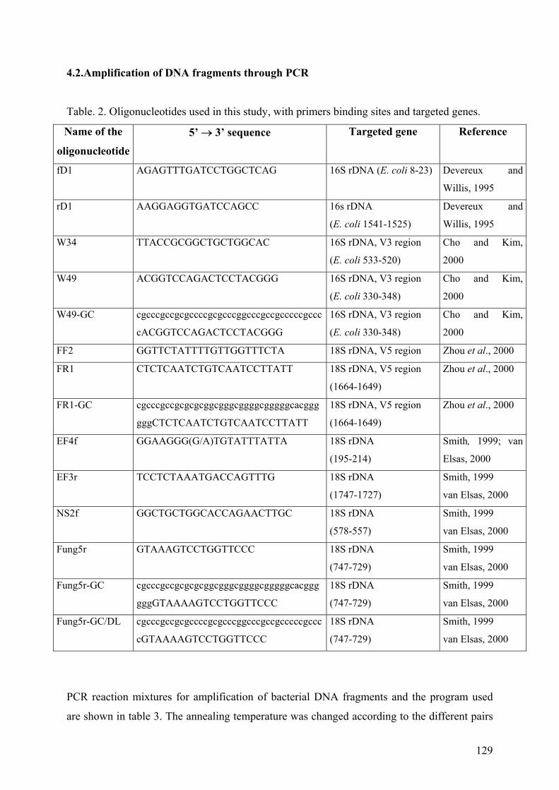
129
4.2.Amplification of DNA fragments through PCR
Table. 2. Oligonucleotides used in this study, with primers binding sites and targeted genes.
Name of the
oligonucleotide
5’ → 3’ sequence Targeted gene Reference
fD1 AGAGTTTGATCCTGGCTCAG 16S rDNA (E. coli 8-23) Devereux and
Willis, 1995
rD1 AAGGAGGTGATCCAGCC 16s rDNA
(E. coli 1541-1525)
Devereux and
Willis, 1995
W34 TTACCGCGGCTGCTGGCAC 16S rDNA, V3 region
(E. coli 533-520)
Cho and Kim,
2000
W49 ACGGTCCAGACTCCTACGGG 16S rDNA, V3 region
(E. coli 330-348)
Cho and Kim,
2000
W49-GC cgcccgccgcgccccgcgcccggcccgccgcccccgccc
cACGGTCCAGACTCCTACGGG
16S rDNA, V3 region
(E. coli 330-348)
Cho and Kim,
2000
FF2 GGTTCTATTTTGTTGGTTTCTA 18S rDNA, V5 region Zhou et al., 2000
FR1 CTCTCAATCTGTCAATCCTTATT 18S rDNA, V5 region
(1664-1649)
Zhou et al., 2000
FR1-GC cgcccgccgcgcgcggcgggcggggcgggggcacggg
gggCTCTCAATCTGTCAATCCTTATT
18S rDNA, V5 region
(1664-1649)
Zhou et al., 2000
EF4f GGAAGGG(G/A)TGTATTTATTA 18S rDNA
(195-214)
Smith, 1999; van
Elsas, 2000
EF3r TCCTCTAAATGACCAGTTTG 18S rDNA
(1747-1727)
Smith, 1999
van Elsas, 2000
NS2f GGCTGCTGGCACCAGAACTTGC 18S rDNA
(578-557)
Smith, 1999
van Elsas, 2000
Fung5r GTAAAGTCCTGGTTCCC 18S rDNA
(747-729)
Smith, 1999
van Elsas, 2000
Fung5r-GC cgcccgccgcgcgcggcgggcggggcgggggcacggg
gggGTAAAAGTCCTGGTTCCC
18S rDNA
(747-729)
Smith, 1999
van Elsas, 2000
Fung5r-GC/DL cgcccgccgcgccccgcgcccggcccgccgcccccgccc
cGTAAAAGTCCTGGTTCCC
18S rDNA
(747-729)
Smith, 1999
van Elsas, 2000
PCR reaction mixtures for amplification of bacterial DNA fragments and the program used
are shown in table 3. The annealing temperature was changed according to the different pairs
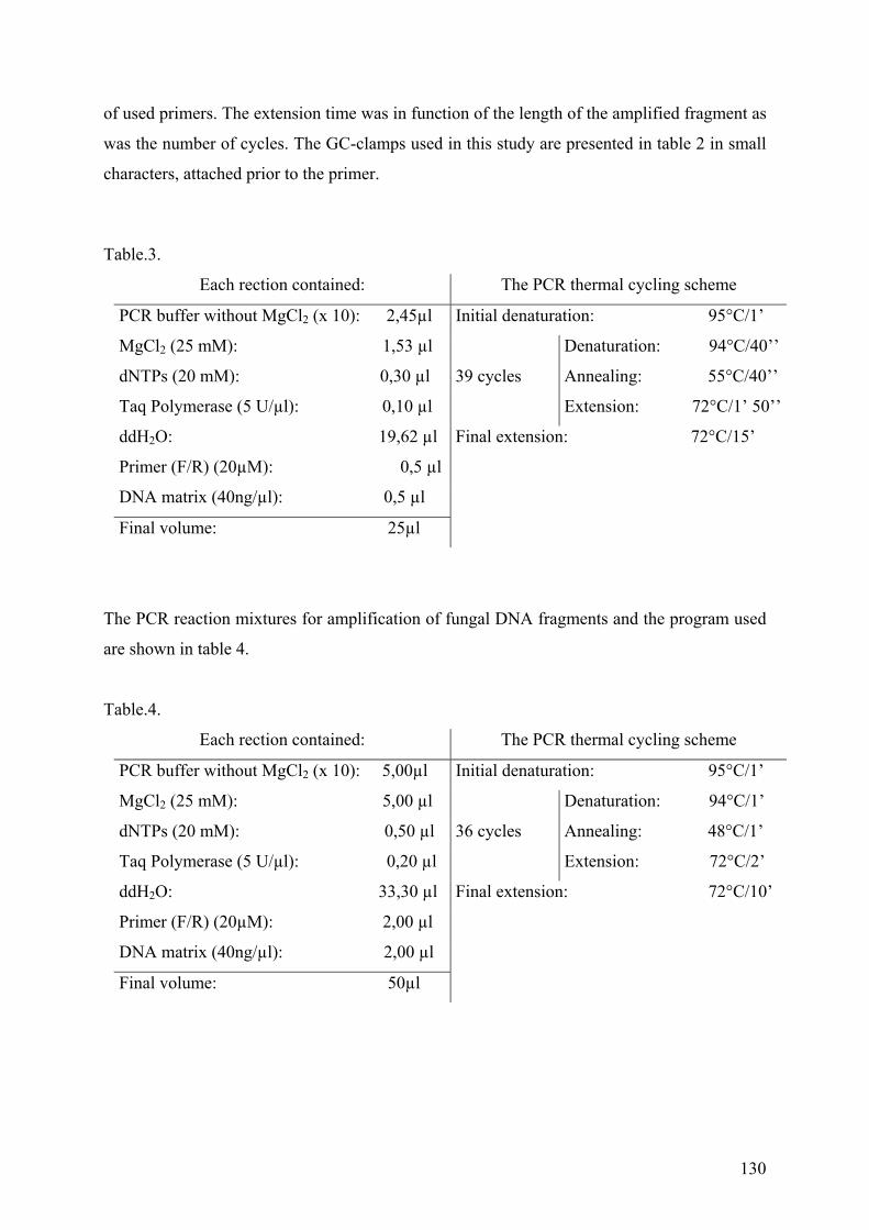
130
of used primers. The extension time was in function of the length of the amplified fragment as
was the number of cycles. The GC-clamps used in this study are presented in table 2 in small
characters, attached prior to the primer.
Table.3.
Each rection contained: The PCR thermal cycling scheme
PCR buffer without MgCl2 (x 10): 2,45µl Initial denaturation: 95°C/1’
MgCl2 (25 mM): 1,53 µl Denaturation: 94°C/40’’
dNTPs (20 mM): 0,30 µl 39 cycles Annealing: 55°C/40’’
Taq Polymerase (5 U/µl): 0,10 µl Extension: 72°C/1’ 50’’
ddH2O: 19,62 µl Final extension: 72°C/15’
Primer (F/R) (20µM): 0,5 µl
DNA matrix (40ng/µl): 0,5 µl
Final volume: 25µl
The PCR reaction mixtures for amplification of fungal DNA fragments and the program used
are shown in table 4.
Table.4.
Each rection contained: The PCR thermal cycling scheme
PCR buffer without MgCl2 (x 10): 5,00µl Initial denaturation: 95°C/1’
MgCl2 (25 mM): 5,00 µl Denaturation: 94°C/1’
dNTPs (20 mM): 0,50 µl 36 cycles Annealing: 48°C/1’
Taq Polymerase (5 U/µl): 0,20 µl Extension: 72°C/2’
ddH2O: 33,30 µl Final extension: 72°C/10’
Primer (F/R) (20µM): 2,00 µl
DNA matrix (40ng/µl): 2,00 µl
Final volume: 50µl
131
4.3. Purification of DNA fragments for sequencing
Each PCR-amplified fragment of 16S bacterial or 18S fungal rDNA was purified prior to
sequencing, according to the protocol of Amersham for the MicroSpinTM Columns
(Amersham Pharmacia Biotech,Inc., USA).
4.4. Temperature gradient gel electrophoresis (TGGE)
This technique is used for the separation of DNA fragments of the same length, but with
different sequences. The separation is based on the decreased electrophoretic mobility of a
partially melted double-stranded DNA fragment in polyacrylamide gels with a linear
temperature gradient. The melting of DNA proceeds in “melting domains”, which are the
stretches of base pairs with an identical melting temperature (Tm). When a domain with a
lowest melting temperature, reaches its melting temperature at a particular position in the
temperature gradient gel, a transition of a helical to a partially melted molecule occurs and the
migration of this molecule stops. The sequence variations in “melting domains” cause the
differentiation between the final positions of the fragments. This method can separate DNA
fragments up to 500 bp. The percentage of the fragments separation is nearly 100%, due to the
attachment of a GC rich sequence (GC-clamp) to one of the sides of the DNA fragment. This
GC-clamp prevents the two DNA strands from complete dissociation and its length can vary
between 30 and 50 nucleotides. The GC-clamps used in this study are presented in table 2 in
small characters.
Each PCR-amplified fragment (10µl) was mixed with 3 µl of loading buffer ( 0,05g l-3
bromophenol blue, 0,05g l-3 xylene cyanol, 4 ml of 100% glycerol, diluted with ddH2O to
final volume of 10ml) and loaded onto the gel. The polyacrylamide gels (200 x 160 x 1 mm3)
contained 8 to 13% of acrylamide/bis, 5 to 7 M urea, 1.25 x TAE, 0.1% TEMED, 0.30ml of
ammonium persulfate of 10% solution.
TGGE were performed at 60, 70, 90 and 100V for 6 and 14 h in BioRad apparatus (Bio-Rad,
Richmond,CA, USA). The range of tested temperatures gradients varied from 41,0°C to 65°C
in total and was in function of the urea quantity and voltage.
132
5. Biochemical techniques
Measurements of lactic acid isomers through the method of Gutmann
To assay the isomers of lactic acid in the SBM, WM and CDM a 0.1 ml portion of each media
supernatant was taken for the determination of D-(-)- and L-(+)-lactic acid by using D- lactate
dehydrogenase and L-lactate dehydrogenase (Boehringer Mannhaim), (Gutmann and
Wahlefeld, 1974)
. The 0.1 ml portion of supernatant was mixed with a solution containing 0.3 ml of Glycine-
Hydrazine buffer (pH 9.2), 0.6 ml of ddH2O, 1.5 mg of NAD and 7.5 µl of L- or D-
dehydrogenase. The prepared samples were incubated for 10 min at ambient temperature (22-
25°C) and then were measured at λ340 with a Uvikon 930 spectrophotometer (Kontron
Instruments). In the control (blank) the supernatant from the samples was replaced with
ddH2O. The quantities of both forms were determined prior to inoculation with C.
arsenoxidans of the three media and after 72 h of growth of the strain.
6. Microscopy techniques
Principles of Luminescence (Fluorescence and Phosphorescence)
The fluorescence is a form of luminescence, which is a consequence of the absorption of a
photon from molecules as flurophores, fluorochromes, or a fluorescent clamp. This reaction is
a part of the luminescence reactions assembly, which consist of: photoluminescence
(fluorescence and phosphorescence) and other types of luminescence (chimiluminescence,
bioluminescence, thermoluminescence, electroluminescence, etc.). The photoluminescence is
based on the excitation and light emission due to the photon energy. The essential
differentiation between the different types of luminescence is the origin of the excitation
energy used.
The fluorescence is characterized as emission of photons due to the absorbency of light. The
emission is due to the transformation of one molecule (at electron level) from excited level
(S1) back to its normal energy level (S0).
The phosphorescence is the emission of light after the end of light excitation of a given
molecule. This reaction differs from the fluorescence by the way of lowering the excitation
133
energy. In this case the average life of the molecule in the S1 energy level is much longer,
when compared to the fluorescence.
A preliminary selection of all 576 gfp mutants, from the three conjugations of C.
arsenoxidans and the donor strains of E.coli S17 λpir (containing one of the 3 different
plasmids used), was performed on a Synergy HT (Bio-Tek). The software used was KC4
supplied with the apparatus from Bio-Tek.
The tracking of gfp mutant of the strain C. arsenoxidans named KDM7, were performed in a
nonsterile sauerkraut brine medium in presence and absence of As[III] with concentration of
50 ppm.
A quantity of 600 µl of each cell suspension was powered in a well and the cells were excited
with UV light for 20 sec. The image acquisition was performed with a Nikon 2000 Eclipse
epifluorescence microscope, equipped with a CCD camera by which gray scale images were
captured. Quantification of the digitized fluorescent signals from KDM7 cells, was
accomplished through the image analysis software package ImageJ
(http://rsb.info.nih.gov/ij/). The count of KDM7 cells emission signals was treated separately
from the images captured with white light. Thus, to obtain the results from both types of
images two different protocols were developed and used.
The fluorescent images were transferred from 16 to 8 bit gray scale images and the threshold
to remove any remaining background noise was established manually. Then the custom
designed plugin “analyze particles” was used, by which the following parameters were
established and measured:
• the numbers of particles for each image
• the area of each particle measured;
• the mean gray value;
• the standard deviation of mean gray value;
• mode
• Min and max gay value for each particle.
The function “area” resulted in the definition of the max size of KDM7 particles. The “mode”
feature gave the intensity of light emission of most represented emitting point and their
standard deviation. Thus the percentage of KDM7 cells was found out from the whole
quantity of fluorescent cell captured in the images.
Second protocol for object count was developed concerning the images captured with white
light. These image processing’s were assayed in order to perform a statistically valid method
134
for count of objects with different shapes and meaning in the gray scale. The following
protocol was used:
• all images were transferred into 8 bit resolution
• the threshold was adjusted manually, which allows separating particles with gray-scale
pixels below or above a fixed level. Additionally the remaining pixels were not fixed to
the background of the picture.
• a second binarisation and adjustment of the pictures threshold were done
• analyses were done automatically, using the process ”analyze particles”
The results obtained, represented the overall quantity of cell per image, including those
possessing the fluorescence and those which were not fluorescent.
7. Immobilization of bacteria
For the immobilization of the C. arsenoxidans cells in Na-alginate beads 0.3g of Sodium
alginate was dissolved in 15 ml of ddH2O. A second solution of 0.5g of CaCl2.2H2O in 75 ml
of ddH2O, was prepared and a 72h of precultured C. arsenoxidans cells in a CDM were used.
Another preculture from C. arsenoxidans cells induced with As[III] were prepared prior to the
encapsulation in the alginate beads. The induced cells were prepared in sauerkraut brine
medium, supplemented with salts solutions and 10ppm of As[III]. The cell suspensions with
an optical density of 0.3-0.4 at 600 nm were used for immobilization and the final
concentration of the alginate was 3-5%. The 200ml of the preculture were centrifuged at
5000rpm, washed twice and finally suspended in 20ml of sterile ddH2O. For the entrapment
of the cells 3 ml of the previously prepared cell culture was added and mixed into the Na-
alginate solution, which was sterilized prior to use at 121°C for 15 min.
The beads formation followed the protocol:
The Na-alginate cell suspension was passed dropwise into the CaCl2.2H2O solution through a
needle with 0.3 mm diameter; the receiver solution was shaken constantly with the forming
beads in it; drops were formed at 10cm distance above the receiver; after formation the beads
were left for 2 to 3h in the CaCl2.2H2O and washed subsequently with a fresh 0.2 M
CaCl2.2H2O solution.
135
8. Chemical analysis (HPLC-ICP-AES)
Arsenic compounds were eluted with a phosphate buffer (7.2 < pH < 7.4) composed of an
mixture of equal volumes of the two following solutions: Solution 1 was prepared by adding
10 mM sodium monohydrogenphosphate (Na2HPO4.12H2O) solution into 10 mM sodium
dihydrogenphosphate (NaH2PO4.2H2O) solution up to pH 6.3. Solution 2 was 0.1M
Na2HPO4.12 H2O (pH 7.8).
Concentrations of As[V] and As[III] in culture supernatants were determined by HPLC-ICP-
AES using an anion exchange polymeric column (PRP-X100, 250 mm X 4.1 mm i.d., particle
size 10 µm, Hamilton). Arsenic compounds were eluted at 1 ml min-1 in phosphate buffer,
degassed and filtered on a 0.65 µm cellulose acetate membrane (Millipore). Detection was at
193.7 nm wavelength using an ICP–AES apparatus (JY138 ULTRACE, Jobin Yvon), coupled
to an HPLC pump (LC – 10 AT, Schimadzu) via a Teflon tube. The ICP-spectrometer was
calibrated before each use and again after every 10 samples by running a blank and samples
of known concentrations. Detection limits were 0.11 mg L-1 and 0.14 mg L-1 for As[III] and
As[V], respectively. Quantification limits were 0.47 mg L-1 and 0.36 mg L-1 for As[III] and
As[V], respectively.
9. Physical and chemical parameters of the nutrient media
Measurements of the main physico-chemical parameters of SBM and WM were made in the
Centre d’Analyse et de Recherche (CAR, Strasbourg, France). The methods used were carried
out as the Norme Française Technique (NFT) and are as follows:
Chemical oxygen demand (COD) (NFT 90 - 101),
Biological oxygen demand in 5 days (BOD5) (NFT 90 - 103),
Ammonium azote (NFT 90 - 015),
Kjeldahl azote NTK (NFT 90 - 110),
Nitrates (NFT 90 – 012)
Nitrites (NFT 90 - 013).
136
CHAPTER V
REFERENCES
137
Abdrashitova, S.A., Abdullina, G.G. and Ilialetdinov, A.N., 1985. [Glucose consumption and
hydrogenase activity of the cells of the arsenite-oxidizing bacterium Pseudomonas
putida] (Article in Russian). Mikrobiologiia, 54: 679-81.
Afkar, E., Lisak, J., Saltikov, C., Basu, P., Oremland, R.S. and Stolz, J.F., 2003. The
respiratory arsenate reductase from Bacillus selenitireducens strain MLS10. FEMS
Microbiology Letters, 226: 107-112.
Alexander, M., 1977. Introduction to Soil Microbiology. Wiley, New York.
Anderson, G.L., Ellis, P.J., Kuhn, P. and Hille, R., 2001. Oxidation of arsenite by Alcaliganes
faecalis. In: Environmental Chemistry of Arsenic. Marcel Dekker, New York, pp. 343-
362.
Arai, Y., Elzinga, E. J., Sparks, D.L., 2001. X-ray Absorption Spectroscopic Investigation of
Arsenite and Arsenate Adsorption at the Aluminium Oxide-Water Interface. Journal
Colloid Interface Science 235: 80-88.
Arienzo, M., Adamo, P., Chiarenzelli, J., Bianco, M.R. and De Martino, A., 2002. Retention
of arsenic on hydrous ferric oxides generated by electrochemical preroxidation.
Chemosphere, 48: 1009-1018.
Atlas, R.M., 1997. Handbook of Microbiological Media. 2-nd ed.
Ayers, R.U. and Ayers, L.W., 1999. Accounting for Resources: The life cycle of Materials.
Edward Elgar Publishing, Inc., UK.
Bentley, R. and Chasteen, T.G., 2002. Microbial Methylation of methalloids: Arsenic,
Antimony and Bismuth. Microbiology and Molecular Biology Reviews, 66: 250-271.
Bledsoe, T.L., Cantafio, A.W. and Macy, J.M., 1999. Fermented whey-an inexpensive feed
source for a laboratory-scale selenium-bioremediation reactor system inoculated with
Thauera selenatis. Applied Microbiology and Biotechnology, 51: 682-685.
Bostick, B.C. and Fendorf, S., 2003. Arsenite sorption on troilite (FeS) and pyrite (FeS2).
Geochimica et Cosmochimica Acta, 67: 909-921.
Brooks, R.R., Lee, J., Reeves, R.D. and Jaffre', T., 1977. Detection of nickeliferous rocks by
analysisof herbarium specimens of indicator plants. Journal of Geochemical
Exploration, 7: 49-57.
Bucher, J.-B., 1999. Etudes des bacterries intervenant dans l'oxydation de l'arsenite (As[III])
en arseniate (As[V]), Université Louis-Pasteur, Laboratoire de Microbiologie et de
Génétique, UPRES-A 7010-CNRS, Strasbourg.
138
Candy, A.C., Hanley, J.E. and Susten, A.S., 1997. ATSDR science panel on bioavalability of
mercury in soil-lessons learned. Risk Analysis, 17: 527-532.
Carter, D.E., Aposhian, H.V. and Gandolfi, A.J., 2003. The metabolism of inorganic arsenic
oxides, gallium arsenide, and arsine: a toxicochemical review. Toxicology and
Applied Pharmacology, 193: 309-334.
Caussy, D., 2003. Case studies of the impact of understanding bioavailability: arsenic.
Ecotoxicology and Environmental Safety, 56: 164-173.
Caussy, D., Gochfeld, M., Gurzau, E., Neagu, C. and Ruedel, H., 2003. Lessons from case
studies of metals: investigating exposure, bioavailability, and risk. Ecotoxicology and
Environmental Safety, 56: 45-51.
Chaffey N., 2002. Sucking out the poison. Trends in Plant Science, 7: 528.
Cheng, C.-N. and Focht, D.D., 1979. Production of arsine and methylarsines in soil and in
culture. Applied and Environmental Microbiology, 38: 494-498.
Cho, J.C. and Kim, S.J., 2000. Computer-assisted PCR-single-strand-conformation
polymorphism analysis for assessing shift in soil bacterial community structure during
bioremediational treatments. World Journal of Microbiology & Biotechnology, 16:
231-235.
Cristiani-Urbina, E., Netzahuatl-Minos, A.R., Manriques-Rojas, F.J., Juarez-Ramirez, C.,
Ruiz-Ordaz, N. and Galindez-Mayer, J. 2000. Batch and fed-batch cultures for the
treatment of whey with mixed yeast cultures. Process Biochemistry, 35: 694-657.
Cullen, W.R. and Reimer, K.J., 1989. Arsenic speciationin the environment. Chemical
Reviews, 89: 713-784.
Del Rio, M., Font, R., Almela, C., Vélez, D., Montoro, R. and Bailon, A.H., 2002. Heavy
metals and arsenic uptake by wild vegetation in the Guadamar river area after the toxic
spill of the Aznacollar mine. Jurnal of Biotechnology 98: 125-137.
Delbecque, A., Recherche des bacteries oxydant l'arsenite en arseniate, Université Louis-
Pasteur, laboratoire de Microbiologie et de Génétique, Strasbourg.
Devereux, R. and Willis, S.G., 1995. Amplification of ribosomal RNA sequences. Molecular
Microbial Ecology Manual. Kluwer Acad. Publishers, Dordrecht.
Dushenko, W.T., Bright, D.A. and Reimer, K.J., 1995. Arsenic bioaccumulation and toxicity
in aquatic macrophytes exposed to gold mine relationships with environmental
partitioning, metal uptake and nutrients. Aquatic Botany, 50: 141-158.
Francesconi, K., Visoottiviseth, P., Sridokchan, W. and Goessler, W., 2001. Arsenic species
in an arsenic hyperaccumulationg fern, Pityrogramma calomelanos: a potentiial
139
phytoremediator of arsenic-contaminated soils. The Science of the Total Environment
284: 27-35.
Gadd, G.M., 2004. Microbial influence on metal mobility and application for bioremediation.
Geoderma, in press.
Gihring, T.M. and Banfield, J.F., 2001 a. Arsenite oxidation and arsenate respiration by a new
Thermus isolate. FEMS Microbiology Letters, 202: 335-340.
Gihring, T.M., Druschel, G.K., McCleskey, R.B., Hammers, R.J. and Banfield, J.F., 2001 b.
Rapid arsenite oxidation by Thermus aquaticus and Thermus thermophilus: Field and
laboratory investigations. Environmental Science and Technology, 35: 3857-3862.
Goldberg S. and Johnston, C.T., 2001. Mechanisms of Arsenic Adsorption on Amorphous
Oxides Evaluated Using Macroscopic Measurements, Vibrational Spectroscopy, and
Surface Complexation Modeling. Journal of Colloid Interface Science 234: 204-216.
Goyer, R.A. and Clarkson, T.W., 2001. Toxic effects of metals. Casaret and Doull's
Toxycollogy, McGraw-Hill, New York, 818-820.
Gregus, Z. and Németi, B., 2002. Purine nucleoside phosphorylase as a cytosolic arsenate
reductase. Toxicological Sciences, 70: 13-19.
Grissom, R.E., Abernathy, C.O., Susten, A.S. and Donohue, J.M., 1999. Estimating total
arsenic exposure in the Unated States. In: J.U. Chapbell, Abernathy, C. O., Calderon,
R. (Editor), Arsenic Exposure and Health Effects, Third International Conference on
Arsenic Exposure and Health Effects. Elsevier, Amsterdam, 51-57.
Groudeva, V.I., Groudev, S.N. and Doycheva, A.S., 2001. Bioremediation of waters
contaminated with crude oil and toxic heavy metals. International Journal of Mineral
Processing, 62: 293-299.
Gutmann, A.B.C. and Wahlefeld, A.W., 1974. L-Lactat Bestimmung mit Lactat-
Dehydrogenase und NAD. Methoden der enzymatischen Analyse, 3-rd edition.
Weinheim, 1510 - 1514.
Hindmarsh, J.T., 2000. Arsenic, its clinical and environmental significance. Journal of Trace
Elements in Experimental Medicine, 13: 165-172.
Huber, R., Sacher, M., Vollmann, A., Huber, H. and Rose, D., 2000. Respiration of arsenate
and selenate by hyperthermophilic archaea. Systematic and Applied Microbiology, 23:
305-314.
Ilialetdinov, A.N. and Abdrashitova, S.A., 1981. [Autotropic oxidation of arsenic by
Pseudomonas arsenitoxidans] (Article in Russian). Mikrobiologiia, 50: 197-204.
140
Ji, G. and Silver, S., 1992. Reduction of arsenate to arsenite by the ArsC protein of the arsenic
resistance operon of Staphylococcus aureus plasmid pI258. Proceedings of the
National Academy of Sciences of the United States of America, 89: 9474-9478.
Jin, Y.L. and Speers, R.A., 1998. Flocculation of Saccharomyces cerevisiae. Food Research
International, 31: 421-440.
Karel, S.F., Libicki, S.B. and Robertson, C.R., 1985. The immobilization of whole cells-
engineering principles. Chemical Engineering Science, 40: 1321-1354.
Kargupta, K., Siddhartha, D. and Sanyal, S.K., 1998. Analysis of the performance of a
countinuous membrane bioreactor with cell recycling during ethanol fermentation.
Biochemical Engineering Journal, 1: 31-37.
Katsoyiannis, I.A. and Zouboulis, A.I., 2004. Application of biological processes for the
removal of arsenic from groundwaters. Water Research, 38: 17-26.
Knauer, K., Behra, R. and Hemond, H., 1999. Toxicity of inorganic and methylated arsenic to
algal communities from lakes along an arsenic contamination gradient. Aquatic
Toxicology, 46: 221-230.
Korte, N.E. and Fernando, Q., 1991. A review of arsenic(III) in ground water. Critical
Review. Environmental Control, 21: 1-39.
Kourkoutas, Y., Bekatorou, A., Banat, I.M., Marchant, R. and Koutinas, A.A., 2004.
Immobilization technologies and support materials suitable in alcohol beverages
production: a review. Food Microbiology, 21: 377-397.
Krafft, T. and Macy, J.M., 1998. Purification and characterization of the respiratory arsenate
reductase of Chrysiogenes arsenatis. European Journal of Biochemistry, 255: 647-653.
Kuroda, K., Yoshida, K., Yasukawa, A., Wanibuchi, H., Fukushima, S. and Endo, G., 2001.
Enteric bacteria may play a role in mammalian arsenic metabolism. Applied
Organometallic Chemistry, 15: 548-552.
Lakovic, J.A., Nikolaidis, N.P. and Dobbs, G.M., 2000. Inorganic arsenic removal by zero-
valent iron. Environmental and Engineering Science, 17: 1487-1492.
Laverman, A.M., Switzer-Blum, J., Schaefer, J.K., Phillips, E.J.P., Lovley, D.R. and
Oremland, R.S., 1995. Growth of strain SES-3 with arsenite and other diverse electron
acceptors. Applied and Environmental Microbiology, 61: 3556-3561.
Lebeau, T., Jouenne, T. and Junter, J.A., 1998. Diffusion of sugars and alcohols through
composite membrane structures immobilizing viableyeast cells. Enzyme and
Microbial Technology, 22: 434-438.
141
Lee, H., Song, M., Yu, Y. and Hwang, S., 2003. Production of Ganoderma lucidum mycelium
using cheese whey as an alternative substrate: response surface analysis and
biokinetics. Biochemical Engineering Journal, 15: 93-99.
Lièvremont D., N'negue, M.-A., Behra, P. and Lett, M.-C., 2003. Biological oxidation of
arsenite: batch reactor experiments in presence of kutnahorite and chabazite.
Chemosphere, 51: 419-428.
Liht, L.A., McCutcheon, S.C., Wolfe, N.L. and Carreira, L.H., 1995. Phytoremediation of
organic and nutrient contaminants. Envronmental Science and Technology, 29: 318-
323.
Liu, Z., Carbrey, J.M., Agre, P. and Rosen, B.P., 2004. Arsenic trioxide uptake by human and
rat aquaglyceroporins*1. Biochemical and Biophysical Research Communications,
316: 1178-1185.
Loukidou, M.X., Matis, K.A., Zouboulis, A.I. and Liakopoulou-Kyriakidou, M., 2003.
Removal of As[V] from wastewaters by chemically modified fungal biomass. Water
Research 37: 4544-4552.
Macy, J.M., Nunan K, Hagen K.D., Dixon D.R., Harbour P.J., Cahill M. and Sly L.I. 1996.
Chrysogenes arsenatis gen. nov., sp. nov., a new arsenate-respiring bacterium isolated
from gold mine wastewater. International Journal of Systematic Bacteriology, 46:
1153-1157.
Macy, J.M. and Santini, J.M., 2002. Unique modes of arsenate respiration by Chrysiogenes
arsenatis and Desulfotomaculum sp. Str. BEN-RB. In: "Environmental Chemistry of
Arsenic". Marcel Dekker, Inc., New York, pp. 297-312.
Macy, J.M., Santini, J.M., Pauling, B.V., O'Neill, A.H. and Sly, L.I., 2000. Two new
arsenate/sulfate-reducing bacteria: mechanisms of arsenate reduction. Archives of
Microbiology, 173: 49-57.
Mandal, B.K. and Suzuki, K.T., 2002. Arsenic round the world: a review. Talanta, 58: 201-
235.
McBride, B.S. and Wolfe, R.S., 1971. Biosynthesis of dimethylarsine by Methanobacterium.
Biochemistry, 10: 4312-4317.
Meng, X., Korfiatis, G.P., Christodoulatos, C. and Bang, S., 2001. Treatment of arsenic in
Bangladesh well water using a hausehold co-precipitation and filtration system. Water
Research, 35: 2805-2810.
142
Micheva K., 2002. Characteristics of the arsenite-oxidation from bacterial strain ULPAs1
immobilized cells in alginate beads, Sofia University "St. Kliment Ohridsky" and
Université Louis-Pasteur, Sofia, Strasbourg.
Mukhopadhyay, R., Rosen, B.P., Phung, L.T. and Silver, S., 2002. Microbial arsenic: from
geocycles to genes and enzymes. FEMS Microbiology Reviews, 26: 311-325.
Muller, D., Lièvremont D., Simeonova D. D., Hubert J.-C. and Lett, M.-C., 2003. Arsenite
oxidase aox genes from a metal-resistant ß-Proteobacterium. Journal of Bacteriology,
185: 135-141.
Navarro, J.M. and Durand, G., 1977. modification of yeast metabolism by immobilization
onto porous glass. European Journal of Applied Microbiology, 4: 234-254.
Newman, D.K. et al., 1997. Dissimilatory arsenate and sulfate reduction in Desulfotomaculum
auripigmentum sp. nov.,. Archives of Microbiology, 168: 380-388.
Nie, L., 2002. Phytoremediation of arsenate contaminated soil by transgenic canola and the
plant growth-promoting bacterium Enterobacter cloacae CAL2. Plant Physiology and
Biochemistry 40: 355-361.
Niggemyer, A., Spring, S., Stackebrandt E. and Rosenzweig, R.F., 2001. Isolation and
characterization of a novel As(V)-reducing bacterium: implications for arsenic
mobilization and genus Desulfitobacterium. Applied and Environmental
Microbiology, 67: 5568-5580.
Nikolaidis, N.P., Dobbs, G.M. and Lackovic, J.A., 2003. Arsenic removal by zero-valent iron:
field, laboratory and modeling studies. Water Research, 37: 1417-1425.
Nishimura, T. and Umetsu, Y., 2001. Oxidative precipitation of arsenic(III) with
manganese(II) and iron(II) in dilute acidic solution by ozone. Hydrometallurgy, 62:
83-92.
Norton, S. and D'Amore, T., 1994. Physiological effects of yeast cell immobilization:
application for brewing. Enzyme and Microbial Technology, 16: 365-375.
Oremland, R.S., Hoeft, S.E., Santini, J.M., Bano, N., Hollibaugh, R.A. and Hollibaugh, J.T.,
2002. Anaerobic oxidation of arsenite in Mono Lake water and by facultative,
arsenite-oxidizing chemoautotroph, strain MLHE-1. Applied and Environmental
Microbiology, 68: 4795-4802.
Osborne, F.H. and Enrlich, H.L., 1976. Oxidation of arsenite by a soil isolate of Alcaligenes.
Journal of Applied Bacteriology, 41: 295-305.
Park, J.K. and Chang, H.N., 2000. Microencapsulation of microbial cells. Biotechnology
Advances, 18: 303-319.
143
Pickett, A.W., McBride, B.S., Cullen, W.R. and Manji, H., 1981. The reduction of
trimethylarsineoxyde by Candida humicola. Canadian Journal of Microbiology, 27:
773-778.
Pilkington, P.H., Margaritis, A., Mensour, N.A. and Russell, I., 1998. Fundamentals of
immobilized yeast cells for continuous beer fermentation: a review. Journal of the
Institute of Brewing, 104: 19-31.
Rehm, B.H.A. and Valla, S., 1997. Bacterial alginates: biosynthesis and applications. Applied
Microbiology and Biotechnology, 48: 281-288.
Robinson, B., Duwig, C., Bolan, N., Kannathasan, M. and Saravanan, A., 2002. Uptake of
arsenic by New Zealand watercress (Lepidium sativum). The Science of The Total
Environment, 301: 67-73.
Rosen, B.P., 2002. Biochemistry of arsenic detoxification. FEBS Letters, 529: 86-92.
Rossman, T.G., 2003. Mechanism of arsenic carcinogenesis: an integrated approach. Mutation
Research/ Fundamental and Molecular Mechanisms of Mutagenesis, 533: 37-65.
Sabra, W., Zeng, A.-P., Deckwer, W.-D., 2001. Bacterial alginate: physiology, product
quality and process aspects. Applied Microbiology and Biotechnology, 56: 315-325.
Sadiq, M., 1990. Arsenic chemistry in marine environments: A comparison between
theoretical and field observation. Marine Chemistry, 31: 285-297.
Sadiq, M., 1997. Arsenic chemistry in soils: An overview of thermodynamic predictions and
field observations. Water Air and Soil Pollution, 93: 117-136.
Saltikov, C.W., Cifuentes, A., Venkateswaran, K. and Newman, D.K., 2003. The ars
detoxification system is advantageous but not required for As(V) respiration by the
genetically tractable Shewanella species strain ANA-3. Applied and Environmental
Microbiology, 69: 2800-2809.
Saltikov, C.W. and Newman, D.K., 2003 a. Genetic identification of a respiratory arsenate
reductase. Proceedings of the National Academy of Sciences of the United States of
America, 100: 10983-10988.
Santini, J.M., Sly, L.I., Schangl, R.D. and Macy, J.M., 2000 a. A new hemolithoautotrophic
arsenite-oxidazing bacterium isolated from a gold mine: Phylogenetic, physiological,
and preliminary biochemical studies. Applied and Environmental Microbiology, 66:
92-97.
Santini, J.M. and vanden Hoven, R.N., 2003. Molybdenum-containing arsenite oxidase of the
chemolitoautotrophic arsenite oxidizer NT-26. Journal of Bacteriology, 186: 1614-
1619.
144
Scott, N., Hatlelid, K.M., MacKenzie, N.E. and Carter, D.E., 1993. Reactions of arsenic (III)
and arsenic (V) species with glutathione. Chemical Research in Toxicology, 6: 102-
106.
Silver, S. and Phung, L.T., 2003. Oxidation and reduction: the microbiology of inorganic
arsenic. Applied and Environmental Microbiology (in press).
Singh, P. and Cameotra S. S., 2004. Enhancement of metal bioremediation by use of
microbial surfactants. Biochemical and Biophysical Research Communications, 319:
293-299.
Smedley, P.L., Edmunds, W.M. and Pelig-Ba, K.B., 1996. Environmental Geochemistry and
Health, 113, London.
Smith, E., Leeflang, P., Glandorf, B., van Elsas, J.D. and Wernars, K., 1999. Analysis of
Fungal Diversity in the Wheat Rhisosphere by Sequencing of Cloned PCR-Amplified
Genes Encoding 18S rRNA and Temperature Gradient Gel Electrophoresis. Applied
and Environmental Microbiology, 65: 2614-2621.
Stolz, J.F., Ellis, D.J., Blum, J.S., Ahmann D., Lovley D.R. and Oremland R.S., 1999.
Sulfurospirillum barnesii sp. nov., Sulfurospirillum clade in the Epsilon
Proteobacteria. International Journal of Systematic Bacteriology, 49: 1177-1180.
Sutherland, I.W.and Ellwood, D.C., 1979. Microbial exopolysaccharides- Industrial polymers
of current and future potential. Symposium of Society of General Microbiology 29:
107-150.
Switzer Blum, J., Burns Bindi A., Buzzeli, J., Stolz, J.F. and Oremland, R.S., 1998. Bacillus
arsenicoselenatis, sp. nov., and Bacillus selenireducens, sp. nov.: two haloalkaliphiles
from Mono Lake, California that respire oxyanions of selenium and arsenic. Archives
in Microbiology, 171: 19-30.
Tang, X., Lu, B.F. and Pan, S.Q., 1999. A bifunctional transposon mini-Tn5gfp-km which can
be used to select for promoter fusions and report gene expression levels in
Agrobacterium tumefaciens. FEMS Microbiology Letters, 179: 37-42.
Thompson, D.J., 1993. A chemical hypothesis for arsenic methylation in mammals. Chemico-
Biological Interactions, 88: 89-114.
van Elsas, J.D., Duarte, G.F., Keijzer-Wolters, A. and Smith, E., 2000. Analysis of the
dynamics of fungal communities in soil via fungal-specific PCR of soil DNA followed
by denaturating gradient gel electrophoresis. Journal of Microbiological Methods, 43:
133-151.
145
Vega, L., Styblo, M., Patterson, R., Cullen, W., Wang, C. and Germolec, D., 2001.
Differential Effects of Trivalent and Pentavalent Arsenicals on Cell Proliferation and
Cytokine Secretion in Normal Human Epidermal Keratinocytes. Toxicology and
Applied Pharmacology, 172: 225-232.
Visoottiviseth, P., Francesconi, K. and Sridokchan, W., 2002. The potential of Thai
indigenous plant species for the phytoremediation of arsenic contaminated land.
Environmental Pollution, 118: 453-461.
Wang, Y. and Reardon, E.J., 2001. A siderite/limestone reactor to remove arsenic and
cadmium from wastewaters. Applied Geochemistry, 16: 1241-1249.
Weeger, W., Lièvremont, D. Perret, M., Lagarde, F., Hubert, J.-C., Leroy, M. and Lett, M.-C.,
1999. Oxidation of arsenite to arsenate by a bacterium isolated from an aquatic
environment. Biometals, 12: 141-9.
WHO, 2001a. Arsenic and arsenic compounds. Environmental Health Criteria. 2-nd Ed.,
World Health Organisation, Geneva.
Xi, C., Lambrecht, M., Vanderleyden, J. and Michielis, J., 1999. Bi-functional gfp-and gusA-
containing mini-Tn5 transposon derivatives for combined gene expression and
bacterial localization studies. Journal of Microbiological Methods, 35: 85-92.
Yamamura, S., Ike, M. and Fujita, M., 2003. Dissimilatory arsenate reduction by a facultative
anaerobe, Bacillus sp. strain SF-1. Journal of Bioscience and Bioengineering, 96: 454-
460.
Zafiriou, O.C., Joussot-Dubien, J., Zepp, R.G. and Zika, R.G., 1984. Photochemistry of
natural waters. Environmental Science and Technology, 18: 358A-371A.
Zaw, M. and Emett, M.T., 2002. Arsenic removal from water using advanced oxidation
processes. Toxicology Letters, 133: 113-118.
Zhang, W., Cai, Y., Downum, K.R. and Ma, L.Q., 2004. Thiol synthesis and arsenic
hyperaccumulation in Pteris vittata (Chinese brake fern). Environmental Pollution
(article in press).
Zhou, G., Whong, W.-Z., Ong, T. and Chen, B., 2000. Development of a fungus-specific PCR
assay for detecting low-level fungi in an indoor environment. Molecular and Cellular
Probes, 14: 339-348.

146
Internet sources
(http://www.lsbu.ac.uk/water/hyalg.html)
(http://www.genialab.de/inventory/alginate.htm).
(http://hyperphysics.phy-astr.gsu.edu/hbase/vision/cie.html)
(http://www.sucre-erstein.com/cadres/presentation.html)
(http://dsmz.de/media/med197.htm)
(http://dsmz.de/media/med198.htm)
(http://dsmz.de/media/med682.htm)
(http://dsmz.de/media/med063.htm)
(http://rsb.info.nih.gov/ij/)

147
CHAPTER VI
APPENDIX
[signalement bibliographique ajouté par : ULP – SCD – Service des thèses électroniques]
Daniel Muller, Didier Lièvremont, Diliana D. Simeonova, Jean-Claude Hubert, and
Marie-Claire Lett
Arsenite oxidase aox genes from a metal-resistant β-proteobacterium.
Journal of Bacteriology, Jan 2003, vol. 185, p. 135-141
Pages 135-141 :
La publication présentée ici dans la thèse est soumise à des droits détenus par un éditeur
commercial.
Pour les utilisateurs ULP, il est possible de consulter cette publication sur le site de
l'éditeur
http://jb.asm.org/cgi/content/full/185/1/135
Il est également possible de consulter la thèse sous sa forme papier ou d'en faire une
demande via le service de prêt entre bibliothèques (PEB), auprès du Service Commun
de Documentation de l'ULP: [email protected].