postmortem examination - ncjrs · postmortem examination thomas t. noguchi, m.d. introduction...
TRANSCRIPT

CHAPTER 3
Postmortem Examination Thomas T. Noguchi, M.D.
INTRODUCTION
Regardless of the suspected cause of death, a medical examiner or coroner's postmortem examination should always be thorough and comprehensive, for deaths which come under the jurisdiction of this office are often extremely complex, dealing as they do mostly with instances of sudden and unexpected demise. The postmortem examination should be approached with a certain degree of suspicion of ot.her possible but hidden etiological factors. The true cause of death may be masked by the obvious findings.
In the simpler ca.ses, the specific cause of death may be accurately determined by any competent pathologist and even by an alert non pathologist physician. In more complex cases, however, those without the trained or ingrained sense of suspicion of a forensic pathologist may be misled by the apparent and may seek no further information beyond the obvious. The average physician is often either unaware of or has a rather lax attitUde toward the potential medicolegal ramifications and significance of the autopsy report in coroner's cases. As an example, a history of heart attack should not automatically lead to the conclusion that a sudden death was due to heart disease. Drug abuse or misuse may be highly unlikely, but adverse reaction to therapeutic drugs is a possibility which should not be overlooked, especially in persons under intensive drug treatment of medical conditions. A nUlnbel' of deaths due to drugs have probably been missed because of bias induced by the medical history of the deceased.
Deaths involving psychoactive drugs may be divided into three categories, each of which calls for a thorough postmortem investigation:
1. Death due to the direct toxic effect of a drug.
2. Death as a result of secondary complica-
15
tions such as hepatitis and other infections due to use of contaminated needles in the administration of a drug.
3. Death, usually violent, from situations created by a person who, under the influence of a high but nonlethal dose of a drug, acts in a manner incompatible with life, exposing himself to life-risking situations, such as running in front of a moving vehicle, jumping from a high place, or aggressively inviting violence from others)
Hospital Versus Forensic Postmortt)m Examination
The primary purpose of a postmortem examination conducted in a hospital is to confirm a known or suspected diagnosis of the disease which caused the death of the patient. In addition, the hospital postmortem may provide knowledge concerning the tissue damage caused by the disease. Emphasis is on the. detailed study of specific internal organ or organs involved in the disease process; only a cursory external examination is conducted.
The forensic pathologist is not only interested in the direct cause of death but also in trying to reconstruct the circumstances and events which led to the death. Whatever the immediate cause of death, he must also ascertain whether the death was an accident, a suicide, or a homicide. Thorough external examination of the clothing worn by the deceased at the time of death and study of the deceased's state of mind just prior to death are as important as the examination of the internal organs. A complete and thorough medical examiner/coroner's postmortem examination must include:
lThese parallel the distinctions represented in categories of Code 3 of the UCI form, i.e., drug induced and drug related (see appendix A),
If you have issues viewing or accessing this file contact us at NCJRS.gov.
16 NOGUCHI
1. Detailed study of the scene of death, noting any signs of disorder, position of the deceased, presence of any possible evidential material, such as pills and prescription bottles in suspected drug deaths (see chapter 2, "Onsitt' Investigation ").
2. Precise and detailed study of the type and extent of any morphological damage or trauma, including microscopic examinations.
3. Determination of the diref't cause of the injury-disease, physical force, chemical agents, etc.
4. Followup toxicological, biochemical, microbiological, and other laboratory tests on selected tissues and body fluids as indicated by gross findings.
5. Analysis of information obtained from family and friends pertaining to the attitudes and lifestyle of the deceased.
The forensic pathologist has the training to recognize the significance of the various information obtained, to put it together like a jigsaw puzzle, interpret the findings, and come up with a reasonable reconstruction of the circumstances and events which led to the death of the individual.
Chain of Evidence
In the handling of any medical examiner/ coroner's case, the proper identification of all materials connected with each case is a MUST. All articles of clothing, contents of pockets, all tissues, and other biological materials removed for testing (e.g., blood), and all records and documents must be properly and legibly identified at the time of removal and examination. Much grief and problems can be avoided when this rule is strictly adhered to (see also chapter 2, "Onsite Investigation").
Docli mentati on
Just as important as a thorough postmortem examination is proper documentation of the findings. The written documentation may be in the form of "check and fill-in" sheets supplemented with short, simple nan'ative reports and picto-charts of body sections and organs showing pertinent findings and dimensiol)s for easy reference. Each document should be identified with:
1. The official name of the medical examiner or coroner's office
2. The name of the deceased 3. The case number 4. The date and time 5. The name of the medical examiner
Finally, each document should be signed by the medical examiner.
In addition to the written reports and charts, photo-documentation is absolutely essential. Photographs should be taken of all pertinent findings, clothing, external body surfaces, including identity photo of the deceased's facial features as well as internal body organs. Color photographs are preferable to black and white. Needle marks and bruises in black-and-white photographs may appear as ambiguous dark areas on the skin which can be misinterpreted. With color photographs fresh injuries can be readily distinguished from old bruises.2
Each photograph should have incorporated within the picture the following clearly visible information: the official name of the medical examiner or coroner's office, the case number, the date and time, a scale (which can be a simple ruler) to indicate dimensions, and the names of the photographer and medical examiner.
Finally, in approaching postmortem procedures, always keep in mind Ont, cardinal rule of forensic postmortem examination: Do not probe, enlarge, cut into, incise, or otherwise distort or destroy any wound, including needle pUllcture site, until it has been completely measured, charted, and photographed.
OUTLINE OF PROCEDURE
1. Review of Reports
The first step in the handling of a case is the complete review of all available documents pertaining to the death, including reports from the police, hospital, mortuary, or coroner's at-scene investigation. Initial and d~ e all reports read.
2. Clothing ;.
Examine clothing for damage and unusual stains. When injury to the deceased is evident,
2Editor's note: Chapter 2 mentions the fact that not all jurisdictions accept color.

---- -- ~~-~---~----,
POSTMORTEM EXMHNATION 17
pay careful attention to the relationship of any damage to the clothing and the pattern of injury, the location of stains from blood or other body fluids. Include as part of the postmortem protocol:
a. Description of the clothing: type, color, size, manufacturer's labeJ, cleaner's marks, etc.
b. Description of pattern, shape, and location of stains, tears, bullet holes, powder burns, knife holes, etc.
c. Description of shoes, any scuffing or unusual markings.
Preserve dirt, glass, paint, spent bullets caught in clothing, or other foreign materials found on the clothing.
Photo-document all significant findings.
3. General External Examination
Unclothe the body. Photograph the unwashed body. Examine the body in a systematic manner starting from the top of the head and going to the tip of the toes. Note the location and extent of any injuries.
4. Detailed Examination of Skin
Closely examine the skin surface of the entire body with a hand or head visor type 6-10 x magnifying lens. Inspect the unwashed body for staining by foreign dyes or commonly identifiable smears such as lipsticks and lip prints. Photo-document findings.
Wash the body and inspect it for scars, ecchymoses, and abrasions as well as gross trauma. Critically examine the skin surface for possible hidden needle marks. Palpate the body surface along with the visual inspection and properly document all significant findings.
5. Examination of !-lead and Facial Structures
a. Head: Chart, measure, describe, and photo-document all trauma.
b. Face: Note color for possible cyanosis or other indications of poisoning: Take identification photograph of facial features.
c. Eyes: Describe and chart trauma. Look for petechial hemorrhages of the conjunctiva. Check both upper and lower lids. Use ophthalmoscope and critically examine the fundus and the vitreous
fluid. Note degree of turbidity of the vitreous fluid. Aspirate the fluid and place in test tube for toxicological and biochemical analyses.
The degree of turbidity of the vitreous fluid can be a clue to the postmortem interval, and in drug-induced deaths, the conjunctiva of the eye may show petechial hemorrhages.
d. Naso-orop!lmYllx: Examine contents of nostrils and the oropharynx for the possible presence of blood and foreign bodies. Palpate the nose for possible fracture or swelling of the subcutaneous tissue.
In cocaine sniffers the nasal mucosa of the septum often shows ulceration.
e. Lips alld oral cavity: Closely examine the upper and lower lips (the mucosal surface as well as the external surface) for any impressions or breaks. Check the buccal cavity and mucosa.
Drug addicts often use the buccal mucosa as the site of injection in an attempt to hide the needle marks. Also, balloons containing heroin have been known to be hidden in the oral cavity.
In cases where problem of identification exists, use dental charts to record condition of teeth.
f. Ear: Check for presence of blood. Describe and chart all trauma noted.
In drug-involved deaths, examination of the ear may not be critical but will eliminate the possibility of other contributing factors to the death.
6. Examination of the Neck
Closely examine and note any unusual markings. Check back and sides of neck as well as the front. Photo-record all significant findings . . Attempts to disguise a drug death as a sui
cide hanging have been Imown to occur. A body hanged after death will show rope marks but no vital tissue reactions.
7. Examination of the Torso
Examine, chart, measure, and describe all trauma, scars, and tattoos. Photo-record all pertinent findings.

18 NOGUCHI
In suspected drug deaths the examiner should always he on the lookout for needle marks.
8. Examination of the Extremities
Carefully inspect fingernails and hands for trauma. Check the antecubital fossae, ankles, dorsum of the hands, etc., for needle marks. Inspect carefully tattoos overlying superficial veins, which are frequently used as camouflage for needle marks. Palpate the veins for possible fibrotic changes. Needle marks are sometimes deliberately defaced by cigarette burns.
Check feet for abrasions. Study 13 brasions for direction of tissue piling. Findings may give indication that the body may have been dragged.
Chart, measure,' describe, and photodocument all significant findings.
AUTOPSY
In processing drug-involved deaths, the coroner or medical examiner will find only two situations in which he may decide to finalize a case without autopsy. The fil'st occurs with a drug overdose patient dying in a hospital, when the hospital toxicology laboratory records indicate lethal drug levels and thel'e are no other circumstances that would warrant an autopsy, The second is a drug overdose case of unequivocal suicide, SUbstantiated by an indisputable suicide note or a history of suicidal tendencies, coupled with definite knowledge of missing medication, etc., and the absence of any suspicion of another possible cause and mode of death.
With those two possible exceptions, all medicolegal postmortem examinations of drug deaths should include an autopsy. It is mandatory in all cases for which there is a suspicion that a drug or poison is involved as a possible cause of death; such cases merit complete and thorough autopsy, including examination of the head and neck structures. A complete autopsy is also indicated, whatever the cU'cumstances, in all cases for which tissues are needed for laboratory confirmation of the suspected cause of death.
Retention of Biological Tissues for Laboratory Testing
Certain precautions should be takrll during the selection and preservation of specimens to be analyzed.
Labelillg contaillers. A specimen should never be placed in an unlabeled or even a partially labeled container. The container should always be fully labeled before the specimen is placed in it, and the label should contain the following information: official name of the medical examiner's or coroner's office name , of the deceased, case number, date and time, name of the medical f'xaminer, [1.nd a descdption of the contents.
Tissues for microscopic examination. Retaining tissues from all organs, even from normal-appearing organs, for microscopic examination should be an integral part of all autopsies and should be routinely done. In the investigation of drug-involverl deaths, microscopic examination of tissues is often neglected. There is a preoccupation with and a tendency to rely solely on chemical and toxicological tests, the results from which are not always clear cut. Microscopic studies can provide supporting or confirmatory evidence.
Tissues for chemical analysis. Each specimen taken for drug or poison analysis should be placed in its own separate container, fully and legibly identified with the proper information. Each specimen should also be accompanied by a test request sheet indicating the specific drug or group of drugs to be tested for.
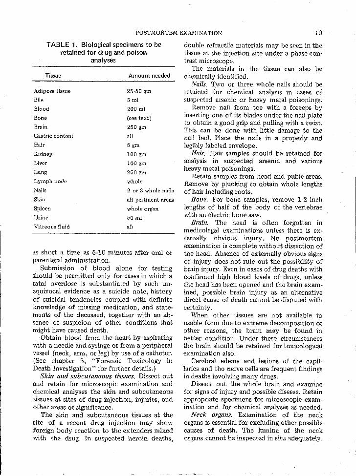
Selection of the tissues and the amounts will vary with the tests to be performed.3 The amounts suggested in table 1 are the approximate minimum"
Blood. Blood should always be saved for testing whenever possible in an medical examiner/coroner's cases. Submission of blood alone, however, for toxicological examination in drug-related deaths is fraught with danger. Many drugs are rapidly removed from circulati~n. Unless .highly sophisticated technological SkIlls and ll1struments are available, some drugs may not be detectable in blood within
3See also chapter 5, "Forensic Toxicology in Death Investigation."
POSTMORTEM EXAl\UNATION 19
TABLE 1. Biological specimens to be retained for drug and poison
analyses
Tissue Amount needed
Adipose tissue 25-50 gm
Bile 5 ml
Blood 200 ml
Bone (see text)
Brain 250 gm
Gastric content all
Hair 5 gm
Kidney 100 gm
Liver 100 gm
Lung 250 gm
Lymph node whole
Nails 2 or 3 whole nails
Skin all pertinent areas
Spleen whole organ
Urine 50 ml
Vitreous fluid all
as short a time as 5-10 minutes after oral or parenteral administration.
Submission of blood alone for testing should be permitted only for cases in which a fatal overdose is substantiated by such unequivocal evidence as a suicide note, history of suicidal tendencies coupled with definite knowledge of missing medication, and statements of the deceased, together with an absence of suspicion of other conditions that might have caused death.
Obtain blood from the heart by aspirating with a needle and syringe or from a peripheral vessel (neck, arm, or leg) by use of a catheter. (See chapter 5, "For~nsic Toxicology in Death Investigation" for fUrther details.)
Skin and subcutaneolls tissues. Dissect out and retain for microscopic examination and chemical analyses the skin and SUbcutaneous tissues at sites of drug injection, injuries, and other areas of significance.
The skin and subcutaneous tissues at the site of a recent drug injection may show foreign body reaction to the extenders mixed with the drug. In suspected heroin deaths,
double refractile materials may be seen in the tissue at the injection site under a phase contrast microscope.
The materials in the tissue can also be chemically identified.
Nails. Two or three whole nails should be retained for chemical analysis in cases of suspected arsenic or heavy metal poisonings.
Remove nail from toe with a forceps by inserting one of its blades under the nail plate to obtain a good grip and pulling with a twist. This can be done with little damage to the nail bed. Place the nails in a properly and legibly labeled envelope.
Hair. Hair samples should be retained for analysis in suspected arsenic and various heavy metal poisonings.
Retain samples from head and pubic areas. Remove by plucking to Qbtain whole lengths of hair including roots.
Bone. For bone samples, remove 1-2 inch lengths of half of the body of the vertebrae with an electric bone saw.
Brain. The head is often forgotten in medicolegal examinations unless there is externally obvious injury. No postmortem examination is complete without dissection of the hdad. Absence of externally obvious signs of injury does not rule out the possibility of brain injury. Even in cases of drug deaths with confirmed high blood levels of drugs, unless the head has been opened and the brain examined, possible brain injury as an alternative direct cause of death cannot be disputed with certainty.
When other tissues are not available in usable form due to extreme decomposition or other reasons, the brain may be found in better condition. Under these circumstances the brain should be retained for toxicological examination also.
Cerebral edema and lesions of the capillaries and the nerve cells are frequent findings in deaths involving many drugs.
Dissect out the whole brain and examine for signs of injury and possible disease. Retain appropriate specimens for microscopic examination and for chemical analyses as needed.
Neck organs. Examination of the neck organs is essential for excluding other possible causes of death. The lumina of the neck organs cannot be inspected in situ adequately.

20 NOGUCHI
Remove by a block dissection the tongue, pharynx, larynx, and the trachea. Dissect and examine for:
1. Evidence of trauma to the soft tissues 2. Fractures of the hyoid or other bony or
cartilaginous structures 3. Submucosal laryngeal or epiglottal hema-
tomata 4. Edema and petechial hemorrhages 5. Other signs of abnormal conditions 6. Foreign body in the airways Cardio)IGSClllar system. The heart and the
aorta should be carefully examined, especially in older persons, to rule out deaths due to cardiovascular diseases. Many drugs, including those used for the treatment of hemi: diseases, will cause changes in the heart tissues.
Remove the aorta and the whole heart from the body. Carefully examine the pericardium and the surface of the heart. Dissect and examine the coronary vessels for arteriosclerotic changes. Dissect and check the linings of the aorta and the heart chambers and the valves for Rr'y pathological changes. Check the whole heart for myocardial abnormalities.
Retain tissues frorn appropriate areas for microscopic examinations.
Lungs. In cases where inhalation of gas or chemical vapor is the suspected cause of death, sections of the lung tissue should be retained for chemical and microscopic analyses.
In deaths due to intravenously injected "street drugs," the lungs often show foreign body reactions to the extenders used in these preparations. Foreign body reactions are also seen in cases of chronic respiratory exposure to many industrial chemicals.
In cases of deaths due to suspected gas poisoning, tie off the bronchus, dissect above the tie, remove the whole lung, and quickly place it in an airtight metal container. Avoid cutting into the lung tissue which would cause the lung to collapse with escape of the gas content. Most gases will readily diffuse out of the lungs into the container. Gas samples may be rel'i.10ved from the container by puncturing it and filling a syringe for analysis by gas chromatography.
Gastrointestinal tract. The lining and contents of the entire digestive tract starting from the oropharynx and proceeding through the
esophagus, stomach, and the intestines should be dissected and carefully examined.
Mm1Y of the commonly ingested drugs are artificially colored, and their presence in the digestive tract may be readily detected. The color, amount, and location of the ingested drug in its original form are important clues in the overall interpretation of the circumstances of drug-related deaths.
Such findings as the type of food and the amount and stage of digestion are also significant and should be noted and recorded as important clues to the sequence of events preceding the death.
In all cases of death suspected of being due to or?lly ingested substances, the entire contents of the stomach and intestine should be retained for chemical and other appropriate laboratory analyses. This protocol applies to deaths suspected of being due to any ingested materials, including foods suspected of being contaminated with bacteria, pesticides, or poisons, as well as to cases due to simple drug ingestion.
Stomach. Tie or clamp off the stomach at both ends, above the diaphragm and at the duodenal junction, and remove intact from body. Dissect and collect entire contents into a lm-ge chemically clean; properly labeled jar. Examine stomach lining and contents. Record by detailed descriptions and photographs all significant findings. Retain tissues from m-eas showing significant deviations from the normal for microscopic examination.
Intestines. Tie off the intestine into easily handled segments (1-2 ft) and remove the whole length from the body. Open each segment separately and retain contents of each segment in separate containers which are properly identified with the location of the segment as well as the information identifying the case. Carefully examine contents and the mucosal lining. Record all significant findings and retain tissues for microscopic examination.
Liver. Liver should be retained in drugrelated deaths, for many drugs are rapidly concentrated in the liver for detoxification, and appreciable quantities may be found in the liver in acute drug deaths while blood concentration may be minimal.
Portal triaditis has been a consistent finding
t , I
POSTMORTEM EXAMINATION 21
in acute narcotism; in heroin addicts, the lymph nodes in the portal region are often enlarged and contain deposits of the drug.
In cases of chronic exposure to hepatotoxic chemicals and drugs, cirrhosis and other degenerative changes of the liver are commonly observed. Prior damage to the liver will prevent the proper detoxification of subsequently administered drugs, resulting in a detrimental reaction to a normally nontoxic dose of a drug. For proper eValuation of drug-induced d'eaths, microscopic study of liver sections is essential.
Dissect out the whole liver; examine for gross pathological changes; retain appropriate areas for microscopic examinations and for drug analyses as needed.
Gallbladder and bile. Many drugs are concentrated and retained in the gallbladder for as long as 48 hours and high concentrations in the bile are usually indicative of chronic drug usage.
Remove the whole gallbladder with contents and retain bile in properly labeled jar for chemical analyses as needed.
Kidneys and urine. Some portion of practically all of the drugs and chemicals taken into the body will be excreted or resorbed into the body through the kidneys. Many of these substances are nephrotoxic and damage the kidneys. In addition, preexisting damage to the kidneys will affect the excretion of a drug and may prevent its proper removal from the body with consequent greater concentration and toxic effect on the body for a given dosage.
High concentration of a drug in the urine is usually indicative of chronic usage.
Use catheter to obtain urine. Measure the total volume in the bladder and retain amount needed for required testing.
Remove both kidneys. Dissect and examine for abnormalities. Retain appropriate specimens for mi.croscopic and chemical analyses.
INTERPRETIVE FORENSIC TOXICOLOGY
Toxicology has a definite place in the investigation of drug-related deaths. The recent advances in technology and instrumentation for chemical analyses of biological materials
have made routine procedures of previously time-consuming and tedious techniques for detecting drugs in body fluids and tissues. Without the proper knowledge to interpret the findings in relation to the functioning of the total living hUman body, all the toxicological data from the analyses of blood and other body fluids and tissues are valueless. Understanding of the physiological action and the metabolic pathways through which the body processes the various foreign chemicals wi th which we intentionally or unintentionally insult our bodily systems is required. The latter is a complex specialized field in itself, and much of the needed information concerning many of the currently used drugs is still incomplete.
For proper interpretation of the postmortem laboratory test findings, the following factors must be taken into conside!'ation;
1. Route of administration 2. Dosage in relation to the size of the indi
vidual 3. Interval between the time of intake and the
time of death 4. Normal route and !'ate of metabolism and
excretion of the drug 5. Physical condition of the individual that
might affect the normal body disposal of the drug such as liver or kidney disease
6. Physiological condition of the individual relative to the specific drug, whether habituated, sensitized, or without previous contact
7. Simultaneous presence in the body of alcohol or other drugs which might have a potentiating effect
8. Susceptibility of the drug to postmortem degradation and the interval of time after death when the specimen was taken and analyzed or treated for preservation
9. Individual genetic variations in enzyme systems which dispose of the drugs and foreign chemicals taken into the body
Due to all of the (bove factors which influ-ence the drug levels in the various body fluids and tissues, the lethal dosage will vary widely with the individual as will the postmortem levels found in the blood and tissues following the ingestion of a given amount of a drug.
'.
22 NOGUCHI
Appendix B lists data on some potentially toxic chemieals.4
4Sce also chapter 5, "Forensic Toxicology in Death Invesligation," and chapter 4, "Treatment for Survival Prior to Death and Interpretation 0: Postmortem 'l'oxicologic Findings," for further discussion of this topie.
Addendum: Toxicological examination of skeletal remains:
In a recent case the five lumbar vertebrae of a dried mummified remains were used to determine a toxicological cause of death. The volume of the marrow was used to estimate the concentration of the drug found.