poster abstracts - news — anatomische...
TRANSCRIPT
Poster Abstracts
Joint Meeting 2009 Anatomische Gesellschaft –
Nederlandse Anatomen Vereniging
104th Annual Meeting of the Anatomische Gesellschaft
March 27-30, 2009, Antwerpen, Belgium

The file includes all abstracts of 153 posters except poster: 140-144 as no abstracts have been submitted. Posters 145-153 have been hand in later and are not integrated in the alphabetical list. You will find them at the end of the file. To find your abstract or an abstract of interest please use the alphabetical list of first authors of lectures and posters starting on next page or use the abstract number which refers to the lecture number given in the meeting program. Example: Rubrik: 3.Methods/Teaching Abstract Nr.:80 This means poster 80.
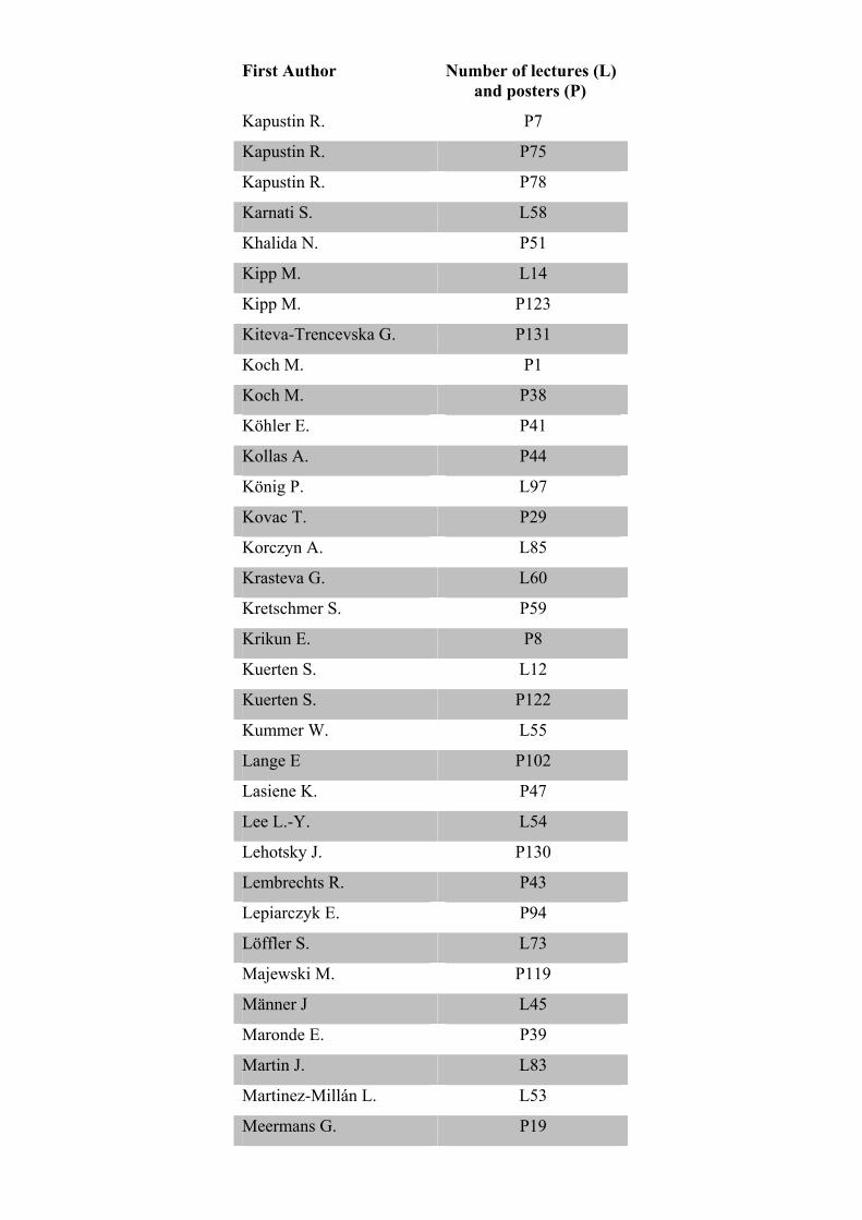
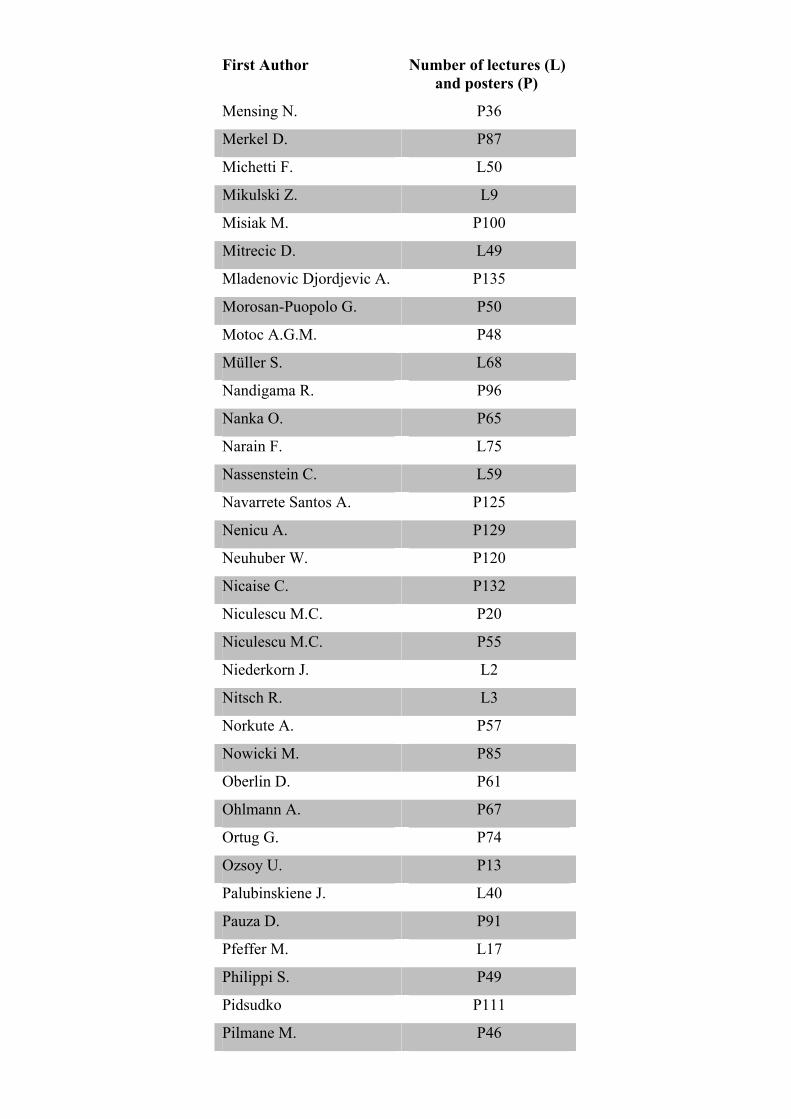
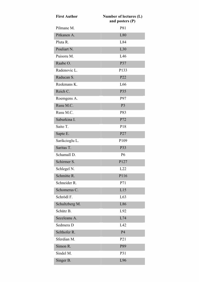
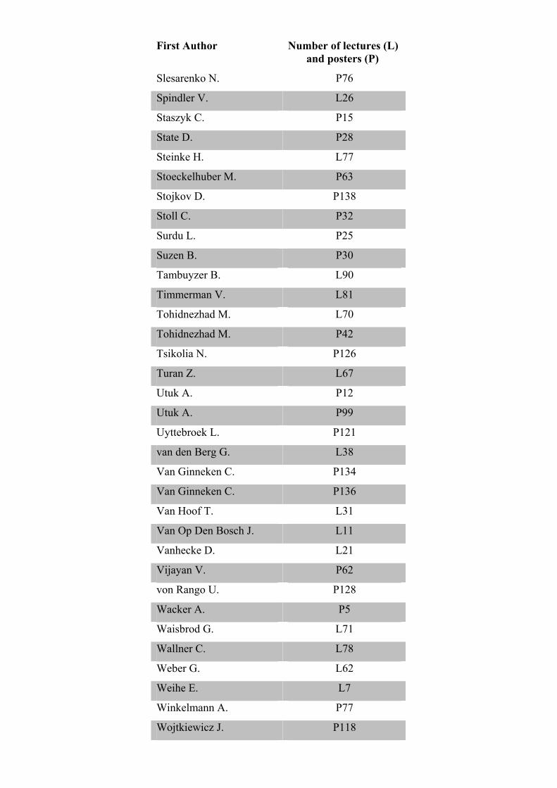
Alphabetical List of First Authors of Lectures and Posters:
First Author Number of lectures (L)
and posters (P)
Adam N. P53
Adriaensen D. L56
Albertine K. L57
Amit B. L32
Antohe D. P103
Antohe D. P104
Antohe D. P110
Antohe I. L76
Arnold S. L65
Avula L. P45
Bakkers J. L35
Balciuniene N. P82
Balnyte I. L43
Banaz-Yasar F. L23
Barcal J. L51
Barham M. L91
Bataveljic D. P139
Baumgart-Vogt L88
Bechmann I. L5
Becker J. L25
Bendella H. P88
Bender R. P105
Bender R. P107
Bergwerf I. L13
Boeckxstaens G. L4
Bömmel H. P115
Bongartz B. P73
Bordei P. P24
Brandenburg L. L69
Bräuer L. P64
Braunger B. M. P106
Brichová H. P108
First Author Number of lectures (L)
and posters (P)
Brion J. L48
Buttler K. P68
Buttler K. P69
Caglayan B. P140
Callebaut M. P58
Calvo A.C. P141
Carmeliet P. L1
Carmichael S. L27
Cengiz M. P23
Chiriac S. P9
Christoffels V. L36
Clarner T. P112
Clemente D. P143
Closhen C. P101
Copray S. L89
Costagliola A. P93
Csaki C. L72
Dai F. P52
Dang J. P79
Davidson B. L34
de Anta J. P66
De Spiegelaere W. L93
De Spiegelaere W. P92
De Wilde L. L29
Demirel B. P11
Dierickx C. L28
Djonov V. L41
Donmez B. P14
Efthymiadis A. L24
El Sayed K. P34
Eppler E. L98
Erdogan A. P2
Ergün S. L39
Ertekin C. P117
First Author Number of lectures (L)
and posters (P)
Fester L. L19
Frandes C. P10
Friederike P. P114
Frindte J.N. L44
Fuchshofer R. P40
Gebert A. L10
Gerrits P. P90
Gajovic S. L99
Glavan G. P137
Gogulescu B. P84
Gomes G. L33
Gonçalves R. P142
Gruart A. L82
Haberberger R. L61
Hammer N. L87
Hannan A. L47
Hausott B. L16
Heimrich B. L18
Hendrix S. L8
Herrler A. P80
Hesse B. L79
Hoelbling-Patscheider D. P70
Horn F. P86
Hueller H. L95
Iliescu M. P26
Illig R. L94
Izquierdo M.A. P144
Jafarpour A. P98
Jay T. L52
Jianu A.M. P54
Johann S. L64
Johann S. P95
Jones K. L6
Kaleczyc J. P113
First Author Number of lectures (L)
and posters (P)
Kapustin R. P7
Kapustin R. P75
Kapustin R. P78
Karnati S. L58
Khalida N. P51
Kipp M. L14
Kipp M. P123
Kiteva-Trencevska G. P131
Koch M. P1
Koch M. P38
Köhler E. P41
Kollas A. P44
König P. L97
Kovac T. P29
Korczyn A. L85
Krasteva G. L60
Kretschmer S. P59
Krikun E. P8
Kuerten S. L12
Kuerten S. P122
Kummer W. L55
Lange E P102
Lasiene K. P47
Lee L.-Y. L54
Lehotsky J. P130
Lembrechts R. P43
Lepiarczyk E. P94
Löffler S. L73
Majewski M. P119
Männer J L45
Maronde E. P39
Martin J. L83
Martinez-Millán L. L53
Meermans G. P19
First Author Number of lectures (L)
and posters (P)
Mensing N. P36
Merkel D. P87
Michetti F. L50
Mikulski Z. L9
Misiak M. P100
Mitrecic D. L49
Mladenovic Djordjevic A. P135
Morosan-Puopolo G. P50
Motoc A.G.M. P48
Müller S. L68
Nandigama R. P96
Nanka O. P65
Narain F. L75
Nassenstein C. L59
Navarrete Santos A. P125
Nenicu A. P129
Neuhuber W. P120
Nicaise C. P132
Niculescu M.C. P20
Niculescu M.C. P55
Niederkorn J. L2
Nitsch R. L3
Norkute A. P57
Nowicki M. P85
Oberlin D. P61
Ohlmann A. P67
Ortug G. P74
Ozsoy U. P13
Palubinskiene J. L40
Pauza D. P91
Pfeffer M. L17
Philippi S. P49
Pidsudko P111
Pilmane M. P46
First Author Number of lectures (L)
and posters (P)
Pilmane M. P81
Pitkanen A. L80
Pluta R. L84
Pouliart N. L30
Puisoru M. L46
Raabe O. P37
Radenovic L. P133
Raducan S. P22
Reekmans K. L66
Reich C. P35
Roemgens A. P97
Rusu M.C. P3
Rusu M.C. P83
Saburkina I. P72
Saito T. P18
Sapte E. P27
Sarikcioglu L. P109
Saritas T. P33
Schamall D. P6
Schirmer S. P127
Schlegel N. L22
Schmitte R. P116
Schneider R. P71
Schomerus C. L15
Schrödl F. L63
Schultzberg M. L86
Schütz B. L92
Seceleanu A. L74
Sedmera D L42
Selthofer R. P4
Sferdian M. P21
Simon R. P89
Sindel M. P31
Singer B. L96
First Author Number of lectures (L)
and posters (P)
Slesarenko N. P76
Spindler V. L26
Staszyk C. P15
State D. P28
Steinke H. L77
Stoeckelhuber M. P63
Stojkov D. P138
Stoll C. P32
Surdu L. P25
Suzen B. P30
Tambuyzer B. L90
Timmerman V. L81
Tohidnezhad M. L70
Tohidnezhad M. P42
Tsikolia N. P126
Turan Z. L67
Utuk A. P12
Utuk A. P99
Uyttebroek L. P121
van den Berg G. L38
Van Ginneken C. P134
Van Ginneken C. P136
Van Hoof T. L31
Van Op Den Bosch J. L11
Vanhecke D. L21
Vijayan V. P62
von Rango U. P128
Wacker A. P5
Waisbrod G. L71
Wallner C. L78
Weber G. L62
Weihe E. L7
Winkelmann A. P77
Wojtkiewicz J. P118

First Author Number of lectures (L)
and posters (P)
Wolff M. P124
Wörl J. P56
Yasuo S. L20
Yutzey L37
Zahoi D. P16
Zahoi D. P17
Zidan M. P60
Abstracts Rubrik: 1.Main Topic I Abstract Nr.:1 Titel:Cannabinoids and their role in neuroprotection Autoren: Koch M.(1),Kreutz S.(1),Böttger C.(1),Ghadban C.(1),Korf H.(1),Dehghani F.(1), Adressen:(1)Dr. Senckenbergische Anatomie, Institut für Anatomie II|Goethe-Universität Frankfurt am Main|Frankfurt|Germany; email:[email protected] Abstract: The neuroprotective potential of cannabinoids varies significantly among the different members of the cannabinoid family. Whereas the endocannabinoid 2-AG is reported to elicit strong neuroprotective effects, the protective role of anandamide and Delta 9-tetrahydrocannabinol (THC) is controversially discussed. Repair mechanisms after brain lesions involve complex interactions between several cell types including neurons, astrocytes and microglial cells. Given this complexity, the cellular targets of endocannabionids need to be identified in order to understand the mechanisms that underly their neuroprotective potential. We have addressed this question by using organotypic hippocampal slice cultures (OHSC). In OHSC, neuronal lesions and glial activation can be induced by application of excitotoxic agents such as NMDA and the effectiveness of pharmacological compounds can readily be monitored. Using OHSC we found that cannabinoids (THC, anandamide or 2-AG) significantly decreased the number of microglial cells in the dentate gyrus after excitotoxic injury. However, the number of degenerating neurons was reduced only by 2-AG treatment. A similar neuroprotective effects was observed after activation of the abn-CBD-sensitive receptor on microglial cells. Application of two abn-CBD-sensitive receptor specific antagonists, O-1918 or cannabidiol (CBD) antagonized this effect. When microglial cells were depleted from the OHSC, 2-AG and abn-CBD lost their neuroprotective potential. These data suggest that the endocannabinoid 2-AG exerts its neuroprotective effects via activation of abn-CBD-sensitive receptors on microglial cells. Our investigations also illustrate that different members of the cannabinoid family elicit specific effects on distinct cell types and allow a better understanding of their role in intrinsic brain repair mechanisms. Kategorie: Poster
Rubrik: 1.Main Topic I Abstract Nr.:2 Titel:A study on the reflex mother rats for saving their babies Autoren: Erdogan A.(1),Cevli C.(2),Aydin M.(3),Sevinc O.(4), Adressen:(1)Anatomy|Çomu Medical Faculty|Canakkale|Turkiye; email:[email protected]; (2)Anatomy|Ataturk University Medical Faculty|Erzurum|Turkiye; (3)Neurosurgery|Ataturk University Medical Faculty|Erzurum|Turkiye; (4)Anatomy|Çomu Medical Faculty|Çanakkale|Turkiye Abstract: There are various studies reported for animal behaviors on subjects such as couple selection for copulation, saving from wild birds, perception limits without escape response from danger and economic decision, decisiveness for fight. There is no study on mother rats for rescuing their babies from a dangerous situation. We studied the instinctive behavior of 17 mother rats for rescuing their babies. Each baby group of mother rat was numbered as a separate group. 17 groups were composed of 4 groups with 3 babies, 3 groups with 4 babies, 3 groups with 5 babies, 2 groups were with 6 babies, 2 groups with 7 babies and 2 groups were with 9 babies, 1 group was with 10 babies. Mother rats and babies were studied separately as they were kept in their fences silently. A disturption of silence and peace was started by knocking their fences in order to check their behaviors. Mother rats sensing the danger and they were found to rescue one of the babies to a place away from the danger either directly or after checking the weight of some others. We weighted all babies including the rescued baby rat. In all the 14 groups except 3 mother rats were found to have rescued the heaviest baby. We predicted that the sex difference was not a point in selection and the weight difference was neglected for following rescues after the first. Kategorie: Poster
Rubrik: 1.Main Topic I Abstract Nr.:3 Titel:A morphological and topographical study of the trigeminal ganglion neurons in humans, with emphasis on the small-diameter trigeminal neurons Autoren: Rusu M.C.(1),Pop F.(2),Ivascu R.V.(1),Ciuluvica R.C.(1), Adressen:(1)Anatomy and Embryology|University of Medicine and Pharmacy Carol Davila|Bucharest|Romania; email:[email protected]; (2)Pathologic Anatomy|University of Medicine and Pharmacy Carol Davila|Bucharest|Romania Abstract: It is debated whether or not neurogenesis/neuronal maturation occur within the sensory peripheral ganglia. We aimed to investigate the human adult trigeminal ganglion (TG) neurons: their topography, morphology and immunoreactions with Neurofilament (NF), substance P (SP), the nociceptive peptide, and, supplementary, with Tyrosin Hydroxylase (TH) and Choline Acetyltransferase (ChAT), in order to get evidences that may be correlated with recent scientific evidences. We dissected free 10 TGs; hematoxylin-eosin staining was followed by immunohistochemistry for NF, SP, TH and ChAT. We identified 2 types of neurons: large and small. The large trigeminal neurons (ltn) showed tendencies to clustering and were strictly located within the macroscopically identified TG. The small trigeminal neurons (stn) were NF(+), TH(-) and ChAT(-). Proximally to the TG, within the triangular plexus, were homogenous clusters of stn, of 6 μm, strongly SP(+); no glial cells were present in these stn populations. Proximal and distal to the ltn population were neuronal clusters and microganglia consisting of heterogeneous populations of neurons (with mild to strong reactions for SP) and glial cells, suggestive for the presence of the processes of maturation; mainly, there were stn of 9 μm but, in some of these clusters/microganglia we identified neurons of 24 μm located within the core of the structure. The presence of 2 types of populations of small neurons within the TG in human, with different cellular compositions, may serve as a morphological basis to consider the occurrence of both processes of neurogenesis and maturation in the adult TG. Kategorie: Poster
Rubrik: 4.Gross Anatomy/Clinical Anatomy Abstract Nr.:4 Titel:Dynamic model of cyclic load on fetal membranes in simulated intrauterine conditions Autoren: Selthofer R.(1),Radic R.(1),Nikolic V.(1),Leksan I.(1),Mrcela T.(2),Dinjar K.(1), Adressen:(1)Department of Anatomy|School of Medicine, University of J. J. Strossmayer|Osijek|Croatia; email:[email protected]; (2)Department of mechanical testing|Faculty of Electrical Engineering, University of J. J. Strosmayer|Osijek|Croatia Abstract: Intact fetal membranes are significant for pregnancy and fetus development. Obstetricians meet with premature rupture of fetal membranes in about 20-25 % of all term pregnancies, and more importantly, with preterm premature rupture of fetal membranes that causes premature childbirth, which occurs in about 2-3 % of all pregnancies. Every rupture of fetal membranes means the beginning of the delivery. Premature childbirth is related to considerably higher perinatal mortality and morbidity. Since the mechanical factor role (regular contractions during the childbirth) in weakening of the membranes in both term and premature childbirth is still unexplained, we designed a dynamic model of cyclic load on fetal membranes in simulated intrauterine conditions. Investigation was performed on 35 fetal membranes from Department of Obstetrics Gynecology in Clinical Hospital Osijek. Biomechanical studies were carried out in a special device for dynamic load of connective structures. Specimens were tested by simulating physiological intrauterine pressures in the rhytm of contractions during normal labour to determine firmness and deformation in gradual structure disruption of fetal membranes by intermitent load. The study included only the membrane specimens obtained from the patients whose bacteriology cervix smear test during pregnancy was negative. Characteristics of preterm and term prematurely ruptured fetal membranes are determined. This study underlyine mechanical factor in membrane weakening in both preterm and term pregnancies, as well as the differences in their biophysical characterictics. Results will help in better understanding pathophysiology of preterm labour. Kategorie: Poster
Rubrik: 4.Gross Anatomy/Clinical Anatomy Abstract Nr.:5 Titel:A prominent fibular artery as outflow vessel for crural bypass Autoren: Wacker A.(1),Löffler S.(2),Ulrich M.(3),Bräunlich S.(3),Adili F.(4),Feja C.(1),Spanel-Borowski K.(1), Adressen:(1)Anatomy|University of Leipzig|Leipzig|Germany; email:[email protected]; (2)Angiology|University of Leipzig|Leipzig|Germany; (3)Angiology|Park-Krankenhaus Leipzig-Südost GmbH|Leipzig|Germany; (4)Vascular and Endovascular Surgery|Goethe-University Frankfurt|Frankfurt|Germany Abstract: A crural bypass utilises the anterior tibial artery, the posterior tibial artery or the fibular artery as outflow vessels. Whether the bypass via the fibular artery is helpful, is under debate. We have recently analysed variations of arteries in the lower leg of alcohol-fixed cadavers because of the clinical implication of the appropriate surgical intervention. Medical imaging is important to detect such individual variations in patients with diabetes foot syndrome. Therefore digital substraction angiography was performed to examine the blood vessels of lower legs after Thiel-fixation. Among all the specimen particularly one displayed a predominant fibular artery communicating with foot arteries which would have been suitable for bypass grafting. Potential inflow vessels are the popliteal-, the superficial femoral- or the common femoral arteries. Each bypass requires the graft from autologous veins derived from the lower leg, the upper leg, the upper and lower body. Even though a prominent fibular artery with connection to the foot arteries is seldom, it allows because of the developmental derived higher calibre its use as runoff vessel. Kategorie: Poster
Rubrik: 4.Gross Anatomy/Clinical Anatomy Abstract Nr.:6 Titel:Computed tomographical analysis of the internal architecture of the first metatarsal bone: a new application of high-resolution radiographic method for age estimation in historic bones. Autoren: Schamall D.(1),Cink V.(1),Teschler-Nicola M.(2),Loewe C.(3),Kainberger F.(4),Pretterklieber M.(1), Adressen:(1)Department of Applied Anatomy|Center of Anatomy and Cell Biology, Medical University of Vienna|Vienna|Austria; (2)Department of Anthropology|Natural History Museum Vienna|Vienna|Austria; (3)Department of Cardiovascular and Interventional Radiology|Medical University of Vienna|Vienna|Austria; (4)Department of Neuroradiology and Musculoskeletal Radiology|Medical University of Vienna|Vienna|Austria; email:[email protected] Abstract: The estimation of age-at-death in (pre-)historic human bones can be carried out either by several macroscopic methods (most accurately by the so-called “Combined Method”) or by microscopic methods. The “Combined Method” includes the evaluation of morphological changes at the facies symphysialis of the pubic bone, the degree of obliteration of the endocranial sutures as well as the structure of the cancellous bone within the proximal end of the humerus and femur. Since the preservation of the epiphyses is often impaired by post-mortem destructions, the reliability of this approach is strongly correlated with the amount of intact skeletal elements. In turn, microscopic techniques base on an invasive access due to sample preparation. This presentation introduces preliminary results of an ongoing study, where we used the potential of the first metatarsal bone for age estimation. This bone is frequently found during archeological recoverings and commonly has a very good preservation status. We used the non-destructive method of computed tomography with images generated on a 64-row-CT-scanner. We also were interested to shed light on the sexual dimorphism and laterality (footedness) on the trabecular pattern in regard to age-related changes. Thus, the data of 200 metatarsal bones of individuals with known age and sex were analyzed. We observed a trend of gradual enlargement of the marrow space during the aging processes, together with manifestation of distinct alterations, i.e. hypointense areas within the subchondral bone. It is discussed that sex of the individual and footedness influence the overall morphology of the first metatarsal bone. Kategorie: Poster

Rubrik: 4.Gross Anatomy/Clinical Anatomy Abstract Nr.:7 Titel:Morphogenesis of bone marrow and clinical anatomy of cattle's liver in early postnatal ontogenesis Autoren: Kapustin R.(1),Vasilyeva A.(1), Adressen:(1)Department of Animal Morphology|Belgorod State Agricultural Academy|Maiskii Belgorodskoi oblasti|Russia; email:[email protected] Abstract Withdrawn Kategorie: Poster
Rubrik: 4.Gross Anatomy/Clinical Anatomy Abstract Nr.:8 Titel:Regional peculiarities in indexes of physical development of Russian children: Moscow and Belgorod region Autoren: Krikun E.(1),Boldyr V.(1),Kapustin R.(2),Krikun Y.(1), Adressen:(1)Department of Human Anatomy and Histology|Belgorod State University|Belgorod|Russia; (2)Department of Animal Morphology|Belgorod State Agricultural Academy|Maiskii Belgorodskoi oblasti|Russia; email:[email protected] Abstract Withdrawn Kategorie: Poster
Rubrik: 4.Gross Anatomy/Clinical Anatomy Abstract Nr.:9 Titel: Progressive evolution of a forearm sarcoma and its functional impact on the affected anatomical muscle groups- a case report Autoren: Chiriac S.(1),Dumitriu A.(1),Iordache I.(1),Unc O.(1), Adressen:(1)Surgery Clinic 2|Faculty of Medicine,University Ovidius|Constanta|Romania; email:[email protected] Abstract: The evolution of a patient diagnosed with sarcoma of the forearm was followed up for a period of 17 years ,from the stage of infracentrimetrical tumor to repeated amputation of forearm,arm and subsequently disarticulation of the scapulohumeral joint.Images are presented in such a way enabling the step by step propagation of the disease in relation to the changes in the anatomical muscle groups from the upper limb.The mechanisms for dissemination of sarcoma, the effective cancer treatment and the evolutionary tumor aggressiviness linked to this particular case are discussed. The specific evolution of a particular sarcoma is hard to predict, but this case report shows that the spread and impact of the tumor on the anatomical distribution and arrangement of the muscles progresses in a centripetal way. Kategorie: Poster

Rubrik: 4.Gross Anatomy/Clinical Anatomy Abstract Nr.:10 Titel:Macroscopic evaluation indexes of placentas from premature births with fetuses presenting developmental abnormalities Autoren: Frandes C.(1),Radu A.(2), Adressen:(1)Anatomy and Embryology|Vest University"Vasile Goldis", Faculty of Medicine|Arad|Romania; email:[email protected]; (2)Pathology|Vest University" Vasile Goldis",Faculty of Medicine|Arad|Romania Abstract: There are statistical proof of a significant increase in the number of pregnancies with fetuses that present growth problems and premature births during the past five years. These facts are in favor of an accurate and detailed research on the fetus – placental unit. The purpose was to establish the importance of the macroscopic placental abnormalities in such cases. The present study was conducted on 160 placentas from premature births that were associated with developmental problems. Our study focuses on the macroscopical features of the placentas. The evaluated parameters are: placental weight, size, shape, surface and the ratio between placental and fetal weight. Regarding weight, size, shape and surface, the values of most of the cases included in this study are in line with earlier literature data. Some cases, however, show discrepant values as far as weight, size, ration between placental and fetal weight are concerned. The parameter ‘shape’ never appeared to be different from data described in the literature so far. After studying such a large number of placentas we must specify that in our opinion is mandatory to introduce a new pregnancy predictor parameter such as the exchange surface which in fact represents the contact surface between the placenta and the uterine wall occupied by it Kategorie: Poster
Rubrik: 4.Gross Anatomy/Clinical Anatomy Abstract Nr.:11 Titel: Three-dimensional digitized location of supraorbital and infraorbital foramens referred to anthropometric and anatomic landmarks: a pilot study Autoren: Demirel B.(1),Ozsoy U.(1),Utuk A.(2),Donmez O.(2), Adressen:(1)Anatomy|Faculty of Medicine|Antalya|Turkey; email:[email protected]; (2)Anatoym|Faculty of Medicine|Antalya|Turkey Abstract: To three dimensionally digitize and measure the distance between supraorbital, infraorbital foramens and the anthropometric landmarks on cranium. Supraorbital foramen, infraorbital foramen, glabella and nasion, as anthropometric landmarks, and also inferior border of the nasal bone and anterior nasal spine were digitized by Microscribe 3D digitizing system on 10 craniums. The distances between the supraorbital foramen and glabella, nasion, inferior border of the nasal bone and anterior nasal spine were measured by digitizing system’s software and the results were evaluated by mean and standard deviation. The same distances were measured and evaluated for the infraorbital foramen. We measured the distances between the supraorbital foramen and glabella, nasion, inferior border of the nasal bone and the anterior nasal spine as 27,28±6,08 mm; 28,00±6,03 mm; 38,23±6,61 mm; 67,03 ± ±5,38 mm, respectively. The same distances were measured as 53,00 ±3,94 mm; 46,70 ±3,45 mm; 39,55 ±3,54 mm; 34,91 ±3,09 mm, respectively for infraorbital foramen. We think that having known the distance of the supraorbital and infraorbital foramens to anthropometric and anatomical landmarks can be useful for craniomaxillofacial surgery to predict the location of the supraorbital and infraorbital nerves. Kategorie: Poster
Rubrik: 4.Gross Anatomy/Clinical Anatomy Abstract Nr.:12 Titel:Digital measurements of the nasal aperture: a pilot study Autoren: Utuk A.(1),Demirel B.(1),Donmez O.(1),Ozsoy U.(1), Adressen:(1)Anatomy|Faculty of Medicine|Antalya|Turkey; email:[email protected] Abstract: Our aim was to three dimensionally digitize the nasal aperture and measure the length, height, upper and lower widths of the nasal aperture on 10 cranium. The nasal aperture was digitized by microscribe digitizing system. In order to obtain the three-dimensioned data of the nasal aperture we choose 8 points on the nasal aperture, which are inferior border of the nasal bone and the anterior nasal spine as anatomical landmarks and other 6 points (3 points on the left and 3 points on the right side) of the aperture. The length, height, upper and lower widths of the nasal aperture were measured by digitizing system’s software and the results were evaluated by mean and standard deviation. We measured the length, height, upper and lower widths of the nasal aperture as 111,16± 7,94 mm; 37,28±4,14 mm; 16,48±1,99 mm and 24,68±2,02 mm respectively. We think that such data will give a reference for surgeons to predict anatomical arrangement of the soft tissue compared to the borders of the nasal aperture. Kategorie: Poster
Rubrik: 4.Gross Anatomy/Clinical Anatomy Abstract Nr.:13 Titel:The comparison of the femoral curve with femoral nails: a digital analysis Autoren: Ozsoy U.(1),Donmez B.(1),Demirel B.(1),Oguz N.(1),Urguden M.(2), Adressen:(1)Department of Anatomy|Faculty of Medicine|Antalya|Turkey; email:[email protected]; (2)Department of Orthopedics and Travmatology|Faculty of Medicine|Antalya|Turkey Abstract: To evaluate the curve of the femur according to anatomical axis of femur and to compare with curve of the femoral nail used in Turkish population. In the present study, 68 left and 66 right femurs were measured. The shaft curve of the femur according to anatomical axis of femur and curve of the four nails was measured using MicroScribe G2X digitizer and the data were obtained by Surfcam Velocity software. In the present study, the mean value of the curve of the femur according to anatomical axis was calculated as 16,713±2,371º in the 66 right femurs and the mean value of the curve of the femur according to anatomical axis was calculated as 16,148±2,689º in the 68 left femurs. We calculated that curve of the four nails were hipocrat 8,65º, trigen 10,20º, orthopro 6,38º and recon 8,70º, respectively. We think that curve of the femur should be kept in mind during surgical procedures and using nail from proximal of femur, and also during nail production, this angle should be considered by medical companies. Kategorie: Poster
Rubrik: 4.Gross Anatomy/Clinical Anatomy Abstract Nr.:14 Titel:The effect of angiotensin receptor blocker on osteoporosis in overiectomized rat’s femurs: a pilot study Autoren: Donmez B.(1),Koc P.(2),Ozdemir S.(3),Yaras N.(3),Demir N.(4),Karayalcin B.(5),Oguz N.(1), Adressen:(1)Department of Anatomy|Faculty of Medicine|Antalya|Turkey; email:[email protected]; (2)Department of Nuclear Medicine|Firat University|Elazig|Turkey; (3)Department of Biophysic|Faculty of Medicine|Antalya|Turkey; (4)Department of Histology and Embriyology|Faculty of Medicine|Antalya|Turkey; (5)Department of Nuclear Medicine|Faculty of Medicine|Antalya|Turkey Abstract: To investigate the effects of angiotensin receptor blocker treatment on bone mineral density of overiectomized rat’s femur. In this study, fifteen female Wistar rats were used. Ten of these animals were overiectomized by ventral incisions and five animals were used as control group. Ovariectomized rats separated into two groups. Losartan (5 mg/kg/day) as angiotensin receptor blocker was dissolved in water and administered via oral gavage after 12 weeks of overiectomy induction and repeated for 8 weeks (OVX-Los). The same amount of vehicle was administered for the same period to the matched control via oral gavage after 12 weeks of ovariectomy induction (OVX). Non-operated control group was also taken water via oral gavage for 8 weeks after 12 weeks housing (CONT). All animals were sacrificed at the end of 20. weeks. Totally thirty femurs were used for calculation of bone mineral density and statistical comparison of groups were done by SIGMASTAT 3.0 software. Bone mineral density values of OVX group were smaller than CONT group (p<0.01). Whereas losartan reversed ovariectomy induced changes of rat femurs effectively, compared to the OVX group (p<0.01). Angiotensin II receptor and its downstream pathway may take a role in generation of osteoporosis and thus inhibition of this signal pathway may have a therapeutic potency for reduction of bone loss. Kategorie: Poster
Rubrik: 4.Gross Anatomy/Clinical Anatomy Abstract Nr.:15 Titel:Histological examinations of the equine periodontal ligament with regard to equine odontoclastic tooth resorption and hypercementosis. Autoren: Staszyk C.(1),Bienert A.(2),Kreutzer R.(3),Wohlsein P.(3),Simhofer H.(4), Adressen:(1)Institute of Anatomy|University of Veterinary Medicine Hannover|Hannover|Germany; email:[email protected]; (2)Clinic for Horses|University of Veterinary Medicine Hannover|Hannover|Germany; (3)Department of Pathology|University of Veterinary Medicine Hannover|Hannover|Germany; (4)Clinic for Surgery, Ophthalmology and Dentistry|University of Veterinary Medicine Vienna|Vienna|Austria Abstract: A poorly described, painful disorder of equine incisor and canine teeth, variably causing periodontitis, with resorptive or proliferative changes of the calcified dental tissues has recently been documented in aged horses. No plausible aetiopathogenesis for this syndrome has been recorded. Forty-two diseased teeth from 14 horses were examined grossly and microscopically paying special attention to the detection of odontoclastic cells by tartrate resistant acid phosphatase (TRAP) staining. A chronological sequence of odontoclastic resorption followed by a reparative reaction by cells of the periodontal ligament causing hypercementosis was demonstrated. Consequently, the term EOTRH (Equine Odontoclastic Tooth Resorption and Hypercementosis) is proposed for this disorder. EOTRH shares many features with similar dental syndromes described in humans (multiple idiopathic root resorption, MIRR) and cats (feline odontoclastic root resorption, FORL). An aetiologic hypothesis proposes mechanical stress of the periodontal ligament as the initiating factor of EORTH. In contrast to MIRR and FORL, there is evidence that the periodontal ligament of the horse is capable of reattaching to the repaired tooth surface. The potent repair mechanism of the equine periodontal ligament reflects the unique capacity of the hypsodont periodontium of the horse for remodelling and renewal. Kategorie: Poster
Rubrik: 4.Gross Anatomy/Clinical Anatomy Abstract Nr.:16 Titel:Peculiar aspects of the renal venous drainage in the case of the single renal vein. a study on corrosion casts. Autoren: Zahoi D.(1),Miclaus G.(2),Sztika D.(1),Pop E.(1), Adressen:(1)Anatomy|UMF "Victor Babes"|Timisoara|Romania; email:[email protected]; (2)-|Neuromed Imagistic and Diagnostic Centre|Timisoara|Romania Abstract: To analyze the formation method of the single renal vein, taking into account the anterior and posterior venous planes. The study was performed on 192 casts with a single renal vein selected from a lot of 200 renal corrosion casts. The pieces were prepared by injecting plastic into the vascular and ductal elements, followed by the corrosion of the renal parenchyma with hydrochloric acid. Among the 192 study casts, 140 (72,87%) present a venous anterior plane and even a venous posterior plane. The venous anterior plane is set up by the confluence of 2-4 venous trunks, most often (68,23%) by the confluence of 3 venous trunks. The posterior plane is represented by 1-4 venous collecting trunks, most frequently (42,18%) by a single venous trunk, continued at the level of the renal sinus with the retropyelic vein. In 27,13% of cases, the venous posterior plane is missing. In these cases, the venous drainage of the posterior parenchima is achieved by anastomotical ways to the constitutive elements of the anterior plane. The anastomotical systems are represented by intraparenchimatous horizontal and vertical arcades. The vertical arcades are well represented at the level of renal extremities, but the horizontal arcades are better represented at the level of the mezorenal portion. The anterior venous plane is better represented and the prepyelic vein is present in all cases, while the retropyelic vein is absent in 27,13% of cases, in which the posterior parenchyma is also drained by the prepyelic vein. Kategorie: Poster
Rubrik: 4.Gross Anatomy/Clinical Anatomy Abstract Nr.:17 Titel:The segmentary distribution specificity of double renal arteries with relation to their parenchyma penetration points. a study on corrosion casts. Autoren: Zahoi D.(1),Miclaus G.(2),Sztika D.(1),Pop E.(1), Adressen:(1)Anatomy|UMF "Victor Babes"|Timisoara|Romania; email:[email protected]; (2)-|Neuromed Imagistic and Diagnostic Centre|Timisoara|Romania Abstract: In the case of double renal arteries, to study the number of renal segments, their penetration of the parenchyma and their distribution therein. 200 pieces of renal corrosion were prepared by injecting plastic into the vascular and ductal elements, followed by the corrosion of the renal parenchyma with hydrochloric acid. Out of the 200, 38 pieces with two renal arteries - superior and inferior - were selected. The number of renal segments varied between five and eight. The distance between the penetration points of the renal arteries in the parenchyma varied between 6 and 52 mm. In six pieces, in which that distance was less than 8mm of one another, the arteries crossed their trajectories and distributed to the opposite renal pole. Concerning the 32 pieces in which the distance between origin of both arteries is larger than 8mm, the quasimetameric distribution of the segmentary branches of the renal arteries became evident. The superior renal artery is specific to the superior segment in all 38 cases (100%) - the superior anterior segment in 26 cases (81%) and anterior inferior in 22 cases (81%). In a single case (3%), it irrigated the inferior segment. The inferior renal artery irrigated the following segments: inferior in 31 cases (97%), inferior anterior in 19 cases (31%), posterior in 6 cases (19%) and superior anterior in 2 cases (6%). Kategorie: Poster
Rubrik: 4.Gross Anatomy/Clinical Anatomy Abstract Nr.:18 Titel:The analysis of the layout of the posterior ramus of the spinal nerve is worthwhile for surgery in the dorsal region Autoren: Saito T.(1),Steinke H.(2),Miyaki T.(3),Sawuti A.(3),Iwabuchi T.(4),Kitayama T.(4),Oi Y.(4),Spanel-Borowski K.(2),Itoh M.(3), Adressen:(1)Department of Anesthesia/Department of Anatomy|Nihon University School of Dentistry/Tokyo Medical University|Tokyo|Japan; email:[email protected]; (2)Institute of Anatomy|University of Leipzig|Leipzig|Germany; (3)Department of Anatomy|Tokyo Medical University|Tokyo|Japan; (4)Department of Anesthesia|Nihon University School of Dentistry|Tokyo|Japan Abstract: The first branching of the posterior ramus of the spinal nerve was studied in the fourteen cadavers. The proximal region of the posterior ramus of the spinal nerve were examined in the thoracic and in the lumbar region. [Results] The layout of the lateral branches changes according to the thoracolumbar transition. After the posterior ramus originated as a short common stem from the spinal nerve, the posterior ramus had the first branching. The first branching was a triplication. This was particularly obvious in the upper thoracic segments. The lateral two branches of the three were currently classified to the lateral branch. The most lateral branch supplied to the iliocostalis muscle. The second lateral branch supplied to the longissimus muscle and dorsal skin lateral to the midline. The medial branch supplied to the multifidus muscle and the skin close to the midline. [Discussion] We consider these supplies to the three muscles and to the two skin regions as the five components of nerve supply. While we could always find two components in the medial branch, the supply of the lateral branch to the lateral three components were not steady. The supply of the lateral cutaneous region was not apparent at the upper thoracic segments. Size and direction of the branch to the iliocostalis muscle changes according to the location of the muscle. [Conclusion] The mode of supply to the three components in the lateral branches changes according to the position of the each of the components. Kategorie: Poster
Rubrik: 4.Gross Anatomy/Clinical Anatomy Abstract Nr.:19 Titel:Evaluation of eccentric femoral broaching in primary hip arthroplasty by medulloscopy Autoren: Meermans G.(1),Govaers K.(2),De Weerdt W.(1),Bortier H.(1), Adressen:(1)Human Anatomy and Embryology|University of Antwerp|Antwerp|Belgium; email:[email protected]; (2)Orthopedic surgery|AZ Sint Blasius|Dendermonde|Belgium Abstract: We studied the use of medulloscopy to improve femoral canal preparation in primary hip arthroplasty. We prospectively evaluated the results of 75 primary hip arthroplasties that were medulloscopically assisted by a standard 10mm laparoscope. The extent of eccentric broaching was standardized on a four point scale. The results of the 3 series of 25 consecutive femoral canal preparations were compared. Statistical analysis was done by means of a non-parametric ANOVA. In the beginning of the study there were 2 grade C (8 percent), 12 grade B (48 percent) and 11 grade A (44 percent) femoral canal preparations. However this declined to no grade C, only 6 grade B (24 percent) and 19 grade A (76 percent). A significant difference between the first series of 25 femoral canal preparations and the following 2 series (p<0.05) could be demonstrated. No statistical difference could be found comparing the second and third series. We could demonstrate an important improvement of the quality of canal preparation with the use of medulloscopy. In our experience the learning curve is rather small. Therefore medulloscopy of the femoral canal is an easy and effective tool for quality control in primary hip arthroplasty. Kategorie: Poster
Rubrik: 4.Gross Anatomy/Clinical Anatomy Abstract Nr.:20 Titel:Anatomical evidences on the origin and number of the sigmoid arteries Autoren: Niculescu M.C.(1),Rusu M.C.(2),Ciobanu I.(1),Stana L.G.(1),Jianu A.M.(1),Motoc A.G.M.(1),Petrescu C.I.(1),Niculescu V.(1), Adressen:(1)Anatomy|University of Medicine and Pharmacy Victor Babes|Timisoara|Romania; (2)Anatomy and Embryology|University of Medicine and Pharmacy Carol Davila|Bucharest|Romania; email:[email protected] Abstract: In the anatomical literature, the sigmoid arteries are the ones meant to irrigate the sigmoid colon and appear described with a large variability in number. The study was performed on 80 human adult cadavers in our departments of anatomy, injected and/or conserved, in order to evaluate the morphological variability in the origin and in the number of the sigmoid arteries. In the present study we frequently found the type with three sigmoid arteries (SA3) (64%), but we also that with two arteries (SA2) (22%), and exceptionally, in 3%, we evidenced four (SA4), five (SA5), or six (SA6) sigmoid arteries. For the SA3 type their origin is from a common trunk (70,5%) and the branching pattern is individually or in primary trunks. In the SA2 type, in 61,2% the origin was direct from the inferior mesenteric artery. As their number increases, in the following types: SA4, SA5, SA6, the sigmoid arteries leave through two or even three common trunks originating from the inferior mesenteric artery. An important percent of our cases (40%) were represented by the sigmoid arteries with the origin via two or three common trunks. It appears that sigmoid arteries seem to be most frequently three, rarely two and exceptional four, five or six; we can mention only a single case with five and also a single one with six sigmoid arteries. Kategorie: Poster
Rubrik: 4.Gross Anatomy/Clinical Anatomy Abstract Nr.:21 Titel:Discovering the most efficient methods of diagnostic of avascular necrosis of the femoral head Autoren: Sferdian M.(1),Frandes C.(2), Adressen:(1)Anatomy|West University"Vasile Goldis" Faculty of Medicine|Arad|Romania; email:[email protected]; (2)Anatomy|West University "Vasile Goldis" , Faculty of Medicine|Arad|Romania Abstract: Discovering the most efficient methods of diagnosis of the avascular necrosis of the femoral head(AVN). The study has been conducted on 1000 patients at the Ortopaedics Clinic of the County Emergency Hospital of Arad, which had hip radiographies. Those who presented risk factors (of developing AVN) were tested by Magnetic Resonance Imaging. We discovered 8 new cases of avascular necrosis of the femoral head: 2 children (aged between 11 – 14 years) and 6 adults (5 women and 1 man). Magnetic resonance imaging is the most accurate and specific method of diagnosis of avascular necrosis of the femoral head. By MRI the disease can be diagnosed even within 5 days after the start of the ischemia. Kategorie: Poster
Rubrik: 4.Gross Anatomy/Clinical Anatomy Abstract Nr.:22 Titel:Anatomic study of the posterior aspect of the knee Autoren: Raducan S.(1),Vermesan D.(2),Bolintineanu S.(1),Prejbeanu R.(2),Moise M.(1),Jianu A.(1),Stana L.(1),Motoc A.(1), Adressen:(1)Anatomy|University of Medicine and Pharmacy Victor Babes|Timisoara|Romania; (2)Orthopaedy and Traumatology|University of Medicine and Pharmacy Victor Babes|Timisoara|Romania Abstract: The anatomy of the posterior aspect of the knee is a complicated network of dynamic and static stabilizers. The purpose of the present study was to provide a detailed description of the anatomy of the posterior aspect of the knee. Detailed dissection of twelve nonpaired knees was performed. Posterior knee structures, that were located between the posterior borders of the posterior oblique ligament and the tibial course of the superficial medial collateral ligament medially and the medial border of the long head of the biceps femoris and fibula laterally were measured according to length, width, and/or distance to reproducible osseous landmarks. The semimembranosus tendon had five attachments distal to the main common tendon. The oblique popliteal ligament formed a broad fascial sheath over the posterior aspect of the knee and measured 46.0 mm in length and 9.4 mm wide at its medial origin. The plantaris muscle, popliteofibular ligament, fabellofibular ligament, and semimembranosus bursa were present in all specimens. The anatomy of the posterior aspect of the knee is quite complex. Important clinical issues that are poorly understood because of a lack of understanding of the anatomy and biomechanics of the posterior aspect of knee is posteromedial rotatory instability of the knee. Kategorie: Poster
Rubrik: 4.Gross Anatomy/Clinical Anatomy Abstract Nr.:23 Titel:The osteoarthrit sighting localization in old cadaver knee joints; radiological, morphological and histopathological comparison Autoren: Cengiz M.(1),Anaç C..(2),Gürer G..(2),Gürer I. (3),Sindel T. (4),Tuncer T. (2),Sindel M. (1), Adressen:(1)Anatomy|Akdeniz University|Antalya|Turkiye; email:[email protected]; (2)Physical Medicine and Rehabilitation|Akdeniz University|Antalya|Turkiye; (3)Department of Pathology|Akdeniz University|Antalya|Turkiye; (4)Department of Radiology|Akdeniz University|Antalya|Turkiye Abstract: Art. genu is a great joint that is formed by medial,lateral and patellofemoral components. Osteoarthrit(OA) is the most frequently seen rheumatic disease in advanced ages and affects this region separately or in different combinations. In this study the general properties such as the location, situation, intensity and the lesion of OA are identified and compared radiologically and morphologically in order to determine possible relationships between them. Radiologic observations: The general properties of OA are studied by antero-posterior and lateral graphies of knee cadaver radiographies and scored according to the Kellgren Lawrence score sheet. Morphological observations: Each joint surface was obtained from formalin fixed cadavers and each surface (ventral, dorsal, medial and central) were analysed. The location of the cartilage downloads ,situation, intensity and the lesion were evaluated. Histopathological observations: The general properties of the knee joint surface and the synovial capsule obtained from formalin fixed cadavers were studied by hematoxylin-eosin and toluene blue stained histological sections. Degenerative alterations of the knee joint of cadavers as a result of an overload of the lower extremities due to overweight,were studied morphologically, histologically and radiologically and the data obtained with the distinct techniques were correlated. In this way we could show that radiological evaluation of the degenerative progress of the knee joint is helpful in diagnosis and additionally extends our knowledge on the prevention of OA in knee joints Kategorie: Poster
Rubrik: 4.Gross Anatomy/Clinical Anatomy Abstract Nr.:24 Titel:Peculiar aspects of the origin of the inferior suprarenal artery Autoren: Bordei P.(1),Sapte E.(1),Antohe D.(2), Adressen:(1)Anatomy|Faculty of Medicine|Constanta|Romania; email:[email protected]; (2)Anatomy|Faculty of Medicine|Iasi|Romania Abstract: Our study was performed on 220 human kidneys, using as study methods the dissection of human cadavers and human organic blocks, the contrast medium injection followed by radiography and the plastic injection followed by corrosion, together with the evaluation of angiographies and Dopller ultrasounds. The origin of the inferior suprarenal artery was assessed from three arterial sources: renal artery (most often), aorta and, in a small number of cases, the genital artery. The aortic origin was described in several variations: single arterial branch, a second inferior suprarenal artery that doubles the one from the renal artery, as a common trunk with the superior polar renal artery (more often) or common trunk with the middle suprarenal artery. Frequently, we discovered several inferior suprarenal arteries (2-3), that could originate from the same arterial source (usually the renal artery) or from different sources, a situation that justifies the terminology of inferior suprarenal pedicle, suggested by some authors. Kategorie: Poster
Rubrik: 4.Gross Anatomy/Clinical Anatomy Abstract Nr.:25 Titel:The origin of the splenic artery in relation with other classic branches of the celiac trunk Autoren: Surdu L.(1),Bordei P.(1),Iliescu D.(1),Antohe D.(1), Adressen:(1)Anatomy|Faculty of Medicine|Constanta|Romania; email:[email protected] Abstract: Our results were obtained from the study of 148 human spleens, using as study methods the dissection of human cadavers and human organic blocks, the contrast medium injection followed by radiography and the plastic injection followed by corrosion together with the evaluation of 34 aortic angiographies. The origin of the splenic artery from an ideal celiac trunk, by trifurcation at the same level, was assessed in approximately 25% of the cases, with different angles between the three branches, mostly between the splenic and the hepatic arteries. For the rest of the cases we encountered the origin of the splenic artery as follows: from different morphological types of celiac trunk (gastrosplenic, gastrohepatic, hepatosplenic, spleno-mesenteric, a celiac trunk with 4 to 6 branches, originating either same level or different levels of the trunk). A peculiar origin, and not a rare one, is the aortic origin of the splenic artery or of all three branches; the latter represents a situation when there is practically no celiac trunk. Kategorie: Poster
Rubrik: 4.Gross Anatomy/Clinical Anatomy Abstract Nr.:26 Titel:Anatomic and imagistic correlations within the lumbar vertebral canal stenosis Autoren: Iliescu M.(1),Bordei P.(1), Adressen:(1)Anatomy|Faculty of Medicine|Constanta|Romania; email:[email protected] Abstract: The stenosis of the lumbar vertebral canal presumes the narrowing of the vertebral canal and/or of one of other vertebral components, with the consecutive compression of the neighboring nervous elements. Our study evaluated, from a dimensional point of view, the elements possible to be included within such modifications. The measurements included linear and angular dimensions of both the evaluated structures. The anatomic morphometrical data was completed and compared with those provided by the imagistic examination. The one above, mostly CT, was performed on 30 patients of both sexes, aged 45 to 55 years, from the Clinical County Hospital of Constanţa. The imagistic evaluation, although oriented on the vertebral canal, also included the possible associated radiological modifications and the presence or absence of some potential vertebral column malformations. When possible, the information was correlated with the data resulted from the clinical examination. The anatomic evaluation was performed on 30 isolated lumbar vertebrae, together with 10 dried lumbar vertebral blocks, harvested from adult cadavers (5 males and 5 females). The morphometrical evaluation included not only the vertebral foramen (respectively the vertebral canal) but also the bilateral evaluation of the pedicles and arches, of the articular facets and of the intervertebral foramen. The results, statistically processed, are presented in tables and charts, together with their interpretation and may provide important data for the evaluation and, mostly, the potential procedures for this entity. Kategorie: Poster
Rubrik: 4.Gross Anatomy/Clinical Anatomy Abstract Nr.:27 Titel:Morphological variations of the renal arteries and their surgical importance Autoren: Sapte E.(1),Bordei P.(1),Indrei A.(2), Adressen:(1)Anatomy|Faculty of Medicine|Constanta|Romania; (2)Anatomy|Faculty of Medicine|Iasi|Romania Abstract: The study was undertaken to study the morphological variations of renal arteries with regard to their origin, number, traject of growth, ending manner and place. For this purpose, 240 human kidneys, fresh or formalin fixed were included in the study and analysed by dissection and plastic injection followed by corrosion vascular cast. We assessed aortic origin of the renal arteries, most often, at the level of lumbal (L)1 (61%), intervertebral disc L1-L2 (17%), and level of vertebra L2 (16%). The maximum number of renal arteries was three for one kidney (15% of cases). Two renal arteries for one kidney appeared in 21% of the cases. As traject variations of single renal arteries, we assessd four possible types: horizontal (22%), ascending oblique (24%), descending oblique (42%) and sinuous, in S italic or multiple curves (12%). In most cases, the renal arteries ended by bifurcation (66%); in 32% of the cases trifurcation was observed, and in 2% four terminal branches were visible. The location of the origin of terminal branches were: outside the renal hilum (51%), next to the renal hilum (26%), and inside the renal hilum (23%). All observed variations in renal arteries reflect a major importance in renal segmentation and thus have major impact for renal segmentectomy and renal transplantion. Kategorie: Poster
Rubrik: 4.Gross Anatomy/Clinical Anatomy Abstract Nr.:28 Titel:Unusual branching pattern of the axillary artery Autoren: State D.(1),Stroica L.(1), Adressen:(1)Anatomy|Faculty of Medicine, "Carol Davila" University of Medicine and Pharmacy|Bucharest|Romania; email:[email protected] Abstract: In the last years, we can observe an extensive use of invasive diagnostic and interventional procedures in cardiovascular diseases. That’s why the type and frequency of vascular anatomical variations, especially in the upper limb, are well known, studied and understood. Two unusual variations in the branching pattern of axillary artery were observed on the left and right arm of a 68 years old female cadaver (this cadaver was dissected in the Department of Anatomy, Faculty of Medicine, Bucharest). We found that, on the right arm, after the axillary artery has given its classical branches (superior thoracic artery, thoracoacromial artery, lateral thoracic artery, subscapular artery, anterior circumflex humeral and posterior circumflex humeral arteries), the profunda brachii artery had also origin in the axillary artery. On the left side, we observed an early branching of the brachial artery. The presence of this kind of anatomic variations must be detected before any surgical or diagnostical procedures. It may prevent surgical tactics errors and avoid complications during the surgery or angiography of the axillary artery. Kategorie: Poster
Rubrik: 4.Gross Anatomy/Clinical Anatomy Abstract Nr.:29 Titel:Experimental investigation of fractures of thyroid cartilage Autoren: Kovac T.(1),Radic R.(1),Popovic B.(1), Adressen:(1)Department of Anatomy|Medical faculty Osijek|Osijek|Croatia; email:[email protected] Abstract: To determine how much force is necessary to break thyroid catilage, to determine place of fracture, and to correlate this results with age and sex of specimens. 68 thyroid cartilages (39 male and 29 female), isolated and fixed in formalin were subjected to pressure. We used a simple apparatus, and the applied pressure was gradually increased. Exact moment of breaking of thyroid cartilage and force that was used were recorded with help of computer software. We determined the exact force needed to break thyroid cartilage. Most of specimens broke at the connection of thyroid laminae. We also made correlations of our results with age and sex of the specimens. Fractures of thyroid cartilage are a frequent finding after a variety of neck injuries, often in manual strangulation or ligature strangulation. Results of our ressearch can be useful in forensic medicine to determine minimal force applied to produce fracture in victims of strangulation. Kategorie: Poster
Rubrik: 4.Gross Anatomy/Clinical Anatomy Abstract Nr.:30 Titel:Bone mineral density, hormonal and biochemicalmeasurements in turkish children with beta-thalassemia major Autoren: Suzen B.(1), Yildirim F.(1), Ozsoy U.(1), Demirel B.(1), Arican R.(1), Sarikcioglu L.(1), Ozturk Z.(2), Keser I.(1), Ilipek A.(3), Ozdem S.(4), Erkilic M.(5), Oguz N.(1), Adressen:(1)Anatomy|Akdeniz University|Antalya|Turkey; email:[email protected]; (2)Pediatric Hematology|Akdeniz University|Antalya|Turkey; (3)Biochemistry|Akdeniz University|Antalya|Turkey; (4)Pharmacology|Akdeniz University|Antalya|Turkey; (5)Nuclear medicine|Akdeniz University|Antalya|Turkey Abstract: Beta-thalasemia is the most common hereditory disease characterized by reduced synthesis or absence of the beta-globin chain of hemoglobin. Beta-thalassemia creates serious health problems including hematologic, endocrinologic, and skeletal deformities in patients with beta-thalassemia major. In the present study, we aimed to determine bone mineral density, hormonal and biochemical alterations of children and adults with a thalasemic phenotype. We used DEXA and hormonal and biochemical markers for measurements. Our patient group consisted of 37 individuals with beta-thalasemia major. A control group of the same number of volunteers with identical age-sex match distribution was also measured by the same method. Bone mineral density, hormonal and biochemical parameters (DEXA, n-telopeptid, serum 25.OH.kolekalsiferol) related to beta-thalasemia were also evaluated. Comparison of the data obtained from control and patient group revealed that there was a significant difference between both groups. We suggest that such measurements will provide the necessary findings for understanding the hormonal and biochemical alterations of the patients with beta-thalassemia major. Kategorie: Poster
Rubrik: 4.Gross Anatomy/Clinical Anatomy Abstract Nr.:31 Titel:Termination of vena saphena magna; a radiological anatomic study Autoren: Sindel M.(1),Sindel T.(2),Arican R.(1),Kabaalioglu A.(2),Coskun N.(1),Ceken K.(2), Adressen:(1)Anatomy|Akdeniz University|Antalya|Turkey; (2)Radiology|Akdeniz University|Antalya|Turkey; email:[email protected] Abstract: The arrangement of the great saphenous vein termination is so variable and complex that anatomical investigation seems to be mandatory to better form its surgical exposition. The objective of the present paper was to carry out an anatomic study of the termination of the great saphenous vein in adult human cadavers by dissection and in adult patients by color doppler ultrasonography. In this study total number of 140 patients (89 Female and 51 Male) were studied by color doppler ultrasonography. In 72 of 140 patients, the circumflex iliac vein , pudental vein, epigastric vein, terminated to the great saphenous vein on the saphenofemoral junction level. In 27 of 140 patients the circumflex iliac vein and the epigastric vein terminated to the great saphenous vein and pudental vein terminated to the common femoral vein bilaterally. In 16 patients two types were seen in one extremity. In 4 patients the vena saphena magna duplicated in the fascia. After a detailed dissection of 44 legs of 22 formaldehyde fixed cadavers the termination types of the Vena saphena magna discussed and compared to existing literature data. Knowledge of the sapheno-femoral junction varieties can be important for the varicose vein surgeries. Keywords: Great saphenous vein, Varicosel, Anatomy, Leg Kategorie: Poster
Rubrik: 8.Cell Biology Abstract Nr.:32 Titel:Extracellular matrix expression in 2d and 3d cultured tenocytes compared to native tendon tissue Autoren: Stoll C.(1),Rosen C.(1),Endres M.(2),Kaps C.(2),John T.(1),Kohl B.(1),Ertel W.(1),Schulze-Tanzil G.(1), Adressen:(1)Department of Traumatology and Reconstructive Surgery inculding Department of Orthopedic Surgery|Charitè Berlin|Berlin|Germany; email:[email protected]; (2)research and development|TransTissueTechnology|Berlin|Germany Abstract: Transplantation of autologous tenocytes, expanded in vitro, might be an appropriate approach to improve healing of tendon defects. Tenocytes cultured in monolayer tend to de-differentiate. Cell-cell- and -matrix-contacts within 3D cultures may counteract that phenomenon. The aim of the present study was to characterize extracellular matrix expression in a long-term 3D tenocyte culture in direct comparison to native tendon to assess whether it might be suitable for tendon tissue engineering. Human tenocytes were expanded and used for a 3D high density air-liquid culture system. At days 0, 14 and 28 semiquantitative mRNA analysis was performed. Furthermore, cell morphology and matrix formation was examined by hematoxyline-eosine and immunofluorescence staining. Despite of high variability in 2D culture the type I collagen gene expression was higher in 3D cultures than in native tissue, whereas type III collagen was increased in 3D cultures. The matrix proteins decorin, elastin and COMP were reduced in 2D and rose in 3D culture almost to tendon level. Sox9, a chondrogenic transcription factor, remained unaltered. The tendon marker scleraxis significantly decreased in monolayer and rose slightly in 3D culture. Additionally, the cell nuclei in 3D culture became more elongated and matrix assembly was enhanced. These results suggest that the high density culture might be a possible link between monolayer and tissue since we found some assimilation of tenocyte gene expression to native tendon (decorin, elastin and COMP) as well as tendon-like tissue formation. Kategorie: Poster
Rubrik: 8.Cell Biology Abstract Nr.:33 Titel:Tamm-horsfall protein (THP) facilitates trafficking and phosphorylation of kidney Na,K,2Cl-cotransporter (nkcc2) Autoren: Saritas T.(1),Mutig K.(1),Kahl T.(1),Böhlick A.(1),Rampoldi L.(2),Bates J.(3),Kumar S.(3),Bachmann S.(1), Adressen:(1)Department of Anatomy|Charité - Universitätsmedizin|Berlin|Germany; email:[email protected]; (2)Molecular Genetics of Renal Disorders Unit|San Raffaele Scientific Institute|Milan|Italy; (3)Department of Medicine/Nephrology|University of Oklahoma Health Science Center|Oklahoma City|USA Abstract: NKCC2 and THP are both expressed exclusively in the thick ascending limb of Henle s loop (TAL). Published data indicate potential interactions between these proteins. Our purpose was to define the effects of THP on NKCC2 with respect to its cellular trafficking and phosphorylation. Cultured TAL cells expressing endogenous NKCC2 were transiently transfected with THP. THP-deficient (THP-/-) and wildtype (WT) mice were used as in vivo models. Biopsies from patients with Medullary Cystic Kidney Disease type 2 (MCKD2) caused by THP mutations and control biopsies were used for evaluation of intracellular NKCC2 distribution. Cells and mice were studied at steady state or treated with AVP for 30 min to 1 h. Trafficking and phosphorylation of NKCC2 were established by Western blot and immunohistochemistry. Transfection of TAL cells with THP was associated with increased baseline NKCC2 phosphorylation (+38%, p&lt;0.05). Immunogold staining revealed intracellular accumulation of NKCC2 (+37%, p&lt;0.05). Western blot results demonstrated increased cytoplasmic NKCC2 immunoreactivity (+75%, p&lt;0.05) and decreased luminal phosphorylation of NKCC2 (-50%, p&lt;0.05) in THP-/- compared to WT mice. Luminal NKCC2 immunoreactivity was decreased and intracellular NKCC2 signal enhanced in biopsies from patients with MCKD2 as compared to control human biopsies. AVP-induced increases of luminal trafficking and phosphorylation of NKCC2 were less pronounced in THP-/- mice than in WT mice. Our data suggest that THP facilitates the activity of NKCC2 by promoting surface expression and phosphorylation of the cotransporter. Kategorie: Poster
Rubrik: 8.Cell Biology Abstract Nr.:34 Titel:Heterotopic chondrocyte co-cultures: an approach for autologous cartilage repair? Autoren: El Sayed K.(1),Kuhne M.(1),Aue A.(1),Marzahn U.(2),Kohl B.(1),John T.(1),Witthuhn A.(2),Haisch A.(2),Stölzel k.(2),Blottner D.(3),Schulze-Tanzil(1), Adressen:(1)Department of Trauma and Reconstructive Surgery|Charité-University of Medicine, CBF, FEM|Berlin|Germany; email:[email protected]; (2)Department of Otorhinolaryngology|University Medical Center Charité, CBF|Berlin|Germany; (3)Centrum of Anatomy, Institute of Vegetative Anatomy|Charité-University of Medicine, CBF|Berlin|Germany Abstract: The restricted availability and poor healing capacity of articular cartilage remains a limiting factor for tissue engineering guided joint cartilage repair. Therefore, the usage of heterotopic non-articular cartilage such as auricular cartilage, as an alternative donor tissue source, becomes a promising approach. So the aim of the present study was to evaluate whether co-cultured articular/auricular chondrocytes exhibit characteristics comparable to mono-cultured articular chondrocytes. Differences between articular and auricular chondrocytes were characterized in regard to: proliferation capacity via CFDA-SE assay and the expression of collagen types I, II, IX, aggrecan, elastin, beta1-integrin and sox 9. Additionally to collagen and proteoglycan production, the survival of both heterotopic chondrocyte populations was monitored in alginate and PGA associated 3D co-culture system by cell tracking. Auricular chondrocyte showed a significant higher proliferative activity and elastin expression compared to articular chondrocytes. Aggrecan expression did not significantly differ. Expression of collagen types I, II, IX, sox 9, beta1-integrin and vinculin was considerably lower in auricular chondrocytes. Cell tracking indicated the survival of heterotopic chondrocyte populations in co-cultures. mRNA and protein analyses revealed a lower collagen type II expression in co-cultures compared to mono-cultured articular chondrocytes, but a higher expression than in mono-cultured auricular chondrocytes. Heterotopic chondrocytes seeded on PGA scaffolds formed an abundant extracellular proteoglycan matrix. Some distinction criteria´s are identified useable as a tool for further analysing behaviour of heterotopic chondrocytes in 3D-co-culture systems. The result indicate an approximation of the collagen type II expression profile of the heterotopic chondrocytes. Kategorie: Poster
Rubrik: 8.Cell Biology Abstract Nr.:35 Titel:Comparison of canine bone marrow and adipose derived mesenchymal stem cells Autoren: Reich C.(1),Raabe O.(1),Wenisch S.(1),Bridger P.(2),Arnhold S.(1), Adressen:(1)Faculty of Veterinary Medicine, Justus-Liebig-University Giessen|Institute for Veterinary-Anatomy, -Histology and –Embryology|Giessen|Germany; email:[email protected]; (2)Faculty of Veterinary Medicine, Justus-Liebig-University Giessen|Institute for Hygiene and Infectious Diseases of Animals|Giessen|Germany Abstract: To today's knowledge multipotent mesenchymal stem cells (MSC) reside not only in the bone marrow but also in various other tissues including fat. However the morphological and functional characteristics of canine adipose-derived MSC (AD-MSC) have not yet been investigated. As adipose tissue is easily accessible, the purpose of this study was to characterize canine AD-MSC and their differentiation potential in comparison to bone marrow-derived stem cells (BM-MSC). BM-MSC were isolated from the femoral neck, AD-MSC from subcutaneous or intraabdominal fat tissue of different dogs with proven isolation protocols. Cells were analyzed for the expression of the CD90 surface protein by FACS and of OCT4-mRNA by RT-PCR. Differentiation into the three commonly used differentiation pathways was conducted. Population doubling time (PDT) was determined and the migration potential of AD-MSCs was investigated by an in vitro wound and healing assay (ivWH-Assay). Cells of both origins yielded in a homogenous cell population. Over 90% of both cell types were CD90-positive whereas OCT4-mRNA was irregularly expressed.Investigation of the PDT revealed a faster proliferation capacity of AD-MSC compared to BM-MSC. In the ivWH-Assay AD-MSC showed a fast migration into the artificial wound area starting at 4–6h, resulting in a closure in-between 12–16h. Concerning the differentiation assays the chondrogenic differentiation potential of AD-MSC was found to be present but weaker than that of BM-MSC. Our investigations show that canine fat tissue can be an attractive source of MSC for tissue engineering approaches albeit their multilineage differentiation capacity needs to be further investigated. Kategorie: Poster
Rubrik: 8.Cell Biology Abstract Nr.:36 Titel:Osteogenic differentiation of equine periodontal cells in vitro: semiquantitative determination of mineralization products and quantitative determination of cellular calcium intake. Autoren: Mensing N.(1),Staszyk C.(2),Gasse H.(1), Adressen:(1)Institute of Anatomy|University of Veterinary Medicine Hannover|Hannover|Germany; (2)Institute of Anatomy|University of Veterinary Medicine Hannover|Hannover,|Germany; email:[email protected] Abstract: With regard to the clinical applications of mesenchymal stem cells (MSC), the osteogenic differentiation is a crucial factor. The osteogenic differentiation is appropriate for the regeneration of mineralized tissues, e.g. bone. However, it is inappropriate for the regeneration of non-mineralized parts, e.g. tendons and ligaments. Our study aims at a better understanding of such processes by investigating the qualitative and quantitative osteogenic differentiation of equine periodontal cells. Cells from the periodontal ligament (pdl) and from the gingiva were cultivated for 21, 28 and 35 days with a standard or with an osteogenic medium. Semiquantitative examination of mineralization: Mineralized nodules were detected and visualized with the Von Kossa staining, and were categorized (-, +, ++, +++, ++++). Quantitative determination of calcium intake: The calcium concentration in the medium was photometrically measured, and the cellular calcium intake was calculated. Both, pdl cells and gingiva cells started mineralization at day 28. The semiquantitative mineralization score of pdl cells (+++) was higher compared to the score of gingiva cells (+/++). Further, the total calcium intake of pdl cells exceeded that of gingiva cells significantly. The demonstrated osteogenic differentiation indicates that pdl and gingival cells posses at least one of the MSC characteristics. The intensity of the mineralization in vitro of pdl cells is relatively low in comparison to MSC from equine bone marrow or blood. Consequently, pdl cells might be suitable for the regeneration of tendons and ligaments rather than for the regeneration of mineralized tissues. Kategorie: Poster
Rubrik: 8.Cell Biology Abstract Nr.:37 Titel:Analysing the chondrogenic differentiation potential of equine adipose tissue-derived stem cells Autoren: Raabe O.(1),Reich C.(1),Dr. Wenisch S.(1),Burg-Roderfeld M.(2),Siebert H.(2),Arnhold S.(1), Adressen:(1)Veterinary -Anatomy, -Histology, and -Embryology|Justus-Liebig University of Giessen|Giessen|Germany; email:[email protected]; (2)Institute of Biochemistry and Endocrinology Faculty of Veterinary Medicine|Justus-Liebig University of Giessen|Giessen|Germany Abstract: Equine adipose tissue presents an alternative source of multipotent stromal cells. Adipose stem cells (ADSCs) are biologically similar, although not identical, to bone marrow derived stem cells. Nevertheless, ADSCs have been reported to show a similar multilineage differentiation capacity. In this study we investigated the chondrogenic differentiation potential of ADSCs. ADSCs were isolated from the subcutaneous fat by liposuction. Chondrogenesis was investigated using two different cell culture protocols: a pellet culture and a three-dimensional alginate gel culture (3D-AG). The differentiation medium for the pellet culture contained fish collagen, whereas ADSCs encapsulated in alginate beads were treated with transforming growth factor (TGF-beta1) for 3 weeks and then cultured for the remainder of 3 weeks in a pellet culture. After the 3 weeks differentiation period Alcian Blue staining and immuno¬histo¬chemical staining for type II collagen were carried out to evaluate the degree of chondrogenic differentiation and matrix production. Application of the 3D-AG system resulted in a homogeneous and rapid synthesis of cartilaginous extracellular matrix. On the other hand Alcian Blue staining for the detection of chondrogenesis in the pellet culture revealed that fish collagen alone has the potential to induce and maintain ADSCs derived chondrogenesis. After 3 weeks of in vitro culture, RT-PCR, and the histological staining demonstrated that chondrogenesis was as effectively induced in the presence of fish collagen as according to the common differentiation protocol using TGF-beta1. These results lend a further support to the application of ADSCs for equine veterinary tissue engineering especially for cartilage repair. Kategorie: Poster
Rubrik: 8.Cell Biology Abstract Nr.:38 Titel:Pancortin-3 enhances substrate adhesion of podocytes Autoren: Koch M.(1),Bauer K.(1),Tamm E.(1), Adressen:(1)Institute of Human Anatomy and Embryology|University Regensburg|Regensburg|Germany; email:[email protected] Abstract: Pancortins are glycoproteins of the olfactomedin family, which are encoded from a single gene. By alternative splicing, pancortins 1-4 are produced that share a middle part B with two different variations at the N-terminal (A1 or A2) and C-terminal (C1 or C2) sides. Pancortin-3, which is constitutively expressed in podocytes of the rat kidney (Kondo et al., JASN 2000) is a secreted variant that contains a C-terminal olfactomedin domain. As other olfactomedin proteins are involved in cell-cell and/or cell-matrix adhesion, we hypothesized that pancortin-3 might play a similar role in the glomerulus. To test our hypothesis, we developed an eukaryotic expression system and purified recombinant pancortin-3 by chromatography. Culture plates were coated with pancortin-3 to test its effects on substrate adhesion of podocytes. We found that pancortin-3 significantly increases substrate adhesion of murine podocytes to fibronectin, collagens I and IV, and laminin I, but has alone no effects on cell adhesion. Cells cultured on mixed pancortin-3 substrates spread and formed focal contacts. The adhesion-promoting effects of pancortin-3 appear to be mediated by focal contact formation as they were blocked by adding RGD-peptides. We conclude that pancortin-3 might contribute to cell-matrix adhesion of podocytes in vivo. Supported by Sonderforschungsbereich 699 Kategorie: Poster
Rubrik: 8.Cell Biology Abstract Nr.:39 Titel:Interaction between MEK-1/2 and PI3K contributes to FGF-1-mediated induction of Egr-1 in hippocampal neurons Autoren: Maronde E.(1),Benz A.(1),Perutzki N.(1),Shajari M.(1),Dehghani F.(2), Adressen:(1)Institut for Anatomy III|Goethe University|Frankfurt|Germany; email:[email protected]; (2)Institut for Anatomy II|Goethe University|Frankfurt|Germany Abstract: Acidic fibroblast growth factor (FGF-1) promotes hippocampal memory consolidation. The regulatory immediate early gene ‘early growth response-1’ (Egr-1) is associated with synaptic plasticity in the hippocampus. Induction of Egr-1 is coupled to activation of MAPK/Erk-kinase (MEK-1/2) but has recently been associated with regulation of the phosphatidyl-inositol-3 kinase (PI3K)/Akt-pathway. However, signaling mechanisms responsible for the regulation of Egr-1 in the hippocampus are not entirely understood. We demonstrate that FGF-1 transiently induces Egr-1 in hippocampal neurons. Time course experiments up to 6 hours showed that Akt and MAPK were initially phosphorylated by FGF-1-treatment but when MAPK reached maximal activation, down-regulation of Akt was observed. This gave reason to assume interaction between these two pathways which was confirmed using specific inhibitors for MEK-1/2 (U0126) and PI3K (LY294002). Inhibition of MEK-1/2 resulted in robust phosphorylation of Akt, which was repressed by increasing doses of LY294002. FGF-1-mediated Egr-1 induction was impaired by MEK-1/2-, but not by PI3K-inhibition. Introducing constitutively active Akt to HT22-cells showed that inactivation of Akt also contributes to FGF-1-mediated induction of Egr-1. Our data reveal a cross-talk of MEK-1/2-signaling and the PI3-cascade in hippocampal neurons upon FGF-1-stimulation leading to the induction of Egr-1 protein and thereby presumably contributing to hippocampal synaptic plasticity. Kategorie: Poster
Rubrik: 8.Cell Biology Abstract Nr.:40 Titel:Connective tissue growth factor induces changes in the actin cytoskeleton of human trabecular meshwork cells Autoren: Fuchshofer R.(1),Junglas B.(1),Tamm E.(1), Adressen:(1)University of Regensburg|Institute of Human Anatomy and Embryology|Regensburg|Germany; email:[email protected] Abstract: The acto-myosin system in the human trabecular meshwork (HTM) might play an important role in modulating trabecular outflow resistance. The information on factors that modulate the HTM actin cytoskeleton is incomplete. CTGF is expressed at high amounts in HTM cells in situ. CTGF is an inducer of extracellular matrix in HTM cells. In this study, we analyzed, if changes in HTM biology induced by CTGF do also affect their actin cytoskeleton. HTM cells were treated with CTGF. Changes in expression and distribution of cytoskeletal proteins were analyzed. Immoratlized human TM cells (HTM5) was stable transfected with a pSilencer(siCTGF)-Vector. Subsequently, the actin cytoskeleton of HTM5-siCTGF cells was compared to HTM5 cells under normal conditions and after stress. CTGF caused an increase of alpha-smooth-muscle-actin, actinin and alphaB-crystallin in HTM cells. In addition more actin stress-fibres were observed that contained increased amounts of the alpha-sm-actin. The number of focal contacts was increased. Knockdown of CTGF in HTM5-siCTGF cells caused a decrease in focal contacts. Stress induced the expression actinin and alphaB-crystallin, which binds to actin, and caused an increase of actin stress-fibers in HTM5 cells. Similar effects were absent in HTM5-siCTGF cells. CTGF is a modulator of the actin cytoskeleton in HTM cells. Together with the inducing effects of CTGF on extracellular matrix in HTM cells, CTGF might lead to an increased stiffness of HTM cells. An increase in HTM stiffness might contribute to an increase in HTM outflow resistance and glaucoma. Support: DFG-FOR 1075 Kategorie: Poster
Rubrik: 8.Cell Biology Abstract Nr.:41 Titel:The human neonatal small intestine has the potential for arginine synthesis Autoren: Köhler E.(1),Sankaranarayanan S.(1),van Ginneken C.(2),Vermeulen J.(3),Ruijter J.(4),Lamers W.(3),Bruder E.(5), Adressen:(1)Anatomy & Embryology|University Maastricht|Maastricht|The Netherlands; email:[email protected]; (2)Department of Veterinary Medicine, Veterinary Anatomy & Embryology|University of Antwerp|Antwerp|Belgium; (3)AMC Liver Center, Academic Medical Center|University of Amsterdam|Amsterdam|The Netherlands; (4)AMC Liver Center, Academic Medical Center|Universtiy of Amsterdam|Amsterdam|The Netherlands; (5)Department of Pathology|Basel University Hospital|Basel|Switzerland Abstract: Milk contains too little arginine for normal growth, but its precursors proline and glutamine are abundant; the small intestine of rodents and piglets produces arginine from proline during the suckling period; and parenterally fed premature human neonates frequently suffer from hypoargininemia. These findings raise the question whether the neonatal human small intestine also expresses the enzymes that enable the synthesis of arginine from proline and/or glutamine. Carbamoylphosphate synthetase (CPS), ornithine aminotransferase (OAT), argininosuccinate synthetase (ASS), arginase-1 (ARG1), arginase-2 (ARG2), and nitric-oxide synthase (NOS) were visualized by semiquantitative immunohistochemistry in 89 small-intestinal specimens. Between 23 weeks of gestation and 3 years after birth, CPS- and ASS-protein content in enterocytes was high and then declined to reach adult levels at 5 years. OAT levels declined more gradually, whereas ARG-1 was not expressed. ARG-2 expression increased neonatally to adult levels. Neurons in the enteric plexus strongly expressed ASS, OAT, NOS1 and ARG2, while varicose nerve fibers in the circular layer of the muscularis propria stained for ASS and NOS1 only. The endothelium of small arterioles expressed ASS and NOS3, while their smooth-muscle layer expressed OAT and ARG2. The human small intestine acquires the potential to produce arginine well before fetuses become viable outside the uterus. The perinatal human intestine therefore resembles that of rodents and pigs. Enteral ASS behaves as a typical suckling enzyme because its expression all but disappears in the putative weaning period of human infants. Kategorie: Poster
Rubrik: 8.Cell Biology Abstract Nr.:42 Titel:Detection of human beta defensin 2 and 3 in thrombocytes Autoren: Tohidnezhad M.(1), Lippross S.(2), Wruck C.(1), Varoga D.(3), Bornemann J.(4), Bovi M.(4), Herrmanns Sachweh B.(4), Brandenburg L.(1), Breuer F.(1), Beckmann R.(1), Jansen S.(1), Pufe T.(1), Adressen:(1)Anatomy und Cellbiology|Anatomy|Aachen|Germany; email:[email protected]; (2)Trauma surgery|UKSH Campus Kiel|Kiel|Germany; (3)Trauma surgery|UKSH Campus Kiel|Aachen|Germany; (4)Pathology|University Hospitals RWTH|Aachen|Germany Abstract: Human beta-defensin-2 (hBD-2) and human beta-defensin-3 (HBD-3) are two subtypes of cationic antimicrobial peptides (AMP). HBD-2 and -3 were first described in human skin. In this study we investigated the content of AMPs in activated thrombocytes. We further compared two different methods in order to evaluate the most efficient technique for thrombocyte activation. Platelets from healthy human donors were collected by low – speed centrifugation from the Blood Donor Centre of the RWTH University Aachen. The release of AMPs was investigated using Western blot and ELISA techniques. Platelets were activated using two different methods. The first group was treated with freeze-thawing at -20°C, the second group was treated using 1U thrombin per ml. The structure of the platelets after different activation procedures was monitored using electron microscopy. In platelets hBD-2 and -3 were detectable. Freeze-thawing treatment leads to a release of 200pg/ml hBD-2 and 1200 pg/ml hBD-3. Thrombin stimulated platelets released five fold concentrations of hBD-2 and -3. Electron microscopical studies revealed that the thrombin activation leads to a most efficient release of dense granules compared to freeze thawing activation. Many groups working on the field of PRP are using the freeze-thawing method. Our results demonstrate that the most efficient method for platelet is the use of thrombin. Especially the release of hBD-2 and hBD-3 was increased after thrombin activation. Several data strengthen the beneficial use of PRP in fracture healing or tendon disorders. For this purpose we would recommend the use of thrombin activation for platelets. Kategorie: Poster
Rubrik: 8.Cell Biology Abstract Nr.:43 Titel:Expression of the two-pore-domain mechanogated potassium channels TREK and TRAAK in lungs. Autoren: Lembrechts R.(1),Brouns I.(1),Pintelon I.(1),Schnorbusch K.(1),Timmermans J.-P (1),Adriaensen D.(1), Adressen:(1)Cell Biology and Histology|University of Antwerp|Antwerp|Belgium; email:[email protected] Abstract: Recently, we suggested the potential involvement of pulmonary neuroepithelial bodies (NEBs) in mechanotransduction from the airways. NEBs are structurally well-defined airway receptors composed of densely innervated groups of neuroendocrine cells located in the airway epithelium and receiving at least two different populations of myelinated vagal afferent nerve terminals, the neurochemical coding of which is indicative for a mechanosensory function. The two-pore-domain K+ (K2P) channels TREK and TRAAK are known to be gated by mechanical stimuli. Physiological evidence, but a clear lack of neurochemical characterization of several classes of vagal mechanosensitive airway receptors, prompted us to focus on the expression of mechanogated K2P channels in mouse airways and more in particular in the NEB micro-environment. Multiple immunolabelling was the method of choice. TREK-1 immunoreactivity was found in lungs but seemed to be restricted to airway smooth muscle cells. TRAAK on the other hand appeared to be mainly expressed in the terminals of vagal myelinated nerves, both intraepithelially in NEBs and in so-called smooth muscle associated airway receptors (SMARs). The observation that the extensive terminals of vagal myelinated afferents in NEBs express mechanogated K2P channels and hence harbor intrinsic mechanosensitive properties strengthens our hypothesis that, in addition to SMARs, the NEB micro-environment likely accounts for subpopulations of the electrophysiologically characterized vagal airway mechanosensors. Moreover, the present data suggest that NEBs may indeed be involved in the transduction of mechanical changes in the airways. Support: FWO grants G.0085.04 and G.0081.08 (DA); UA grants GOA BOF 2007 (DA) and KP BOF 2006 (IB) Kategorie: Poster
Rubrik: 8.Cell Biology Abstract Nr.:44 Titel:Effects of starvation and phorbolesters on the peroxisomal compartment of RAW264.7 murine macrophages Autoren: Kollas A.(1),Baumgart-Vogt E.(1), Adressen:(1)Institute for Anatomy and Cell Biology II|Division of Medical Cell Biology|Gießen|Germany; email:[email protected] Abstract: Proinflammatory macrophage functions are induced by phorbolesters, which are generally applied in serum-free medium. However, this could significantly influence metabolic pathways, leading to altered cellular reactions after proinflammatory stimuli. Therefore, we investigated the effect of a proinflammatory stimulus in presence/absence of fetal calf serum (FCS) on the peroxisomal compartment in RAW264.7 murine macrophages. RAW264.7 cells were treated with/without 0,5µM 12-O-tetradecanoylphorbol-13-acetate (TPA) in the presence/absence of 10% FCS (24h to 72h). Expression of mRNAs for proteins involved in peroxisomal biogenesis (Pex14p, Pex13p), ROS- or lipid-metabolism (catalase or 3-oxo-acyl-CoA-thiolase) were analyzed by RT-PCR. Levels of corresponding proteins were determined by Western blot analyses. Our results show that FCS withdrawal alone already led to significant changes of the expression levels of mRNAs and of protein abundance, such as for the beta-oxidation enzyme thiolase or less strongly also Pex14p, a protein of the docking complex for matrix protein import. Analysis of TPA-treated samples revealed, that most peroxisomal enzymes and biogenesis proteins were downregulated. This downregulation was most pronounced in serum-deprived TPA-treated samples in comparison to their corresponding controls. FCS-deprivation significantly influences lipid metabolic pathways, possibly leading to false interpretation of data if not noticed. Additionally peroxisomal metabolism and biogenesis is significantly affected by a proinflammatory stimuli. Therefore, alterations in peroxisomal metabolism might play a role in the pathogenesis of acute and chronic inflammatory diseases with altered lipid metabolism, such as sepsis or inflammation of blood vessel walls, leading to atherosclerosis. Kategorie: Poster
Rubrik: 8.Cell Biology Abstract Nr.:45 Titel:Expression of mas-related genes (mrgs) in the normal and inflamed murine ileum. Autoren: Avula L.(1),Knapen D.(2),Van Op den bosch J.(1),Vergauwen L.(2),Blust R.(2),Van Nassauw L.(1),Timmermans J.(1), Adressen:(1)laboratory of Cell biology and Histology|University of Antwerp|Antwerp|Belgium; email:[email protected]; (2)Laboratory of Ecophysiology, Biochemistry and Toxicology|University of Antwerp|Antwerp|Belgium; (1)Laboratory of Cell biology and Histology|University of Antwerp|Antwerp|Belgium Abstract: Due to the lack of detailed data on the intestinal expression of Mrg receptors (a family of G-protein-coupled receptors), of which some are implicated in nociception, we aimed to reveal the presence and distribution of these receptors in the murine ileum. We used two animal models for intestinal inflammation, namely intestinal schistosomiasis and TNBS-induced ileitis. To unravel which Mrg receptors are present or differentially expressed in the ileum and to obtain a more extensive view on affected molecular pathways in the control versus inflamed animals, we performed gene expression analysis of the full transcriptome using the Agilent Whole-Mouse Genome Oligo Microarrays, which consisted of about 44,000 probes including those for 20 Mrg receptors already sequenced in mice. Additionally, Real-Time PCR and immunohistochemical analyses with commercial antibodies against MrgE and MrgF were performed. Preliminary analyses of Microarray resulted in ~5000 and ~3000 differentially expressed genes in intestinal schistosomiasis and TNBS-induced ileitis, respectively. Microarray analysis did not reveal altered expression levels of MrgE and MrgF, which is in line with the immunohistochemistry and Real-Time PCR results, suggesting that these receptors have no major role in intestinal inflammation. Both MrgE and MrgF were detected in a subpopulation of enteric neurons, while Real-Time PCR indicated that there was no significant differential response of these receptors during inflammation. These data indicate that, in contrast to what has been proposed earlier, the above mentioned MrgE and MrgF receptors appear not to be crucial in the inflammatory pathways in the two intestinal inflammation models studied. Kategorie: Poster
Rubrik: 9.Developmental Biology Abstract Nr.:46 Titel:Growth factors in the proximal and distal ends of esophagus in children with esophageal atresia (ea) Autoren: Pilmane M.(1),Abola Z.(2),Petersons A.(2), Adressen:(1)Institute of Anatomy and Anthropology|Riga Stradins University|Riga|Latvia; email:[email protected]; (2)Clinic of Children Surgery|Children Clinical University Hospital|Riga|Latvia Abstract: The pathogenesis of EA remains unknown despite relatively high incidence of this anomaly in population. Aim of study was examination of relative distribution of growth factors, tissue degradation markers and neuropeptide-containing innervation in the proximal and distal end of esophagus. Histopathological study was conducted on 15 EA patients. Tissues were processed for NGFR p75, PGP 9.5, TGFbeta, FGFR, VEGF, EGFR and MMP-2 by use of biotin-streptavidin immunohistochemistry. In control and EA affected distal esophagus numerous and abundant NGFR-containing structures were detected with decrease of their number in proximal part of organ in patients. Also PGP 9.5 marked neuronal structures similarly. TGFbeta was found in occasional cells in EA esophagus, while controls demonstrated moderate to numerous TGFbeta-containing structures. Abundance of FGFR and occasional appearance of VEGF positive cells was found in both controls and patients. Moderate connective tissue cells in controls contained EGFR. Compared to controls, in EA tissues the number of MMP-2 expressing cells was decreased in the proximal esophagus. Using nonparametric Kruskal-Wallis Test statistical significant differences between groups were for PGP95, TGFbeta, EGFR and MMP-2. The decrease of PGP 9.5-containing neuronal structures in proximal esophagus supports insufficient innervation of this part of organ in EA. Decrease of MMP-2 positive cells in EA affected proximal esophagus indicates also the possible decrease of tissue adaptive reactions. Low expression of TGFbeta and almost absence of EGFR may deal with disturbances in cell growth, proliferation and differentiation indicating significant role of these substances in morphopathogenesis of EA. Kategorie: Poster
Rubrik: 9.Developmental Biology Abstract Nr.:47 Titel:Morphometric analysis of 2-2.5 years old guinea pigs oocytes Autoren: Lasiene K.(1),Valanciute A.(1),Vitkus A.(1),Lasys V.(2),Salomskaite Davalgiene S.(1), Adressen:(1)Department of Histology and Embryology|Kaunas University of Medicine|Kaunas|Lithuania; email:[email protected]; (2)Department of Anatomy and Physiology|Lithuanian Veterinary Academy|Kaunas|Lithuania Abstract: Aim of work was to investigate the morphometric peculiarities of 2-2.5 years old guinea pigs oocytes. Ovaries were taken from 3 guinea pigs groups: group 1 (2 years old), group 2 (2.2 years old) and group 3 (2.5 years old). Microphotographs of oocytes were made from histological slides stained with hematoxylin and eosin. The diameter of the primary oocytes, which were in antral ovarian follicles of investigated guinea pigs group 1 (81.09±0.75 micrometers), was major than in guinea pigs group 2 (80.76±0.32) and group 3 (80.18±0.63) (differed not significant). The area of oocytes and the thickness of zona pelucida was similar in all 3 guinea pigs groups (differed not significant). The diameter and area of the cytoplasm of oocytes of all 3 guinea pigs groups was similar (differed not significant). The nuclei were found in the oocytes of guinea pigs group 2 and No. 3, but the meiotic spindles were found in the oocytes of guinea pigs group 1. The area of meiotic spindles of guinea pigs group 1 was smaller (55.18±1.0 square micrometers) than the area of nuclei in the oocytes of guinea pigs groups 2 and 3 (differed significant). The diameter and area of oocytes and cytoplasm and the thickness of zona pellucida were similar in the oocytes of 2-2.5 years old guinea pigs. The area of meiotic spindles of guinea pigs’ group 1 was smaller than the area of nuclei, which were found in the oocytes of guinea pigs groups 2 and 3. Kategorie: Poster
Rubrik: 9.Developmental Biology Abstract Nr.:48 Titel:Fetal condylar morphology that prefigures the bifid mandibular condyle Autoren: Motoc A.G.M.(1),Pop F.(2),Rusu M.C.(3), Adressen:(1)Anatomy|University of Medicine and Pharmacy Victor Babes|Timisoara|Romania; (2)Pathology|University of Medicine and Pharmacy Carol Davila|Bucharest|Romania; (3)Anatomy and Embryology|University of Medicine and Pharmacy Carol Davila|Bucharest|Romania; email:[email protected] Abstract: The bifid mandibular condyle (BMC) is an extremely rare anomaly. Unfortunately the papers dealing with the details of this anomaly in human fetuses are very few and thus we aimed to fill this gap and to investigate on human fetal material the developmental morphological features that may lead to BMC. For this we used 10 human fetuses with CRL’s from 24 to 38 cm (n=20 mandibular condyles); slides were prepared and stained either with hematoxylin-eosin, or with the Van Gieson reactive. Two condyles presented each a central V-shaped defect: (a) in a fetus of 27 cm. CRL that defect was superficially opened and also a septum was identified within the anterior part of the condyle; (b) in a fetus of 33 cm.CRL the V-shaped defect was superficially closed by the proliferative layer of the condyle and was strongly vascularized. These evidences, in human fetuses, strongly encourage those theories involving developmental alterations in the etiology of the BMC. Kategorie: Poster
Rubrik: 9.Developmental Biology Abstract Nr.:49 Titel:Expression pattern and functional analysis of the flightless-i gene in myogenesis in the chicken embryo Autoren: Philippi S.(1),Redinger-Kraus B.(1),Brand-Saberi B.(1), Adressen:(1)Molecular Embryology|Anatomy and Cell Biology|Freiburg|Germany; email:[email protected] Abstract: To define the expression pattern of the Flightless-I gene in the chicken embryo and to investigate the role of the gene in the network of myogenic regulatory factors (MRFs) like Pax3, Myf5, MyoD and Myogenin. An expression pattern of Flightless-I was established by whole-mount and section in-situ hybridization in the chicken embryo. Overexpression and downregulation were performed by injection and electroporation of DNA constructs into the somite, the muscle yielding tissue in the chicken embryo. Downregulation is achieved by RNAi employing shRNA. Manipulated expression patterns of Flightless-I and the MRFs are shown by in-situ hybridization. Flightless-I was shown to be expressed in the neural tube, the primitive streak and Hensen’s node, the featherbuds, in tissues of eye and ear and highly in neurons, connective tissue and the somites during embryonic development of the chicken embryo. Overexpression of Flightless-I in the somites lead to a disturbance in the elongation of myoblasts, cells that later on fuse to form myotubes. The downregulation of the gene by RNAi was verified but no further investigations have been performed to date. The Flightless-I protein consists of 6 gelsolin-like domains and 16 leucin-rich repeats (LRRs). Gelsolins are known to be regulating and interacting with actin-filaments. LRRs are mediating protein-protein interactions. We assume that its regulative role in actin-dynamics explains the disturbance of myoblast elongation in myogenesis in cells overexpressing the protein. Its role as a cytoskeletal modulator and protein-protein interactor is thought to be connected with its widespread expression. Kategorie: Poster
Rubrik: 9.Developmental Biology Abstract Nr.:50 Titel:The role of Wnt11 in dermis development Autoren: Morosan-Puopolo G.(1),Dai F.(1),Yusuf F.(1),Rehimi R.(1),Brand-Saberi B.(1), Adressen:(1)Molecular Embryology|Anatomy and Cell Biology|Freiburg|Germany; email:[email protected] Abstract: To decipher the role of Wnt11 in dermis development. We constructed a shRNA construct coupled with an EGFP sequence. The Wnt11 shRNA-EGFP construct was injected into the somites of stage HH16-17 embryos and subsequently electroporated. After 24 hrs of reincubation, the embryos were analyzed in ovo under fluorescence to detect the transfected region indicated by EGFP. For the possible effects on dermal markers, the transfected embryos were reincubated for a longer period of time. The successfully transfected embryos were submitted for in situ hybridization with specific RNA probes for dermal, myogenic, dermomyotomal and EMT markers. We noticed that the transfected site or GFP fluorescent site correlated with the silencing seen in the hybridized samples with Wnt11 probe. In situ hybridisation for c-Dermo1, Shh and myogenic markers like MyoD and Myf5 showed a downregulation following RNAi targeting Wnt11. No change in Paraxis expression, but a strong upregulation of Pax3 was observed. Cdc42, an EMT marker, was remarkably upregulated. Investigated knock out mice for Wnt11 put into evidence a decrease in dermis thickness and reduce number of hair follicles in comparison with the WT littermates. We propose a role of Wnt11 in orientation of the cells which de-epithelialize from the dermomyotome. We could show in our experiments that Wnt11 is involved in dense dermis development of the trunk region. Kategorie: Poster
Rubrik: 9.Developmental Biology Abstract Nr.:51 Titel:A novel role of CXCR4 and SDF-1 during cloacal muscle formation in the developing chick embryo Autoren: Khalida N.(1),Rehimi R.(1),Yusuf F.(1),Dai F.(1),Morosan-Puopolo G.(1),Brand-Saberi B.(1), Adressen:(1)Molecular Embryology|Anatomy and Cell Biology|Freiburg|Germany; (1)Molecular Embryology|Anatomy and Cell Biology|Freiburg|Germany; email:[email protected] Abstract: To investigate the role of CXCR4/SDF-1 in the formation of the cloacal muscle. Normal expression of CXCR4, SDF-1, MyoD and Pax7 at stage HH29 were analysed by using specific probes. Additionally, we interrupted the cloacal myoblasts migration by misexpressing SDF-1 (SDF-1-EGFP expressing cells) and by using an inhibitor of CXCR4 (T140/TN14003) in the proximal ventral side of the hind limb. Furthermore, we used a vascular marker, QH-1 antibody to observe the effect of CXCR4 inhibitors on vessel formation in the hind limb of the quail embryo. In our experiments, we observed an agglomeration of CXCR4, Pax7 and MyoD expressing cells around the SDF-1-EGFP expressing cells as compared to the control side. Moreover, the expression of CXCR4, Pax7 and MyoD were prominently reduced in the ventral proximal side of the hind limb where the inhibitor soaked beads were placed and the extension band towards the cloaca also was reduced considerably. In addition to interrupting the migration of CXCR4+ cells, we found more blood vessels suggesting that CXCR4 inhibitor also forced an angiogenic fate in the cloacal myoblasts precursors. We have shown that interfering with CXCR4 signalling results in a compromised development of cloacal muscles. Based on these results, we suggest a novel role of CXCR4 and SDF-1 in the cloacal muscle formation in the developing chick embryo. Kategorie: Poster
Rubrik: 9.Developmental Biology Abstract Nr.:52 Titel:The role of bHLH transcription factor ATOH8 in myogenesis Autoren: Dai F.(1),Morosan-Puopolo G.(1),Balakrishnan-Renuka A.(1),Philippi S.(1),Zhao W.(1),Yusuf F.(1),Diana Runkel E.(1),Brand-Saberi B.(1), Adressen:(1)Molecular Embryology|Anatomy and Cell Biology|Freiburg|Germany; email:[email protected] Abstract: Previously we have reported that a bHLH transcription factor, ATOH8, is down-regulated in a patient who suffered from severe myopathy. To understand whether this transcription factor is involved in myogenesis, we have investigated its expression and role in in myoblasts and during embryonic myogenesis. The ATOH8 gene expression pattern has been investigated by in situ hybridization in embryogenesis. Vector-based RNAi for knock-down of the ATOH8 gene has been constructed and delivered into chicken embryos in vivo after gene transfection via in ovo electroporation. The knock-down effects of ATOH8 on several myogenesis related genes have been investigated. Further, we have analysed the expression of Atoh8 in human proliferating and differentiating myoblast in culture by Real Time PCR and immunohistochemistry. The role in myogenesis has been investigated by shRNA in human myoblast cultures. The results from in situ hybridization show that ATOH8 is expressed not only in the retina, neural tissues, but also in the myotomes of somites during the development of the chicken and mouse embryo. Knock-down of cATOH8 mRNA by RNAi resulted in down-regulated expression of ATOH8, MyoD, Myf5, accompanied with a decrease in myosin heavy chain expression and an up-regulated expression of Pax3. Real Time PCR and immunohistochemistry studies reveal the expression of ATOH8 in human myoblasts. Our results show that early myogenesis requires the bHLH transcription factor ATOH8. ATOH8 may thus be another bHLH transcription factor joining the network controlling gene expression in myogenesis. Kategorie: Poster
Rubrik: 9.Developmental Biology Abstract Nr.:53 Titel:Development of vaginal epithelium: a histologic and immunohistochemical view Autoren: Adam N.(1),Höckel M.(2),Fritsch H.(1), Adressen:(1)Department of Anatomy, Histology and Embryology|Division of Clinical and Functional Anatomy|Innsbruck|Austria; email:[email protected]; (2)Women's and Children's Centre|Department of Obstetrics and Gynaecology|Leipzig|Germany Abstract: It is still not clear whether the vagina arises from the urogenital sinus, Müllerian or Wolffian ducts or from a combination of two or three of these components. To gain a deeper insight what is the origin of the epithelial layers in the uterovaginal canal expression patterns of several proteins have been examined. A total of 13 female fetuses between 10th week and newborn were investigated by histological and immunohistochemical staining. Expression of cytokeratins 8, 18, 13, 19, E-Cadherin, p63, Laminin, Vimentin, Smooth Muscle Actin, Ki67 and Caspase3 was examined. Expression of cytokeratins 8 and 18 was present in all epithelia in early fetuses but gets restricted to the cervical canal during further development. Cytokeratin 13 could be detected in the sinus and in the canal first and later on it is present in vaginal epithelium and in fornices only. Cytokeratin 19 is expressed in all epithelia in all stages examined. Immunostaining for p63 in 10th-12th week fetuses shows an expression mostly in basal cells of sinus and canal epithelia. In the newborn p63 is only present in basal layers of vagina, portio and fornix. In a 10th week fetus Vimentin was expressed in canal epithelium only; sinus and Müllerian ducts were negative. In later stages Vimentin expression was restricted to single cells. These data help to gain deeper insights in cellular origin and differentiation of uterovaginal epithelia. The expression of Vimentin points to a possible mesenchymal-epithelial transition within large parts of the uterovaginal canal. Kategorie: Poster
Rubrik: 9.Developmental Biology Abstract Nr.:54 Titel:Evidences of the arterial microvasculature of the fetal temporomandibular joint (TMJ) Autoren: Jianu A.M.(1),Rusu M.C.(2),Sisu A.M.(1),Niculescu M.C.(1),Motoc A.G.M.(1), Adressen:(1)Anatomy|University of Medicine and Pharmacy Victor Babes|Timisoara|Romania; (2)Anatomy and Embryology|University of Medicine and Pharmacy Carol Davila|Bucharest|Romania; email:[email protected] Abstract: Based upon the poor descriptions available on this topic we aimed to bring original evidences on the fetal temporomandibular joint arterial microvasculature. For this, we used 6 human fetuses resulted after spontaneous abortions, with the agreement of the Ethical Committees of our institutions. There were specimens with ages between 6 and 8 gestational months. The unfixed specimens were injected with black ink in the ascending aorta and after fixation the mandibular condyles and TMJ disks were dissected out and prepared for diaphanizations. The microscopic study leaded us to the following evidences: (1) the fetal TMJ disk seems to be a vascular structure, with better represented capillary plexuses and tufts at its antero-inferior and postero-inferior parts; (2) the main suppliers of the TMJ disk are represented by the lateral pterygoid and retrodiscal vessels; (3) common vascular resources of the lateral pterygoid muscle and mandibular condyle were identified; (4) segmental lateral pterygoid arterioles with a transverse disposition supply a distinctive vascular layer at the limit of the respective muscle and the fibrous layer of the mandibular condyle. It can be considered that the common mesenchymal blastemata involved in the TMJ development justify the common arterial resources we evidenced and also that the centripetally evolving angiogenesis in the aged and altered adult TMJ disks will restore a pattern already present during the fetal life. Kategorie: Poster
Rubrik: 9.Developmental Biology Abstract Nr.:55 Titel:Considerations on the causative factors in the morphogenesis of the mandible’s lingula and antilingula Autoren: Niculescu M.C.(1),Jianu A.M.(1),Sisu A.M.(1),Stana L.G.(1),Niculescu V.(1),Motoc A.G.M.(1),Folescu R.(1),Rusu M.C.(2), Adressen:(1)Anatomy|University of Medicine and Pharmacy Victor Babes|Timisoara|Romania; (2)Anatomy and Embryology|University of Medicine and Pharmacy Carol Davila|Bucharest|Romania; email:[email protected] Abstract: The sphenomandibular ligament, the medial pterygoid muscle, the alveolar nerve (or dental inferior) and the dynamics of the temporo-mandibular joint represent the causal factors in the morphogenesis of mandible`s lingula and antilingula. During the movement of descending and lateral motion of the mandible, the sphenomandibular ligament is stretched and does some mechanical traction on its insertion area, onto the medial face of the ramus of the mandible. The stronger actions are the tractions of the anterior fascicle because its obliquity is bigger while the tractions of the posterior fascicle are reduced, its obliquity being smaller. That way, these unequal tractions, will determine an anterior prominence, bigger, the future lingula and a posterior one, smaller or even missing, antilingula. During the appearance of the antilingula could interfere the medial pterygoid muscle through its most posterior fibers and having the most superior insertion. The dynamics of the temporomandibular joint makes the alveolar nerve to deviate the space between lingula and antilingula. This space is getting narrow towards inferior, in ”V” shape, with its top pointed to the opening of the alveolar canal. We have to mention that none of these bulges exist on fetus or on new born. Lingula or Spix` s spine is present in all the cases (100%) and can be large (12%), medium-sized (43%) and small (45%). Antilingula appears only in one third of the cases (37%) and is always smaller than lingula. Kategorie: Poster
Rubrik: 9.Developmental Biology Abstract Nr.:56 Titel:Deletion of Pax7 changes the tunica muscularis of the mouse esophagus from an entirely striated into a mixed phenotype Autoren: Wörl J.(1),Breuer C.(2),Neuhuber W.(1), Adressen:(1)Institute of Anatomy|University of Erlangen-Nuremberg|Erlangen|Germany; email:[email protected]; (2)Children and Youth Hospital|University of Erlangen-Nuremberg|Erlangen|Germany Abstract: The mechanisms responsible for the different amounts of striated muscle in mammalian esophagi are still enigmatic. A recent ultrastructural analysis in mouse esophagus pointed to a particular role of satellite cells during postnatal growth of striated muscle (Wörl and Neuhuber, Dev Dyn, 2005). The aim of this study was to investigate satellite cell development and the influence of Pax7 on this process. Developing and adult esophagi of wildtype and mice carrying a targeted mutation in Pax7 were analyzed by electron microscopy. We found a gene dose dependent delayed development of striated muscle and a severe loss of satellite cells in Pax7+/- and Pax7-/- esophagi. In contrast to the entirely striated wildtype esophagus, Pax7-/- mutants developed a mixed phenotype with predominantly smooth muscle caudally. We conclude that Pax7-dependent myogenic progenitor cells are of prime importance for striated muscle formation and the degree of smooth-to-striated muscle conversion during esophageal ontogeny. Supported by ELAN-Fonds and DFG Ne 534/3-1. Kategorie: Poster

Rubrik: 9.Developmental Biology Abstract Nr.:57 Titel:Early formation of a GFAP-positive cell population during chicken brain development Autoren: Norkute A.(1),Kipp M.(2),Graf von Keyserlingk D.(1),Valanciute A.(1),Beyer C.(2), Adressen:(1)Department of histology and embryology|Kaunas university of medicine|Kaunas|Lithuania; email:[email protected]; (2)Institute of Neuroanatomy|RWTH Aachen|Aachen|Germany Abstract Withdrawn Kategorie: Poster
Rubrik: 9.Developmental Biology Abstract Nr.:58 Titel:Mechanisms of regulation or mosaicism in avian blastoderm parts Autoren: Callebaut M.(1),Van Nueten E.(1),Harrisson F.(1),Hubens G.(1), Adressen:(1)Lab. Anatomy & Embryology|University of Antwerp|Antwerpen|Belgium; email:[email protected] Abstract: Mediosagittaly hemi-sectioned avian blastoderms form after culture hemi-embryos with half-primitive streaks, indicating mosaicism. Paradoxically, hemi-blastoderms from which the median part of the Rauber’s sickle was removed develop normally with rectilinear symmetrical primitive streak formation, indicating regulation. Thus such half blastoderms can develop unilaterally and autonomously, without interaction with the contra-lateral side. In obliquely hemi-sectioned blastoderms analogous phenomena were observed: after scraping away Rauber’s sickle material alongside the incision line, the embryonic development is much improved, even with primary heart tube formation. Thus proportionally more upper layer and lesser median Rauber’s sickle material gives more development. The formation of (hemi)-primitive streaks blastoderms (indicating mosaic versus regulation development) after sectioning unincubated and culture can be explained by the medially directed sliding of the upper layer cells in the concavity of Rauber’s sickle. Kategorie: Poster
Rubrik: 11.Immune Biology Abstract Nr.:59 Titel:An ex-vivo airway model to study cellular dynamics and tissue morphology using two-photon laser scanning microscopy Autoren: Kretschmer S.(1),Gebert A.(1),Orzekowsky-Schroeder R.(2),Hüttmann G.(2),König P.(1), Adressen:(1)Institute of Anatomy|University of Lübeck|Lübeck|Germany; (2)Institute of Biomedical Optics|University of Lübeck|Lübeck|Germany Abstract: Understanding immunological processes greatly benefits from directly observing the dynamics of immune cells and their environment. This study aimed to develop an ex-vivo system to follow the motion of immune cells in the airways over hours in combination with imaging the tissue morphology. The experiments were performed using an explanted mouse trachea that was kept in Hepes-Ringer solution at 37°C and a two-photon laser scanning microscope equipped with a water immersion objective. Imaging through the whole tracheal wall was possible in the area between the tracheal cartilages without application of exogenous fluorophores. Airway epithelial cells, blood and lymph vessels, immune cells, and elastic and collagen fibres could be visualized giving a detailed morphology of the tissue. Furthermore, the motion of various cell types in the epithelium and in the subepithelial connective tissue could be tracked over a period of several hours allowing also the detection of interactions between individual leukocytes and between leukocytes and epithelial cells. In addition, the contraction and relaxation of blood vessels were occasionally noticed. Addition of the DNA-binding dye SYTO 9 to the bath solution labelled cell nuclei permitting the identification of moving lymphocytes in the airway epithelium. During the observation time, the beating of ciliated cells and the movement of leukocytes were preserved indicating the functional integrity of the tracheal tissue. This model can be used to visualize the tracheal morphology and observe cell motility and cell-cell interaction providing the possibility to follow the dynamics of an immune reaction ex-vivo. Kategorie: Poster
Rubrik: 11.Immune Biology Abstract Nr.:60 Titel:The microanatomy of the palatine tonsils of the one humped camel (Camelus dromedarius) Autoren: Zidan M.(1),Pabst R.(2), Adressen:(1)Histology and Cytology|Alexandria University|Edfina|Egypt; (2)Functional and Applied Anatomy|Medical School of Hannover|Hannover|Germany; email:[email protected] Abstract: Tonsils form a first line of defense against foreign antigens and are also a route of entry and a replication site for some pathogens. The palatine tonsils form a significant part of the tonsils. Despite their importance little is known about the microanatomy of the palatine tonsils of the one humped camel. Palatine tonsils of 10 clinically healthy male camels (3-5 or 20-25 years of age) were obtained directly after slaughtering. The tonsils were examined macroscopically and by light, scanning and transmission electron microscopy. Palatine tonsils were uniquely formed from several spherical macroscopic masses (nodules) bulging into the pharyngeal lumen. These nodules were numerous and close together in the lateral oropharyngeal wall. A few solitary nodules were observed in the ventral wall. Each nodule had one or two apical openings leading to crypts. The nodule was enclosed by an incomplete connective tissue capsule and covered apically with stratified squamous epithelium. The tonsillar crypt was lined with stratified non keratinized epithelium. Several lymphocytes infiltrated the epithelial cell layer. Lymphoid follicles with clear germinal centers extended under the epithelial surface. Diffuse lymphocytes were seen in the interfollicular region. High endothelial venules, dendritic cells, macrophages and plasma cells were observed among the diffuse lymphocytes. The unique arrangement of palatine tonsils in individual units with separate crypts results in a very large surface area exposed to antigens and indicates a more important role for immune reactions in the camel than in other mammals. Kategorie: Poster
Rubrik: 11.Immune Biology Abstract Nr.:61 Titel:IGF-I is distinctly located in human lymph node Autoren: Oberlin D.(1),Fellbaum C.(2),Eppler E.(1), Adressen:(1)Research Group Neuro-endocrine-immune Interactions, Institute of Anatomy|University of Zürich|Zürich|Switzerland; (2)Institute of Pathology|Hegau-Klinik|Singen|Germany Abstract: Insulin-like growth factor (IGF)-I is a potent hormone that stimulates growth and differentiation and inhibits apoptosis in numerous tissues. Some evidence suggests that IGF-I exerts differentiating, mitogenic and restoring activities in the immune system but the synthesis sites of local IGF-I are unknown. Knowledge on the production sites, however, would allow to conclude on the functional role of local IGF-I. The presence of IGF-I in supporting cells would suggest its function as trophic factor while its occurrence in subtypes of lymphocytes or in antigen-presenting cells would suggest paracrine/autocrine regulatory involvements of IGF-I in human immune response. The present study investigates the cellular sites of IGF-I in non-neoplastic human lymph node by double immunofluorescence using antisera specific for human IGF-I and CD3 (T-lymphocytes), CD20 (B-lymphocytes), CD68 (macrophages), CD21 (follicular dendritic cells), S100 (interdigitating dendritic cells) and podoplanin (fibroblastic reticular cells). Numerous cells within the B- and T-cell compartments show IGF-I-immunoreactivity, the majority macrophages. Only solitary follicular dendritic cells, T lymphocytes and fibroblastic reticular cells contained IGF-I. Thus, the main task of IGF-I in human non-neoplastic lymph node may be regulation of the lymphatic cells and proliferation of macrophages. Further studies are needed to investigate the role of IGF-I in infection, tumour metastasis, and neoplastic malignancies of the immune system such as leukemia and Hodgkin’s disease. Kategorie: Poster
Rubrik: 11.Immune Biology Abstract Nr.:62 Titel:Cross-talk of pro-inflammatory and anti-inflammatory signal transduction pathways in lipopolysaccharide (LPS) induced RAW264.7 murine macrophages. Autoren: Vijayan V.(1),Immenschuh S.(2),Baumgart-Vogt E.(1), Adressen:(1)Institute for Anatomy and Cell Biology II|Division of Medical Cell Biology|Giessen|Germany; (2)Institute for clinical immunology and transfusions medicine|Department of immunology|Giessen|Germany Abstract: Heme oxygenase-1 (HO-1) is critical for the resolution of inflammation since HO-1 knockout mice are lethal to endotoxin challenge. The purpose of this study was to elucidate the signalling mechanisms, leading to the LPS-induction of HO-1 in an experimental model system with RAW264.7 murine macrophages. The induction pattern of HO-1 was analysed at the mRNA level by RT-PCR and at the protein level by Western blots. Chemical enzyme inhibitors were used to inhibit potential target candidates and luciferase reporter assays were used to study promoter regulation. LPS induction of COX-2 (3hrs) preceeded the ones for HO-1 and iNOS (9hrs). Inhibition of COX-2 or Bruton’s tyrosine kinase (Btk) blocked the LPS-induction of HO-1. Although lower doses of an iNOS-inhibitor blocked the LPS-induced NO production, it failed to block the induction of HO-1. Promoter studies using mouse HO-1 promoter series revealed that the transcriptional factor responsible for the HO-1 induction lies in the first 120 bp of the HO-1 promoter. In RAW264.7 murine macrophages, LPS does not signal HO-1 directly. Instead COX-2, a pro-inflammatory enzyme seems to act as a trigger to switch on the transcription of the anti-inflammatory HO-1 gene. Additionally, Bruton’s tyrosine kinase might be involved in the LPS-induction of HO-1 in macrophages, suggesting an important role for Btk in macrophage functions. Our study provides a better understanding of LPS-induced HO-1-induction and might help in designing the optimal strategy to target HO-1 for therapy of sepsis or other severe inflammatory diseases. Kategorie: Poster
Rubrik: 11.Immune Biology Abstract Nr.:63 Titel:Immunolocalization of defensins and cathelicidin in human glands of Moll Autoren: Stoeckelhuber M.(1),Messmer E.(2),Schubert C.(3),Stoeckelhuber B.(4),Koehler C.(1),Welsch U.(1),Bals R.(5), Adressen:(1)Institute for Anatomy and Cellbiology|LMU Munich|Munich|Germany; email:[email protected]; (2)Department of Ophthalmology|LMU Munich|Munich|Germany; (3)Institute for Dermatopathology|Dermatopathology|Buchholz|Germany; (4)Department of Radiology|University of Luebeck|Luebeck|Germany; (5)Department of Internal Medicine|Philipps-University of Marburg|Marburg|Germany Abstract: The human gland of Moll located at the margin of the eyelids is a specialized apocrine gland, the function of which is not exactly known. The presence of antimicrobial proteins was identified in this gland recently, suggesting a function in the external ocular defense barrier against pathogens. In this study, we have demonstrated beta-defensin-1, beta-defensin-2 and cathelicidin (LL-37) in the secretory endpieces of the glands of Moll using immunohistochemical methods. beta-Defensin-1, beta-defensin-2 and cathelicidin (LL-37) showed a weak to moderately intensive staining pattern. The strongest immunolocalization of beta-defensin-1 was observed in the apical protrusions of the gland, which could also be observed but to a lesser extent in the case of beta-defensin-2 and cathelicidin. In active glandular cells, a granular staining pattern could be observed. beta-Defensin-1 and beta-defensin-2 varied in staining intensities, and even within one section strongly and weakly stained cells can coexist side by side. Also cells that, according to morphological criteria, appeared to be inactive still had an apical beta-defensin-1 immunolabeling. We assume that beta-defensin-1, beta-defensin-2 and cathelicidin (LL-37) work together with other antimicrobial peptides and proteins to create a defensive barrier against microbial invasion at the ocular surface. Kategorie: Poster
Rubrik: 11.Immune Biology Abstract Nr.:64 Titel: Detection and localization of the surfactant proteins A, B, C and D in human salivary glands and saliva Autoren: Bräuer L. (1),Möschter S. (1),Beileke S. (1),Paulsen FP.(1), Adressen:(1)Institute for Anatomy and Cellbiology|Martin Luther University Halle-Wittenberg|Halle|Germany; email:[email protected] Abstract: To evaluate the expression and presence of surfactant proteins (SP) A, B, C, and D in human salivary glands and saliva. Expression of mRNA for SP-A, -B, -C and -D was analyzed by RT-PCR in healthy parotid, submandibular and sublingual glands. Deposition of all surfactant proteins was determined with monoclonal antibodies by means of Western blot analysis and immunohistochemistry in healthy tissues and saliva of voluteers. Presence of SP-A, -B, -C, and -D was evidenced on mRNA and protein levels in all investigated tissue specimen. Moreover, all proteins were present in saliva. Immunohistochemistry revealed production of all four proteins by serous cells of parotid gland and serous parts of submandibular and sublingual glands. Morevoer, the lining epithelial cells of the excretory duct system in all glands stained positive for all four proteins. Our results show that all four surfactant proteins SP-A, SP-B, SP-C and SP-D are peptides of saliva and salivary glands. Based on the known direct and indirect antimicrobial effects of collectins, the surfactant-associated proteins A and D seem to be involved in immune defense inside the oral cavity. Furthermore, by lowering surface tension between saliva and the epithelial lining of excretory ducts, SP-B and SP-C could assist in drainage and outflow into the oral cavity. Further functions such as pellikel formation on teeth need to be determined. Kategorie: Poster
Rubrik: 2.Main Topic II Abstract Nr.:65 Titel:Pathogenesis of catecholamine induced cardiomyopathy in quail embryo Autoren: Nanka O.(1),Petrovova E.(2),Fikrle M.(1),Sedmera D.(1), Adressen:(1)Inst of Anatomy|Charles University in Prague, First Faculty of Medicine|Prague|Czech Republic; email:[email protected]; (2)Institute of Animal Physiology and Genetics|Academy of Sciences of The Czech Republic|Prague|Czech Republic Abstract: Repeated doses of beta-mimetic drugs lead to diffuse loss of cardiomyocytes and scarring in the adult. Studies performed on embryonic chick showed regions of impaired vascularization, no which where supposed to be responsible for pathogenesis of this cardiomyopathy in the fetus. We have re-created this model in quail embryo in order to study early changes in myocardial wall following isoprotenerol administration. Isoprotenerol was administrated on embryonic day (ED) 5 and 7, and sampling was performed on ED 6, 9, and 13. There was dose dependent mortality, and phenotypic severity. The heart showed dilatation, and we found a significant thinning of both left and right ventricular free wall and interventricular septum. In some cases we observed subepicardial hemorrhage. On ED 6, we found a significant decrease of proliferative activity detected by bromodeoxyuridine incorporation. However, we did not see any changes in cell death detected by supravital staining with Lysotracker Red. Immunohistochemical detection of developing capillary bed with QH1 antibody was hard to interpret because of extremely thin compact layer; however, no coronary anomalies were observed at ED 13. We conclude that excessive adrenergic stimulation of developing cardiomyocytes interferes with their normal proliferative activity, leading to cardiac dilatation and failure. Supported by MSMT VZ 0021620806, LC 06061, AS CR AV0Z50450515, and Purkinje Fellowship of the Academy of Sciences of the Czech Republic (DS). Kategorie: Poster
Rubrik: 2.Main Topic II Abstract Nr.:66 Titel:VEGFA and KDR single nucleotide polymorphisms and coronary artery variations in hearts from human cadaver specimens Autoren: de Anta J.(1),Ramírez M.(2),Miguel M.(2),Buxeda M.(2),Sanchez E.(2),Götzens V.(2),Duran J.(2),Ortiz J.(3), Adressen:(1)Human Anatomy and Embryology Unit. Department of Pathology and Experimental Therapeutics|Faculty of Medicine. Bellvitge’s Health Sciences Campus. IDIBELL. University of Barcelona|L’Hospitalet de Llobregat.|Spain; email:[email protected]; (2)Human Anatomy and Embryology Unit. Department of Pathology and Experimental Therapeutics|Faculty of Medicine. Bellvitge’s Health Sciences Campus. IDIBELL. University of Barcelona|L’Hospitalet de Llobregat|Spain; (3)Anaesthesia and Resuscitation Unit|Fundació Puigvert|Barcelona|Spain Abstract: To ascertain if coronary artery variations may be associated with certain polymorphic markers in angiogenic genes, we search for statistical associations between VEGFA and KDR gene polymorphisms and several coronary artery variations in human hearts. Main branches of left and right coronary arteries were dissected from 83 human hearts, and the numbers of divisions of left coronary main trunk, the anterior intraventricular, circumflex, and right coronary artery types, or the coronary dominance, were determined. -2578C/A, -1154G/A, +405C/G, -460C/T VEGFA, and -604C/T, 297Val/Ile, 472Gln/His KDR single gene polymorphism genotyping was performed by Real-time PCR Taqman genotyping assays (Applied Biosystems) from genomic DNA isolated from right ventricular myocardium. SNPStats, a simple, ready-to-use software designed to analyze genetic-epidemiology studies of association using SNPs (http://bioinfo.iconcologia.net/index.php?module=Snpstats) was used to find correlations between VEGFA and KDR gene polymorphisms and the type of coronary distribution. Kategorie: Poster
Rubrik: 2.Main Topic II Abstract Nr.:67 Titel:Norrin promotes vascular regrowth after oxygen-induced retinal vessel loss and suppresses retinopathy in mice Autoren: Ohlmann A.(1),Seitz R.(1),Braunger B. M.(1),Seitz D.(1),Bösl M. R.(2),Tamm E. R.(1), Adressen:(1)Institute of Human Anatomy and Embryology|University of Regensburg|Regensburg|Germany; email:[email protected]; (2)Max Planck Institute|Department of Neurobiology|Martinsried|Germany Abstract: Norrin is a secreted protein that activates the classical Wnt-signaling pathway. Ndp(y/-) mutant mice that are deficient in norrin show a distinct failure in retinal angiogenesis, and completely lack the deep capillary layers of the retina. To analyze the functional roles of norrin, recombinant human norrin was isolated and used to treat cultured human retinal microvascular endothelial cells (HRMEC). In parallel in vivo experiments, (beta)B1-Norrin and Rpe65-Norrin mice with ocular overexpression of norrin were exposed to high oxygen (75%) at postnatal day 7 in order to analyze the role of norrin during oxygen-induced retinopathy as a model of retinopathy of prematurity. Norrin significantly increased proliferation, viability, migration and tube formation in HRMEC. Vasoobliterated areas following acute (18 hrs) or prolonged (5 days) oxygen exposure were significantly smaller in retinae of (beta)B1-Norrin and Rpe65-Norrin mice as compared to wild-type littermates. In addition, regrowth of vessels on the retinal surface and formation of deep retinal capillary layers following prolonged hyperoxia were significantly increased in both transgenic mouse strains. In cultured HRMEC, treatment with norrin caused a substantial increase in angiopoietin-2 (Ang-2) expression. When inhibitory antibodies against Ang-2 were added to norrin treated HRMEC, the proliferative effects of norrin were significantly suppressed. We conclude that norrin is a potent factor to induce angiogenesis in microvascular endothelial cells, which has the distinct potential to suppress the damaging effects of hyperoxia. The effects of norrin on microvascular endothelial cells appear to be mediated, at least partially, via Ang-2. Supported by DFG-Forschergruppe FOR1075. Kategorie: Poster
Rubrik: 2.Main Topic II Abstract Nr.:68 Titel:Human lymphangioma: comparison with animal models Autoren: Buttler K.(1),Wilting J.(2), Adressen:(1)Centre of Anatomy, Department of Anatomy and Cell Biology,|University Medicine Goettingen,|Goettinge|Germany; (2)Centre of Anatomy, Department of Anatomy and Cell Biology,|University Medicine Goettingen,|Goettingen|Germany; email:[email protected] Abstract: The etiology of human lymphangioma (LA) is poorly understood. Macroscopically LAs are solitary or interconnected multicystic masses, containing serous or chylous fluid. 90% of them appear before 2 years of age. The causes for their formation and their origin are unknown. We performed immunohistological studies of LA tissue of two newborns and isolated their LECs for comparative molecular studies with normal dermal LECs. We compared our findings with those obtained in an experimental mouse model where LA is induced by intra-peritoneal injections of incomplete Freund’s adjuvant. We observed expression of Vascular Endothelial Growth Factor Receptor-2 (VEGFR-2) and VEGFR-3 in both blood vessels and lymphatics of the patients. We found much higher values of VEGFR-3 expression in lymphangioma LECs (30 - 40 ng/mg total protein) as compared to normal LECs (20 ng/mg total protein). Furthermore, we observed disintegration of the tunica media of lymphatic collectors in association with MMP2 expression, suggesting a mechanism for abnormal cyst formation. Furthermore, we show that - in contrast to published data - the mouse model represents an oil-granuloma, which is penetrated by blood and lymph vessels, rather than a LA. VEGFR-2 is confined to the activated blood vessels in this model, and is not found on lymphatics. Our studies reveal complex mechanisms of human LA formation, a possible function of VEGFR-3 in the disease, and molecular differences between the human disease and the experimental mouse model. Kategorie: Poster
Rubrik: 2.Main Topic II Abstract Nr.:69 Titel:Lymphangiogenic potential of endothelial precursor cells in the mouse embryo Autoren: Buttler K.(1),Wilting J.(1), Adressen:(1)Anatomy and Cell Biology|University of Göttingen|Göttingen|Deutschland; email:[email protected] Abstract: Various opinions about the development of the lymphatic vascular system subsist for a long time. The expression of markers for lymphatic endothelial cells (LEC) in specific segments of embryonic veins argue for a venous origin whereas the observation of mesenchymal precursor cells (lymphangioblasts) suggests another contribution to lymphangiogenesis. To evaluate the latter hypothesis, we investigated lymphangioblasts in murine embryonic development. We characterized the mesoderm of murine embryos with LEC markers Prox1, Lyve-1 and LA102 in combination with macrophage markers CD11b and F4/80. Furthermore, we investigated lymphangiogenesis in vitro in different areas of murine embryos. Cells co-expressing both types of markers are observed in the mesoderm, immediately adjacent to, and within lymph vessels. By the use of slice cultures development of lymphatic vessels could be observed in most parts of the embryo. Application of VEGF-C to these tissue cultures revealed lymphangiogenesis in additional sections of the embryo. Our data indicate a contribution of mesenchymal cells to the formation of the lymphatics by integration into the endothelium of the lymph sacs and lymphatic vessels and, therefore, a dual origin in the mouse. Wide areas of the mouse embryo possess lymphangiogenic potential independently of each other. Kategorie: Poster
Rubrik: 2.Main Topic II Abstract Nr.:70 Titel:The “forgotten” pericardial fat depot: an immunohistochemical view Autoren: Hoelbling-Patscheider D.(1),Schottkowsky S.(1),Falkeis C.(2),Klein H.(3),Fritsch H.(1), Adressen:(1)Department of Anatomy, Histology and Embryology; Division of Clinical and Functional Anatomy|Innsbruck Medical University|Innsbruck|Austria; email:[email protected]; (2)Department of Pathology|Innsbruck Medical University|Innsbruck|Austria; (3)Medical Hospital 1, Bergmannsheil University Hospitals|Ruhr University of Bochum|Bochum|Germany Abstract: The possible high amount of the pericardial adipose tissue and its newly detected association with cardiovascular risks was the reason to get a deeper insight into this fat depot. The aim of our study was to investigate and compare the hormone and cytokine expression of these two depots. Paraffin-embedded sections of epi- and pericardial adipose tissue obtained from 7 non fixed human cadavers underwent histological and immunohistochemical staining using the following markers: leptin, visfatin, cytokines IL-1beta and TNF-alpha as well as CD68. All hormones and cytokines were expressed in both depots. However, differences were observed: the cytokines and visfatin were mostly, and with a local accumulation, present in the pericardial fat. There was no significant difference in the leptin expression. The amount of CD68 positive cells was mostly higher in the pericardial adipose tissue compared to the epicardial fat. Although the differences are not totally clear, it seems evident that the pericardial fat has an immunological function, whereas the epicardial adipose tissue has an endocrine one. This assumption, however, will yet have to be verified in a clinical trail we are about to plan. Kategorie: Poster
Rubrik: 2.Main Topic II Abstract Nr.:71 Titel:The influence of cardiac ischemia and reperfusion in spontaneously diabetic rats with and without application of egb 761 on myocardial microvascular compartment Autoren: Schneider R.(1),Welt K.(2),Fitzl G.(2), Adressen:(1)Department of Surgery II|University of Leipzig|Leipzig|Germany; (2)Institute of Anatomy|University of Leipzig|Leipzig|Germany; email:[email protected] Abstract: We have investigated the effects of Ginkgo biloba extract (EGb 761), a radical scavenger, against diabetes-induced damage of myocardial microvasculature, and against additional ischemia/ reperfusion injury in spontaneously diabetic BioBreeding/Ottawa Karlsburg (BB/OK) rats as a model of cardiac infarction in diabetic condition. Morphological and morphometric parameters of microvessels and interstitium of the heart muscle were evaluated by light and electron microscopy. We used immunohistochemistry to investigate endothelial nitric oxide synthase (eNOS) protein expression as a marker of endothelial-dependent vasodilation, collagen expression and quantified the occurrence of mast cells. A) The microvessels and interstitium of diabetic myocardium show significant alterations compared to normal myocardium with regard to ultrastructure as well as perivascular mast cell accumulation, myocardial eNOS protein- and collagen expression; B) Some ultrastructural microvascular parameters of diabetic rats were stronger altered after ischemia/reperfusion than normal ones. C) Pre-treatment of diabetic myocardium with EGb 761 reduced the diabetic and ischemia/reperfusion induced alterations, f. e. regulation of vasodilation, mast cell accumulation and ultrastructure parameters of microvessels. Diabetes deteriorates the ischemia/reperfusion tolerance of microvascular endothelium. EGb 761 may act as a potent therapeutic adjuvant in diabetics with respect to ischemic myocardial injury, and may contribute to preventing late complications in diabetic cardiopathy. Kategorie: Poster

Rubrik: 2.Main Topic II Abstract Nr.:72 Titel:Relationship of the ovine epicardial ganglionated nerve plexus with sympathetic trunks and vagal nerves. stereomicroscopic study Autoren: Saburkina I.(1),Pauza D.(1),Rysevaite K.(1),Vaitkevicius R.(1),Geguzis V.(1),Juodis E.(1),Pauziene N.(1), Adressen:(1)Institute for Anatomy|Kaunas University of Medicine|Kaunas|Lithuania; email:[email protected] Abstract: In respect of further investigation of the ovine model, a stereomicroscopic study of the ovine extrinsic cardiac nerve plexus was performed in order to identify the relationship of the epicardial ganglionated nerve plexus (EGNP) with sympathetic chains and vagal nerves. EGNP was revealed by a histochemical method for acetylcholinesterase on whole heart while extrinsic cardiac nerves were stereomicroscopically tracked using a microdissection of 18 newborn German black-faced lambs. The present study shows that the dorsal and ventral right atrial, and the middle epicardial nerve subplexuses received the extrinsic neural input from the right cervicothoracic (stellate) and the right thoracic T2, T3 ganglia of the sympathetic chain as well as from the right vagus nerve. The lamb left dorsal epicardial nerve subplexus is supplied by extrinsic nerves from the left thoracic (T5, T6) sympathetic ganglia and the left vagus nerve. The cardiac branches from the both vagal nerves, both stellate ganglia, left superior cervical and left middle cervical ganglia proceed into the left and right coronary epicardial nerve subplexuses. The sources of neural inputs as well as the course of extrinsic cardiac nerves into the ovine heart are sharply distinct compared to schema of the human heart innervation. Kategorie: Poster
Rubrik: 8.Cell Biology Abstract Nr.:73 Titel:Role of line-1-mediated retrotransposition events and the function of line-1-encoded ORF-1p in angiogenesis Autoren: Bongartz B.(1),Banaz-Yasar F.(1),Scheffrahn I.(1),Schumann G.(2),Ergün S.(1), Adressen:(1)Anatomy|University Hospital Essen|Essen|Germany; email:[email protected]; (2)Federal Agency for Sera and Vaccines|Paul Ehrlich Institute|Langen|Germany Abstract: Retrotransposons are mobile elements that insert into new genomic locations by reverse transcription of an RNA intermediate, causing potential deleterious mutations by inserting into genes. Long interspersed nuclear element-1 (LINE-1) belongs to the family of retrotransposons. The human genome contains over 500.000 LINE-1 copies, but only 80-100 are active. LINE-1 has been found in embryonic carcinoma cells, testicular germ line tumors and ovarian carcinomas. Ergün et al. (2004) described an accumulation of LINE-1-encoded proteins, ORF-1p and ORF-2p, in endothelial cells of human mature blood vessels. The aim of this study was to explore the role of LINE-1-mediated retrotransposition events and the function of the LINE-1 encoded ORF-1p in cell proliferation, differentiation and maturation of endothelial cells in normal and tumor blood vessels. Here we report the recombinant expression of hLINE-1 and hORF-1p in the endothelial cell line EA.hy926 (fusion cell line of human umbilical vein endothelial and lung carcinoma A549 cells) and describe the effect of LINE-1 mediated retrotransposition events in contrast to the LINE-1 encoded protein ORF-1p on cell-proliferation. Furthermore, we demonstrate the localization of ORF1 in hLINE-1 and ORF-1p expressing EA.hy926 cells. Our first results show that LINE-1 mediated retrotransposition events result in increased cell proliferation of EA.hy926 cells. Thus, LINE-1 mediated retrotransposition events may interfere with angiogenic properties of endothelial cells and hereby influence tumor vascularisation. Kategorie: Poster
Rubrik: 3.Methods/Teaching Abstract Nr.:74 Titel:European anatomists honored with a medal from ottoman empire. Autoren: Ortug G.(1),Yucel F.(1), Adressen:(1)Anatomy|Medicine|Eskisehir|Turkey; email:[email protected] Abstract: In Ottoman-Turkish Medicine, anatomy education traditionally continued for ages. In medical education Galenos (199-200) and Avicenna’s (980-1037) applications were main keystones of medical approach. The period until the beginning of 19th century anatomy education was only theoretic and no cadaver dissection was performed. Modernization of educational systems of medicine in Ottoman Empire started with the reign Sultan Selim 3rd (1789-1807. In 1839 Galatasaray Medical School was established and some Austrian anatomist gave lectures in this school. Dr. Bernard (1808-1844) and Dr. Spitzer (1813-1895) made very essential renovation to anatomy education in Ottoman Empire. Especially, Dr. Spitzer (1813-1895) concentrated on anatomy dissections and in his class all students also attended dissections directly. The lecturers mentioned above were recommended by the famous anatomist Dr. Hyrtl (1810-1894) from Austria. All these lectures and Dr. Hyrtl were honored with the medal of the Empire by Sultan Abdulmecid (1823-1861). Oh the other hand, Willam Henry Flower (1831-1899) was the another anatomist and surgeon who had the medal from Ottoman Empire. This medal was given to him on his medical efforts in Great Britain army. There was no contribution of him to the Ottoman-Turkish medical education but Great Britain and Ottoman Empire were allied forces against Russian army in 1854 Crimean war. In the first half of 19th century, modernization of educational systems of medicine in Ottoman Empire showed fast improvement by the effects of European physicians. Kategorie: Poster

Rubrik: 3.Methods/Teaching Abstract Nr.:75 Titel:Factor plans in morphological investigations of structural transformations of rabbits' skin in an experiment Autoren: Kapustin R.(1),Korobeynikova M.(1), Adressen:(1)Department of Animal Morphology|Belgorod State Agricultural Academy|Maiskii Belgorodskoi oblasti|Russia; email:[email protected] Abstract Withdrawn Kategorie: Poster

Rubrik: 3.Methods/Teaching Abstract Nr.:76 Titel:Invariant relationship as an integral criterion of evaluation of individual and typological reactions of bone’s system of mammals’ organisms Autoren: Slesarenko N.(1),Nosovsky A.(2),Kapustin R.(3), Adressen:(1)Department of Animal Anatomy and Histology|Moscow State Academy of Veterinary Medicine and Biotechnology named after K.I. Skryabin|Moscow|Russia; (2)Department of Elaboration and Realization of Scientific Programmes|Institute for Biomedical Problems|Moscow|Russia; (3)Department of Animal Morphology|Belgorod State Agricultural Academy|Maiskii Belgorodskoi oblasti|Russia; email:[email protected] Abstract Withdrawn Kategorie: Poster
Rubrik: 3.Methods/Teaching Abstract Nr.:77 Titel:Progression of anatomical knowledge in traditional and reformed curricula Autoren: Winkelmann A.(1), Adressen:(1)Institut für Zell- und Neurobiologie|Charité - Universitätsmedizin Berlin|Berlin|Germany; email:[email protected] Abstract: In traditional curricula, anatomy teaching is usually restricted to the first (mostly two) years. There is a lack of data about how anatomical knowledge develops during later years in these curricula. Since 1999, two parallel tracks, a traditional one and a reformed one, exist in Berlin within the same medical school. We wanted to know whether the progression of students' anatomical knowledge over the whole curriculum reflects the different curricula and whether the final outcome is the same. We used data from the "Progress Test", a written Multiple Choice-test performed by all students over the whole length of the curriculum. Each test includes about 10 questions concerning anatomy (gross anatomy and histology). Preliminary data from four tests show that in the reformed track, anatomical knowledge grows continually over the years of the curriculum while in the traditional track, there is a peak of anatomical knowledge at the end of the basic science years. Test results then decline for about a year, but rise again thereafter. Even after 10 semesters, test results in the traditional curriculum remain above those of the reformed curriculum. The preliminary data so far allow a qualitative description only. The differences of the two parallel curricula obviously have an effect on the progression of students' anatomical knowledge. It will be important to discuss which effect sizes of curricular organisation we expect and which type of "knowledge curve" is desirable for basic sciences in medical school. Kategorie: Poster

Rubrik: 3.Methods/Teaching Abstract Nr.:78 Titel:The historical and archival analysis of anatomic and histologic expositions of veterinary high schools in russia Autoren: Kapustin R.(1),Kashuba A.(1),Datsenko E.(1), Adressen:(1)Department of Animal Morphology|Belgorod State Agricultural Academy|Maiskii Belgorodskoi oblasti|Russia; email:[email protected] Abstract Withdrawn Kategorie: Poster Rubrik: 3.Methods/Teaching
Abstract Nr.:79 Titel:Tissue staining with ttc does not influence gene or protein expression analysis Autoren: Dang J.(1),Baertling F.(1),Kramer M.(1),Beyer C.(1),Kipp M.(1), Adressen:(1)Institute of Neuroanatomy, Faculty of medicine|RWTH Aachen University|Aachen|Germany; email:[email protected] Abstract: TTC (2,3,5-triphenyltetrazolium chloride) is irreversibly converted into a red formazan salt in the presence of free electrons. In ischemic stroke models, TTC staining is a fast and reliable method to visualize hypoxic tissue. It is not known whether TTC-stained sections can be further used for quantitative gene and protein analysis. Adult rat brains were cut into 2 mm coronal sections and split (one hemisphere was used as control, the other stained with TTC). RNA was isolated using conventional phenol/chloroform extraction or ready-to-use columns. Proteins were extracted by lysis buffer or columns. RNA quality was tested by gel electrophoresis. Efficiency of cDNA synthesis was analyzed by gene expression using different house keeping genes. Western blot analysis was performed to exclude effects on protein retrieval and detection. TTC staining did neither affect RNA nor protein quality. Gene expression analysis by cDNA synthesis and subsequent real-time PCR did not differ between the control and TTC group. No interference in western blot analysis was detected using ß-actin and GFAP as markers. Our study demonstrates that TTC-stained tissue can be utilized in a reliable manner for quantitative and qualitative gene and protein expression analysis. Visualisation of infarct borders and extraction of appropriate tissue specimens in TTC-stained sections will help to better understand underlying mechanisms of tissue loss in peri-infarcted areas. Kategorie: Poster
Rubrik: 3.Methods/Teaching Abstract Nr.:80 Titel:Teaching clinical anatomy – arthroscopy from the anatomical and clinical point of view Autoren: Herrler A.(1),Müller-Rath R.(2),Prescher A.(3), Adressen:(1)Anatomy and Embryology|Uni Maastricht|Maastricht|Netherlands; email:[email protected]; (2)Sportmedizin|Orthopädische Praxis–Klinik Neuss|Neuss|Germany; (3)Institut für molekulare und zelluläre Anatomie|UK Aachen|Aachen|Germany Abstract: The conversion of theoretical anatomical knowledge to practical use is mostly a problem. Arthroscopy is a commonly used method. Nevertheless only few practitioners have seen one live. Experiencing this live will make possibilities, efforts and limitations of this method more clearly to all. During the organ block “Orthopedics and Trauma surgery” (3. year students, n=341; 3 weeks, 55 contact-hours) a practical part “Clinical Anatomy – Arthroscopy shoulder and knee” was introduced (contact-hours: 5; group-size: 9 students). Following half an hour anatomical studies, immediately the same structures where presented by live arthroscopy using briefly fixed specimens. A pre/post-test resulted in an increase of knowledge from 60.9±6.7% to 86.3±3.1% (> 60% done; p<0.001); anatomy/knee +25.8%, anatomy/shoulder +31.1%, orthopedics +19.3%. This block part was rated as 1.4 (grades 1-6, 1=best; mean 2.5/min-max=2-3.9: anatomy/orthopedics alone 2.7). The qualitative evaluation did show that 94.4% of all participants rated this course as an exemplary didactical module which should be preceded and incorporated into other. 95% of all positive statements referenced on this block-part. 30% stated as only ‘negative’ rating that they would like to do the arthroscopic investigation by themselves. Although this was the third time learning these joints (general/year 1; special/year 2) the presentation of anatomical circumstances in a timely direct combination with clinical use, lead to an extreme increase of knowledge (anatomical even more than clinical). Therefore, a combination of special anatomical knowledge in timely direct combination with clinical use leads to better understanding as well as learning results. Kategorie: Poster
Rubrik: 5.Experimental Morphology Abstract Nr.:81 Titel:Time-depending changes of surrounding tissue and implants after different biomaterial implantation in experimental animals Autoren: Pilmane M.(1), Skagers A.(2), Berzina-Cimdina L.(3), Adressen:(1)Institute of Anatomy and Anthropology|Riga Stradins University|Riga|Latvia; email:[email protected]; (2)Institute of Stomatology|Riga Stradins University|Riga|Latvia; (3)Lab of Biomaterials|Riga Technical University|Riga|Latvia Abstract: The reaction of the surrounding tissue on implanted biomaterials is still unclear. Our aim was to investigate possible changes in growth factors, neuropeptides, matrix metalloproteinases (MMP), apoptotic factors, and antimicrobial proteins of tissue around the implants. Soft tissues were investigated for PGP9.5, VIP, SP, TGFbeta, FGFR1, MMP2, apoptosis and beta defensin II at different time points after subcutaneous implants of hydroxyapatite (HAP), glass ceramic (4N, 4NK) in Wistar rats by use of immunohistochemistry. Native slides were prepared by use of Exact Grinding (Heidelberg, Germany) for analyzing the biomaterial and surrounding soft tissue. Part of the connective tissue cells expressed TGFbeta, while FGFR1 was seen almost in all structures. Neuropeptide-containing nerves were seen after longer implantation time. MMP2 marked white blood cells, connective tissue cells and glandulocytes. Neoangiogenesis, proliferation of nerves and sclerotisation of blood vessels were observed near the implants. Apoptosis affected all soft tissues. All implants were surrounded by a fibrous capsule. Inflammation was seen mainly after 3 weeks in cases of 4H and 4NK. Defensin appeared in tissue after 3 months NK and HAP implants. The largest pore size was for 4N material with following decrease by prolongation of implantation time. Decrease of quality in innervation and expression of defensin by surrounding tissue correlates with increased implantation time of the biomaterial. Degenerative changes of tissue with selective expression of FGFR1 are characteristic around different implants. Thickness of the connective tissue capsule around the implants varies and does not depend on implantation time. Number of pores of biomaterial increases, but diameter of pores decreases with implantation time. Kategorie: Poster
Rubrik: 5.Experimental Morphology Abstract Nr.:82 Titel:Main histological features of glioblastoma transplanted on chicken chorioallantoic membrane: comparison with original glioblastoma Autoren: Balciuniene N.(1),Valanciute A.(2),Graf von Keyserlingk D.(2),Tamasauskas A.(1), Adressen:(1)Neurosurgery|Kaunas Medical University|Kaunas|Lithuania; email:[email protected]; (2)Histology-Embriology|Kaunas Medical University|Kaunas|Lithuania Abstract: Glioblastoma is the most common neuroectodermal tumor and the most malignant in the range of cerebral astrocytic gliomas. The patient’s course fully reflects the biological aggressiveness of the tumor. This is the reason why glioblastoma is still under research. A lot of experimental models are used to evaluate various properties of glioblastoma, the chicken chorioallantoic model being one of them. Objective: to compare histology of glioblastoma tumors transplanted on chicken chorioallantoic membrane with original glioblastoma taken immediately from the operating theatre. Materials and methods: glioblastoma samples taken from 10 patients were transplanted onto 200 eggs. On the whole we used 15 tumors; only 5 of them, as it was revealed later, were not glioblastomas. Results: the transplanted tumors survive up till 6 days. Ending of survival is programmed by drying of the nourishing membrane that anticipates hatching. Transplanted glioblastomas exhibited the same features as original glioblastomas - necrosis, endothelium proliferation, cellular polymorphism, while transplanted glioblastomas also showed glial fibrillary acidic protein (GFAP), vimentin, Ki67, S100 protein, neurofilament immunoreactivity and infiltration of macrophages (CD68) and T-cells (CD3+, CD8+). Transplanted glioblastomas didn’t show any immunoreactivity of p53. Conclusion: our data show that transplanted pieces of glioblastoma does survive with all cytological features. The presence of macrophages (marker CD68) and T-cells (markers CD3+ and CD8+) can be registered in the transplant. The features of original tumor-host reaction of the patient survived too. Kategorie: Poster
Rubrik: 6.Neuroanatomy/Neurobiology Abstract Nr.:83 Titel:Considerations on the sympathetic fibers at the level of the pterygopalatine fossa Autoren: Rusu M.C.(1),Pop F.(2), Adressen:(1)Anatomy and Embryology|Carol Davila University, Faculty of Medicine and Pharmacy|Bucharest|Romania; email:[email protected]; (2)Pathologic Anatomy| Carol Davila University, Faculty of Medicine and Pharmacy|Bucharest|Romania Abstract: There is general consent on the anatomy of the pterygopalatine ganglion (PPG). Although being parasympathetic, traditional anatomy further considers the PPG harbouring proper postganglionic fibers, somehow silencing the evolution of the postganglionic sympathetic fibers distally to the pterygopalatine fossa (PPF). Based on studies by dissection of the PPF in 10 cadavers, that evidenced distinctive periarterial plexuses leaving the PPG, we aimed to investigate the sympathetic anatomy in humans at this level. For this we applied the ABC method using tyrosine hydroxylase (TH) antibodies on 5 human adult samples of PPF contents drawn at autopsy. On the slides we identified TH(+) nerves consisting of varicose fibers (with vesicular/granular content), coursing adjacent to the PPG, supplying it and also supplying all the periarterial plexuses of the PPF and TH(+) reactions at the level of the PPG neuronal capsules. Very few TH(+) neurons were detected. We consider: (a) at least a part the postganglionic sympathetic fibers within the PPF are catecholaminergic; (b) only a small part of the sympathetic input traverses the PPG; (c) most of the sympathetic outflow of the PPF avoids the PPG either to continue directly within ganglionic branches, or to distribute via periarterial plexuses in the territory of the maxillary artery; (d) it seems that the orbital and nasal branches of the PPG may carry sympathetic fibers to the orbit and nasal fossa but our results strongly suggest that the sympathetic distribution in the palate is mainly ensured by the periarterial plexuses and not by the palatine nerves. Grant UEFISCSU 317/2007 Kategorie: Poster

Rubrik: 5.Experimental Morphology Abstract Nr.:84 Titel:The three dimensional model of gleno-humeral joint stability Autoren: Gogulescu B.(1), Adressen:(1)Orthopedy|Faculty of Medicine - University|Galati|Romania; email:[email protected] Abstract: Finite element analysis was used to investigate, from a mechanical point of view, biomechanics of cervical spine injuries, modeling of posture, stresses of coracoclavicular ligaments, the orientation of hydroxyapatite cristals, simulation of making up the vertebral sindesmofit. We studied the glenohumeral joint stability using finite element analysis in a two-dimensional model (PLANE2D axisymmetric elements were used for humerus head and glenoid fossa). The contact between the surfaces of head humerus and glenoid fossa was simulated using very stiff elements TRUSS2D. In the three-dimensional model of glenohumeral joint were used solid elements TETRA4R for humerus, SHELL3T elements for glenoid fossa and very stiff TRUSS3D elements to simulate the contact. This joint is more stable at higher value of its first natural frequency. By analyzing the variation of the first natural frequency versus rotation of humerus in the three planes, in the range 0-1800, it turns out that the instability positions of the humerus are: between 900-1800 (for INFERIOR-SUPERIOR rotation), 600 and 1800 (for LATERAL – MEDIAL rotation) and 1200 (for ANTERIOR – POSTERIOR rotation). These positions are in incidences of glenoid loosening. For numerical model, with material properties of human bone, the value of subluxation force calculated using FEM was of 500N, close to experimental values of Anglin et al. (510N-540N). This level of subluxation force is possible because of elastic ligaments and muscles. Kategorie: Lecture Rubrik: 6.Neuroanatomy/Neurobiology
Abstract Nr.:85 Titel:Expression of lectin-like oxidized lipoprotein receptor-1 (lox-1) and toll-like receptor-4 (tlr4) in rat dorsal root ganglion (drg) cell cultures – a highway to hell or fight for survival? Autoren: Nowicki M.(1),Müller K.(1),Hüller H.(1),Spanel-Borowski K.(1), Adressen:(1)Institute of Anatomy|University of Leipzig|Leipzig|Germany; email:[email protected] Abstract: DRG cells have been found to undergo apoptosis and necrosis after oxidized low density lipoprotein (oxLDL) stimulation in vitro. However, the mechanism of oxLDL-induced DRG cell death is unclear. For this reason, we studied the expression of two potential oxLDL receptors: lectin-like oxidized low density lipoprotein receptor-1 (LOX-1) and toll-like receptor-4 (TLR4) in DRG cell cultures from postnatal rats. Cells were cultivated with and without oxLDL. In oxLDL-treated DRG cell cultures, the increase of cleaved caspase-3 protein was observed as a sign of enhanced apoptosis. Untreated and oxLDL-treated DRG cell cultures expressed LOX-1 and TLR4 at similar levels. The LOX-1 expression remained unchanged after receptor blockade. Yet the inhibition of LOX-1 caused a significant increase of cleaved caspase-3 and a decrease of TLR4 levels. The TLR4-inhibited DRG cell cultures lacked changes in LOX-1 expression for all experimental groups. The inhibition of TLR4 caused a significant decrease of cleaved caspase-3, but did not change the TLR4 level. We conclude that LOX-1 and TLR4 activation leads either to survival or to cell death in DRG cell cultures. Which way is chosen, seems to depend on culture conditions with small or high concentrations of ROS or of oxidized lipid fractions. Kategorie: Poster
Rubrik: 6.Neuroanatomy/Neurobiology Abstract Nr.:86 Titel:Electrical stimulation of paralyzed vibrissal muscles reduces endplate reinnervation and does not promote motor recovery after facial nerve repair in rats Autoren: Horn F.(1),Genchev B.(1),Sinis N.(2),Igelmund P.(3),Schaller H.(2),Irintchev A.(4),Dunlop S.(5),Angelov D.(1), Adressen:(1)Department of Anatomy I|University of Cologne|Cologne|FR Germany; (2)Department of Hand-, Plastic-, and Reconstructive Surgery with Burn Unit, BG-Trauma Centre|University of Tübingen|Tübingen|FR Germany; (3)Ear-Nose-Throat Department|University of Cologne|Cologne|FR Germany; (4)Ear-Nose-Throat Department|Friedrich-Schiller University Jena|Jena|FR Germany; (5)School of Animal Biology and Western Australian Institute for Medical Research|The University of Western Australia|Crawley/Perth|Australia; email:[email protected] Abstract: The outcome of peripheral nerve injuries requiring surgical repair is poor. Recent work suggested that electrical stimulation (ES) of denervated muscles could be beneficial. Here we tested whether ES has a positive influence on functional recovery after injury and surgical repair of the facial nerve. Outcomes at 2 months were compared to animals receiving sham ES or our recently developed non-invasive manual stimulation (MS) protocol. Starting on the first day after end-to-end suture (facial-facial anastomosis), electrical stimulation (square 0.1 ms pulses at 5 Hz at an ex tempore established threshold amplitude of between 3.0 and 5.0 V) was delivered to the vibrissal muscles for 5 minutes a day 3 times a week. Manual stimulation involved daily rhythmic stroking of the whisker pads. Restoration of vibrissal motor performance following ES or MS was evaluated using video-based motion analysis and correlated with the degree of collateral axonal branching at the lesion site, the number of motor end-plates in the target musculature and the quality of their reinnervation, i.e. the degree of mono- versus poly-innervation. Neither protocol reduced collateral branching. ES did not improve functional outcome, reduced the number of innervated motor end-plates to approximately one fifth of normal values and failed to reduce the proportion of polyinnervated motor end-plates. By contrast, MS was associated with restoration of normal whisking function, a normal number of motor endplates and reduced poly-innervation. We conclude that, whereas ES is not beneficial for recovery of mimic function after facial nerve repair, MS provides long-term benefits. Kategorie: Poster
Rubrik: 6.Neuroanatomy/Neurobiology Abstract Nr.:87 Titel:Manual stimulation, but not acute electrical stimulation prior to reconstructive surgery, improves functional recovery after facial nerve injury in rats Autoren: Merkel D.(1),Skouras E.(2),Grosheva M.(3),Igelmund P.(3),Dunlop S.(4),Irintchev A.(5),Angelov D.(1), Adressen:(1)Department of Anatomy I|University of Cologne|Cologne|FR Germany; (2)Department of Trauma, Hand and Reconstructive Surgery|University of Cologne|Cologne|FR Germany; (3)Ear-Nose-Throat Department|University of Cologne|Cologne|FR Germany; (4)School of Animal Biology and Western Australian Institute for Medical Research|The University of Western Australia|Crawley/Perth|Australia; (5)Ear-Nose-Throat Department|Friedrich-Schiller University Jena|Jena|FR Germany; email:[email protected] Abstract: The outcome of peripheral nerve injuries requiring surgical repair is poor. Recent work suggested that electrical stimulation (ES) of the proximal nerve stump to produce repeated discharges of the parent motoneurons for one hour could be a beneficial therapy if delivered immediately prior to reconstructive surgery of mixed peripheral nerves. We tested whether ES has a positive influence on functional recovery after repair of a purely motor nerve, the facial nerve. Electrical stimulation (20 Hz) was delivered to the proximal nerve stump of the transected facial nerve for 1 hour prior to nerve reconstruction by end-to-end suture (facial-facial anastomosis, FFA). For manual stimulation (MS), animals received daily rhythmic stroking of the whisker pads. Restoration of vibrissal motor performance following ES or MS was evaluated using video-based motion analysis. We also assessed the degree of collateral axonal branching at the lesion site, by counting motoneuronal perikarya after triple retrograde labeling, and estimated the quality of motor end-plate reinnervation in the target musculature. Outcomes at 4 months were compared to animals receiving sham stimulation (SS) or MS. Neither protocol reduced the degree of collateral sprouting. ES did not improve functional outcome and failed to reduce the proportion of polyinnervated motor end-plates. By contrast, MS restored normal whisking function and reduced polyinnervation. Whereas acute ES is not beneficial for facial nerve repair, MS provides long-term benefits. Kategorie: Poster
Rubrik: 6.Neuroanatomy/Neurobiology Abstract Nr.:88 Titel:Sequential sensory and mechanical stimulation of rat whisker pad improves recovery of whisking function after combined lesion of the trigeminal and facial nerves Autoren: Bendella H.(1),Pavlov S.(2),Grosheva M.(3),Merkel D.(1),Irintchev A.(4),Dunlop S.(5),Angelov D.(1), Adressen:(1)Department of Anatomy I|University of Cologne|Cologne|FR Germany; (2)Department of Anatomy, Histology, Embryology|Medical University|Varna|Bulgaria; (3)Ear-Nose-Throat Department|University of Cologne|Cologne|FR Germany; (4)Ear-Nose-Throat Department|Friedrich-Schiller University Jena|Jena|FR Germany; (5)School of Animal Biology and Western Australian Institute for Medical Research|The University of Western Australia|Crawley/Perth|Australia; email:[email protected] Abstract: Recently we showed that the beneficial effect of manual stimulation (MS) on recovery of whisking after facial-facial anastomosis (FFA) was completely dependent on the integrity of the trigeminal sensory system. MS is thus beneficial only after surgical repair of pure motor nerves (n. facialis, n. hypoglossus), but has no effect after transection and suture of a mixed nerve (n. medianus, n. ischiadicus). Unfortunately the vast majority of peripheral nerves contain motor and sensory components. In the present report we performed simultaneous unilateral cut-and-suture lesions on the facial (FFA) and infraorbital (trigeminal, TTA) nerves and examined whether a sensory stimulation (SS) of the vibrissae followed by MS of the whiskerpad muscles would improve recovery of whsking. Four months after surgery and stimulations we (1) evaluated the motor performance of the vibrissal hairs by computerized video based motion analysis, (2) determined the degree of collateral axonal branching at the site of lesion by triple retrograde labeling, (3) quantified the extent of total synaptic input to the facial motoneurons using synaptophysin immunocytochemistry, and (3) estimated the pattern of end-plate reinnervation (mono- vs. polyinnervated) in m. levator labii superiors. Our results show that whereas the degree of collateral axonal branching remained as aberrant as after FFA or FFA+TTA without any stimulation, the vibrissal motor performance, synaptic covering and pattern of end-plate reinnervation were improved after SS, MS and SS/MS. We conclude that performance of sensory, mechanical or combined stimulation is mandatory after combined injury on the facial and trigeminal nerves. Kategorie: Poster
Rubrik: 6.Neuroanatomy/Neurobiology Abstract Nr.:89 Titel:Homeobox gene sax2 is required for diet-induced obesity Autoren: Simon R.(1),Britsch S.(1),Bergemann A.(2), Adressen:(1)Institute for Molecular and Cellular Anatomy|University of Ulm|ulm|Germany; email:[email protected]; (1)Institute for Molecular and Cellular Anatomy|University of Ulm|Ulm|Germany; (2)Pathology|Mount Sinai School of Medicine|New York, NY|USA Abstract: The brain, in particular the hypothalamus and the brainstem, plays a critical role in the regulation of energy homeostasis by incorporating signals from the periphery and translating them into feeding behavior. The homeobox gene Sax2 is expressed predominantly in the brainstem, in the vicinity of serotonergic neurons, and in the ventral neural tube starting during early development. Deleting the Sax2 gene causes growth retardation starting at birth and a high rate of postnatal lethality (Simon and Lufkin, 2003) as well as a dramatic metabolic phenotype (Simon et al., 2007). To further determine a role for Sax2 in energy homeostasis age matched adult wild-type, Sax2 heterozygous and null mutants were exposed to a high fat diet. Although there is no significant difference in food uptake among the different groups Sax2 null mutants fed a high fat diet exhibit a significantly lower weight gain. Unlike their counter parts Sax2 null mutants do not develop insulin resistance and exhibit significantly lower leptin levels under both, control as well as high fat diet conditions. Furthermore NPY is significantly decreased in the forebrain of Sax2 null mutants on a high fat diet. These data strongly suggest a critical role of Sax2 gene expression in brain development allowing the coordinated crosstalk of factors involved in the maintenance of energy homeostasis. Kategorie: Poster
Rubrik: 6.Neuroanatomy/Neurobiology Abstract Nr.:90 Titel:Neural plasticity in the female golden hamster; estrogen sensitive descending projections from the periaqueductal gray to the caudal ventrolateral medulla Autoren: Gerrits P.(1),Veening J.(2), Adressen:(1)Dept. of Anatomy|UMCG/University of Groningen|Groningen|The Netherlands; email:[email protected]; (2)Dept. of Anatomy|University Medical Center St Radboud|Nijmegen|The Netherlands Abstract: Estrogen receptor-alpha-immunoreactive neurons (ER-alpha-IR) are present in the nucleus para-retroambiguus (NPRA), located ventrolaterally to the nucleus retroambiguus (NRA) in the CVLM. NPRA neurons project to mainly to the IML of the thoracic and upper lumbar cord. The periaqueductal gray (PAG) projecting to the CVLM also contains ER-alpha-IR neurons. The organization of ER-alpha-IR PAG neurons projecting to the CVLM (lightmicroscopy), and axo-dendritic relationships (electronmicroscopy) in the CVLM in estrous, diestrous and OVX hamsters were studied. Retrograde tracing. WGA-HRP-injections in the NRA resulted in relatively small numbers of neurons in the ipsilateral caudal PAG. WGA-HRP-injections in NRA and NPRA resulted in numerous labeled neurons in the ipsilateral rostral, intermediate and caudal PAG. Anterograde tracing. Intermediate/lateral PAG neurons projected to both NPRA and NRA. The rostral PAG projected almost exclusively to the NRA. The NPRA received ER-alpha-IR projections from mainly the caudal PAG. Cells of origin were located mainly ipsilaterally to the injection site, in two separate columns, laterally and ventrolaterally in the caudal half of the PAG. Ultrastructural analysis. The ratio ”axon terminals surface/dendrite surface” was significantly increased in the NPRA during estrous compared to OVX and diestrous. Enlargement of the axon terminals contacting more dendrites was the main cause for the “axonal terminal-dendritic-ratio” shift. Columnar organized PAG neurons project to NRA and NPRA. The PAG-NPRA- pathway is ER-alpha-sensitive. Our findings suggest that estrogen-sensitive neuronal networks in the CVLM display structural plasticity, probably to modulate steroid hormone dependent behaviors, or autonomic adaptations during successive phases of the estrous cycle. Kategorie: Poster
Rubrik: 6.Neuroanatomy/Neurobiology Abstract Nr.:91 Titel:Distribution of tyrosine hydroxylase and choline acetyltransferase positive nerve fibers within epicardial ganglionated nerve plexus in guinea pig and dog Autoren: Pauza D.(1),Rysevaite K.(1),Saburkina I.(1),Pauziene N.(1), Adressen:(1)Institute for Anatomy|Kaunas University of Medicine|Kaunas|Lithuania; email:[email protected] Abstract: Since the distribution of sympathetic and vagal efferent nerve fibers within the mammalian epicardial ganglionated nerve plexus (EGNP) is unknown, the present study was aimed to identify the access sites and course of sympathetic and parasympathetic nerve fibers within the guinea pig and canine EGNP. EGNP was immunohistochemically revealed by a multiple labeling of nerve fibers positive for choline acetyltransferase (ChAT) and tyrosine hydroxylase (TH) on the guinea pig whole-mount hearts and on cryo-sections of the canine hearts. ChAT(+) and TH(+) nerve fibers are plentifully distributed within the atrial and ventricular EGNP both in guinea pig and dog. All ganglionic cells revealed in this study were ChAT(+) only. The guinea pig EGNP, nevertheless, exhibits the evident predominance of TH(+) nerve fibers. The cross-sections of epicardial nerves from the left dorsal atrial neural subplexus at the level of cardiac hilum display the ChAT(+)nerve fibers distributed diffusively between TH(+) fibers in guinea pig, while ChAT(+) nerve fibers in the canine epicardial nerves were recurrently arranged into their own bundles. The canine left dorsal atrial subplexus below the pericardial reflection into epicardium involves the regular tiny nerves that contain exceptionally ChAT(+) nerve fibers. The present findings demonstrate that (1) in general sympathetic TH(+) and parasympathetic ChAT(+) nerve fibers access the EGNP and distribute within it as the mixed nerves, (2) a small number of comparatively thin nerves in the proximal portion of the canine epicardial neural subplexuses may be regarded as the separate parasympathetic ChAT(+) ones. Kategorie: Poster
Rubrik: 6.Neuroanatomy/Neurobiology Abstract Nr.:92 Titel:On the function of epithelial cells with large cave-like depressions present on the lateral choroid plexus Autoren: De Spiegelaere W.(1),Casteleyn C.(1),Van den Broeck W.(1),Simoens P.(1), Adressen:(1)Morphology|Faculty of Veterinary Medicine, Ghent University|Merelbeke|Belgium; email:[email protected] Abstract: The choroid plexus is a highly vascularized organ in the brain ventricles that acts as the main producer of cerebrospinal fluid. In a study of the surface ultrastructure of the porcine and rabbit choroid plexus, special cells with large cave like depressions were observed. These cells were reminiscent to cells with a similar morphology that were reported earlier in the monkey (Ling, 1983). Small cells could occasionally be recognized inside these depressions. It has been suggested that these small cells are epiplexus cells which migrate through the choroid plexus by penetrating through the epithelial cells, instead of passing through the inter-epithelial cell space. This process, in which one cell moves through another cell by means of penetration, is called emperipolesis. This mechanism could facilitate the migration of dendritic epiplexus cells which trap antigens inside the ventricle and move through the choroid plexus by means of emperipolesis in order to reach the follicles of the cervical lymph nodes. Ling, E.A., 1983: Scanning Electron-Microscopic study of epiplexus cells in the lateral ventricles of the monkey (Macaca fascicularis). J. Anat. 137, 645-652. Kategorie: Poster
Rubrik: 6.Neuroanatomy/Neurobiology Abstract Nr.:93 Titel:Expression of the voltage-gated delayed-rectifier potassium subunits kv1.1 and kv1.2 in the murine intestine. Autoren: Costagliola A.(1),Van Nassauw L.(2),Snyders D.(3),Adriaensen D.(2),Timmermans J.-P.(2), Adressen:(1)Dept. of Biological Structures, Functions and Technology|University of Naples Federico II|Naples|Italy; email:[email protected]; (2)1Laboratory of Cell Biology and Histology|University of Antwerp|Antwerp|Belgium; (3)2Laboratory for Molecular Biophysics|University of Antwerp|Antwerp|Belgium Abstract: To unravel the expression of the Shaker-related delayed rectifier potassium subunits Kv1.1 and Kv1.2 in the ileum of three murine strains. Immunofluorescence using cell type markers was performed on cryosections and whole mounts, containing the myenteric or submucous plexus, which were analysed by fluorescence and confocal microscopy. Distribution of Kv1.1 and Kv1.2 was similar in the ileum of the three murine strains. Kv1.1 was found in myenteric ganglionic S100-immunoreactive (ir) glial cells, in almost all extraganglionic S100-ir glial cells, in S100-ir glial cells of the submucosa. Kv1.1 was also detected in some GFAP-ir glial cells of the myenteric ganglia and in the GFAP-ir glial cells of the submucous internodal strands. Kv1.2 was found in intramuscular and submucous S100-ir glial cells, and in a few GFAP-ir glial cells. Kv1.1 or Kv1.2 were not found in interstitial cells of Cajal. Kv1.2 was expressed in some ganglionic nerve cell bodies. A part of these neurons was cholinergic. Kv1.1 and Kv1.2 are predominantly expressed in distinct phenotypes of the ileal enteroglial cells. Kv1.2 is also expressed on the somata of distinct enteric subpopulations. These results support the active role of enteroglial cells in intestinal motility. Kategorie: Poster
Rubrik: 6.Neuroanatomy/Neurobiology Abstract Nr.:94 Titel:The influence of tetrodotoxin (ttx) on the adrenergic (sympathetic) and cholinergic (parasympathetic) innervation pattern of the porcine urinary bladder wall. Autoren: Lepiarczyk E.(1),Bossowska A.(1),Wojtkiewicz J.(1),Majewski M.(1), Adressen:(1)department of Human Physiology|University of Warmia and Mazury|Olsztyn|Poland; email:[email protected]; (1)Department of Human Physiology|University of Warmia and Mazury|Olsztyn|Poland Abstract: The present study was designed to identify relative frequency and chemical coding of adrenergic and cholinergic axons in juvenile female pigs after intravesical TTX instillation. Six animals were treated by intravesical TTX instillation. Six animals formed a control group. After one week, all pigs were anaesthetized and transcardially perfused. Samples of the urinary bladder wall were processed for dabble-labelling immunofluorescence. Noradrenergic innervation pattern was disclosed using primary antibody against dopamine beta hydroxylase (DBH) while the cholinergic innervation pattern was revealed using primary antibody against vesicular acetylcholine transporter (VAChT). In control animals, a small number of DBH-immunoreactive (IR) axons was distributed in the muscle coat. A moderate number of these nerve terminals was observed in the submucosa and single axons were found beneath the urothelium. Some of the DBH-positive fibers stained for SOM, VIP, NPY, GAL and CGRP. TTX treatment caused a slight increase in the number of DBH-IR axons in the submucosa. Some of the DBH-IR nerve terminals stained also for L-ENK and NOS. In control group a very dense meshwork of VAChT-IR axons was observed in a muscle layer, many of these nerve fibers were distributed in the submucosa, and single axons were found under the urothelium. TTX treatment caused a significant decrease in VAChT-IR axons in all three layers of the urinary bladder wall. Some of the cholinergic axons stained also for VIP, SOM, NPY. TTX is able to profoundly change the neurochemical architecture of noradrenergic and cholinergic limb of peripheral micturition reflexes. Kategorie: Poster
Rubrik: 6.Neuroanatomy/Neurobiology Abstract Nr.:95 Titel:Effect of sex steroids on mitochondrial gene expression in spinal cord astrocytes and neurons Autoren: Johann S.(1),Dahm M.(1),Beyer C.(1),Arnold S.(1), Adressen:(1)Medical Clinic RWTH Aachen|Neuroanatomy|Aachen|Germany; email:[email protected] Abstract: The regulation of mitochondrial energy metabolism is essential for the function and protection of the CNS. Mitochondrial defects are implicated in the development of many neurodegenerative diseases. Since sex hormones are well-known to mediate neuroprotection in the brain and spinal cord, we have assessed their role in the regulation of mitochondrial function in spinal cord cells. The expression of mitochondria-encoded catalytic subunits of proton-translocating respiratory chain complexes was analyzed by real-time RT-PCR in cultured mouse embryonic spinal cord neurons and neonatal astrocytes. To assure the feasibility of a sex-specific regulation, male and female cells were cultured separately. The application of estrogen but not testosterone significantly increased the expression of four investigated catalytic subunits (ND1, CytB, Cox2, and ATP6) in spinal cord neurons. The estrogen receptor antagonist ICI 182780 abrogated estrogen effects on neurons suggesting classical genomic signaling. In female astrocytes, testosterone increased the expression of ND1 and CytB but not Cox2 and ATP6. No steroid-dependent regulation of subunits was observed in male astrocytes. The androgen receptor antagonist cyproterone acetate was ineffective in abolishing testosterone effects. Our results show that sex steroids are able to increase the expression of mitochondrial-encoded subunits of the respiratory chain in the spinal cord. Estrogen effects were more pronounced in neurons compared to astrocytes. However, astrocytes revealed a gender-specific reaction. The potency of steroid-mediated neuroprotection in the spinal cord remains to be scrutinized in relevant animal models such as ALS- and other motor neuron degeneration mouse models. Kategorie: Poster
Rubrik: 6.Neuroanatomy/Neurobiology Abstract Nr.:96 Titel:Expression of muscarinic and nicotinic acetylcholine receptors in lumbosacral bladder afferent neurons in control and bladder outlet obstructed mice Autoren: Nandigama R.(1),Bonitz M.(1),Papadakis T.(1),Möller S.(2),Illig C.(2),Schwantes U.(3),Bschleipfer T.(2),Kummer W.(1), Adressen:(1)Instiute for Anatomy and Cell Biology|Justus-Liebig-University|Giessen|Germany; email:[email protected] ; (2)Department of Urology and Pediatric Urology|Justus-Liebig-University|Giessen|Germany; (3)Department of medicine/clinical research|Dr. R. Pfleger GmbH|Bamberg|Germany Abstract: To obtain knowledge on cholinergic regulation of the bladder afferent pathways, we investigated the expression of cholinergic receptors in bladder afferents and determined to which extent the expression of cholinergic receptors tends to change in bladder outlet obstructed (BOO) mice. Retrogradely labelled DiI or Fast Blue positive bladder afferents were either subjected to laser microdissection with subsequent RT-PCR or immunohistochemistry to study muscarinic (M1-M5) and nicotinic (alpha4-10) receptors. BOO was performed by tying a suture around the proximal urethra. After 5 weeks, L5-S2 DRGs were prepared from native, sham and BOO mice and processed for real-time PCR to check the expression of cholinergic receptors. RT-PCR studies in laser microdissected DiI positive bladder afferents revealed expression of muscarinic receptors M3 and M4, nicotinic receptors alpha6, 7 and weak alpha4 expression, whereas we were unable to detect muscarinic receptors M1 and M5, nicotinic receptors alpha5, 9 and 10. Our previous studies on GFP-nicotinic alpha3 transgenic mice showed that 69% of bladder afferents were positive for nicotinic alpha3. Immunolabelling for M2 receptor showed that 27% of Fast Blue labelled bladder afferents were M2-immunoreactive. Real-time PCR experiments in BOO showed no significant difference in the muscarinic receptors mRNA expression between native, sham and BOO, whereas nicotinic receptors alpha3, 5, 6 and 10 showed significant down regulation in BOO and sham compared to native. Bladder afferent neurons express different subtypes of both muscarinic and nicotinic receptors, and their expression qualitatively and quantitatively persists in BOO. Distribution of these cholinergic receptors might involve in the bladder sensory transduction. Kategorie: Poster
Rubrik: 6.Neuroanatomy/Neurobiology Abstract Nr.:97 Titel:Gender-specific regulation of cytochrome c oxidase IV isoform and viability of astrocytes by chemical hypoxia Autoren: Roemgens A.(1),Beyer C.(1),Arnold S.(1), Adressen:(1)Neuroanatomy|Faculty of Medicine, RWTH Aachen University|Aachen|Germany; email:[email protected] Abstract: Oxygen is the ultimate electron acceptor for mitochondrial respiration, a process catalyzed by cytochrome c oxidase (COX). Cox4 isoform gene transcription is regulated depending on the oxygen concentration. In this study, we demonstrate that chemical hypoxia by inhibition of mitochondrial respiration through the application of the COX inhibitor cobalt affects Cox4i1 and Cox4i2 transcription in a gender- and brain region specific way. Quantitative RT-PCR and cell viability analysis were performed in primary astrocytes from mouse brain cortex and striatum after treatment with 100 µM cobalt sulphate for 6 h. Treatment of male cortical astrocytes with cobalt induced a decrease of Cox4i1 and Cox4i2 transcription, whereas female astrocytes showed an increase of both isoforms, thereby counteracting any net effect resulting from changes in isoform transcription. Cortical astrocyte viability in both genders remained unchanged. Striatal astrocytes, however, demonstrated a gender-specific transcription pattern of Cox4 isoforms upon cobalt treatment. Transcription of Cox4i2 was increased in males accompanied by a decrease in cell viability, whereas Cox4i1 was elevated in females and no changes in cell viability were observed. Striatal but not cortical astrocytes showed a gender-specific sensitivity in cell viability towards cobalt. This effect is correlated with an increased transcription of Cox4i2. It is assumed that such gender differences account for the known male-related vulnerability against hypoxic conditions. Kategorie: Poster
Rubrik: 6.Neuroanatomy/Neurobiology Abstract Nr.:98 Titel:Immunohistochemical demonstration of an endocannabinoid system in the adrenal gland of the syrian hamster Autoren: Jafarpour A.(1),Dehghani F.(1),Korf E.(1),Korf H.(1), Adressen:(1)Dr. Senckenbergische Anatomie, Inst. f. Anatomie II|Goethe-Universität|Frankfurt am Main|Germany; email:[email protected] Abstract: Endocannabinoids play multiple roles in intercellular communication either in autocrine, paracrine or endocrine fashion. Recent data from our laboratory have suggested that endocannabinoids may play a role as signaling molecules in noradrenergic sympathetic neurons (M. Koch et al., J. Pineal Res. 45: 351-360, 2008). Here we investigated in Syrian hamsters whether an endocannabinoid system is also present in the adrenal medulla which is closely related to the sympathetic nervous system. After transcardial perfusion of deeply anaesthetized animals with paraformaldehyde their adrenals were removed and cut into frozen sections. Tyrosine hydroxylase (TH), enzymes involved in biosynthesis and degradation of anandamide (AEA), N-acylphosphatidylethanolamide-phospholipase D (NAPE-PLD), fatty acid amide hydrolase (FAAH) and cannabinoid (CB) receptor proteins were demonstrated by means of immunohistochemistry. Virtually all TH-immunoreactive cells in the adrenal medulla displayed immunoreactions for NAPE-PLD, FAAH and the CB receptor 1. These immunoreactions were also prominent in the zona glomerulosa of the adrenal cortex and in TH-immunoreactive nerve fibers running in the organ capsule and penetrating into the adrenal cortex. Our data suggest that endocannabinoids may modulate the functional activity of the adrenal medulla and cortex. This study is supported by LOEWE Lipid Signaling Forschungszentrum Frankfurt (LiFF) Kategorie: Poster
Rubrik: 6.Neuroanatomy/Neurobiology Abstract Nr.:99 Titel:Effect of fk506 administration on focal ischemic injury in the rat sciatic nerve. Autoren: Utuk A.(1),Sarikcioglu L.(1),Demirel B.(1),Demir N.(2), Adressen:(1)Department of Anatomy|Faculty of Medicine|Antalya|Turkey; email:[email protected]; (2)Department of Histology and Embriyology|Faculty of Medicine|Antalya|Turkey Abstract: In this study, effect of the FK506 administration on ischemic injury of the rat scatic nerve was investigated. A total number of 48 Wistar rats were used and the animals were divided into four groups (control, sham-operated, FK506-treated, Vehicle-treated). The rats were anesthetized with a mixture of ketamine/xylazin HCl. Sciatic nerves were approached by femoral and gluteal muscle splitting. Then, epineural vessels around the sciatic nerve were stripped in the FK506-treated and Vehicle-treated groups. To mark the stripped area two sutures were tied to the adjacent muscle. After the operation, 5mg/kg/day FK506 administration was initiated by subcutaneous injection until animal sacrifice. The same volume of saline were administrated to the vehicle-treated group. The functional and sensory recovery were tested by walking pattern analysis and pinch test in the every postoperative week. The animals were sacrificed in the end of the fourth postoperative weeks and sciatic nerve samples were harvested and processed for electron microscopic evaluation. Our data revealed that FK506 administration showed beneficial effect on subperineural degeneration/demyelinization from functional, sensorial, and ultrastructural points of view. The sciatic nerve samples in the FK506-treated group had several remyelinated fibers compared to the vehicle-treated group. Our literature searches revealed that FK506 administration has not, to our knowledge, been reported in focal ischemic degeneration produced by stripping of the epineural vessels. We also think that inflammatory process after the focal injury in the rat sciatic nerve might be affected from the FK506 administration. Kategorie: Poster
Rubrik: 6.Neuroanatomy/Neurobiology Abstract Nr.:100 Titel:The role of cytochrome c oxidase subunit IV isoforms for viability of 3-nitropropionic acid-treated cortical and midbrain astrocytes Autoren: Misiak M.(1),Singh S.(1),Beyer C.(1),Arnold S.(1), Adressen:(1)Institute for Neuroanatomy|Faculty of Medicine, RWTH Aachen University|Aachen|Germany; email:[email protected] Abstract: Cytochrome c oxidase subunit IV exists in two isoforms (COX IV-1 and COX IV-2) which play a crucial role in regulating enzyme activity, ATP and reactive oxygen species production. COX IV-1 is ubiquitously expressed, whereas COX IV-2 is not present in astrocytes under physiological conditions, but is up-regulated after treatment with 3-nitropropionic acid (NPA). We investigated the effect of the respective COX isoforms on the viability of NPA-treated primary astrocytes from mouse cortex and midbrain. A siRNA system against Cox4i1 and Cox4i2 was applied. Quantitative RT-PCR data were correlated with intracellular ATP, ROS levels, astrocyte apoptosis and necrosis. In NPA-treated astrocytes, transcription of either isoform was efficiently decreased by siRNA without affecting the other isoform. In cortical astrocytes, knock-down of Cox4i1 caused decreased intracellular ATP levels paralleled by increased apoptosis. Intracellular ROS and necrosis levels remained unaffected. Knock-down of Cox4i2 led to a reversal of NPA-mediated increase of intracellular ATP/ROS levels and necrosis without affecting apoptosis. In midbrain astrocytes, suppression of Cox4i1 or Cox4i2 transcription resulted in increased ATP levels, but only Cox4i2 knock-down was paralleled by elevated apoptosis and decreased necrosis. Our data suggest that under toxic conditions, astrocyte viability is affected by Cox4 isoform transcription pattern which appears to be variably regulated in different brain regions. Kategorie: Poster
Rubrik: 6.Neuroanatomy/Neurobiology Abstract Nr.:101 Titel:Comparative morphological study of the olivocochlear pathway in albinotic and pigmented rats Autoren: Closhen C.(1),Reuss(1), Adressen:(1)Anatomy and Cell Biology|Johannes Gutenberg-University|Mainz|Germany; email:[email protected] Abstract: Since functional studies have shown differences in auditory perception, the present study was conducted to examine possible morphological differences in the efferent olivocochlear system between albinotic and pigmented rats. The retrograde neuronal tracer Fluorogold was injected into the left cochlea of eight Wistar and eight Brown Norway rats. After a survival time of five days, the animals were fixed by intracardiac perfusion. Frozen sections of the brainstem were prepared and retrogradly labelled cells in the superior olivary complex of both sides were counted. There was no difference in the projection pattern of intrinsic lateral olivocochlear neurons, located in the lateral superior olivary nucleus (LSO). Shell neurons, located in the dorsal periolivary region (DPO), the lateral nucleus of the trapezoid body (LNTB) and the caudal periolivary region (CPO), projected predominantely ipsilaterally in both groups. In the medial olivocochlear system (rostral periolivary region (RPO), ventral nucleus of the trapezoid body (VNTB)) of albinotic rats the contralateral projection outbalances the ipsilateral projection. This was not found in pigmented rats. We provide evidence for morphological differences in the projection pattern of the olivocochlear pathway (medial oc system) between albinotic and pigmented rats. These may account for the differences in auditory perception. Kategorie: Poster
Rubrik: 6.Neuroanatomy/Neurobiology Abstract Nr.:102 Titel:Vestibular cortex in rats: a micro-pet study Autoren: Lange E(1),Buchholz HG(2),Bausbacher N(2),Best C(3), Dieterich M(4),Schreckenberger M(2), Reuss S(1) Adressen:(1)Anatomy and Cell Biology|Johannes Gutenberg-University|Mainz|Germany; email:[email protected]; (2)Nuclear Medicine|Hospital|Mainz|Germany; (3)Neurology|Hospital|Mainz|Germany; (4)Neurology|Hospital|Munich|Germany Abstract: To identify vestibular cortex areas in rats by functional brain imaging, using Micro-Positron Emission Tomography (PET). The glucose metabolism during vestibular stimulation was investigated by using 18F-Fluorodesoxyglucose (FDG) tracer. The metabolic brain activity of eight male SD rats was measured by PET under 2 conditions in Micro-PET Focus 120: (I) with vestibular stimulation (t=50’, 0.2 mA, 1 Hz) during FDG uptake and (II) with sham stimulation. Vestibular stimulation was performed with the galvanic vestibular stimulation method. Electrical stimuli were applied via electrodes above the external auditory meatal cartilage and the parietal subperiost. PET imaging started 60 min after i.p. injection of 25-32 MBq FDG for 30 min. Using the image pre-processing routines of SPM, the images were realigned to a Magnet Resonance Tomograph rat brain in Paxinos orientation, and a FDG rat template was generated. After spatial normalisation to FDG template voxelwise analyses (paired t-tests) with SPM were made. The FDG-PET showed a significantly increased glucose metabolism in the right temporo-parietal cortex during right galvanic vestibular stimulation (p&lt;0.001, Z=4.0) whereas no significant clusters in cortical areas of the left hemisphere were found. The results indicate the presence of a vestibular processing cortical region in the parieto-temporal cortex. Kategorie: Poster
Rubrik: 6.Neuroanatomy/Neurobiology Abstract Nr.:103 Titel:The sexual dimorphism of anterior white commissure Autoren: Antohe D.(1),Antohe I.(2),Zanoschi C.(2), Adressen:(1)Department of Anatomy|University “Gr. T. Popa”,|Iasi|Romania; email:[email protected]; (2)Department of Anatomy|University “Gr. T. Popa”|Iasi|Romania Abstract: The brain, and especially its regions concerned with sexual behavior, are recognized as having sexual dimorphism both anatomical and neurochemical. Our work proposes to asses the male/female quantitative differences of commissura alba anterior. The anatomical material studied consists of 156 adult brains (312 hemispheres) harvested from patients who died from nonneurological diseases. The brains were stored in 10% formaline for six month and midsagitall sections were used for further investigation. The images of diencephalo-mesencephalic regions, calibrated with millimetrical strip, were recorded on the Sony video line of Zeiss surgical microscope or on Sony 717 digital camera. Our study pointed to significant differences between the morphometrical parameters (sectional area, vertical and anteroposterior diameters) that are graphically and statistical presented. The anterior white commissure is the most important structure that units the olfactory regions, septal areas and amygdaloid nuclei and the higher values of morphometrical parameters we have found on female brains might reflect the richness of left/right limbic interconnections. Kategorie: Poster
Rubrik: 6.Neuroanatomy/Neurobiology Abstract Nr.:104 Titel:The variations of heubner's artery origin Autoren: Antohe D.(1),Varlam H.(1),Bordei P.(2), Adressen:(1)Department of Anatomy|University “Gr. T. Popa”|Iasi|Romania; email:[email protected]; (2)Department of Anatomy|University “Gr. T. Popa”,|Iasi|Romania Abstract: Due to their complicate early morphogenesis and complex definitive anatomy, the anterior cerebral and anterior communicating arteries are known for their variability. The purpose of our study is to investigate and to classify the variations as number and origin of Heubner's artery. The anatomical material studied consists of 156 adult brains(312 hemispheres) harvested from patients dead by nonneurological diseases. The brains were 10% formaline fixed for six month and the arterial polygon of Willis was dissected with the aid of a Zeiss surgical microscope. The images of the targeted area were recorded on a Sony video line or with Sony 717 digital camera. Our study pointed to the great variability of the A.cerebri longa. From the viewpoint of number we have demonstrated the presence of multiple cerebri longa arteries which were classified as dominant and accessories. The origin of unique or multiple arteries was reported to the segments of anterior cerebral artery and we have realized a map of variants. Conclusions.The anatomy of the anterior cerebral, anterior communicating arteries and of their branches is considerably complicate and the surgery of this area is extremely difficult. The arterial patterns we have described might contribute to establishment of a anatomical variants library that might express the anatomical status of romanian population. Kategorie: Poster
Rubrik: 6.Neuroanatomy/Neurobiology Abstract Nr.:105 Titel:Inhibition of hippocampal estradiol synthesis results in up-regulation of TrkB expression in CA1 Autoren: Bender R.(1),Mechsner M.(1),Nottebohm M.(1),Glassmeier G.(2),Rune G.(1), Adressen:(1)Institute of Anatomy I|University of Hamburg, Medical Center|Hamburg|Germany; email:[email protected]; (2)Dept. Vegetative Physiology|University of Hamburg, Medical Center|Hamburg|Germany Abstract: Inhibiting hippocampal 17-beta-estradiol (E2) synthesis results in loss of spine synapses, reduced expression of synaptic proteins and impairment of long-term potentiation (LTP) in hippocampal CA1, suggesting that endogenous E2 is an important regulator of hippocampal plasticity (Rune et al., 2006, Zhou et al., 2008). In order to identify molecular mediators of this regulation, we studied how hippocampal E2-levels affect the expression of receptor types known to be critically involved in hippocampal synaptic plasticity: the glutamate receptors GluR1 and NR1, and the neurotrophin receptor TrkB. Organotypic slice cultures were prepared from 5-day-old rats. After a brief recovery period (4 days), exogenous E2 or the aromatase-inhibitor letrozole was applied for 4-7 days to the cultures. Matched (untreated) slices served as controls. GluR1, NR1 and TrkB receptors were detected using specific antibodies, and expression levels in CA1 stratum radiatum were analyzed using a cell imaging system. Exogenous application of E2 did not significantly affect expression of any of the receptors. In contrast, reduction of endogenous hippocampal E2 synthesis using letrozole resulted in a significant increase of TrkB expression in CA1. NR1 was slightly (but not significantly) reduced, whereas no change of expression was observed for GluR1 after letrozole treatment. Our results indicate that TrkB expression in CA1 is regulated by E2 levels. Because TrkB is a well-known mediator of synaptic plasticity, this regulation could point to a mechanism by which hippocampus-derived E2 modulates hippocampal plasticity, including LTP. Support: Deutsche Forschungsgemeinschaft: Ru436/1-4 Kategorie: Poster
Rubrik: 6.Neuroanatomy/Neurobiology Abstract Nr.:106 Titel:Norrin protects against light-induced photoreceptor damage Autoren: Braunger B. M.(1),Ohlmann A.(1),Cvekl A.(2),Bösl M. R.(3),Tamm E. R.(1), Adressen:(1)Institute of Human Anatomy and Embryology|University of Regensburg|Regensburg|Germany; email:[email protected]; (2)The Departments of Ophthalmology and Visual Sciences and Genetics, 123 Ullmann|Albert Einstein-College of Medicine, Bronx|New York|USA; (3)Neurobiology|Max Planck Institute|Martinsried|Germany Abstract: Norrie disease which leads to congenital blindness, inner ear defects and mental retardation is caused by mutations in the NDP gene which encodes for norrin, a secreted protein. To test, if norrin has neuroprotective effects on retinal neurons, we generated a transgenic mouse model with overexpression of norrin in retinal pigmented epithelial (RPE) cells. Light-induced photoreceptor damage was induced in transgenic mice and their control littermates, and the effects on apoptotic photoreceptor cell death were quantitatively analyzed. Transgenic mice (Rpe65-Norrin) that express norrin under the control of the Rpe65 promoter with specific activity in RPE cells were developed. Transgenic expression was analyzed by Northern and Western blot analyses. After light damage with white light (5000 Lux, 1h) apoptotic cell death in the retina was analyzed by TUNEL- labeling. Thickness of the outer nuclear layer throughout the entire retina was measured on semithin sections and statistically evaluated. Rpe65-Norrin mice showed a normal phenotype. By Northern and Western Blot analyses, a marked increased in the expression of transgenic norrin was observed in the posterior eye. 24h after light damage, retinae of Rpe65-Norrin mice expressed fewer TUNEL-positive cells as compared to wild-type littermates. Moreover, 7 days after light damage, the outer nuclear layer was significantly thinner in control mice than in Rpe65-Norrin mice. Transgenic overexpression of norrin via the RPE protects photoreceptors from light induced apoptotic cell death indicating a neuroprotective role of Norrin. Supported by DFG Research Unit (Forschergruppe) FOR1075 Kategorie: Poster
Rubrik: 6.Neuroanatomy/Neurobiology Abstract Nr.:107 Titel:Developmental regulation of potassium channel alpha- and beta-subunit expression in rat perforant path Autoren: Bender R.(1), Adressen:(1)Institute of Anatomy I|University of Hamburg, Medical Center|Hamburg|Germany; email:[email protected] Abstract: The probability of transmitter release at synapses is to a considerable extent determined by the ensemble of ion channels located in the axon terminals. This ensemble – and thus the presynaptic properties - may change during development. Such a developmental shift in expression was recently shown for hyperpolarization-activated cyclic nucleotide-gated (HCN) channels in axon terminals of the perforant path (Bender et al., 2007). In order to complement this previous study, I examined the developmental expression of other ion channels known to be part of the ion channel ensemble in perforant path axon terminals: potassium channels Kv 1.1, Kv 1.2, Kv 1.4, and their corresponding beta-subunits. Subtype-specific monoclonal antibodies (NeuroMab) were used to detect alpha- and beta-subunits of Kv-channels in hippocampal sections from rats of different ages (P5, P10, P20, P60). Contrary to our findings on HCN channel expression which decreased with age, potassium channel expression increased with age in perforant path axon terminals. The maturational expression shift of ion channels in perforant path (HCN-channels: down; Kv-channels: up) indicates that presynaptic properties of perforant path synapses on dentate gyrus granule cells change significantly during development. It is intended to further characterize the nature of these changes in studies including electrophysiology. Support: Deutsche Forschungsgemeinschaft: BE 4107/2-1 Kategorie: Poster
Rubrik: 6.Neuroanatomy/Neurobiology Abstract Nr.:108 Titel:Differentiation of mesenchymal-neuroectodermal cell interaction in fetal alcohol syndrome (fas) Autoren: Brichová H.(1),Jiroutek P.(2),Zima T.(3), Adressen:(1)Institute of Histology and Embryology|1st Faculty of Medicine, Charles University|Prague|Czech Republic; email:[email protected]; (2)Department of Neurology|1st Faculty of Medicine, Charles University|Prague|Czech Republic; (3)Institute of Clinical Chemistry|1st Faculty of Medicine, Charles University|Prague|Czech Republic Abstract: The effect of chronic maternal alcohol abuse on early prenatal development in the affected Wistar rat offsprings was investigated using immunocytochemical (vimentin, beta-III-tubulin, GFAP, Ulex Europaeus agglutinin) and electronmicroscopical (EM) methods. Attention was directed to the differentiation of the mesenchyme appropriate to the prosencephalon and developing telencephalon neuroectodermal cells. On E10 -12, brain vesicle wall, formed by neuroepithelial cells of ventricular zone, was enfolded in mesenchyme in which blood vessels of perineural vascular plexus were originated. Vascular sprouts, anti-vimentin labeled, expanded in neuroepithelium, but not in the layer of stem cells with the mitotic figures. The course of anti-vimentin marked radial glia fibers - regular in controls - was irregular in FAS. On E14 -15, an extracellular oedema was developed in stem cells and their progenies layer. On E15 -16, in FAS, volume of the extracellular spaces substantially decreased. Degenerative ultrastructural changes in tissue were obvious. On E18 -19, in FAS, a thick layer of meninges was formed in a highly condensed external surface mesenchyme. Differentiation of cortical plate was retarded and its architecture was changed. Expression of beta-III-tubulin and GFAP was delayed. Extracellular spaces were markedly reduced, i.e. neuroblast migration, synaptic contact differentiation and vascular sprout growth were impaired. Development of specialized contacts between neuroectodermal and mesenchymal elements was defective. Kategorie: Poster
Rubrik: 6.Neuroanatomy/Neurobiology Abstract Nr.:109 Titel:Ligation of the two sources of vasa nervorum did not produce watershed zones in sciatic nerve: a methodological study Autoren: Sarikcioglu L.(1),Demirel B.(1),Yildirim F.(1),Utuk A.(1),Demir N.(2),Oguz N.(1), Adressen:(1)Department of Anatomy|Akdeniz University Faculty of Medicine|Antalya|Turkey; email:[email protected]; (2)Department of Histology and Embryology|Akdeniz University Faculty of Medicine|Antalya|Turkey Abstract: Sciatic nerve in the thigh is nourished by two main blood vessels, popliteal inferior gluteal arteries. In the present study we aimed to simulate ischemic injury in the rat sciatic nerve model. A total number of 50 male rats were used for this study. The animals were divided into five groups (Control, Sham, Group 1, Group 2, Group 3). Epineurial vessels contributing to the formation of the vasa nervorum of the sciatic nerve were ligated from the inferior gluteal, popliteal, and both arteries in Group 1, Group 2, Group 3, respectively. Functional and sensorial analyses, and light and electron microscopic evaluation of the sciatic nerve samples revealed that subperineurial demyelinization/degeneration was not observed in all experimental groups. We showed that ligation of the epineurial vessels from their origination did not create a watershed zone in the sciatic nerve. Therefore we think that sciatic nerve is relatively resistant to the ligation of the epineurial vessels because of its extensive anastomosis coming from above and/or below of the ligation levels. Kategorie: Poster
Rubrik: 6.Neuroanatomy/Neurobiology Abstract Nr.:110 Titel:Anatomomorphometrical study of the cerebral aqueduct Autoren: Antohe D.(1), Antohe I.(2), Nedelcu A.(2), Varlam H.(3), Adressen:(1)Department of anatomy|University “Gr. T. Popa”,|Iasi|Romania; email:[email protected]; (2)Department of Anatomy|University “Gr. T. Popa|iasi|Romania; (2)Department of Anatomy|University “Gr. T. Popa|Iasi|Romania; (3)University “Gr. T. Popa|Department of Anatomy|Iasi|Romania Abstract: Our study proposes to realize a comparative, qualitative and quantitative assesment of cerebral aqueduct on cadaveric brains and IRM images. The anatomical material studied consist of 20 IRM midline slices and 100 formaline fixed adult brains harvested from nonneurological patients. The brains were mediosagittally sectioned and the images, calibrated with a millimetric strip were observed by means of Zeiss surgical microscope and recorded on Sony video line or Sony F717 digital camera. We have successively examined the following details of the aqueduct. Apertura superior is estuary like organized. Its diameter progressively diminishes caudally, allowing us to describe a minimal and maximal diameter. Its traject is irregularly shaped and presents superior and inferior quadrigeminal enlargements separated by a narrow isthmus. This anatomical condition allowed us to describe subcomissural (SC), anterior subquadrigeminal (SQA), posterior subquadrigeminal (SQP) and subvellar (SV) segments. The proximal and distal diameters of the apertura inferior, also estuary shaped,with lumen gradually increasing caudally, were measured. The morphometrical parameters considered were luminal (apertura magna superior D1, apertura minima superior D2, isthmus D3, apertura minima inferior D4, apertura magna inferior D5) and linear (diameter maximus DM, diameter minimus Dm, intercomissuralis distance DIC) and the findings were statistically processed. Our study demonstrated the correspondence of anatomical and imagistical data. We have proposed an original regionalization of the cerebral aqueduct and described a new functional organization of the aqueduct’s aperture and isthmus. Our findings may be useful to appreciate the localization and extension of various mesencephalodiencephalic pathological conditions. Kategorie: Poster
Rubrik: 6.Neuroanatomy/Neurobiology Abstract Nr.:111 Titel:Distribution and chemical coding of neurons from the anterior pelvic ganglion supplying the urinary bladder trigone in the male pig Autoren: Pidsudko Z.(1), Adressen:(1)Department of Animal Anatomy|Faculty of Veterinary Medicine, University of Warmia and Mazury in Olsztyn|Olsztyn|Poland Abstract: The aim of this study was to investigate the distribution and chemical coding of neurones in the anterior pelvic ganglion (APG) supplying the urinary bladder trigone in the male pig using combined retrograde tracing and double-labelling immunohistochemistry. Retrograde fluorescent tracer Fast Blue (FB) was injected into the wall of both the left and right side of the bladder trigone during laparatomy performed under pentobarbital anaesthesia. Ten-µm-thick cryostat sections were processed for double-labelling immunofluorescence with antibodies against TH, DBH, VAChT, NPY, SOM, GAL, VIP, NOS, CGRP and SP. The APG was found to contain many FB-positive neuron projecting to the urinary bladder trigone (UBT-PN) which were distributed bilaterally, i.e. within both the left and right ganglia. However, some left-right variation in the number of these nerve cells was observed. Immunohistochemistry revealed that UBT-PN formed two main population of neurones : TH/DBH - (approx. 40 %) and VAChT – immunoreactive (-IR; approx. 44 %). Some of the FB+/TH/DBH –IR neurones contained also immunoreactivity to NPY, SOM and GAL. The FB+/VAChT – IR neurones partly stained to NPY, SOM and NOS. A prominent subpopulation (approx. 16 %) of FB+ neurones was found to be immunonegative for all the antigens studied. The APG has been found to contain many neurones projecting to the UBT. Some left-right differences in their number have been observed. Immunohistochemistry has revealed three major subpopulation of these nerve cells (1) cholinergic neurons (VAChT – IR), (2) adrenergic neurones (TH/DBH – IR) and (3) non-immunoreactive neurones. Kategorie: Poster
Rubrik: 6.Neuroanatomy/Neurobiology Abstract Nr.:112 Titel:Dexamethasone stimulates oligodendrocyte maturation and alters growth factor expression in astrocytes Autoren: Clarner T.(1),Gingele S.(1),Pott F.(1),Beyer C.(1),Kipp M.(1), Adressen:(1)Institute of Neuroanatomy|RWTH Aachen University|Aachen|Germany; email:[email protected] Abstract: Glucocorticoids are used for the treatment of relapsing remitting multiple sclerosis courses. Short-term positive effects of glucocorticoid application are the result from their known anti-inflammatory effects. Retrospective studies, however, reveal that glucocorticoids might additionally have neuroprotective and regenerative functions in the injured brain. The underlying mechanisms of these effects are presently not understood. An oligodendrocyte cell line (OLN93) and primary astrocyte and oligodendrocyte cultures prepared from the rat cortex were treated with dexamethasone. The influence of dexamethasone on the proliferation of precursor cells and their differentiation was determined by proliferation assays, immunhistochemistry, and gene expression studies. Dex stimulated the expression of proteins known to be characteristic for mature oligodendrocytes (myelin basic protein and proteolipid protein) in premature oligo-dendrocytes and in OLN93 cells. In addition, the treatment caused morphological changes indicating cell maturation. In astrocytes, dexamethasone increased the expression and release of different growth factors such as IGF-1 and VEGF which are discussed in the context of oligodendrocyte maturation. Our findings indicate that glucocorticoids affect oligodendrocyte precursor cell maturation directly by interacting with oligodendrocyte precursors and indirectly by modulating astrocyte function. We assume that both effects are responsible for beneficial effects of glucocorticoids on the long-term outcome of multiple sclerosis. Kategorie: Poster
Rubrik: 6.Neuroanatomy/Neurobiology Abstract Nr.:113 Titel:Distribution and chemical coding of neurons in the celiac-superior mesenteric ganglion complex supplying the pancreas in the pig Autoren: Kaleczyc J.(1),Pidsudko Z.(1),Pytlos A.(1),Nakielski A.(1),Sienkiewicz W.(1),Lakomy M.(1), Adressen:(1)Department of Animal Anatomy|University of Warmia and Mazury in Olsztyn|Olsztyn|Poland; email:[email protected] Abstract: This study investigated the distribution and chemical coding of neurones in the celiac-superior mesenteric ganglion (CSMG)-complex supplying the pancreas in the pig (n=4) using combined retrograde tracing and double-labelling immunohistochemistry. Retrograde racer Fast Blue (FB) was injected into the right lobe of the pancreas during laparatomy performed under pentobarbital anaesthesia. The cryostat sections were processed for double-labelling immunofluorescence with antibodies against TH, DBH, VAChT, NPY, SOM, GAL, VIP, CGRP and SP. CSMG-complex contained many FB-positive (FB+) neurones. They formed three distinct clusters, one located in the celiac ganglion and two found in the left and right superior mesenteric ganglia, respectively. Immunohistochemistry revealed two major populations of retrogradely labelled nerve cells: TH/DBH- (approx. 70 %) or VAChT–immunoreactive (IR; approx. 20 %). Some of these neurones stained also for NPY, SOM and GAL or for NPY and SOM, respectively. A prominent subpopulation (approx. 10 %) of FB+ neurones was found to be immunonegative for all the substances investigated. With respect to their surrounding nerve fibres, two subpopulations of the dye-labelled neurons could be distinguished. The small one consisted of solitary neurons receiving a strong CGRP-IR innervation. The remaining neurons were poorly supplied by singular CGRP-positive nerve fibres. The present results suggest that CSMG-complex constitutes an important element of the neuro-endocrine system involved in the regulation of the porcine pancreas function. Kategorie: Poster
Rubrik: 6.Neuroanatomy/Neurobiology Abstract Nr.:114 Titel:Cuprizone treatment induces demyelination, microglia cell proliferation, and astrocytosis in the deep grey matter of mouse cortex Autoren: Friederike P.(1),Gingele S.(1),Clarner T.(1),Beyer C.(1),Kipp M.(1), Adressen:(1)Institute of Neuroanatomy, Faculty of Medicine|RWTH Aachen University|Aachen|Germany; email:[email protected] Abstract: Multiple sclerosis affects more than two million people worldwide and is recognized as the leading cause of neurological disability in young adults. Although multiple sclerosis is generally considered as a “white matter disease”, pathological processes are also found in grey matter structures. Involvement of basal ganglia is related to a set of symptoms such as fatigue, impaired cognition, and movement disabilities. No appropriate animal model for the study of cortical deep grey matter demyelination is established. We used the cuprizone mouse model to investigated demyelination in the basal ganglia of young and aged mice. Mice were fed cuprizone for different time intervals (2-13 weeks). Myelination was assessed by histological staining and immunocytochemistry. Specific markers for oligodendrocytes, astroglial and microglia were investigated to receive information about cellular pathology. Cuprizone intoxication induced an extensive demyelination of deep grey matter sub-regions. The caudate putamen displayed intense demyelination, whereas the globus pallidus was not affected. Intact myelin sheets were seen in the “head region” of the caudate nucleus. Intense demyelination occurred in the ventral part. Besides demyelination, the damaged areas revealed hypertrophic and hyperplastic astrocytosis and local microglia cell invasion and proliferation. No differences were found between young and old mice. We conclude that cuprizone-induced demyelination provides an adequate animal model to investigate appropriate therapy strategies for the prevention of cortical deep grey matter demyelination. The heterogeneity in local distribution of demyelinated areas in the vicinity of liquor cavitae suggests a protective role for liquor cerebrospinalis. Kategorie: Poster

Rubrik: 6.Neuroanatomy/Neurobiology Abstract Nr.115 Titel:Comparison of neurite length supporting properties of wildtype and ciliary neurotrophic factor (CNTF)-deficient olfactory ensheathing cells (OEC) Autoren: Bömmel H.(1),Asan E.(1), Adressen:(1)University of Wuerzburg|Institute of Anatomy and Cell Biology|Würzburg|Germany; email:[email protected] Abstract: CNTF production in OEC was suggested to be involved in axon outgrowth from olfactory receptor neurons (ORN). In vitro, application of exogenous CNTF differentially affects neurite growth in different neurons. We have established an ORN/OEC coculture system for investigating neuron/glia interactions, especially with respect to expression and possible functions of CNTF. In previous studies, we found that OEC from neonatal rats and mice cultured according to established methods ceased to synthesize CNTF. CNTF-immunoreactivity was reinduced in late passage OEC by contact with ORN in overnight cocultures. We now complemented these findings by real-time PCR showing that CNTF mRNA levels were reduced in late passage rat OEC cultures, and confirmed preliminary data indicating that ORN cultured on CNTF-deficient mouse OEC displayed longer neurites than ORN cultured on wildtype mouse OEC. After application of CNTF antibodies to rat ORN/OEC cocultures, ORN displayed increased neurite length. Preliminary experiments indicated that neurite outgrowth in differentiating PC12 cells, which is increased by exogenous CNTF, was supported in cocultures with both wildtype and CNTF-deficient mouse OEC. Neurite length of PC12 cells was significantly increased in cocultures with wildtype OEC compared to cocultures with CNTF-/- OEC. The data indicate that reinduction of CNTF production in ORN/OEC requires neuronal contact-induced signal transduction on the transcripitonal level. Experiments using CNTF antibodies indicate that effects are mediated by CNTF released into the medium. The mechanisms underlying differential neurite length supporting properties of wildtype and CNTF-deficient OEC for ORN and PC12 cells require further analyses. Kategorie: Poster
Rubrik: 6.Neuroanatomy/Neurobiology Abstract Nr.:116 Titel:Intraoperative electrostimulation: a way to enhance axonal regeneration and functional recovery after reconstruction of long peripheral nerve gaps? Autoren: Schmitte R.(1),Haastert K.(1),Grothe C.(1), Adressen:(1)Neuroanatomy|Medical School of Hannover|Hannover|Germany; email:[email protected] Abstract: Electrical stimulation (ES) of the proximal peripheral nerve stump prior to end-to-end coaptation increased preferential motor innervation(1) and functional motor recovery(2). Here we investigated the effects of ES on regeneration across nerve gaps. Furthermore, a combination of ES and gene therapy with fibroblast growth factor-2 (FGF-2-21/23kD) was evaluated. Synergistic beneficial effects of FGF-2-21/23kD gene therapy on axonal regeneration were demonstrated for sciatic nerve gap repair followed by motor enriched rehabilitation, but functional recovery was still insufficient(3). A short and midterm observation period was chosen. Some of the adult female Sprague Dawley rats were kept w/o ES after transection for control. Proximal sciatic nerve stumps received ES (20 Hz, 0.3mA) for 1h. After ES or w/o ES, a 13 mm gap was reconstructed by differentially filled silicone tubes: (I) 4 weeks - half of the rats w/o ES: (A) cell-free, matrigel alone (n=18 rats), (B) neonatal, naïve rat Schwann cells (SC) (n=16 rats); (II) 8 weeks: (C) naïve SC (n=10); (D) naïve SC w/o ES (n=7), (E) empty vector transfected SC, (F) SC over-expressing FGF-2. Rats which received ES and transplantation of naïve SC showed better macroscopic tissue regeneration than w/o ES controls. Furthermore, functional motor recovery of lower limb muscles could be found in few but only electrically stimulated animals. Currently, the regenerated myelinated axons as well as the retrogradely labelled regenerating motor and sensory neurons are quantified. (1)Al-Majed et al. J Neurosci 2000; (2)Ahlborn et al. Exp Neurol. 2007, (3)Haastert et al. Neurosci Lett 2008 Kategorie: Poster
Rubrik: 6.Neuroanatomy/Neurobiology Abstract Nr.:117 Titel:Proximal motor conduction time along the lumbar plexus Autoren: Ertekin C.(1),Uysal H.(2),Bademkiran F.(1),Albayrak N.(3),Esmer AF.(4),Coskun N.(5),Sindel M.(5),Tekdemir I.(4),Kizilay F.(2),Yalin S.(1),Karapinar N.(1), Adressen:(1)Neurology and Neurophysiology|Ege University|Izmir|Turkiye; (2)Neurology|Akdeniz University|Antalya|Turkiye; (3)Neurology|Physical Therapy Rehabilitation Educational and Research Hospital|Ankara|Turkiye; (4)Anatomy|Ankara University|Ankara|Turkiye; (5)Anatomy|Akdeniz University|Antalya|Turkiye; email:[email protected] Abstract: Instead of exclusively investigating the distal segment of the nerve from the groin, it might be indicated to record electrophysiologically from both proximal and distal parts of the nerves originating from the lumbar plexus. Two methods have been studied in different normal subjects and the electrophysiological results have been compared with the anatomical cadaver studies. Electrophysiological studies have been performed on 109 healthy adult human subjects. One of the two methods is proximal motor conduction time of femoral and obturator and genitofemoral nerve along the lumbar plexus using lumbar MS and peripheral ES. The second method is to evaluate the proximal motor conduction time along the lumbar plexus by using the H-reflex methods in adductor and quadriceps muscle group. The anatomical aspects of the study have been applied on the 20 human adult cadavers by dissecting femoral and obturator nerve and roots proximally. With anatomic dissection, the total distance from ligamentum inguinale to spinal level was 392.4±13.3mm in femoral nerve. The total distance was approximately 382.8±11.1mm in obturator nerve. Proximal conduction velocity in femoral nerve was 59.1±13.3m/sec, proximal conduction velocity in obturator nerve and genitofemoral nerve was 52.7±14.9 and 58.7±0.8 m/sec respectively using lumbar MS and the peripheral ES. In this paper two methods have been studied in different normal subjects and electrophysiological results have been compared by the cadaver studies. There was no statistical difference between the proximal conduction velocities which are elicited by either methods. Both kind of approaches can be considered as valid tools to show the pathology. Kategorie: Poster
Rubrik: 6.Neuroanatomy/Neurobiology Abstract Nr.:118 Titel:Conantokin g (CNTG)-induced changes in the chemical coding of sympathetic neurons in the inferior mesenteric ganglion (IMG) supplying the porcine urinary bladder Autoren: Wojtkiewicz J.(1),Bossowska A.(1),Lepiarczyk E.(1),Borkowski A.(2),Radziszewski P.(2),Majewski M.(1), Adressen:(1)Department of Human Physiology, Faculty of Medical Sciences|University of Warmia and Mazury|Olsztyn|Poland; email:[email protected]; (2)Department and Clinic of Urology|Warsaw Medical University|Warsaw|Poland Abstract: The aim of present study was to reveal the chemical coding of IMG neurons supplying the urinary bladder after intravesical CNTG treatment. The study was performed on twelve pigs. Retrograde tracer injections were made into the urinary bladder wall in all animals, and after three weeks, animals were randomly divided into 2 groups. Animals of the control group were injected with saline, while experimental animals were injected with CNTG. One week later, interior mesenteric ganglia (IMGs) were collected from all animals and were processed for routine double-immunofluorescence labelings on serial cryostat sections. FB-positive neurons were found in bilateral IMGs of all studied animals. In the control animals, the vast majority of FB+ neurons were tyrosine hydroxylase(TH)- and/or neuropeptide Y-immunoreactive (NPY-IR; 95% and 85% respectively), while somatostatin (SOM), vasoactive intestinal polypeptide (VIP), calbindin (CB) or galanin (GAL; 2.2%, 2%, 1.7% and 1.2% of all FB+ cells, respectively) neurons were distinctly less numerous. After CNTG treatment, a distinct decrease in the number of neurons containing NPY and TH (40%, 87%; respectively), and a significant increase in the number of SOM-, CB-, VIP- and GAL-IR traced neurons (14%, 10%, 3% and 4%; respectively) was observed. As may be judged from the present study, all the three neurotoxins are able to influence the chemical coding of IMG neurons supplying porcine urinary bladder; however the effects of their action are distinctly different. The physiological relevance of the obtained data needs, however, to be elucidated in detail. Kategorie: Poster
Rubrik: 6.Neuroanatomy/Neurobiology Abstract Nr.:119 Titel:Conantokin g (CNTG)-induced changes in the chemical coding of paracervical ganglion (PCG) neurons supplying the porcine urinary bladder Autoren: Majewski M.(1),Wojtkiewicz J.(1),Majewska M.(1),Bossowska A.(1),Lepiarczyk E.(1),Burlinski P.(2),Borkowski A.(3),Radziszewski P.(3), Adressen:(1)Department of Human Physiology, Faculty of Medical Sciences|University of Warmia and Mazury|Olsztyn|Poland; email:[email protected]; (2)Division of Clinical Phusiology, Faculty of Veterynary Medicine|University of Warmia and Mazury|Olsztyn|Poland; (3)Department and Clinic of Urology|Warsaw Medical University|Warsaw|Poland Abstract: The study was aimed at disclosing CNTG-induced changes in the chemical coding of urinary bladder-projecting paracervical ganglion neurons by means of a combined retrograde neuronal tracing and double-immunolabelling techniques. The study was performed on twelve pigs. Retrograde tracer injections were made into the urinary bladder wall in all animals, and after three weeks, animals were divided into 2 groups. Animals of the control group were injected with saline, while experimental animals were injected with CNTG. One week later all animals were sacrificed and bilatetal paracervical ganglia were collected. Afterwards, the neurochemical characteristics of FB+ neurons was studied by means of double-immunofluorescence labeling using antibodies directed towards choline acetyltransferase (ChAT), nitric oxide synthase (NOS), somatostatin (SOM) and pituitary adenylate cyclase-activating peptide (PACAP). FB-positive neurons were found in bilateral paracervical ganglia of all studied animals. In the control group, ChAT-, NOS-, SOM- or PACAP-immunoreactive FB+ neurons constituted 75%, 24%, 64% and 6% of all retrogradelly traced PCG neurons, respectively. The treatment with CNTG lead to a significant increase in the number of NOS-, SOM- and PACAP-IR cells (82%, 86% and 60%, respectively). On the other hand, an insignificant decrease in the number of cells containing ChAT (72%) was observed. As may be judged from the present study, all the three neurotoxins are able to influence the chemical coding of PCG neurons supplying porcine urinary bladder; however the effects of their action are distinctly different. The physiological relevance of the obtained data needs, however, to be elucidated in detail. Kategorie: Poster
Rubrik: 6.Neuroanatomy/Neurobiology Abstract Nr.:120 Titel:Phylogeny of enteric co-innervation of striated muscle in the esophagus Autoren: Neuhuber W.(1),Hempfling C.(1),Wörl J.(1),Seibold R.(1),Shiina T.(2),Heimler W.(3), Adressen:(1)Institute of Anatomy|University of Erlangen-Nuremberg|Erlangen|Germany; email:[email protected]; (2)Basic Veterinary Science, Lab. of Physiology|The United Graduate School, Gifu University|Gifu|Japan; (3)Developmental Biology|University of Erlangen-Nuremberg|Erlangen|Germany Abstract: Striated muscle of the esophagus is innervated by both vagal cholinergic motoneurons and nitrergic/peptidergic enteric neurons. This peculiar innervation pattern was originally described in rat and meanwhile confirmed in a variety of mammals including human (Wörl and Neuhuber 2005, Histochem Cell Biol 123, 117-130; Kallmünzer et al. 2008, Neurogastroenterol Motil 20, 597-610). Results from functional studies in vitro indicated inhibitory modulatin of vagally induced striated muscel contraction by enteric co-innervating neurons (Izumi et al. 2003, J Physiol 551, 287-294; Boudaka et al. 2007, Eur J Pharmacol 556, 157-165). However, studies in vivo hitherto failed to provide conclusive data as to functional significance of enteric co-innervation. Thus, we decided to look at phylogeny. The bat Glossophaga soricina and the shrew Suncus murinus were chosen as examples of phylogenetically old mammals. To complete the data on mammals, three ruminant species were included, i.e., the antelope Tragelaphus imberbis, the he-goat Capra falconeri and the sheep Ovis aries. As non-mammals, the frog Xenopus laevis and the rainbow trout Oncorhynchus mykiss were investigated. NADPH diaphorase combined with AChE histochemsitry or VIP immunofluorescence combined with alpha-bungarotoxin staining were applied to demonstrate enteric nerve fibers and motor endplates, respectively. Enteric co-innervation was observed in both mammals and non-mammals, although at varying co-innervation rates ranging from 5% (he-goat and antelope) to 40% (sheep). This data indicates that enteric co-innervation is phylogenetically conserved from non-mammals to mammals and suggests an important role of this unorthodox innervation in esophageal peristalsis control. (Supported by DFG Ne 534/3-1). Kategorie: Poster
Rubrik: 9.Developmental Biology Abstract Nr.:121 Titel:Neuronal subpopulations of the enteric nervous system (ENS) of larval and adult zebrafish (Danio rerio) Autoren: Uyttebroek L.(1),Dirckx M.(1),Harrisson F.(1),Hubens G.(1),Shepherd I.(2),Timmermans J-P.(3),Van Nassauw L.(1), Adressen:(1)Human Anatomy & Embryology|University of Antwerp|Antwerpen|Belgium; email:[email protected]; (2) Dept of Biology|Emory University|Atlanta|USA; (3)Laboratory of Cell Biology & Histology|University of Antwerp|Antwerpen|Belgium Abstract: In the last decade, the zebrafish emerged as a leading model organism in experimental research, including studies of gastrointestinal congenital diseases. While general morphology and development of the ENS of the zebrafish are already known, specific characteristics of enteric neurons is still incomplete. This study aimed to unravel the neurochemical coding of zebrafish enteric neurons, revealing specific subpopulations. Using immunoenzymatic and multiple immunofluorescent staining methods on isolated intestines from adult and larval zebrafish, we demonstrated and quantified the expression of different neurochemical markers. Tyrosine hydroxylase, VIP, and PACAP were only observed in enteric nerve fibres, while other markers were also detected in neuronal cell bodies. In all segments of the adult intestine, ±50% of the neurons expressed calretinin, while ±40% expressed calbindin and ChAT and ±20% nNOS. The proportion of 5HT(+) neurons significantly and progressively decreased from the anterior (±23%) to the posterior part (±11%) of the gut. No colocalization was observed between 5HT and calretinin, calbindin nor ChAT. All calretinin(+) neurons expressed calbindin. ChAT colocalized with calretinin and calbindin, but not with 5HT nor nNOS. In embryo’s VIP, PACAP and nNOS were present from 72 hpf on in the mid- and hindgut, while calretinin and calbindin were expressed in the midgut. From 96 hpf on, also 5HT was expressed. These results support previous data that the ENS is well-developed before the start of feeding. In the adult intestine, the results are indicative of the presence of several subpopulations of enteric neurons, and of the existence of regional differences. Kategorie: Poster
Rubrik: 7.Neuroimmunology Abstract Nr.:122 Titel:MP4-induced experimental autoimmune encephalomyelitis is BDNF-dependent Autoren: Kuerten S.(1),Rodi M.(1),Javeri S.(1),Kirch C.(1),Nichlos C.(1),Lehmann P.(2),Addicks K.(1), Adressen:(1)Department of Anatomy I|University of Cologne|Cologne|Germany; email:[email protected]; (2)Department of Pathology|Case Western Reserve University|Cleveland|USA Abstract: In MS and its animal model EAE, the role of BDNF is still not understood. Results are ambiguous with studies pointing to a neuroprotective role while others implied neurodestructive components. The use of gene-modified mice could help elucidate the mechanisms BDNF plays in the disease. Thus far, however, such studies have not been done. BDNF deficient B6.129S4-BdnftmJae/J and wild-type mice were immunized with the MBP-PLP fusion protein MP4. Clinical EAE was monitored daily. Cytokine levels were measured by ELISPOT. CNS histology was evaluated in brain, spinal cord and cerebellum analyzing lesion frequencies as well as sizes via HE staining in addition to the cellular composition of infiltrates using immunohistochemistry. Demyelination was assessed by Luxol Fast Blue staining. BDNF deficient B6.129S4-BdnftmJae/J mice showed significantly attenuated disease compared to the wild-type mice. While wild-type mice displayed a classical TH1 cytokine profile, levels of antigen-specific IFN-gamma and IL-2 were undetectable in the BDNF-deficient mice. Lesion frequencies in the CNS were lower in these mice as well as demyelination was less severe. Analysis of the cellular lesion composition revealed significantly higher numbers of B cells and CD8+ T cells in the wild-type mice. Our results suggest an important role for BDNF in the pathogenesis of MP4-induced EAE pointing more to a disease sustaining than to a neuroprotective function. Our initial immunological and histological studies suggest the involvement of this factor not only in mediating peripheral T cell responses, but also CNS pathology itself. Kategorie: Poster
Rubrik: 7.Neuroimmunology Abstract Nr.:123 Titel:Sex steroids prevent cuprizone-provoked demyelination of the mouse corpus callosum Autoren: Kipp M.(1),Acs P.(2),Johann S.(1),Norkute A.(1),Braun A.(1),Clarner T.(1),Komoly S.(2),Beyer C.(1), Adressen:(1)Institute of Neuroanatomy, Faculty of Medicine|RWTH Aachen University|Aachen|Germany; email:[email protected]; (2)Department of Neurology|University of Pécs, Faculty of Medicine|Pecs|Hungary Abstract: Estrogen and progesterone, known to be neuroprotective, are thought to delay progression of multiple sclerosis in pregnant women. We have investigated the potency of both steroids to inhibit demyelination and axonal destruction in a MS-related animal model. Adult male mice were fed cuprizone for defined time intervals and simultaneously treated with steroids by repeated neck injections. Cuprizone induces a massive demyelination in the brain similar to the morphological appearance of human multiple sclerosis. The status of myelination was analyzed by MRI, conventional histological staining, and the expression of myelin-relevant proteins in the corpus callosum. Cuprizone induced a significant degree of demyelination. The combined treatment with both steroids almost completely prevented demyelination. Single steroid applications only yielded moderate positive effects. The expression of proteolipoprotein and myelin basic protein (mature oligodendrocytes) and platelet-derived growth factor-receptor&#61537; (premature oligodendrocytes) were significantly increased after hormone application. In addition, both hormones stimulated astrogliosis and the expression of astroglial IGF-1. Microglial invasion in the demyelinated area was pronounced and localized in the midline of the corpus callosum. Our data indicate that sex steroids protect the brain from demyelination and stimulate remyelination under toxic conditions. These steroid effects require interactions with oligodendrocytes by preventing their degeneration and/or recruitment of precursors which subsequently re-myelinate axons. Hormonal treatment may be a prospective therapeutically strategy for multiple sclerosis treatment. Kategorie: Poster
Rubrik: 10.Reproductive Biology Abstract Nr.:124 Titel:The cholinergic system in the mouse oviduct. Autoren: Wolff M.(1),Lips K.(2),Kölle S.(3),Wessler I.(4),Kummer W.(1), Adressen:(1)Institute for Anatomy and Cell Biology|Justus-Liebig-University Giessen|Giessen|Germany; email:[email protected]; (2)Department of Trauma Surgery|Justus-Liebig-University Giessen|Giessen|Germany; (3)Institute of Veterinary Anatomy, Histology and Embryology|Justus-Liebig-University Giessen|Giessen|Germany; (4)Institute of Pathology|Johannes-Gutenberg-University Mainz|Mainz|Germany Abstract: To analyze the distribution of cholinergic nerve fibers and the expression of components of the cholinergic system in the various segments of the mouse oviduct in both cycling and pregnant animals. ACh was measured by HPLC combined with bioreactors and electrochemical detection. We used acetylcholine esterase histochemistry and a choline acetyltransferase (ChAT)-eGFP transgenic mouse to detect cholinergic nerve fibers. Expression of the components of the cholinergic system was analyzed by RT-PCR in the different segments of pregnant and cycling animals and by laser-assisted microdissection and RT-PCR in epithelium and smooth muscle layer. ACh amounts to 0.32 pmol/mg oviduct. Using acetylcholine esterase histochemistry and the transgenic mouse we could not detect cholinergic nerve fibers. ChAT, the vesicular ACh transporter and the high-affinity choline transporter-1 were not consistently expressed, whereas organic cation transporters 1-3 were regularly detected. Of the 5 muscarinic receptor subtypes, expression of M1 and M3 receptors was predominant. They were found in the epithelium by laser-assisted microdissection. Expression levels of M1-M5 receptores were not significantly different in samples from cycling and pregnant animals. Among the nAChR subunits, alpha4 and alpha5 were predominantly expressed. Expression of nAChR subunit 7 was significantly reduced in pregnant animals and was detected in the oviductal epithelium. In absence of cholinergic innervation and ChAT, various components of the cholinergic system and ACh itself are present in the oviduct. Thus, there is a non-neuronal cholinergic system in the oviduct that differs in its molecular machinery from the neuronal system. Kategorie: Poster
Rubrik: 10.Reproductive Biology Abstract Nr.:125 Titel:Metabolic genes are dysregulated in blastocysts grown in diabetic rabbits Autoren: Navarrete Santos A.(1),Ramin N.(2),Thieme R.(3),Fischer S.(3),Fischer B.(3), Adressen:(1)Martin Luther University Faculty of Medicine|Department of Anatomy and Cell Biology|Halle (Saale)|Germany; email:[email protected]; (2)Technische Universität München|Else Kröner-Fresenius Zentrum für Ernährungsmedizin|Freising-Weihenstephan|Germany; (3)Department of Anatomy and Cell Biology|Martin Luther University Faculty of Medicine|Halle (Saale)|Germany Abstract: Maternal diabetes is a potential risk factor for fertility and congenital malformations of the foetus. In humans, type 1 or insulin-dependent diabetes has been found to negatively affect pregnancy by causing poor prenatal outcomes and early miscarriage. The objective of this study was to investigate diabetogenous effects on expression of metabolically relevant genes in blastocysts. Therefore we established a type 1 diabetic rabbit model. Acute hyperglycemia was induced in female rabbits by a single alloxan treatment 10 days before mating. Alloxan destroys the pancreatic beta cells approx. 12h after application. 48h after alloxan treatment the rabbits developed a diabetic metabolism. The circulating insulin level decreased (6.5fold) to approx. 15pM and the blood glucose levels increased up to 20mM. The fertility rate of the diabetic rabbits clearly mirrored subfertility. Six day old blastocysts from diabetic and normoglycaemic mothers were analysed for mRNA expression of hexokinase (HK), phosphoenolpyruvat carboxykinase (PEPCK) and glucose transporter 4 by real time PCR. These genes code for key metabolic enzymes, HK for glycolysis and PEPCK for gluconeogenesis. Both genes were downregulated. Furthermore the expression of the insulin-dependent glucose transporter 4 was decreased. Taken together we demonstrate significant changes in transcription of metabolic genes in blastocysts as a result of diabetic conditions during early development, indicating a disruption of embryonic insulin-dependent glucose metabolism. This dysfunction of the embryonic glucose metabolism may potentially be involved in the mechanism of diabetogenous embryopathies. Supported by the German Research Council (DFG; NA 418/4-2) Kategorie: Poster
Rubrik: 10.Reproductive Biology Abstract Nr.:126 Titel:Characterization of KIT-positive stromal cells in bovine fetal gonads Autoren: Tsikolia N.(1),Hummitzsch K.(1),Sass K.(1),Sygnecka K.(1),Spanel-Borowski K.(1),Ricken A.(1), Adressen:(1)Anatomy|University of Leipzig|Leipzig|Germany; email:[email protected] Abstract: Activation of the KIT receptor is essential for germ cell migration and survival. Little is known about KIT expression in somatic cells of the fetal gonad. We collected gonads from bovine fetuses with crown rump length (CRL) from 2.5 to 85 cm and determined gender-specific genes by PCR analysis. The immunostaining of gonads for KIT showed gender-specific differences. In the female gonads, the strong immunoresponse was mainly associated with germ cells within the epithelial cords extending from the KIT-positive surface epithelium towards the KIT-negative inner stroma. In the male gonads, KIT expression was strong in cells between the epithelial cords. These interstitial cells were apart from the surface epithelium. KIT-positive germ cells were scattered within the stroma adjacent to the surface epithelium and within the cords. The KIT-positive interstitial cells expressed CRL-dependent steroidogenic proteins and enzymes. In very early gonads without epithelial cords, KIT-positive cells were located around anti-Müllerian hormone positive cells. The platelet derived growth factor receptor alpha, a mediator of cord organization and of Leydig cell differentiation, was not expressed. By magnetic bead separation using an antibody against the ectodomain of KIT, KIT-positive cells could be isolated from male gonads. The cultured spindle-shaped cells secreted androstenedione, but no testosterone like adult Leydig cells. Conclusion: KIT-positive stromal cells in bovine fetal gonads represent fetal Leydig cells. Kategorie: Poster
Rubrik: 10.Reproductive Biology Abstract Nr.:127 Titel:The expression of the cholinergic system in rat testis is predominantly of non-neuronal origin Autoren: Schirmer S.(1),DeGraaf Y.(2),Gibbins I.(2),Meinhardt A.(3),Haberberger R.(2), Adressen:(1)Anatomy & Histology|Flinders University of South Australia, Justus-Liebig-University Giessen|Adelaide|Australia; email:[email protected]; (2)Anatomy & Histology|Flinders University of South Australia|Adelaide|Australia; (3)Anatomy and Cell Biology|Justus-Liebig-University|Giessen|Germany Abstract: Recent advances propose a "cholinergic anti-inflammatory pathway" that inhibits proinflammatory cytokine release through acetylcholine (ACh) and nicotinic ACh receptors (nAChR) activation. The testicular paradox with immune privilege on one side and painful inflammation-based infertility on the other side prompted us to identify potential sources of ACh and AChR in the rat testis. Total RNA from testicular parenchyma (TP) and the tunica albuginea (TA) of rats (Wistar Firth) was used for analysis of the mRNA expression profiles of nAChR subunits (nAChR), muscarinic-ACh receptor subtypes (MR1-5), the ACh-synthesizing enzyme choline acetyltransferase (ChAT), and the ACh transporters OCT2, VAChT and CHT1. The cellular localisation of individual mRNAs was also investigated by In-Situ-Hybridisation (ISH) in TP. CHT1, VAChT and ChAT proteins were localised using immunohistochemistry. We could detect mRNAs for ChAT, the ACh transporters and for MR1-5 in TP and TA. TA contained mRNAs for all nAChR subunits, whereas the beta4 and alpha6 subunits were not present in TP. Both the mRNA and protein of the ACh-synthesizing enzyme ChAT was present in seminiferous tubules with the mRNA stronger in spermatogonia and primary spermatocytes and the protein mainly in spermatocytes and elongated spermatids. Immunoreactivity for ChAT and VAChT could be detected in subsets of spermatogonia. A non-neuronal cholinergic system is present in rat testis with different expression profiles in parenchyma and tunica albuginea. The molecules responsible for ACh synthesis, release and receptor subunits are present in the seminiferous tubules in a cell type-specific molecular diversity. Kategorie: Poster
Rubrik: 10.Reproductive Biology Abstract Nr.:128 Titel:Foxp3+ T-regulatory cells may allow uncontrolled EVT invasion in placenta accreta/increta Autoren: von Rango U.(1),Schwede S.(2),Alfer J.(3), Adressen:(1)Anatomy and Embryology|University Maastricht|Maastricht|Netherlands; email:[email protected]; (2)Molecular and Cellular Anatomy|RWTH Aachen|Aachen|Germany; (3)Pathology|Krankenhaus Düren|Düren|Germany Abstract: CD4+CD25+Foxp3+ Treg cells are discussed to promote tumor cell migration and metastasis. The CD4+CD25+ T-cells found in human decidua are suggested to be regulatory T (Treg)-cells promoting trophoblast invasion. In mice reduced numbers of Tregs seem to be responsible for spontaneous abortions. Recently it was shown that human trophoblast cells induce antigen-specific CD8+ Treg cells. We analysed Foxp3 m-RNA and protein expression in human endometrium, 1st and 2nd trimester decidua, menstrual cycle Fallopian tube, ectopic tubal pregnancy (characterized by ectopic EVT over-invasion) and decidual tissue from placenta accreta/increta (characterized by eutopic EVT over-invasion). Foxp3+Treg cells are present within the endometrium. Their number is reduced during the 1st trimester of pregnancy and re-increases at the beginning of the 2nd trimester. Within the Fallopian tube rare Foxp3+ Treg cells are found during the menstrual cycle as well as in case of ectopic tubal pregnancy. In placenta accreta/increta significantly more Foxp3+ Treg cells were detected compared to normal pregnancy. Foxp3+ Treg cells were CD8-. During the menstrual cycle Foxp3+ Treg cells may establish an endometrial tolerance probably to paternal semen. At beginning of pregnancy this tolerance is possibly reduced to limit EVT invasion. In the second trimester, EVT invasiveness is reduced and the number of Foxp3+ Treg cells rises again. In placenta accreta/increta, higher levels of Foxp3+ Treg cells may be responsible for EVT over-invasion. In ectopic tubal pregnancies, Foxp3+ Treg cells are not responsible for the overinvasion of the tubal wall. Kategorie: Poster
Rubrik: 10.Reproductive Biology Abstract Nr.:129 Titel:Molecular pathogenesis of infertility in mice with Sertoli-cell-specific peroxisome dysfunction Autoren: Nenicu A.(1),Okun J.(2),Wudy S.(3),Guillou F.(4),Crane D.(5),Baumgart-Vogt E.(1), Adressen:(1)Institute for Anatomy and Cell Biology II, Division of Medical Cell Biology|Justus-Liebig-University Giessen|Giessen|Germany; email:[email protected]; (2)Division of Inherited Metabolic Diseases|University Children´s Hospital|Heidelberg|Germany; (3)Steroid Research Unit, Steroid Laboratory, Department of Pediatrics|Justus-Liebig-University Giessen|Giessen|Germany; (4)Station de Physiologie de la Reproduction et des Comportements (PRC)|Institut National de la Recherche Agronomique, Université de Tours|Nouzilly|France; (5)School of Biomolecular and Biophysical Science|Nathan Campus, Griffith University|Brisbane|Australia Abstract: Peroxisomes are organelles with vital importance for fertility in men. This is accentuated already in peroxisomal single-enzyme deficiencies, such as X-linked adrenoleukodystrophy, leading to impaired spermatogenesis in juvenile patients and infertility in adult patients. We have used a mouse model with Sertoli cell-specific knockout of the PEX13 gene (scsPEX13-/-), exhibiting a complete disruption of peroxisomal metabolic pathways, induced by deficient peroxisome biogenesis. We analyzed scsPEX13-/- in comparison to heterozygous and wild type mice using light microscopy, CLSM and EM analyses, microdissection, selective isolation of interstitial, peritubular and tubular cells with subsequent RT-PCR, Western blot , steroid derivative-/peroxisome-metabolized fatty acid quantification and PEX13-siRNA experiments with isolated Sertoli cells. The scsPEX13-/- mice exhibit a gradual deterioration of spermatogenesis during the course of postnatal development, leading to loss of germ cells and a “Sertoli cell only” phenotype at age of 4 months. Sertoli cells exhibited strong lipid accumulation, induction of SOD2 and the proinflammatory enzymes iNOS and COX2 as well as IL1alpha and IL6 proinflammatory cytokine induction. Leydig cells were proliferating, exhibiting VLCFA-crystals and showing severe alterations of mRNAs and corresponding peroxisomal proteins, involved in fatty acid degradation, mitochondrial cholesterol transport and steroid synthesis. Testosteron levels were normal, however, alterations of steroid precursor were noted, e.g. DHEA accumulation. ROS levels were significantly increased in primary Sertoli cells treated with PEX13 siRNA. Our data demonstrate that peroxisomes in Sertoli cells are an absolute requirement for male fertility. Peroxisomes protect testis against accumulation of prooxidative lipids, regulate ROS metabolism and steroid precursor levels. Kategorie: Poster
Rubrik: 13.COST B30 symposium Abstract Nr.:130 Titel:Cerebral ischemic preconditioning: effect on stress response of endoplasmic reticulum Autoren: Lehotsky J.(1),Urban P.(1),Pavlikova M.(1),Kaminska B.(2),Peter Kaplan(1), Adressen:(1)Med Biochem|Comenius Univ Jessenius Fac Med|MARTIN|Slovakia; email:[email protected]; (2)Mol Biol|Nencki Institute of Exp Biol|Warsaw|Poland Abstract: Tolerance to ischemia can be developed by prior ischemic non-lethal stimulus -preconditioning. The molecular mechanisms underlying ischemic tolerance are not yet fully understood. The purpose of this study is to evaluate the effect of preconditioning/pre-ischemia on ischemic brain injury. We examined the endoplasmic reticulum stress response (UPR /unfolded protein response), by measuring the mRNA and protein levels of specific genes such as ATF6, GRP78 and XBP1 after 15 minutes 4-VO ischemia and different times of reperfusion (1, 3 and 24h). The data from the group of naïve ischemic rats were compared with data from the group of preconditioned animals. The results of the experiments showed significant changes in the gene expression at the mRNA level in all ischemic/reperfusion phases. The influence of pre-ischemia on protein level of XBP was significant in later ischemic times and at the 3 hours of reperfusion reached 230% of the controls. The protein levels of GRP78 in pre-ischemic animals showed a significant increase in ischemic and reperfusion times and exceed to 50% levels of corresponding naïve ischemic/reperfusion groups. Preconditioning also induced remarkable changes in the levels of ATF6 protein in the ischemic phase (about 170%), the levels of ATF6 remained elevated in earlier reperfusion times (37% and 62% respectively) and persisted significantly elevated after 24 hour of reperfusion. The results of experiments suggest that preconditioning paradigm (preischemia) underlies its neuroprotective effect by the attenuation of ER stress response after acute ischemic/reperfusion insult. Kategorie: Poster
Rubrik: 13.COST B30 symposium Abstract Nr.:131 Titel:Post-traumatic epilepsy and neuropsychological deterioration: a case report Autoren: Kiteva-Trencevska G.(1),Demerdzieva A.(2),Kiteva-Trencevska G.(1), Adressen:(1)neurology|University clinic of neurology|Skopje|R. Macedonia; email:[email protected]; (2)psychophysiology|University clinic of pediatrics|Skopje|R. Macedonia Abstract: To report the neuropsychological deterioration in conjunction with seizures in a case of peripartal post-traumatic epilepsy. A 41 year old female started to manifest secondary generalized tonic-clonic seizures (sGTCS) at the age of 26 years, long after peripartial brain injury. EEG, brain computerized tomography (CT ), magnetic resonance imaging (MRI) and neuropsychological testing were performed. The diagnose of post-traumatic epilepsy was established at the age of 26 years. CT and MRI showed peripartial post-traumatic left hemisphere atrophy. Antiepileptic drug (AED) monotherapy was started and the patient was seizure free for one year. Then seizure relapse happened and kept repeating with a frequency of 1-3 seizures yearly. Despite the existing left brain atrophy and seizure relapses, the patient was compensated, her neurological and psychological functioning was good, until the age of 40, when series of a few sGTCS appeared in period of a few hours, provoked by fatigue and AED reduction. Post-ictally motor dysphasia and right sided mild hemiparesis developed lasting 2 months. EEG performed one month postictally showed focal left sided slowing. Two months postictally the EEG improved, alpha and beta background activity reappeared. The patient gradually improved. Another seizure relapse returned her neuropsychological functioning back to deterioration with motor dysphasia, cognitive impairment and depression. A combined AED treatment was introduced resulting in a seizure free period, but without the patient’s full recovery in the next few months. Seizure control improved patient's neuropsychological functioning. Seizure relapses with series of seizures were provocative factors for neuropsychological deterioration in the patient with peripartal post-traumatic epilepsy. Kategorie: Poster
Rubrik: 13.COST B30 symposium Abstract Nr.:132 Titel:Modified aquaporin-4 expression in the SOD1 (G93A) rat model of amyotrophic lateral sclerosis Autoren: Nicaise C.(1),Pochet R.(1), Adressen:(1)Histology, Neuroanatomy and Neuropathology|Université Libre de Bruxelles|Brussels|Belgium; email:[email protected] Abstract: Amyotrophic lateral sclerosis (ALS) is a neurodegenerative disorder resulting in the progressive loss of motoneurons in the CNS. AQP4 is a transmembrane protein present on astrocyte end-feet and responsible through water movements of CNS homeostasis and blood-brain-barrier (BBB) function. Novel findings in ALS field highlighted the BBB disruption as an early event in the disease process. As BBB impairment occurs, we decided to compare AQP4 localisation and expression in lumbar spinal cord from SOD1(G93A) rat ALS model. RT-PCR, Western blotting and immunohistochemistry were used to investigate AQP4 in SOD1(G93A) rat spinal cord at end-stage disease. Throughout spinal cord tissue, AQP4 was predominantly found in the white matter radial glia, around blood vessels and around motoneurons in the grey matter. AQP4 immunoreactivity was greatly increased in SOD1(G93A) rats as compared to control rats. Double immunofluorescence showed that AQP4 expression was high in astroglial end-feet surrounding both microvessels and motoneurons soma. AQP4 mRNA and protein expressions were significantly higher, specifically in the grey matter of the SOD1(G93A) spinal cord. By ultrastructure analysis and pre-embedding AQP4-immunostaining, we pointed out swollen astrocyte end-feet AQP4-immunoreactive around microvasculature in SOD1(G93A) rat. Endothelial cell degeneration, IgG immunoreactivity and Blue Evans extravasation in the spinal cord tissue confirmed the BBB dysfunction. The demonstration of AQP4 overexpression in the rat model for ALS may indicate that BBB permeability but also water homeostasis change might be due to AQP4 change. Kategorie: Poster
Rubrik: 13.COST B30 symposium Abstract Nr.:133 Titel:The mongolian gerbil model for cerebral ischemia Autoren: Radenovic L.(1),Selakovic V.(2), Adressen:(1)Center for Laser Microscopy, Department of Physiology and Biochemistry|Faculty of Biology, University of Belgrade|Belgrade|Serbia; email:[email protected]; (2)Institute for Medical Research|MMA|Belgrade|Serbia Abstract: Global cerebral ischemia in Mongolian gerbils is an established model in experimental research on cerebral ischemia which is characterized morphologically by a selective neuronal damage in the hippocampus, striatum and cortex. Stroke or cerebral ischemia is a leading cause of death and permanent disability for which there is currently no effective treatment. Transient global cerebral ischemia occurs during cardiac arrest, cardiopulmonary bypass surgery and other situations that deprive the brain of oxygen and glucose for short time periods. Global cerebral ischemia leads to a cascade of pathophysiological processes, which contribute to the ischemic cell damage. In both humans and animals, ischemia of this type damages neurons in vulnerable regions of the brain including the hippocampus, which plays a very important role in learning and memory. Adult male Mongolian gerbils (Meriones unguiculatus, 60 - 75 g) were submitted to different duration of cerebral ischemia. The common carotid arteries of gerbils were occluded for either 5, 10, or 15 minutes. Because mature gerbils lack of posterior communicating arteries, that normally connect the posterior circulation of the brain from the vertebral arteries with the anterior circulation from the carotid arteries within the circle of Willis, occlusion of both common carotid arteries results, reproducibly, in global forebrain ischemia. This animal model of experimental ischemia is very useful for neuroprotective, behavioral, biochemical and histopathological studies. Kategorie: Poster
Rubrik: 13.COST B30 symposium Abstract Nr.:134 Titel:Stam2 expression in the enteric nervous system Autoren: Van Ginneken C.(1),Kapuralin K.(2),Timmermans J.-P(3),Gajovic S.(2), Adressen:(1)Lab. Veterinary Anatomy & Embryology, Dept of Veterinary Medicine|University of Antwerp|Wilrijk|België; email:[email protected]; (2)Croatian Institute for Brain Research|School of Medicine University of Zagreb|Zagreb|Croatia; (3) Lab Of Cell Biology & Histology, Dept of Veterinary Medicine|University of Antwerp|Antwerp|belgium Abstract: Stam2 (Signal transducing adaptor molecule 2) is as a phosphotyrosine protein involved in the cell’s response to certain growth factors and cytokines. Together with HRS (hepatocyte growth factor regulated tyrosine kinase substrate) it forms the ESCRT-0 complex implicated in sorting of ubiqutinated receptors toward late endosomes/multivesicular bodies and subsequent degradation in the lysosome. A mouse carrying a gene trap modification of Stam2 gene was created to document the expression of Stam2. Its presence in the central nervous system and the importance of the regulation of growth factor and cytokine signalling for the maintenance of a healthy gut, motivated to study Stam2 in the enteric nervous system (ENS). Stam2-expression in the ENS was determined by monitoring the activity of the introduced lacZ gene either in whole-mount preparations or on tissue sections of the oesophagus, stomach, small and large intestine via histochemical staining for ß-galactosidase. The presence of PGP9.5 in ß-galactosidase-stained cells was observed in the various plexuses and gastrointestinal regions. Labelling against c-kit and GFAP was carried out in order to determine the nature of the cells that were PGP9.5 negative but ß-galactosidase positive. No glial cells (GFAP) contained Stam2. Regarding its presence in interstitial cells of Cajal (via c-kit), co-staining could be observed in some of the interstitial cells of Cajal associated with the myenteric plexus. The presence of Stam2 in enteric neurons and Cajal cells indicate the involvement of the ESCRT-0 complex in the receptor downregulation and degradation in the ENS. Kategorie: Poster
Rubrik: 13.COST B30 symposium Abstract Nr.:135 Titel:Dietary restriction modulates age related changes of cholesterol content and Cyp46 expression in the rat hippocampus Autoren: Mladenovic Djordjevic A.(1),Smiljanic K.(1),Perovic M.(1),Tesic V.(1),Kanazir S.(1), Adressen:(1)Neurobiology|Institute for Biological Research|Belgrade|Serbia; email:[email protected] Abstract: Neurodegeneration and development of neurological disorders such as Alzheimer Disease (AD) are associated with disturbances in cholesterol homeostasis in the brain. Conversion of cholesterol to 24S-hydroxycholesterol by neuron specific enzyme cholesterol 24S-hydroxylase (Cyp46) is an important mechanism that controls cholesterol turnover in the brain. Cyp46 is highly expressed in the hippocampus, brain region responsible for learning and memory and vulnerable to aging and neurodegeneration. We have followed the effect of aging on Cyp46 expression and cholesterol content in the rat hippocampus and examined if dietary restriction (DR) could modulate those changes. We used 6-, 12-, and 24-months old male Wistar rats. One group was fed ad libitum (AL), while the other group was exposed to long-term DR starting from 6 months up to 24 months of age. The level of Cyp46 was assayed by Western blot (WB) and immunohistochemistry (IHC), while the cholesterol content was determined by HPLC analysis. HPLC has shown increase in cholesterol amount during aging. In contrast, dietary restricted rats have shown no changes in cholesterol level. WB and IHC analysis has shown a decrease in Cyp46 protein level during aging, starting from 12 months (23%), suggesting that decrease in Cyp46 expression could be connected with increased cholesterol content. DR inhibited decrease in protein level during aging, maintaining the cholesterol amount at the control value measured in 6 months old animals. Our results have shown that DR counteracts age-related changes in cholesterol metabolism, suggesting that DR may have a beneficial role in brain cholesterol homeostasis. Kategorie: Poster
Rubrik: 13.COST B30 symposium Abstract Nr.:136 Titel:What happens in the enteric nervous system of Alzheimer mice? Autoren: Van Ginneken C.(1),Brône H.(1),Van Dam D.(2),De Deyn P.(2), Adressen:(1)Departement of Veterinary Medicine|University of Antwerp|Wilrijk|belgium; email:[email protected]; (2)Biomedical Sciences|university of Antwerp|wilrijk|belgium Abstract: Constipation and fecal incontinence are common and debilitating conditions in elderly patients. Often dementia is a risk factor. This study looked whether the enteric nervous system (ENS) is implicated in Alzheimer’s disease (AD). In 6- and 12 month old transgenic mice, which overproduce Aß42 and Aß40, and in age-matched wild type mice the large and small intestine is dissected. In tissue sections, enteric neurons are visualised via ß-III tubulin-, vasoactive intestinal peptide- (VIP) or Substance P-immunohistochemistry and glial cells via GFAP- or S100-immunohistochemistry. Stereologic measurements yielded quantitative data regarding these cells. The density of the ßIII-tubulin-IR myenteric neurons was lowest in 12-month old mice (P = 0.04). Similarly the density of VIP-ergic myenteric neurons (P = 0.01) and nerve fibres in the muscle layers (P = 0.009) dropped to ± 70% of their density in 6-month old mice. In addition more GFAP-IR relative to S100-IR glial cells were noticed in the myenteric plexus of 12 month old mice (P = 0.005). The ENS is most dense in the large intestine (P = 0.001). Within the submucous plexus the density of VIP-ergic (P = 0.01) and nitrergic neurons (P = 0.004) is highest in the large intestine. Moreover more large intestinal myenteric neurons contain NOS when compared with in the small intestine (P = 0.005). Changes in the ENS with age seem unrelated to the disease process of AD. However we cannot exclude that changes will appear at a later stage. Kategorie: Poster
Rubrik: 13.COST B30 symposium Abstract Nr.:137 Titel:Upregulation of mRNA for synaptotagmins 2, 4 and 7 in the Tg2576 mouse model of Alzheimer’s disease Autoren: Glavan G.(1),Schliebs R.(2),Zivin M.(1), Adressen:(1)Institute of Pathophysiology|Medical Faculty, University of Ljubljana|Ljubljana|Slovenia; (2)Paul Flechsig Institute for Brain Research|University of Leipzig|Leipzig|Germany Abstract: In this study we used 19-month-old transgenic Tg2576 mice containing as transgene the Swedish double mutation of human amyloid precursor protein 695 to evaluate the expression of synaptotagmins 1, 2, 4 , 7, mRNAs in brain regions with Beta-amyloid plaques. Expression of synaptotagmins 1, 2, 4 and 7 was analysed by autoradiographic method of in situ hybridization histochemistry, using 35S labelled 45-mer DNA antisense oligonucleotides. Protein levels were visualized by immunohystochemistry with Syt 4 and Syt 7 antibodies. Beta-amyloid plaques were visualized by thioflavin-S staining. In situ hybridization revealed individual spots on the x-ray film autoradiograms corresponding to Syt 2, 4 and 7, but not Syt1 mRNAs that were apparently co-localized in the hippocampus and cerebral cortex of Tg2576 mice. Their distribution resembled the distribution of beta-amyloid plaques, as visualized by thioflavin-S staining. Microscopic examination of emulsion autoradiograms counterstained with methylene blue probably indicated the presence of glial cells surrounding beta-amyloid plaques. Immunostaining demonstrated the upregulation of Syt 4 but not Syt 7 protein in cells surrounding beta-amyloid plaques. Our findings suggest that the beta-amyloid plaque-associated induction of reactive astrocytes involves also the transcriptional activation of glial Syt 4 and 7 genes. Presumably, Syt 4 and 7 may play a role in the glial release of immune mediators that exacerbate plaque-associated local inflammatory events. Kategorie: Poster
Rubrik: 13.COST B30 symposium Abstract Nr.:138 Titel:In vitro studies of purine nucleoside analogues effects on microglial cell culture in resting condition and upon activation – STSM report Autoren: Stojkov D.(1),Herdegen T.(2),Panayides A.(2),Waetzig V.(2),Stojiljkovic M.(1),Pekovic S.(1), Adressen:(1)Neurobiology|Inst Biol Res "Sinisa Stankovic", Univ Belgrade|Belgrade|Serbia; (2)Neuropharmacology|Inst Pharmacol, Univ Hosp Schleswig-Holstein|Kiel|Germany; email:[email protected] Abstract: Purine nucleoside analogues (PNAs) are drugs with antiproliferative and immunosuppressive activity. We have already shown beneficial effect of PNAs in experimental autoimmune encephalomyelitis (EAE) - animal model of human CNS disorder multiple sclerosis and in suppression of reactive astrogliosis after brain injury. It is known that microglial cells have very important role in pathogenesis of EAE and MS. Thus, the first purpose of this STSM was to evaluate the effects of different doses of two PNAs (ribavirin - R, tiazofurin - T) given alone or in combination in primary microglia culture from neonatal rats. Microglial cells were exposed to PNAs doses ranging from 0.001 to 50 micromol/L at three different incubation times (24h, 48h, 72h) under resting conditions, as well as following immunogenic stimulation with LPS (25 ng/ml). After the incubation, morphology of microglial cells was analyzed by inverse microscopy and immunocytochemistry. Effects of PNAs on morphology of microglia cells upon LPS stimulation (for 48h or 72h) were noticed only at higher doses of R (10, 20, 50 micromol/L), T (10, 20, 50 micromol/L) and their combination R+T (10+10, 20+10 micromol/L), respectively. PNAs reduced the size of stimulated microglia and their number as well. Additionally, following the PNAs treatment activated/resting microglia ratio was shifted to resting forms. Results gained in this study indicated that R and T downregulate activation of LPS stimulated microglia in vitro. This work was supported by European Cooperation in the Field of Scientific and Technical Research (COST), action B30, COST-STSM-B30-3338. Kategorie: Poster
Rubrik: 13.COST B30 symposium Abstract Nr.:139 Titel:Ischemia-reperfusion long-term survival model: a MRI follow-up study Autoren: Bataveljic D.(1),Zupunski L.(2),Ulamek M.(3),Januszewski S.(4),Pluta R.(5),Bacic G.(6),Andjus P.(1), Adressen:(1)Dept. of Physiology and Biochemistry|Sch. of Biology, Univ. of Belgrade|Belgrade|Serbia; (2)Dept. of Physiology and Biochemistry|Sch. of Biology; Univ. of Belgrade|Belgrade|Serbia; (3)Dept of Neurodeg. Disorders|Med. Res. Ctr, Polish Acad Sci|Warsaw|Poland; (4)Dept. Neurodeg. Disorders|Med. Res. Ctr, Polish Acad. Sci.|Warsaw|Poland; (5)Dept. Neurodeg. Disorders|Med. Res. Ctr, Polish Acad. Sci|Warsaw|Poland; (6)Faculty for Physical Chemistry|University of Belgrade|Belgrade|Serbia; email:[email protected] Abstract: Magnetic resonance imaging was employed to follow the neurodegenerative foci and to monitor the localization of inflammatory cells by magnetically labeled CD4 or CD8 lymphocytes (ultra small paramagnetic iron oxide - tagged antibodies; MACS®) in the ischemia-reperfusion (after cardiac arrest) long-lived rats (9 and 12 months after 10 min of cardiac arrest). MRI scanning (wide bore 1.5 T Siemens Avanto Imager) of anesthetized long-lived post-ischemic rats showed the following characteristics: a) blood brain barrier (BBB) leakage (with Gd-contrast) in the area of the dorsal hippocampus and brainstem-hindbrain level in basal cerebellum; b) anti-CD8 magnetic antibodies did not give an apparent signal while anti-CD4 MACS® antibodies revealed hypointense T2*W areas in the brainstem-interbrain region (caudoputamen) likely to originate from paramagnetic iron since they were not found in animals not injected with MACS® antibodies; c) dilation of the fourth and third ventricle. These observations point to a hampered BBB that in long-term may still lead to infiltration of immune cells that are predominantly of helper function (CD4+ T cells). These creeping degenerative phenomena specifically in the hippocampal area along with ventricle dilation may lead to formation of amyloid plaques and Alzheimer’s type dementia which has already been proposed for this long-term ischemia survival animal model. Kategorie: Poster
Poster abstracts hand in later Rubrik:Cell Biology Abstract Nr.:145 Title: Inflammation-dependent regulation of the Akt/PKB in the human epithelial rests of Malassez Authors: Ulbrich H.(1), Korkmaz Y.(2), Klinz F.-J.(1), Bloch W.(3), Raab W. H.-M.(2), Addicks K. (1) Addresses: (1) Department I of Anatomy, University of Cologne, Cologne, Germany; email: [email protected] (2) Department of Operative and Preventive Dentistry, Heinrich-Heine-University, Düsseldorf, Germany, (3) Molecular and Cellular Sports Medicine, German Sports University, Cologne, Germany Abstract: The epithelial rests of Malassez (ERM) are the developmental residues of epithelial cells derived from Hertwig’s root sheath that remain in the adult periodontal ligament (PDL). Despite numerous investigations, the physiological and pathophysiological functions of the ERM in the PDL has still not been elucidated. The ERM are known to be an epithelial component of cysts and odontogenic tumors induced by stimulation of inflammatory cytokines. The serine/threonine kinase Akt/PKB is activated by extracellular stimuli via intracellular enzymes to regulate cell differentiation, cell proliferation and apoptosis. To test the inflammation-dependent regulation of the Akt/PKB in the ERM of the healthy and inflamed human molar PDL, the Akt1, Akt2, Akt3, p-Akt at Ser473 and p-Akt at Thr308 were investigated at protein levels in decalcified and frozen-sectioned free-floating sections by quantitative immunohistochemistry. In comparison to the ERM of the healthy PDL, the numbers and staining intensities for Akt1, Akt2, Akt3, p-Akt at Ser473 and p-Akt at Thr308 were decreased in the ERM of the inflamed PDL. These results suggest that ERM may be not differentiated to cyst epithelium via Akt/PKB signaling in case of an inflammation of the PDL. Whether inflammation-dependent reduction in number and staining intensities of the ERM may be associated with apoptosis via Akt/PKB signaling, remains to be established. Category: Poster
Rubrik: Cell Biology Abstract Nr.:146 Titel: Transmission of Mastication Forces by Activation of the Akt/PKB in Cells of the Muscle-Tendon-Bone Unit Authors: Korkmaz Y.(1), Raab W. H.-M.(1), Klinz F.-J.(2), Ulbrich H.(2), Moghbeli M.(3), Bloch W.(3), Addicks K.(2) Addresses: (1)Department of Operative and Preventive Dentistry and Endodontics, Heinrich-Heine-University, Düsseldorf, Germany; email: [email protected] (2)Department I of Anatomy, University of Cologne, Cologne, Germany (3)Department of Molecular and Cellular Sports Medicine, German Sports University, Cologne, Germany Abstract: The muscle-tendon-bone unit is composed of myocytes, fibroblasts, nerve fibers, blood vessels, osteoblasts, osteoclasts, osteocytes and extracellular matrix. In comparison to studies which were performed only in muscle, tendon or bone cells separately, there are no reports about complete transmission of the forces generated by muscle cells and further transmit via tendon cells to the bone cells within the muscle-tendon-bone unit. The serine/threonine kinase Akt/PKB is activated by extracellular stimuli in these cell types regulating cell differentiation, cell proliferation and apoptosis. However, the activity regulation of the Akt/PKB during transmission of the mechanical forces in cells of the muscle-tendon-bone unit is unknown. The influence of the physiological mechanical load generated from mastication on the cells of the decalcified, frozen-sectioned muscle-tendon-bone unit sections of mandible were investigated by quantitative immunohistochemistry and confocal double immunofluorescence analysis using antibodies against Akt1, p-Akt at Ser473, p-Akt at Thr308. The p-Akt/PKB at Ser473, p-Akt/PKB at Thr308 are increased at the myotendinous junction (MTJ) area of the masticatory muscle cells. In osteoclasts, Akt/PKB is activated at Ser473 and Thr308 in the alveolar bone area to the near of the osteotendinous junction (OTJ). Mastication induced forces in muscle cells at the MTJ are transmitted by tendon cells to osteoclasts of the alveolar bone via activation of the Akt/PKB at Ser473/Thr308. It was concluded that mastication induced mechanical loading of skeletal muscle cells is transmitted via tendon cells to osteoclasts by activation of Akt/PKB within the muscle-tendon-bone unit. Category: Poster
Rubrik: Neurobiology Abstract Nr.:147 Titel: The activation of ERK1/2 is involved in nociception and mechanoception in nerve fibers of the periodontal ligament Authors: Klinz F.-J.(1), Korkmaz Y.(2), Rojak S.(1), Lambertz T.(1), Bloch W.(3), Raab W.H.-M.(2), Addicks K.(1) Addresses: (1)Department I of Anatomy, University of Cologne, Cologne, Germany; email: [email protected] (2)Department of Operative and Preventive Dentistry and Endodontics, Heinrich-Heine-University, Düsseldorf, Germany (3)Department of Molecular and Cellular Sports Medicine, German Sports University, Cologne, Germany Abstract: In addition to the regulation of gene expression, cell proliferation and cell differentiation, the extracellular signal-regulated kinases 1 and 2 (ERK1/2) have been implicated in the inflammation-dependent sensitization of nociceptors. The constitutive activation of ERK1/2 was detected in nerve fibers of the periodontal ligament (PDL) by our previously experiments. However, it is unknown in which type/s of nerve fibers ERK1/2 is activated. Because PDL contains numerous nociceptors and mechanoceptors, activation of ERK1/2 was investigated in nociceptive and mechanoceptive nerve fibers of the PDL. In decalcified and frozen-sectioned free-floating sections of the rat molar PDL, double-immunofluorescence experiments were performed using p-ERK1/2 in combination with calcitonin gene-related peptide (CGRP) (neuropeptide marker for nociceptors) or calretinin (marker protein for mechanoceptors). Using confocal analysis, we found that p-ERK1/2 was co-localized with CGRP and with calretinin in nerve fibers of the cervical, midroot and apical areas of the PDL. The double-staining of CGRP with p-ERK1/2 in the PDL nerve terminals indicated that ERK1/2 is activated in nociceptors of the PDL. The co-localization of calretinin with p-ERK1/2 in nerve fibers of the PDL is compatible with the activation of ERK1/2 in mechanoceptors of the PDL. The constitutive phosphorylation of ERK1/2 in peripheral nerve fibers and nerve terminals of the PDL leads to the hypothesis that local constitutive activation of ERK1/2 may contribute to neurotransmission of p-ERK1/2 in nerve fibers and nerve terminals of the PDL regulating nociception and mechanoception. Category: Poster
Rubrik: Neurobiology Abstract Nr.:148 Titel:Influence of Dexamethasone on LPS induced changes in rat glial cocultures: An experimental cell culture model for independent immunoregulation in E. coli meningitis Autoren: Schöbel A.(1),Hinkerohe D.(1,2), Smikalla D.(1),Haghikia A.(1,3),Berthold C.(1),Zülow E.(1),Dambach H.(1),Schlegel U.(2),Faustmann P.(1) Adressen:(1)Neuroanatomy and Molecular Brain Research|Ruhr-University|Bochum|Germany; email:[email protected]; (2) Neurology|Knappschafts-Hospital, Ruhr-University|Bochum|Germany; (3) Neurology|St. Josefshospital, Ruhr-University|Bochum|Germany
Abstract: The aim of this study was to analyse the effect of Dexamethasone on LPS induced inflammatory reactions in mixed astroglial and microglial cocultures. We explored the potential of a rat in vitro coculture model consisting of microglia and astroglia. Inflammation was simulated by application of Lipopolysaccharide (LPS), a cell wall component of gram negative bacteria like E. coli. We investigated the cellular and molecular responses of microglial and astroglial cells to an LPS (1µg/ml/24h) induced inflammatory stimulus and the protective effect of Dexamethasone (10µM/ml/24h). Microglial activation was detected using a monoclonal anti-ED1 antibody. The astroglial membrane resting potential (MRP) and the gap junctional intercellular astroglial coupling were measured by patch clamp technique (whole cell). The presence of Connexin 43 (Cx43) was analyzed by Western blotting and immunocytochemistry. At the cellular level, LPS led to a switch from the resting to an activated microglial phenotype. Furthermore, functional astroglial properties like membrane resting potential and intracellular coupling were compromised under LPS. At the molecular level Cx43 protein expression was reduced in LPS treated cocultures. Coincubation of Dexamethasone with LPS prevented these LPS induced changes within our glial coculture model. In summary, this study demonstrates that Dexamethasone could prevent the molecular and cellular changes after extrinsic induced inflammation with LPS. We conclude that cocultures of rat astrocytes and rat microglia can serve as a model to study the molecular and cellular changes in glial cells related to the pathophysiology of meningitis induced by gram- negative bacteria. Kategorie:Poster
Rubrik: Cell Biology Abstract Nr.:149 Titel:Studies on dairy cows' bone structure Autoren:Pilmane M.(1),Zitare1.(2), Jemeljanovs A.(2) Adressen:(1)Anatomy and Anthropology|Riga Stradins University|Riga|Latvia; (2) Biotechnology and Veterinary Medicine "Sigra"|Latvia University of Agriculture|Sigulda|Latvia Abstract: Bone routine morphology and factors able to influence bone structure in dairy cows were investigated. Humerus bone in 5-6 years lactating cows were examined after their compulsory slaughtering. The Cutting-Grinding Technique was used for dissection of bone. Also mineral density test was used for cows' bones. Growth factors - BMP2/4, FGFR, were used to detect cell growth and cellular differentiation by immunohistochemistry (IMH). TUNEL method was performed to detect cell death and for matrix degradation we used MMP2 and MMP9 IMH detection. Bone showed thin trabecules with variable number of osteocytes from 20.30±3.79 to 54.30±5.66 per mm2. Osteones also presented different diameter - from 0.0668±0.0183 to 0.1596±0.0285 mm. Intensive proliferation of connective tissue and small capillaries were seen in osteon channels. Regions with granular, optically intensively stained basophilic substance were observed regionally in bone with density from 2206.45±714 to 3017.94±744 g cm2. Fragments of articular cartilage were not changed in routine histological sections. Few BMP2/4-containing cells were detected in all chondrocytes of articular cartilage in all animals and in main part of bone of cows. Numerous to abundance of chondrocytes expressed FGFR1 in articular cartilage, but only few osteocytes of spongy bone contained these receptors. Total apoptosis affected mainly chondrocytes. Both matrix metalloproteinases mainly degraded cartilage. Healthy dairy cows bone demonstrated various number and diameter osteocytes of osteones, different density, proliferation of connective tissue and small capillaries in osteon channels that proves regional osteoporosis. BMPs were expressed in articular cartilage. FGFR, apoptosis and MMP more affected the articular cartilage. Kategorie:Poster
Rubrik: Cell Biology Abstract Nr.:150 Titel:eNOS dependency of endothelial progenitor cell (EPC) mobilization, proliferation and homing Autoren:Everaert B.(1), Hoymans V.(2), Timmermans J.-P.(1), Vrints C.(2)
Adressen:(1) Cell Biology and Histology|University of Antwerp|Antwerp|Belgium; email:[email protected]; (2) Cell Therapy and Regenerative Medicine (CCRG)|University Hospital|Edegem|Belgium Abstract: Endothelial progenitor cells (EPCs) are implicated in cardiovascular homeostasis and were shown to induce angiogenesis in ischemically compromised tissue. Efficient homing of EPCs to their target tissue depends on (1) their ability to mobilize out of the bone marrow, (2) their migration potential towards signals of tissue ischemia, such as the chemokine stromal cell-derived factor-1α (SDF-1α), (3) upregulation of specific subsets of adhesion molecules on EPCs complementary to the adhesion molecule profile on endothelial cells at the ischemic site and (4) the release of the matrix proteases and growth factors both by EPCs and at the level of the target tissue, facilitating transendothelial EPC migration, proliferation and differentiation. The PI3K-Akt-eNOS pathway has been reported to play a pivotal role in the process of EPC mobilization out of the bone marrow. Based on a review of recent literature we conclude that this pathway is also essential for efficient peripheral homing of EPCs. In EPCs under hypoxic conditions, the PI3K-Akt-eNOS pathway becomes upregulated by the combined action of the intracellular HIF-1α hypoxia signaling pathway and by the response to ischemic tissue-derived SDF-1α, which leads to the expression of vascular adhesion molecules, such as PECAM-1 and α- and β-integrins, and to the release of matrix metalloproteinases and growth factors, such as VEGF and FGF-2, necessary for efficient progenitor cell homing. We conclude that pharmaceutical targeting of this pathway could be beneficial for patients with cardiovascular disease by enhancing EPC-dependent tissue restoration. Kategorie:Poster
Rubrik: Cell Biology Abstract Nr.:151 Titel:Effects of progenitor cell transplantation on capillary density in a mouse model of hind limb ischemia. Autoren:Everaert B.(1,2), Timmermans J.-P.(2), Vrints C.(1)
Adressen:(1) Cardiology|University Hospital|Edegem|Belgium; email:[email protected]; (2) Cell Biology and Histology|University of Antwerp|Antwerp|Belgium Abstract: Recently, bone marrow-derived and G-CSF mobilized progenitor cells have been shown to be capable of differentiating in vitro into an endothelial cell phenotype and of inducing in vivo neovascularisation at the level of ischemically compromised tissue. We set up a mouse model of peripheral tissue ischemia to study the effects of (human) progenitor cell infusion on angiogenesis induction and capillary vessel formation in an ischemically compromized hind limb. Furthermore we looked at the angiogenic capacity of a specific subset of progenitor cells, charactarized by the expression of the early hematopoietic stem cell marker CD133. To study whether ischemia-directed homing of progenitor cells could be observed in this animal model, we used fluorescent nanoparticles (Qdots) for progenitor cell tracking. Macroscopically, increased vascularization and collateral formation was observed in progenitor cell-treated animals compared to the respective contralateral non-ischemic control limbs or to control animals injected with normal saline. Microscopically, capillary density, measured by PECAM-1 staining, was significantly increased one week after progenitor cell transplantation (p=0,02). CD133+ cell selection showed no higher potential for angiogenesis induction compared to non-selected progenitor cells. Although an increase in capillary density was observed, no ischemia-directed homing of Qdot-labeled cells could be shown in our study. Progenitor cell transplantation in the setting of ischemia seems to increase capillary density. However, selection for an early hematopoietic stem cell marker does not seem to augment this angiogenic potential. Furthermore, we did not observe any Qdot-labeled progenitor cells in situ. In brief, our results hint at a paracrine mechanism of action of progenitor cell infusion in the setting of hind limb ischemia. Kategorie:Poster
Rubrik: Developmental Biology Abstract Nr.:152 Titel:Effects of folic acid on the development of heart failure in a mouse model of adriamycin-induced cardiomyopathy. Autoren:Everaert B.(1,2), Timmermans J.-P.(2), Vrints C.(1)
Adressen:(1) Cardiology|University Hospital|Edegem|Belgium; email:[email protected]; (2) Cell Biology and Histology|University of Antwerp|Antwerp|Belgium Abstract: Heart failure affects about 6-10% of the elderly population over the age of 65. Adriamycin induces a chronic heart muscle disease characterized by cavity enlargement and impaired systolic function, leading to heart failure. We report on the use of a mouse model of adriamycin-induced cardiomyopathy to study whether treatment with folic acid could prevent the development of heart failure.Adriamycin was injected intraperitoneally at a dose of 25mg/kg. Mice were randomized whether or not to receive treatment with 10mg folic acid daily from one week before the injection to the end of the study. Study endpoints were survival, cardiac function, estimated by weekly echocardiographic measurements of left ventricular function, and the expression profile a set of genes, known to be altered by heart failure syndroms (atrial natriuretic peptide, brain natriuretic peptide, α-skeletal actin, β-myosin heavy chain, phospholamban). Survival did not differ significantly between the treated and control groups. Cardiac function, expressed by ejection fraction and fractional shortening, decreased in both groups compared to baseline after adriamycin treatment, and this decrease could not prevented by treatment with folic acid. Gene expression profile of folic acid treated animals did not show a favourable gene expression pattern compatible with the prevention of cardiac remoddeling. We conclude that folic acid treatment does not seem to prevent the induction of heart failure in this animal model of adriamycin-induced cardiomyopathy. Kategorie:Poster
Rubrik: 8.Cell Biology Abstract Nr.:152 Titel:Expression of mas-related genes (Mrg) in the normal and inflamed murine ileum. Autoren: Avula L.(1),Knapen D.(2),Van Op den bosch J.(1),Vergauwen L.(2),Blust R.(2),Van Nassauw L.(1),Timmermans J.-P.(1), Adressen:(1)Laboratory of Cell Biology and Histology|University of Antwerp|Antwerp|Belgium; email:[email protected]; (2)Laboratory of Ecophysiology, Biochemistry and Toxicology|University of Antwerp|Antwerp|Belgium; Abstract: Due to the lack of detailed data on the intestinal expression of Mrg receptors (a family of G-protein-coupled receptors), of which some are implicated in nociception, we aimed to reveal the presence and distribution of these receptors in the murine ileum. We used two animal models for intestinal inflammation, namely intestinal schistosomiasis and TNBS-induced ileitis. To unravel which Mrg receptors are present or differentially expressed in the ileum and to obtain a more extensive view on affected molecular pathways in the control versus inflamed animals, we performed gene expression analysis of the full transcriptome using the Agilent Whole-Mouse Genome Oligo Microarrays, which consisted of about 44,000 probes including those for 20 Mrg receptors already sequenced in mice. Additionally, Real-Time PCR and immunohistochemical analyses with commercial antibodies against MrgE and MrgF were performed. Preliminary analyses of Microarray resulted in ~5000 and ~3000 differentially expressed genes in intestinal schistosomiasis and TNBS-induced ileitis, respectively. Microarray analysis did not reveal altered expression levels of MrgE and MrgF, which is in line with the immunohistochemistry and Real-Time PCR results, suggesting that these receptors have no major role in intestinal inflammation. Both MrgE and MrgF were detected in a subpopulation of enteric neurons, while Real-Time PCR indicated that there was no significant differential response of these receptors during inflammation. These data indicate that, in contrast to what has been proposed earlier, the above mentioned MrgE and MrgF receptors appear not to be crucial in the inflammatory pathways in the two intestinal inflammation models studied. Kategorie: Poster
Rubrik: 13.COST B30 symposium Abstract Nr.:153 Titel:Tail bud development and its stem cell properties in the mouse embryo Autoren: Gajovic S.(1),Zizic M.(2),Mitrecic D.(3),Pochet R.(3), Adressen:(1)Croatian Institute for Brain Research|School of Medicine University of Zagreb|Zagreb|Croatia; email:[email protected]; (2)Croatian Institute for Brain Researcg|School of Medicine University of Zagreb|Zagreb|Croatia; (3)Laboratoire d'histologie générale, de neuroanatomie et de neuropathologie|ULB|Brussels|Belgium Abstract: After gastrulation was completed, the further elongation of the vertebrate embryo is achieved through development of the tail bud. Whether the tail bud represents a stem cell blastema is still a metter of controversy. The tail bud development and its stem cell properties were analyzed in mouse embryos (E11.5). Six markers of early cellular differentiation were tested for the presence in the tail bud: Oct4 and Sox2, early markers of stem cells; nestin, early marker of neural stem cells, Map2, marker of neurons, Gfap, marker of astrocytes, and Noto, marker of caudal notochord. Morphogenetic movements in the wild type embryos revealed differentiation of the undifferentiated cells of the tail bud and formation of the neural tube in dorsal, and the notochord and the tail gut in the ventral portion of the region. The main result obtained was recognizing of Oct4 positive cells in the tail bud of the 11.5 days old mouse embryo. The very tip of the tail showed homogenous expression of Oct4 cells, while more cranial segment showed stronger positivity in cells at the periphery of the tail bud region. Segment in which was possible to recognize the medullary cord and the tail cord did not reveal Oct4 positive cells, confirming the presence of this protein only in the early step of differentiation. Undifferentiated cells of the tail bud express stem cell marker Oct4. Tail bud differentiation is important for the development of tail structures. Kategorie: Poster