poster abstracts - mrna health...
TRANSCRIPT

November 1-2, 2016Westin Boston Waterfront
Boston, MA, USA
www.mrna-conference.com • #mrna2016 • @mrnaconference
POSTER ABSTRACTS
2 | 4th International mRNA Health Conference - Poster Abstracts Nov. 1-2, 2016 • Westin Boston Waterfront • Boston, MA, USA | 3
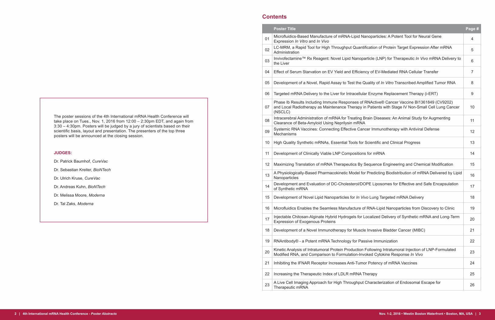
Contents
Poster Title Page #
01 Microfluidics-Based Manufacture of mRNA-Lipid Nanoparticles: A Potent Tool for Neural Gene Expression In Vitro and In Vivo 4
02 LC-MRM, a Rapid Tool for High Throughput Quantification of Protein Target Expression After mRNA Administration 5
03 Invivofectamine™ Rx Reagent: Novel Lipid Nanoparticle (LNP) for Therapeutic In Vivo mRNA Delivery to the Liver 6
04 Effect of Serum Starvation on EV Yield and Efficiency of EV-Mediated RNA Cellular Transfer 7
05 Development of a Novel, Rapid Assay to Test the Quality of In Vitro Transcribed Amplified Tumor RNA 8
06 Targeted mRNA Delivery to the Liver for Intracellular Enzyme Replacement Therapy (i-ERT) 9
07Phase Ib Results Including Immune Responses of RNActive® Cancer Vaccine BI1361849 (CV9202) and Local Radiotherapy as Maintenance Therapy in Patients with Stage IV Non-Small Cell Lung Cancer (NSCLC)
10
08 Intracerebral Administration of mRNA for Treating Brain Diseases: An Animal Study for Augmenting Clearance of Beta-Amyloid Using Neprilysin mRNA 11
09 Systemic RNA Vaccines: Connecting Effective Cancer Immunotherapy with Antiviral Defense Mechanisms 12
10 High Quality Synthetic mRNAs, Essential Tools for Scientific and Clinical Progress 13
11 Development of Clinically Viable LNP Compositions for mRNA 14
12 Maximizing Translation of mRNA Therapeutics By Sequence Engineering and Chemical Modification 15
13 A Physiologically-Based Pharmacokinetic Model for Predicting Biodistribution of mRNA Delivered by Lipid Nanoparticles 16
14 Development and Evaluation of DC-Cholesterol/DOPE Liposomes for Effective and Safe Encapsulation of Synthetic mRNA 17
15 Development of Novel Lipid Nanoparticles for In Vivo Lung Targeted mRNA Delivery 18
16 Microfluidics Enables the Seamless Manufacture of RNA-Lipid Nanoparticles from Discovery to Clinic 19
17 Injectable Chitosan-Alginate Hybrid Hydrogels for Localized Delivery of Synthetic mRNA and Long-Term Expression of Exogenous Proteins 20
18 Development of a Novel Immunotherapy for Muscle Invasive Bladder Cancer (MIBC) 21
19 RNAntibody® - a Potent mRNA Technology for Passive Immunization 22
20 Kinetic Analysis of Intratumoral Protein Production Following Intratumoral Injection of LNP-Formulated Modified RNA, and Comparison to Formulation-Invoked Cytokine Response In Vivo 23
21 Inhibiting the IFNAR Receptor Increases Anti-Tumor Potency of mRNA Vaccines 24
22 Increasing the Therapeutic Index of LDLR mRNA Therapy 25
23 A Live Cell Imaging Approach for High Throughput Characterization of Endosomal Escape for Therapeutic mRNA 26
The poster sessions of the 4th International mRNA Health Conference will take place on Tues., Nov. 1, 2016 from 12:00 – 2:30pm EDT, and again from 3:30 – 4:30pm. Posters will be judged by a jury of scientists based on their scientific basis, layout and presentation. The presenters of the top three posters will be announced at the closing session.
JUDGES:
Dr. Patrick Baumhof, CureVac
Dr. Sebastian Kreiter, BioNTech
Dr. Ulrich Kruse, CureVac
Dr. Andreas Kuhn, BioNTech
Dr. Melissa Moore, Moderna
Dr. Tal Zaks, Moderna

4 | 4th International mRNA Health Conference - Poster Abstracts Nov. 1-2, 2016 • Westin Boston Waterfront • Boston, MA, USA | 5
01 - Microfluidics-Based Manufacture of mRNA-Lipid Nanoparticles: A Potent Tool for Neural Gene Expression In Vitro and In Vivo
Rebecca DeSouza, Anitha Thomas, Grace Tharmarajah, Shyam Garg, Eric Ouellet, Peter Johnson, Tim Leaver, Andre Wild, James Taylor, Euan Ramsay
Precision NanoSystems, Inc., Vancouver, BC, Canada
Purpose: The use of RNA to manipulate gene expression in neuroscience has been limited due to the lack of an effective delivery tool. Recently, lipid nanoparticles (LNPs) have gained interest as safe and effective RNA delivery vehicles both in vitro and in vivo. However, traditional methods for the manufacturing of LNPs pose numerous challenges such as operator variability, and poor scalability. Here, we describe the robust and reproducible manufacture of mRNA-LNPs using the scalable microfluidics-based NanoAssemblr™ platform. The mRNA-LNPs are shown to effectively deliver and promote expression of mRNA in difficult-to-transfect primary neurons and astrocytes.
Methods: GFP mRNA was encapsulated into LNPs using the NanoAssemblr™ bench-top instrument (Precision NanoSystems, Inc., Vancouver, Canada). The mRNA-LNPs were characterized for their size, PDI, and GFP mRNA encapsulation efficiency. GFP mRNA-LNP uptake and GFP expression in neural cell cultures was investigated using flow cytometry, ELISA, and confocal microscopy. Cell viability of neurons and astrocytes was investigated using the Presto Blue Assay. The effectiveness of mRNA-LNPs was investigated in vivo following local administration of GFP mRNA-LNPs in the brain of mice.
Results: GFP mRNA was successfully encapsulated into LNPs using the NanoAssemblr™ platform. The LNPs exhibited a size of ~120 nm, low PDI (<0.2), and high mRNA encapsulation efficiency (>95%). Flow cytometry analysis after a 48 h treatment of 2.5 µg/mL GFP mRNA-LNP in rat primary neurons at DIV 7 showed >95% uptake of nanoparticles (based on a fluorescent probe within the nanoparticle) leading to >75% cells expressing GFP. Similar results for GFP expression were observed using ELISA. The high expression and uptake did not significantly impact cell viability even at doses of 5 µg/mL of GFP mRNA as measured using Presto Blue Cell Viability Assay. Further, GFP mRNA-LNPs exhibited >80% GFP expression in both human iPS derived neurons and primary mouse neurons. Treatment of primary rat astrocytes for 24 h with 2 µg/mL GFP mRNA showed >95% of cellular association leading to >80% cells expressing GFP. Similar results were observed using ELISA after 24 h treatment. Doses as high as 5 µg/mL did not affect the viability of primary astrocytes after 24 h treatment.
Conclusions: The NanoAssemblr microfluidics-based technology reproducibly manufactured mRNA-LNP that effectively mediated gene expression in primary neurons and astrocytes, and iPS-derived neurons, demonstrating mRNA-LNPs as an effective delivery tool for neuroscience applications.
02 - LC-MRM, a Rapid Tool for High Throughput Quantification of Protein Target Expression After mRNA Administration
Claudia Escher1, Jeffrey Silva2, Matthias John3
1Biognosys AG, Wagistrasse 25, Schlieren, 8952, Switzerland2Biognosys Inc., One Kendall Square, Building 200, Suite 2203, Cambridge, MA 02139, USA3Moderna Therapeutics, Inc., 200 Technology Square, Cambridge MA 20139, USA
Protein quantification after the administration of mRNA therapeutics is a critical step in efficacy evaluation of any treatment. Traditionally, this has been done using antibody-based methods such as ELISA or Western blots. These sensitive methods have several limitations. An antibody may not always be available, especially for non-human or non-mouse species, and method development often can’t be achieved in a time-frame acceptable to R&D or the pre-clinical process. Mass spectrometry based LC-MRM methods are able to generate highly specific and sensitive assays for the quantification of hundreds of protein targets within days. Protein targets from uncommon species or minor sequence differences with as little as one amino acid difference between the target protein and the host species (e.g. human APOA1 expressed in APOA1 knockout mice) can be resolved due to the high specificity of the mass spectrometer.
Here, we show examples for protein quantification of highly similar non-human proteins, for overexpression of human proteins and demonstrate the feasibility of a rapid assay generation tool for proteins where no antibody assay is available or evaluated.
In a large experiment series, LC-MRM assays for 114 target proteins were generated for the quantification in 480 samples in several batches. The time between sample receipt and results was only one week, making the method a valuable rapid tool in the development of more efficient versions of therapeutic mRNAs. When absolute quantification of the target protein is required, stable isotope-labelled reference peptides can by synthesized within a few weeks at low cost. The method can be easily multiplexed with no effect on specificity or the time required to carry out the assay, and multiple peptides per protein can be analysed further enhancing the confidence in results. The complex background that is typically present in the samples provides a virtually unlimited source of assays for “housekeeping” or normalizing proteins from where more than 2000 candidates could be selected.
Notes Notes
6 | 4th International mRNA Health Conference - Poster Abstracts Nov. 1-2, 2016 • Westin Boston Waterfront • Boston, MA, USA | 7
03 - Invivofectamine™ Rx Reagent: Novel Lipid Nanoparticle (LNP) for Therapeutic In Vivo mRNA Delivery to the Liver
Sean Essex1, Shikha Mishra1, Mu Li1, Nektaria Andronikou1, Xavier de Mollerat du Jeu1*
1Author affiliations- Thermo Fisher Scientific, Carlsbad, CA, USA*Corresponding author
Messenger RNA (mRNA) has attracted a lot of attention recently as a new drug class to deliver genetic information. This excitement has been fueled by the fact that mRNA-based cancer immunotherapies and infectious disease vaccines have already entered clinical development in a very short span of time. The focus has been on the use of mRNA in treating liver diseases, metabolic diseases, cancer, neurological diseases, mRNA vaccine development, CRISPR gene editing and gene therapy. However, efficient delivery of mRNA is the major bottleneck that needs to be addressed and overcome in order for this field to explode and meet its exciting potential.
We have developed novel Lipid nanoparticles (LNPs) through a Design of Experiment (DOE) approach to maximize the in vivo delivery efficiency of the mRNA. Our approach involves systematically designing an optimal DOE that takes into account both main and interaction effects, analyzing the experimental data using appropriate statistical techniques and generating response predictions from all potential combinations of the parameters. This iterative process is repeated till the optimal formulation is discovered. Adopting this approach provides us the flexibility to optimize formulations for organ specific delivery – Liver, Spleen, Lungs and Muscle (vaccines).
The process of formulating these LNPs is simple, scalable and results in uniform-sized LNPs with a narrow PDI. The LNPs efficiently encapsulate and protect the mRNA from degradation and facilitate cellular uptake which translates into efficient delivery and reduced toxicity in vivo. We have made significant progress moving through several generations of formulations where our current generation liver delivery LNP is 15,000X more potent than its first generation predecessor. Upon in vivo administration of our LNPs complexed with a chemically modified, luciferase-encoding mRNA (Luc mRNA) we have demonstrated high luciferase activity in vivo - bioluminescence of 10^10 p/sec. We have also shown a significant boost in therapeutically relevant proteins - 4000X serum mouse erythropoietin (EPO) production as compared to baseline murine EPO levels in as little as 4 hours post-injection and high human Factor IX (hFIX) levels in mice with a single LNP dose. Additionally, we have generated a comprehensive panel of elicited cytokines and liver-function related enzymes and shown that these LNPs are well-tolerated in a murine model.
04 - Effect of Serum Starvation on EV Yield and Efficiency of EV-Mediated RNA Cellular Transfer
Reka Agnes Haraszti1, Marie Didiot1, Andrew Coles1, Matthew Hassler1, Julia Alterman1, Neil Aronin2, Anastasia Khvorova1
1RNA Therapeutics Institute, University of Massachusetts Medical School, Worcester, MA, USA2Department of Medicine, University of Massachusetts Medical School, Worcester, MA, USA
Introduction: Extracellular vesicles (EVs) are known as natural intercellular carriers of various RNA classes, including mRNAs and small RNAs. Therefore EVs may be attractive means to further develop into delivery vehicles of exogenously synthesized RNAs. Mesenchymal stem cell (MSC) are a clinically safe and well characterized source of EVs. Here we studied how the source and metabolic state (serum starvation) of MSCs affected EV production and the ability of produced EVs to transfer small RNAs between cells.
Methods: Exosomes and microvesicles (MVs) were purified from different mesenchymal stem cell (MSC) types: umbilical cord-derived (UCD), adipose-derived (AD) and bone marrow-derived (BMD) via differential ultracentrifugation. EV yields were assessed by Nanoparticle Tracking Analysis. Purified vesicles were loaded with hydrophobically modified siRNA (hsiRNA) and applied to primary neurons in culture. hsiRNA distribution and mRNA silencing were quantified by PNA (peptide nucleic acid)-hybridization and QuantiGene® assays, respectively.
Results: UCD MSCs showed the fastest doubling time yielding the most and the largest exosomes and microvesicles. BMD and AD MSCs’ EV production significantly decreased upon serum starvation, while UCD EV yield was not affected. Unexpectedly, hTERT-immortalization was detrimental to EV yield. All tested EVs efficiently loaded hsiRNA and delivered them to primary neurons resulting in silencing. Serum starvation of source cells improved hsiRNA delivering activity of exosomes but abolished that of microvesicles. Hence RNA delivering activity depends on metabolic state and possibly on membrane composition of the EV class.
Conclusions: Source and metabolic state of MSCs (serum starvation) affected EV yield, and exosomes to microvesicles ratio. Exosomes from serum-starved cells were more efficient delivery vesicles for therapeutic siRNA, while serum starvation abolishes small RNA delivering activity of microvesicles. EVs could further be utilized to transport and deliver mRNAs to cells and organs.
Notes Notes
8 | 4th International mRNA Health Conference - Poster Abstracts Nov. 1-2, 2016 • Westin Boston Waterfront • Boston, MA, USA | 9
05 - Development of a Novel, Rapid Assay to Test the Quality of In Vitro Transcribed Amplified Tumor RNA
Jason Harris, Xiaorong Feng, Charles Nicolette and Irina Tcherepanova
Argos Therapeutics, Durham NC USA
Background: Amplified tumor RNA is used to enable autologous tumor antigen expression in electroporated DCs, which are then used in individualized immunotherapy for renal cell carcinoma (RCC). The development of an assay to further characterize the integrity of amplified RNA is challenging, as the material is composed of transcripts with diverse molecular weights and stabilities. A qPCR assay that quantitatively measured the ratio between 5’ and 3’ ends of a marker transcript was evaluated as a rapid, objective measurement of RNA quality.
Materials and Methods: A database of housekeeping genes was queried for a suitable marker that could be used in determining RNA quality. The PSMD1 transcript (3 kb) was chosen as a surrogate marker because it is greater in size than the average transcript (1.4 kb) and because of its ubiquitous nature and low variance in expression levels among various tissue types. To test the RT-qPCR assay, five different amplified RNA samples were degraded following incubation at 80°C for 0, 10, 20, 40, and 60 minutes. Their quality was evaluated by appearance following gel electrophoresis and PSMD1 Northern blot analysis. Two separate RT-qPCR assays were designed for the 5’ and 3’ ends of PSMD1. The copy number of 5’ and 3’ ends was established, and the qPCR 5’:3’ ratio calculated using the formula 2-∆Ct, where ∆Ct is defined as 5’ end Ct values – 3’ end Ct values. The initial findings were confirmed by testing 104 additional RNA samples with known quality, based on Northern blot analysis.
Results: The five amplified RNAs, and their cognate degraded material, show a time-dependent shift toward smaller molecular weights; as a smear by gel analysis and the gradual disappearance of the 3 kb PSMD1 transcript by Northern blot analysis. The ratio of 5’:3’ ends using the new RT-qPCR assay show a strong correlation with quality assessments by gel and Northern blot analyses, with decreasing values with longer degradation times (Table 1).
Degradation Time, min 0 10 20 40 60Sample 1 0.28 0.18 0.11 0.05 0.02Sample 2 0.28 0.20 0.12 0.05 0.02Sample 3 0.28 0.18 0.11 0.05 0.02Sample 4 0.16 0.10 0.06 0.03 0.02Sample 5 0.14 0.09 0.05 0.05 0.04
Shading indicates bands that do not pass quality assessment in an independent Northern blot assay
The additional data from 104 RNA samples amplified from various tumor with known qualities that were also examined using the 5’: 3’ assay, support that a ratio value may be used as an objective measure of RNA quality.
Conclusions: A novel, rapid assay for amplified tumor RNA quality was developed using the 5’:3’ ratio of the PSMD1 transcript. This assay replaces gel electrophoresis and Northern Blot analyses and is high through put and objective.
06 - Targeted mRNA Delivery to the Liver for Intracellular Enzyme Replacement Therapy (i-ERT)
Pierrot Harvie, Amber Paschal, Allen Li, Teri Blevins, Eric Bell, Debashish Roy, Anna Galperin, Matt Waldheim, Jean-Rene Ella-Menye, Sean Monahan, Mary Prieve and Mike Houston
PhaseRx, Inc., 410 West Harrison Street, Suite 300, Seattle WA 98119 USA
PhaseRx’s technology replaces missing or defective enzymes inside liver cells in certain inherited liver diseases. This technology is achieved by intracellular enzyme replacement therapy (i-ERT) which targets synthesis of delivered mRNA specifically to liver cells. PhaseRx’s Hybrid mRNA TechnologyTM platform using an endosomolytic polymer plus an inert mRNA lipid nanoparticle led to the normalization of two well-validated therapeutic biomarkers (urinary orotic acid and ammonia) in a hyperammonemia mouse disease model for ornithine transcarbamylase (OTC) deficiency. Therapeutic mRNA expression was detected in the liver after a single mRNA dose with good duration of expression. The treatment was well tolerated, with no toxicities associated with both single and multiple dosing regimens. PhaseRx nanoparticles were also well tolerated in rats and significant protein expression can be detected at a dose ranging from 0.1 to 1 mg/kg mRNA. PhaseRx’s Hybrid mRNA TechnologyTM platform provides a significant opportunity for the treatment of urea cycle disorders and other orphan liver diseases.
Notes Notes
10 | 4th International mRNA Health Conference - Poster Abstracts Nov. 1-2, 2016 • Westin Boston Waterfront • Boston, MA, USA | 11
07 - Phase Ib Results Including Immune Responses of RNActive® Cancer Vaccine BI1361849 (CV9202) and Local Radiotherapy as Maintenance Therapy in Patients with Stage IV Non-Small Cell Lung Cancer (NSCLC)2Hipp MM, 1Sebastian M, 1Weiss C, 3Früh M, 4Pless M, 5Cathomas R, 6Hilbe W, 6Pall G, 7Papachristofilou A, 2Doener F, 2Fotin-Mleczek M, 2Hong HS, 2Kallen KJ, 2Klinkhardt U, 2Koch SD, 2Scheel B, 2Schröder A, 2Seibel T, 2Gnad-Vogt U, 7Zippelius A
1University Hospital Frankfurt, Frankfurt, Germany 5Hospital Graubünden, Chur, Switzerland2CureVac AG, Tübingen, Germany 6University Hospital Innsbruck, Innsbruck, Austria 3Hospital of St Gallen, St Gallen, Switzerland 7University Hospital Basel, Basel, Switzerland4Hospital of Winterthur, Winterthur, Switzerland
Background: Preclinical studies demonstrated that local radiotherapy (RT) acts synergistically with RNActive® mRNA vaccines to enhance anti-tumor effects and increase tumor-infiltrating lymphocytes. BI1361849 is a therapeutic vaccine comprising optimized mRNA constituents encoding six NSCLC-associated antigens. We report final results of a phase Ib study including immune response analyses.
Methods: Patients (pts) with stage IV NSCLC were enrolled in three cohorts based on histological and molecular NSCLC subtypes. Pts received two vaccinations with BI1361849 prior to local RT to a single lesion (four consecutive daily fractions of 5Gy). Vaccination continued until start of subsequent anti-cancer therapy, maintenance pemetrexed (mP) and EGFR tyrosine kinase inhibitors (EGFR-TKIs) were given in parallel according to the label where indicated. Primary endpoint was safety; secondary endpoints included objective response, progression-free survival (PFS) and overall survival (OS). Cellular and humoral immune responses were measured ex vivo by intracellular cytokine staining, IFN-γ ELISpot, and ELISA in pre- and post-treatment blood samples.
Results: 26 pts were enrolled. 15 pts received mP, two received EGFR TKIs. Most frequent AEs were mild to moderate injection-site reactions and flu-like symptoms. No BI1361849-related SAEs were reported. One confirmed PR was observed in a pt on mP, 12/25 evaluable pts (46%) experienced SD (8 pts on mP, 1 pt on EGFR-TKI and 3 pts without concomitant maintenance treatment). Substantial decrease in tumor burden and long-lasting disease stabilization was observed in two pts receiving mP. Shrinkage of lesions outside the irradiated field of ≥15% occurred in 7 pts, all but one receiving mP. 25 pts were available for immune response analysis. Preliminary data indicate that BI1361849 elicited antigen-specific cellular and humoral immune responses in the majority of the patients
Conclusion: BI1361849 can be safely combined with local RT and mP treatment. Shrinkage of non-irradiated lesions and prolonged disease stabilization was observed in a subset of pts, mainly in combination with mP. BI1361849 induces humoral and cellular immune responses.
08 - Intracerebral Administration of mRNA for Treating Brain Diseases: An Animal Study for Augmenting Clearance of Beta-Amyloid Using Neprilysin mRNA
Keiji Itaka1,2, Chin-Yu Lin2, Satoshi Uchida1,2, Kazunori Kataoka2
1Laboratory of Clinical Biotechnology, Center for Disease Biology and Integrative Medicine, Graduate School of Medicine, The University of Tokyo, Tokyo, Japan2Innovation Center of NanoMedicine, Kawasaki Institute of Industry Promotion, Kawasaki, Kanagawa, Japan
Brain diseases are the most challenging medical field due to their complex pathogenetic mechanism, difficulty in delivering drugs, and the low restorative capacity. mRNA-based therapy has potentials as a means of introducing therapeutic any protein(s) and peptide(s) into neural cells for providing disease-modifying effects.
In this study, we applied mRNA for regulating the metabolic imbalance between anabolism and clearance of amyloid-beta (Aβ), that is considered to be the pathogenesis of Alzheimer’s disease (AD). Breaking the equilibrium between soluble and insoluble forms of Aβ is likely to help prevent the progression of AD. Neprilysin (NEP) plays a major role in the clearance of Aβ in the brain, and its supplementation using viral vectors has shown to decrease Aβ deposition and prevent pathogenic changes in the brain.
We constructed mRNA encoding the mouse NEP protein and evaluated its ability to degrade Aβ. In vitro transfection of NEP mRNA to primary neurons exhibited the activity of degrading Amyloid Precursor Protein (APP) superior to that of plasmid DNA encoding NEP. We then evaluated the in vivo activity of NEP mRNA by intracerebroventricular (i.c.v.) infusion using a cationic polymer-based PEGylated nanocarrier, polyplex nanomicelles, which have been shown to have a high potential to deliver mRNA to various target tissues and organs. Nanomicelles carrying a GFP-NEP fusion mRNA produced efficient protein expression in a diffusive manner surrounding the ventricular space. An ELISA evaluation revealed that the mRNA infusion significantly augmented NEP level and effectively reduced the concentration of Aβ that had been supplemented in the mouse brain. It should be noted that NEP is not a secreted protein, but a membrane-integrated exoenzyme with a large portion of the extracellular domain responsible for Aβ degradation on the cell surface. Thus, the significant decrease in Aβ concentration in the brain represents the potential of mRNA-based therapy by introducing therapeutic gene(s) in a diffusive manner, that would be more effective at degrading pericellular Aβ.
In conclusion, mRNA has the therapeutic potential for the treatment of brain diseases, opening the new era of mRNA-based therapeutics.
Notes Notes

12 | 4th International mRNA Health Conference - Poster Abstracts Nov. 1-2, 2016 • Westin Boston Waterfront • Boston, MA, USA | 13
09 - Systemic RNA Vaccines: Connecting Effective Cancer Immunotherapy with Antiviral Defense Mechanisms
Lena M. Kranz1,2*, Mustafa Diken1,3*, Heinrich Haas3, Sebastian Kreiter1,3, Carmen Loquai4, Kerstin C. Reuter3, Martin Meng3, Daniel Fritz3, Fulvia Vascotto1, Hossam Hefesha3, Christian Grunwitz2,3, Mathias Vormehr2,3, Yves Hüsemann3, Abderraouf Selmi1,2, Andreas N. Kuhn3, Janina Buck3, Evelyna Derhovanessian3, Richard Rae1, Sebastian Attig1,2, Jan Diekmann3, Robert A. Jabulowsky3, Sandra Heesch3, Jessica Hassel5, Peter Langguth6, Stephan Grabbe4, Christoph Huber1,3, Özlem Türeci7§, Ugur Sahin1,2,3§
1Translational Oncology at the University Medical Center (UMC) of the Johannes Gutenberg University (JGU) 2Research Center for Immunotherapy (FZI), UMC of JGU 3Biopharmaceutical New Technologies (BioNTech) Corporation 4Department of Dermatology, UMC of JGU 5Department of Dermatology, Heidelberg University Hospital 6Institute of Pharmacy and Biochemistry, JGU 7Cluster for Individualized Immune Intervention*These authors contributed equally to this work. §These authors jointly supervised this work.
Mechanisms of antiviral host defense are important for survival and evolutionarily optimized for high sensitivity and potency. Intending to harvest these pathogen immune defense programs for cancer immunotherapy, we simulated a systemic pathogen intrusion into the blood stream by intravenous injection of lipid-formulated, tumor antigen-encoding mRNA nanoparticles. These RNA-lipoplexes (RNA-LPX) were directed to various lymphoid tissues including the spleen, lymph nodes and bone marrow, which provide the ideal microenvironment for efficient priming and proliferation of T cells. The RNA-to-lipid ratio was discovered to rule the biodistribution of RNA-LPX, irrespective of the types of lipids used, and a slightly negative particle net charge was able to specifically transfect lymphoid organ-resident antigen presenting cells (APCs). Following uptake by dendritic cells (DC) and macrophages in spleen and in other lymphoid organs, predominantly by macropinocytosis, RNA recognition via TLR7 triggered two transient waves of type I IFN production by pDCs (early response) and macrophages (delayed response), which established an inflammatory, lymphocyte-activating milieu reminiscent of that initiated during the early systemic phase of viral infection. IFNα receptor (IFNAR)-dependent immune mechanisms were required for DCs to mature, migrate into the T cell zones and express RNA-encoded tumor antigens. Presentation of these antigens on MHC class I and II by mature DCs elicited strong effector and memory CD8 and CD4 T cell immunity against viral, mutant neo-antigens or self-antigens, which was able to reject progressive tumors in therapeutic mouse models of melanoma, colon carcinoma and human papilloma virus (HPV)-associated cancer. In an ongoing phase I dose escalation study, the first cohort of three patients with advanced melanoma received RNA-LPX encoding four shared tumor antigens at doses lower than those used in the mouse studies. All patients showed a dose-dependent IFNα- and IP-10-dominated cytokine response, developed de novo CD4 and CD8 T cell responses or enhanced pre-existing immunity against the encoded self-antigens NY-ESO-I, Tyrosinase and MAGE-A3. These results support the preclinically identified mode of action and strong potency of this approach in the clinical setting. Our study presents a novel class of systemically administered nanoparticulate RNA vaccines acting by body-wide delivery of encoded antigens to APCs and simultaneous initiation of a strong type I IFN-driven immunostimulatory program. RNA-LPX vaccines mimic the infectious non-self and thus mobilize both adaptive and innate immune mechanisms. The simple but highly versatile design allows vaccine preparation with any type of RNA-encoded antigen and may thus be regarded as a universally applicable, first-in-class vaccine platform for cancer immunotherapy.
10 - High Quality Synthetic mRNAs, Essential Tools for Scientific and Clinical Progress
Guido Krupp1, Hristina Dineva1, Andreas Hanne1, Lisa Heinrich1, Patricia Grzechnik1, Wanda Laugell-Wunsch1, Susanne Quabius2, Peter Scheinert1
1AmpTec GmbH, Hamburg, Germany2Department of Otorhinolaryngology & Institute of Immunology, University Hospital UKSH, Kiel, Germany
Synthetic mRNAs from AmpTec have achieved a world-wide acceptance by users in Europe, North America, Near and Far East and Australia. Application data by a user consortium were published recently by Petrova et al. (2014) in Stem Cell Reports.
AmpTec’s mRNAs are available as GMP-grade products and are currently used in several DC(Dendritic Cell)-based clinical trials in Europe, Far East and Australia. This demonstrates that the availability of high quality synthetic mRNAs is very important in enabling significant progress in this field. With a limited number of world-wide active manufacturers, AmpTec continues to realize its obligation to support the entry of new players by providing customized, high quality mRNA products.
Important mRNA features and alternative technical options for large-scale high-quality mRNA production and for GMP-compliant manufacturing and quality control are presented.
Synthetic mRNAs are generated by in vitro transcription (IVT) with T7 RNA polymerase from defined templates containing the synthetic gene of interest. Synthetic genes can be obtained in the form of plasmid clones from commercial suppliers like Eurofins Genomics. In principle, linearised plasmids (with a restriction enzyme) can be used directly as templates in IVT reactions. This procedure has disadvantages: (i) incomplete plasmid cleavage results in poor reproducibility; (ii) along with high amounts of plasmid DNA, undesired bacterial components can be introduced into mRNA products; (iii) optimal mRNA activity depends on a long, unmasked poly(A) tail, i.e. a tail with more than 100 A’s. However, this long hompolymeric sequence is not reliably propagated in E.coli. Our alternative procedure was presented by Quabius&Krupp (2015) in New Biotechnology: well defined PCR products are used as IVT-templates. This approach with example results and observed problems - and problem solutions - will be presented. We will also present a detailed list of quality requirements for high-quality synthetic mRNAs. As important milestone, AmpTec has achieved a production scale of several hundred milligrams per batch.
Notes Notes
14 | 4th International mRNA Health Conference - Poster Abstracts Nov. 1-2, 2016 • Westin Boston Waterfront • Boston, MA, USA | 15
11 - Development of Clinically Viable LNP Compositions for mRNA
K. Lam, J. Heyes, A. Judge, L. Palmer, H. Yuen, P. Schreiner, J. Bechard, M. Wood, M. Abrams
Arbutus Biopharma Corp., Burnaby, BC, Canada
Arbutus’ Lipid Nanoparticle (LNP) platform is the leading nucleic acid delivery technology platform, enabling a number of early and late stage clinical trials. It is designed to deliver their payloads to sites of disease and have been used to target both viral and endogenous gene targets. We continuously seek to broaden the Therapeutic Index (TI) of the LNP platform, from both potency and tolerability perspectives.
It is important to recognize that nucleic acid drugs can stimulate cytokine release that may be accentuated by the delivery vehicle. This is sporadically observed with siRNA-LNP products in the clinic and can be managed with steroid premedication. It would be advantageous to address these drug properties at the compositional level in advance. This is particularly true for mRNA payloads, that preclinical data suggests are more likely to provoke inflammatory responses than smaller oligonucleotides. mRNA has garnered increasing interest for a range of therapeutic strategies, and its safe and successful formulation demands appropriate attention.
New formulation strategies were tested to address the challenge of immune stimulation. Initial activity/tolerability screens were run with siRNA payloads in murine models, with compositions of interest advanced to porcine and non-human primate (NHP) models. The results were consistent across these models, with a similar hierarchy of the compositions’ tendency to provoke an inflammatory response. The NHP model also helped identify a lead LNP composition that was significantly more potent than those currently in the clinic. These strategies were equally applicable to LNP bearing mRNA payloads. We further demonstrated that the mRNA purification method plays a significant role in immune stimulation.
Based on our clinical experience, the safe and successful translation of mRNA-LNP into man will require careful attention to both potency and immune stimulation. New lipid compositions identified are significantly more potent than those currently in the clinic. More importantly, they possess an immune stimulatory profile similar to saline controls at doses far greater than those required for efficacy. This unprecedented degree of immune silence, a profile which was preserved in higher species such as pig and NHP, will be imperative in providing the necessary TI for mRNA therapeutics in man.
12 - Maximizing Translation of mRNA Therapeutics By Sequence Engineering and Chemical Modification
Anton P. McCaffrey, Krist T. Azizian, Dongwon Shin, Sabrina Shore, Alexander Lebedev, Richard I. Hogrefe
TriLink BioTechnologies, San Diego, CA, USA
For maximal expression in cells or target organs, transfected mRNAs must avoid detection by pattern recognition receptors (PRRs) that evolved to sense pathogenic non-self RNAs. These include PRRs that recognize improperly capped RNAs (RIG-I, IFITs) and double stranded RNA (PKR, OAS, RIG-I, TLR3). PRR activation leads to cytokine production, translational arrest and cell toxicity or death. Mammalian mRNAs are modified post-transcriptionally to contain nucleotides with 2’-O-methyl residues, pseudouridine (Ψ) and N6-methyladenosine (m6A). Interestingly, these modifications can reduce activation of PRRs and allow maximal translation of the transfected mRNA. For mRNA drugs to achieve their potential, methods to produce multigram batches of properly capped, highly purified and non-immunogenic mRNA will be needed. For vaccines, some immunogenicity may be desirable to serve as an adjuvant.
During RNA capping, Cap0 (m7GpppN) is formed as an intermediate. Methylation of the 2’ position of the first and sometimes second nucleotide forms Cap1 and Cap2, respectively. Recombinant enzymes used to generate Cap1 mRNA are expensive, do not always go to completion and the RNA must be purified prior to capping. We developed a novel co-transcriptional capping method called CleanCap™ that yields Cap1 with high efficiency and lower costs in a “one pot” reaction. CleanCap also facilitates modification of the 5’ end of mRNAs with diverse functional groups, including m6A. We developed a capping assay that allows direct assessment of mRNA capping. Capping efficiencies as high as 99% can be obtained.
Previously, we identified 5-methoxyuridine (5moU) as a promising modification to avoid innate immune stimulation while supporting efficient translation. To further explore chemically modified bases, we synthesized several Ψ derivatives: N1-ethyl-Ψ (Et1Ψ), N1-fluoroethyl-Ψ (FE1Ψ), N1-propyl-Ψ (Pr1Ψ), N1-isopropyl-Ψ (iPr1Ψ) and N1-methoxylmethyl-Ψ (MOM1Ψ) 5’-triphosphates. Luciferase mRNAs were fully substituted with these modifications and translational potential was monitored in wheat germ extracts. Activity was also measured in the THP-1 monocyte cell line, which is a sensitive model for innate immune activation. A recent report showed that minimizing uridine content in mRNAs reduced immune stimulation by unmodified mRNAs. Here we show that incorporation of our modified uridine residues or 5moU into uridine depleted luciferase resulted in equivalentactivity relative to Ψ in THP-1 cells. We are currently evaluating the use of 5moU in U depleted Renilla, beta-galactosidase, erythropoietin, mCherry and Cas9 mRNAs
Notes Notes

16 | 4th International mRNA Health Conference - Poster Abstracts Nov. 1-2, 2016 • Westin Boston Waterfront • Boston, MA, USA | 17
13 - A Physiologically-Based Pharmacokinetic Model for Predicting Biodistribu-tion of mRNA Delivered by Lipid Nanoparticles
Robin McDougall1, Alex Bulychev2, Pete Smith2, Jay Mettetal1
1Drug Safety & Metabolism, Astrazeneca, Waltham MA2DMPK, Moderna Therapeutics, Cambridge MA
Background: Compared to the pharmacokinetics of traditional small molecule drugs, the biodistribution of LNP delivered mRNA is quite complex. The exposure profiles of the nanoparticle carriers, the mRNA cargo and expressed protein can vary significantly between key tissues and plasma - largely due to differential extravasation rates between vasculature and different tissues and in the rates of uptake into different cell types.
Physiologically based pharmacokinetic (PBPK) models provide an ideal platform to capture these nonlinearities in a computational framework which can then be used to make predictions for the dose and administration route dependent tissue exposures of different mRNA therapies. Furthermore, because the model is characterized by distinct sets of physiological and formulation dependent parameters, calibrated PBPK models offer the potential for more robust translation across administration routes and species.
Objectives: We designed and built a hierarchical PBPK model for the biodistribution of LNPs and mRNA into individual tissues. Here, we demonstrate the application of this model for the quantitative translation of disposition across routes of administration and across preclinical species using data for human EPO mRNA delivered via ionizable cationic lipid-based LNPs (herein LNP-mRNA).
Methods: A typical PBPK model was extended into multiple integrated “levels” – one to track each of the three key relevant states of the LNP-mRNA construct: Intact LNPs complete with mRNA cargo, Detectable LNPs no longer carrying mRNA, and mRNA released from the LNP.
By fixing the physiological parameters for each tissue compartment to the literature we fit the formulation specific parameters for IV data “Murine-LNP-mRNA-hEPO” exposure scenario, yielding complimentary sets of physiological parameters and formulation parameters. PBPK model predictions were generated from the calibrated model and compared to the observed data set. The formulation-specific parameters were used to predict the disposition for other dosing routes and species.
Results: The PBPK model was able to accurately describe the LNP (ionizable cationic lipid component) and mRNA levels in various key tissues in mice dosed with LNP-hEPO. The model was used to generate prospective predictions for other LNP dosed mRNA showing significant promise for predicting mRNA distribution in other preclinical and clinical settings.
Conclusion: The hierarchical PBPK model constructed for describing the biodistribution of LNP-mRNA constructs was able to successfully describe the non-linear pharmacokinetics of the dosed LNP-hEPO construct in all the key tissues in mice. Applications of the model to ionizable cationic lipid-based LNP exposures in other scenarios have been encouraging.
14 - Development and Evaluation of DC-Cholesterol/DOPE Liposomes for Effective and Safe Encapsulation of Synthetic mRNA
Tatjana Michel, Meike-Kristin Abraham, Daniel Luft, Julia Kurz, Martha L. Salinas Medina, Christian Schlensak, Hans Peter Wendel, Stefanie Krajewski
Department of Thoracic and Cardiovascular Surgery, Clinical Research Laboratory, University Medical Center, Tuebingen, Germany
Synthetic modified mRNA is a novel and highly promising bioactive agent for different therapeutic applications based on the induction of de novo synthesis of specific and functional protein. It can be used for example for infectious diseases or cancer vaccination or the treatment of single-gene disorders. In case of single-gene disorders, the mRNA should be delivered to the target cells, where the desired and fully functional protein is expressed. However, so far cells need to be transfected in short time intervals, because the mRNA has a short intracellular half-life. With regard to mRNA application in vivo, a delivery system with slow release characteristics, specific targeting and mRNA protective properties is desirable. For these reasons, we generated cationic DC-Cholesterol/DOPE liposomes using dry-film-method and extrusion in order to achieve an effective encapsulation as well as a high transfection efficiency and prolonged release of therapeutic mRNA. The transfection efficiency of the liposomes was analyzed using enhanced green fluorescence protein (eGFP) and alpha-1-antitrypsin (AAT) mRNA. Furthermore, immunogenicity and haemocompatibility was analyzed.
The results indicate that the liposomes possess good transfection efficiencies and also prolonged protein expression. Moreover, the liposomes show no immunogenic reaction in cells at different concentrations. When tested with fresh human blood, the liposomes have no impact on platelets, the coagulation cascade or immune cells. With regard to the therapeutic application of mRNA for the treatment of genetic disorders, an AAT-encoding mRNA was encapsulated in liposomes and transfection was performed in vitro. The concentration of AAT protein was measured 24 h and 72 h after transfection. The results show that the AAT mRNA-loaded liposomes result in a prolonged release of AAT protein when compared to a commercially available transfection reagent.
This innovative project presents a highly promising and inexpensive therapeutic approach for the delivery of mRNA using liposomes, which have a great potential for a therapeutic mRNA delivery system in vivo.
Notes Notes
18 | 4th International mRNA Health Conference - Poster Abstracts Nov. 1-2, 2016 • Westin Boston Waterfront • Boston, MA, USA | 19
15 - Development of Novel Lipid Nanoparticles for In Vivo Lung Targeted mRNA Delivery
Shikha Mishra, Sean Essex, Prerana Malwadkar1, Mu Li, Xavier de Mollerat du Jeu
Thermofisher Scientific, Carlsbad, California, USA
The rapidly expanding utilization of mRNA as a therapeutic tool has presented the field with the task of optimizing and innovating delivery methods. As the applications for mRNA based therapeutics continues to rise, a parallel emerging need to improve upon and develop novel technology has come to the forefront. Lipid nanoparticles are a common delivery vehicle for mRNA due to their ability to facilitate cellular uptake while protecting the mRNA from extracellular enzyme degradation. Organ and tissue specific delivery of mRNA is of great importance in tailoring therapeutic functionality. This is commonly achieved using biomolecular targeting via ligand or receptor expression on the surface of the nanoparticle.
We have developed novel lipid nanoparticles that are specifically optimized for use in vivo, and engineered to inherently target and deliver mRNA to murine lungs and spleen without the use of biomolecular targeting. Development of the lipid nanoparticle was performed using multivariable Design of Experiment modeling, which allowed us to understand and optimize the lipid formulation components and composition. Characterization of the lipid nanoparticles showed uniform size and high encapsulation efficiency. Following multiple design iterations, in vivo functional testing to assess biodistribution identified a novel formulation capable of exclusively targeting the lung tissue and achieving highly efficient mRNA transfection. Transfection efficiency was measured following in vivo systemic delivery of a chemically modified luciferase encoding mRNA. Quantification was performed using the IVIS imaging system to measure in vivo and ex vivo bioluminescence measurements. Lung specific expression levels could be modulated by varying the dose of mRNA, and significant protein expression was sustained over the course of 48 hours following a single administration. This novel lipid nanoparticle is well tolerated in vivo, with no qualitative gross toxicity, and quantitatively analyzed by a comprehensive cytokine profiling performed on murine serum samples. Further optimization of biodistribution to achieve exclusive targeting to the lung is currently underway, and preliminary data indicates that variance in charge ratio enhances delivery and expression exclusively to the lung, and depletes expression in other organs. Finally, flow cytometry analysis was used to analyze mRNA delivery to specific cell populations within the lung and spleen.
16 - Microfluidics Enables the Seamless Manufacture of RNA-Lipid Nanoparticles from Discovery to Clinic
Kevin Ou, Jagbir Singh, Aine Rooney, Mark Ma, Suki Sidhu, Anitha Thomas, Shyam Garg, Eric Ouellet, Peter Johnson, Tim Leaver, Andre Wild, James Taylor, Ray Lockard, Euan Ramsay
Precision NanoSystems, Inc., Vancouver, BC, Canada
Purpose: Lipid nanoparticles (LNPs) have gained interest in gene therapy as safe, efficient vectors for the delivery of nucleic acids both, in vitro and in vivo, as well as providing the basis for the development of RNA nanomedicines. However, their transition from discovery to clinic has been limited due to challenges in optimization of these candidates as manufacturing scales. Here, we describe the robust manufacture of mRNA-LNPs using the microfluidics-based NanoAssemblr™ platform, and the ease in translating this technology from discovery through clinical-scale manufacturing.
Methods: Luciferase mRNA was encapsulated into LNPs using a single microfluidic mixer on the NanoAssemblr™ bench-top instrument (Precision NanoSystems, Inc., Vancouver, Canada) at desired flow rate ratios and rates to optimize process parameters. Using the same parameters, mRNA was encapsulated into LNPs using a pre-clinical continuous flow single microfluidic mixer system i.e. NanoAssemblr Blaze (Precision NanoSystems, Inc., Vancouver, Canada). Lead candidate formulations and optimized process conditions can then be transferred to a scale-up system comprising a continuous flow pumping system with 8 single-mixer (8x) microfluidic chips arrayed in parallel using a manifold. This scale-up system utilizes parallelized microfluidic mixers to maintain identical reaction conditions and increase production throughput.
Results: Luciferase mRNA-LNPs prepared using both, the NanoAssemblr Benchtop and NanoAssemblr Blaze were compared for their size, PDI, mRNA encapsulation, and luciferase expression in vivo in mice post i.v. administration. Lead mRNA candidate formulations prepared on the NanoAssemblr Benchtop and NanoAssemblr Blaze can be seamlessly transferred to GMP scale manufacturing. Previous studies using siRNA-LNP prepared using a single mixer at total flow rate through the microfluidic chip of 12 mL/min were compared to siRNA-LNP made using 8x microfluidic chips arrayed in parallel. No change in particle size and PDI, and biological activity was observed for Factor VII siRNA-LNPs manufactured using the NanoAssemblr Benchtop, NanoAssemblr Blaze, and the 8x GMP scale-up system, which further validates microfluidic parallelization as a mechanism for rapid and seamless scale-up.
Conclusions: This study reflects the potential of microfluidics-based technology in the development of nanoparticle candidates from discovery to clinic.
Notes Notes

20 | 4th International mRNA Health Conference - Poster Abstracts Nov. 1-2, 2016 • Westin Boston Waterfront • Boston, MA, USA | 21
17- Injectable Chitosan-Alginate Hybrid Hydrogels for Localized Delivery of Synthetic mRNA and Long-Term Expression of Exogenous Proteins
Heidrun Steinle, Tudor-Mihai Ionescu, Selina Schenk, Sonia Golombek, Alexander Reifschneider, Christian Schlensak, Hans Peter Wendel, Meltem Avci-Adali
University Hospital Tuebingen, Department of Thoracic and Cardiovascular Surgery, Calwerstraße 7/1, 72076 Tuebingen, Germany
Clinical approaches with synthetic messenger RNA (mRNA) exhibit many advantages, such as the efficient posttranslational expression of desired proteins without genomic integration. Despite several modifications of synthetic mRNA, protein expression in cells only lasts a few days in physiological conditions after a single mRNA transfection. To overcome this hurdle, injectable hydrogels that are highly biocompatible and biodegradable, are suitable candidates for long-term delivery of synthetic mRNA in vivo. Numerous applications, such as protein replacement therapy, reprogramming and (trans-) differentiation strategies would benefit from an appropriate in vivo system for the extended release of mRNA over weeks. In this study, the in vitro release of Cy3 labeled firefly luciferase encoding mRNA, integrated into four different types of hydrogels, alginate, fibrin, chitosan, and chitosan-alginate, was quantified over a period of three weeks. To investigate the stability of these hydrogels, viscoelasticity was rheological examined. Because of its rheological stability properties and the prolonged mRNA release (70% after 3 weeks), chitosan-alginate hydrogels were further used to test the biofunctionality of included luciferase mRNA. HEK293 cells were incorporated into the mRNA-loaded hydrogels and luciferase activity could be detected over a time course of 3 weeks. The feasibility to continuously release synthetic mRNA from chitosan-alginate hydrogels would allow extended protein expression in desired tissues by minimal invasive local injections.
18 - Development of a Novel Immunotherapy for Muscle Invasive Bladder Cancer (MIBC)
Irina Tcherepanova1, Bradley Leibovich2, Jacoba Slagter-Jager1, Patrick Dillon1, Shawn Leland1, Lance Mynderse2, Brian Costello2, Charles Nicolette1
1Argos Therapeutics Durham NC, USA2Mayo Clinic Rochester NY USA
Background: AGS-003 is an autologous tumor RNA-loaded dendritic cell-based immunotherapy being tested in advanced renal cell carcinoma (RCC) patients. The immunologic specificity for the patient’s own tumor is achieved using amplified tumor RNA electroporated into monocyte derived dendritic cells. To produce AGS-003 for RCC patients the RNA is amplified from a 100 mg primary tumor sample obtained via nephrectomy. In this non treatment study we evaluated the feasibility of RNA amplification from MIBC tissue acquired by transurethral resection of bladder tumor (TURBT).
Methods: MIBC tissue was obtained via TURBT. Following draining of the irrigant used during the TURBT procedure, tissue was placed into an RNA preservative solution to prevent RNA degradation until processing. Methods developed for the amplification of RCC tumor RNA and RNA quality testing were applied to the RNA extracted from the MIBC specimens.
Results: This work demonstrated that the methods developed for the amplification and testing of RNA from RCC can be successfully employed for the amplification of RNA from MIBC specimens collected by TURBT. Results demonstrated that the total RNA yields obtained from bladder tissue exceed that of similar masses of primary RCC tumors. This finding enabled the reduction in the amount of starting material from 100 mg to 50 mg of bladder tissue. The amount of amplified RNA produced from the lower bladder cancer masses were sufficient to support the production of immunotherapy at full scale (17 doses average).
SID Target PST86992 PST06052 PST69180 PST70242 PST76245Tumor amount(mg) 50 50.4 50.4 49.2 49.6 51.4Total RNA concentration(ng/µL) ≥ 84 591.5 852.4 636.3 381.8 209.8cDNA yield (µg) ≥ 32 80.1 88.4 75.1 97.1 72.2amplified RNA yield (mg) 2 2.87 2.84 2.63 3.03 2.91
Conclusion: These data demonstrate the feasibility of RNA extraction and amplification from muscle invasive bladder cancer tissue and supports our planned Phase II clinical study.
Notes Notes
22 | 4th International mRNA Health Conference - Poster Abstracts Nov. 1-2, 2016 • Westin Boston Waterfront • Boston, MA, USA | 23
19 - RNAntibody® - a Potent mRNA Technology for Passive Immunization
Moritz Thran1, Marion Pönisch1, Barbara L. Mui2, Michael J. Hope2, Mariola Fotin-Mleczek1, Thomas Schlake1
1CureVac AG, Tübingen, Germany2Acuitas Therapeutics, Vancouver, British Columbia, Canada
The delivery of genetic information has emerged as a promising alternative to overcome substantial drawbacks associated with the use of recombinant proteins when applied in protein therapies. Until recently, these approaches mainly pursued the development of DNA-based gene therapies whereas RNA was not considered as an appropriate genetic carrier due to its transient and unstable nature. Against all odds it could be demonstrated that RNA, besides its remarkable potential to act as a vaccine, can promote a strong and robust protein expression and thus be utilized in gene therapy approaches. In this line, we recently demonstrated that the application of RNA can boost the serum levels of erythropoietin and consistently leads to long-lasting biologically-relevant effects, even in large animals. An important field of protein therapies is passive immunization with recombinant antibodies against threats such as infectious diseases or intoxication. In the present study we asked whether mRNA can fulfil its promise and be utilized even in scenarios where a prompt and strong response is crucial. In animals, a single injection of mRNA immediately leads to high neutralizing antibody titers, sufficient to provide fast protection in lethal challenge situations. We postulate that the mRNA technology provides a potent alternative/means for passive immunization.
20 - Kinetic Analysis of Intratumoral Protein Production Following Intratumoral Injection of LNP-Formulated Modified RNA, and Comparison to Formulation-Invoked Cytokine Response In Vivo
Yoav Timsit1, Mark Pietras2, Shenghua Wen4, Michael Grondine4, Nakpangi Johnson2, Nicola Derbyshire5, Sue Bickerton5, Herbert Barthlow1, Arpan Desai7, Catherine Betts6, Matthew Wagoner3, Clay Scott1
1Discovery Safety2Lab Animal Sciences3New Modalities, Drug Safety and Metabolism4Oncology iMED, AstraZeneca Pharmaceuticals, 35 Gatehouse Park Drive, Waltham, MA, USA 024515Pathology, Drug Safety and Metabolism, AstraZeneca Pharmaceuticals, Alderly Park, Macclesfield, United Kingdom SK10 4TG6Pathology, Drug Safety and Metabolism7Advanced Drug Delivery, Pharmaceutical Sciences, AstraZeneca Pharmaceuticals, Cambridge Science Park, Milton Road, Cambridge, United Kingdom CB4 0FZ
Modified RNA (modRNA)-based therapeutics require formulation in nanoparticle carriers for effective delivery of modRNA into cells. While the modRNA molecule itself contains modifications designed to avoid immune detection, a nanoparticle formulation that avoids immune detection has yet to be developed. Characterization of existing nanoparticle formulations injected directly into tumors show that certain classes of nanoparticles elicit potent inflammatory cytokine response, and so understanding the mechanistic basis for this would be very informative for developing new formulations that do not invoke an immune response. Important to this mechanistic understanding is to determine whether a relationship exists between the immune response and protein expression. To this end, whole-body bioluminescent imaging (BLI) technique was implemented to measure kinetics of modRNA-directed protein production in tumors in a syngeneic tumor model, using luciferase as the expressed target protein. To test for a relationship between protein production and immune response, two formulations known to produce different levels of protein expression were used, and serial plasma sampling was performed to measure circulating cytokines. By serial scanning following intratumoral (iTu) dosing, protein expression kinetics were clearly different between the two formulations (2.5-fold difference in AUC). In contrast, inflammatory cytokine response after iTu dosing were either similar between the two formulations, or the formulation giving greater protein expression had a lower cytokine response. Interestingly, repeat dosing revealed attenuated IL-6 response, or as seen with MCP-1 and KC, peak levels were reached sooner but not exceeding peak levels observed after the first dose. These changes to cytokine responses with repeat dosing may be indicative of immune tolerance to both LNP formulations, but further investigation is needed. Thus, BLI has enhanced our insights into tumor modRNA protein expression, and our results show that increased tumor protein production did not translate into greater cytokine response, suggesting that other factors are likely key drivers of inflammation. Further characterization of protein production kinetics will feed into PBPK models, and combining with mathematical models of immune response, would enable prediction of intratumoral protein expression
Notes Notes

24 | 4th International mRNA Health Conference - Poster Abstracts Nov. 1-2, 2016 • Westin Boston Waterfront • Boston, MA, USA | 25
21 - Inhibiting the IFNAR Receptor Increases Anti-Tumor Potency of mRNA Vaccines
Lien Van Hoecke1,2, Ans De Beuckelaer3, Charlotte Pollard3, Xavier Saelens1,2, Johan Grooten3, Stefaan De Koker3
1Medical Biotechnology Center, VIB, Ghent, Belgium2Department of Biomedical Molecular Biology, Laboratory of Molecular Virology, Ghent University, Ghent, Belgium3Department of Biomedical Molecular Biology, Laboratory Molecular Immunology, Ghent University, Ghent, Belgium
mRNA-based vaccines have great potential to elicit cytotoxic T cell responses. Therefore tumor antigen-encoding mRNA vaccines are now being intensively explored as therapeutic cancer vaccines. It’s been described in vitro – and proved by us in vivo - that mRNA vaccines evoke a strong type I IFN response upon immunization. With this poster, we demonstrate that mRNA lipoplexes induce a potent type I IFN response upon subcutaneous, intradermal and intranodal injection. Using mice that lack type Interferon α/β receptor I (IFNAR-/-), we have identified that this type I IFN activity limits the induction of cytotoxic CD8+T cell responses to antigens encoded by lipoplex-based mRNA vaccines. Immunization of IFNAR-/- mice dramatically increased priming of naïve T cells as well as the quantity of antigen-specific CD8+ T cells and cytolytic effector function in the blood. Contrariwise, type I IFNs have been shown to be vital to the endogenous anti-tumour response. In order to unravel these opposing functions of type I IFN in the context of mRNA based melanoma vaccination, we show that a topical and transient blocking of type I IFN signalling at the site of mRNA vaccination by co-injection of an IFNAR-blocking antibody dramatically augmented the magnitude of a vaccine elicited CD8+ T cell immunity, resulting in prolonged survival in case of prophylactic and therapeutic melanoma vaccination in mice.
Notes
22 - Increasing the Therapeutic Index of LDLR mRNA Therapy
Justin Guild1, Jeffrey Pimentel2, Summar Siddiqui1, Alex Cavedon2, Kerry Benenato2, William Butcher2, Andrea Frassetto1, Ling Yin1, Shi Liang1, Shawn Hillier1, Barry Ticho2, Paolo Martini1
1Elpidera, Cambridge MA2Moderna, Cambridge MA
Background: Homozygous Familial Hypercholesterolemia (HoFH) is defined by the absence of functional low density lipoprotein receptor (LDLR) leading to severely elevated low density lipoprotein cholesterol (LDL-c). As a consequence, patients develop atherosclerosis at an early age and have an increased risk of heart attack, stroke, and peripheral vascular disease1.
Further compounding the LDLR dysfunction, HoFH patients also have elevated levels of the LDLR negative regulator, PCSK9, which binds to and targets LDLR for lysosomal digestion. Another regulator of LDLR, IDOL (Inducible Degrader of the LDLR), is an E3 ubiquitin ligase which has been observed to increase LDLR degradation and reduce plasma LDL-c2. Hepatocyte delivery of mRNA is typically conducted with lipid nanoparticles (LNP) that shuttle cargo through the LDLR receptor. As HoFH are deficient in LDLR, delivery is particularly challenging.
Current treatments do not meet HoFH patient needs as statins and other lipid lowering agents are only marginally effective at reducing LDL-c levels. Instead, costly, invasive and time consuming LDL apheresis is often required to filter LDL-c from the blood. It is our hope that the LDLR restoration with sequence engineered mRNA in novel non-LDLR mediated LNP formulations will improve the quality of life of HoFH patients.
Objectives: LDLR sequence engineering was conducted to confer resistance to PCSK9 and IDOL. It is our hope that implementation of these variants in an mRNA therapy context will produce significant improvement to receptor stability and function. Together with the application of novel delivery vehicles, we strive to successfully treat HoFH patients and alleviate the need for LDL apheresis.
Methods: In vitro LDLR mRNA transfection assays in HEK293, LDLR knockout mouse hepatocytes, and LDLR CRISPR knockout haploid cells were developed to quantify LDLR expression and LDL uptake by flow cytometry. These assays were further used to evaluate LDLR mRNA variants in the presence of recombinant PCSK9 as well as endogenously expressed PCSK9 and IDOL.
Results: Several LDLR mRNA variants were assessed in vitro and for their ability to control LDL. In HEK293 cells, a PCSK9 variant conferred significantly better resistance to recombinant PCSK9 down modulation over wild type LDLR as measured by LDL uptake. In LDLR knockout Hap1 cells, an IDOL variant produced nearly two-fold better LDLR expression and LDL uptake.
Conclusions: LDLR sequence engineering has produced significant gains in expression of LDLR and uptake of LDL over wild type mRNA. An LDLR PCSK9 mutation has consistently improved biomarkers of efficacy across all studies and outperformed the wild type and other screened PCSK9 LDLR variant sequences. The LDLR IDOL resistant variant bolsters LDLR stability through a complementary pathway to PCSK9 regulation of cholesterol uptake. Mitigation of PCSK9 and IDOL mediated LDLR degradation will be essential in the treatment of HoFH patients. Additional efforts are underway testing these variants in LDLR knockout mice using novel, LDLR-independent LNP.
Notes
1Nordestgaard BG, Chapman MJ, Humphries SE, et al. Familial hypercholesterolemia is underdiagnosed and undertreated in the general population: guidance for clinicians to prevent coronary heart disease: consensus statement of the European Atherosclerosis Society. Eur Heart J. (2013.)
2Zelcer N, Hong C, Boyadjian R, Tontonoz P. 2009. LXR regulates cholesterol uptake through Idol-dependent ubiquitination of the LDL receptor. Science. (2009)

26 | 4th International mRNA Health Conference - Poster Abstracts Nov. 1-2, 2016 • Westin Boston Waterfront • Boston, MA, USA | 27
23 - A Live Cell Imaging Approach for High Throughput Characterization of Endosomal Escape for Therapeutic mRNA
Kara Hoar, Elena Casado, Tatiana Ketova, Cosmin Mihai
Moderna Therapeutics, Cambridge, MA, USA
Background: Efficient delivery to the cytosol remains one of the major challenges of mRNA therapeutics. A critical component of the mRNA delivery mechanism is endosomal escape, with reports in the literature indicating that the majority of the delivered material (99%) is trapped in the endocytic compartments (Gilleron J. et all. 2013, Nat Bio 31(7) 638-46). Further compounding the issue is the lack of reliable methods for high-throughput characterization of endosomal escape for therapeutic mRNA. Here we outline the use of live cell microscopy and high-throughput image analysis to derive metrics describing the endosomal escape of lipid nanoparticle (LNP) encapsulated mRNA.
Methods & Materials: IVT mRNA encoding an eGFP reporter was encapsulated in cationic lipid-based LNP containing cationic lipid, Rhodamine-DOPE, phospholipid, cholesterol and PEG-lipid at a molar ratio of 50:9.9:0.1:38.5:1.5. Hela cells were pre-loaded with DAPI and Cell Tracker Red for cell segmentation, and incubated with LNP (25 ng mRNA per well) in complete culture media. Live cell imaging was done using a 20X water immersion objective (NA 1.0) on an Opera Phenix high throughput spinning disk confocal system (Perkin Elmer) equipped with environmental control. Alternatively cells were electroporated (EP) with unformulated mRNA, and imaged in a similar fashion. eGFP expression and LNP uptake was quantified at the single cell level, with data being shown either as well averages (time-lapse graphs) or single cell values (correlation analysis).
Results: Significant time lag in eGFP expression can be observed for the LNP-treated cells compared to the Electroporated (EP) cells, indicating the role of endosomal escape. Further analysis of the correlation between mRNA uptake and eGFP expression revealed significant difference between the LNP treated (r2 = 0.1) and electroporated samples (r2 = 0.7).
Conclusion: The use of live cell microscopy and high-throughput image analysis can generate useful metrics for quantitative characterization of endosomal escape for LNP delivered mRNA. This methodology can be applied to development of in vitro assays for high-throughput screening of novel-delivery vectors.
Notes
Thank you to all who submitted posters for the 4th International mRNA
Health Conference.
We hope to see you in Germany next year!

November 1-2, 2016Westin Boston Waterfront
Boston, MA, USA
www.mrna-conference.com • #mrna2016 • @mrnaconference