post-translational modifications of hepatoma-derived...
TRANSCRIPT
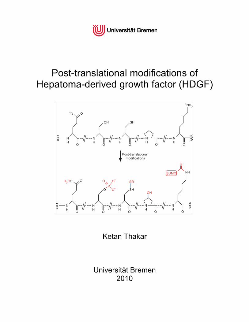
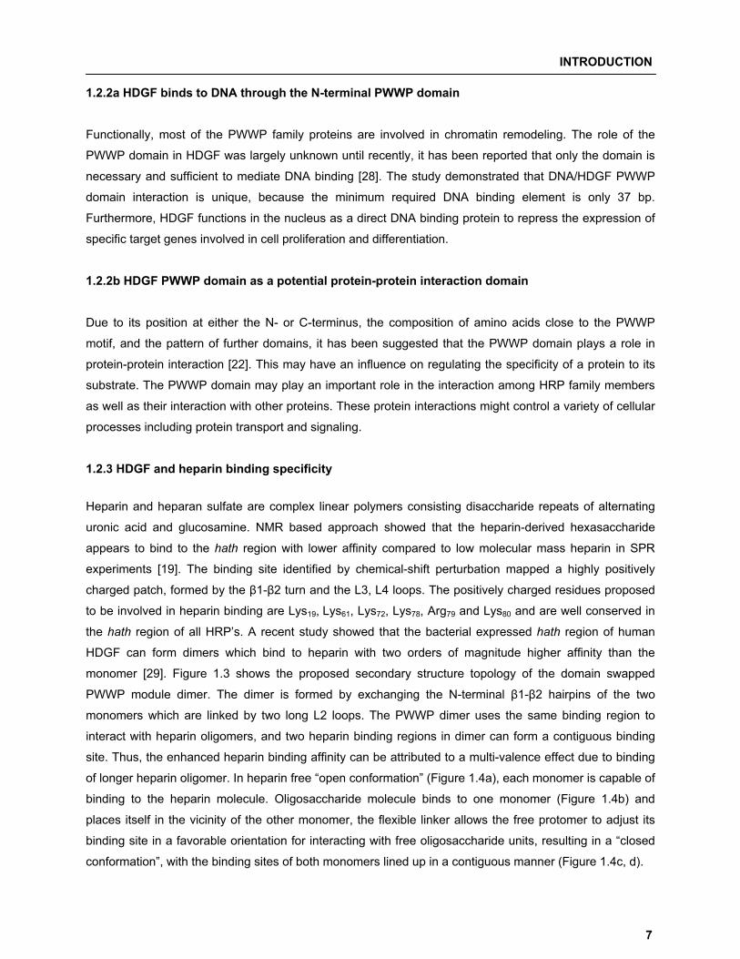
Post-translational modifications of
Hepatoma-derived growth factor (HDGF)
�Post-translational modifications
NH
O
O-O
NH
O
OH
NH
O
SH
NH
O
NH
O
+NH3
NH
O
OH3CO
NH
O
O
NH
O
SH
NH
O
SR
OH
P
-O
-O
O
NH
O
NH�SUMO
O
Ketan Thakar
Universität Bremen 2010
Post-translational modifications of Hepatoma-derived growth factor (HDGF)
Dissertation submitted as a partial fulfillment for procuring the degree Doctor of Natural Science (Dr. rer. nat.)
Submitted to
Fachbereich 2 Biologie/Chemie Universität Bremen
Submitted by
Ketan Thakar M.Sc. Biochemistry and Molecular Biology
Bremen 2010

Examination Committee
Reviewers: 1. Prof. Dr. Sørge Kelm
Universität Bremen, Fachbereich 2, Biochemie
Postfach 330440, 28334 Bremen
2. Dr. Kathrin Mädler Universität Bremen, Fachbereich 2, Biochemie
Leobener Straße NW2, B2060, 28359 Bremen
Examiners: 1. Dr. Frank Dietz
Universität Bremen, Fachbereich 2, Biochemie
Postfach 330440, 28334 Bremen
2. Prof. Dr. Reimer Stick Universität Bremen, Fachbereich 2, Institut für Zellbiologie
Leobener Straße NW2 A3290, 28359 Bremen
Date of Public defense: 28 May, 2010
TABLE OF CONTENTS
iv
Table of Contents I. Summary………………………………………………………………………………………………….. vii II. Zusammenfassung…………………………………………………………………………………… viii
1. Introduction…………………………………………………............................................................. 1 1.1 The HDGF related protein (HRP) family………………………………………………………… 1 1.1.1 Hepatoma-derived growth factor……………………………………………………………. 2
1.1.2 Hepatoma-derived growth factor related protein-1 (HRP-1)……………………………… 2
1.1.3 Hepatoma-derived growth factor related protein-2 (HRP-2) …………………………….. 2
1.1.4 Hepatoma-derived growth factor related protein-3 (HRP-3) …………………………….. 3
1.1.5 Hepatoma-derived growth factor related protein-4 (HRP-4)……………………………… 3
1.1.6 Lens epithelium derived growth factor (LEDGF)…………………………………………... 3
1.2 HDGF structure-function relationship………………………………………………………….. 4 1.2.1 HDGF is a modular protein with two structurally independent domains………………… 4
1.2.2 HDGF PWWP domain………………………………………………………………………... 4
1.2.2a HDGF binds to DNA through the N-terminal PWWP domain……………………………. 7
1.2.2b HDGF PWWP domain as a potential protein-protein interaction domain………………. 7
1.2.3 HDGF and heparin binding specificity………………………………………………………. 7
1.3 Biological roles of HDGF………………………………………………………………………….. 9 1.3.1 HDGF in cancer development, prognosis and diagnosis…………………………………. 9
1.3.2 HDGF and its role in developmental regulation…………………………………………… 10
1.3.3 HDGF in organ remodeling after injury…………………………………………………….. 10
1.3.4 HDGF involvement in apoptosis…………………………………………………………….. 11
1.4 Concept of the thesis ……………………………………………………………………………… 12
2. Materials and Methods…………………………………………………………………………….. 13 2.1 Cell lines……………………………………………………………………………………………… 14 2.2 Plasmid vectors and Primers…………………………………………………………………….. 14 2.3 Kits…………………………………………………………………………………………………….. 14 2.4 Antibodies……………………………………………………………………………………………. 15 2.5 Growth media……………………………………………………………………………………….. 16 2.6 Solutions and buffers……………………………………………………………………………… 16 2.7 Molecular cloning and plasmid isolation………………………………………………………. 18 2.7.1 Bacterial Transformation……………………………………………………………………... 18
2.7.2 Colony PCR……………………………………………………………………………………. 18
2.7.3 Plasmid Purification…………………………………………………………………………… 18
TABLE OF CONTENTS
v
2.7.3a Plasmid purification using the NucleoBond Xtra Midi kit (Macherey-Nagel) …………… 19
2.7.3b Plasmid purification using the GeneJET Plasmid Miniprep kit (Fermentas) …………… 19
2.7.4 Measurement of plasmid concentration and level of purity………………………………. 19
2.7.5 Agarose gel electrophoresis…………………………………………………………………. 19
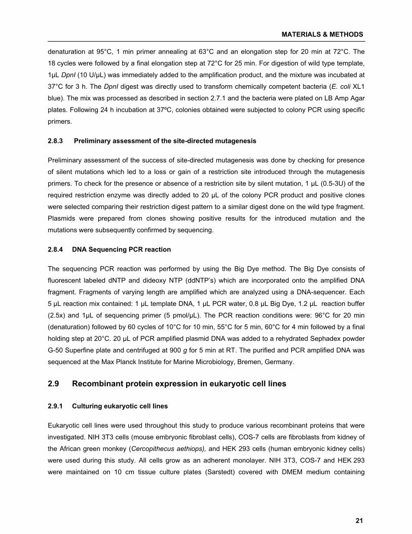
2.8 Plasmid construction………………………………………………………………………………. 20 2.8.1 Production of untagged mHDGF D205G and hHDGF G205D mutants………………… 20
2.8.2 Site-directed mutagenesis …………………………………………………………………... 20
2.8.3 Preliminary assessment of the site-directed mutagenesis……………………………….. 21
2.8.4 DNA Sequencing PCR reaction……………………………………………………………... 21
2.9 Recombinant protein expression in eukaryotic cell lines…………………………………... 21 2.9.1 Culturing eukaryotic cell lines………………………………………………………………... 21
2.9.2 In vitro transfection……………………………………………………………………………. 22
2.9.3 Cell Harvesting………………………………………………………………………………… 22
2.10 Protein Analysis…………………………………………………………………………………….. 22 2.10.1 Sodium dodecyl sulphate-Polyacrylamide gel electrophoresis (SDS-PAGE) …………. 22
2.10.2 Western blotting……………………………………………………………………………….. 23
2.10.2a Transferring resolved proteins on PVDF membrane……………………………………… 23
2.10.2b Immunodetection……………………………………………………………………………… 23
3. Publications……………………………………………………………………………………………. 25 3.1 Publication 1………………………………………………………………………………………… 27 3.2 Publication 2………………………………………………………………………………………… 44 3.3 Publication 3………………………………………………………………………………………… 66
4. Additional results…………………………………………………………………………………….. 85 4.1 SUMOylation of HDGF by SUMO isoforms in mammalian cells…………………………… 86 4.2 HDGF is processed C-terminally at a potential caspase cleavage site…………………… 87
5. Discussion………………………………………………………………………………………………. 89 5.1 SUMOylation of HDGF…………………………………………………………………………….. 89 5.2 Phosphorylation dependent regulation of HDGF secretion and processing……………. 91 5.3 HDGF dimerisation…………………………………………………………………………………. 92
5.4 HDGF�HRP-2 interaction………………………………………………………………………….. 93
5.4.1 Differential expression of HRP-2 corresponds to alternatively spliced isoforms………. 93
5.4.2 HDGF/HRP-2 interaction studies……………………………………………………………. 94
5.5 Caspase dependent cleavage of HDGF………………………………………………………… 95 5.6 Conclusion and perspectives…………………...................................................................... 96

TABLE OF CONTENTS
vi
6. References………………………………………………………………………………………………. 99
A. Appendix…………………………………………………………………………………………………. 107 A.1 Abbreviations…………………………………………………………………………………………108 A.2 List of Tables and Figures………………………………………………………………………… 109 A.2.1 List of Tables……………………………………………………………………………...…… 109
A.2.2 List of Figures…………………………………………………………………………...…….. 109
A.3 Vector maps…………………………………………………………………………...…………….. 110 A.4 List of Manufacturers…………………………………………………………………………........ 112 A.4.1 Chemicals and consumables……………………………………………………….............. 112
A.4.2 Devices………………………………………………………............................................... 113
Acknowledgements……………………………………………………………………........................... 114
Erklarüng……………………………………………………………………................................................ 115
SUMMARY
vii
I. Summary
Post-translational modifications (PTM’s) are modifications that occur during or after protein translation.
Nascent or folded protein can be subjected to an array of specific enzyme-catalyzed modifications on the
amino acid side chains or the peptide backbone. Two broad categories of protein PTM’s occur; the first
includes all enzyme-catalyzed covalent additions of different lower molecular chemical groups up to
complex proteins to amino acid side chains in the target protein, whereas the second category comprises
structural changes and the cleavage of peptide backbones in proteins either by action of proteases or,
less commonly, by autocatalytic cleavage. PTM’s can modulate the function of proteins by altering their
activity state, localization, turnover, and interactions with other proteins.
Hepatoma-derived growth factor (HDGF) is the prototype of a family of six proteins comprising HDGF, the
four HDGF-related proteins (HRP-1–4), and the lens epithelium-derived growth factor (LEDGF). HDGF
exhibits growth factor properties and has been implicated in organ development and tissue differentiation
of the intestine, kidney, liver, and cardiovascular system. Recently, the role of HDGF in cancer biology has
become a main focus of its research. HDGF was found to be over-expressed in a large number of
different tumor types. Although a direct influence of HDGF on tumor biology is still unclear, its expression
is correlated with metastasis and tumor recurrence in multiple studies. HDGF appears to be a novel
prognostic marker for different types of cancer. Growth promoting as well as other activities of HDGF, like
the suppression of differentiation; possible role in apoptotic processes; or its angiogenic properties have
been suggested to play a role in tumor induction and/or cancer progression.
Interestingly, until now, very little is known about how PTM’s are involved in modulating HDGF function.
Therefore, the main aim of the thesis was the identification of HDGF PTM’s and their consequence on its
function. At first, we have identified that HDGF is post-translationally modified by SUMO-1 at a
non-consensus site and SUMOylated HDGF does not associate with chromatin in contrast to the
unSUMOylated form. Further, we show that HDGF secretion is regulated by the presence or absence of a
serine phosphorylation site and loss in secretion is attributed to N-terminal processing of the protein.
Additionally, the two cysteine residues in HDGF are involved in formation of intra-and inter-molecular
disulfide bonds and N-terminal processing favors dimer formation. Furthermore, we demonstrated the
presence of HRP-2 isoforms with their developmentally regulated expression in different rat brain regions
and were able to show that HDGF can interact with HRP-2. Moreover, a new HRP-2 isoform can
exclusively interact and enrich a processed form of HDGF. Finally, we identified the presence of a
functional caspase cleavage site in the C-terminal region of murine HDGF. The results complied provide
substantial new knowledge concerning PTM’s of HDGF and affected mechanisms, and will help to add
new perspectives for research in the field of HDGF and its related proteins.
ZUSAMMENFASSUNG
viii
II. Zusammenfassung Posttranslationale Modifikationen (PTM’s) sind Modifikationen, die während oder nach erfolgter
Proteintranslation auftreten. Das entstehende oder schon gefaltete Protein kann an verschiedenen
Aminosäureseitenketten oder den Peptidbindungen über eine Vielzahl von Enzym-katalysierten
Reaktionen modifiziert werden. Im wesentlichen können zwei Kategorien von PTM’s auftreten; die erste
umfasst alle Enzym-katalysierten, kovalenten Bindungen verschiedener niedermolekularer, chemischer
Gruppen bis hin zu komplexen Proteinen an Aminosäureseitenketten von Zielproteinen, während die
zweite Kategorie Reaktionen umfasst, die zu Strukturänderungen oder zur Spaltung von Peptidbindungen
durch Proteasen oder weniger häufig, durch Autokatalyse führen. PTM’s können die Funktion von
Proteinen modulieren indem sie deren Aktivitätstatus, die Lokalisation, Halbwertszeit und z.B. die
Interaktion mit anderen Proteinen beeinflussen.
Der Hepatoma-derived growth factor (HDGF) ist der Prototyp einer Proteinfamilie, die sechs Mitglieder
umfasst und zu denen neben HDGF die vier HDGF-ähnlichen Proteine (HRP-1–4) und der lens
epithelium-derived growth factor (LEDGF), gehören. HDGF zeigt Wachstumsfaktor-Eigenschaften und ist
in Zusammenhang mit der Organ-Entwicklung und Gewebe-Differenzierung im Dünndarm, Niere, Leber
und dem Kardiovaskulären-System, gebracht worden. Ein weiterer Fokus der HDGF-Forschung befasst
sich mit der Rolle des Proteins in der Krebsbiologie. Es konnte gezeigt werden, dass HDGF in einer
Vielzahl verschiedener Tumor-Typen überexprimiert wird. Obwohl der direkte Einfluss von HDGF auf die
Tumorentwicklung noch immer unklar ist, korreliert die Expression des Proteins mit dem Auftreten von
Metastasen und dem erneuten Auftreten von Tumoren in mehreren Studien. HDGF scheint damit ein
neuer prognostischer Marker für verschiedene Krebsformen zu sein. Seine Wachstums-fördernde, als
auch andere Eigenschaften, wie die Unterdrückung der Differenzierung, eine mögliche Rolle in
apoptotischen Prozessen oder seine angiogenen Eigenschaften wurden mit einer möglichen Rolle bei der
Tumor-Induktion und /oder bei dem Fortschreiten der Krebsentwicklung, in Zusammenhang gebracht.
Weil bis heute wenig aufgeklärt ist, wie PTM’s die Funktionalität des HDGF beeinflußen können, lag der
Fokus dieser Arbeit in der Identifikation von HDGF PTM’s und deren Konsequenzen auf die
Protein-Funktion. Zunächst haben wir gezeigt, dass HDGF posttranslational über SUMO-1 an einem
nicht-Konsensusmotiv modifiziert werden kann und das SUMOyliertes HDGF im Gegensatz zu der
nicht-SUMOylierten Form, nicht mehr an Chromatin bindet. Ferner, konnten wir zeigen, dass
HDGF-Sekretion über das Vorhandensein einer vorausgesagten Serin-Phosphorylierungsstelle reguliert
wird und das ein Verlust der Sekretion mit einer N-terminalen Prozessierung des Proteins
zusammenhängt. Außerdem, sind die zwei Cystein-Reste im HDGF in der Bildung von intra- und
intermolekularen Disulfidbrücken involviert und N-terminale Prozessierung unterstützt die Bildung von
Homodimeren. Desweiteren, konnten wir das Vorhandensein von verschiedenen HRP-2 Isoformen zeigen
ZUSAMMENFASSUNG
ix
und deren entwicklungsabhängige Expression in verschiedenen Rattengehirn-Regionen. In diesem
Zusammenhang, konnten wir nachweisen, dass HDGF mit HRP-2 interagiert und das eine neu
identifizierte HRP-2 Isoform exklusiv mit einer prozessierten Form von HDGF interagiert und diese
spezifisch anreichern kann. Darüber hinaus, haben wir das Vorhandensein einer funktionellen Caspase
Schnittstelle in der C-terminalen Region von HDGF identifiziert.
Die Ergebnisse dieser Arbeit tragen wesentlich zu einer Erweiterung des Wissens um die Funktion von
posttranslationalen Modifikationen des HDGF und dadurch beeinflusster Mechanismen bei und helfen
damit, in diesem Feld, die Forschung an dem HDGF und den HDGF-ähnlichen Proteinen um neue
Ansätze zu bereichern.
INTRODUCTION
1
11 IInnttrroodduuccttiioonn
1.1 The HDGF related protein (HRP) family
Hepatoma-derived growth factor (HDGF) and HDGF related proteins (HRPs) belong to a gene family with
a well-conserved amino acid sequence at the N-terminus. HDGF forms the prototype of a new family of
growth factors called HDGF-related proteins (HRPs), which includes HRP-1, HRP-2, HRP-3, HRP-4 and
lens epithelium-derived growth factor (LEDGF/p75) (Table 1.1). For all HRP members the following
common features are described: (1) homology in the first N-terminal 98 amino acid residues or the hath
region (homologous to amino terminus of HDGF) (2) a PWWP domain in the hath region (3) a canonical
bipartite nuclear localization signal in the non-hath C-terminal region (4) lack of a hydrophobic signal
peptide and (5) altered electrophoretic running behavior.
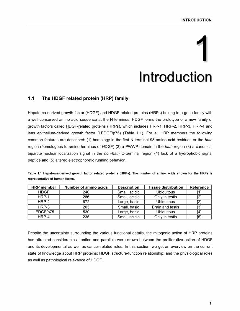
Table 1.1 Hepatoma-derived growth factor related proteins (HRPs). The number of amino acids shown for the HRPs is representative of human forms.
HRP member Number of amino acids Description Tissue distribution Reference HDGF 240 Small, acidic Ubiquitous [1] HRP-1 286 Small, acidic Only in testis [2] HRP-2 672 Large, basic Ubiquitous [2] HRP-3 203 Small, basic Brain and testis [3]
LEDGF/p75 530 Large, basic Ubiquitous [4] HRP-4 235 Small, acidic Only in testis [5]
Despite the uncertainty surrounding the various functional details, the mitogenic action of HRP proteins
has attracted considerable attention and parallels were drawn between the proliferative action of HDGF
and its developmental as well as cancer-related roles. In this section, we get an overview on the current
state of knowledge about HRP proteins; HDGF structure-function relationship; and the physiological roles
as well as pathological relevance of HDGF.
INTRODUCTION
2
1.1.1 Hepatoma-derived growth factor Hepatoma-derived growth factor (HDGF) was originally isolated from conditioned medium of the human
hepatoma-derived cell line, HuH-7, as a heparin-binding and proliferation promoting factor [6].
Recombinant cloning of HDGF derived from HuH-7 cDNA and protein sequencing showed that HDGF
shares homology (23.4% identity and 35.6% homology) with the high mobility group (HMG)-1 protein [1].
HDGF is an acidic polypeptide with mitogenic activity for fibroblasts [1], endothelial cells [7], vascular
smooth muscle cells [8], and hepatoma cells [9].
Nuclear targeting of HDGF was reported to be required for mitogenesis [10, 11]. HDGF contains a nuclear
localization signal (NLS); 155KRRAGLLEDSPKRPK170 (basic residues underlined) {NLS2}, homologous to
the reported consensus sequences for a bipartite NLS which consists of two clusters of basic residues
separated by 10-12 amino acids including proline residues [1]. Additionally, another NLS like basic amino
acid-rich region, 75KPNKRK80 (basic residues underlined) {NLS1}, is found in the hath region [12]. The
cellular trafficking of HDGF is considered atypical, as compared to the majority of described growth
factors, due to the fact that HDGF lacks a classical secretion signal but is still able to exit the cell by a yet
unknown route. Extracellular HDGF undergoes receptor mediated internalization after which it is targeted
to the nucleus via NLS2. The extracellular HDGF receptor and HDGF sub-nuclear target(s) are however
not fully described so far.
1.1.2 Hepatoma-derived growth factor related protein-1 (HRP-1) Izumoto et al. (1997) screened a mouse testis cDNA library with the 0.9 kb cDNA of human HDGF [2].
Isolated clones were grouped into three distinct classes named, HDGF, HRP-1 and HRP-2 based on
sequence relationship. HRP-1 was observed to be 46 amino acid longer as compared to HDGF, showing
a significantly high negative net charge (Lys + Arg: 8.4%, Glu +Asp: 26.4%) and is rich in proline residues
(9.5%). The tissue specific expression pattern of HRP-1 shows expression restricted only to testis.
Interestingly, nothing is known about the function of this HRP family member.
1.1.3 Hepatoma-derived growth factor related protein-2 (HRP-2) With an open reading frame of 2007 bp, encoding a protein of 669 amino acids, HRP-2 is the largest
member of the HRP family and has a predicted Mr of ~74 kDa and pI 9.14 [2]. HRP-2 shows significant
homology to the N-terminal region of HDGF (68%) within the 98-amino acid hath region. Found on
chromosome 17 in mice, the HRP-2 gene encodes 16 exons and is expressed in a wide variety of tissues.
It carries a high level of basic and acidic residues, with Lys + Arg making nearly 20.6% and Glu + Asp
nearly 19.7% of the total protein. HRP-2 is also rich in proline and serine residues which is characteristic
of nuclear proteins [2] and hints towards a nuclear site of action of the protein. Another feature of HRP-2 is
the presence of a mixed charge cluster, a segment of the protein with alternating positive and negative
residues. Eukaryotic regulatory proteins, including transcription and replication factors, are characterized
INTRODUCTION
3
by the presence of charge clusters; and it may well be possible that HRP-2 protein functions as a
regulatory protein [2]. A novel domain, the integrase binding domain (IBD), has been discovered in
HRP-2. The ~80 amino acid residue spanning domain is necessary to bind HIV-1 integrase (HIV-1 IN).
Another HRP family member, LEDGF (p75) contains the homologous sequence, and the IBDs of HRP-2
as well as LEDGF/p75 are capable of binding and stimulating HIV-1 IN in vitro [13]. However, unlike
LEDGF, HRP-2 lacks the ability to tether HIV-1 IN to chromatin. Apart from this function, the molecular
functional mechanisms; specificity or regulation; and physiological relevance of HRP-2 is largely unknown.
1.1.4 Hepatoma-derived growth factor related protein-3 (HRP-3) Discovered by Ikegame et al. (1999), HRP-3 shows 81.4% identity to the HDGF hath region [3]. Mapped
to chromosome 15, region q25 in humans, the HRP-3 gene encodes a protein of 203 amino acid residues
with a basic iso-electric point. HRP-3, like HDGF, was found to have growth-stimulating activity. With its
expression mainly restricted to nervous tissue, HRP-3 may have a specialized function [14].
El-Tahir et al. (2009) showed that HRP-3 is the first member of the HRP family to interact with the
cytoskeleton. The results suggest that HRP-3 is involved in neurite growth and exerts its effects through
interaction with tubulin and microtubules. Also, HRP-3 is redistributed from an extra-nuclear to a nuclear
localization during development suggesting a role of HRP-3 during neuritogenesis. The neuritogenic effect
is due to the interaction of HRP-3 with tubulin dimers and assembled microtubules. It promotes tubulin
assembly into microtubules and stabilizes microtubules once they have been formed. In addition, HRP-3
promotes bundling of microtubules. The stabilization and bundling of cytoskeletal components was
attributed to the dimerisation of HRP-3 [15].
1.1.5 Hepatoma-derived growth factor related protein-4 (HRP-4) HRP-4 was discovered by Dietz et al. (2002) from a bovine cDNA library and codes for a 36 kDa protein
with 235 amino acids [5]. HRP-4, like HDGF, can bind to the glycoaminoglycans heparin and heparan
sulphate and shows growth promoting activity. Amino acid sequence comparison of the N-terminal
91 amino acids of bHRP-4 to other HRP members showed that it shares 86 and 89% amino acids with
HDGF and mHRP-1 respectively, but only 70 and 69% amino acids with mHRP-2/hHRP-3 and LEDGF
respectively. Like HRP-1, HRP-4 is exclusively expressed in testis but its function still remains unclear.
1.1.6 Lens epithelium derived growth factor (LEDGF) LEDGF is an ubiquitously expressed nuclear protein, tightly associated with chromatin throughout the cell
cycle. Chromatin association is primarily mediated by three conserved sequence elements within the
N-terminal half of the protein: the PWWP domain, nuclear localization signal (NLS), and a dual copy of the
AT-hook DNA binding motif. Recently, study revealed that the association with chromatin is essential for
LEDGF function during HIV-1 infection, highlighting its significance in chromatin binding [16]. Like HRP-2

INTRODUCTION
4
LEDGF contains a second evolutionarily conserved domain within its extended C-terminus. It is this
domain (IBD) that mediates the interaction with HIV-1 integrase. Two alternatively spliced isoforms,
LEDGF/p75 and LEDGF/p52 are expressed from the same gene (human PSIP1). The smaller p52 isoform
lacks the IBD and fails to engage HIV-1 IN in vitro or in living cells. The cellular functions of LEDGF
remain largely uncharacterized, although initial reports have indicated a role for LEDGF/p75 in
transcriptional regulation. LEDGF is not essential for cell survival, although the majority of LEDGF-null
mice died soon after birth or showed a range of developmental abnormalities in adulthood [17]. LEDGF
also binds to heparin which protects it from heat, acid-base deactivation, and proteolytic degradation with
trypsin and chymotrypsin. Heparin-bound LEDGF greatly potentiates survival of mouse LECs in culture
[18].
1.2 HDGF structure-function relationship Analysis of the HDGF structure showed that it contains two structurally independent regions, (i) a
N-terminal PWWP domain within the hath region, and (ii) an unstructured C-terminal part [19].
1.2.1 HDGF is a modular protein with two structurally independent domains
The few functional data available on HDGF gave a clue that it might be a modular protein in which the
N- and C-terminal regions have distinctive roles. The N-terminal region was shown to be responsible for
cell surface binding, promoting proliferation through triggering a membrane receptor-mediated pathway
[20] and HDGF internalization [11], while the C-terminal region was found to be responsible for mediating
mitogenic effects after nuclear entry [11]. A functional distinction between the N-and C-terminal regions of
HDGF was supported by a recent study demonstrating, that the N-terminal hath region of human HDGF is
a well structured domain while the C-terminal non-hath region is disordered and does not pursue any
defined secondary structure [21]. Moreover, the ordered and disordered structures of the N- and
C-terminal regions, respectively, were shown to be independent of linkage of the two regions within the full
length HDGF or their presence in two separate constructs [21].
1.2.2 HDGF PWWP domain
HRPs are members of a family of proteins with a conserved PWWP domain. The PWWP domain was first
characterized from the WHSC1 gene. It contains a conserved 70 amino acid sequence and has been
found in around 60 eukaryotic proteins. Initially, the PWWP domain was hypothesized to be a site for
protein-protein interaction [22]. However, the PWWP domain of DNA-methyltransferase 3b (Dnmt3b) has
been shown to interact with DNA [23-25]. In addition to the PWWP domain, proteins in this family are
frequently known to contain chromatin association domains such as the bromodomain, chromodomain,
SET domain, and the Cys-rich Zn-binding domains [22]. This strongly suggests a role in chromatin
INTRODUCTION
5
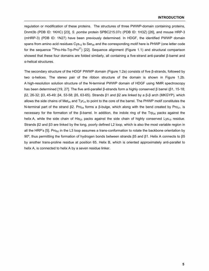
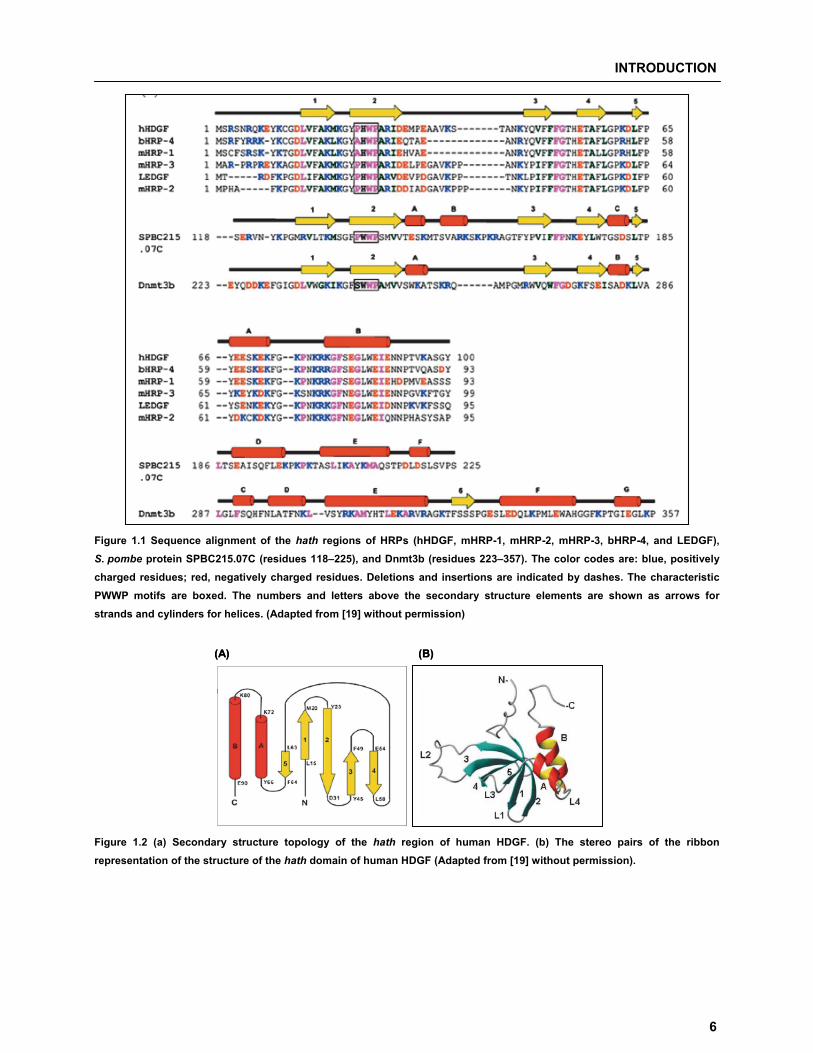
regulation or modification of these proteins. The structures of three PWWP-domain containing proteins,
Dnmt3b (PDB ID: 1KHC) [23], S. pombe protein SPBC215.07c (PDB ID: 1H3Z) [26], and mouse HRP-3
(mHRP-3) (PDB ID: 1N27) have been previously determined. In HDGF, the identified PWWP domain
spans from amino acid residues Cys12 to Ser68 and the corresponding motif here is PHWP (one letter code
for the sequence 24Pro-His-Trp-Pro27) [22]. Sequence alignment (Figure 1.1) and structural comparison
showed that these four domains are folded similarly, all containing a five-strand anti-parallel �-barrel and
�-helical structures.
The secondary structure of the HDGF PWWP domain (Figure 1.2a) consists of five �-strands, followed by
two �-helices. The stereo pair of the ribbon structure of the domain is shown in Figure 1.2b.
A high-resolution solution structure of the N-terminal PWWP domain of HDGF using NMR spectroscopy
has been determined [19, 27]. The five anti-parallel �-strands form a highly conserved � barrel (�1, 15-18;
�2, 26-32; �3, 45-49; �4, 53-58; �5, 63-65). Strands �1 and �2 are linked by a �-� arch (MKGYP), which
allows the side chains of Met20 and Tyr23 to point to the core of the barrel. The PHWP motif constitutes the
N-terminal part of the strand �2. Pro24 forms a �-bulge, which along with the bend created by Pro27, is
necessary for the formation of the �-barrel. In addition, the indole ring of the Trp26 packs against the
helix A, while the side chain of His25 packs against the side chain of highly conserved Lys19 residue.
Strands �2 and �3 are linked by the long, poorly defined L2 loop, which is also the most variable region in
all the HRP’s [5]. Pro60 in the L3 loop assumes a trans-conformation to rotate the backbone orientation by
90º, thus permitting the formation of hydrogen bonds between strands �5 and �1. Helix A connects to �5
by another trans-proline residue at position 65. Helix B, which is oriented approximately anti-parallel to
helix A, is connected to helix A by a seven residue linker.
INTRODUCTION
6
Figure 1.1 Sequence alignment of the hath regions of HRPs (hHDGF, mHRP-1, mHRP-2, mHRP-3, bHRP-4, and LEDGF), S. pombe protein SPBC215.07C (residues 118–225), and Dnmt3b (residues 223–357). The color codes are: blue, positively charged residues; red, negatively charged residues. Deletions and insertions are indicated by dashes. The characteristic PWWP motifs are boxed. The numbers and letters above the secondary structure elements are shown as arrows for strands and cylinders for helices. (Adapted from [19] without permission)
(A) (B)(A) (B)
Figure 1.2 (a) Secondary structure topology of the hath region of human HDGF. (b) The stereo pairs of the ribbon representation of the structure of the hath domain of human HDGF (Adapted from [19] without permission).
INTRODUCTION
7
1.2.2a HDGF binds to DNA through the N-terminal PWWP domain Functionally, most of the PWWP family proteins are involved in chromatin remodeling. The role of the
PWWP domain in HDGF was largely unknown until recently, it has been reported that only the domain is
necessary and sufficient to mediate DNA binding [28]. The study demonstrated that DNA/HDGF PWWP
domain interaction is unique, because the minimum required DNA binding element is only 37 bp.
Furthermore, HDGF functions in the nucleus as a direct DNA binding protein to repress the expression of
specific target genes involved in cell proliferation and differentiation. 1.2.2b HDGF PWWP domain as a potential protein-protein interaction domain Due to its position at either the N- or C-terminus, the composition of amino acids close to the PWWP
motif, and the pattern of further domains, it has been suggested that the PWWP domain plays a role in
protein-protein interaction [22]. This may have an influence on regulating the specificity of a protein to its
substrate. The PWWP domain may play an important role in the interaction among HRP family members
as well as their interaction with other proteins. These protein interactions might control a variety of cellular
processes including protein transport and signaling.
1.2.3 HDGF and heparin binding specificity
Heparin and heparan sulfate are complex linear polymers consisting disaccharide repeats of alternating
uronic acid and glucosamine. NMR based approach showed that the heparin-derived hexasaccharide
appears to bind to the hath region with lower affinity compared to low molecular mass heparin in SPR
experiments [19]. The binding site identified by chemical-shift perturbation mapped a highly positively
charged patch, formed by the �1-�2 turn and the L3, L4 loops. The positively charged residues proposed
to be involved in heparin binding are Lys19, Lys61, Lys72, Lys78, Arg79 and Lys80 and are well conserved in
the hath region of all HRP’s. A recent study showed that the bacterial expressed hath region of human
HDGF can form dimers which bind to heparin with two orders of magnitude higher affinity than the
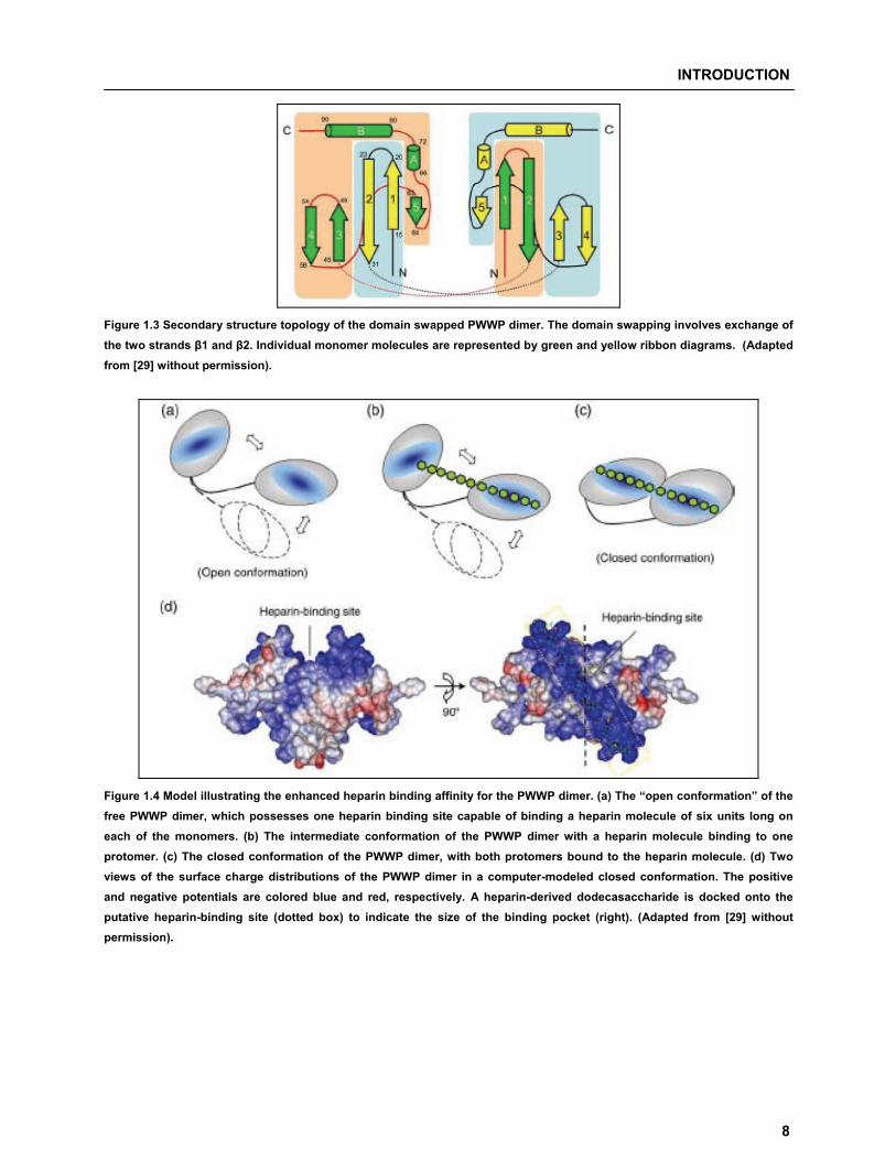
monomer [29]. Figure 1.3 shows the proposed secondary structure topology of the domain swapped
PWWP module dimer. The dimer is formed by exchanging the N-terminal �1-�2 hairpins of the two
monomers which are linked by two long L2 loops. The PWWP dimer uses the same binding region to
interact with heparin oligomers, and two heparin binding regions in dimer can form a contiguous binding
site. Thus, the enhanced heparin binding affinity can be attributed to a multi-valence effect due to binding
of longer heparin oligomer. In heparin free “open conformation” (Figure 1.4a), each monomer is capable of
binding to the heparin molecule. Oligosaccharide molecule binds to one monomer (Figure 1.4b) and
places itself in the vicinity of the other monomer, the flexible linker allows the free protomer to adjust its
binding site in a favorable orientation for interacting with free oligosaccharide units, resulting in a “closed
conformation”, with the binding sites of both monomers lined up in a contiguous manner (Figure 1.4c, d).
INTRODUCTION
8
Figure 1.3 Secondary structure topology of the domain swapped PWWP dimer. The domain swapping involves exchange of the two strands �1 and �2. Individual monomer molecules are represented by green and yellow ribbon diagrams. (Adapted from [29] without permission).
Figure 1.4 Model illustrating the enhanced heparin binding affinity for the PWWP dimer. (a) The “open conformation” of the free PWWP dimer, which possesses one heparin binding site capable of binding a heparin molecule of six units long on each of the monomers. (b) The intermediate conformation of the PWWP dimer with a heparin molecule binding to one protomer. (c) The closed conformation of the PWWP dimer, with both protomers bound to the heparin molecule. (d) Two views of the surface charge distributions of the PWWP dimer in a computer-modeled closed conformation. The positive and negative potentials are colored blue and red, respectively. A heparin-derived dodecasaccharide is docked onto the putative heparin-binding site (dotted box) to indicate the size of the binding pocket (right). (Adapted from [29] without permission).
INTRODUCTION
9
1.3 Biological roles of HDGF The first biological effect described for HDGF was its ability to stimulate DNA synthesis in Swiss 3T3 cells
as assessed by the rate of incorporation of (3H) thymidine [1]. Due to the fact that HDGF was first isolated
from a cancerous cell line, led to intensive investigations that mainly dealt with; (i) links between HDGF
under pathological conditions that are related to abnormal cellular proliferation such as tumorigenesis and
neoplastic angiogenesis, (ii) the relation between the proliferative action of HDGF and development (iii) its
role in organ remodeling after injury (iv) its involvement in apoptosis. Other biological functions were also
attributed to HDGF like serving as a neurotrophic factor preventing neuron death [30] or as trophic factor
for motor neurons up-regulated in the spinal cord of transgenic mice before onset of degeneration [31].
1.3.1 HDGF in cancer development, prognosis and diagnosis Several studies implicated HDGF in the development and progression of neoplasias. A strong correlation
was observed between the up-regulation of HDGF and hepatic malignancy [32, 33]. Elevated level of
HDGF was detected in the highly proliferative hepatocellular carcinoma compared to adjacent normal
hepatocytes, and was associated with loss in differentiation [32]. It was suggested that HDGF exerts such
an effect on hepatic cells through a combination of autocrine and paracrine actions [33]. Such data are
also supported by an earlier study showing that HDGF antisense oligonucleotides inhibit the growth of
cultured hepatoma cells [9]. Such a correlation, similar to that with hepatocellular carcinoma, was also
found between HDGF and melanoma [34]. Using a functional proteomics approach, HDGF was found to
be one of eight candidates that are differentially regulated in transformed cells, where the levels of HDGF
expression increases in early and late stage melanomas compared to low levels in normal melanocytes
[34]. The correlative behavior between the increased HDGF levels and cancer development encouraged
efforts attempting to establish HDGF as a diagnostic and prognostic tool. It was possible to show that
HDGF is a strong prognostic predictor for patients with non-small cell lung cancer (NSCLC). Two research
groups demonstrated that there is a direct and strong correlation between the increased levels of HDGF
expression (immunohistochemically assessed) and the poor overall survival of NSCLC patients after
curative therapy [35, 36]. In another study it was shown that HDGF is one of the proteins that can be
useful in the determination of the regressive (where HDGF is down-regulated) versus progressive status
of tumors [37]. Furthermore, HDGF expression significantly increases in human colorectal cancers,
especially in tumors capable in DNA mismatch repair, and thus represented a novel marker for specific
tumor subtypes [38]. Different studies established association of HDGF with the recurrence and
prognosis of gastric carcinoma and pancreatic ductal carcinoma [39, 40].
The ability of HDGF to induce tumorigenesis in vivo is attributed direct angiogenic activity and stimulates
the potent angiogenic factor VEGF (vascular endothelial growth factor). Through this combined action,
HDGF is supposed to assist in the formation of new blood vessels that are required for tumor
INTRODUCTION
10
development and progression [41, 42]. Such findings, on the angiogenesis-mediated role of HDGF in
tumorigenesis, were also supported by data showing HDGF stimulated proliferation of endothelial cells,
including those of the kidney and the cardiovascular systems [7, 43]. However, the molecular mechanism
of HDGF in cancer incidence and development are still unknown. One study illustrated that the
independent abnormal expression of HDGF can promote cell proliferation but was insufficient to cause
cancer. Increasing rates in carcinogenesis were attributed to the cumulative effects produced by aberrant
cell signal transduction activated by gene mutation and epigenetic modification. Supporting this
hypothesis it has been shown that HDGF over-expression activates Erk1/2 and promotes growth of
human gastric cancer AGS cells in an anchorage-independent manner [44].
1.3.2 HDGF and its role in developmental regulation
Many antigens are highly and broadly expressed during embryogenesis and then normally show lower
levels and limited distribution after birth, to be only again highly expressed in case of cancer development
or in response to tissue damage. HDGF was observed to be developmentally regulated and plays a
putative role in renal, cardiovascular, hepatic and intestinal development. HDGF is widely distributed in
the kidney at early stages of development but eventually becomes confined to sites of active
morphogenesis and, except for renal tubules, disappears from the adult kidney [7]. In another study,
HDGF was detected in the nuclei of smooth muscle cells (SMC) and endothelial cells from 19-day fetal,
but not in the adult, rat aorta and thus concluded that HDGF helps to regulate SMC growth during
development [10]. HDGF is also expressed early in embryonic heart and fetal gut suggesting that it may
play a role in cardiovascular growth and differentiation and represented the first described
nuclear-targeted mitogen in the developing heart [43]. HDGF expression in hepatocytes decreased with
differentiation suggesting that HDGF might also participate in the regulation of hepatocyte proliferation
during liver development [45]. Also, HDGF is highly expressed in early gut tissue with dramatically
reduced levels after villous epithelial differentiation [38].
1.3.3 HDGF in organ remodeling after injury
Different studies also demonstrated that HDGF play a role in processes of organ repair and remodeling
after injury. For example, HDGF helps to regulate the proliferation of SMC in response to vascular injury
[8]. Smooth muscles normally show low proliferative rate and their rapid proliferation and migration after
vascular injury might result in narrowing of the vascular lumen leading to vascular stenosis [46]. Many
growth factors are released in the process of vascular remodeling. Interestingly, HDGF is not expressed in
the vascular wall until it is injured and thus represents an attractive target for therapeutic intervention [47].
A similar role of HDGF in lung remodeling was provided by stimulating lung epithelial growth after injury
[48]. Furthermore, HDGF seems to play a role in the renewal of intestinal epithelium and that the presence
of auto anti-HDGF antibodies in patients with chronic ulcerative colitis might be a reason for their delayed
INTRODUCTION
11
mucosal healing [49]. HDGF expression is induced in hepatocytes before induction of DNA synthesis in
two liver regeneration models suggesting that it might also be involved in liver regeneration possibly as an
autocrine factor. In addition, HDGF participates in human liver regeneration after drug-induced liver
damage and surgical resection thus making it a candidate for the protection and enhancement of the
recovery from liver damage as a result of surgical resection, viral infection or drug intoxicity [50].
1.3.4 HDGF involvement in apoptosis
Induction of apoptosis via the TNF receptor requires the release of the IAP antagonist Smac/Diablo from
the mitochondria, which subsequently disrupts the TRAF2-cIAP1 complex and permits activation of
caspase-8 [51]. RNAi-induced silencing of the HDGF gene prevented the release of Smac/Diablo upon
TNF-� treatment, suggesting that lack of HDGF interferes with the release of pro-apoptotic factors from
the mitochondria [52]. Clermont et al. (2008) showed that triggering of endothelial cell apoptosis by TNF�
and cycloheximide leads to an early dephosphorylation of HDGF which occurs before mitochondrial
membrane permeabilization and downstream from an initiator caspase [53]. Knock-down of HDGF not
only induced apoptosis in human cancer cells through the Bad-mediated intrinsic apoptotic pathway [54],
but also the Fas-mediated extrinsic apoptotic pathway, eventually suppressing anchorage-independent
growth of cancer cells [55].
INTRODUCTION
12
1.4 Concept of the thesis The aim of the present thesis is to improve the knowledge about HDGF and its functional regulation. The
thesis specifically centers on post-translational modifications and protein-protein interactions for HDGF.
Post-translational modifications are known to modulate cellular pathways by causing dynamic and
reversible alterations in the interaction profile of proteins. More detailed insights in this area would greatly
increase our understanding about molecular mechanisms and their impact on HDGF function.
In particular the following questions were addressed and elaborated:
� Is HDGF modified by SUMOylation and does it affect HDGF function? The fact that many nuclear/DNA interacting proteins are post-translationally modified by small
ubiquitin-like modifier (SUMO) thereby regulating their function makes it plausible that HDGF might also
be a candidate for SUMO modification. In this part of the thesis we investigate if HDGF is SUMO modified
and if so, where is the site of modification. We were able to show that HDGF is SUMOylated at a
non-consensus motif and the modification negatively influences the ability of HDGF to bind to chromatin.
� Does post-translational modification play a role in regulating HDGF secretion? The cellular trafficking of HDGF is considered atypical due to the fact that HDGF lacks a classical
secretion signal but is still able to exit the cell by a yet unknown route. In this part of the thesis we
investigated regulation of HDGF secretion. We show for the first time that the HDGF N-terminal region
mediates secretion and that loss of a potential serine phosphorylation site in HDGF regulates N-terminally
processing and secretion of the protein.
� Does HDGF interact with other members of the HRP family of proteins? Protein-protein interactions play a pivotal role in regulation of function. Recently, HDGF dimer formation
via a novel domain swapping mechanism through interaction of the N-terminal hath region of the protein
was demonstrated. The fact that all HRP members share structural homology in the N-terminal region led
to the basic hypothesis of interaction of HDGF with other members of the HRP family. We discovered a
new HRP-2 splice variant and showed specific interaction of the different HRP-2 splice variants with
HDGF.

22 MMaatteerriiaallss && MMeetthhooddss
MATERIALS & METHODS
14
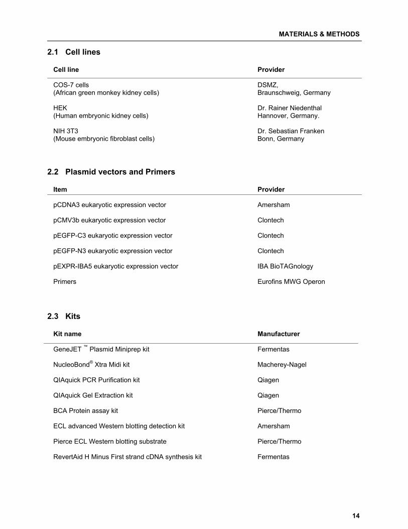
2.1 Cell lines
Cell line Provider
COS-7 cells DSMZ, (African green monkey kidney cells) Braunschweig, Germany HEK Dr. Rainer Niedenthal (Human embryonic kidney cells) Hannover, Germany. NIH 3T3 Dr. Sebastian Franken (Mouse embryonic fibroblast cells) Bonn, Germany
2.2 Plasmid vectors and Primers
Item Provider
pCDNA3 eukaryotic expression vector Amersham pCMV3b eukaryotic expression vector Clontech pEGFP-C3 eukaryotic expression vector Clontech pEGFP-N3 eukaryotic expression vector Clontech pEXPR-IBA5 eukaryotic expression vector IBA BioTAGnology Primers Eurofins MWG Operon
2.3 Kits
Kit name Manufacturer
GeneJET ™ Plasmid Miniprep kit Fermentas NucleoBond® Xtra Midi kit Macherey-Nagel QIAquick PCR Purification kit Qiagen
QIAquick Gel Extraction kit Qiagen BCA Protein assay kit Pierce/Thermo ECL advanced Western blotting detection kit Amersham Pierce ECL Western blotting substrate Pierce/Thermo RevertAid H Minus First strand cDNA synthesis kit Fermentas
MATERIALS & METHODS
15
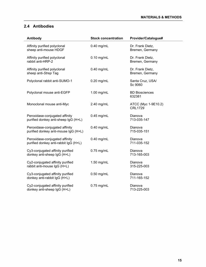
2.4 Antibodies Antibody Stock concentration Provider/Catalogue# Affinity purified polyclonal 0.40 mg/mL Dr. Frank Dietz, sheep anti-mouse HDGF Bremen, Germany Affinity purified polyclonal 0.10 mg/mL Dr. Frank Dietz, rabbit anti-HRP-2 Bremen, Germany Affinity purified polyclonal 0.40 mg/mL Dr. Frank Dietz, sheep anti-Strep Tag Bremen, Germany Polyclonal rabbit anti-SUMO-1 0.20 mg/mL Santa Cruz, USA/ Sc 9060 Polyclonal mouse anti-EGFP 1.00 mg/mL BD Biosciences 632381 Monoclonal mouse anti-Myc 2.40 mg/mL ATCC (Myc 1-9E10.2) CRL1729 Peroxidase-conjugated affinity 0.45 mg/mL Dianova purified donkey anti-sheep IgG (H+L) 713-035-147 Peroxidase-conjugated affinity 0.40 mg/mL Dianova purified donkey anti-mouse IgG (H+L) 715-035-151 Peroxidase-conjugated affinity 0.40 mg/mL Dianova purified donkey anti-rabbit IgG (H+L) 711-035-152 Cy3-conjugated affinity purified 0.75 mg/mL Dianova donkey anti-sheep IgG (H+L) 713-165-003 Cy2-conjugated affinity purified 1.50 mg/mL Dianova rabbit anti-mouse IgG (H+L) 315-225-003 Cy3-conjugated affinity purified 0.50 mg/mL Dianova donkey anti-rabbit IgG (H+L) 711-165-152 Cy2-conjugated affinity purified 0.75 mg/mL Dianova donkey anti-sheep IgG (H+L) 713-225-003
MATERIALS & METHODS
16
2.5 Growth media
Media* Composition
DMEM 13.37 g DMEM (Dulbecco’s Modified Eagle’s Medium) 03.70 g NaHCO3 add to 1 L with ddH2O LB medium 15.00 g Tryptone (Liquid Broth) 05.00 g Yeast extract 05.00 g NaCl add to 1 L with ddH2O
LB ampicillin agar 15.00 g agar
1 L LB medium 50 μg/mL ampicillin
LB kanamycin agar 15.00 g agar 1 L LB medium 25 μg/mL kanamycin SOC medium 20.00 g tryptone 05.00 g yeast extract 00.50 g NaCl 02.50 mL KCl (1M) add to 1 L with ddH2O
2.6 Solutions and buffers
Solutions/buffer Composition
Agarose gel electrophoresis loading buffer (6X) 00.09% Bromophenol blue 60.00% Glycerol 60.00 mM EDTA Ampicillin stock solution 500 mg/mL in ddH2O Ethidium bromide stock solution 10 mg/mL in ddH2O Glycine Solution 0.10 M Glycine HCl, pH 3.0 Kanamycin stock solution 50 mg/mL in ddH2O
PBS buffer 37.00 mM NaCl (Phosphate Buffer Saline) 02.70 mM Na2HPO4 01.50 mM K2HPO4 SDS-PAGE electrophoresis (running) buffer (10X) 30.30 g Tris base 144.0 g Glycine 10.00 g SDS to 1 L with ddH2O
* All growth media listed here were sterilized by autoclaving.
MATERIALS & METHODS
17
SDS-PAGE stacking gel buffer 0.5 M Tris-HCl, pH 6.8 0.4% SDS in ddH2O SDS-PAGE separation gel buffer 1.5 M Tris-HCl, pH 8.8 0.4 % SDS in ddH2O SDS-PAGE sample buffer (2x) 0.125 M Tris-HCl, pH 6.8 4% SDS 20% glycerol (v/v) 0.02% bromophenol blue TAE buffer (10x) 0.4 M Tris acetate (Agarose gel electrophoresis running buffer) 10 mM EDTA, pH 8.3 TBS 10 mM Tris HCl, pH 7.4 (Tris Buffer Saline) 150 mM NaCl TNE lysis buffer 20 mM Tris (NP-40 lysis buffer) 150 mM NaCl 5 mM EDTA 1% NP-40, pH 7.4 Trypsin/EDTA 0.5 g Trypsin 0.2 g EDTA to 1 L using sterile 1XPBS
Western blotting buffer 25 mM Tris-HCl 192 mM glycine 20% (v/v) Methanol Protease inhibitor cocktail-tablet (1 tablet/10 mL lysis buffer) Chromatin binding assay buffers: CSK I 10 mM Pipes, pH 6.8 100 mM NaCl 1 mM EDTA 300 mM Sucrose 1 mM MgCl2 1 mM DTT 0.5% Triton X-100
Protease inhibitor -tablet 1 mM PMSF RIPA 150 mM Tris-HCl, pH 8.0 150 mM NaCl 0.5% Sodium deoxycholate 0.1% SDS 1% (v/v) NP-40 CSK II 10 mM Pipes, pH 6.8 50 mM NaCl 300 mM Sucrose 6 mM MgCl2 1 mM DTT
MATERIALS & METHODS
18
2.7 Molecular cloning and plasmid isolation† 2.7.1 Bacterial Transformation Around 12 μL of the ligation mix were added to 200 μL half thawed chemically competent E. coli XL1 blue
cells obtained from -80°C storage. After keeping the mix for 10 min on ice, the tube was transferred to a
37�C heat block for 90 s (heat shock) and then back on ice for 2 min. Pre-warmed SOC medium (1 mL)
was then added to the transformation mixture and incubated in the 37°C shaker for 45 min to allow the
expression of the resistance gene under non-selective conditions. The culture was then aseptically spread
on a pre-warmed agar plate containing the specified antibiotic (depending on the resistance gene included
in the host plasmid). The plate was incubated overnight at 37°C to allow the growth of transformed
bacteria.
2.7.2 Colony PCR Colony PCR was performed to distinguish colonies arising from bacteria in which only the re-ligated
plasmid had been transformed from those into which the plasmid containing the insert of interest had been
transferred. Each 25 μL PCR reaction mix contained: a portion of the colony under test , 0.25 μL sense
primer, 0.25 μL antisense primer (primers concentration 100 pmol/μL), 1.5 μL MgCl2 (25 mM), 2.5 μL of
10x Taq buffer, 0.5 μL dNTPs (10 mM each), 0.10 μL Taq polymerase (5 U/μL). The PCR reaction took
place in a thermocycler (Eppendorf) using following conditions: 3 min at 95°C, 29 cycles of 30 s at 95°C
(denaturation), 1 min at 56°C (annealing) and 1 min at 72°C (extension). The 29 cycles were followed by a
final extension step of 3 min at 72°C. 10 μL of the PCR product were then mixed with 2 μL of 6x agarose
gel loading buffer and loaded to a 1% (w/v) agarose gel containing ethidium bromide (0.1�L/mL). The
samples were allowed to run at constant voltage (100 V) for adequate time. The bands were visualized
under UV light and the size of the insert was determined in comparison to the DNA mass ruler ladder mix
(Fermentas).
2.7.3 Plasmid Purification A positive clone for the insert was aseptically transferred to 5 mL or 200 mL sterile LB medium containing
the antibiotic which depends on the resistance gene in the plasmid vector. The clones were allowed to
grow overnight in a 37°C incubator with shaking at 250 revolutions min-1. Plasmids were purified from
bacterial cultures using the NucleoBond Xtra Midi kit (Macherey-Nagel) for the 200 mL cultures or the
GeneJET Plasmid Miniprep kit (Fermentas) for the 5 mL cultures.
† This section only contains methods not described or elaborated in publications
MATERIALS & METHODS
19
2.7.3a Plasmid purification using the NucleoBond Xtra Midi kit (Macherey-Nagel) A 250 mL overnight bacterial culture was centrifuged at 6000 g for 15 min at 4ºC. The supernatant was
decanted and residual medium was removed by placing the tubes in an inverted position. The pellet was
re-suspended in 8 mL of the provided Buffer RES (ice cold with RNase A) and the homogenized lysate
was transferred to a 50 mL tube. Alkaline lysis was done by adding 8 mL of the Buffer LYS followed by
incubating the mix for 5 min at RT. The lysate was neutralized by adding 6 mL of the Buffer NEU and the
fluffy precipitate was allowed to flow through the column, pre-equilibrated with 12 mL Buffer EQU, by
gravity. The column filter was first washed twice with 5 mL Buffer EQU, filter then discarded and the
column now washed with 8 mL of buffer WASH. The bound DNA was eluted using 5 mL Buffer ELU. For
DNA precipitation, 3.5 mL isopropanol was added to the eluate and the mix was incubated for 5 min. The
eluate/isopropanol mix was centrifuged at 15000 g for 30 min at 4ºC, the pellet obtained was washed with
2 mL 70% ethanol and re-centrifuged at 15000 g for 15 min at RT and left to dry. Finally the DNA pellet
was reconstituted in 1 mL buffer TE.
2.7.3b Plasmid purification using the GeneJET Plasmid Miniprep kit (Fermentas) A 5 mL overnight bacterial culture was centrifuged at 12000 g for 10 min at 4ºC, the supernatant was
decanted and residual medium was removed. The pellet was re-suspended in 250 μL of the provided
resuspension buffer (ice-cold) and suspension was made homogenous by continuous vortexing for 30 s.
Following this, 250 μL of lysis solution was added to the tube and mixed thoroughly by inverting the tube
4-6 times. Neutralization solution (350 �L) was then added to the tube and mixed thoroughly by inverting
the tube 4-6 times. This was followed by a centrifugation step for 10 min on a table top centrifuge. The
resulting supernatant is applied to GeneJET spin column and centrifuged again at 13000 rpm for 30-60 s.
The GeneJET spin column was then washed using 500 �L of wash solution, centrifuged for 30-60 s. The
flow through is discarded and the assembly spun once again to remove the residual wash buffer. The
column was then transferred to a new tube and the DNA eluted with 50 �L of elution buffer by
centrifugation for 1 min at full speed to obtain plasmid DNA.
2.7.4 Measurement of plasmid concentration and level of purity The plasmid concentrations were determined by measuring the absorbance of the eluates at 260 nm
using the Eppendorf photometer. The purity (level of protein contamination) was assessed from the
A 260/280 value.
2.7.5 Agarose gel electrophoresis Agarose gels (1%) containing ethidium bromide was pre-casted. For sample preparation, 20 μL of each
sample under test was mixed with 5 μL of the agarose gel loading buffer and loaded to the wells. 10 μL of
MATERIALS & METHODS
20
the molecular weight marker (Mass Ruler DNA Ladder Mix, Fermentas) was loaded in parallel. The
electrophoresis was performed at 100 volts for approximately 30 min after which the DNA was visualized
under the UV transilluminator (Herolab).
2.8 Plasmid construction 2.8.1 Production of untagged mHDGF D205G and hHDGF G205D mutants Plasmids were constructed using standard recombinant cloning techniques and all changes were verified
by DNA sequencing. HDGF mutants were generated by site-directed mutagenesis with the Quickchange
Multi-Site Direct Mutagenesis kit (Stratagene, La Jolla, CA, U.S.A.) according to the manufacturer’s
protocol. Untagged wild type mouse HDGF and human HDGF cloned in pcDNA3 Amp was used as a
template and amplified using the primers listed below in Table 2.1. Table 2.1 List of primers used for site directed mutagenesis. In case a restriction site is created, such site is shown in parentheses in the description lane and in bold in sequence lane. The base changes leading to the appearance or disappearance of restriction sites are underlined. The shaded triplets represent changed codons in mutants. The mutated base leading to mutations are underlined in shaded codons.
Name Description Sequence (5´�3´) OSK 1290
sense primer mHDGF D205G BamHI (G^GATCC) appears
CCCTCTGAGCCAGGATCCGGCCAGGGACCT
OSK 1289
antisense primer mHDGF D205G BamHI appears
AGGTCCCTGGCCGGATCCTGGCTCAGAGGG
OSK 1292
sense primer hHDGF G205D (silent mutation)
CCCTCTGAGCCCGACTCTGGCCGGGGGCCT
OSK 1291
antisense primer hHDGF G205D (silent mutation)
AGGCCCCCGGCCAGAGTCGGGCTCAGAGGG
Plasmids encoding for HA-SUMO-2 and HA-SUMO-3 were provided by Dr. Rainer Niedenthal (Hannover,
Germany). Other cloning strategies, plasmids used and mutants produced during the course of the entire
study have been elaborated under the materials and methods section of individual publications in the next
chapter of this thesis.
2.8.2 Site-directed mutagenesis The insertion of point mutations was performed based on the QuickChange site-directed mutagenesis
method (Stratagene). The method is based on nonstrand-displacing action of the Pfu DNA polymerase, as
well as on the selective action of the DpnI endonuclease on methylated and hemimethylated DNA. Each
reaction tube contained 100 ng of the plasmid with the wt HDGF insert as a template, 1.25 �L of the sense
and antisense primer (100 pmol/�L each) containing the nucleotide exchanges for the mutation which has
to be introduced, 1 �L Pfu polymerase (5U/�L), 5�L of 10x Pfu polymerase buffer and 1 �L dNTPs (10 mM
each). The following profile was used for amplification: 5 min at 95°C followed by 18 cycles of 1 min
MATERIALS & METHODS
21
denaturation at 95°C, 1 min primer annealing at 63°C and an elongation step for 20 min at 72°C. The
18 cycles were followed by a final elongation step at 72°C for 25 min. For digestion of wild type template,
1�L DpnI (10 U/�L) was immediately added to the amplification product, and the mixture was incubated at
37°C for 3 h. The DpnI digest was directly used to transform chemically competent bacteria (E. coli XL1
blue). The mix was processed as described in section 2.7.1 and the bacteria were plated on LB Amp Agar
plates. Following 24 h incubation at 37ºC, colonies obtained were subjected to colony PCR using specific
primers.
2.8.3 Preliminary assessment of the site-directed mutagenesis Preliminary assessment of the success of site-directed mutagenesis was done by checking for presence
of silent mutations which led to a loss or gain of a restriction site introduced through the mutagenesis
primers. To check for the presence or absence of a restriction site by silent mutation, 1 μL (0.5-3U) of the
required restriction enzyme was directly added to 20 μL of the colony PCR product and positive clones
were selected comparing their restriction digest pattern to a similar digest done on the wild type fragment.
Plasmids were prepared from clones showing positive results for the introduced mutation and the
mutations were subsequently confirmed by sequencing.
2.8.4 DNA Sequencing PCR reaction The sequencing PCR reaction was performed by using the Big Dye method. The Big Dye consists of
fluorescent labeled dNTP and dideoxy NTP (ddNTP’s) which are incorporated onto the amplified DNA
fragment. Fragments of varying length are amplified which are analyzed using a DNA-sequencer. Each
5 μL reaction mix contained: 1 �L template DNA, 1 μL PCR water, 0.8 μL Big Dye, 1.2 �L reaction buffer
(2.5x) and 1�L of sequencing primer (5 pmol/�L). The PCR reaction conditions were: 96°C for 20 min
(denaturation) followed by 60 cycles of 10°C for 10 min, 55°C for 5 min, 60°C for 4 min followed by a final
holding step at 20°C. 20 �L of PCR amplified plasmid DNA was added to a rehydrated Sephadex powder
G-50 Superfine plate and centrifuged at 900 g for 5 min at RT. The purified and PCR amplified DNA was
sequenced at the Max Planck Institute for Marine Microbiology, Bremen, Germany. 2.9 Recombinant protein expression in eukaryotic cell lines 2.9.1 Culturing eukaryotic cell lines Eukaryotic cell lines were used throughout this study to produce various recombinant proteins that were
investigated. NIH 3T3 cells (mouse embryonic fibroblast cells), COS-7 cells are fibroblasts from kidney of
the African green monkey (Cercopithecus aethiops), and HEK 293 cells (human embryonic kidney cells)
were used during this study. All cells grow as an adherent monolayer. NIH 3T3, COS-7 and HEK 293
were maintained on 10 cm tissue culture plates (Sarstedt) covered with DMEM medium containing
MATERIALS & METHODS
22
10% fetal calf serum (FCS) and 10 μg/mL antibiotic gentamycin. Cells grown for in vitro transfections were
transfected when adherent cells had reached a subconfluent state (~ 70 % coverage of the plate surface).
2.9.2 In vitro transfection Transient transfections were done by adding DNA plasmids, containing an insert coding for the protein of
interest, together with the ExGen 500 gene delivery reagent (Fermentas) to adherent cultured cells. Due
to charge interaction the ExGen 500 and DNA form complexes that settle by gravity onto the cell and are
absorbed by endocytosis. Transfections were carried out according to manufacturer’s protocol. The
average post-transfection incubation time was 24 h, after which the cells were either harvested or
processed for immunofluorescence.
2.9.3 Cell Harvesting Twenty four hours after transfection, culture medium was removed using a pasteur pipette connected to a
bench aspirator. The cells were briefly washed twice using 1xPBS to remove residual culture medium
containing fetal calf serum (FCS). The specified volume of TNE lysis buffer with protease inhibitor tablet
(1 tablet/10 mL of lysis buffer) with or without 20 mM each of NEM and IAA, according to the setup of
experiment, was then applied and plates were placed for 10 min on ice to allow cell lysis. The cells were
then scraped off the plate using a rubber scrapper, disaggregated using a pipette tip and then transferred
to 1.5 mL cap. Following 10 min incubation on ice, the cell lysates were centrifuged (15 min, 4°C,
14000 g) to pellet unlysed cells and debris. The resulting supernatants were transferred into fresh 1.5 mL
tubes and used for subsequent investigations.
2.10 Protein Analysis 2.10.1 Sodium dodecyl sulphate-Polyacrylamide gel electrophoresis (SDS-PAGE) SDS-PAGE is an electrophorectic technique used for separation of proteins on the basis of their molecular
weights irrespective of their native charge or their 3D structure. Introduction of the anionic surfactant SDS
and treatment of the protein sample with reducing agents like dithiothreitol (DTT) causes the linearization
of proteins at 95ºC and imparts a uniform negative charge on the protein. The separation of the proteins
takes place due the difference in their masses. 10 μL of the protein sample to be analyzed was mixed with
10 μL of the 2x SDS-PAGE sample buffer (with or without DTT). The mix was heated for 5 min at 95°C
and loaded to the discontinuous SDS polyacrylamide gel casted in miniprotean 3 system (Bio-Rad). A pre-
stained molecular weight marker (Fermentas) was loaded in a separate lane. The electrophoresis was
performed at constant current of 20 mA with the voltage set to maximum of 200 V
MATERIALS & METHODS
23
2.10.2 Western blotting Western blotting is a 3-step process in which SDS-PAGE as a first step is used to resolve a mixture of
proteins by size. The second step is the transfer of the resolved proteins from the gel to a membrane
support (E.g. polyvinylidene difluoride (PVDF)) via electroelution. The third and final step is processing of
the blot for detection of specific proteins with an antibody. 2.10.2a Transferring resolved proteins on PVDF membrane Following SDS PAGE, the gels were equilibrated for 10 min in chilled (4°C) Western blotting buffer. A
cut-to-size PVDF membrane was briefly activated by immersing in methanol for few seconds; washed
briefly in ddH2O and then equilibrated in blotting buffer. The gel and activated membrane were
sandwiched between a fiber pad and two filter papers from each side. The assembled gel/blot sandwich
was placed in the blotting cassette, installed into the cassette holder and placed into the blotting tank
(Bio-Rad, Miniprotean III tank blot). The tank was filled with blotting buffer and electro-elution was allowed
to occur at constant voltage (100V) for 60 min.
2.10.2b Immunodetection Disassembling the blot sandwich, the PVDF membrane was briefly washed in 1xTBS/Tween 0.05% in
order to remove residual methanol and gel pieces. The membrane was then incubated on the shaker with
5% skimmed milk or 5% BSA (in 1xTBS/Tween 0.05%) for 1 h at RT to block unspecific binding. The
membrane, covered with the primary antibody solution, was sealed with parafilm and incubated at RT for
3 h or 4 °C overnight (1 mL solution at the dilutions specified in Table 2.2). The primary antibody solution
was washed 3 times, 10 min each, using 1xTBS/Tween 0.05%. The membrane was incubated with 20 mL
of the POD-conjugated secondary antibody solution (at the dilutions specified in Table 2.2). The
secondary antibody solution was washed with the same procedure as described for the primary antibody.
For detection, 750 μL ECL solution A/solution B (1:1 mix) were applied to each membrane and allowed to
stand for 5 min. The membrane was sealed in plastic and exposed to an X-ray film (Amersham). The
protein bands were visualized after developing the X-ray film (Agfa developer).
MATERIALS & METHODS
24
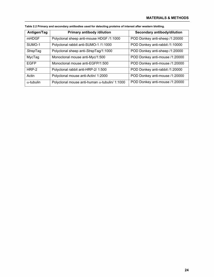
Table 2.2 Primary and secondary antibodies used for detecting proteins of interest after western blotting.
Antigen/Tag Primary antibody /dilution Secondary antibody/dilution
mHDGF Polyclonal sheep anti-mouse HDGF /1:1000 POD Donkey anti-sheep /1:20000
SUMO-1 Polyclonal rabbit anti-SUMO-1 /1:1000 POD Donkey anti-rabbit /1:10000
StrepTag Polyclonal sheep anti-StrepTag/1:1000 POD Donkey anti-sheep /1:20000
MycTag Monoclonal mouse anti-Myc/1:500 POD Donkey anti-mouse /1:20000
EGFP Monoclonal mouse anti-EGFP/1:500 POD Donkey anti-mouse /1:20000
HRP-2 Polyclonal rabbit anti-HRP-2/ 1:500 POD Donkey anti-rabbit /1:20000
Actin Polyclonal mouse anti-Actin/ 1:2000 POD Donkey anti-mouse /1:20000
�-tubulin Polyclonal mouse anti-human �-tubulin/ 1:1000 POD Donkey anti-mouse /1:20000

33 PPuubblliiccaattiioonnss
PUBLICATIONS
26
List of publications and my contribution towards them � Publication 1
Ketan Thakar, Rainer Niedenthal, Elwy Okaz, Sebastian Franken, Astrid Jakobs, Shivangi Gupta,
Sørge Kelm and Frank Dietz (2008)
SUMOylation of the hepatoma-derived growth factor negatively influences its binding to chromatin FEBS Journal 275: 1411–1426
I developed the concept of the study in co-operation with FD. I conducted the experiments supported
by FD, EO, and SG. RN provided SUMO-1 expression plasmid used in work. SF and AJ provided the
MALDI-TOF data for the study. Along with FD, I analyzed the data and was involved in writing and
revision of manuscript together with FD. SK, RN and SF also provided assistance in revision the
manuscript
� Publication 2
Ketan Thakar, Tim Kröcher, Soniya Savant, Doron Gollnast, Sørge Kelm, Frank Dietz (2010)
Secretion of hepatoma-derived growth factor is regulated by N-terminal processing Biological Chemistry (submitted)
I developed the concept of the study and designed the experiments in co-operation with FD. I carried
out the experiments supported by FD, TK, DG and SS. I analyzed the data and wrote the manuscript,
which was revised together with FD and SK.
� Publication 3
Ketan Thakar , Dipti Kelkar, Teja Shidore, Shivangi Gupta, Sørge Kelm, Frank Dietz (2010)
Interaction of HRP-2 splice variants with Hepatoma-derived growth factor Biological Chemistry (submitted) I developed the concept of the study, designed and carried out the experiments supported by FD, DK,
SG, and TS. I analyzed the data and wrote the manuscript, which was revised together with FD and
SK.
3.1 Publication 1
SUMOylation of the hepatoma-derived growth factor negatively influences its binding to chromatin
K Thakar, R Niedenthal, E Okaz, S Franken, A Jakobs, S Gupta, S Kelm and F Dietz
2008
FEBS Journal 275: 1411–1426
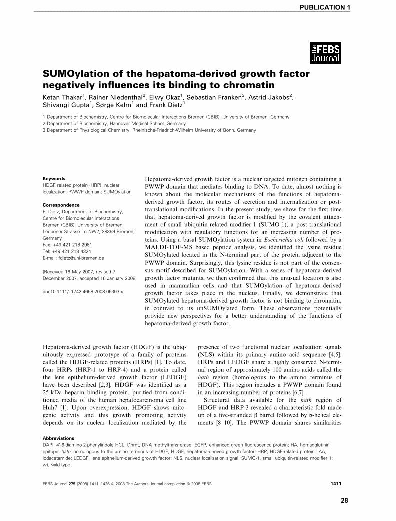
SUMOylation of the hepatoma-derived growth factornegatively influences its binding to chromatinKetan Thakar1, Rainer Niedenthal2, Elwy Okaz1, Sebastian Franken3, Astrid Jakobs2,Shivangi Gupta1, Sørge Kelm1 and Frank Dietz1
1 Department of Biochemistry, Centre for Biomolecular Interactions Bremen (CBIB), University of Bremen, Germany
2 Department of Biochemistry, Hannover Medical School, Germany
3 Department of Physiological Chemistry, Rheinische-Friedrich-Wilhelm University of Bonn, Germany
Hepatoma-derived growth factor (HDGF) is the ubiq-
uitously expressed prototype of a family of proteins
called the HDGF-related proteins (HRPs) [1]. To date,
four HRPs (HRP-1 to HRP-4) and a protein called
the lens epithelium-derived growth factor (LEDGF)
have been described [2,3]. HDGF was identified as a
25 kDa heparin binding protein, purified from condi-
tioned media of the human hepatocarcinoma cell line
Huh7 [1]. Upon overexpression, HDGF shows mito-
genic activity and this growth promoting activity
depends on its nuclear localization mediated by the
presence of two functional nuclear localization signals
(NLS) within its primary amino acid sequence [4,5].
HRPs and LEDGF share a highly conserved N-termi-
nal region of approximately 100 amino acids called the
hath region (homologous to the amino terminus of
HDGF). This region includes a PWWP domain found
in an increasing number of proteins [6,7].
Structural data available for the hath region of
HDGF and HRP-3 revealed a characteristic fold made
up of a five-stranded b barrel followed by a-helical ele-ments [8–10]. The PWWP domain shares similarities
Keywords
HDGF related protein (HRP); nuclear
localization; PWWP domain; SUMOylation
Correspondence
F. Dietz, Department of Biochemistry,
Centre for Biomolecular Interactions
Bremen (CBIB), University of Bremen,
Leobener Strasse im NW2, 28359 Bremen,
Germany
Fax: +49 421 218 2981
Tel: +49 421 218 4324
E-mail: [email protected]
(Received 16 May 2007, revised 7
December 2007, accepted 16 January 2008)
doi:10.1111/j.1742-4658.2008.06303.x
Hepatoma-derived growth factor is a nuclear targeted mitogen containing a
PWWP domain that mediates binding to DNA. To date, almost nothing is
known about the molecular mechanisms of the functions of hepatoma-
derived growth factor, its routes of secretion and internalization or post-
translational modifications. In the present study, we show for the first time
that hepatoma-derived growth factor is modified by the covalent attach-
ment of small ubiquitin-related modifier 1 (SUMO-1), a post-translational
modification with regulatory functions for an increasing number of pro-
teins. Using a basal SUMOylation system in Escherichia coli followed by a
MALDI-TOF-MS based peptide analysis, we identified the lysine residue
SUMOylated located in the N-terminal part of the protein adjacent to the
PWWP domain. Surprisingly, this lysine residue is not part of the consen-
sus motif described for SUMOylation. With a series of hepatoma-derived
growth factor mutants, we then confirmed that this unusual location is also
used in mammalian cells and that SUMOylation of hepatoma-derived
growth factor takes place in the nucleus. Finally, we demonstrate that
SUMOylated hepatoma-derived growth factor is not binding to chromatin,
in contrast to its unSUMOylated form. These observations potentially
provide new perspectives for a better understanding of the functions of
hepatoma-derived growth factor.
Abbreviations
DAPI, 4¢-6-diamino-2-phenylindole HCL; Dnmt, DNA methyltransferase; EGFP, enhanced green fluorescence protein; HA, hemagglutinin
epitope; hath, homologous to the amino terminus of HDGF; HDGF, hepatoma-derived growth factor; HRP, HDGF-related protein; IAA,
iodacetamide; LEDGF, lens epithelium-derived growth factor; NLS, nuclear localization signal; SUMO-1, small ubiquitin-related modifier 1;
wt, wild-type.
FEBS Journal 275 (2008) 1411–1426 ª 2008 The Authors Journal compilation ª 2008 FEBS 1411
___________________________________________________________________________________________________________PUBLICATION 1
28
with the well known Tudor and Chromo domain and,
like these domains, it has been proposed to play a role
in DNA-binding and ⁄or protein–protein interactions.
In the case of HDGF, the PWWP domain may have a
dual function in binding double-stranded DNA as well
as the glycosaminoglycan heparin [2,8–10]. DNA-bind-
ing via the PWWP domain of HDGF appears to be
specific for a region covering approximately 40 bp
found in potential target genes of HDGF [11],
although it has not been clarified whether further spec-
ificity may be mediated by the C-terminal portion of
the protein [8]. A recent study by Sue et al. [12] dem-
onstrated that dimerization of the PWWP domain by
an unusual domain-swapping leads to an increased
binding affinity for heparin. However, the physiologi-
cal role of this phenomenon is unclear.
Hepatoma-derived growth factor is secreted from
cells. The mechanism for externalization remains
unclear because HDGF, like the other HRPs, has no
obvious signal peptide. Extracellular HDGF appears
to be internalized by binding to heparan sulfate or
other mechanisms [5]. Recent studies have provided
evidence for a potential receptor specifically binding
extracellular HDGF, leading to the activation of intra-
cellular signalling cascades [13].
The expression of HDGF changes during develop-
ment, as shown for kidney, liver, heart and vascular
tissue [14–20]. In addition, recent studies have demon-
strated that HDGF is differentially expressed in the
brain [21] and also can function as a potent neuro-
trophic factor [22,23]. Furthermore, different studies
have shown that HDGF can serve as a prognostic
marker in a variety of human cancers [24–31] and that
it probably promotes angiogenesis and tumor progres-
sion [32].
Phosphorylation prediction programs have identified
HDGF as a good candidate for phosphorylation on sev-
eral serine and threonine residues, but only one mass
spectroscopy based approach has confirmed the use of
serine residues S132, S133 and S165 [33], although no
evidence for functional relevance was provided.
A post-translational modification found in several
nuclear proteins comprises the attachment of the small
ubiquitin-related modifier 1 (SUMO-1). The modifica-
tion by this 11 kDa protein is mechanistically related
to that of ubiquitin, with which it shares a high degree
of structural similarity. Like ubiquitination, SUMOyla-
tion is a dynamic process that is mediated by activat-
ing (E1), conjugating (E2) and ligating (E3) enzymes
and can be reversed by the action of SUMO specific
proteases [34,35]. Despite these similarities, the func-
tions of both modifications differ. SUMOylation of
target proteins usually occurs on lysine residues in the
context of a highly conserved recognition motif
YKxE ⁄D (where Y stands for a large hydrophobic
amino acid, K is the lysine modified, x is any amino
acid and E ⁄D are the negatively charged amino acids
glutamate or aspartate). Well documented functions of
SUMOylation are the regulation of subcellular distri-
bution, DNA repair, transcriptional regulation, stabil-
ization, RNA metabolism and cell signalling [34–38].
SUMO itself can further serve as a docking site for the
binding of other proteins containing SUMO binding
motifs [39–42].
Based on the knowledge that HDGF is a nuclear
targeted mitogen with DNA binding capacity, we
investigated whether HDGF is also modified by the
addition of SUMO-1. In the present study, we show
for the first time that HDGF serves as a template for
SUMO-1 conjugation, although it does not contain a
suitable consensus site for SUMOylation. Using a
basal SUMOylation system in Escherichia coli [43]
followed by a MALDI-based peptide analysis of the
SUMOylated HDGF wild-type (wt) and a series of
HDGF mutants, we identified an unusual SUMOyla-
tion site located in the N-terminal hath region. Fur-
thermore, we discovered that SUMOylated HDGF
does not bind to chromatin, in contrast to its
unSUMOylated form.
Results
SUMOylation of HDGF
For most SUMOylated proteins, overexpression of
the target protein together with SUMO-1 is necessary
to detect SUMOylation. When untagged HDGFwt
(apparent molecular mass ¼ 40 kDa) is overexpressed
together with enhanced green fluorescence protein
(EGFP)-SUMO-1 in COS-7 cells (Fig. 1A) or human
embryonic kidney cells (HEK293) (data not shown) we
use the advantage of the much higher molecular mass
shift of the EGFP-SUMO-1 SUMOylated proteins.
We detected an extra protein band reacting with a spe-
cific anti-HDGF serum in the molecular weight range
expected for EGFP-SUMO-1 conjugated HDGF
(apparent molecular weight ¼ 100 kDa). This observa-
tion suggests that HDGF can be modified by SUMOy-
lation. In several systems, it has been shown that
SUMOylation is a highly dynamic, reversible modifi-
cation, which is sensitve to the action of specific
isopeptidases. Since these are cysteine proteases, de-
SUMOylation can be partially blocked by lysing the
cells in the presence of N-ethylmaleimide and iodaceta-
mide (IAA) to alkylate the free SH-group in the active
centres of the isopeptidases. In agreement with this
SUMOylation of HDGF K. Thakar et al.
1412 FEBS Journal 275 (2008) 1411–1426 ª 2008 The Authors Journal compilation ª 2008 FEBS
___________________________________________________________________________________________________________PUBLICATION 1
29
phenomenon, we found that most of the suspected
HDGF-EGFP-SUMO-1 band migrating at 100 kDa is
lost if the cell lysates are incubated for 15 min on ice
in the absence of N-ethylmaleimide and IAA (Fig. 1B),
whereas almost no loss occurs if these SH-alkylating
reagents are included.
To further investigate whether the additional protein
bands are indeed due to the covalent modification of
HDGF with SUMO-1, we used C-terminally Strep-
tagged HDGF, which can be precipitated using
StrepTactin� beads. Similar to the experiment with
untagged HDGFwt, the C-terminal Strep-tag-labelled
HDGF appears to be SUMOylated (Fig. 1C) because,
in StrepTactin� precipitates, a similar upshifted
HDGF-band occurs, which reacts with antibody
against SUMO-1 (Fig. 1B). As negative controls, we
cotransfected either EGFP alone or an EGFP-SUMO-
1 chimera lacking the C-terminal di-glycine motif
(EGFP-SUMO-1DGG) required for the isopeptide
bond formation of SUMO-1 with the acceptor lysine.
Only EGFP-SUMO-1 including the terminal di-gly-
cine was covalently attached to HDGF, whereas EGFP
alone or EGFP-SUMO-1DGG was not (Fig. 1C).
To overcome problems in the detection of HDGF
mutants due to the restricted specificity of the polyclonal
anti-HDGF antiserum, in all further experiments, Strep-
tag-labelled HDGF constructs were employed, provid-
ing the possibility of using identical StrepTactin�precipitation and detection assays for all HDGF
mutants. For all these experiments, similar results were
obtained if HEK293 cells were utilized instead of COS-7
cells, supporting the notion that SUMOylation of
HDGF is not a COS-7 cell restricted modification.
Overexpression of the conjugating enzyme Ubc-9
(E2) is commonly used to obtain detectable amounts
of SUMOylated products. However, this was not
required for the SUMOylation of HDGF because
omission of the hemagglutinin epitope (HA)-Ubc-9
encoding plasmid did not reduce the level of SUMOy-
lation (Fig. 1C). These data suggested that endogenous
levels of Ubc-9 in COS-7 cells are sufficient to generate
detectable levels of SUMOylated HDGF.
SUMOylation site of HDGF in E. coli
Screening the primary amino acid sequence of HDGF
from different mammalian species (Fig. 2) using the
prediction programs for SUMOylation motifs SUMO-
plot� (http://www.abgent.com/doc/sumoplot) and the
SUMOsp–SUMOylation sites prediction program
(http://bioinformatics.lcd-ustc.org/sumosp/), we found
only three motifs that weakly match the postulated
consensus motif YKxE ⁄D (Fig. 2). The highest score
by SUMO site prediction was obtained for K223 in
the motif AK223EE of mouse HDGF (> 0.8), which is
TKED in human and chimpanzee or AKED in bovine
HDGF (Fig. 2). Other potential motifs predicted with
low scores (0.5) are EK148NE and PK167RP. Single K
to R mutations in these three motifs and the expres-
sion of the mutants together with EGFP-SUMO-1 in
COS-7 cells did not lead to any detectable loss in
SUMOylation compared to HDGFwt (Fig. 3). This
strongly suggested that other lysine residues than these
are SUMOylated in HDGF.
Since no obvious consensus motif was found in
HDGF, we aimed to identify the SUMOylation site(s)
of HDGF by MS after tryptic digestion. In order to
obtain sufficient amounts of SUMOylated HDGF for
MALDI-TOF-MS analysis, we expressed the protein
in E. coli. Essential compounds of the SUMOylation
machinery, such as the activating (E1) and conjugating
(E2) enzymes but not the ligating enzyme (E3) [43],
were coexpressed with Strep-tagged mHDGFwt in
E. coli strain BL21 DE3. This artificial bacterial
SUMOylation system enabled us to purify high levels
of SUMOylated and unmodified HDGF from bacterial
lysates via StrepTactin� precipitation. After 2D elec-
trophoresis of the purified protein, spots representing
unmodified HDGFStrep-tag appearing at a molecular
weight of 40 kDa (Fig. 4A, spot 1) and a protein spot
appearing at a molecular weight of 63 kDa (Fig. 4A,
spot 2) were cut out, digested with trypsin and used
for MALDI-TOF-MS analysis. The protein spot at
63 kDa is only observed if HDGF is coexpressed with
the SUMOylation machinery (data not shown). In the
spectra of both protein spots, we found peptide masses
perfectly matching the expected peptides from HDGF.
However, only in spot 2 did we recognize additional
peptide masses corresponding to peptides derived from
huSUMO-1 (Fig. 4C).
Furthermore, comparison of the obtained peptide
spectra of both spots clearly showed an almost com-
plete loss of two mass peaks in the chromatogram
derived from the digest of spot 2, most probably corre-
sponding to monoSUMOylated HDGF (Fig. 4D,E).
These mass peaks perfectly match the HDGF peptides
K80-K96 (K80GFSEGLWEINNPTVK96) and G81-K96
(G81FSEGLWEINNPTVK96) of the hath region
(Fig. 2). This observation strongly suggested that
either K80 or K96 is modified by SUMOylation.
SUMOylation of HDGF at Lys80 in mammalian
cell lines
To investigate whether K80 or K96 is SUMOylated in
mammalian cells as suggested by the bacterial system,
K. Thakar et al. SUMOylation of HDGF
FEBS Journal 275 (2008) 1411–1426 ª 2008 The Authors Journal compilation ª 2008 FEBS 1413
___________________________________________________________________________________________________________PUBLICATION 1
30
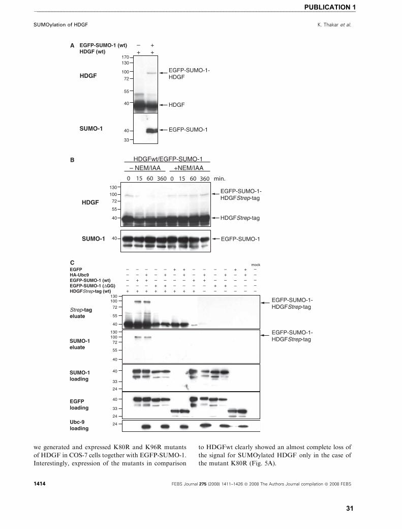
we generated and expressed K80R and K96R mutants
of HDGF in COS-7 cells together with EGFP-SUMO-1.
Interestingly, expression of the mutants in comparison
to HDGFwt clearly showed an almost complete loss of
the signal for SUMOylated HDGF only in the case of
the mutant K80R (Fig. 5A).
A
B
C
SUMOylation of HDGF K. Thakar et al.
1414 FEBS Journal 275 (2008) 1411–1426 ª 2008 The Authors Journal compilation ª 2008 FEBS
___________________________________________________________________________________________________________PUBLICATION 1
31
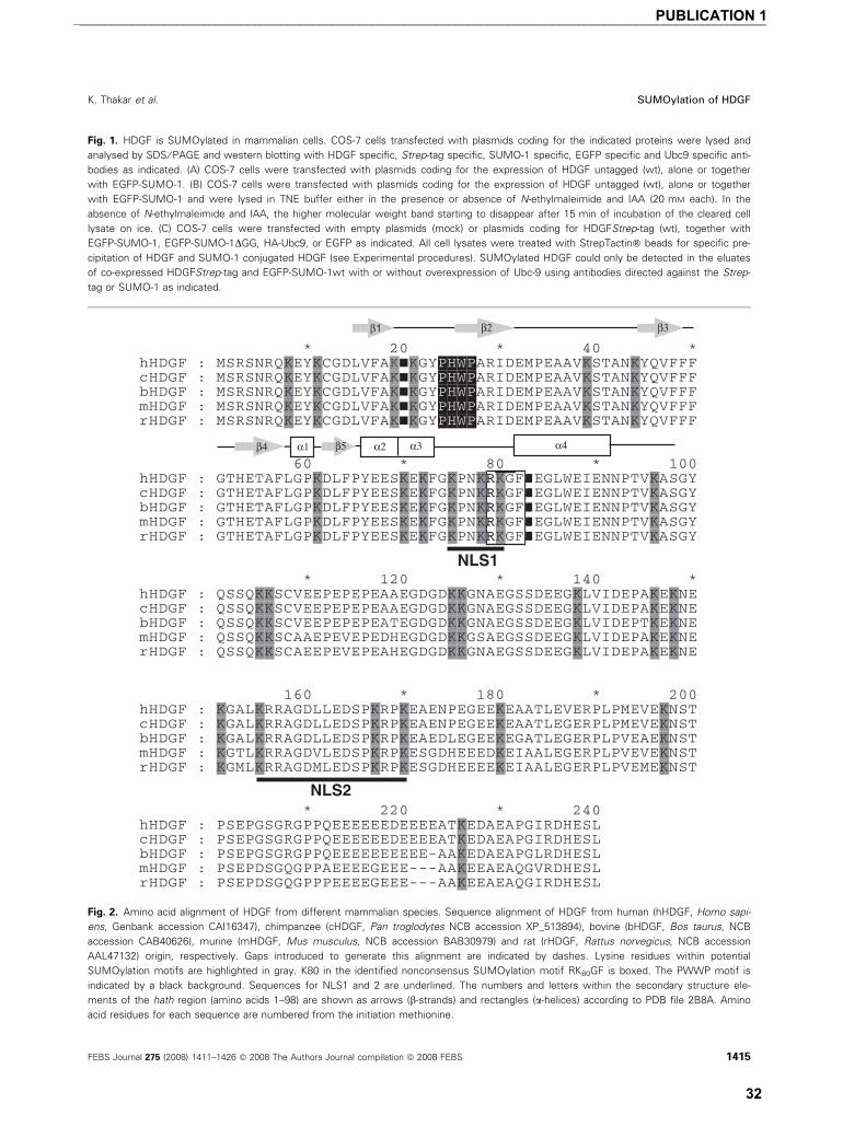
Fig. 1. HDGF is SUMOylated in mammalian cells. COS-7 cells transfected with plasmids coding for the indicated proteins were lysed and
analysed by SDS ⁄ PAGE and western blotting with HDGF specific, Strep-tag specific, SUMO-1 specific, EGFP specific and Ubc9 specific anti-
bodies as indicated. (A) COS-7 cells were transfected with plasmids coding for the expression of HDGF untagged (wt), alone or together
with EGFP-SUMO-1. (B) COS-7 cells were transfected with plasmids coding for the expression of HDGF untagged (wt), alone or together
with EGFP-SUMO-1 and were lysed in TNE buffer either in the presence or absence of N-ethylmaleimide and IAA (20 mM each). In the
absence of N-ethylmaleimide and IAA, the higher molecular weight band starting to disappear after 15 min of incubation of the cleared cell
lysate on ice. (C) COS-7 cells were transfected with empty plasmids (mock) or plasmids coding for HDGFStrep-tag (wt), together with
EGFP-SUMO-1, EGFP-SUMO-1DGG, HA-Ubc9, or EGFP as indicated. All cell lysates were treated with StrepTactin� beads for specific pre-
cipitation of HDGF and SUMO-1 conjugated HDGF (see Experimental procedures). SUMOylated HDGF could only be detected in the eluates
of co-expressed HDGFStrep-tag and EGFP-SUMO-1wt with or without overexpression of Ubc-9 using antibodies directed against the Strep-
tag or SUMO-1 as indicated.
Fig. 2. Amino acid alignment of HDGF from different mammalian species. Sequence alignment of HDGF from human (hHDGF, Homo sapi-
ens, Genbank accession CAI16347), chimpanzee (cHDGF, Pan troglodytes NCB accession XP_513894), bovine (bHDGF, Bos taurus, NCB
accession CAB40626), murine (mHDGF, Mus musculus, NCB accession BAB30979) and rat (rHDGF, Rattus norvegicus, NCB accession
AAL47132) origin, respectively. Gaps introduced to generate this alignment are indicated by dashes. Lysine residues within potential
SUMOylation motifs are highlighted in gray. K80 in the identified nonconsensus SUMOylation motif RK80GF is boxed. The PWWP motif is
indicated by a black background. Sequences for NLS1 and 2 are underlined. The numbers and letters within the secondary structure ele-
ments of the hath region (amino acids 1–98) are shown as arrows (b-strands) and rectangles (a-helices) according to PDB file 2B8A. Amino
acid residues for each sequence are numbered from the initiation methionine.
K. Thakar et al. SUMOylation of HDGF
FEBS Journal 275 (2008) 1411–1426 ª 2008 The Authors Journal compilation ª 2008 FEBS 1415
___________________________________________________________________________________________________________PUBLICATION 1
32
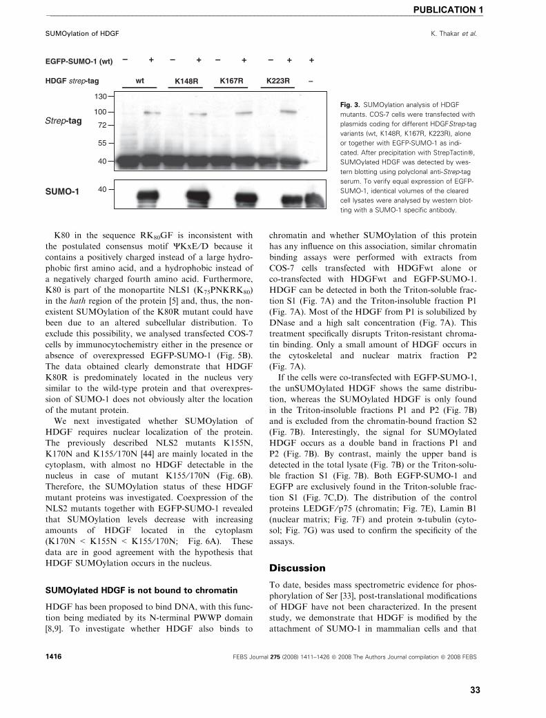
K80 in the sequence RK80GF is inconsistent with
the postulated consensus motif YKxE ⁄D because it
contains a positively charged instead of a large hydro-
phobic first amino acid, and a hydrophobic instead of
a negatively charged fourth amino acid. Furthermore,
K80 is part of the monopartite NLS1 (K75PNKRK80)
in the hath region of the protein [5] and, thus, the non-
existent SUMOylation of the K80R mutant could have
been due to an altered subcellular distribution. To
exclude this possibility, we analysed transfected COS-7
cells by immunocytochemistry either in the presence or
absence of overexpressed EGFP-SUMO-1 (Fig. 5B).
The data obtained clearly demonstrate that HDGF
K80R is predominately located in the nucleus very
similar to the wild-type protein and that overexpres-
sion of SUMO-1 does not obviously alter the location
of the mutant protein.
We next investigated whether SUMOylation of
HDGF requires nuclear localization of the protein.
The previously described NLS2 mutants K155N,
K170N and K155 ⁄ 170N [44] are mainly located in the
cytoplasm, with almost no HDGF detectable in the
nucleus in case of mutant K155 ⁄ 170N (Fig. 6B).
Therefore, the SUMOylation status of these HDGF
mutant proteins was investigated. Coexpression of the
NLS2 mutants together with EGFP-SUMO-1 revealed
that SUMOylation levels decrease with increasing
amounts of HDGF located in the cytoplasm
(K170N < K155N < K155 ⁄ 170N; Fig. 6A). These
data are in good agreement with the hypothesis that
HDGF SUMOylation occurs in the nucleus.
SUMOylated HDGF is not bound to chromatin
HDGF has been proposed to bind DNA, with this func-
tion being mediated by its N-terminal PWWP domain
[8,9]. To investigate whether HDGF also binds to
chromatin and whether SUMOylation of this protein
has any influence on this association, similar chromatin
binding assays were performed with extracts from
COS-7 cells transfected with HDGFwt alone or
co-transfected with HDGFwt and EGFP-SUMO-1.
HDGF can be detected in both the Triton-soluble frac-
tion S1 (Fig. 7A) and the Triton-insoluble fraction P1
(Fig. 7A). Most of the HDGF from P1 is solubilized by
DNase and a high salt concentration (Fig. 7A). This
treatment specifically disrupts Triton-resistant chroma-
tin binding. Only a small amount of HDGF occurs in
the cytoskeletal and nuclear matrix fraction P2
(Fig. 7A).
If the cells were co-transfected with EGFP-SUMO-1,
the unSUMOylated HDGF shows the same distribu-
tion, whereas the SUMOylated HDGF is only found
in the Triton-insoluble fractions P1 and P2 (Fig. 7B)
and is excluded from the chromatin-bound fraction S2
(Fig. 7B). Interestingly, the signal for SUMOylated
HDGF occurs as a double band in fractions P1 and
P2 (Fig. 7B). By contrast, mainly the upper band is
detected in the total lysate (Fig. 7B) or the Triton-solu-
ble fraction S1 (Fig. 7B). Both EGFP-SUMO-1 and
EGFP are exclusively found in the Triton-soluble frac-
tion S1 (Fig. 7C,D). The distribution of the control
proteins LEDGF ⁄p75 (chromatin; Fig. 7E), Lamin B1
(nuclear matrix; Fig. 7F) and protein a-tubulin (cyto-
sol; Fig. 7G) was used to confirm the specificity of the
assays.
Discussion
To date, besides mass spectrometric evidence for phos-
phorylation of Ser [33], post-translational modifications
of HDGF have not been characterized. In the present
study, we demonstrate that HDGF is modified by the
attachment of SUMO-1 in mammalian cells and that
Fig. 3. SUMOylation analysis of HDGF
mutants. COS-7 cells were transfected with
plasmids coding for different HDGFStrep-tag
variants (wt, K148R, K167R, K223R), alone
or together with EGFP-SUMO-1 as indi-
cated. After precipitation with StrepTactin�,
SUMOylated HDGF was detected by wes-
tern blotting using polyclonal anti-Strep-tag
serum. To verify equal expression of EGFP-
SUMO-1, identical volumes of the cleared
cell lysates were analysed by western blot-
ting with a SUMO-1 specific antibody.
SUMOylation of HDGF K. Thakar et al.
1416 FEBS Journal 275 (2008) 1411–1426 ª 2008 The Authors Journal compilation ª 2008 FEBS
___________________________________________________________________________________________________________PUBLICATION 1
33
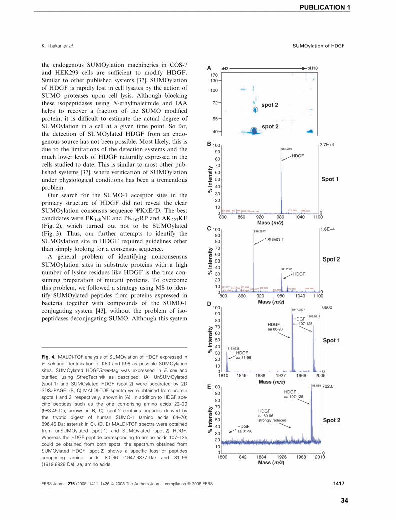
the endogenous SUMOylation machineries in COS-7
and HEK293 cells are sufficient to modify HDGF.
Similar to other published systems [37], SUMOylation
of HDGF is rapidly lost in cell lysates by the action of
SUMO proteases upon cell lysis. Although blocking
these isopeptidases using N-ethylmaleimide and IAA
helps to recover a fraction of the SUMO modified
protein, it is difficult to estimate the actual degree of
SUMOylation in a cell at a given time point. So far,
the detection of SUMOylated HDGF from an endo-
genous source has not been possible. Most likely, this is
due to the limitations of the detection systems and the
much lower levels of HDGF naturally expressed in the
cells studied to date. This is similar to most other pub-
lished systems [37], where verification of SUMOylation
under physiological conditions has been a tremendous
problem.
Our search for the SUMO-1 acceptor sites in the
primary structure of HDGF did not reveal the clear
SUMOylation consensus sequence YKxE ⁄D. The best
candidates were EK148NE and PK167RP and AK223KE
(Fig. 2), which turned out not to be SUMOylated
(Fig. 3). Thus, our further attempts to identify the
SUMOylation site in HDGF required guidelines other
than simply looking for a consensus sequence.
A general problem of identifying nonconsensus
SUMOylation sites in substrate proteins with a high
number of lysine residues like HDGF is the time con-
suming preparation of mutant proteins. To overcome
this problem, we followed a strategy using MS to iden-
tify SUMOylated peptides from proteins expressed in
bacteria together with compounds of the SUMO-1
conjugating system [43], without the problem of iso-
peptidases deconjugating SUMO. Although this system
Fig. 4. MALDI-TOF analysis of SUMOylation of HDGF expressed in
E. coli and identification of K80 and K96 as possible SUMOylation
sites. SUMOylated HDGFStrep-tag was expressed in E. coli and
purified using StrepTactin� as described. (A) UnSUMOylated
(spot 1) and SUMOylated HDGF (spot 2) were separated by 2D
SDS ⁄ PAGE. (B, C) MALDI-TOF spectra were obtained from protein
spots 1 and 2, respectively, shown in (A). In addition to HDGF spe-
cific peptides such as the one comprising amino acids 22–29
(983.49 Da; arrows in B, C), spot 2 contains peptides derived by
the tryptic digest of human SUMO-1 (amino acids 64–70;
896.46 Da; asterisk in C). (D, E) MALDI-TOF spectra were obtained
from unSUMOylated (spot 1) and SUMOylated (spot 2) HDGF.
Whereas the HDGF peptide corresponding to amino acids 107–125
could be obtained from both spots, the spectrum obtained from
SUMOylated HDGF (spot 2) shows a specific loss of peptides
comprising amino acids 80–96 (1947.9877 Da) and 81–96
(1819.8928 Da). aa, amino acids.
K. Thakar et al. SUMOylation of HDGF
FEBS Journal 275 (2008) 1411–1426 ª 2008 The Authors Journal compilation ª 2008 FEBS 1417
___________________________________________________________________________________________________________PUBLICATION 1
34
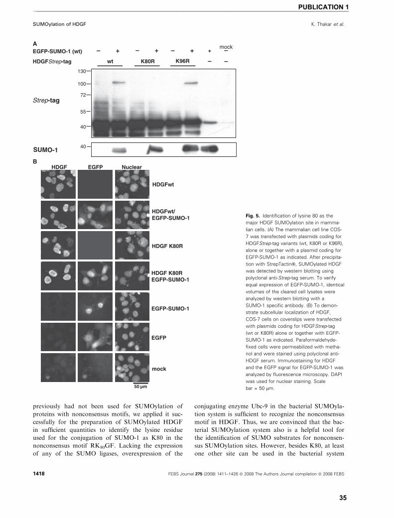
previously had not been used for SUMOylation of
proteins with nonconsensus motifs, we applied it suc-
cessfully for the preparation of SUMOylated HDGF
in sufficient quantities to identify the lysine residue
used for the conjugation of SUMO-1 as K80 in the
nonconsensus motif RK80GF. Lacking the expression
of any of the SUMO ligases, overexpression of the
conjugating enzyme Ubc-9 in the bacterial SUMOyla-
tion system is sufficient to recognize the nonconsensus
motif in HDGF. Thus, we are convinced that the bac-
terial SUMOylation system also is a helpful tool for
the identification of SUMO substrates for nonconsen-
sus SUMOylation sites. However, besides K80, at least
one other site can be used in the bacterial system
Fig. 5. Identification of lysine 80 as the
major HDGF SUMOylation site in mamma-
lian cells. (A) The mammalian cell line COS-
7 was transfected with plasmids coding for
HDGFStrep-tag variants (wt, K80R or K96R),
alone or together with a plasmid coding for
EGFP-SUMO-1 as indicated. After precipita-
tion with StrepTactin�, SUMOylated HDGF
was detected by western blotting using
polyclonal anti-Strep-tag serum. To verify
equal expression of EGFP-SUMO-1, identical
volumes of the cleared cell lysates were
analyzed by western blotting with a
SUMO-1 specific antibody. (B) To demon-
strate subcellular localization of HDGF,
COS-7 cells on coverslips were transfected
with plasmids coding for HDGFStrep-tag
(wt or K80R) alone or together with EGFP-
SUMO-1 as indicated. Paraformaldehyde-
fixed cells were permeabilized with metha-
nol and were stained using polyclonal anti-
HDGF serum. Immunostaining for HDGF
and the EGFP signal for EGFP-SUMO-1 was
analyzed by fluorescence microscopy. DAPI
was used for nuclear staining. Scale
bar = 50 lm.
SUMOylation of HDGF K. Thakar et al.
1418 FEBS Journal 275 (2008) 1411–1426 ª 2008 The Authors Journal compilation ª 2008 FEBS
___________________________________________________________________________________________________________PUBLICATION 1
35
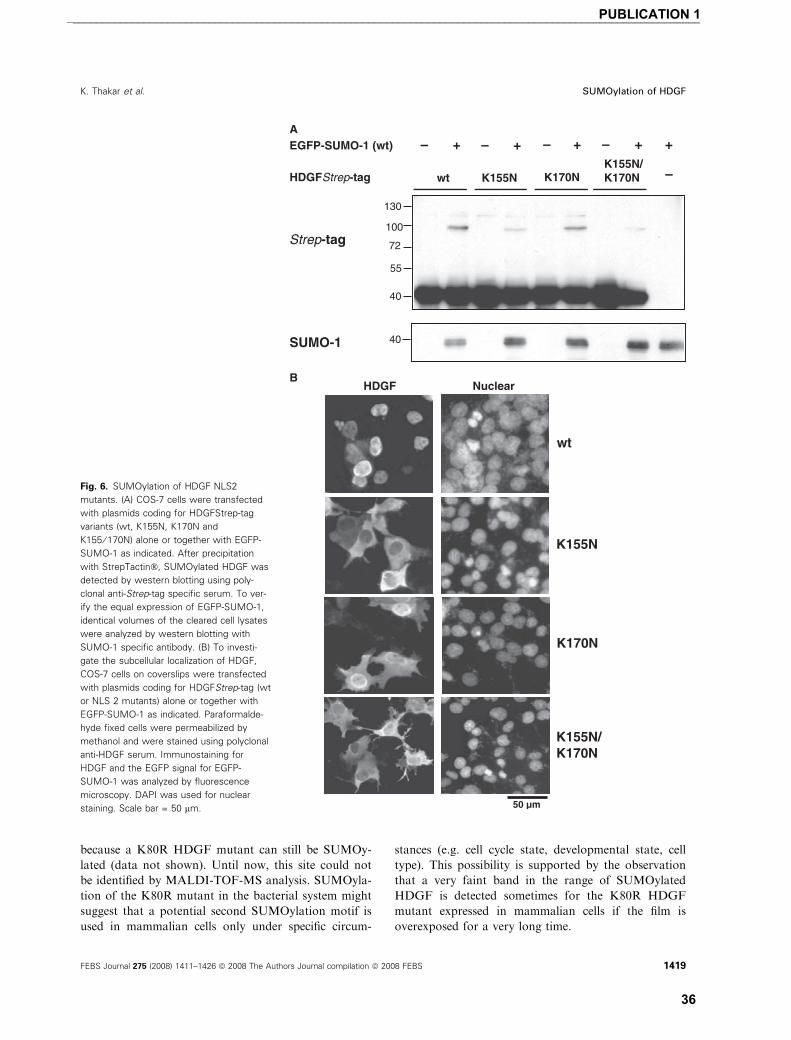
because a K80R HDGF mutant can still be SUMOy-
lated (data not shown). Until now, this site could not
be identified by MALDI-TOF-MS analysis. SUMOyla-
tion of the K80R mutant in the bacterial system might
suggest that a potential second SUMOylation motif is
used in mammalian cells only under specific circum-
stances (e.g. cell cycle state, developmental state, cell
type). This possibility is supported by the observation
that a very faint band in the range of SUMOylated
HDGF is detected sometimes for the K80R HDGF
mutant expressed in mammalian cells if the film is
overexposed for a very long time.
Fig. 6. SUMOylation of HDGF NLS2
mutants. (A) COS-7 cells were transfected
with plasmids coding for HDGFStrep-tag
variants (wt, K155N, K170N and
K155 ⁄ 170N) alone or together with EGFP-
SUMO-1 as indicated. After precipitation
with StrepTactin�, SUMOylated HDGF was
detected by western blotting using poly-
clonal anti-Strep-tag specific serum. To ver-
ify the equal expression of EGFP-SUMO-1,
identical volumes of the cleared cell lysates
were analyzed by western blotting with
SUMO-1 specific antibody. (B) To investi-
gate the subcellular localization of HDGF,
COS-7 cells on coverslips were transfected
with plasmids coding for HDGFStrep-tag (wt
or NLS 2 mutants) alone or together with
EGFP-SUMO-1 as indicated. Paraformalde-
hyde fixed cells were permeabilized by
methanol and were stained using polyclonal
anti-HDGF serum. Immunostaining for
HDGF and the EGFP signal for EGFP-
SUMO-1 was analyzed by fluorescence
microscopy. DAPI was used for nuclear
staining. Scale bar = 50 lm.
K. Thakar et al. SUMOylation of HDGF
FEBS Journal 275 (2008) 1411–1426 ª 2008 The Authors Journal compilation ª 2008 FEBS 1419
___________________________________________________________________________________________________________PUBLICATION 1
36
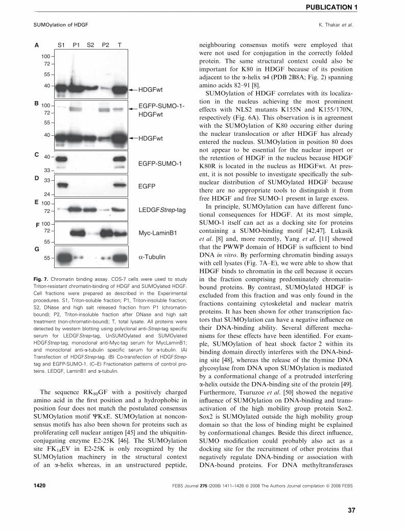
The sequence RK80GF with a positively charged
amino acid in the first position and a hydrophobic in
position four does not match the postulated consensus
SUMOylation motif YKxE. SUMOylation at noncon-
sensus motifs has also been shown for proteins such as
proliferating cell nuclear antigen [45] and the ubiquitin-
conjugating enzyme E2-25K [46]. The SUMOylation
site FK14EV in E2-25K is only recognized by the
SUMOylation machinery in the structural context
of an a-helix whereas, in an unstructured peptide,
neighbouring consensus motifs were employed that
were not used for conjugation in the correctly folded
protein. The same structural context could also be
important for K80 in HDGF because of its position
adjacent to the a-helix a4 (PDB 2B8A; Fig. 2) spanning
amino acids 82–91 [8].
SUMOylation of HDGF correlates with its localiza-
tion in the nucleus achieving the most prominent
effects with NLS2 mutants K155N and K155 ⁄ 170N,
respectively (Fig. 6A). This observation is in agreement
with the SUMOylation of K80 occuring either during
the nuclear translocation or after HDGF has already
entered the nucleus. SUMOylation in position 80 does
not appear to be essential for the nuclear import or
the retention of HDGF in the nucleus because HDGF
K80R is located in the nucleus as HDGFwt. At pres-
ent, it is not possible to investigate specifically the sub-
nuclear distribution of SUMOylated HDGF because
there are no appropriate tools to distinguish it from
free HDGF and free SUMO-1 present in large excess.
In principle, SUMOylation can have different func-
tional consequences for HDGF. At its most simple,
SUMO-1 itself can act as a docking site for proteins
containing a SUMO-binding motif [42,47]. Lukasik
et al. [8] and, more recently, Yang et al. [11] showed
that the PWWP domain of HDGF is sufficient to bind
DNA in vitro. By performing chromatin binding assays
with cell lysates (Fig. 7A–E), we were able to show that
HDGF binds to chromatin in the cell because it occurs
in the fraction comprising predominately chromatin-
bound proteins. By contrast, SUMOylated HDGF is
excluded from this fraction and was only found in the
fractions containing cytoskeletal and nuclear matrix
proteins. It has been shown for other transcription fac-
tors that SUMOylation can have a negative influence on
their DNA-binding ability. Several different mecha-
nisms for these effects have been identified. For exam-
ple, SUMOylation of heat shock factor 2 within its
binding domain directly interferes with the DNA-bind-
ing site [48], whereas the release of the thymine DNA
glycosylase from DNA upon SUMOylation is mediated
by a conformational change of a protruded interfering
a-helix outside the DNA-binding site of the protein [49].
Furthermore, Tsuruzoe et al. [50] showed the negative
influence of SUMOylation on DNA-binding and trans-
activation of the high mobility group protein Sox2.
Sox2 is SUMOylated outside the high mobility group
domain so that the loss of binding might be explained
by conformational changes. Beside this direct influence,
SUMO modification could probably also act as a
docking site for the recruitment of other proteins that
negatively regulate DNA-binding or association with
DNA-bound proteins. For DNA methyltransferases
Fig. 7. Chromatin binding assay. COS-7 cells were used to study
Triton-resistant chromatin-binding of HDGF and SUMOylated HDGF.
Cell fractions were prepared as described in the Experimental
procedures. S1, Triton-soluble fraction; P1, Triton-insoluble fraction;
S2, DNase and high salt released fraction from P1 (chromatin-
bound); P2, Triton-insoluble fraction after DNase and high salt
treatment (non-chromatin-bound); T, total lysate. All proteins were
detected by western blotting using polyclonal anti-Strep-tag specific
serum for LEDGFStrep-tag, UnSUMOylated and SUMOylated
HDGFStrep-tag; monoclonal anti-Myc-tag serum for MycLaminB1;
and monoclonal anti-a-tubulin specific serum for a-tubulin. (A)
Transfection of HDGFStrep-tag. (B) Co-transfection of HDGFStrep-
tag and EGFP-SUMO-1. (C–E) Fractionation patterns of control pro-
teins. LEDGF, LaminB1 and a-tubulin.
SUMOylation of HDGF K. Thakar et al.
1420 FEBS Journal 275 (2008) 1411–1426 ª 2008 The Authors Journal compilation ª 2008 FEBS
___________________________________________________________________________________________________________PUBLICATION 1
37
(Dnmt) 3a and 3b and the HDGF family member
LEDGF ⁄p75, a PWWP domain mediated DNA-bind-
ing activity and chromatin targeting function has been
described [51–53]. Very recently, Li et al. [54] were able
to show SUMOylation of Dnmt3a within an extended
PWWP region depending on the interaction with the
polycomb group protein chromobox 4 acting as an
E3 ligase. The responsible lysine for SUMOylation in
Dnmt3a has not yet been identified but, in a similar
sequence context to that of the RK80GF motif in
HDGF, a RK363AI motif can be found in Dnmt3a.
Chemical shift changes in NMR spectra of the HDGF
PWWP domain indicated that the putative DNA-bind-
ing site overlaps with a patch built by a number of
solvent-exposed Lys residues, including K80. Thus,
SUMOylation of K80 might have a direct influence on
the ability of HDGF to bind to DNA. In the model
structure of PDB file 1RI0 [10], the helix is placed in a
groove between b2 and the b3 ⁄b4 loop in a fixed direc-
tion. Interestingly, W26 of the PHWP motif vertically
stacks against the aromatic ring of F82 within the helix,
whereas K80 is solvent accessible. It is possible that
SUMOylation at K80 might alter this conformation or
it might just sterically hinder the interaction with the
DNA.
Although the data presented in the present study
clearly demonstrate that K80 is the main SUMOyla-
tion site in HDGF, we cannot exclude with certainty
that other SUMOylation sites are used under specific
circumstances (e.g. cell cycle state, cell type, develop-
ment, stress conditions, etc.). Initial evidence for this
possibility comes from the observation that, upon
overexposure of the films, we noticed a very faint band
in the range of SUMOylated HDGF in the case of the
K80R mutant in mammalian cells (data not shown).
The C-terminal region of the PWWP domain includ-
ing a-helix 82–91 of HDGF differs strongly between
PWWP domain containing proteins [6,7]. It could be
speculated that this region of the HDGF protein is
involved in specific protein–protein interactions
responsible for targeting the proteins to an appropriate
region of DNA. In this case, SUMOylation at position
K80 could modify such protein–protein interaction,
either by inhibiting or promoting it. For example, the
structural data suggests that the decrease in ubiquitin
conjugation of E2-25K after SUMO-1 binding is medi-
ated by sterically blocking the binding site of the acti-
vating enzyme (E1) instead of affecting the cysteine
responsible for ubiquitin conjugation directly [46].
It should be noted that the RKGF motif is also
found in other HRP members, such as HRP-2, HRP-3
and LEDGF, whereas, in the testis specific HRP-4 and
HRP-1, the corresponding Lys residues are changed to
an Arg. Thus, it would be interesting to investigate
other HRP family members for their ability to be
SUMOylated.
In summary, the present study provides a new per-
spective for our future understanding of the molecular
mechanisms involved in the biological functions of
HDGF, and possibly other HRPs, through SUMOyla-
tion.
Experimental procedures
Cell culture and transfections
COS-7 (kidney fibroblasts from the African green monkey
Cercopithecus aethiops) and HEK293 (human embryonic
kidney cells) were grown in DMEM supplemented with
10% fetal bovine serum, 100 UÆmL)1 penicillin and
50 lgÆmL)1 streptomycin at 37 �C in a humidified atmo-
sphere of 5% (vol ⁄ vol) CO2. For transient transfection,
cells were grown to a density of 80% confluence. Directly
before transfection, the media was changed from 10% fetal
bovine serum to 2% fetal bovine serum. Transfection was
carried out with the polyethylenimine ExGEN 500 accord-
ing to the manufacturer’s instructions (Fermentas, St Leon-
Rot, Germany). For protein expression, COS-7 and
HEK293 cells were transfected with the indicated amount
of the different plasmids and were harvested after 24 h.
Plasmids
Plasmids were constructed by standard recombinant cloning
techniques and all changes were verified by DNA sequenc-
ing. For the addition of a Strep-tag II peptide (NH2-
WSHPQFEK-COOH) [55] to the C-terminus of HDGF,
sense (5¢-AATTCGATATCGGTACCTGGAGCCACCCG
CAGTTCGAAAAATAAGC-3¢) and antisense (5¢-GGC
CGCTTATTTTTCGAACTGCGGGTGGCTCCAGGTAC
CGATATCG-3¢) oligonucleotides containing the sequence
coding for the Tag and EcoRI and NotI sites were annealed
and cloned into EcoRI ⁄NotI digested pcDNA3 Amp vector
(Invitrogen, Carlsbad, CA, USA).
The coding sequence of HDGFwt (GenBank accession
number BC021654) was amplified using murine adult brain
cDNA as a source and subcloned in frame into HindIII ⁄BamHI digested pcDNA3 Amp Strep-tag N-terminal. The
primers used for amplification were: 5¢-CGAAGCTTAT
GTCGCGATCCAACCGGCAG-3¢ and 5¢-CGGGATCCC
TACAGGCTCTCATGATCTCT-3¢.For the expression of untagged HDGF, we amplified
mouse adult brain cDNA with the same sense primer and
an antisense primer containing an internal Stop codon
(5¢CGGGATCCTCACTACAGGCTCTCATGATCTCT).
The coding sequence HDGF was cloned into HindIII ⁄BamHI digested pcDNA3 Amp.
K. Thakar et al. SUMOylation of HDGF
FEBS Journal 275 (2008) 1411–1426 ª 2008 The Authors Journal compilation ª 2008 FEBS 1421
___________________________________________________________________________________________________________PUBLICATION 1
38

HDGF mutants were generated by site-directed mutagen-
esis with the Quickchange Multi-Site Direct Mutagenesis
kit (Stratagene, La Jolla, CA, USA) according to the man-
ufacturer‘s instructions using the plasmid HDGFStrep-tag
wt cloned in pcDNA3 Amp as a template. Positive clones
were identified by inserting or removing restriction sites
by silent mutations within the following primer pairs:
HDGFStrep-tag K80R (HhaI appears) 5¢-GGCA
AGCCCAACAAGCGCAGAGGGTTCAGCGAG-3¢, 5¢-CTCGCTGAACCCTCTGCGCTTGTTGGGCTTGCC-3¢;HDGFStrep-tag K96R (EcoRV appears) 5¢-CTACAGT
CAGGGCCTCTGGATATCAGTC-3¢, 5¢-GACTGATAT
CCAGAGGCCCTGACTGTAG-3¢; HDGFStrep-tag
K148R (StyI disappears) 5¢-AACCAGCCAAAGAGCG
GAACGAAAAGGGC-3¢, 5¢-TTGGTCGGTTTCTCGCC
TTGCTTTTCCCG-3¢; HDGFStrep-tag K167R (TaqI dis-
appears) 5¢-GAGGACTCCCCCCGACGTCCCAAGGA
G-3¢, 5¢-CTCCTGAGGGGGGCTGCAGGGTTCCTG-3¢;and HDGFStrep-tag K223R (SmaI appears) 5¢-GAG
GAAGAGGCTGCCCGGGAAGAGGGCTGAAGCC-3¢,5¢-GGCTTCAGCCTCTTCCCGGGCAGCCTCTTCCTC-3¢.The primers used for mutagenesis of lysine residues in
the NLS 2 were: HDGFStrep-tag K155N (Eco57I disap-
pears) 5¢-GAAAAGGGCACGCTGAATAGGAGAGCAG
GG-3¢, 5¢-CCCTGCTCTCCTATTCAGCGTGCCCTTTT
C-3¢ and HDGFStrep-tag K170N (SmaI appears) 5¢-CCTAAACGTCCCCGGGAGTCAGGAGAC-3¢, 5¢-GTC
TCCTGACTCCCGGGGACGTTTAGG-3¢.For the double mutant HDGFStrep-tag K155 ⁄ 170N,
K155N was used as a template for mutagenesis.
For the expression of HDGFStrep-tag in E. coli, the cod-
ing sequence was cloned into the bacterial expression vector
pET28a (Merck, Schwalbach, Germany) digested with NcoI
and XhoI using the plasmid encoding HDGFStrep-tag as a
template and the primers: 5¢-CGCCATGGCGCGATCC
AACCGGCAG-3¢ and 5¢-CGCTCGAGTTATTTTTCGA
ACTGCGGGTGGC-3¢.Mouse SUMO-1wt, mouse LEDGF ⁄p75wt and mouse
LaminB1wt were amplified using mouse adult brain cDNA
as a template and the following primers derived from the
published cDNA sequences for SUMO-1 (GenBank acces-
sion number BC083158): 5¢-CGGGATCCATGTCTGACC
AGGAGGCA-3¢, 5¢-ACGCGTCGACCTAAACCGTCG
AGTGACC-3¢; for LEDGF ⁄ p75 (GenBank accession num-
ber AF339083): 5¢-CGGGATCCATGACTCGCGATTT
CAAACCT-3¢, 5¢-CGGAATTCGTTATCTAGTGTAGA
CTC-3¢; and for LaminB1 (GenBank accession number
NM_010721): 5¢-CGAAGATCTATGGCGACCGCGACC
CCCGTGC-3¢, 5¢-CGCTCGAGTCACATAATGGCAC
AGCTT-3¢.Mouse SUMO-1DGG was amplified using the specific
anti sense primer: 5¢-ACGCGTCGACCGTTTGTTCCTG
ATAAAC-3¢.The fragments for SUMO-1wt and SUMO-1DGG were
cloned C-terminal in frame to EGFP into BglII and SalI
digested pEGFP-C1 (Clontech, Palo Alto, CA, USA). The
fragment for LEDGF ⁄ p75wt was cloned N-terminal in
frame to the Strep-tag into BamHI and EcoRI digested
pcDNA III Strep-tag vector and LaminB1wt was cloned
C-terminal in frame to the Myc-tag into BamHI and XhoI
digested pCMV3B (Clontech). For the expression of EGFP,
the eukaryotic expression plasmid pEGFP C3 (Clontech)
was used. N-terminal hemagglutinin HA-tagged murine
Ubc9 cloned in pcDNA III Amp was a gift from R. Bernards
(Netherlands Cancer Institute, Amesterdam, the Nether-
lands).
Cell extracts
Cells were washed twice with NaCl ⁄Pi before treatment with
ice cold TNE lysis buffer containing 20 mm Tris ⁄Cl, pH 7.4,
150 mm NaCl, 5 mm EDTA, 1% NP-40 and freshly added
20 mm N-ethylmaleimide (Sigma-Aldrich, Steinheim, Ger-
many), 20 mm IAA (Sigma-Aldrich) + protease inhibitors
(Complete Mini, EDTA free; Roche, Mannheim, Germany)
on ice. After 10 min on ice, cells were scraped from the dish,
transferred to a 1.5 mL reaction tube and incubated on ice
for an additional 10 min. Cell debris was separated by cen-
trifugation at 20 000 g at 4 �C for 20 min and the superna-
tant transferred to a new 1.5 mL reaction tube. The cleared
lysate was used directly for SDS ⁄PAGE and western blot or
for the purification of the recombinant Strep-tagged HDGF
using StrepTactin� (IBA, Gottingen, Germany).
StrepTactin� purification
Cleared lysate (900 lL) derived from transiently transfected
COS-7 cells on a 10 cm diameter cell culture dish (Sarstedt,
Numbrecht, Germany) was incubated in a batch procedure
with 30 lL of StrepTactin� Sepharose beads on a rotor
shaker for 6 h at 4 �C. After incubation, beads were centri-
fuged at 240 g at 4 �C for 15 s and the supernatant was
transferred to a new 1.5 mL reaction tube. Beads were
washed four times with 1 mL of TNE lysis buffer. After the
final washing step, bound proteins were eluted by directly
boiling the beads at 95 �C for 10 min in 80 lL2 · SDS ⁄PAGE sample buffer. After centrifugation, the
supernatant was transferred to a fresh 1.5 mL reaction tube.
A 20 lL fraction of the eluate was used for SDS ⁄PAGE.
Chromatin binding assay
The method was performed as previously described [53]
with slight modification. Twenty-four hours after transfec-
tion, COS-7 was lysed for 10 min on ice in 1 mL of ice cold
CSK I buffer (10 mm Pipes, pH 6.8, 100 mm NaCl, 1 mm
EDTA, 300 mm sucrose, 1 mm MgCl2, 1 mm dithithreitol)
supplemented with 0.5% Triton X-100, protease inhibitors
(Complete Mini; Roche). Ten percent of the lysate was
SUMOylation of HDGF K. Thakar et al.
1422 FEBS Journal 275 (2008) 1411–1426 ª 2008 The Authors Journal compilation ª 2008 FEBS
___________________________________________________________________________________________________________PUBLICATION 1
39

mixed with RIPA buffer [150 mm Tris–HCl, pH 8.0,
150 mm NaCl, 0.5% DOC, 0.1% (w ⁄ v) SDS, 1% (v ⁄ v)NP-40] and kept for further analysis as total lysate (T). The
remaining cell lysate was divided into two equal portions,
which were centrifuged at 450 g at 4 �C for 3 min. The su-
pernatants where pooled and represent the Triton-soluble
protein fraction (S1). The pellets contain chromatin-bound,
nuclear matrix bound and insoluble proteins. One of the
pellets was resuspended in RIPA buffer (P1). The second
pellet was resuspended in CSK II buffer (10 mm Pipes, pH
6.8, 50 mm NaCl, 300 mm sucrose, 6 mm MgCl2, 1 mm
dithiothreitol), treated with 6 UÆlL)1 DNase (Boehringer,
Mannheim, Germany) for 30 min followed by extraction
with 250 mm NH4SO4 for 10 min at 25 �C. The sample
treated with DNase and salt was then centrifuged at 1200 g
for 6 min at 4 �C and the supernatant containing DNase-
released chromatin-associated proteins (S2) and the pellet
containing insoluble, cytoskeletal and nuclear matrix pro-
teins (P2) were both collected. P2 was resuspended in
100 lL RIPA buffer. A total of 20 lg protein of each frac-
tion was loaded for SDS ⁄PAGE.
Immunofluorescence
For transfection and subsequent immunofluorescence, cells
(3 · 104) were plated on glass coverslips in a 24-well plate
and grown overnight. Cells were washed twice in NaCl ⁄Pi
and then fixed in 4% paraformaldehyde in NaCl ⁄Pi for
10 min. For permeabilization, cells were treated for 2 min
with ice-cold methanol on ice and subsequently washed
twice with NaCl ⁄Pi ⁄ 2% BSA before blocking unspecific
binding sites with DMEM ⁄ 10% fetal bovine serum for
30 min. HDGF was detected using polyclonal sheep anti-
HDGF serum as described previously [56]. Cells were incu-
bated with primary antibody for 90 min in NaCl ⁄Pi ⁄ 2%BSA at room temperature. After washing twice with
NaCl ⁄Pi ⁄ 2% BSA, cells were treated for 90 min in
NaCl ⁄Pi ⁄ 2% BSA at room temperature in the dark with the
corresponding fluorophor-conjugated secondary antibody
(Dianova, Hamburg, Germany). Cells were washed in
NaCl ⁄Pi ⁄ 2% BSA and then treated with the nuclear stain
4¢-6-diamino-2-phenylindole HCL (DAPI) (Roche) for
1 min at room temperature. After additional washing, cells
were embedded in mounting media (Sigma, Deisenhofen,
Germany) and images were captured on a fluorescence
microscope (Olympus, Hamburg, Germany). EGFP-SUMO-
1 was detected by autofluorescence of the EGFP-tag.
Expression of SUMOylated HDGF from E. coli
The system of overexpression of SUMOylated proteins in
E. coli has been described previously [43]. The chemical
competent E. coli BL21 DE3 (Pharmacia, Uppsala,
Sweden) transformed with the plasmid containing the
SUMOylation machinery (pE1E2SUMO-1) was then
transformed in a second step with the plasmid coding for
HDGFwtStrep-tag. Overnight cultures were used to inocu-
late fresh medium (1 : 100, vol ⁄ vol); the new cultures were
subsequently grown until D600 of 0.5 was reached to be
induced with a final concentration of 0.5 mm isopropyl-
1-thio-b-D-galactopyranoside (Sigma) and grown for an
additional 4 h. Cells were collected by centrifugation at
5400 g at 4 �C for 15 min and resuspended in 2% of the
original volume with 100 mm Tris–HCl (pH 8.0 at 4 �C),150 mm NaCl, 1 mm EDTA. The bacteria were sonicated
on ice until complete lysis and lysates were clarified by
centrifugation at 20 000 g for 15 min at 4 �C. The recom-
binant Strep-tagged HDGF was affinity purified using
StrepTactin� beads according to the manufacturer’s
instructions.
Western blot analysis
Samples were separated by SDS ⁄PAGE and transferred
onto poly(vinylidene difluoride) (Millipore, Schwalbach,
Germany) membranes (MiniProtean III; BioRad, Munchen,
Germany). The membranes were blocked with 5% nonfat
dry milk powder or 5% BSA in Tris-buffered saline 0.15%
Tween 20� for 1 h. Immunodetection was performed by
incubating the membranes with the different primary anti-
bodies diluted in blocking buffer overnight at 4 �C. After
four washes with Tris-buffered saline 0.15% Tween 20�,
the membranes were incubated with secondary antibody
conjugated with horseradish peroxidase for 2 h. After four
washes, blots were developed with the enhanced chemilumi-
nescence system (Amersham, Braunschweig, Germany) and
membranes were exposed to X-ray film (Amersham).
The following antibodies were used: polyclonal sheep
anti-HDGF (1 : 1000), rabbit polyclonal anti-Strep-tag
(1 : 1000; IBA, Gottingen, Germany), polyclonal rabbit
anti-SUMO-1 (sc-9060; 1 : 500; Santa Cruz Biotechnology,
Santa Cruz, CA, USA), mouse monoclonal anti-EGFP
(JL-8; 1 : 5000; Clontech), polyclonal rabbit anti-Ubc9
(sc-10759; 1 : 500; Santa Cruz Biotechnology), polyclonal
mouse anti-human a-tubulin (DM1A; 1 : 1000; Sigma) and
monoclonal mouse anti-Myc-tag (9E10; 1 : 500; Sigma).
2D electrophoresis
IEF was carried out according to the manufacturer’s instruc-
tions (BioRad) using immobilized pH gradient strips (7 cm;
BioRad) with a linear pH gradient of 3–10. For IEF, 30 lgaliquots of the StrepTactin� purified HDGFStrep-tag and
SUMO-1 conjugated HDGFStrep-tag from the bacterial
expression system were acetone precipitated by adding ace-
tone (four times the sample volume) and incubated at )20 �Cfor 4 h. Precipitated proteins were collected by centrifugation
at 20 000 g at 4 �C for 15 min and dissolved in 125 lL 8 m
urea, 2% CHAPS, 50 mm dithiothreitol, 0.2% Bio-Lyte
ampholytes (Bio-Rad, Hercules, CA, USA).
K. Thakar et al. SUMOylation of HDGF
FEBS Journal 275 (2008) 1411–1426 ª 2008 The Authors Journal compilation ª 2008 FEBS 1423
___________________________________________________________________________________________________________PUBLICATION 1
40
MALDI-TOF-MS
Following 2D SDS ⁄PAGE, Coomassise Blue-stained protein
spots were excised from gels and processed using a Trypsin
Profile IGD Kit according to the manufacturer’s instructions
(Sigma). After extraction, peptides were premixed with a sat-
urated matrix solution of a-cyano-4-hydroxycinnamic acid
in 60% acetonitrile with 0.1% formic acid for spotting onto
the target plate. MALDI-TOF-MS analysis was performed
on an Applied Biosystem 4700 Proteomics Analyzer mass
spectrometer in reflectron positive ion mode (Applied Bio-
systems, Darmstadt, Germany). For each spectrum, a mini-
mum of 500 shots were accumulated in a mass range of
700–5000 Da. For calibration, a standard peptide mixture
procured from Applied Biosystem was spotted next to each
sample and used as an external control. After identifying
HDGF-derived peptides via a search against the swiss-prot
database, internal recalibration was performed using these
peptides to reach highest mass accuracy.
Acknowledgements
Our thanks go to Dr Yasuhiro Uchimura from the
Graduate School of Science and Technology of the
Department of Biological Science at the University of
Kumamoto for kindly providing the pE1E2S1 plasmid
and to Dr R. Bernards from the Netherlands Cancer
Institute for kindly providing the HA-Ubc9 expression
plasmid. We would also like to thank Mrs Nazila
Isakovic for her excellent technical support. This work
is supported by the University of Bremen (BFK-
Nr.02 ⁄ 608 ⁄3 and 02 ⁄ 100 ⁄ 1) and the Tonjes-Vagt-Stif-
tung.
References
1 Nakamura H, Izumoto Y, Kambe H, Kuroda T, Mori
T, Kawamura K, Yamamoto H & Kishimoto T (1994)
Molecular cloning of complementary DNA for a novel
human hepatoma-derived growth factor. Its homology
with high mobility group-1 protein. J Biol Chem 269,
25143–25149.
2 Dietz F, Franken S, Yoshida K, Nakamura H, Kappler
J & Gieselmann V (2002) The family of hepatoma-
derived growth factor proteins: characterization of a
new member HRP-4 and classification of its subfamilies.
Biochem J 366, 491–500.
3 Izumoto Y, Kuroda T, Harada H, Kishimoto T &
Nakamura H (1997) Hepatoma-derived growth factor
belongs to a gene family in mice showing significant
homology in the amino terminus. Biochem Biophys Res
Commun 238, 26–32.
4 Everett AD, Stoops T & McNamara CA (2001) Nuclear
targeting is required for hepatoma-derived growth
factor-stimulated mitogenesis in vascular smooth muscle
cells. J Biol Chem 276, 37564–37568.
5 Kishima Y, Yamamoto H, Izumoto Y, Yoshida K,
Enomoto H, Yamamoto M, Kuroda T, Ito H,
Yoshizaki K & Nakamura H (2002) Hepatoma-derived
growth factor stimulates cell growth after translocation
to the nucleus by nuclear localization signals. J Biol
Chem 277, 10315–10322.
6 Slater LM, Allen MD & Bycroft M (2003) Structural
variation in PWWP domains. J Mol Biol 330, 571–576.
7 Stec I, Nagl SB, van Ommen GJB & den Dunnen JT
(2000) The PWWP domain: a potential protein-protein
interaction domain in nuclear proteins influencing dif-
ferentiation? FEBS Lett 473, 1–5.
8 Lukasik SM, Cierpicki T, Borloz M, Grembecka J,
Everett A & Bushweller JH (2006) High resolution
structure of the HDGF PWWP domain: a potential
DNA binding domain. Protein Sci 15, 314–323.
9 Nameki N, Tochio N, Koshiba S, Inoue M, Yabuki T,
Aoki M, Seki E, Matsuda T, Fujikura Y, Saito M et al.
(2005) Solution structure of the PWWP domain of the
hepatoma-derived growth factor family. Protein Sci 14,
756–764.
10 Sue SC, Chen JY, Lee SC, Wu WG & Huang TH
(2004) Solution structure and heparin interaction of
human hepatoma-derived growth factor. J Mol Biol
343, 1365–1377.
11 Yang J & Everett AD (2007) Hepatoma derived growth
factor binds DNA through the N-terminal PWWP
domain. BMC Mol Biol 8, 101.
12 Sue SC, Lee WT, Tien SC, Lee SC, Yu JG, Wu WJ,
Wu WG & Huang TH (2007) PWWP module of human
hepatoma-derived growth factor forms a domain-
swapped dimer with much higher affinity for heparin.
J Mol Biol 367, 456–472.
13 Abouzied MM, El Tahir HM, Prenner L, Haberlein H,
Gieselmann V & Franken S (2005) Hepatoma-derived
growth factor. Significance of amino acid residues 81-
100 in cell surface interaction and proliferative activity.
J Biol Chem 280, 10945–10954.
14 Enomoto H, Yoshida K, Kishima Y, Okuda Y &
Nakamura H (2002) Participation of hepatoma-derived
growth factor in the regulation of fetal hepatocyte pro-
liferation. J Gastroenterol 37(Suppl. 14), 158–161.
15 Enomoto H, Yoshida K, Kishima Y, Kinoshita T,
Yamamoto M, Everett AD, Miyajima A & Nakamura
H (2002) Hepatoma-derived growth factor is highly
expressed in developing liver and promotes fetal hepato-
cyte proliferation. Hepatology 36, 1519–1527.
16 Everett AD, Lobe DR, Matsumura ME, Nakamura H
& McNamara CA (2000) Hepatoma-derived growth
factor stimulates smooth muscle cell growth and is
expressed in vascular development. J Clin Invest 105,
567–575.
SUMOylation of HDGF K. Thakar et al.
1424 FEBS Journal 275 (2008) 1411–1426 ª 2008 The Authors Journal compilation ª 2008 FEBS
___________________________________________________________________________________________________________PUBLICATION 1
41
17 Everett AD (2001) Identification, cloning, and develop-
mental expression of hepatoma-derived growth factor in
the developing rat heart. Dev Dyn 222, 450–458.
18 Everett AD, Narron JV, Stoops T, Nakamura H &
Tucker A (2004) Hepatoma-derived growth factor is a
pulmonary endothelial cell-expressed angiogenic factor.
Am J Physiol Lung Cell Mol Physiol 286, L1194–L1201.
19 Mori M, Morishita H, Nakamura H, Matsuoka H,
Yoshida K, Kishima Y, Zhou Z, Kida H, Funakoshi T,
Goya S et al. (2004) Hepatoma-derived growth factor is
involved in lung remodeling by stimulating epithelial
growth. Am J Respir Cell Mol Biol 30, 459–469.
20 Oliver JA & Al Awqati Q (1998) An endothelial growth
factor involved in rat renal development. J Clin Invest
102, 1208–1219.
21 Abouzied MM, Baader SL, Dietz F, Kappler J, Giesel-
mann V & Franken S (2004) Expression patterns and
different subcellular localization of the growth factors
HDGF (hepatoma-derived growth factor) and HRP-3
(HDGF-related protein-3) suggest functions in addition
to their mitogenic activity. Biochem J 378, 169–176.
22 Marubuchi S, Okuda T, Tagawa K, Enokido Y, Horiu-
chi D, Shimokawa R, Tamura T, Qi ML, Eishi Y,
Watabe K et al. (2006) Hepatoma-derived growth fac-
tor, a new trophic factor for motor neurons, is up-regu-
lated in the spinal cord of PQBP-1 transgenic mice
before onset of degeneration. J Neurochem 99, 70–83.
23 Zhou Z, Yamamoto Y, Sugai F, Yoshida K, Kishima
Y, Sumi H, Nakamura H & Sakoda S (2004) Hepa-
toma-derived growth factor is a neurotrophic factor
harbored in the nucleus. J Biol Chem 279, 27320–27326.
24 Hu TH, Huang CC, Liu LF, Lin PR, Liu SY, Chang
HW, Changchien CS, Lee CM, Chuang JH & Tai MH
(2003) Expression of hepatoma-derived growth factor in
hepatocellular carcinoma. Cancer 98, 1444–1456.
25 Iwasaki T, Nakagawa K, Nakamura H, Takada Y, Mat-
sui K & Kawahara K (2005) Hepatoma-derived growth
factor as a prognostic marker in completely resected
non-small-cell lung cancer. Oncol Rep 13, 1075–1080.
26 Lepourcelet M, Tou L, Cai L, Sawada J, Lazar AJ,
Glickman JN, Williamson JA, Everett AD, Redston M,
Fox EA et al. (2005) Insights into developmental mech-
anisms and cancers in the mammalian intestine derived
from serial analysis of gene expression and study of the
hepatoma-derived growth factor (HDGF). Development
132, 415–427.
27 Ren HN, Tang XM, Lee JJ, Feng L, Everett AD, Hong
WK, Khuri FR & Mao L (2004) Expression of hepa-
toma-derived growth factor is a strong prognostic pre-
dictor for patients with early-stage non-small-cell lung
cancer. J Clin Oncol 22, 3230–3237.
28 Yamamoto S, Tomita Y, Hoshida Y, Takiguchi S,
Fujiwara Y, Yasuda T, Doki Y, Yoshida K, Aozasa K,
Nakamura H et al. (2006) Expression of hepatoma-
derived growth factor is correlated with lymph node
metastasis and prognosis of gastric carcinoma. Clin
Cancer Res 12, 117–122.
29 Yoshida K, Nakamura H, Okuda Y, Enomoto H,
Kishima Y, Uyama H, Ito H, Hirasawa T, Inagaki S
& Kawase I (2003) Expression of hepatoma-derived
growth factor in hepatocarcinogenesis. J Gastroen
Hepatol 18, 1293–1301.
30 Yoshida K, Tomita Y, Okuda Y, Yamamoto S, Eno-
moto H, Uyama H, Ito H, Hoshida Y, Aozasa K,
Nagano H et al. (2006) Hepatoma-derived growth fac-
tor is a novel prognostic factor for hepatocellular carci-
noma. Ann Surg Oncol 13, 159–167.
31 Zhang J, Ren H, Yuan P, Lang W, Zhang L & Mao L
(2006) Down-regulation of hepatoma-derived growth
factor inhibits anchorage-independent growth and inva-
sion of non-small cell lung cancer cells. Cancer Res 66,
18–23.
32 Okuda Y, Nakamura H, Yoshida K, Enomoto H, Uy-
ama H, Hirotani T, Funamoto M, Ito H, Everett AD,
Hada T et al. (2003) Hepatoma-derived growth factor
induces tumorigenesis in vivo through both direct
angiogenic activity and induction of vascular endothe-
lial growth factor. Cancer Sci 94, 1034–1041.
33 Shu HJ, Chen S, Bi Q, Mumby M & Brekken DL
(2004) Identification of phosphoproteins and their phos-
phorylation sites in the WEHI-231 B lymphoma cell
line. Mol Cell Proteom 3, 279–286.
34 Hay RT (2005) SUMO: A history of modification. Mol
Cell 18, 1–12.
35 Johnson ES (2004) Protein modification by SUMO.
Annu Rev Biochem 73, 355–382.
36 Dohmen RJ (2004) SUMO protein modification.
BBA-Mol Cell Res 1695, 113–131.
37 Melchior F, Schergaut M & Pichler A (2003) SUMO:
ligases, isopeptidases and nuclear pores. Trends Biochem
Sci 28, 612–618.
38 Muller S, Ledl A & Schmidt D (2004) SUMO: a regula-
tor of gene expression and genome integrity. Oncogene
23, 1998–2008.
39 Hannich JT, Lewis A, Kroetz MB, Li SJ, Heide H,
Emili A & Hochstrasser M (2005) Defining the SUMO-
modified proteome by multiple approaches in Saccharo-
myces cerevisiae. J Biol Chem 280, 4102–4110.
40 Hecker CM, Rabiller M, Haglund K, Bayer P & Dikic
I (2006) Specification of SUMO1- and SUMO2-interact-
ing motifs. J Biol Chem 281, 16117–16127.
41 Minty A, Dumont X, Kaghad M & Caput D (2000)
Covalent modification of p73 alpha by SUMO-1 – two-
hybrid screening with p73 identifies novel SUMO-1-
interacting proteins and a SUMO-1 interaction motif.
J Biol Chem 275, 36316–36323.
42 Song J, Zhang ZM, Hu WD & Chen Y (2005) Small
ubiquitin-like modifier (SUMO) recognition of a SUMO
binding motif – a reversal of the bound orientation.
J Biol Chem 280, 40122–40129.
K. Thakar et al. SUMOylation of HDGF
FEBS Journal 275 (2008) 1411–1426 ª 2008 The Authors Journal compilation ª 2008 FEBS 1425
___________________________________________________________________________________________________________PUBLICATION 1
42
43 Uchimura Y, Nakamura M, Sugasawa K, Nakao M &
Saitoh H (2004) Overproduction of eukaryotic SUMO-
1- and SUMO-2-conjugated proteins in Escherichia coli.
Anal Biochem 331, 204–206.
44 Everett AD & Bushweller J (2003) Hepatoma derived
growth factor is a nuclear targeted mitogen. Curr Drug
Targets 4, 367–371.
45 Hoege C, Pfander B, Moldovan GL, Pyrowolakis G &
Jentsch S (2002) RAD6-dependent DNA repair is linked
to modification of PCNA by ubiquitin and SUMO.
Nature 419, 135–141.
46 Pichler A, Knipscheer P, Oberhofer E, van Dijk WJ,
Korner R, Olsen JV, Jentsch S, Melchior F & Sixma TK
(2005) SUMO modification of the ubiquitin-conjugating
enzyme E2-25K. Nat Struct Mol Biol 12, 264–269.
47 Song J, Durrin LK, Wilkinson TA, Krontiris TG &
Chen Y (2004) Identification of a SUMO-Binding motif
that recognizes SUMO modified proteins. Mol Biol Cell
15, 119A.
48 Baba D, Maita N, Jee JG, Uchimura Y, Saitoh H,
Sugasawa K, Hanaoka F, Tochio H, Hiroaki H &
Shirakawa M (2005) Crystal structure of thymine DNA
glycosylase conjugated to SUMO-1. Nature 435, 979–
982.
49 Anckar J, Hietakangas V, Denessiouk K, Thiele DJ,
Johnson MS & Sistonen L (2006) Inhibition of DNA
binding by differential sumoylation of heat shock fac-
tors. Mol Cell Biol 26, 955–964.
50 Tsuruzoe S, Ishihara K, Uchimura Y, Watanabe S, Sek-
ita Y, Aoto T, Saitoh H, Yuasa Y, Niwa H, Kawasuji
M et al. (2006) Inhibition of DNA binding of Sox2 by
the SUMO conjugation. Biochem Biophys Res Commun
351, 920–926.
51 Ge YZ, Pu MT, Gowher H, Wu HP, Ding JP, Jeltsch
A & Xu GL (2004) Chromatin targeting of de novo
DNA methyltransferases by the PWWP domain. J Biol
Chem 279, 25447–25454.
52 Qiu C, Sawada K, Zhang X & Cheng XD (2002) The
PWWP domain of mammalian DNA methyltransferase
Dnmt3b defines a new family of DNA-binding folds.
Nat Struct Biol 9, 217–224.
53 Llano M, Vanegas M, Hutchins N, Thompson D, Del-
gado S & Poeschla EM (2006) Identification and char-
acterization of the chromatin-binding domains of the
HIV-1 integrase interactor LEDGF ⁄p75. J Mol Biol
360, 760–773.
54 Li B, Zhou J, Liu P, Hu J, Jin H, Shimono Y,
Takahashi M & Xu G (2007) Polycomb protein
Cbx4 promotes SUMO modification of de novo
DNA methyltransferase Dnmt3a. Biochem J 405,
369–378.
55 Schmidt TGM, Koepke J, Frank R & Skerra A (1996)
Molecular interaction between the Strep-tag affinity
peptide and its cognate target, streptavidin. J Mol Biol
255, 753–766.
56 El Tahir HM, Dietz F, Dringen R, Schwabe K, Strenge
K, Kelm S, Abouzied MM, Gieselmann V & Franken S
(2006) Expression of hepatoma-derived growth factor
family members in the adult central nervous system.
BMC Neurosci 7, 6.
SUMOylation of HDGF K. Thakar et al.
1426 FEBS Journal 275 (2008) 1411–1426 ª 2008 The Authors Journal compilation ª 2008 FEBS
___________________________________________________________________________________________________________PUBLICATION 1
43
3.2 Publication 2
Secretion of hepatoma-derived growth factor is regulated by N-terminal processing
K Thakar, T Kröcher, S Savant, D Gollnast, S Kelm, F Dietz
2010
Biological Chemistry (Submitted; MS no. BIOLCHEM-10-181)
PUBLICATION 2
45
BIOLOGICAL CHEMISTRY
Secretion of hepatoma-derived growth factor is regulated by N-terminal processing
Ketan Thakar, Tim Kröcher, Doron Gollnast, Soniya Savant, Sørge Kelm, Frank Dietz 1
Centre for Biomolecular Interactions Bremen (CBIB), Department of Biochemistry, University of Bremen, Leobener Str. NW2, 28359 Bremen, Germany
1To whom correspondence should be addressed:
Centre for Biomolecular Interactions Bremen (CBIB)
Department of Biochemistry, University of Bremen
Leobener Str. im NW2, 28359 Bremen, Germany,
Phone ++4942121863223
FAX ++4942121863221
E-mail: [email protected]
15 text pages (including references), 6 Figure pages; Total word count: 5830; Abstract: 160; Total no. of characters (no spaces): 34,012
PUBLICATION 2
46
Abstract
Hepatoma-derived growth factor (HDGF) was first purified as a growth factor secreted by hepatoma cells.
It promotes angiogenesis and has been related to tumorigenesis. To date, little is known about the
molecular mechanisms of HDGF functions and especially its routes or regulation of secretion. Here we
show that secretion of HDGF requires the N-terminal 10 amino acids and that this peptide can mediate
secretion of other proteins like enhanced green fluorescent protein (EGFP) if fused to their N-terminus.
Our results further demonstrate that cysteine residues at positions 12 and 108 are linked via an
intra-molecular disulfide bridge. Surprisingly, phosphorylation of serine 165 in the C-terminal part of HDGF
plays a critical role in the secretion process. If this serine is replaced by alanine, the N-terminus is
truncated, the intra-molecular disulfide bridge is not formed and the protein is not secreted. In summary,
these observations provide a model how phosphorylation, a disulfide bridge and proteolytic cleavage are
involved in HDGF secretion.
Key words
Intra-molecular disulfide bridge; non-classical secretion; N-terminal processing; phosphorylation.
PUBLICATION 2
47
Introduction
Hepatoma-derived growth factor (HDGF) is a heparin binding factor originally purified from culture
supernatants of the human hepatoma cell line Huh-7 [Nakamura et al., 1989] or from the rat
metanephrogenic mesenchymal cell line 7.1.1 [Nakamura et al., 1994]. HDGF forms the prototype of a
growth factor family called HDGF-related proteins (HRPs), which includes HRP-1 [Kuroda et al., 1999],
HRP-2 [Izumoto et al., 1997], HRP-3 [Ikegame et al., 1999], HRP-4 [Dietz et al., 2002] and lens
epithelium-derived growth factor (LEDGF/p75/p52) [Singh et al., 2000]
HDGF stimulates growth of several cell types including fibroblasts [Nakamura et al., 1994], endothelial
cells [Oliver and Al-Awqati, 1998], smooth muscle cells [Everett et al., 2000] and lung epithelial cells [Mori
et al., 2004]; and is highly expressed in fetal hepatocytes promoting their growth, suggesting its role in
liver development [Enomoto et al., 2002]. Furthermore, HDGF is tumorigenic and a prognostic factor in
various cancer forms like in human hepatocellular carcinoma (HCC) [Yoshida et al., 2003], non-small-cell
lung cancer (NSCLC) [Ren et al., 2004], gastric carcinoma [Yamamoto et al., 2006], gastrointestinal
stromal tumor [Chang et al., 2007], colorectal stromal tumors [Hu et al., 2009]. As reported previously,
HDGF may act as a survival factor for cancer cells at least through the regulation of the apoptotic
pathways. Knock down experiments of HDGF have demonstrated that it not only influences the
Bad-mediate intrinsic apoptotic pathway but also the extrinsic Fas-mediate apoptotic pathway, eventually
suppressing anchorage dependent growth of cancerous cells [Tsang et al., 2008; Tsang et al., 2009].
HDGF trafficking is complex. Its translocation into the nucleus is mediated by its mono- and bipartite
nuclear localization signals and is required to stimulate DNA replication [Everett et al., 2001; Kishima et
al., 2002]. Furthermore, HDGF is secreted by a yet unknown route as it lacks a classical secretion signal.
Extracellular HDGF appears to be internalized by binding to heparin sulfate or other mechanisms [Kishima
et al., 2002]. Evidence for a potential receptor which specifically binds to extracellular HDGF leading to
activation of an intracellular signal cascade has also been demonstrated [Abouzied et al., 2005]. Little is
know about post-translational mechanisms regulating HDGF function. Recently, we demonstrated that
HDGF can be modified by SUMO-1 [Thakar et al., 2008] and that this occurs at a non-consensus motif
leading to a strongly reduced binding of HDGF to chromatin. Phosphorylation of three serine residues
(S132, S133 and S165) was identified by mass spectroscopy [Shu et al., 2004]. These modifications may
regulate HDGF function since it is rapidly dephosphorylated in response to different apoptotic inducers in
endothelial cells [Clermont et al., 2008].
The N-terminal region (residues 1-100) of HDGF is structurally well defined, contains a PWWP domain
and is called hath region as it is similar in all HRPs. It is made up of a five stranded �-barrel followed by
�-��helical elements. [Nameki et al., 2005; Lukasik et al., 2006; Sue et al., 2007] and binds double stranded

PUBLICATION 2
48
DNA as well as the glycosaminoglycan heparin. If expressed in bacteria, the hath region can dimerise by
an unusual domain-swapping mechanism leading to an increased binding affinity for heparin [Sue et al.,
2004]. However, the physiological relevance of this phenomenon has remained unclear. The structure of
the C-terminal part has remained unknown so far.
In the present study, we demonstrate that the N-terminal peptide of HDGF is essential for its secretion and
provide evidence for the participation of serine phosphorylation; an intramolecular disulfide bridge; and
proteolytic cleavage in the N-terminus.
Results
Serine 165 is essential for the secretion of HDGF
As no functions have been described for the phosphorylation of HDGF, we wanted to investigate the
hypothesis that it regulates secretion of this protein using ectopic expression in fibroblasts. Comparing
murine NIH3T3 cells, human HEK 293 cells and monkey COS-7 cells high levels of secreted HDGF was
detected by a polyclonal anti-murine HDGF antibody [El-Tahir et al., 2006] only in NIH3T3 cells
(Figure 1A) whereas none was detected in the tissue culture supernatants of the other two cell lines (data
not shown). Upon transfection of COS-7 or HEK 293 cells with plasmids encoding murine HDGF, secreted
protein can easily be detected in cell supernatant (Figure 1B). This is not due to increased cell death as
determined by LDH assay (Figure 1C).
We generated HDGF variants in which potential serine phosphorylation sites had been mutated to alanine
and expressed these in HEK 293 cells. The expression pattern and levels of secretion for HDGF wt and
these mutants are shown in Figure 2A. All mutants were expressed and secreted similar to HDGF wt with
the exception of HDGF S165A which was not detected in cell culture supernatants. In addition, most of
this mutant appeared to have a lower molecular mass. Next we addressed the question whether the
molecular mass change was due to either N- or C-terminal processing. For this, untagged, C-terminal
Strep-tagged or N-terminal Strep-tagged HDGF wt and S165A mutants were expressed in HEK 293 cells.
The results shown in Figure 2B clearly indicate that the molecular mass change is due to a processing of
the N-terminal region of HDGF. Interestingly, the presence of a C-terminal StrepTag seems to protect the
S165A mutant from this processing. Therefore, it was necessary to use untagged proteins in all further
secretion studies.
PUBLICATION 2
49
The N-terminus of HDGF mediates secretion
The above described results raised the interesting question whether the N-terminal stretch of HDGF is
required for secretion. To explore this idea, we created N-terminally truncated HDGF variants (�N11,
�N13 and �N15, indicating the number of N-terminal amino acids deleted) of HDGF wt and analysed their
expression and secretion. The results shown in Figure 3 provide clear evidence that the truncation of
HDGF wt by 11 or more amino acids prevents its secretion. Furthermore, the data indicate that the
N-terminal truncation of HDGF S165A corresponds to the loss of the first 11 or more amino acids.
N-terminal amino acid sequencing of the processed S165A mutant (performed by Proteome Factory AG,
Berlin, Germany), provided evidence for heterogeneous N-termini starting with 13GDL15, 17AK18 or 22GY23
suggesting the loss of the 12 to 21 N-terminal amino acids.
The observation that the N-terminal stretch of HDGF is essential for its secretion led to the idea that this
part may be sufficient to mediate secretion also of other proteins. To investigate this possibility, we
prepared reporter protein (EGFP and SNAP-StrepTag) chimeras extended at their N-termini with peptides
representing the N-terminal 10 or 19 amino acids of HDGF. Expression and secretion of these protein
chimeras (EGFP, HDGF N10-EGFP, HDGF N19-EGFP, SNAP-Strep, HDGF N10-SNAP-Strep and HDGF
N19-SNAP-Strep) by transfected HEK 293 cells was investigated. The results (Figure 4A-B) indicated that
the first 10 amino acids of HDGF can mediate the secretion of reporter proteins, while tagging these with
the first 19 amino acids of HDGF strongly reduces the amounts of secreted protein.
Disulfide bridges in HDGF
Previously, dimerisation of the HDGF hath region (residues 1-100) by an unusual domain-swapping
mechanism within the PWWP domain has been reported [Sue et al., 2004]. The involvement of a disulfide
bridge via cysteine 12 had been excluded in that study. However, the protein investigated did not include
cysteine 108. Since our data suggested that cysteine 12 is lost in HDGF S165A, we reconsidered the
possibility of intra- or inter-molecular disulfide bonds in HDGF wt and the S165A mutant expressed in
mammalian cells. To investigate this, C12A and C108A mutants of these proteins were expressed in
HEK 293 cells and analyzed by SDS-PAGE under reducing and non-reducing conditions. The data
presented in Figure 5A suggest that intra- and inter-molecular disulfide bonds occur in HDGF. Under
non-reducing conditions the HDGF wt monomer shows a higher electrophoretic mobility than the
corresponding C12A and the C108A mutants, whereas under reducing conditions, wt and the mutants
migration is identical. Moreover, under non-reducing conditions a distinct band at a molecular mass of
about 85kDa is clearly detected for the HDGF C12A but not the C108 mutant. This band most likely
represents the homodimer linked via Cys108. Furthermore, N-terminal truncations of the HDGF S165A or
C12A/S165A mutants appear to enhance dimer formation. Interestingly, the HDGF �N15 variant behaves
exactly like the S165A mutant, strongly supporting the idea that N-terminal truncation favors dimer
PUBLICATION 2
50
formation. It appears that disulfide-linked HDGF dimers are not secreted, even if they are not truncated,
since no dimers of the C12A mutant was detected in cell supernatants (Figure 5A). Interestingly, secreted
HDGF wt, C12A or C108A mutants appear as two bands if analyzed in the presence of DTT, whereas
only a single band is detected for HDGF wt under non-reducing conditions. This suggests that some
protein molecules are proteolytically cleaved at their N-terminus. The resulting fragments are held
together by the Cys12-Cys108 disulfide bridge in HDGF wt but not in the cysteine mutants. This cleavage
seems not to be an artifact of over-expression as it is also observed for HDGF endogenously expressed in
NIH3T3 cells (Figure 5B).
Discussion
The cellular trafficking of HDGF is considered to be atypical due to the fact that HDGF lacks a classical
secretion signal but is still secreted by a yet unknown pathway.
Here we show for the first time that the N-terminal stretch of HDGF is necessary for secretion.
Furthermore, we demonstrate that a peptide comprising the first 10 N-terminal amino acids of HDGF are
sufficient to mediate secretion of other proteins as shown for EGFP or SNAP. Surprisingly, extending this
“secretion-peptide” to the first 19 amino acids of HDGF strongly reduces this effect. According to the
known structure of the HDGF hath region, the first 10 amino acids are unstructured and exposed to the
solvent while the first structural element of HDGF begins with a �-sheet at Leucine 15 [Sue et al., 2004].
Possibly, the first 10 amino acids of HDGF fused to the reporter protein function as freely accessible
peptide as in HDGF, whereas the additional 9 amino acids may lead to a structural element together with
the reporter proteins used interfering with the secretion process.
An unexpected observation of this study was that the HDGF S165A mutant is not secreted but truncated
at the N-terminus. Phosphorylation of HDGF at serine residues, including S165, has been described
previously [Shu et al., 2004] without providing evidence for functions of these modifications. Here we
demonstrate that loss of one of these phosphorylation sites, S165, interferes with secretion probably by
promoting N-terminal truncation. Furthermore, in this study we demonstrate that HDGF is able to form
intra- and inter-molecular disulfide bonds. Indirect evidence for disulfide bonds being essential for HDGF
function was already provided in the original description of the mitogenic activity of HDGF enriched from
cell culture supernatants [Nakamura et al., 1989].
Based on our analysis of the band pattern observed for intracellular and extracellular HDGF wt and
mutants, we propose a model for the possible mechanisms involved in secretion and N-terminal
processing as illustrated in Figure 6. According to this model, a conformational change requiring the
presence of S165 is necessary to build an intra-molecular disulfide bond between C12 and C108. Some,
but not all of the HDGF molecules, are proteolytically cleaved in the N-terminus and the two resulting
PUBLICATION 2
51
fragments are held together by the internal disulfide bridge (Figure 6A). This proteolytic step does not
seem to be essential for secretion, since processed HDGF molecules are found both inside and outside of
the cell. The intramolecular disulfide bridge itself appears not to be essential for secretion, since both
cysteine mutants (HDGF C12A and HDGF C108A) are secreted as monomers (Figure 6B). Surprisingly,
also for these mutants the N-terminally truncated forms are found in the supernatant despite the fact that
the N-terminus is essential for secretion. Besides the obvious explanation that the cleavage occurred later
in the secretion process, it is possible that the N-terminal fragment remains bound non-covalently and
thus can support secretion. Both, intramolecular disulfide bridge formation and secretion do not occur in
the absence of S165 (Figure 6C). The failure to be secreted can easily be explained by a loss of the
N-terminal sequence required for secretion in the corresponding mutants. Obviously, the intramolecular
disulfide bridge cannot be formed if the N-terminus is truncated beyond C12 leaving free C108 residues
which then leads to the increased amount of disulfide bonded dimers found for HDGF S165A. Recently,
an unusual domain swapping mechanism involving the loop between amino acids 31 and 45 has been
proposed for the dimerisation of HDGF hath region expressed in bacteria [Sue et al., 2007]. In principle,
such dimer formation is not in conflict with our model. However, whether such domain swapped dimers
exist in fibroblasts and whether they are secreted is unclear. In this context it should be mentioned that we
observed that HDGF interacts with HRP-2 and that in particular a HRP-2 splice variant selectively binds to
a processed form of HDGF, possibly representing non-phosphorylated at S165 (refer Publication 3).
An interesting and difficult to answer question is how this is structurally related to S165, since the structure
of the C-terminal part beyond the hath region is unknown. One possible scenario is that phosphorylation
of S165 induces a conformational change leading to an interaction of the C-terminal part with the
N-terminus of HDGF preventing the loss of the N-terminus and supporting the formation of an
intra-molecular disulfide bond between C12 and C108. We noticed that S165 is followed by a proline
residue. If phosphorylated such S/T-P motifs are recognized by Pin1 [Landrieu et al., 2001; Sudol et al.,
2001], a peptidyl-prolyl isomerase (PPI) catalysing cis/trans isomerisation of peptide bonds at proline
residues, causing a conformational change in the protein substrate [Shaw, 2002]. For example, Pin1 has
profound effects on amyloid precursor protein (APP) processing and amyloid-� peptides (A�) secretion
[Pastorino et al., 2006]. This observation opens the possibility that HDGF secretion might also be
regulated by phosphorylation. In the context of the SP motif, S165 is a potential substrate for
mitogen-activated protein kinases (MAPK). Interestingly, a recent report provided evidence for a link
between HDGF and Erk1/2, a member of the MAPK family [Mao et al., 2008]. Furthermore, in HDGF the
sequence 8KEYKCGDLVF17 represents a possible MAPK docking motif with 100% identity to the
consensus sequence (ELM server to identify functional sites on proteins (http://elm.eu.org/)) [Kallunki et
al., 1996; Bardwell et al., 2001]. In this context it is relevant to point out that the mutation of S165 leads to
a truncated N-terminus resulting in the loss of this potential MAPK docking site. Further on, Pin1 interacts
with Cdc2 [Crenshaw et al., 1998] which is another candidate kinase for the phosphorylation of S165
(based on KinasePhos prediction server analysis [Huang et al., 2005]). In conclusion, cis/trans
PUBLICATION 2
52
isomerization of P166 might be a mechanism that could be involved in HDGF processing. However,
further studies are necessary to identify the kinase(s) responsible for HDGF phosphorylation and/or the
protease(s) involved.
The relevance of interactions between N- and C-terminal parts for maintaining the structure and function
have also been described for HMGB1, a protein closely related to HDGF [Thomsen et al., 2004]. The
study revealed that the HMGB1 acidic C-terminal domain interacts with the basic N-terminal domain and
the intramolecular interaction between the two oppositely charged termini is enhanced when serine
residues in the acidic tail of HMGB1 are phosphorylated. Interestingly, HDGF shares 36% protein
sequence homology with HMGB1, with particularly high similarity in the C-terminal acidic region, the
so-called acidic tail [Nakamura et al., 1994] and has a highly positively charged N-terminal region [Sue et
al., 2004], which might lead to the same kind of interaction like observed for HMGB1.
In summary, the N-terminal 10 amino acids of HDGF appear to be responsible and sufficient to mediate
non-classical secretion of HDGF and other proteins. Furthermore, a serine residue in the C-terminal part
of HDGF is essential for maintaining this N-terminal part, probably by influencing the structure of HDGF.
Materials and Methods
Expression plasmids and recombinant proteins
Plasmids were constructed using standard recombinant cloning techniques and all changes were verified
by DNA sequencing. Plasmids coding for C-terminally Strep-tagged HDGF wild type (HDGF wt) and
untagged HDGF wt were prepared as described before [Thakar et al., 2008]. N-terminally Strep-tagged
HDGF wt was prepared using the untagged HDGF wt as template, and then subcloned in frame to the
N-terminal StrepTag sequence using EcoRI/XhoI restriction sites of the multiple cloning site of the
pEXPR-IBA vector (IBA, Göttingen, Germany). The primers used for amplification were
Sense 5’CGGAATTCGATGTCGCGATCCAACCGGCAG and
antisense 5’CGCTCGAGCTACAGGCTCTCTCATGATCT
HDGF mutants were generated by site-directed mutagenesis with the Quickchange Multi-Site Direct
Mutagenesis kit (Stratagene, La Jolla, CA, U.S.A.) according to the manufacturer’s protocol using the
untagged HDGF wt plasmid cloned in pcDNA3 Amp as a template. Positive clones were identified by
silent mutations within the following primer pairs
HDGFuntagged S98A (KpnI silent)
Sense 5´ACAGTCAAGGCCGCTGGGTACCAGTCCTCCCAG
Antisense 5´CTGGGAGGACTGGTACCCAGCGGCCTTGACTGT
PUBLICATION 2
53
HDGFuntagged S102/103A (without restriction site)
Sense 5´TCTGGCTACCAGGCCGCGCAGAAAAAGAGTTGT
Antisense 5´ACAACTCTTTTTCTGCGCGGCCTGGTAGCCAGA
HDGFuntagged 128A (PstI silent)
Sense 5´GATAAGAAGGGCGCTGCAGAGGGCAGC
Antisense 5´CTATTCTTCCCGCGACGTCTCCCGTCG
HDGFuntagged S132A (PstI silent)
Sense 5´AAGGGCTCTGCAGAGGGCGCCAGCGACGAAGAA
Antisense 5´TTCTTCGTCGCTGGCGCCCTCTGCAGCGCCCTT
HDGFuntagged S133A (PstI silent)
Sense 5´AAGGGCTCTGCAGAGGGCAGCGCCGACGAAGAAGGG
Antisense 5´CCCTTCTTCGTGGCGCTGCCCTCTGCAGCGCCCTT
HDGFuntagged S165A (XhoI silent)
Sense 5´GATGTGCTCGAGGACGCCCCTAAACGTCCC
Antisense 5´GGGACGTTTAGGGGCGTCCTCGAGCACATC
HDGFuntagged S202A (PstI silent)
Sense 5´GAGAAGAACAGCACCCCTGCAGAGCCAGACGC
Antisense 5´GCGTCTGGCTCTGCAGGGGTGCTGTTCTTCTC
HDGFuntagged S206A (NaeI silent)
Sense 5´GAGCCAGACGCCGGCCAGGGACCTCCT
Antisense 5´AGGAGGTCCCTGGCCGGCGTCTGGCTC
HDGFuntagged C12A (without restriction site)
Sense 5´CAGAAAGAGTACAAGGCTGGAGACCTGGTG
Antisense 5´CACCAGGTCTCCAGCCTTGTACTCTTCTG
HDGFuntagged C108A (without restriction site)
Sense 5´TCCCAGAAAAAGAGTGCTGCTGCGGCAGAGCCC
Antisense 5´GGGCTCTGCCGCAGCACTCTTTTTCTGGA
For the double mutant HDGF untagged C12A/S165A and C108A/S165A, HDGF S165A untagged was
used as a template for mutagenesis.
For construction of HDGF N-terminally truncated mutants HDGF wt in pcDNA3 was used as the template
for amplification. HDGF N-terminally truncated fragments were amplified double digested and sub cloned
into BamHI/EcoRI pre-cut pcDNA3 vector (Invitrogen, CA, USA) using the antisense primer
5´CGGAATTCCTACAGGCTCTCATGATCTCT and the following sense primers:
HDGF untagged �N11
5´CGGGATCCATGTGCGGAGACCTGGTGTTTGCG
PUBLICATION 2
54
HDGF untagged �N13
5´CGGGATCCATGGACCTGGTGTTTGCGAAGATG
HDGF untagged �N15
5´CGGGATCCATGGTGTTTGCGAAGATGAAAGGA
HDGF wt with N-terminal StrepTag was used as the template to amplify varying lengths of peptides
representing the N-terminal amino acids of HDGF. The PCR amplified product was BglII/SalI double
digested, cleaned and ligated to BglII/SalI pre-cut pEGFP-N3 vector (Clontech, CA, USA) to obtain EGFP
chimeras extended at its N-termini with peptides representing the N-terminal amino acids of HDGF
(N-terminal StrepTag HDGF peptide-EGFP plasmids). Amplification was carried out using the sense primer
5´CGAGATCTATGGCTAGCTGGAGCCACCCG and antisense primer specific for different lengths of the
peptide as follows:
HDGF N10
5´ GCGTCGACGTACTCTTTCTGCCGGTTGGA
HDGF N15
5´ GCGTCGACCAGGTCTTCGCACTTGTACTC
HDGF N19
5´ GCGTCGACGTACTCTTTCTGCCGGTTGGA
Further, these constructs were EcoRI/NotI double digested, cleaned and ligated to EcoRI/NotI pre-cut
pcDNA3 vector to obtain untagged HDGF peptide-EGFP plasmids.
For production of HDGF-SNAP-StrepTag in pcDNA3, the coding sequence of the hAGT protein (SNAP)
was amplified using the pSNAP-tag® (m) vector (NEB, Ipswich, MA, USA) as a template and subcloned in
frame into BamHI/EcoRI digested pcDNA3 Amp Strep-tag. The primers used for amplification were sense
5´CGGGATCCCTGGAGGTGCTGTTCCAGGGCCCCATGGACAAAGACTGCGAAATGAAGCG-3´
including the coding sequence for the 3C protease recognition site of the human rhinovirus HRV 3C
(LEVLFQGP, coding sequence underlined) and antisense primer
5´CGGAATTCACCCAGCCCAGGCTTGCCCAGA.
HDGF peptide-EGFP constructs in pcDNA3 vector were BamHI/NotI double digested to remove EGFP.
Simultaneously HDGF-SNAP-StrepTag in pcDNA3 was BamHI/NotI double digested to remove
SNAP-StrepTag and ligated to previously cut pcDNA3 to obtain HDGF peptide -SNAP-StrepTag constructs
in pcDNA3.
PUBLICATION 2
55
Cell culture and transfections
NIH3T3 (Mouse embryonic fibroblast cell line), COS-7 (kidney fibroblasts from the African green monkey
Cercopithecus aethiops) and HEK 293 (human embryonic kidney cells) were grown in Dulbecco’s
modified Eagle medium (DMEM) supplemented with 10% foetal calf serum (FCS), 100 U/mL penicillin and
50μg/mL streptomycin at 37°C in a humidified atmosphere of 5% (vol/vol) CO2. For transient transfection
cells were grown to a density of 80% confluency. Directly before transfection the media was changed from
10% to 2% FCS. Transfection was carried out with the polyethylenimine ExGen 500 according to the
manufacturer’s protocol (Fermentas, St. Leon-Rot, Germany). For protein expression, COS-7 and
HEK 293 cells were transfected with the indicated amount of the different plasmids and were harvested
after 24 h.
Preparation of protein extracts and supernatant samples
Cells were washed twice with PBS before treatment with ice cold (1mL/10 cm plate) TNE lysis buffer
(20mM Tris/Cl pH 7.4, 150 mM NaCl, 5mM EDTA, 1% NP-40 + protease inhibitors (Roche, Mannheim,
Germany)) on ice. After 10 min on ice, cells were scraped from the dish, transferred to a 1.5 mL reaction
tube and incubated on ice for additional 10 min. Cell debris was separated by centrifugation, 12000 g at
4°C for 20 min, and the supernatant transferred to a new 1.5 mL reaction tube. The cleared lysate was
used directly for SDS-PAGE and western blot. Supernatant from cell cultures transiently transfected was
collected after 24 h; centrifuged, 12000 g at 4°C for 20 min, to clarify cell debris. Four times the volume of
acetone was mixed with 250 �L clarified supernatant and incubated at -20°C for 2 h to precipitate proteins.
The mixture was then centrifuged, 12000 g at 4°C for 15 min. Precipitated protein pellet was resuspended
in 80 μL 2x SDS-PAGE sample buffer, incubated at 95°C for 5 min and was used directly for SDS-PAGE
and Western blot.
Western Blot analysis
Samples were separated by SDS-PAGE (MiniProtean III; BioRad, München, Germany) and transferred
onto poly (vinylidene difluoride) (PVDF; Millepore, Schwalbach, Germany) membranes. The membranes
were blocked with 5% non-fat dry milk powder or 5% bovine serum albumin (BSA) in Tris-buffered saline
0.15% Tween20™ for 1h. Immunodetection was performed by incubating the membranes with the
different primary antibodies diluted in blocking buffer over-night at 4°C. After four washes with
Tris-buffered saline 0.15% Tween 20™, the membranes were incubated with secondary antibody
conjugated with horseradish peroxidase for 2 h. After four washes, blots were developed with the
enhanced chemiluminescence system (Amersham, Braunschweig, Germany), and membranes were
exposed to X-ray film (Amersham, Braunschweig, Germany). The following antibodies were used:
polyclonal sheep anti HDGF (1:1000), polyclonal sheep anti Strep-tag (1:1000; IBA, Göttingen, Germany),
PUBLICATION 2
56
monoclonal mouse anti EGFP (JL-8; 1:5000; Clontech, Palo Alto, CA, U.S.A.) monoclonal mouse anti
Actin (1:2000; Dianova, Hamburg, Germany)
Determination of cell viability
Cell viability was analyzed by determining the activity of lactate dehydrogenase (LDH) released into
culture supernatants within 24 h incubation [Dringen and Hamprecht, 1998]. Briefly, 10 �L of the
incubation medium were added to 190 �L 80 mM TrisHCl buffer pH 7.2 containing 200 mM NaCl followed
by 200 �L reaction mixture (80 mM Tris HCl buffer pH 7.2 containing 200 mM NaCl, 3.2 mM pyruvate and
0.4 mM NADH (final concentration of NADH: 0.2 mM)). The decrease in the absorbance at 340 nm was
recorded for up to 10 min in 30 s intervals at room temperature. The total LDH activity present in the cells
was determined using Triton X-100 (1% (w/v) for 30 min) extracts of the cells. LDH activities released in
supernatants exceeding 15% were considered as indication for increased cell lysis.
Acknowledgments
Our thanks go to Mrs. Nazila Isakovic for her excellent technical support. This work is supported by the
Central Research Funding (ZFK, BFK NaWi), University of Bremen, and the Tönjes-Vagt-Stiftung, Project
XXI.
References
Abouzied, M.M., El-Tahir, H.M., Prenner, L., Haberlein, H., Gieselmann, V., Franken, S. (2005).
Hepatoma-derived growth factor. Significance of amino acid residues 81-100 in cell surface interaction
and proliferative activity. J Biol Chem 280, 10945-10954.
Bardwell, A.J., Flatauer, L.J., Matsukuma, K., Thorner, J., Bardwell, L. (2001). A conserved docking site in
MEKs mediates high-affinity binding to MAP kinases and cooperates with a scaffold protein to enhance
signal transmission. J Biol Chem 276, 10374-10386.
Chang, K.C., Tai, M.H., Lin, J.W., Wang, C.C., Huang, C.C., Hung, C.H., Chen, C.H., Lu, S.N., Lee, C.M.,
Changchien, C.S., Hu, T.H. (2007). Hepatoma-derived growth factor is a novel prognostic factor for
gastrointestinal stromal tumors. Int J Cancer 121, 1059-1065.
Clermont, F., Gonzalez, N.S., Communi, D., Franken, S., Dumont, J.E., Robaye, B. (2008). HDGF is
dephosphorylated during the early steps of endothelial cell apoptosis in a caspase-dependent way.
J Cell Biochem 104, 1161-1171.
Crenshaw, D.G., Yang, J., Means, A.R., Kornbluth, S. (1998). The mitotic peptidyl-prolyl isomerase, Pin1,
interacts with Cdc25 and Plx1. EMBO J 17, 1315-1327.
PUBLICATION 2
57
Dietz, F., Franken, S., Yoshida, K., Nakamura, H., Kappler, J., Gieselmann, V. (2002). The family of
hepatoma-derived growth factor proteins: characterization of a new member HRP-4 and classification
of its subfamilies. Biochem J 366, 491-500.
Dringen, R., Hamprecht, B. (1998). Glutathione restoration as indicator for cellular metabolism of astroglial
cells. Dev Neurosci 20, 401-407.
El-Tahir, H.M., Dietz, F., Dringen, R., Schwabe, K., Strenge, K., Kelm, S., Abouzied, M.M., Gieselmann,
V., Franken, S. (2006). Expression of hepatoma-derived growth factor family members in the adult
central nervous system. BMC Neurosci 7, 6.
Enomoto, H., Yoshida, K., Kishima, Y., Kinoshita, T., Yamamoto, M., Everett, A.D., Miyajima, A.,
Nakamura, H. (2002). Hepatoma-derived growth factor is highly expressed in developing liver and
promotes fetal hepatocyte proliferation. Hepatology 36, 1519-1527.
Everett, A.D., Lobe, D.R., Matsumura, M.E., Nakamura, H., McNamara, C.A. (2000). Hepatoma-derived
growth factor stimulates smooth muscle cell growth and is expressed in vascular development. J Clin
Invest 105, 567-575.
Everett, A.D., Stoops, T., McNamara, C.A. (2001). Nuclear targeting is required for hepatoma-derived
growth factor-stimulated mitogenesis in vascular smooth muscle cells. J Biol Chem 276, 37564-37568.
Hu, T.H., Lin, J.W., Chen, H.H., Liu, L.F., Chuah, S.K., Tai, M.H. (2009). The expression and prognostic
role of hepatoma-derived growth factor in colorectal stromal tumors. Dis Colon Rectum 52, 319-326.
Huang, H.D., Lee, T.Y., Tzeng, S.W., Horng, J.T. (2005). KinasePhos: a web tool for identifying protein
kinase-specific phosphorylation sites. Nucleic Acids Res 33, W226-229.
Ikegame, K., Yamamoto, M., Kishima, Y., Enomoto, H., Yoshida, K., Suemura, M., Kishimoto, T.,
Nakamura, H. (1999). A new member of a hepatoma-derived growth factor gene family can translocate
to the nucleus. Biochem Biophys Res Commun 266, 81-87.
Izumoto, Y., Kuroda, T., Harada, H., Kishimoto, T., Nakamura, H. (1997). Hepatoma-derived growth factor
belongs to a gene family in mice showing significant homology in the amino terminus. Biochem
Biophys Res Commun 238, 26-32.
Kallunki, T., Deng, T., Hibi, M., Karin, M. (1996). c-Jun can recruit JNK to phosphorylate dimerization
partners via specific docking interactions. Cell 87, 929-939.
Kishima, Y., Yamamoto, H., Izumoto, Y., Yoshida, K., Enomoto, H., Yamamoto, M., Kuroda, T., Ito, H.,
Yoshizaki, K., Nakamura, H. (2002). Hepatoma-derived growth factor stimulates cell growth after
translocation to the nucleus by nuclear localization signals. J Biol Chem 277, 10315-10322.
Kuroda, T., Tanaka, H., Nakamura, H., Nishimune, Y., Kishimoto, T. (1999). Hepatoma-derived growth
factor-related protein (HRP)-1 gene in spermatogenesis in mice. Biochem Biophys Res Commun 262,
433-437.
Landrieu, I., Odaert, B., Wieruszeski, J.M., Drobecq, H., Rousselot-Pailley, P., Inze, D., Lippens, G.
(2001). p13(SUC1) and the WW domain of PIN1 bind to the same phosphothreonine-proline epitope.
J Biol Chem 276, 1434-1438.
PUBLICATION 2
58
Lukasik, S.M., Cierpicki, T., Borloz, M., Grembecka, J., Everett, A., Bushweller, J.H. (2006). High
resolution structure of the HDGF PWWP domain: a potential DNA binding domain. Protein Sci 15,
314-323.
Mao, J., Xu, Z., Fang, Y., Wang, H., Xu, J., Ye, J., Zheng, S., Zhu, Y. (2008). Hepatoma-derived growth
factor involved in the carcinogenesis of gastric epithelial cells through promotion of cell proliferation by
Erk1/2 activation. Cancer Sci 99, 2120-2127.
Mori, M., Morishita, H., Nakamura, H., Matsuoka, H., Yoshida, K., Kishima, Y., Zhou, Z., Kida, H.,
Funakoshi, T., Goya, S., Yoshida, M., Kumagai, T., Tachibana, I., Yamamoto, Y., Kawase, I., Hayashi,
S. (2004). Hepatoma-derived growth factor is involved in lung remodeling by stimulating epithelial
growth. Am J Respir Cell Mol Biol 30, 459-469.
Nakamura, H., Izumoto, Y., Kambe, H., Kuroda, T., Mori, T., Kawamura, K., Yamamoto, H., Kishimoto, T.
(1994). Molecular cloning of complementary DNA for a novel human hepatoma-derived growth factor.
Its homology with high mobility group-1 protein. J Biol Chem 269, 25143-25149.
Nakamura, H., Kambe, H., Egawa, T., Kimura, Y., Ito, H., Hayashi, E., Yamamoto, H., Sato, J., Kishimoto,
S. (1989). Partial purification and characterization of human hepatoma-derived growth factor. Clin
Chim Acta 183, 273-284.
Nameki, N., Tochio, N., Koshiba, S., Inoue, M., Yabuki, T., Aoki, M., Seki, E., Matsuda, T., Fujikura, Y.,
Saito, M., Ikari, M., Watanabe, M., Terada, T., Shirouzu, M., Yoshida, M., Hirota, H., Tanaka, A.,
Hayashizaki, Y., Guntert, P., Kigawa, T., Yokoyama, S. (2005). Solution structure of the PWWP
domain of the hepatoma-derived growth factor family. Protein Sci 14, 756-764.
Oliver, J.A., Al-Awqati, Q. (1998). An endothelial growth factor involved in rat renal development. J Clin
Invest 102, 1208-1219.
Pastorino, L., Sun, A., Lu, P.J., Zhou, X.Z., Balastik, M., Finn, G., Wulf, G., Lim, J., Li, S.H., Li, X., Xia, W.,
Nicholson, L.K., Lu, K.P. (2006). The prolyl isomerase Pin1 regulates amyloid precursor protein
processing and amyloid-beta production. Nature 440, 528-534.
Ren, H., Tang, X., Lee, J.J., Feng, L., Everett, A.D., Hong, W.K., Khuri, F.R., Mao, L. (2004). Expression
of hepatoma-derived growth factor is a strong prognostic predictor for patients with early-stage
non-small-cell lung cancer. J Clin Oncol 22, 3230-3237.
Shaw, P.E. (2002). Peptidyl-prolyl isomerases: a new twist to transcription. EMBO Rep 3, 521-526.
Shu, H., Chen, S., Bi, Q., Mumby, M., Brekken, D.L. (2004). Identification of phosphoproteins and their
phosphorylation sites in the WEHI-231 B lymphoma cell line. Mol Cell Proteomics 3, 279-286.
Singh, D.P., Kimura, A., Chylack, L.T., Jr., Shinohara, T. (2000). Lens epithelium-derived growth factor
(LEDGF/p75) and p52 are derived from a single gene by alternative splicing. Gene 242, 265-273.
Sudol, M., Sliwa, K., Russo, T. (2001). Functions of WW domains in the nucleus. FEBS Lett 490, 190-195.
Sue, S.C., Chen, J.Y., Lee, S.C., Wu, W.G., Huang, T.H. (2004). Solution structure and heparin
interaction of human hepatoma-derived growth factor. J Mol Biol 343, 1365-1377.
PUBLICATION 2
59
Sue, S.C., Lee, W.T., Tien, S.C., Lee, S.C., Yu, J.G., Wu, W.J., Wu, W.G., Huang, T.H. (2007). PWWP
module of human hepatoma-derived growth factor forms a domain-swapped dimer with much higher
affinity for heparin. J Mol Biol 367, 456-472.
Thakar, K., Niedenthal, R., Okaz, E., Franken, S., Jakobs, A., Gupta, S., Kelm, S., Dietz, F. (2008).
SUMOylation of the hepatoma-derived growth factor negatively influences its binding to chromatin.
FEBS J 275, 1411-1426.
Thomsen, M.S., Franssen, L., Launholt, D., Fojan, P., Grasser, K.D. (2004). Interactions of the basic
N-terminal and the acidic C-terminal domains of the maize chromosomal HMGB1 protein. Biochemistry
43, 8029-8037.
Tsang, T.Y., Tang, W.Y., Tsang, W.P., Co, N.N., Kong, S.K., Kwok, T.T. (2008). Downregulation of
hepatoma-derived growth factor activates the Bad-mediated apoptotic pathway in human cancer cells.
Apoptosis 13, 1135-1147.
Tsang, T.Y., Tang, W.Y., Tsang, W.P., Co, N.N., Kong, S.K., Kwok, T.T. (2009). Mechanistic study on
growth suppression and apoptosis induction by targeting hepatoma-derived growth factor in human
hepatocellular carcinoma HepG2 cells. Cell Physiol Biochem 24, 253-262.
Yamamoto, S., Tomita, Y., Hoshida, Y., Takiguchi, S., Fujiwara, Y., Yasuda, T., Doki, Y., Yoshida, K.,
Aozasa, K., Nakamura, H., Monden, M. (2006). Expression of hepatoma-derived growth factor is
correlated with lymph node metastasis and prognosis of gastric carcinoma. Clin Cancer Res 12,
117-122.
Yoshida, K., Nakamura, H., Okuda, Y., Enomoto, H., Kishima, Y., Uyama, H., Ito, H., Hirasawa, T.,
Inagaki, S., Kawase, I. (2003). Expression of hepatoma-derived growth factor in hepatocarcinogenesis.
J Gastroenterol Hepatol 18, 1293-1301.

PUBLICATION 2
60
Figures
72
55
43
34
95130170
CL CM
NIH 3T3 (A)
72
55
43
34
95130170
CL CM
HEK 293
CL CM
COS 7
HDGF wt
(B)
(C)
NIH 3T3 11,7870 ± 2,8086HEK 293 8,3330 ± 3,6673COS-7 7,8444 ± 1,5288
LDH level (% max. release)
Figure 1 HDGF secretion of under endogenous and upon over-expression. Cell lysates (CL) and media (CM) for (A) NIH3T3 cells and (B) HEK 293 and COS-7 cells transfected with
plasmids coding for the HDGF wt were lysed and analysed by SDS-PAGE and Western blotting with
HDGF specific antibody (C) LDH assay results for cell media (CM) of NIH 3T3, HEK 293 and COS-7 to
assess cell lysis.
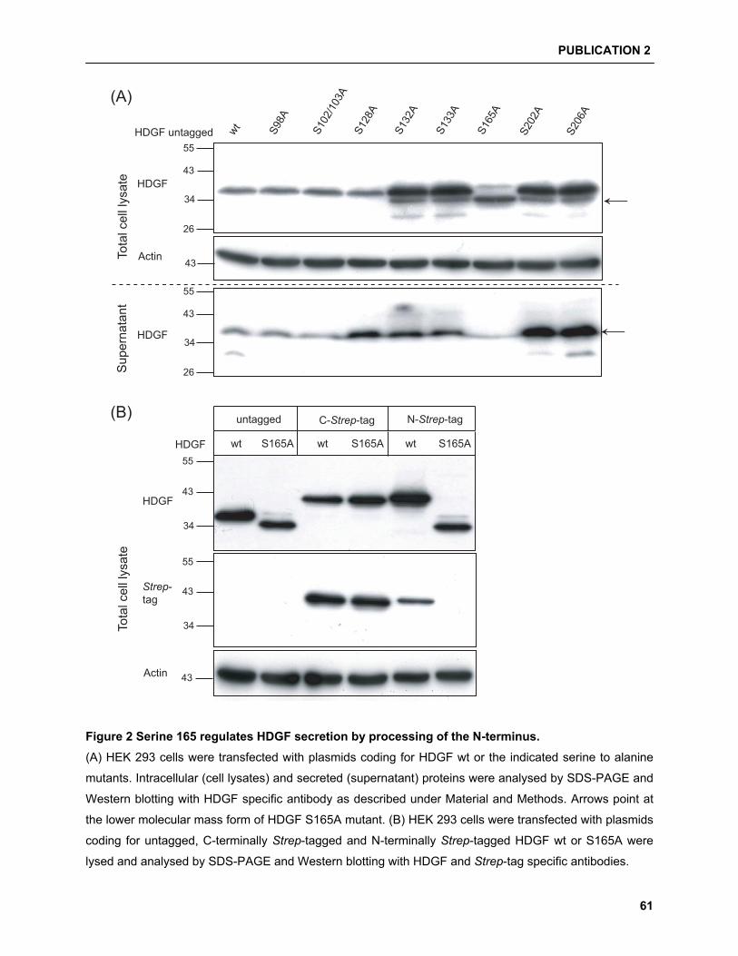
PUBLICATION 2
61
55
43
34
26
43
HDGF untagged wt S98A
S102
/103
AS1
28A
S132
A
S133
A
S165
A
S202
A
S206
A
←
55
43
34
26
←
55
43
34
43
wt S165A wt S165A wt S165A55
43
34
untagged C-Strep-tag N-Strep-tag
HDGF
(B)
HDGF
Actin
Strep-tag
(A)To
tal c
ell l
ysat
e HDGF
Actin
Sup
erna
tant
HDGF
Tota
l cel
l lys
ate
Figure 2 Serine 165 regulates HDGF secretion by processing of the N-terminus. (A) HEK 293 cells were transfected with plasmids coding for HDGF wt or the indicated serine to alanine
mutants. Intracellular (cell lysates) and secreted (supernatant) proteins were analysed by SDS-PAGE and
Western blotting with HDGF specific antibody as described under Material and Methods. Arrows point at
the lower molecular mass form of HDGF S165A mutant. (B) HEK 293 cells were transfected with plasmids
coding for untagged, C-terminally Strep-tagged and N-terminally Strep-tagged HDGF wt or S165A were
lysed and analysed by SDS-PAGE and Western blotting with HDGF and Strep-tag specific antibodies.
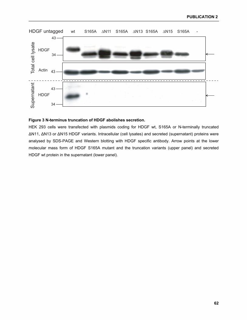
PUBLICATION 2
62
43
34
HDGF untagged wt S165A S165A S165A S165A∆N11 ∆N13 -
←
∆N15
43
43
34
←
Tota
l cel
l lys
ate
HDGF
Actin
Sup
erna
tant
HDGF
Figure 3 N-terminus truncation of HDGF abolishes secretion. HEK 293 cells were transfected with plasmids coding for HDGF wt, S165A or N-terminally truncated
�N11, �N13 or �N15 HDGF variants. Intracellular (cell lysates) and secreted (supernatant) proteins were
analysed by SDS-PAGE and Western blotting with HDGF specific antibody. Arrow points at the lower
molecular mass form of HDGF S165A mutant and the truncation variants (upper panel) and secreted
HDGF wt protein in the supernatant (lower panel).

PUBLICATION 2
63
(A)34
26
17
←
34
26
EGFP HDGF
N10-
EGFP HDGF
N19-
EGFP
17
(B)26
1726
17
HDGF N10
-
SNAP
-Stre
p-ta
gHDGF
N19-
SNAP
-Stre
p-ta
g
SNAP
-Stre
p-ta
g
←
Total cell lysate
Supernatantanti-
EG
FP
anti-
Stre
p-ta
g Total cell lysate
Supernatant
EGFP 16,1870 ± 2.0167HDGF N10-EGFP 10,4260 ± 2.2252HDGF N19- EGFP 13,4260 ± 1.2252
LDH level (% max. release)
SNAP-Strep-tag 18,1870 ± 1.3167HDGF N10-SNAP-Strep-tag 13,0260 ± 1.2287HDGF N19- SNAP-Strep-tag 11,4260 ± 1.4452
(C)
Figure 4 HDGF N-terminal 10 amino acids of mediate secretion of other proteins. (A) HEK 293 cells were transfected with plasmids coding EGFP, HDGF N10-EGFP or HDGF N19-EGFP
plasmids. Intracellular (cell lysates) and secreted (supernatant) proteins were analysed by SDS-PAGE
and Western blotting with EGFP specific antibody. Arrow points out secreted EGFP detected in the
supernatant. (B) HEK 293 cells transfected with plasmids coding SNAP-StrepTag, HDGF
N10-SNAP-StrepTag or HDGF N19-SNAP-StrepTag plasmids wt were lysed and analysed by SDS-PAGE
and Western blotting with StrepTag specific antibody. Arrow points out secreted SNAP-StrepTag detected
in the supernatant.

PUBLICATION 2
64
HDGF untagged wt C12A C108AC108AS165AS165A
+DTT
∆N15
95
72
55
43
34
wt C12A C108AC108AS165AS165A ∆N15
95
72
55
43
34
95
72
55
43
34
95
72
55
43
34
C12AS165A
C12AS165A
-DTT(A)
(B)
←
+DTT-DTT
NIH3T3
72
55
43
34
95
Tota
l cel
l lys
ate
Sup
erna
tant
Tota
l cel
l lys
ate
Figure 5 Disulfide bridges in HDGF. (A) HEK 293 cells transfected with plasmids coding HDGF wt, C12A, C108A, C12A/S165A, C108A/S165A
or �N15 mutants were lysed; cleared lysate was subjected to immunoprecipitation, as described under
Materials and Methods, and analysed (under reducing and non-reducing conditions) along with secreted
(supernatant) proteins by SDS-PAGE and Western blotting with HDGF specific antibody. Arrows point at
the HDGF dimer band under non-reducing conditions (B) HDGF expression in NIH 3T3 cells was
analysed (under reducing and non-reducing conditions) by SDS-PAGE and Western blotting with HDGF
specific antibody.

PUBLICATION 2
65
INTRACELLULAR
EXTRACELLULAR
�Secretion
�Dimerisation
No secretion
Uncleaved and cleavedHDGF
Cleaved HDGF
�Dimerisation
Uncleaved HDGF
Cleaved HDGF
)?
?
Cleavage
�Secretion
)?
?
Cleavage
N-terminal processing
�
(A) (B) (C)
OR?
OR?
S S
S165A S165A
N´
C´
S S
S165A
Ala165
N´
A S
C12A
N´
S A
C12A
Ser165
C12A C12AC´
N´
S S
WTC´
165Ser
++++
----
++++
----
----
----
++++
++++
S
S165A
Ala165
----
C´
N´
S S
WTC´
165Ser
++++
----
N´
S S
C´
165Ser
++++
----
WT
S
C´
165Ser
----
WT
S
C´
165Ser
----
C12A C´
165Ser
----
C108A
N´
S
C12AC´
165Ser
++++
----
N´
S
C108AC´
165Ser
++++
----
N´
S
C12AC´
165Ser
++++
----
N´
S
C108AC´
165Ser
++++
----
C´
165Ser
S S
Ser165
C´C´
165Ser
C´
165Ala
----
C´
Ala165
----
Figure 6 Proposed model for HDGF secretion and dimerisation. (A) HDGF wt; (B) HDGF C12A and
C108A mutants; and (C) HDGF S165A mutant.
3.3 Publication 3
Interaction of HRP-2 splice variants with Hepatoma-derived growth factor
K Thakar, D Kelkar, T Shidore, S Gupta, S Kelm, F Dietz
2010
Biological Chemistry (Submitted; MS no. BIOLCHEM-10-180)
PUBLICATION 3
67
BIOLOGICAL CHEMISTRY
Interaction of HRP-2 splice variants with Hepatoma-derived growth factor
Ketan Thakar , Dipti Kelkar, Teja Shidore, Shivangi Gupta, Sørge Kelm, Frank Dietz 1
Centre for Biomolecular Interactions Bremen (CBIB), Department of Biochemistry, University of Bremen, Leobener Str. NW2, 28359 Bremen, Germany
1To whom correspondence should be addressed:
Centre for Biomolecular Interactions Bremen (CBIB)
Department of Biochemistry, University of Bremen
Leobener Str. im NW2, 28359 Bremen, Germany,
Phone ++4942121863223
FAX ++4942121863221
E-mail: [email protected]
12 text pages (including references), 6 Figure pages; Total word count: 4878; Abstract: 149; Total no. of characters (no spaces): 27,096
PUBLICATION 3
68
Abstract
Hepatoma-derived growth factor related protein 2 (HRP-2) belongs to a family with five additional
members: hepatoma-derived growth factor (HDGF); lens epithelium derived growth factor (LEDGF); and
the HDGF related proteins 1, 3 and 4. Especially for HRP-2 very little is known regarding its function. Here
we demonstrate that HRP-2 interacts with HDGF. Furthermore, we identified a new splice variant of
HRP-2 having a 53 amino acid deletion in the N-terminal region of the protein. This deletion outside of the
PWWP domain causes a loss of an �-helix within the hath region. Moreover, the specific interaction of the
new HRP-2 splice variant with a processed HDGF form is attributed to the loss of this �-helix, suggesting
a critical role of this element. These observations provide evidence for a previously unknown interplay
between different members of the HRP family and open a new perspective for a better understanding of
HRP functions.
Key words
Alternative splicing; hath (homologous to the amino terminus of HDGF) region; N-terminal processing;
phosphorylation; PWWP domain.
PUBLICATION 3
69
Introduction
Hepatoma-derived growth factor related proteins (HRPs) comprise a family of proteins shown to be
mitogenic to various cell lines. Besides the prototype of the family, hepatoma-derived growth factor
(HDGF) [Nakamura et al., 1989], five HRPs have been identified so far; HRP-1 [Izumoto et al., 1997],
HRP-2 [Izumoto et al., 1997], HRP-3 [Ikegame et al., 1999; Abouzied et al., 2004], HRP-4 [Dietz et al.,
2002], and LEDGF (lens epithelium-derived growth factor) [Singh et al., 2000a]. All HRPs share the
following features: (i) high degree of sequence identity in the first N-terminal 98 amino acid residues,
abbreviated as hath (homologous to the amino terminus of HDGF) region; (ii) a PWWP domain in the hath
region; (iii) a canonical bipartite nuclear localization signal in the C-terminal region; and (iv) lack of a
hydrophobic signal peptide.
HDGF was originally purified as a heparin binding growth factor stimulating growth of a variety of cell
types [Nakamura et al., 1994; Oliver and Al-Awqati, 1998; Everett et al., 2000; Mori et al., 2004;
Lepourcelet et al., 2005]. Apart from HDGF, mitogenic activity has been confirmed for HRP-4 in fibroblasts
[Dietz et al., 2002]; and LEDGF in lens epithelial cells, keratinocytes, and fibroblasts [Singh et al., 2000b].
Only HRP-2 and HDGF are ubiquitously expressed, whereas HRP-1/4 and HRP-3 are restricted to testis
and brain, respectively [Nakamura et al., 1994; Izumoto et al., 1997; Ikegame et al., 1999; Dietz et al.,
2002].
HRP-2 is the largest HRP containing 669 amino acids with a high level of basic and acidic residues, a
typical feature of nuclear proteins, and a mixed charge cluster found in eukaryotic regulatory proteins,
including transcription and replication factors [Izumoto et al., 1997]. Another feature of HRP-2 is the
presence of an integrase binding domain (IBD), which has a high degree of similarity to the IBD of
LEDGF. For both proteins it was shown that they bind to and enhance the activity of HIV integrase,
however unlike LEDGF/p75, HRP-2 lacks the ability to tether HIV-1 IN to chromatin [Cherepanov et al.,
2004]. Alternatively spliced gene products (p75 and p52) have been shown for LEDGF [Singh et al.,
2000a]. Analysis of the murine genome revealed that the HRP-2 gene encompasses at least 16 exons.
Furthermore, mRNA entries for 2 isoforms, one with an insertion of 9 amino acids, suggest that also
HRP-2 pre-mRNA is alternatively spliced.
The PWWP domain of HRPs comprises a conserved 70-amino acid sequence found in ~60 eukaryotic
proteins. Mainly roles in interactions with other proteins [Stec et al., 2000] or with DNA [Qiu et al., 2002;
Chen et al., 2004; Ge et al., 2004] have been attributed to the PWWP domain. Interestingly, the PWWP
domain of HDGF binds not only to DNA [Yang and Everett, 2007] but also the heparin-binding activity of
HDGF [Nakamura et al., 1989] has been assigned to its PWWP domain [Sue et al., 2007]. A
high-resolution structure of the HDGF hath region determined by NMR revealed the core structure
PUBLICATION 3
70
consisting of �-helical elements and a �-barrel with five anti-parallel �-strands [Lukasik et al., 2006].
HDGF forms homodimer by an unusual mechanism exchanging �-strands 1 and 2 between two HDGF
PWWP domains [Sue et al., 2007]. This dimerisation leads to an increased binding affinity towards
heparin.
In this study we addressed the question whether HDGF also interacts with other HRPs like HRP-2. We
show that (1) HRP-2 binds to HDGF, that (2) a HRP-2 isoform with a 53 amino acid deletion in the hath
region preferentially interact with a HDGF variant, and (3) this specificity can be attributed to the loss of an
�-helix within the deleted region.
Results Differential expression of HRP-2 correspond to alternatively spliced isoforms
Previous studies investigating HRP-2 expression in different brain regions have shown distinct bands with
a specific anti HRP-2 polyclonal antibody [El-Tahir et al., 2006]. Extending this observation we analyzed
the changes in HRP-2 expression during the development of rats (P= post natal days 2–30, adult) in
different brain regions (Figure 1). The intensities of the four distinct bands at 100-130 kDa identified by the
polyclonal antibody suggested area-specific changes in expression pattern of HRP-2 forms during the
development of rat brain.
The heterogeneity of HRP-2 proteins detected by the antibody could be the result of either specific
proteolytic processing or alternative splicing of the HRP-2 pre-mRNA. To further explore the hypothesis of
alternatively spliced HRP-2 variants, we amplified total HRP-2 mRNA by RT-PCR using mouse adult brain
cDNA as a template with sense and antisense primers flanking the coding region. By sequencing the PCR
products we discovered a new splice variant of HRP-2, besides the already described HRP-2 coding
sequences referred to as HRP-2 isoform a in this study (669 aa, accession number BAA22896), and a
variant referred to as HRP-2 isoform b (678 aa, accession number BC003741). Figure 2 shows a
schematic representation of the known intron and exon borders and resulting alternatively spliced HRP-2
gene products. The new splice variant is characterized by an in-frame deletion spanning parts of exons 3
and 4. During this study we refer this new isoform as HRP-2 isoform c (616 aa, accession number
FN687734).
In HRP-2 isoform c the deletion of 53 amino acids (aa 66–118) leaves the predicted �-barrel intact, but
skips the remaining part of the conserved hath region including a regular �-helix which seems to be a
characteristic feature of the PWWP domain of all HRPs according to the solved structure of the hath
region of HDGF (PDB ID: 1RI0) [Sue et al., 2004] and HRP-3 (PDB ID: 1N27) [Nameki et al., 2005]
PUBLICATION 3
71
All HRP-2 isoforms were expressed in COS-7 cells and analyzed by Western blotting (Figure 3). While the
HRP-2 isoforms a and/or b have an electrophoretic mobility similar to the bands just below 130 kDa found
in tissue samples (frontal lobe P5 in Figure 3) isoform c migrates similar to the lower bands.
Interaction of HRP-2 with HDGF
HDGF has been shown to form homo- and hetero-dimers [Sue et al., 2007; Yang and Everett, 2007].
Therefore, we have tested the hypothesis that different HRPs interact with each other. To investigate the
interaction of HRP-2 isoforms with HDGF, HRP-2 isoforms a, b or c and HDGF wt were co-expressed in
COS-7 cells. For affinity precipitation of the interacting HRPs with StrepTactin® beads, either HRP-2
(Figure 4A) or HDGF wt (Figure 4B) contained a Strep-tag at its C-terminus. The Western blot results of
these precipitates clearly indicate interaction of HDGF with all HRP-2 isoforms. Interestingly, only HRP-2
isoform c interacts additionally with a lower molecular mass form of HDGF which is almost not detectable
in the total cell lysate fraction.
Immunocytochemical analysis revealed a predominant nuclear localization of HDGF and HRP-2 isoform a,
b and c, providing the spatial requirement for the interaction between these proteins in the cell (Figure 5).
In the context of a study addressing the secretion mechanism of HDGF (see accompanying manuscript),
we generated eight HDGF variants in which potential serine phosphorylation sites had been mutated to
alanine and expressed these in HEK 293 cells. One of these mutants, HDGF S165A, is truncated at its
N-terminus leading to a higher electrophoretic mobility similar to the lower molecular mass band
co-precipitating with HRP-2 isoform c.
The 53 amino acid deletion (aa 66-118) in HRP-2 isoform c includes a 11-residue long �-helical element.
To test whether this modification is the structural basis for the altered binding specificity, we prepared a
deletion mutant of HRP-2 isoform a lacking only these amino acids stretch (HRP-2 isoform a�helix).
Co-precipitation studies were performed as described above. The data shown in Figure 6 confirms that all
HRP-2 isoforms and the �helix mutant interact with both, HDGF wt and HDGF S165A mutant.
Furthermore, like HRP-2 isoform c, the HRP-2 isoform a�helix mutant also co-precipitates the lower
molecular mass form of HDGF. Although no precise quantification is possible from these experiments, it
appears that HDGF S165A is co-precipitated by HRP-2 isoform c and the isoform a�helix mutant at a much
higher rate than HDGF wt.
PUBLICATION 3
72
Discussion
In this study we demonstrate that HRP-2 and HDGF bind to each other and explored the specificity of this
reaction as a prototype for interactions between HRPs. HDGF acts as a transcriptional co-repressor for
SMYD1 gene expression by recruiting CtBP [Yang and Everett, 2009]. It is feasible that HRP-2/HDGF
hetero-dimerisation modulates DNA binding thus regulating gene expression. One important result of this
study has been the observation that HRP-2 isoform c selectively binds to a subset of HDGF molecules
with a lower molecular mass. This opens the possibility that interactions between HRPs, and consequently
their functions, are controlled by differential expression of such isoforms. Here we have shown that the
expression pattern of HRP-2 variants is changing during development, extending a previous investigation
describing the regional differences of HRPs expression in rat brain [El-Tahir et al., 2006]. Obviously, the
observed band pattern can have different origins, such as proteolysis, post-translational modification or
the occurrence of different splice variants. All these events can be regulated during developmental
processes.
We further provide strong evidence that at least three HRP-2 variants, isoforms a and b, and a so far
unknown isoform c, are generated by alternative mRNA splicing. Alternative splicing is not unusual for
HRPs. It has been described for LEDGF giving two isoforms p75 and p52 [Singh et al., 2000a] and splice
variants of other members like HDGF or HRP-3 can be found in databases. However, the corresponding
proteins have not been described.
The new isoform c is a 616 aa protein with a deletion in its N-terminal region extending from amino acids
66 to 118. Studies on HDGF have highlighted the importance of this region in regulating or defining HDGF
function. For example, amino acids 81-100 of HDGF appear to be relevant for its cell surface interaction
and proliferative function [Abouzied et al., 2005] and SUMOylation of K80 has been shown to affect HDGF
binding to chromatin [Thakar et al., 2008]. Recombinant HRP-2 isoforms a and b migrate like proteins with
a molecular mass of 120-130 kDa, whereas HRP-2 isoform c migrates like a protein of 100-110 kDa
correlating with the lower molecular weight bands from the tissue samples. For HRPs it is difficult to
directly correlate the predicted molecular mass of the splice variants with bands in SDS-PAGE. As already
shown for HDGF and HRP-3 [Abouzied et al., 2004], HRP-2 is migrating at a molecular weight
significantly higher than predicted (74 kDa) by its primary amino acid sequence. Since also proteins
expressed in bacteria (data not shown) show the same unusual low electrophoretic mobility, it is unlikely
to result from post-translational modifications.
The occurrence of various isoforms suggests specialized and specific functions of these proteins related
to their structural characteristics. Sequence alignments and structural comparison between different HRPs
suggest that the hath region is folded similarly in all these proteins, consisting of a five stranded
PUBLICATION 3
73
anti-parallel �-barrel and �-helices [Sue et al., 2004]. The hath region of HDGF can form dimers mediated
by an unusual exchange of between the two monomers [Sue et al., 2007]. It is tempting to speculate that
a similar mechanism mediates the interaction between HRP-2 and HDGF described in this study. In this
context it is important to note that the specificity of HRP-2 isoform c for interaction with HDGF variants
appears to be due to the loss of 53 amino acids including the �-helix named B [Sue et al., 2004] or �4
[Lukasik et al., 2006]) which is in direct contact with the �-strands 1 and 2 involved in HDGF dimerisation
[Sue et al., 2007]. Along this line, an explanation for the binding preferences of HRP-2 isoform c could be
that the exchange of �-strands 1 and 2 is facilitated, as these are exposed due to the lack of the �-helix
covering them in isoform a or b. This hypothesis is supported by our data, since HRP-2 isoform a�helix
lacking only this �-helix has the same binding preferences as isoform c.
Moreover, the apparently preferred HDGF binding partner of HRP-2 isoform c is a variant with an
electrophoretic mobility similar to the HDGF S165A mutant which is truncated at the N-terminus.
Interestingly, this truncation leads to the loss of the intramolecular disulfide bridge between C12 and C108
(see accompanying manuscript) which may stabilize a more compact conformation of the hath region
preventing the exchange of �-strands 1 and 2 in HDGF wt.
In summary, this study shows for the first time heteromerisation between different members of the HRP
family; HDGF and HRP-2. Additionally, we discovered a new splice variant of HRP-2 with a deletion in its
hath region which interacts preferentially with a processed form of HDGF probably due to the loss of an
�-helix in HRP-2. In conclusion, these observations provide a new perspective for understanding the
biological functions of HDGF and related proteins.
Materials and Methods
RNA isolation and cDNA preparation from brain tissue sample
Total RNA was isolated by homogenization of brain tissue samples according to manufacture’s protocol
(peqGOLD TriFast kit, PEQLAB, Erlangen, Germany). Briefly; 1 mL TriFast for every 50-100 mg of tissue
was used. The nucleoprotein complexes were incubated at room temperature for 5 min followed by
addition of 0.2 mL chloroform and further incubation for 10 min at room temperature. The samples were
then centrifuged at 12000 g to separate phases. RNA containing aqueous phase was then separated and
precipitated using 500 �L of isopropanol mL-1 of TriFast. These samples were further incubated on ice for
15 min and centrifuged for 10 min at 12000 g, 4°C. RNA pellet obtained was subsequently washed with
75% ethanol, centrifuged for 10 min at 7500 g, 4°C and re-suspended in RNA free water. cDNA synthesis
was carried out according to manufactures protocol (RevertAid™ First Strand cDNA Synthesis Kit,
Fermentas, St. Leon-Rot, Germany).
PUBLICATION 3
74
Production of recombinant proteins
Plasmids were constructed using standard recombinant cloning techniques and all changes were verified
by DNA sequencing. HRP-2 was amplified from mouse brain cDNA using the following nested primers:
Sense 5’ CCTCCGCCGACAGCATGCCG and
antisense 5’ GGCTGGAGCTCAGCTGTCCT followed by
Sense 5´CGAAGCTTATGCCGCACGCCTTCAAGCCC and
antisense 5´GCGAATTCGCTGTCCTCATTGTCAT primer
containing HindIII and EcoRI restriction sites and cloned in frame to the StrepTag coding sequence into
HindIII/EcoRI digested pcDNA3 Amp StrepTag vector as described previously [Thakar et al., 2008].
Untagged HRP-2 isoforms were prepared by amplifying the coding sequence from the respective
C-terminally Strep-tagged constructs using the same sense primer and antisense primer
5´GCGAATTCTCAGCTGTCCTCATTGTCAT
The PCR amplification product was cloned using the HindIII and EcoRI restriction sites in the pcDNA3
Amp vector.
The cloning strategy to generate C-terminally Strep-tagged HRP-2 isoform a�helix was as follows: The
cDNAs coding for the N-terminal (1 to 74) and the C-terminal region of HRP-2 isoform a (87 to 669) were
amplified separately using C-terminally Strep-tagged HRP-2 isoform a as templates. The 3' and 5' ends of
the amplification products were designed to allow an in frame ligation using EcoRI and subcloned in frame
into HindIII/EcoRI or EcoRI/KpnI digested pcDNA3 Amp. The following primers were used for
amplification:
Sense primer for N-terminal region 5’ CCCAAGCTTATGCCGCACGCCTTCAAGCCC,
antisense primer for N-terminal region 5’ CGGAATTCGCCTTTCCTCTTGTTGGG,
Sense primer for C-terminal region 5’ CGGAATTCAACCCCCATGCCAGCTAC,
antisense primer for C-terminal region 5’ CGGGTACCGCTGTCCTCATTGTCATC.
The plasmid encoding for HDGF S165A was described elsewhere (see accompanying manuscript).
Cell culture and transfections
For transient transfection COS-7 cells were maintained, transfected using the polyethylenimine
ExGen 500 according to the manufacturer’s protocol (Fermentas, St. Leon-Rot, Germany) and harvested
after 24 h as described previously [Thakar et al., 2008].
PUBLICATION 3
75
Preparation of proteins extracts
Cells were washed twice with PBS before treatment with ice cold (1mL/10 cm plate) TNE lysis buffer
containing 20mM Tris/Cl pH 7.4, 150 mM NaCl, 5mM EDTA, 1% NP-40 + protease inhibitors (complete
mini, EDTA free from Roche, Mannheim, Germany) on ice. After 10 min on ice, cells were scraped from
the dish, transferred to a 1.5 mL reaction tube and incubated on ice for additional 10 min. Cell debris was
separated by centrifugation at 12000 g, 4ºC for 20 min. The cleared lysate was used directly for
SDS-PAGE and Western blot or for the purification of the recombinant Strep-tagged HDGF using
StrepTactin® (IBA, Göttingen, Germany).
StrepTactin® purification
Cleared lysate (900 μL from each 10 cm � cell culture dish) were incubated in a batch procedure with
30 μL of StrepTactin® Sepharose beads on a rotor shaker for 6 h at 4°C. After incubation beads were
centrifuged at 1200 g, 4ºC for 15 s and the supernatant was transferred to a new 1.5 mL reaction tube.
Beads were washed 4 times with 1 mL of TNE lysis buffer. After the final washing step, bound proteins
were eluted by incubating the beads at 95°C for 10 min in 80 μL 2x SDS-PAGE sample buffer. 20 μL of
the eluate was used for SDS-PAGE.
Western Blot analysis
In principle, Western blot analysis was performed as described elsewhere (see accompanying
manuscript). The following antibodies were used: rabbit anti-HRP-2 polyclonal (1:250) and sheep
anti-HDGF polyclonal (1:1000) [Abouzied et al., 2004], sheep anti-Strep-tag polyclonal (1:1000; IBA,
Göttingen, Germany), mouse anti-Actin monoclonal (1:2000; Dianova, Hamburg, Germany).
Immunofluorescence
For analysis of sub-cellular localization by immunofluorescence COS-7 cells (3 x 10⁴) were plated on
cover-slips in a 24 well plate were grown overnight. Post transfection cells were washed twice in
phosphate buffered saline (PBS) solution and then fixed in 4% paraformaldehyde in PBS for 10 min. For
permeabilization, cells were treated for 2 min with ice cold methanol on ice and afterwards washed twice
with PBS/2% BSA before blocking unspecific binding sites with DMEM/10% FCS for 30 min. The cells
were immunostained with polyclonal sheep anti-HDGF and rabbit anti-HRP-2 as primary antibodies for
90 min in PBS/2% BSA at room temperature. After washing twice with PBS/2% BSA, cells were treated
for 90 min in PBS/2% BSA at room temperature in the dark with donkey anti-rabbit antibody-Cy3
conjugate and donkey anti-sheep antibody-Cy2 conjugate (Dianova, Hamburg, Germany). Cells were

PUBLICATION 3
76
washed in PBS/2% BSA and then treated with the nuclear stain DAPI (Roche, Mannheim, Germany) for
1 min at room temperature. After additional washing cells were embedded in immuno-mount (Thermo
Shandon, Pittsburgh, PA, USA) and images were captured on the fluorescence microscope (Olympus,
Hamburg, Germany).
Acknowledgments
Our thanks go to Dr. Sebastian Franken from the Department of Physiological Chemistry, University of
Bonn, Germany for kindly providing the rabbit anti-HRP-2 polyclonal antibody. We also like to thank
Mrs. Nazila Isakovic for excellent technical support. This work is supported by the Central Research
Funding (ZFK, BFK NaWi), University of Bremen, and the Tönjes-Vagt-Stiftung, Project XXI.
References
Abouzied, M.M., Baader, S.L., Dietz, F., Kappler, J., Gieselmann, V., Franken, S. (2004). Expression
patterns and different subcellular localization of the growth factors HDGF (hepatoma-derived
growth factor) and HRP-3 (HDGF-related protein-3) suggest functions in addition to their
mitogenic activity. Biochem J 378, 169-176.
Abouzied, M.M., El-Tahir, H.M., Prenner, L., Haberlein, H., Gieselmann, V., Franken, S. (2005).
Hepatoma-derived growth factor. Significance of amino acid residues 81-100 in cell surface
interaction and proliferative activity. J Biol Chem 280, 10945-10954.
Chen, T., Tsujimoto, N., Li, E. (2004). The PWWP domain of Dnmt3a and Dnmt3b is required for directing
DNA methylation to the major satellite repeats at pericentric heterochromatin. Mol Cell Biol 24,
9048-9058.
Cherepanov, P., Devroe, E., Silver, P.A., Engelman, A. (2004). Identification of an evolutionarily
conserved domain in human lens epithelium-derived growth factor/transcriptional co-activator p75
(LEDGF/p75) that binds HIV-1 integrase. J Biol Chem 279, 48883-48892.
Dietz, F., Franken, S., Yoshida, K., Nakamura, H., Kappler, J., Gieselmann, V. (2002). The family of
hepatoma-derived growth factor proteins: characterization of a new member HRP-4 and
classification of its subfamilies. Biochem J 366, 491-500.
El-Tahir, H.M., Dietz, F., Dringen, R., Schwabe, K., Strenge, K., Kelm, S., Abouzied, M.M., Gieselmann,
V., Franken, S. (2006). Expression of hepatoma-derived growth factor family members in the adult
central nervous system. BMC Neurosci 7, 6.
Everett, A.D., Lobe, D.R., Matsumura, M.E., Nakamura, H., McNamara, C.A. (2000). Hepatoma-derived
growth factor stimulates smooth muscle cell growth and is expressed in vascular development.
J Clin Invest 105, 567-575.
PUBLICATION 3
77
Ge, Y.Z., Pu, M.T., Gowher, H., Wu, H.P., Ding, J.P., Jeltsch, A., Xu, G.L. (2004). Chromatin targeting of
de novo DNA methyltransferases by the PWWP domain. J Biol Chem 279, 25447-25454.
Huang, H.D., Lee, T.Y., Tzeng, S.W., Horng, J.T. (2005). KinasePhos: a web tool for identifying protein
kinase-specific phosphorylation sites. Nucleic Acids Res 33, W226-229.
Ikegame, K., Yamamoto, M., Kishima, Y., Enomoto, H., Yoshida, K., Suemura, M., Kishimoto, T.,
Nakamura, H. (1999). A new member of a hepatoma-derived growth factor gene family can
translocate to the nucleus. Biochem Biophys Res Commun 266, 81-87.
Izumoto, Y., Kuroda, T., Harada, H., Kishimoto, T., Nakamura, H. (1997). Hepatoma-derived growth factor
belongs to a gene family in mice showing significant homology in the amino terminus. Biochem
Biophys Res Commun 238, 26-32.
Lepourcelet, M., Tou, L., Cai, L., Sawada, J., Lazar, A.J., Glickman, J.N., Williamson, J.A., Everett, A.D.,
Redston, M., Fox, E.A., Nakatani, Y., Shivdasani, R.A. (2005). Insights into developmental
mechanisms and cancers in the mammalian intestine derived from serial analysis of gene
expression and study of the hepatoma-derived growth factor (HDGF). Development 132, 415-427.
Lukasik, S.M., Cierpicki, T., Borloz, M., Grembecka, J., Everett, A., Bushweller, J.H. (2006). High
resolution structure of the HDGF PWWP domain: a potential DNA binding domain. Protein Sci 15,
314-323.
Mense, M., Vergani, P., White, D.M., Altberg, G., Nairn, A.C., Gadsby, D.C. (2006). In vivo
phosphorylation of CFTR promotes formation of a nucleotide-binding domain heterodimer.
EMBO J 25, 4728-4739.
Mori, M., Morishita, H., Nakamura, H., Matsuoka, H., Yoshida, K., Kishima, Y., Zhou, Z., Kida, H.,
Funakoshi, T., Goya, S., Yoshida, M., Kumagai, T., Tachibana, I., Yamamoto, Y., Kawase, I.,
Hayashi, S. (2004). Hepatoma-derived growth factor is involved in lung remodeling by stimulating
epithelial growth. Am J Respir Cell Mol Biol 30, 459-469.
Nakamura, H., Izumoto, Y., Kambe, H., Kuroda, T., Mori, T., Kawamura, K., Yamamoto, H., Kishimoto, T.
(1994). Molecular cloning of complementary DNA for a novel human hepatoma-derived growth
factor. Its homology with high mobility group-1 protein. J Biol Chem 269, 25143-25149.
Nakamura, H., Kambe, H., Egawa, T., Kimura, Y., Ito, H., Hayashi, E., Yamamoto, H., Sato, J., Kishimoto,
S. (1989). Partial purification and characterization of human hepatoma-derived growth factor. Clin
Chim Acta 183, 273-284.
Nameki, N., Tochio, N., Koshiba, S., Inoue, M., Yabuki, T., Aoki, M., Seki, E., Matsuda, T., Fujikura, Y.,
Saito, M., Ikari, M., Watanabe, M., Terada, T., Shirouzu, M., Yoshida, M., Hirota, H., Tanaka, A.,
Hayashizaki, Y., Guntert, P., Kigawa, T., Yokoyama, S. (2005). Solution structure of the PWWP
domain of the hepatoma-derived growth factor family. Protein Sci 14, 756-764.
Oliver, J.A., Al-Awqati, Q. (1998). An endothelial growth factor involved in rat renal development. J Clin
Invest 102, 1208-1219.
Qiu, C., Sawada, K., Zhang, X., Cheng, X. (2002). The PWWP domain of mammalian DNA
methyltransferase Dnmt3b defines a new family of DNA-binding folds. Nat Struct Biol 9, 217-224.
PUBLICATION 3
78
Rubin, S.M., Gall, A.L., Zheng, N., Pavletich, N.P. (2005). Structure of the Rb C-terminal domain bound to
E2F1-DP1: a mechanism for phosphorylation-induced E2F release. Cell 123, 1093-1106.
Singh, D.P., Kimura, A., Chylack, L.T., Jr., Shinohara, T. (2000a). Lens epithelium-derived growth factor
(LEDGF/p75) and p52 are derived from a single gene by alternative splicing. Gene 242, 265-273.
Singh, D.P., Ohguro, N., Kikuchi, T., Sueno, T., Reddy, V.N., Yuge, K., Chylack, L.T., Jr., Shinohara, T.
(2000b). Lens epithelium-derived growth factor: effects on growth and survival of lens epithelial
cells, keratinocytes, and fibroblasts. Biochem Biophys Res Commun 267, 373-381.
Stec, I., Nagl, S.B., van Ommen, G.J., den Dunnen, J.T. (2000). The PWWP domain: a potential
protein-protein interaction domain in nuclear proteins influencing differentiation? FEBS Lett 473,
1-5.
Sue, S.C., Chen, J.Y., Lee, S.C., Wu, W.G., Huang, T.H. (2004). Solution structure and heparin
interaction of human hepatoma-derived growth factor. J Mol Biol 343, 1365-1377.
Sue, S.C., Lee, W.T., Tien, S.C., Lee, S.C., Yu, J.G., Wu, W.J., Wu, W.G., Huang, T.H. (2007). PWWP
module of human hepatoma-derived growth factor forms a domain-swapped dimer with much
higher affinity for heparin. J Mol Biol 367, 456-472.
Thakar, K., Niedenthal, R., Okaz, E., Franken, S., Jakobs, A., Gupta, S., Kelm, S., Dietz, F. (2008).
SUMOylation of the hepatoma-derived growth factor negatively influences its binding to
chromatin. FEBS J 275, 1411-1426.
Yang, J., Everett, A.D. (2007). Hepatoma-derived growth factor binds DNA through the N-terminal PWWP
domain. BMC Mol Biol 8, 101.
Yang, J., Everett, A.D. (2009). Hepatoma-derived growth factor represses SET and MYND domain
containing 1 gene expression through interaction with C-terminal binding protein. J Mol Biol 386,
938-950.
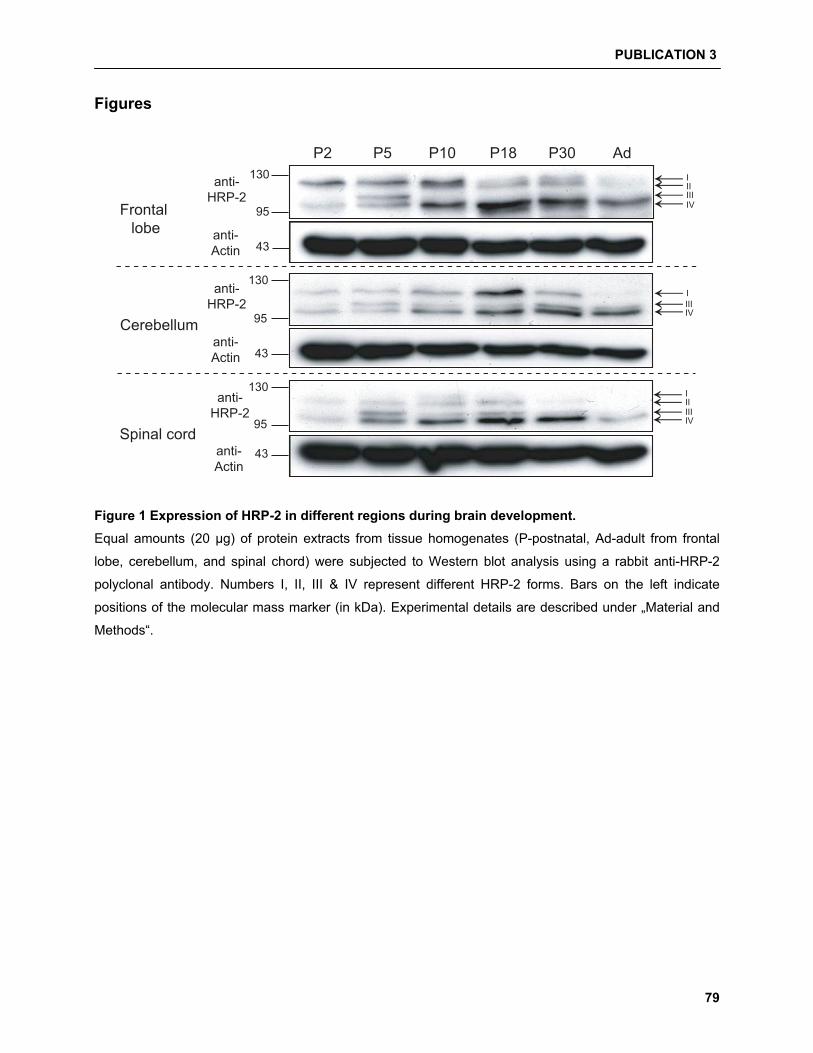
PUBLICATION 3
79
Figures
P2 P5 P10 P18 P30 Ad
Cerebellum
130
95
43
←←←IIIIIV
Frontal lobe
130
95
43
←←←←II
I
IIIIV
Spinal cord
130
95
43
←←←←II
I
IIIIV
anti-HRP-2
anti-Actin
anti-HRP-2
anti-Actin
anti-HRP-2
anti-Actin
Figure 1 Expression of HRP-2 in different regions during brain development. Equal amounts (20 �g) of protein extracts from tissue homogenates (P-postnatal, Ad-adult from frontal
lobe, cerebellum, and spinal chord) were subjected to Western blot analysis using a rabbit anti-HRP-2
polyclonal antibody. Numbers I, II, III & IV represent different HRP-2 forms. Bars on the left indicate
positions of the molecular mass marker (in kDa). Experimental details are described under „Material and
Methods“.
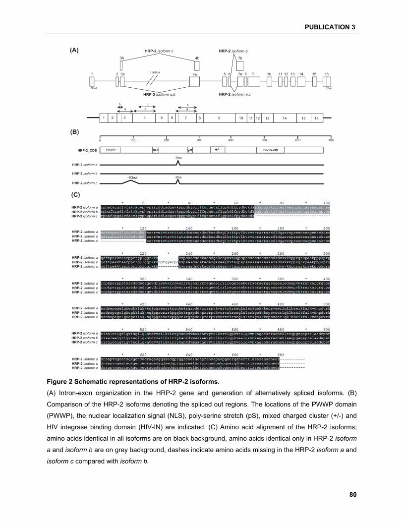
PUBLICATION 3
80
-53aa
-9aa
-9aa
HRP-2 isoform a
0 100 200 300 400 500 600 700
HRP-2_CDS PWWP NLS pS +/- HIV IN-BD
1 2 3 4 5 6 7 8 9 10 11 12 13 14 15 16
a a a
b b b
11105nt1 2 3a 4a 5 6 7a 8 9 10 11 12 13 14 15 16
HRP-2 isoform c
HRP-2 isoform a,c
3b 4b 7b
HRP-2 isoform b
HRP-2 isoform a,b
Start Stop
HRP-2 isoform b
HRP-2 isoform c
(A)
HRP-2 isoform aHRP-2 isoform bHRP-2 isoform c
(B)
(C)
HRP-2 isoform aHRP-2 isoform bHRP-2 isoform c
HRP-2 isoform aHRP-2 isoform bHRP-2 isoform c
HRP-2 isoform aHRP-2 isoform bHRP-2 isoform c
HRP-2 isoform aHRP-2 isoform bHRP-2 isoform c
HRP-2 isoform aHRP-2 isoform bHRP-2 isoform c
HRP-2 isoform aHRP-2 isoform bHRP-2 isoform c
Figure 2 Schematic representations of HRP-2 isoforms. (A) Intron-exon organization in the HRP-2 gene and generation of alternatively spliced isoforms. (B)
Comparison of the HRP-2 isoforms denoting the spliced out regions. The locations of the PWWP domain
(PWWP), the nuclear localization signal (NLS), poly-serine stretch (pS), mixed charged cluster (+/-) and
HIV integrase binding domain (HIV-IN) are indicated. (C) Amino acid alignment of the HRP-2 isoforms;
amino acids identical in all isoforms are on black background, amino acids identical only in HRP-2 isoform
a and isoform b are on grey background, dashes indicate amino acids missing in the HRP-2 isoform a and
isoform c compared with isoform b.
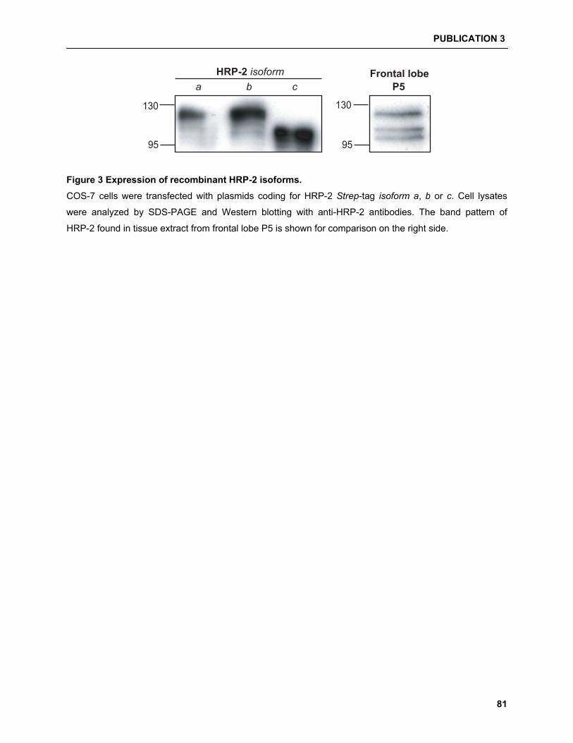
PUBLICATION 3
81
130
95
Frontal lobeP5
130
95
HRP-2 isoforma b c
Figure 3 Expression of recombinant HRP-2 isoforms. COS-7 cells were transfected with plasmids coding for HRP-2 Strep-tag isoform a, b or c. Cell lysates
were analyzed by SDS-PAGE and Western blotting with anti-HRP-2 antibodies. The band pattern of
HRP-2 found in tissue extract from frontal lobe P5 is shown for comparison on the right side.
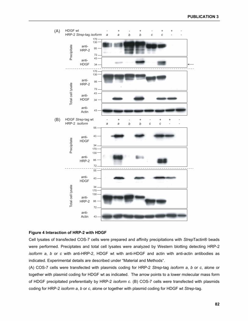
PUBLICATION 3
82
170130
95
72
anti-HRP-2
43
34
43
HRP-2 Strep-tag isoformHDGF wt
a a b b c c - -- + - + + + --
43
34 ←170130
95
72
(A)
43
34
55
43
34
55
43
170130
95
72
170130
95
72
(B)
Pre
cipi
tate
Tota
l cel
l lys
ate
HRP-2 isoformHDGF Strep-tag wt
a a b b c c - -- + - + + + --
Pre
cipi
tate
Tota
l cel
l lys
ate
anti-HDGF
anti-HRP-2
anti-HDGF
anti-Actin
anti-HDGF
anti-HRP-2
anti-HDGF
anti-HRP-2
anti-Actin
Figure 4 Interaction of HRP-2 with HDGF Cell lysates of transfected COS-7 cells were prepared and affinity precipitations with StrepTactin® beads
were performed. Preciptates and total cell lysates were analyzed by Western blotting detecting HRP-2
isoform a, b or c with anti-HRP-2, HDGF wt with anti-HDGF and actin with anti-actin antibodies as
indicated. Experimental details are described under “Material and Methods“.
(A) COS-7 cells were transfected with plasmids coding for HRP-2 Strep-tag isoform a, b or c, alone or
together with plasmid coding for HDGF wt as indicated. The arrow points to a lower molecular mass form
of HDGF precipitated preferentially by HRP-2 isoform c. (B) COS-7 cells were transfected with plasmids
coding for HRP-2 isoform a, b or c, alone or together with plasmid coding for HDGF wt Strep-tag.
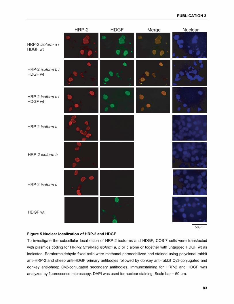
PUBLICATION 3
83
HRP-2 HDGF Merge Nuclear
50μm
HRP-2 isoform a /HDGF wt
HRP-2 isoform b /HDGF wt
HRP-2 isoform c /HDGF wt
HRP-2 isoform a
HRP-2 isoform b
HRP-2 isoform c
HDGF wt
Figure 5 Nuclear localization of HRP-2 and HDGF. To investigate the subcellular localization of HRP-2 isoforms and HDGF, COS-7 cells were transfected
with plasmids coding for HRP-2 Strep-tag isoform a, b or c alone or together with untagged HDGF wt as
indicated. Paraformaldehyde fixed cells were methanol permeabilized and stained using polyclonal rabbit
anti-HRP-2 and sheep anti-HDGF primary antibodies followed by donkey anti-rabbit Cy3-conjugated and
donkey anti-sheep Cy2-conjugated secondary antibodies. Immunostaining for HRP-2 and HDGF was
analyzed by fluorescence microscopy. DAPI was used for nuclear staining. Scale bar = 50 �m.
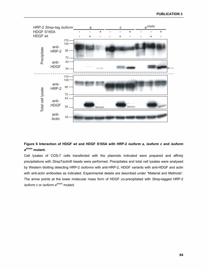
PUBLICATION 3
84
170130
95
7243
34
43
43
34
HDGF wt - + -HDGF S165A - +
- + - - + -- - +- - +-
←
170130
95
72
HRP-2 Strep-tag isoform a c a∆helix
Pre
cipi
tate
Tota
l cel
l lys
ate
anti-HRP-2
anti-HDGF
anti-HRP-2
anti-HDGF
anti-Actin
Figure 6 Interaction of HDGF wt and HDGF S165A with HRP-2 isoform a, isoform c and isoform
a�helix mutant. Cell lysates of COS-7 cells transfected with the plasmids indicated were prepared and affinity
precipitations with StrepTactin® beads were performed. Precipitates and total cell lysates were analysed
by Western blotting detecting HRP-2 isoforms with anti-HRP-2, HDGF variants with anti-HDGF and actin
with anti-actin antibodies as indicated. Experimental details are described under “Material and Methods“.
The arrow points at the lower molecular mass form of HDGF co-precipitated with Strep-tagged HRP-2
isoform c or isoform a�helix mutant.

44 AAddddiittiioonnaall RReessuullttss
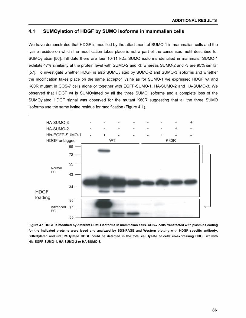
ADDITIONAL RESULTS
86
4.1 SUMOylation of HDGF by SUMO isoforms in mammalian cells We have demonstrated that HDGF is modified by the attachment of SUMO-1 in mammalian cells and the
lysine residue on which the modification takes place is not a part of the consensus motif described for
SUMOylation [56]. Till date there are four 10-11 kDa SUMO isoforms identified in mammals. SUMO-1
exhibits 47% similarity at the protein level with SUMO-2 and -3, whereas SUMO-2 and -3 are 95% similar
[57]. To investigate whether HDGF is also SUMOylated by SUMO-2 and SUMO-3 isoforms and whether
the modification takes place on the same acceptor lysine as for SUMO-1 we expressed HDGF wt and
K80R mutant in COS-7 cells alone or together with EGFP-SUMO-1, HA-SUMO-2 and HA-SUMO-3. We
observed that HDGF wt is SUMOylated by all the three SUMO isoforms and a complete loss of the
SUMOylated HDGF signal was observed for the mutant K80R suggesting that all the three SUMO
isoforms use the same lysine residue for modification (Figure 4.1).
.
95
72
43
34
55
HDGF untagged-His-EGFP-SUMO-1
HA-SUMO-2 HA-SUMO-3
WT K80R+ - - - + - -
- - + - - - + -- - - + - - - +
95
72
55
←
HDGFloading
Normal ECL
Advanced ECL
Figure 4.1 HDGF is modified by different SUMO isoforms in mammalian cells. COS-7 cells transfected with plasmids coding for the indicated proteins were lysed and analyzed by SDS-PAGE and Western blotting with HDGF specific antibody. SUMOylated and unSUMOylated HDGF could be detected in the total cell lysate of cells co-expressing HDGF wt with His-EGFP-SUMO-1, HA-SUMO-2 or HA-SUMO-3.
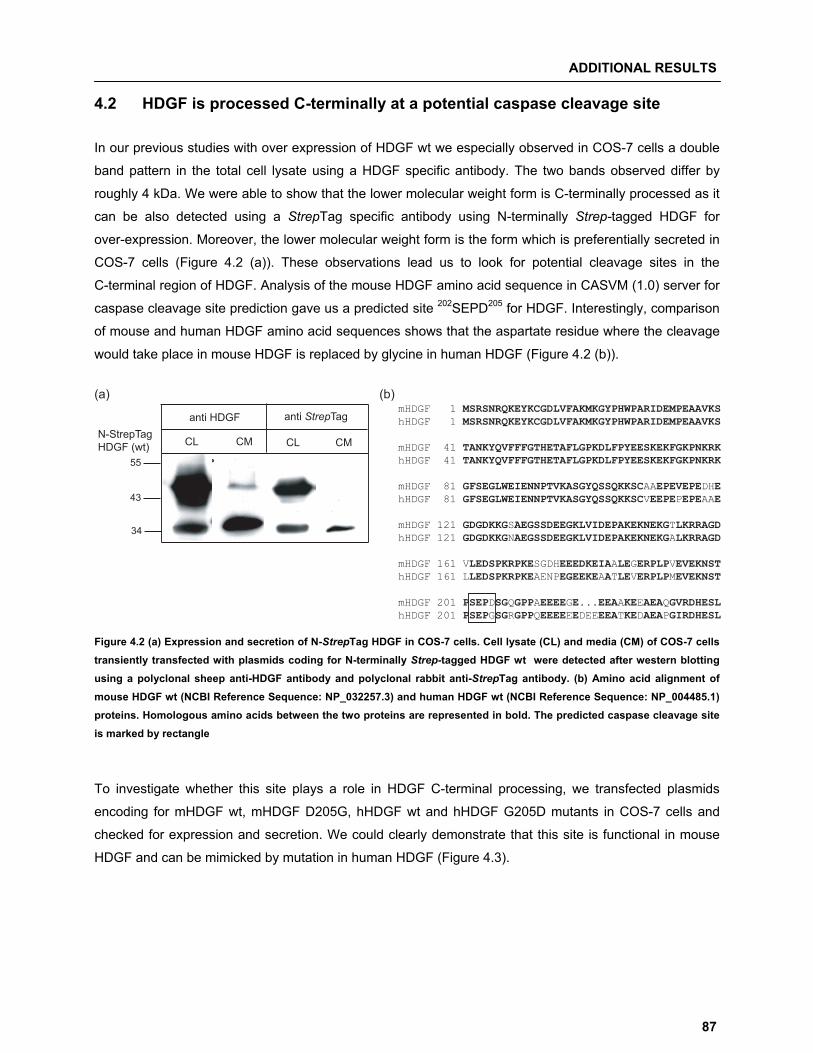
ADDITIONAL RESULTS
87
4.2 HDGF is processed C-terminally at a potential caspase cleavage site In our previous studies with over expression of HDGF wt we especially observed in COS-7 cells a double
band pattern in the total cell lysate using a HDGF specific antibody. The two bands observed differ by
roughly 4 kDa. We were able to show that the lower molecular weight form is C-terminally processed as it
can be also detected using a StrepTag specific antibody using N-terminally Strep-tagged HDGF for
over-expression. Moreover, the lower molecular weight form is the form which is preferentially secreted in
COS-7 cells (Figure 4.2 (a)). These observations lead us to look for potential cleavage sites in the
C-terminal region of HDGF. Analysis of the mouse HDGF amino acid sequence in CASVM (1.0) server for
caspase cleavage site prediction gave us a predicted site 202SEPD205 for HDGF. Interestingly, comparison
of mouse and human HDGF amino acid sequences shows that the aspartate residue where the cleavage
would take place in mouse HDGF is replaced by glycine in human HDGF (Figure 4.2 (b)).
CL CM
anti HDGF
CL CM
anti StrepTag
55
43
34
N-StrepTag HDGF (wt)
(a)mHDGF 1 MSRSNRQKEYKCGDLVFAKMKGYPHWPARIDEMPEAAVKS hHDGF 1 MSRSNRQKEYKCGDLVFAKMKGYPHWPARIDEMPEAAVKS
mHDGF 41 TANKYQVFFFGTHETAFLGPKDLFPYEESKEKFGKPNKRK hHDGF 41 TANKYQVFFFGTHETAFLGPKDLFPYEESKEKFGKPNKRK
mHDGF 81 GFSEGLWEIENNPTVKASGYQSSQKKSCAAEPEVEPEDHE hHDGF 81 GFSEGLWEIENNPTVKASGYQSSQKKSCVEEPEPEPEAAE
mHDGF 121 GDGDKKGSAEGSSDEEGKLVIDEPAKEKNEKGTLKRRAGD hHDGF 121 GDGDKKGNAEGSSDEEGKLVIDEPAKEKNEKGALKRRAGD
mHDGF 161 VLEDSPKRPKESGDHEEEDKEIAALEGERPLPVEVEKNST hHDGF 161 LLEDSPKRPKEAENPEGEEKEAATLEVERPLPMEVEKNST
mHDGF 201 PSEPDSGQGPPAEEEEGE...EEAAKEEAEAQGVRDHESL hHDGF 201 PSEPGSGRGPPQEEEEEEDEEEEATKEDAEAPGIRDHESL
(b)
Figure 4.2 (a) Expression and secretion of N-StrepTag HDGF in COS-7 cells. Cell lysate (CL) and media (CM) of COS-7 cells transiently transfected with plasmids coding for N-terminally Strep-tagged HDGF wt were detected after western blotting using a polyclonal sheep anti-HDGF antibody and polyclonal rabbit anti-StrepTag antibody. (b) Amino acid alignment of mouse HDGF wt (NCBI Reference Sequence: NP_032257.3) and human HDGF wt (NCBI Reference Sequence: NP_004485.1) proteins. Homologous amino acids between the two proteins are represented in bold. The predicted caspase cleavage site is marked by rectangle
To investigate whether this site plays a role in HDGF C-terminal processing, we transfected plasmids
encoding for mHDGF wt, mHDGF D205G, hHDGF wt and hHDGF G205D mutants in COS-7 cells and
checked for expression and secretion. We could clearly demonstrate that this site is functional in mouse
HDGF and can be mimicked by mutation in human HDGF (Figure 4.3).
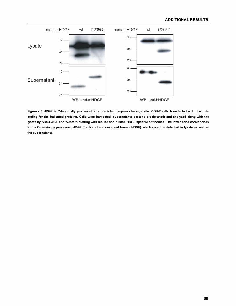
ADDITIONAL RESULTS
88
43
34
26
43
34
26
43
34
26
43
34
26
mouse HDGF wt D205G human HDGF wt G205D
Lysate
Supernatant
WB: anti-mHDGF WB: anti-hHDGF Figure 4.3 HDGF is C-terminally processed at a predicted caspase cleavage site. COS-7 cells transfected with plasmids coding for the indicated proteins. Cells were harvested; supernatants acetone precipitated; and analyzed along with the lysate by SDS-PAGE and Western blotting with mouse and human HDGF specific antibodies. The lower band corresponds to the C-terminally processed HDGF (for both the mouse and human HDGF) which could be detected in lysate as well as the supernatants.
DISCUSSION
89
55 DDiissccuussssiioonn
Post-translational modifications (PTM’s) are modifications occurring on proteins, like enzyme-catalyzed
covalent additions; structural changes; and proteolytic cleavage of peptide backbones. So far, more than
200 PTM’s have been characterized. PTM’s modulate the function of most eukaryotic proteins by altering
their activity state, localization, turnover, and interactions with other proteins. HDGF is a nuclear-targeted
mitogen implicated in fetal development, cancer progression and organ remodeling. HDGF was predicted
to be modified by phosphorylation on serine and threonine residues and mass spectroscopy based
approach also confirmed the use of serine 132, 133 and 165 [58]. Furthermore, HDGF is rapidly
dephosphorylated in response to apoptotic inducers in endothelial cells [53]. However, conclusive
evidence for these modifications on HDGF and their effects on its function has not yet been determined. It
is therefore important to establish how HDGF activity is regulated by PTM’s. The present thesis is based
on three major projects described in the introduction and undertaken in publications 1 to 3, addressing the
question of PTM’s of HDGF.
5.1 SUMOylation of HDGF SUMO, a ~10kDa protein, is a member of the ubiquitin-like protein super-family and is post-translationally
conjugated to various cellular proteins in a process that is mechanistically analogous to ubiquitination.
Chemically, SUMOylation involves the formation of an iso-peptide bond between the C-terminal carboxyl
group of a mature SUMO and the -amino group of a lysine residue in the substrate protein. The target
lysine residue is generally embedded within an internal motif with the consensus KXE/D (where is any
hydrophobic amino acid; K is lysine; X can be any amino acid; E is glutamate and D is aspartate) that is
conserved in most, but not all, proteins that are targeted by SUMO-1 [59]. So far, four 10-11 kDa SUMO
isoforms in mammals have been described. SUMO-1 exhibits 47% similarity at the protein level with
SUMO-2 and -3, whereas SUMO-2 and -3 share 95% similarity [57]. Since the identification of the first
SUMO-modified substrate, RanGAP, in 1996 [60], a large number of proteins have been shown to be
modified by SUMO and new substrates for SUMO-modification continue to be identified. Many of the
known SUMO-modified proteins are located in the nucleus with important roles in regulating transcription,
DISCUSSION
90
chromatin structure assembly, and DNA repair. PTM by SUMO is also important for regulation and/or
localization of proteins with roles in cell proliferation, differentiation, and apoptosis.
We demonstrated that HDGF is modified by the attachment of SUMO-1 in mammalian cells
(Publication 1). Our search for the SUMO-1 acceptor sites in HDGF did not reveal a clear SUMOylation
consensus sequence �KxE/D. Moreover, mutation of all predicted SUMOylation sites did not lead to the
loss of SUMOylation. This was coupled with the problem of a high number of lysine residues in HDGF
making the search time consuming. We overcame this problem by expressing HDGF in bacteria together
with components of the SUMO-1 conjugating system [61]; and later analyzed the SUMO-conjugated HDGF
peptide in a mass spectrometry based analysis, to identify the lysine residue used for the conjugation of
SUMO-1 as K80 in the non-consensus motif RK80GF. In principle, SUMOylation can have varied functional
consequences for HDGF. Previous studies showed that the PWWP domain of HDGF is sufficient to bind
DNA in vitro [27, 28]. To check for the effect of SUMOylation on HDGF ability to bind DNA, we performed
chromatin binding assay and were able to show that unSUMOylated HDGF indeed binds to chromatin or
chromatin-associated proteins. In contrast, SUMOylated HDGF is excluded from the chromatin-bound
fraction and was only found in the cytoskeleton and nuclear matrix proteins containing fraction
As mentioned previously, there are four SUMO isoforms in mammals. For a few substrates, isoform
preferences have been documented. RanGAP1 is modified almost exclusively by SUMO-1 in vivo [62],
while other substrates, such as the pro-myelocytic leukemia protein (PML), have been reported to be
conjugated with both SUMO-1 [63] and SUMO-2/3 [64]. Analysis of functional differences between the
SUMO isoforms in mammalian cells has been previously studied by comparing the localization and
dynamic behavior of YFP-SUMO fusion proteins [65]. The results showed that YFP-SUMO-1 behaves in a
manner that is highly distinct from either YFP-SUMO-2 or YFP-SUMO-3. Although the pattern of
localization of the isoforms was overlapping in the nucleoplasm and in PML bodies, YFP-SUMO-1 was
also uniquely localized to nucleoli, the nuclear envelope, and cytoplasmic foci. Furthermore, YFP-SUMO-1
showed different dynamics from YFP-SUMO- 2 or -3, with slower rates of both recovery and depletion, and
also had substantially different responses to physiological stimuli, like heat stress, a known fact from
previous SUMO modified protein studies [62]. These findings make it intriguing to study the complexity of
SUMO modification on HDGF. To access whether HDGF can be SUMOylated by different SUMO isoforms,
we performed co-expression experiments and demonstrated that SUMO-2 and SUMO-3 are also able to
modify HDGF. Moreover, they utilize the same non-consensus motif RK80GF for SUMOylation (refer Section 4.1; Figure 4.1). The functional relevance of this modification however needs to be elucidated. It
would be interesting to check if there is competition or preference for the use of the site between the
SUMO isoforms and if modification of HDGF by different isoforms leads to different functional
consequences.
DISCUSSION
91
5.2 Phosphorylation dependent regulation of HDGF secretion and processing Considering its implicated involvement in fetal development, cancer progression and organ remodeling, it
is necessary to understand the mechanism of HDGF secretion and its regulation. In this part of the study
we focused on HDGF secretion with respect to PTM. We assessed HDGF wt and S�A mutants for
differences in expression and secretion. Surprisingly, we could observe a loss of secretion if a
phosphorylation site, serine 165, was mutated. Additionally, we have shown that HDGF secretion is
attributed to processing of N-terminal region, which is also influenced by the loss of serine 165
phosphorylation site (Publication 2). A kinase that might be involved in phosphorylation of HDGF at position S165 can be the mitogen-activated
protein kinases (MAPK). MAPKs are serine/threonine kinases that tend to phosphorylate their substrates
on S/T-P motif. MAPK signaling pathways are characterized by a cascade of multiple kinases that
contribute to regulate a variety of extracellular stimuli, thereby control the cellular environment. The MAPK
family includes the ERKs (extracellular signal-regulated kinase), the JNKs (c-Jun N-terminal kinase), the
p38 MAPKs and ERK5. The ERKs generally regulate cellular proliferation and differentiation processes in
response to growth factors and hormones; while the JNKs and p38 MAPKs are primarily activated in
response to extracellular stresses, such as U.V. irradiation, osmotic stress or inflammatory stimuli [66]. As
the sequence S/T-P is found in ~ 80% of all proteins, additional factors are required to direct individual
kinases towards the correct substrates [67]. MAPKs ensure their substrate specificity by interaction
through docking motifs, short amino acid stretches located on MAPK-interacting proteins. The classic
MAPK docking site was first identified in c-JUN, a substrate of JNK [68]. It is characterized by a cluster of
at least two positively charged amino acids followed by a spacer of 2-to-6 residues from a
hydrophobic-X-hydrophobic sequence, where the hydrophobic residues are long chain aliphatics (usually
Leu, Ile). Both in the spacer and in the sequence immediately C-terminal to the
hydrophobic-X-hydrophobic element, there is a high tendency for the presence of Pro, Asn, and/or Gly,
which are residues that are both turn-forming and helix-breaking [69]. Interestingly, such a MAPK docking
motif is predicted with 100% identity for amino acids 8KEYKCGDLVF17 in HDGF (ELM server to identify
functional sites on proteins (http://elm.eu.org/)). Moreover, S165 in HDGF is a part of a SP motif which
might be phosphorylated by a MAPK. Supporting this speculation, it has already been shown that HDGF
can activate Erk1/2, a member of the MAPK family. The study suggested that HDGF is involved in the
carcinogenesis of gastric epithelial cells and promotes cell proliferation by Erk1/2 activation [44].
Furthermore, we have demonstrated that HDGF secretion is attributed to a processing of N-terminal region
of the protein and the processing of HDGF S165A mutant leads to a loss of 13 or more amino acids
(analyzed by N-terminal amino acid sequencing). This would lead to the loss of the potential MAPK
docking site.
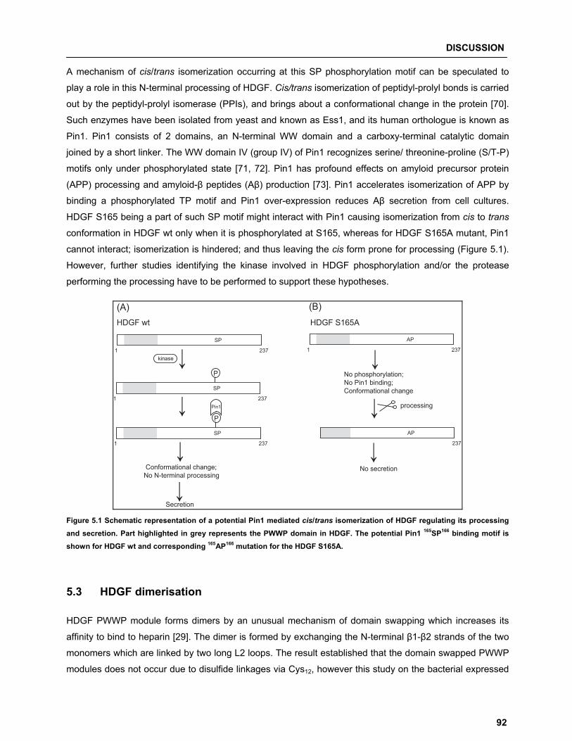
DISCUSSION
92
A mechanism of cis/trans isomerization occurring at this SP phosphorylation motif can be speculated to
play a role in this N-terminal processing of HDGF. Cis/trans isomerization of peptidyl-prolyl bonds is carried
out by the peptidyl-prolyl isomerase (PPIs), and brings about a conformational change in the protein [70].
Such enzymes have been isolated from yeast and known as Ess1, and its human orthologue is known as
Pin1. Pin1 consists of 2 domains, an N-terminal WW domain and a carboxy-terminal catalytic domain
joined by a short linker. The WW domain IV (group IV) of Pin1 recognizes serine/ threonine-proline (S/T-P)
motifs only under phosphorylated state [71, 72]. Pin1 has profound effects on amyloid precursor protein
(APP) processing and amyloid-� peptides (A�) production [73]. Pin1 accelerates isomerization of APP by
binding a phosphorylated TP motif and Pin1 over-expression reduces A� secretion from cell cultures.
HDGF S165 being a part of such SP motif might interact with Pin1 causing isomerization from cis to trans
conformation in HDGF wt only when it is phosphorylated at S165, whereas for HDGF S165A mutant, Pin1
cannot interact; isomerization is hindered; and thus leaving the cis form prone for processing (Figure 5.1).
However, further studies identifying the kinase involved in HDGF phosphorylation and/or the protease
performing the processing have to be performed to support these hypotheses.
1 237
SP
1 237
SP
P
↓kinase
1 237
SP
P
Pin1
Conformational change;No N-terminal processing
Secretion
1 237
AP
No phosphorylation;No Pin1 binding;Conformational change
237
AP
No secretion
processing
HDGF wt HDGF S165A
(B) (A)
↓
↓
↓
↓
↓
↓
Figure 5.1 Schematic representation of a potential Pin1 mediated cis/trans isomerization of HDGF regulating its processing and secretion. Part highlighted in grey represents the PWWP domain in HDGF. The potential Pin1 165SP166 binding motif is shown for HDGF wt and corresponding 165AP166 mutation for the HDGF S165A.
5.3 HDGF dimerisation HDGF PWWP module forms dimers by an unusual mechanism of domain swapping which increases its
affinity to bind to heparin [29]. The dimer is formed by exchanging the N-terminal �1-�2 strands of the two
monomers which are linked by two long L2 loops. The result established that the domain swapped PWWP
modules does not occur due to disulfide linkages via Cys12, however this study on the bacterial expressed
DISCUSSION
93
PWWP module and excludes the Cys108. Evidence for the existence of a disulfide bond was provided in the
original study for HDGF. The mitogenic activity of secreted HDGF was completely lost after treatment by
reducing agent [6]. We have demonstrated in our study that HDGF is able to form intra- and
inter-molecular disulfide bonds (Publication 2). We provided evidence for HDGF dimerisation is mediated
exclusively via Cys108, and N-terminal processing in the S165A mutant or N-terminal truncation of HDGF
favors dimer formation. The results suggest that phosphorylation of Ser165 in HDGF is required to introduce
a structural change in the molecule which might then lead to an interaction of the C-terminal and
N-Terminal regions, and further to the formation of an intra-molecular disulfide bond between Cys12 and
Cys108. In the HDGF S165A mutant the initial structural change cannot occur (possibly by the absence of
the motif recognized by a PPI like Pin1) resulting in a loss of the C- and N-terminal interaction and the
intra-molecular disulfide bond. Based on these results we propose a model illustrating the possible
mechanism (refer Publication 2; Figure 6). N- and C-terminal interactions have been demonstrated
HMGB-1, a closely related protein to HDGF, and play a crucial role in maintaining its structure and
function. The study revealed that the HMGB-1 acidic C-terminal domain interacts with the basic N-terminal
domain and the intramolecular interaction between the two oppositely charged termini is enhanced when
serine residues in the acidic tail of HMGB-1 are phosphorylated [74].
In summary, the N-terminal amino acids of HDGF appear to be responsible to mediate non-classical
secretion of HDGF. Furthermore, a serine 165 is essential for maintaining N-terminal part, probably by
influencing the structure of HDGF.
5.4 HDGF�HRP-2 interaction 5.4.1 Differential expression of HRP-2 correspond to alternatively spliced isoforms Tissue specific data for expression of HRP-2 was only available at the RNA level which shows ubiquitous
distribution [2], while area and cell type specific expression of HRP-2 in the rat brain is also demonstrated
at the protein level [14]. In this study we have shown differential expression of HRP-2 protein in brain in a
developmental dependent manner (Publication 3). HRP-2 has a wide distribution in cerebrum, heart,
muscle, liver, frontal lobe, thymus whereas low levels of expression in lung and spleen tissue (Dietz et al.,
unpublished data). By screening mouse brain cDNA; along with the two variants already described in the
database (referred in this study HRP-2 isoform a and b); we could also identify a new variant of HRP-2
(referred in this study as HRP-2 isoform c). The three different isoforms arise from alternative splicing
(Publication 3). The newly discovered isoform is a 616 amino acid protein which shows a deletion in the
conserved hath region. The 53 amino acid deletion; spanning from aa 66-118; might affect the proper
folding of the PWWP domain, which has been suggested as a protein-protein interaction domain. The only
other member of the family for which presence of spliced variants has been described in the literature is
DISCUSSION
94
LEDGF, occurring as LEDGF/p75 and p52 [75]. All the three HRP-2 isoforms contain two putative
helix-turn-helix motifs. The helix-turn-helix motifs described for LEDGF, which are reported to be essential
for DNA binding and trans-activation [76], show a high degree of similarity to the putative motifs in HRP-2.
The three HRP-2 isoforms displaying the occurrence of the putative helix-turn-helix motifs may well
function like LEDGF in modulating gene expression. Also, the presence of a mixed charge cluster, a
characteristic of eukaryotic regulatory proteins, makes it plausible that HRP-2 functions as regulatory
protein [2]. In vitro as well as in vivo cells expressing LEDGF exhibit increased resistance to environmental
stress and improved survival [76]. Also, LEDGF is produced at higher levels under thermal and oxidative
stress [77] and is important in up-regulating stress-associated genes by binding to the stress response
element and heat shock elements present in these genes [76]. Considering its high similarity to LEDGF,
HRP-2 might also play a role in defense mechanisms of cells against stress. In future experiments should
be carried out to test the role of HRP-2 in the stress response of cells. The appearance of various bands in
tissue may be attributed to the regulated function of different HRP-2 isoforms and further studies in this
area should be undertaken to investigate the precise roles played by the various isoforms in different
tissues and stages.
5.4.2 HDGF/HRP-2 interaction studies As mentioned before, HDGF can form domain swapped dimers derived from the interaction of the
structured N-terminal part [29]. Sequence and structural identity between N-terminal regions of all HRPs
family proteins raises the possibility for the interplay and heteromerisation between different HRP
members. Moreover, a recent study indicates that HDGF acts as a transcriptional co-repressor for SMYD1
gene expression by recruiting CtBP and this interaction is critical for nuclear function of HDGF [78]. It might
be that HRP-2/HDGF heteromerisation acts in similar manner modulating DNA binding to specific targets
and regulate gene expression. By co-precipitation studies we were able to show that HRP-2 and HDGF
interact and the new HRP-2 isoform c can additionally interact with a lower molecular mass form of HDGF,
which correlates with the N-terminally processed HDGF S165A mutant (Publication 3). From our results we hypothesize that protein-protein interaction between different members of the HRP
family might play a very important role in their functions. This finding opens a new perspective in the field
of HRP family research.
DISCUSSION
95
5.5 Caspase dependent cleavage of HDGF Caspases are a family of cysteine proteases expressed in virtually all animal cells and play an essential
role in many forms of cell death by apoptosis [79]. A total of 12 cysteine proteases known as caspases
have been identified in mammals: caspase-1 to -10, caspase-12 and caspase-14. [80]. There are two main
pathways of caspase activation leading to apoptotic cell death. The intrinsic pathway; which involves
proteins of the Bcl-2 family, which in response to a cellular damage migrate to the mitochondria to either
antagonize anti-apoptotic proteins or activate multi-domain pro-apoptotic proteins Bax and Bak [81]; and
the extrinsic pathway, which starts at the plasma membrane by engagement of transmembrane death
receptors such as Fas/CD95, tumor necrosis factor receptor 1 (TNF-R1) and TNF-related
apoptosis-inducing ligand (TRAIL) receptors. Multi-protein complexes are formed in which caspases are
recruited by the adaptor molecule Fas-associated death domain protein (FADD) and activated. In turn,
these enzymes either directly activate the caspase cascade or connect the extrinsic and the intrinsic
pathway [82]. Previous studies suggests that HDGF is involved in apoptosis and also is regulated by
caspase dependent mechanism [53]. Knock-down of HDGF not only induced apoptosis in human cancer
cells through the Bad-mediated intrinsic apoptotic pathway, but also the Fas-mediated extrinsic apoptotic
pathway suppressing anchorage-independent growth of cancer cells [54, 55]. RNAi-induced silencing of
the HDGF prevented the release of pro-apoptotic factors, Smac/Diablo, from the mitochondria upon TNF-�
treatment [52]. Triggering endothelial cell apoptosis by TNF� and cycloheximide leads to an early
dephosphorylation of HDGF before mitochondrial membrane permeabilization and downstream from an
initiator caspase [53]. All these studies suggest involvement and regulation of HDGF by components of the
apoptotic pathway.
Based on our previous knowledge for over-expression of mouse HDGF wt we always observed a
characteristic double band pattern in the total cell lysate exclusively in COS-7 cells (refer Section 4.2, Figure 4.2(a)). This pattern can be only observed for over-expression of the untagged mouse HDGF wt,
but interestingly not for the untagged human HDGF wt (data not shown). This lead to the hypothesis that
occurrence of such a pattern is not unspecific or not derived from degradation, but involves a specific
cleavage product upon over-expression. Our results show that the two forms differ by 4 kDa; the lower
molecular mass form is C-terminally processed; and it is preferentially secreted and suggest that
C-terminal cleavage of mouse HDGF might have a regulated function under specific conditions. It is
important to note that, the lower molecular mass form of HDGF observed here is not same as the
processed form of HDGF demonstrated earlier which is observed in regulation of HDGF secretion.
Considering the difference in the molecular mass in HDGF, we searched for cleavage motifs which might
be relevant and lead us to search of a potential caspase cleavage site in the C-terminal region of HDGF.
Caspases typically recognize tetra-peptide (P4-P3-P2-P1) motifs in their substrates and cleave between the
P1 amino acid residue and the adjacent C-terminal amino acid in the peptide chain. Analysis of mouse
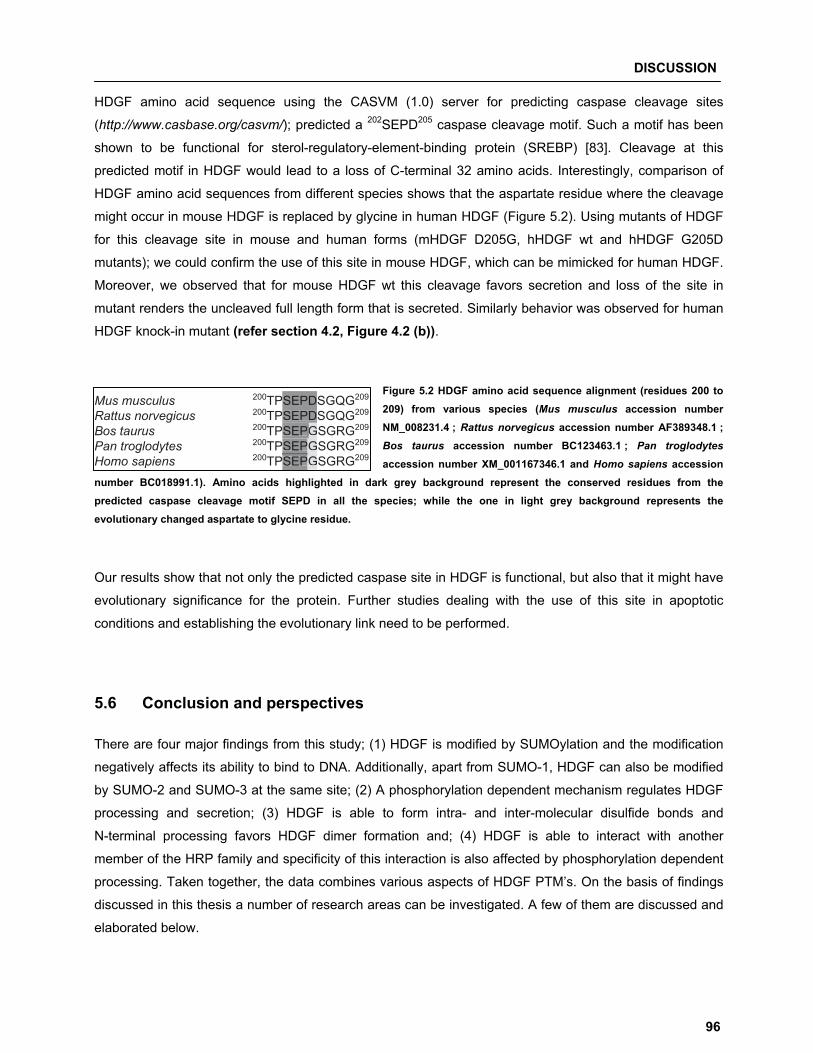
DISCUSSION
96
HDGF amino acid sequence using the CASVM (1.0) server for predicting caspase cleavage sites
(http://www.casbase.org/casvm/); predicted a 202SEPD205 caspase cleavage motif. Such a motif has been
shown to be functional for sterol-regulatory-element-binding protein (SREBP) [83]. Cleavage at this
predicted motif in HDGF would lead to a loss of C-terminal 32 amino acids. Interestingly, comparison of
HDGF amino acid sequences from different species shows that the aspartate residue where the cleavage
might occur in mouse HDGF is replaced by glycine in human HDGF (Figure 5.2). Using mutants of HDGF
for this cleavage site in mouse and human forms (mHDGF D205G, hHDGF wt and hHDGF G205D
mutants); we could confirm the use of this site in mouse HDGF, which can be mimicked for human HDGF.
Moreover, we observed that for mouse HDGF wt this cleavage favors secretion and loss of the site in
mutant renders the uncleaved full length form that is secreted. Similarly behavior was observed for human
HDGF knock-in mutant (refer section 4.2, Figure 4.2 (b)).
Figure 5.2 HDGF amino acid sequence alignment (residues 200 to 209) from various species (Mus musculus accession number NM_008231.4 ; Rattus norvegicus accession number AF389348.1 ;Bos taurus accession number BC123463.1 ; Pan troglodytes
accession number XM_001167346.1 and Homo sapiens accession number BC018991.1). Amino acids highlighted in dark grey background represent the conserved residues from the predicted caspase cleavage motif SEPD in all the species; while the one in light grey background represents the evolutionary changed aspartate to glycine residue.
Our results show that not only the predicted caspase site in HDGF is functional, but also that it might have
evolutionary significance for the protein. Further studies dealing with the use of this site in apoptotic
conditions and establishing the evolutionary link need to be performed.
5.6 Conclusion and perspectives There are four major findings from this study; (1) HDGF is modified by SUMOylation and the modification
negatively affects its ability to bind to DNA. Additionally, apart from SUMO-1, HDGF can also be modified
by SUMO-2 and SUMO-3 at the same site; (2) A phosphorylation dependent mechanism regulates HDGF
processing and secretion; (3) HDGF is able to form intra- and inter-molecular disulfide bonds and
N-terminal processing favors HDGF dimer formation and; (4) HDGF is able to interact with another
member of the HRP family and specificity of this interaction is also affected by phosphorylation dependent
processing. Taken together, the data combines various aspects of HDGF PTM’s. On the basis of findings
discussed in this thesis a number of research areas can be investigated. A few of them are discussed and
elaborated below.
Mus musculus 200TPSEPDSGQG209
Rattus norvegicus 200TPSEPDSGQG209
Bos taurus 200TPSEPGSGRG209
Pan troglodytes 200TPSEPGSGRG209
Homo sapiens 200TPSEPGSGRG209
GSEPDSEPSEPG
G
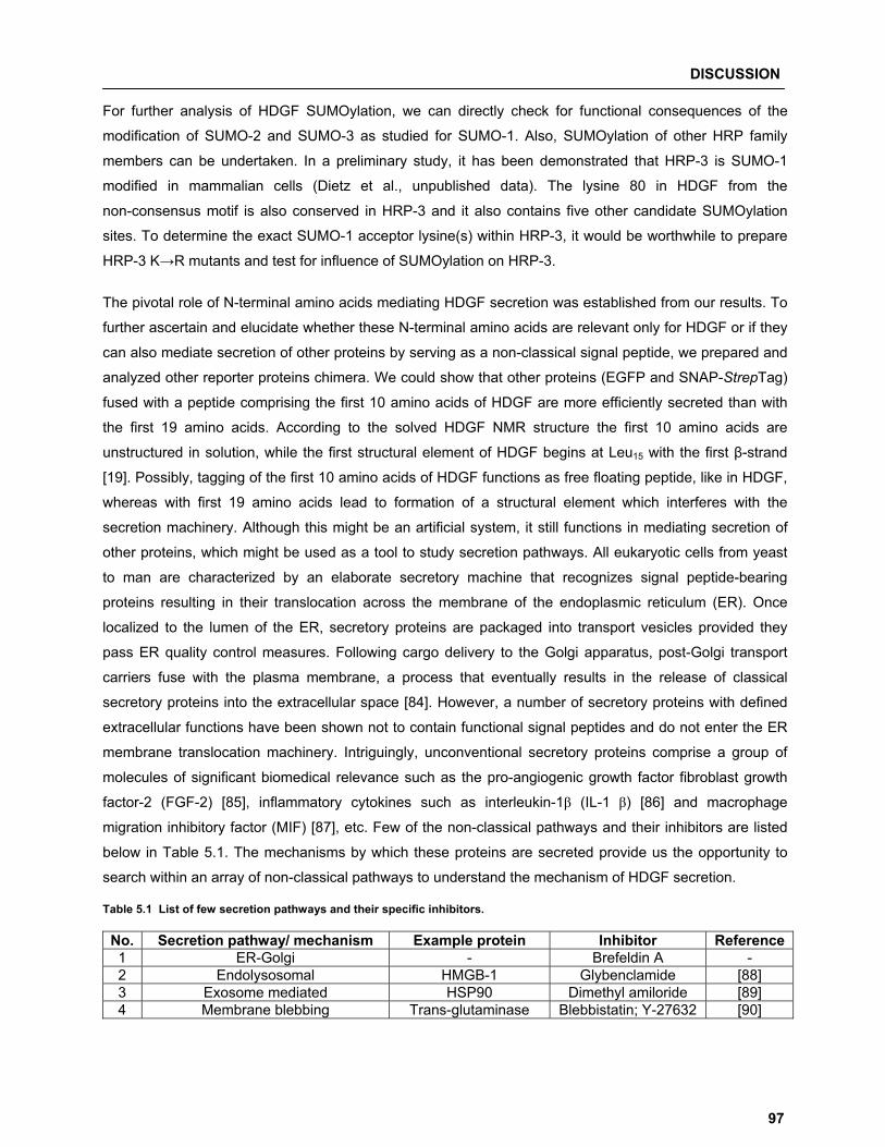
DISCUSSION
97
For further analysis of HDGF SUMOylation, we can directly check for functional consequences of the
modification of SUMO-2 and SUMO-3 as studied for SUMO-1. Also, SUMOylation of other HRP family
members can be undertaken. In a preliminary study, it has been demonstrated that HRP-3 is SUMO-1
modified in mammalian cells (Dietz et al., unpublished data). The lysine 80 in HDGF from the
non-consensus motif is also conserved in HRP-3 and it also contains five other candidate SUMOylation
sites. To determine the exact SUMO-1 acceptor lysine(s) within HRP-3, it would be worthwhile to prepare
HRP-3 K�R mutants and test for influence of SUMOylation on HRP-3.
The pivotal role of N-terminal amino acids mediating HDGF secretion was established from our results. To
further ascertain and elucidate whether these N-terminal amino acids are relevant only for HDGF or if they
can also mediate secretion of other proteins by serving as a non-classical signal peptide, we prepared and
analyzed other reporter proteins chimera. We could show that other proteins (EGFP and SNAP-StrepTag)
fused with a peptide comprising the first 10 amino acids of HDGF are more efficiently secreted than with
the first 19 amino acids. According to the solved HDGF NMR structure the first 10 amino acids are
unstructured in solution, while the first structural element of HDGF begins at Leu15 with the first �-strand
[19]. Possibly, tagging of the first 10 amino acids of HDGF functions as free floating peptide, like in HDGF,
whereas with first 19 amino acids lead to formation of a structural element which interferes with the
secretion machinery. Although this might be an artificial system, it still functions in mediating secretion of
other proteins, which might be used as a tool to study secretion pathways. All eukaryotic cells from yeast
to man are characterized by an elaborate secretory machine that recognizes signal peptide-bearing
proteins resulting in their translocation across the membrane of the endoplasmic reticulum (ER). Once
localized to the lumen of the ER, secretory proteins are packaged into transport vesicles provided they
pass ER quality control measures. Following cargo delivery to the Golgi apparatus, post-Golgi transport
carriers fuse with the plasma membrane, a process that eventually results in the release of classical
secretory proteins into the extracellular space [84]. However, a number of secretory proteins with defined
extracellular functions have been shown not to contain functional signal peptides and do not enter the ER
membrane translocation machinery. Intriguingly, unconventional secretory proteins comprise a group of
molecules of significant biomedical relevance such as the pro-angiogenic growth factor fibroblast growth
factor-2 (FGF-2) [85], inflammatory cytokines such as interleukin-1� (IL-1 �) [86] and macrophage
migration inhibitory factor (MIF) [87], etc. Few of the non-classical pathways and their inhibitors are listed
below in Table 5.1. The mechanisms by which these proteins are secreted provide us the opportunity to
search within an array of non-classical pathways to understand the mechanism of HDGF secretion.
Table 5.1 List of few secretion pathways and their specific inhibitors.
No. Secretion pathway/ mechanism Example protein Inhibitor Reference 1 ER-Golgi - Brefeldin A - 2 Endolysosomal HMGB-1 Glybenclamide [88] 3 Exosome mediated HSP90 Dimethyl amiloride [89] 4 Membrane blebbing Trans-glutaminase Blebbistatin; Y-27632 [90]
DISCUSSION
98
The discovery of new HRP-2 splice variants and their differential expression opens a way for better
understanding the HRP family functions. Confirming the expression of splice variants using alternative
method like real-time PCR, and analyzing the functional behavior of the various isoforms are steps in
future direction. Estimating the relative abundance of each variant is also necessary in studying the
biological functions of the isoforms. We have shown that there is interaction of HRP-2 isoforms with HDGF.
It might be that the interaction within the HRP family acts as central regulator controlling the mode of action
of these proteins. The consequences of such an interaction on cellular pathways offer an interesting area
of research.
Our results demonstrate that HDGF can also be cleaved at the C-terminus at a potential caspase cleavage
site 202SEPD205. However, drawing a conclusive link between the cleavage of a particular protein and a
cellular change that takes place has not been straightforward. While the cleavage of certain proteins is
probably vital to achieve the controlled and complete collapse of the cell, the cleavage of many other
proteins may ultimately be of little consequence to the death process. Such “bystander substrates” may
simply contain the appropriate caspase-consensus cleavage site(s) by chance alone [91]. Also, a number
of proteins with the ability to repress the cell death program have been discovered. Perhaps the most well
known of these is the Bcl-2 protein [92]. A whole family of Bcl-2 related proteins have been discovered,
some of which are repressors of apoptosis while others, paradoxically, can promote entry into the death
program. Many proteins that block cell death appear to do so by protecting endogenous substrates from
the destructive action of the caspases by offering themselves up for proteolysis, often neutralizing the
activity of the caspase. In the case of Bcl-2, many binding partners have been discovered in recent years.
One study indicates that the apoptosis induced by knock-down of HDGF is mediated by Bad
(a pro-apoptotic Bcl-2 family protein), which trigger apoptosis by dimerising with Bcl-2 and Bcl-xL, and
neutralize their anti-apoptotic effects. This suggests role of HDGF as a survival factor, although the
mechanism to regulate the intracellular signaling pathways is yet to be defined [54]. Investigating the
caspase involved in HDGF cleavage, its functional effect on HDGF and checking for HDGF ability to act as
cell survival factor by acting in co-operation with other repressor proteins such as Bcl-2 should be a
pursued in the future.
Finally, it can be concluded that the advances in the understanding of post-translational modifications of
HDGF demonstrated in this thesis, would lead to the realization of importance of these modifications to
control and regulate HDGF and HRP family protein functions.

66 RReeffeerreenncceess
REFERENCES
100
1. Nakamura H, Izumoto Y, Kambe H, Kuroda T, Mori T, Kawamura K, Yamamoto H & Kishimoto T
(1994) Molecular cloning of complementary DNA for a novel human hepatoma-derived growth factor. Its
homology with high mobility group-1 protein. J Biol Chem 269, 25143-25149.
2. Izumoto Y, Kuroda T, Harada H, Kishimoto T & Nakamura H (1997) Hepatoma-derived growth
factor belongs to a gene family in mice showing significant homology in the amino terminus. Biochem
Biophys Res Commun 238, 26-32.
3. Ikegame K, Yamamoto M, Kishima Y, Enomoto H, Yoshida K, Suemura M, Kishimoto T &
Nakamura H (1999) A new member of a hepatoma-derived growth factor gene family can translocate to
the nucleus. Biochem Biophys Res Commun 266, 81-87.
4. Ge H, Si Y & Roeder RG (1998) Isolation of cDNAs encoding novel transcription coactivators p52
and p75 reveals an alternate regulatory mechanism of transcriptional activation. EMBO J 17, 6723-6729.
5. Dietz F, Franken S, Yoshida K, Nakamura H, Kappler J & Gieselmann V (2002) The family of
hepatoma-derived growth factor proteins: characterization of a new member HRP-4 and classification of
its subfamilies. Biochem J 366, 491-500.
6. Nakamura H, Kambe H, Egawa T, Kimura Y, Ito H, Hayashi E, Yamamoto H, Sato J & Kishimoto
S (1989) Partial purification and characterization of human hepatoma-derived growth factor. Clin Chim
Acta 183, 273-284.
7. Oliver JA & Al-Awqati Q (1998) An endothelial growth factor involved in rat renal development.
J Clin Invest 102, 1208-1219.
8. Everett AD, Lobe DR, Matsumura ME, Nakamura H & McNamara CA (2000) Hepatoma-derived
growth factor stimulates smooth muscle cell growth and is expressed in vascular development. J Clin
Invest 105, 567-575.
9. Kishima Y, Yoshida K, Enomoto H, Yamamoto M, Kuroda T, Okuda Y, Uyama H & Nakamura H
(2002) Antisense oligonucleotides of hepatoma-derived growth factor (HDGF) suppress the proliferation of
hepatoma cells. Hepatogastroenterology 49, 1639-1644.
10. Everett AD, Stoops T & McNamara CA (2001) Nuclear targeting is required for hepatoma-derived
growth factor-stimulated mitogenesis in vascular smooth muscle cells. J Biol Chem 276, 37564-37568.
11. Kishima Y, Yamamoto H, Izumoto Y, Yoshida K, Enomoto H, Yamamoto M, Kuroda T, Ito H,
Yoshizaki K & Nakamura H (2002) Hepatoma-derived growth factor stimulates cell growth after
translocation to the nucleus by nuclear localization signals. J Biol Chem 277, 10315-10322.
12. Mosevitsky MI, Novitskaya VA, Iogannsen MG & Zabezhinsky MA (1989) Tissue specificity of
nucleo-cytoplasmic distribution of HMG1 and HMG2 proteins and their probable functions. Eur J Biochem
185, 303-310.
13. Cherepanov P, Devroe E, Silver PA & Engelman A (2004) Identification of an evolutionarily
conserved domain in human lens epithelium-derived growth factor/transcriptional co-activator p75
(LEDGF/p75) that binds HIV-1 integrase. J Biol Chem 279, 48883-48892.
REFERENCES
101
14. El-Tahir HM, Dietz F, Dringen R, Schwabe K, Strenge K, Kelm S, Abouzied MM, Gieselmann V &
Franken S (2006) Expression of hepatoma-derived growth factor family members in the adult central
nervous system. BMC Neurosci 7, 6.
15. El-Tahir HM, Abouzied MM, Gallitzendoerfer R, Gieselmann V & Franken S (2009)
Hepatoma-derived growth factor-related protein-3 interacts with microtubules and promotes neurite
outgrowth in mouse cortical neurons. J Biol Chem 284, 11637-11651.
16. Engelman A & Cherepanov P (2008) The lentiviral integrase binding protein LEDGF/p75 and
HIV-1 replication. PLoS Pathog 4, e1000046.
17. Sutherland HG, Newton K, Brownstein DG, Holmes MC, Kress C, Semple CA & Bickmore WA
(2006) Disruption of Ledgf/Psip1 results in perinatal mortality and homeotic skeletal transformations. Mol
Cell Biol 26, 7201-7210.
18. Fatma N, Singh DP, Shinohara T & Chylack LT, Jr. (2001) Transcriptional regulation of the
antioxidant protein 2 gene, a thiol-specific antioxidant, by lens epithelium-derived growth factor to protect
cells from oxidative stress. J Biol Chem 276, 48899-48907.
19. Sue SC, Chen JY, Lee SC, Wu WG & Huang TH (2004) Solution structure and heparin interaction
of human hepatoma-derived growth factor. J Mol Biol 343, 1365-1377.
20. Abouzied MM, Baader SL, Dietz F, Kappler J, Gieselmann V & Franken S (2004) Expression
patterns and different subcellular localization of the growth factors HDGF (hepatoma-derived growth
factor) and HRP-3 (HDGF-related protein-3) suggest functions in addition to their mitogenic activity.
Biochem J 378, 169-176.
21. Sue SC, Chen JY & Huang TH (2004) Sequence specific 1H, 13C and 15N resonance
assignments of the hath-domain of human hepatoma-derived growth factor. J Biomol NMR 29, 95-96.
22. Stec I, Nagl SB, van Ommen GJ & den Dunnen JT (2000) The PWWP domain: a potential
protein-protein interaction domain in nuclear proteins influencing differentiation? FEBS Lett 473, 1-5.
23. Qiu C, Sawada K, Zhang X & Cheng X (2002) The PWWP domain of mammalian DNA
methyltransferase Dnmt3b defines a new family of DNA-binding folds. Nat Struct Biol 9, 217-224.
24. Chen T, Tsujimoto N & Li E (2004) The PWWP domain of Dnmt3a and Dnmt3b is required for
directing DNA methylation to the major satellite repeats at pericentric heterochromatin. Mol Cell Biol 24,
9048-9058.
25. Ge YZ, Pu MT, Gowher H, Wu HP, Ding JP, Jeltsch A & Xu GL (2004) Chromatin targeting of de
novo DNA methyltransferases by the PWWP domain. J Biol Chem 279, 25447-25454.
26. Slater LM, Allen MD & Bycroft M (2003) Structural variation in PWWP domains. J Mol Biol 330,
571-576.
27. Lukasik SM, Cierpicki T, Borloz M, Grembecka J, Everett A & Bushweller JH (2006) High
resolution structure of the HDGF PWWP domain: a potential DNA binding domain. Protein Sci 15,
314-323.
28. Yang J & Everett AD (2007) Hepatoma-derived growth factor binds DNA through the N-terminal
PWWP domain. BMC Mol Biol 8, 101.
REFERENCES
102
29. Sue SC, Lee WT, Tien SC, Lee SC, Yu JG, Wu WJ, Wu WG & Huang TH (2007) PWWP module
of human hepatoma-derived growth factor forms a domain-swapped dimer with much higher affinity for
heparin. J Mol Biol 367, 456-472.
30. Zhou Z, Yamamoto Y, Sugai F, Yoshida K, Kishima Y, Sumi H, Nakamura H & Sakoda S (2004)
Hepatoma-derived growth factor is a neurotrophic factor harbored in the nucleus. J Biol Chem 279,
27320-27326.
31. Marubuchi S, Okuda T, Tagawa K, Enokido Y, Horiuchi D, Shimokawa R, Tamura T, Qi ML, Eishi
Y, Watabe K, Shibata M, Nakagawa M & Okazawa H (2006) Hepatoma-derived growth factor, a new
trophic factor for motor neurons, is up-regulated in the spinal cord of PQBP-1 transgenic mice before
onset of degeneration. J Neurochem 99, 70-83.
32. Hu TH, Huang CC, Liu LF, Lin PR, Liu SY, Chang HW, Changchien CS, Lee CM, Chuang JH &
Tai MH (2003) Expression of hepatoma-derived growth factor in hepatocellular carcinoma. Cancer 98,
1444-1456.
33. Yoshida K, Nakamura H, Okuda Y, Enomoto H, Kishima Y, Uyama H, Ito H, Hirasawa T, Inagaki
S & Kawase I (2003) Expression of hepatoma-derived growth factor in hepatocarcinogenesis.
J Gastroenterol Hepatol 18, 1293-1301.
34. Bernard K, Litman E, Fitzpatrick JL, Shellman YG, Argast G, Polvinen K, Everett AD, Fukasawa
K, Norris DA, Ahn NG & Resing KA (2003) Functional proteomic analysis of melanoma progression.
Cancer Res 63, 6716-6725.
35. Ren H, Tang X, Lee JJ, Feng L, Everett AD, Hong WK, Khuri FR & Mao L (2004) Expression of
hepatoma-derived growth factor is a strong prognostic predictor for patients with early-stage
non-small-cell lung cancer. J Clin Oncol 22, 3230-3237.
36. Iwasaki T, Nakagawa K, Nakamura H, Takada Y, Matsui K & Kawahara K (2005)
Hepatoma-derived growth factor as a prognostic marker in completely resected non-small-cell lung
cancer. Oncol Rep 13, 1075-1080.
37. Hayashi E, Kuramitsu Y, Okada F, Fujimoto M, Zhang X, Kobayashi M, Iizuka N, Ueyama Y &
Nakamura K (2005) Proteomic profiling for cancer progression: Differential display analysis for the
expression of intracellular proteins between regressive and progressive cancer cell lines. Proteomics 5,
1024-1032.
38. Lepourcelet M, Tou L, Cai L, Sawada J, Lazar AJ, Glickman JN, Williamson JA, Everett AD,
Redston M, Fox EA, Nakatani Y & Shivdasani RA (2005) Insights into developmental mechanisms and
cancers in the mammalian intestine derived from serial analysis of gene expression and study of the
hepatoma-derived growth factor (HDGF). Development 132, 415-427.
39. Uyama H, Tomita Y, Nakamura H, Nakamori S, Zhang B, Hoshida Y, Enomoto H, Okuda Y,
Sakon M, Aozasa K, Kawase I, Hayashi N & Monden M (2006) Hepatoma-derived growth factor is a novel
prognostic factor for patients with pancreatic cancer. Clin Cancer Res 12, 6043-6048.
REFERENCES
103
40. Yamamoto S, Tomita Y, Hoshida Y, Takiguchi S, Fujiwara Y, Yasuda T, Doki Y, Yoshida K,
Aozasa K, Nakamura H & Monden M (2006) Expression of hepatoma-derived growth factor is correlated
with lymph node metastasis and prognosis of gastric carcinoma. Clin Cancer Res 12, 117-122.
41. Folkman J (1995) Angiogenesis in cancer, vascular, rheumatoid and other disease. Nat Med 1,
27-31.
42. Okuda Y, Nakamura H, Yoshida K, Enomoto H, Uyama H, Hirotani T, Funamoto M, Ito H, Everett
AD, Hada T & Kawase I (2003) Hepatoma-derived growth factor induces tumorigenesis in vivo through
both direct angiogenic activity and induction of vascular endothelial growth factor. Cancer Sci 94,
1034-1041.
43. Everett AD (2001) Identification, cloning, and developmental expression of hepatoma-derived
growth factor in the developing rat heart. Dev Dyn 222, 450-458.
44. Mao J, Xu Z, Fang Y, Wang H, Xu J, Ye J, Zheng S & Zhu Y (2008) Hepatoma-derived growth
factor involved in the carcinogenesis of gastric epithelial cells through promotion of cell proliferation by
Erk1/2 activation. Cancer Sci 99, 2120-2127.
45. Enomoto H, Yoshida K, Kishima Y, Okuda Y & Nakamura H (2002) Participation of
hepatoma-derived growth factor in the regulation of fetal hepatocyte proliferation. J Gastroenterol 37 Suppl 14, 158-161.
46. Raines EW & Ross R (1993) Smooth muscle cells and the pathogenesis of the lesions of
atherosclerosis. Br Heart J 69, S30-37.
47. Everett AD & Bushweller J (2003) Hepatoma derived growth factor is a nuclear targeted mitogen.
Curr Drug Targets 4, 367-371.
48. Mori M, Morishita H, Nakamura H, Matsuoka H, Yoshida K, Kishima Y, Zhou Z, Kida H, Funakoshi
T, Goya S, Yoshida M, Kumagai T, Tachibana I, Yamamoto Y, Kawase I & Hayashi S (2004)
Hepatoma-derived growth factor is involved in lung remodeling by stimulating epithelial growth. Am J
Respir Cell Mol Biol 30, 459-469.
49. Nakamura H, Yoshida K, Ikegame K, Kishima Y, Uyama H & Enomoto H (2002) Antibodies
against hepatoma-derived growth factor and mucosal repair in ulcerative colitis. J Gastroenterol 37 Suppl 14, 8-14.
50. Enomoto H, Nakamura H, Liu W, Yoshida K, Okuda Y, Imanishi H, Saito M, Shimomura S, Hada
T & Nishiguchi S (2009) Hepatoma-derived growth factor is induced in liver regeneration. Hepatol Res 39,
988-997.
51. Deng Y, Ren X, Yang L, Lin Y & Wu X (2003) A JNK-dependent pathway is required for
TNFalpha-induced apoptosis. Cell 115, 61-70.
52. Machuy N, Thiede B, Rajalingam K, Dimmler C, Thieck O, Meyer TF & Rudel T (2005) A global
approach combining proteome analysis and phenotypic screening with RNA interference yields novel
apoptosis regulators. Mol Cell Proteomics 4, 44-55.
REFERENCES
104
53. Clermont F, Gonzalez NS, Communi D, Franken S, Dumont JE & Robaye B (2008) HDGF is
dephosphorylated during the early steps of endothelial cell apoptosis in a caspase-dependent way. J Cell
Biochem 104, 1161-1171.
54. Tsang TY, Tang WY, Tsang WP, Co NN, Kong SK & Kwok TT (2008) Downregulation of
hepatoma-derived growth factor activates the Bad-mediated apoptotic pathway in human cancer cells.
Apoptosis 13, 1135-1147.
55. Tsang TY, Tang WY, Tsang WP, Co NN, Kong SK & Kwok TT (2009) Mechanistic study on
growth suppression and apoptosis induction by targeting hepatoma-derived growth factor in human
hepatocellular carcinoma HepG2 cells. Cell Physiol Biochem 24, 253-262.
56. Thakar K, Niedenthal R, Okaz E, Franken S, Jakobs A, Gupta S, Kelm S & Dietz F (2008)
SUMOylation of the hepatoma-derived growth factor negatively influences its binding to chromatin.
FEBS J 275, 1411-1426.
57. Kim KI, Baek SH & Chung CH (2002) Versatile protein tag, SUMO: its enzymology and biological
function. J Cell Physiol 191, 257-268.
58. Shu H, Chen S, Bi Q, Mumby M & Brekken DL (2004) Identification of phosphoproteins and their
phosphorylation sites in the WEHI-231 B lymphoma cell line. Mol Cell Proteomics 3, 279-286.
59. Melchior F, Schergaut M & Pichler A (2003) SUMO: ligases, isopeptidases and nuclear pores.
Trends Biochem Sci 28, 612-618.
60. Matunis MJ, Coutavas E & Blobel G (1996) A novel ubiquitin-like modification modulates the
partitioning of the Ran-GTPase-activating protein RanGAP1 between the cytosol and the nuclear pore
complex. J Cell Biol 135, 1457-1470.
61. Uchimura Y, Nakamura M, Sugasawa K, Nakao M & Saitoh H (2004) Overproduction of
eukaryotic SUMO-1- and SUMO-2-conjugated proteins in Escherichia coli. Anal Biochem 331, 204-206.
62. Saitoh H & Hinchey J (2000) Functional heterogeneity of small ubiquitin-related protein modifiers
SUMO-1 versus SUMO-2/3. J Biol Chem 275, 6252-6258.
63. Sternsdorf T, Puccetti E, Jensen K, Hoelzer D, Will H, Ottmann OG & Ruthardt M (1999)
PIC-1/SUMO-1-modified PML-retinoic acid receptor alpha mediates arsenic trioxide-induced apoptosis in
acute promyelocytic leukemia. Mol Cell Biol 19, 5170-5178.
64. Kamitani T, Nguyen HP, Kito K, Fukuda-Kamitani T & Yeh ET (1998) Covalent modification of
PML by the sentrin family of ubiquitin-like proteins. J Biol Chem 273, 3117-3120.
65. Ayaydin F & Dasso M (2004) Distinct in vivo dynamics of vertebrate SUMO paralogues. Mol Biol
Cell 15, 5208-5218.
66. Biondi RM & Nebreda AR (2003) Signalling specificity of Ser/Thr protein kinases through
docking-site-mediated interactions. Biochem J 372, 1-13.
67. Bardwell L (2006) Mechanisms of MAPK signalling specificity. Biochem Soc Trans 34, 837-841.
68. Kallunki T, Deng T, Hibi M & Karin M (1996) c-Jun can recruit JNK to phosphorylate dimerization
partners via specific docking interactions. Cell 87, 929-939.
REFERENCES
105
69. Bardwell AJ, Flatauer LJ, Matsukuma K, Thorner J & Bardwell L (2001) A conserved docking site
in MEKs mediates high-affinity binding to MAP kinases and cooperates with a scaffold protein to enhance
signal transmission. J Biol Chem 276, 10374-10386.
70. Shaw PE (2002) Peptidyl-prolyl isomerases: a new twist to transcription. EMBO Rep 3, 521-526.
71. Sudol M, Sliwa K & Russo T (2001) Functions of WW domains in the nucleus. FEBS Lett 490,
190-195.
72. Landrieu I, Odaert B, Wieruszeski JM, Drobecq H, Rousselot-Pailley P, Inze D & Lippens G
(2001) p13(SUC1) and the WW domain of PIN1 bind to the same phosphothreonine-proline epitope. J Biol
Chem 276, 1434-1438.
73. Pastorino L, Sun A, Lu PJ, Zhou XZ, Balastik M, Finn G, Wulf G, Lim J, Li SH, Li X, Xia W,
Nicholson LK & Lu KP (2006) The prolyl isomerase Pin1 regulates amyloid precursor protein processing
and amyloid-beta production. Nature 440, 528-534.
74. Thomsen MS, Franssen L, Launholt D, Fojan P & Grasser KD (2004) Interactions of the basic
N-terminal and the acidic C-terminal domains of the maize chromosomal HMGB1 protein. Biochemistry
43, 8029-8037.
75. Singh DP, Kimura A, Chylack LT, Jr. & Shinohara T (2000) Lens epithelium-derived growth factor
(LEDGF/p75) and p52 are derived from a single gene by alternative splicing. Gene 242, 265-273.
76. Singh DP, Kubo E, Takamura Y, Shinohara T, Kumar A, Chylack LT, Jr. & Fatma N (2006) DNA
binding domains and nuclear localization signal of LEDGF: contribution of two helix-turn-helix (HTH)-like
domains and a stretch of 58 amino acids of the N-terminal to the trans-activation potential of LEDGF.
J Mol Biol 355, 379-394.
77. Sharma P, Singh DP, Fatma N, Chylack LT, Jr. & Shinohara T (2000) Activation of LEDGF gene
by thermal-and oxidative-stresses. Biochem Biophys Res Commun 276, 1320-1324.
78. Yang J & Everett AD (2009) Hepatoma-derived growth factor represses SET and MYND domain
containing 1 gene expression through interaction with C-terminal binding protein. J Mol Biol 386, 938-950.
79. Nicholson DW & Thornberry NA (1997) Caspases: killer proteases. Trends Biochem Sci 22,
299-306.
80. Salvesen GS & Abrams JM (2004) Caspase activation - stepping on the gas or releasing the
brakes? Lessons from humans and flies. Oncogene 23, 2774-2784.
81. Marsden VS & Strasser A (2003) Control of apoptosis in the immune system: Bcl-2, BH3-only
proteins and more. Annu Rev Immunol 21, 71-105.
82. Launay S, Hermine O, Fontenay M, Kroemer G, Solary E & Garrido C (2005) Vital functions for
lethal caspases. Oncogene 24, 5137-5148.
83. Wang X, Zelenski NG, Yang J, Sakai J, Brown MS & Goldstein JL (1996) Cleavage of sterol
regulatory element binding proteins (SREBPs) by CPP32 during apoptosis. EMBO J 15, 1012-1020.
84. Schatz G & Dobberstein B (1996) Common principles of protein translocation across membranes.
Science 271, 1519-1526.
85. Nugent MA & Iozzo RV (2000) Fibroblast growth factor-2. Int J Biochem Cell Biol 32, 115-120.

REFERENCES
106
86. Braddock M & Quinn A (2004) Targeting IL-1 in inflammatory disease: new opportunities for
therapeutic intervention. Nat Rev Drug Discov 3, 330-339.
87. Lue H, Kleemann R, Calandra T, Roger T & Bernhagen J (2002) Macrophage migration inhibitory
factor (MIF): mechanisms of action and role in disease. Microbes Infect 4, 449-460.
88. Gardella S, Andrei C, Ferrera D, Lotti LV, Torrisi MR, Bianchi ME & Rubartelli A (2002) The
nuclear protein HMGB1 is secreted by monocytes via a non-classical, vesicle-mediated secretory
pathway. EMBO Rep 3, 995-1001.
89. Yu X, Harris SL & Levine AJ (2006) The regulation of exosome secretion: a novel function of the
p53 protein. Cancer Res 66, 4795-4801.
90. Aumuller G, Wilhelm B & Seitz J (1999) Apocrine secretion--fact or artifact? Ann Anat 181,
437-446.
91. Martin SJ & Green DR (1995) Protease activation during apoptosis: death by a thousand cuts?
Cell 82, 349-352.
92. Reed JC (1997) Double identity for proteins of the Bcl-2 family. Nature 387, 773-776.
AA AAppppeennddiixx
APPENDIX
108
A.1 Abbreviations APP amyloid precursor protein ATP adenosine triphosphate BSA bovine serum albumin CAMKII calmodulin independent protein kinase II cAMP cyclic adenosine monophosphate cDNA complementary deoxyribonucleic acid DAPI 4´-6-diamidino-2-phenylindole dihydrochloride dd double distilled dNTP deoxy nucleotide triphophasphate ddNTP dideoxy nucleotide triphophasphate DMEM Dulbecco’s modified Eagle’s medium DNA deoxyribonucleic acid Dnmt DNA methyltransferase homology domain dsDNA double stranded DNA DTT dithiothretol ECL enhanced chemiluminescence EDTA ethylene diamine tetraacetic acid EGFP enhanced green fluorescent protein ERK extracellular signal-regulated kinase FCS fetal calf serum hath homologous to the amino terminus of HDGF HEK human embryonic kidney HDGF hepatoma-derived growth factor HMG-1 high mobility group protein-1 HRP HDGF-related protein HSF-2 heat shock factor-2 IAA iodoacetic acid IL-1� interleukin-1� JNK c-Jun N-terminal kinase KDS potassium dodecyl sulfate kDa kilo Daltons LEDGF lens epithelium-derived growth factor MAPK mitogen-activated protein kinases NEM N-ethylmaleimide NLS nuclear localization signal NMR nuclear magnetic resonance NSCLC non-small cell lung cancer NTP nucleoside triphosphate OD optical density PBS phosphate buffered saline PCNA proliferating cell nuclear antigen PCR polymerase chain reaction PKA protein kinase A PKC protein kinase C PML promyelocytic leukemia protein PPi peptidyl-prolyl isomerase PTM post-translational modifications PVDF polyvinylidene fluoride RanGAP RanGTPase-activating protein RNA ribonucleic acid rpm rounds per minute RT room temperature RT-PCR reverse transcriptase PCR SUMO small ubiquitin-like modifier SDS sodium dodecyl sulfate SDS-PAGE SDS-polyacrylamide gel electrophoresis SMC smooth muscle cell SPR surface plasmon resonance SUMO small ubiquitin-like modifier TBS tris-buffered saline TGF transforming growth factor Ubc-9 ubiquitin conjugating V volts VEGF vascular endothelium growth factor WB western blot wt wild type YFP yellow fluorescent protein
APPENDIX
109
A.2 List of Tables and Figures A.2.1 List of Tables Table 1.1 Hepatoma-derived growth factor related proteins (HRPs)…………………………………. 1
Table 2.1 List of primers used for site directed mutagenesis………………………………………….. 20
Table 2.2 Primary and secondary antibodies used after Western blotting…………………………… 24
Table 5.1 List of few secretion pathways and their specific inhibitors………………………………… 97
A.2.2 List of Figures Figure 1.1 Sequence alignment of the hath regions of HRPs, S. pombe protein SPBC215.07C
(residues 118–225), and Dnmt3b (residues 223–357)……………………………………… 6
Figure 1.2(a) Secondary structure topology of the hath region of human HDGF………………………… 6
Figure 1.2(b) The stereo pairs of the ribbon representation of the structure of the hath domain of
human HDGF……………………………………………………………………………………. 6
Figure 1.3 Secondary structure topology of the domain swapped PWWP dimer……………………. 8
Figure 1.4 Model illustrating the enhanced heparin binding affinity for the PWWP dimer…………... 8
Figure 4.1 HDGF is modified by different SUMO isoforms in mammalian cells………………………. 86
Figure 4.2(a) Expression and secretion of N-StrepTag HDGF in COS-7 cells…………………………... 87
Figure 4.2(b) Amino acid alignment of mouse HDGF wt and human HDGF wt proteins……………….. 87
Figure 4.3 HDGF is C-terminally processed at a predicted caspase cleavage site………………….. 88
Figure 5.1 Schematic representation of a potential Pin1 mediated cis/trans isomerization of
HDGF regulating its processing and secretion………………………………………………. 92
Figure 5.2 HDGF amino acid sequence alignment (residues 200 to 209) from various species……. 96
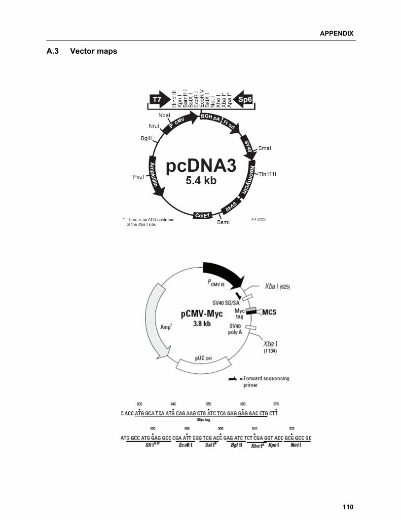
APPENDIX
110
A.3 Vector maps
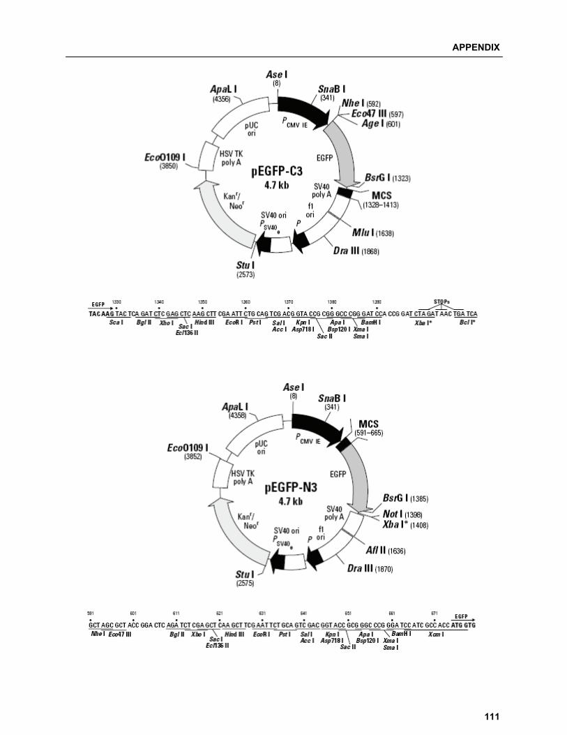
APPENDIX
111

APPENDIX
112
A.4 List of Manufacturers A.4.1 Chemicals and consumables
Item Provider Acrylamide, 30 % (Acrylamid / Bisacrylamid 37, 5:1) Bio-Rad Agarose (SeaKem) Cambrex Ammoniumpersulfate (APS) Serva Ampicillin Roth Amersham Hyperfilm ECL GE healthcare Blotting paper Whatman Bovine Serum Albumin (BSA fraction V) PAA laboratories Bromophenol blue Sigma Cover-slips Starstedt DAPI (4´-6-Diamidino-2-phenylindole dihydrochloride) Fluka Disposable pipette tips Sarstedt Double distilled water (dd H2O) UltraClear UV+ system DMEM (Dulbecco’s modified Eagle’s medium) Invitrogen DpnI endonuclease Fermentas DTT (Dithiothreitol) Fermentas Embedding media Fluka Enhanced ChemiLuminescence (ECL) Amersham Biosciences Epi-cups (1.5 mL /2 mL) Eppendorf Ethanol, 99.8 % Roth Ethylendiamintetraacetatic acid (EDTA) Sigma Ethidium Bromide Sigma ExGen 500 Fermentas Falcon tubes (15 and 50 mL) Sarstedt Fetal calf serum (FCS) Invitrogen Gentamycin, 50 mg/mL PAA laboratories Glycerol, 86-88 % Riedel-deHaen Glycine Riedel-deHaen Hydrochloric acid (HCl) Fischer Scientific Iodoacetamide Sigma Isopropanol (99.7 %) Roth Kanamycin (10,000 mcg/mL) Gibco Magnesium dichloride (Hexahydrate, 99 %) Acros Organics Mass Ruler DNA Ladder Mix (80-10000 base pairs) Fermentas Methanol (99.6 %) Fluka Microcentrifuge tubes, 1.5 mL Sarstedt N-Ethylmaleimide Sigma NP-40 Sigma Parafilm American National Can Pasture pipette (Glass) Brand Pfu DNA polymerase Fermentas Piperazine-1, 4-bis (2-ethanesulfonic acid) (PIPES) Roth Phenylmethylsulfonyl fluoride (PMSF) Sigma Potassium chloride Merck Prestained protein molecular weight marker MBI Fermentas Protease inhibitor Cocktails tabs EDTA free Roche PVDF membrane Hybond P Roth Restriction endonucleases (used in this project) Fermentas Serological pipettes (5, 10 and 25 mL) Sarstedt Skimmed milk Heirler Sodium chloride Fluka Sodium deoxycholate Merck Sodium dodecyl sulfate (SDS) Biomol Sodium hydrogen carbonate Riedel-deHaen Sodium hydroxide Fluka Sucrose Acros Organics T4 DNA ligase Fermentas Taq DNA polymerase Fermentas TEMED (Tetramethylendiamine) Roth Tisuue cell culture plates (10 cm) Sarstedt Tisuue cell culture plates (6, 24 and 96 well) Greiner Tris-(hydoxymethyl-) aminomethane (Tris) Sigma Triton X-100 USB
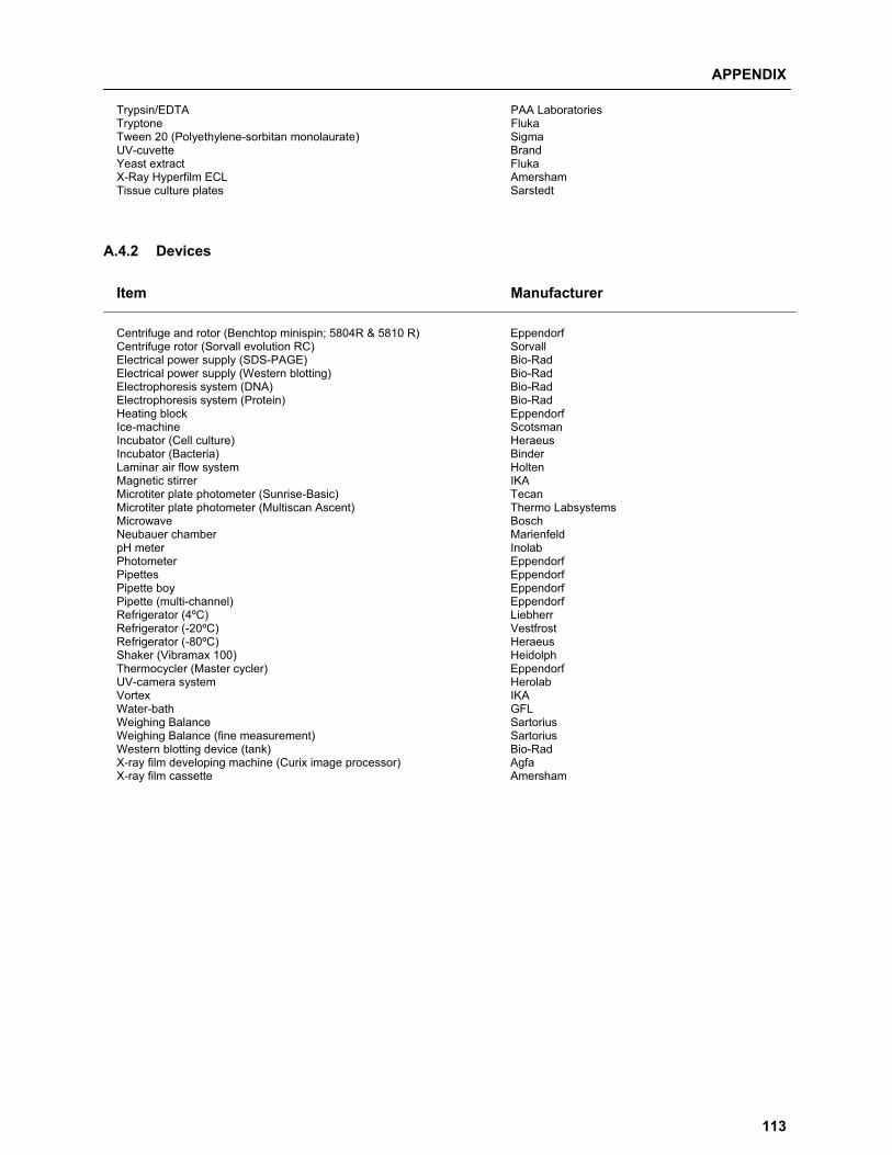
APPENDIX
113
Trypsin/EDTA PAA Laboratories Tryptone Fluka Tween 20 (Polyethylene-sorbitan monolaurate) Sigma UV-cuvette Brand Yeast extract Fluka X-Ray Hyperfilm ECL Amersham Tissue culture plates Sarstedt
A.4.2 Devices
Item Manufacturer
Centrifuge and rotor (Benchtop minispin; 5804R & 5810 R) Eppendorf Centrifuge rotor (Sorvall evolution RC) Sorvall Electrical power supply (SDS-PAGE) Bio-Rad Electrical power supply (Western blotting) Bio-Rad Electrophoresis system (DNA) Bio-Rad Electrophoresis system (Protein) Bio-Rad Heating block Eppendorf Ice-machine Scotsman Incubator (Cell culture) Heraeus Incubator (Bacteria) Binder Laminar air flow system Holten Magnetic stirrer IKA Microtiter plate photometer (Sunrise-Basic) Tecan Microtiter plate photometer (Multiscan Ascent) Thermo Labsystems Microwave Bosch Neubauer chamber Marienfeld pH meter Inolab Photometer Eppendorf Pipettes Eppendorf Pipette boy Eppendorf Pipette (multi-channel) Eppendorf Refrigerator (4ºC) Liebherr Refrigerator (-20ºC) Vestfrost Refrigerator (-80ºC) Heraeus Shaker (Vibramax 100) Heidolph Thermocycler (Master cycler) Eppendorf UV-camera system Herolab Vortex IKA Water-bath GFL Weighing Balance Sartorius Weighing Balance (fine measurement) Sartorius Western blotting device (tank) Bio-Rad X-ray film developing machine (Curix image processor) Agfa X-ray film cassette Amersham

ACKNOWLEDGEMENTS
114
Acknowledgements Dr. Frank Dietz, thank you for giving me the opportunity to work on this project and also for your expert
supervision over the entire period of the thesis. I thank you for friendly working environment and thought
stimulating discussions. You have been a great mentor, your work style and dedication will keep on
inspiring me throughout my life. I specially would like to show appreciation for your excellent, quick and
prompt reviewing during the writing of the manuscripts and the thesis. I owe a lot to your perseverance
regarding the project and keeping faith in my abilities.
Prof. Dr. Sørge Kelm, thank you for reviewing my thesis work and giving me the opportunity to work in
your working group. I thank you for excellent reviewing of the manuscripts. I am also grateful for all the
guidance and support you have showed during entire period of the thesis. I highly appreciate the fact that
you were always eager to expand the boundaries of work in different quadrants, encouraging the use of
various methods and tools, which in turn benefited the project and also my personal development.
Prof. Dr. Reimer Stick, thank you for showing interest in the project and reviewing my thesis work.
Dr. Kathrin Mädler, thank you for being willing to be an examiner for my thesis.
I thank Dr. Sebastian Franken, Bonn, Germany and Dr. Rainer Niedenthal, Hannover, Germany for their
collaboration and Dr. Jens Harder, Max Planck Institute for Marine Microbiology, Bremen, Germany for
providing access to the DNA sequencing facility. Also, I thank Prof. Dr. Ralf Dringen, Bremen, Germany
for allowing use of experimental facilities in his laboratory. I personally would like to mention gratitude of
the International office, University Bremen, Germany for providing additional financial assistance.
Mrs. Nazilla Isakovic and Mrs. Petra Berger, thank you for your excellent technical assistance.
Mr. Thaddeus T. Gbem, thank you for all the discussions and also for your encouragement and
friendship. I like to mention gratitude towards Mrs. Sabine Limberg for always helping with administrative
work. I thank all the members of AG Kelm for providing friendly working environment. I also thank
members of AG Mädler, with special mention to Dr. Federico Paroni and Mr. Amin Ardestani, for friendly
atmosphere and interactions. I thank Mr. Shaunak Khandekar and Mr. Thaddeus T. Gbem for
proof-reading my thesis.
I am grateful and indebted for the support and motivation received from my parents, parents-in-law, and
my family throughout my studies. Last; but certainly not the least; I thank my wonderful wife, Teja, for all
her support, encouragement, perseverance and love.
ERKLÄRUNG
115
Erklärung Gemäß § 6 der Promotionsordnung der Universität Bremen für die mathematischen, natur- und
ingenieurswissenschaftlichen Fachbereiche vom 14 März 2007 versichere ich, dass
1. die Arbeit ohne unerlaubte fremde Hilfe angefertigt wurde
2. keine anderen als die angegebenen Quellen und Hilfsmittel benutzt werden
3. die den benutzten Werken wörtlich oder inhaltlich entnommenen Stellen als solche kenntlich
gemacht wurden.
Bremen, 27 April 2010
Ketan Thakar

END
116