point of care ultrasound - cdn.ymaws.com · 1. describe clinical scenarios in which poc us will...
TRANSCRIPT
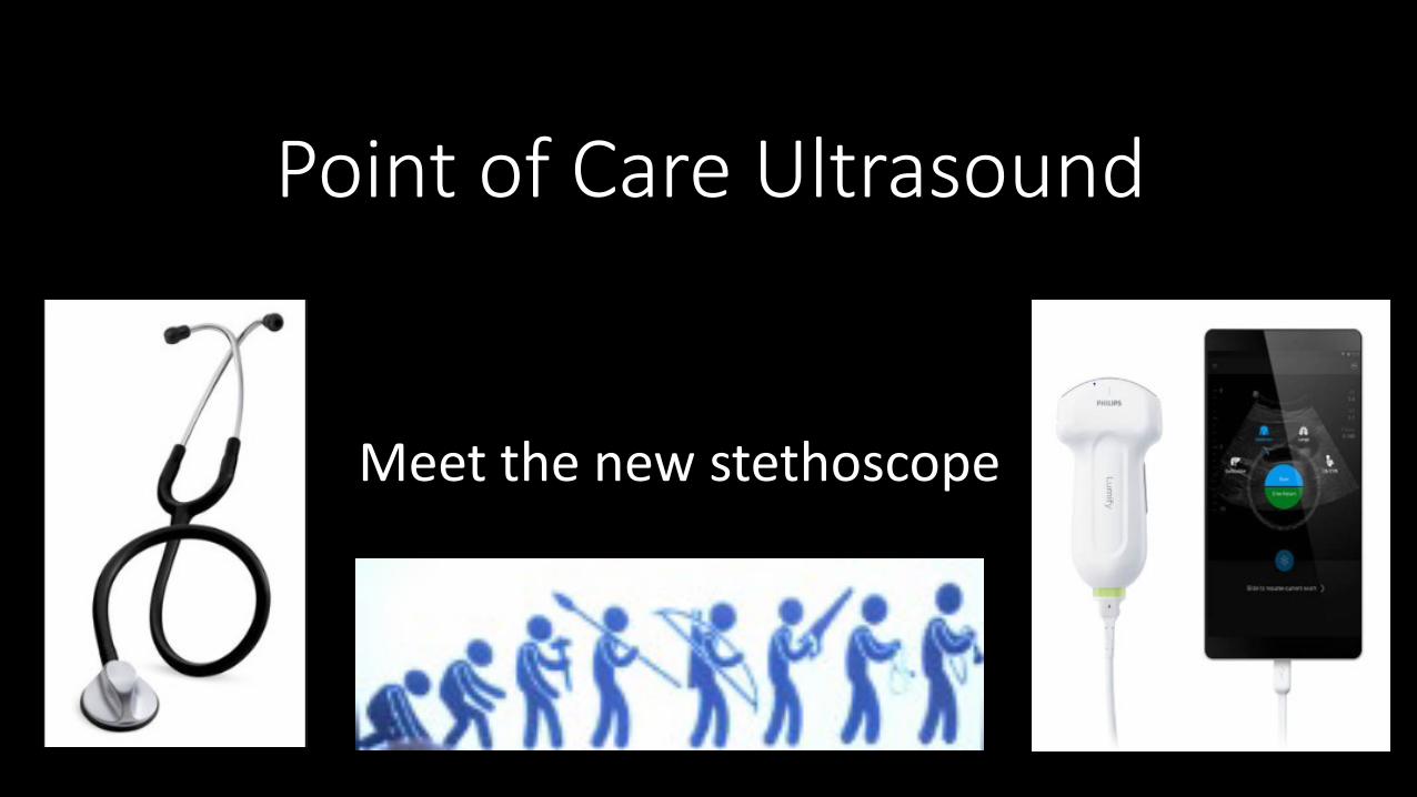
Point of Care Ultrasound
Meet the new stethoscope
1. Describe clinical scenarios in which POC US will improve patient care
2. Recognize the differences between diagnostic and POC ultrasound
3. Identify effusions versus cellulitis and when drainage is indicated
4. Lung POC case study
Objectives
What can POCUS be used for?
• Abdominal Aortic Aneurysm
● Abdominal Trauma
● Cardiac
● Gastrointestinal
● Hepatobiliary/Spleen
Lower Extremity Deep Vein Thrombosis
● Lung
● Musculoskeletal (MSK) Soft Tissue
● Obstetrics/First Trimester
● Renal/Genitourinary
Any other clinical applications?
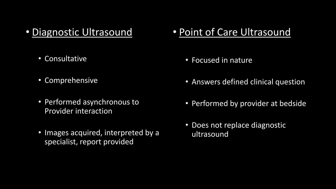
• Diagnostic Ultrasound
• Consultative
• Comprehensive
• Performed asynchronous to Provider interaction
• Images acquired, interpreted by a specialist, report provided
• Point of Care Ultrasound
• Focused in nature
• Answers defined clinical question
• Performed by provider at bedside
• Does not replace diagnostic ultrasound
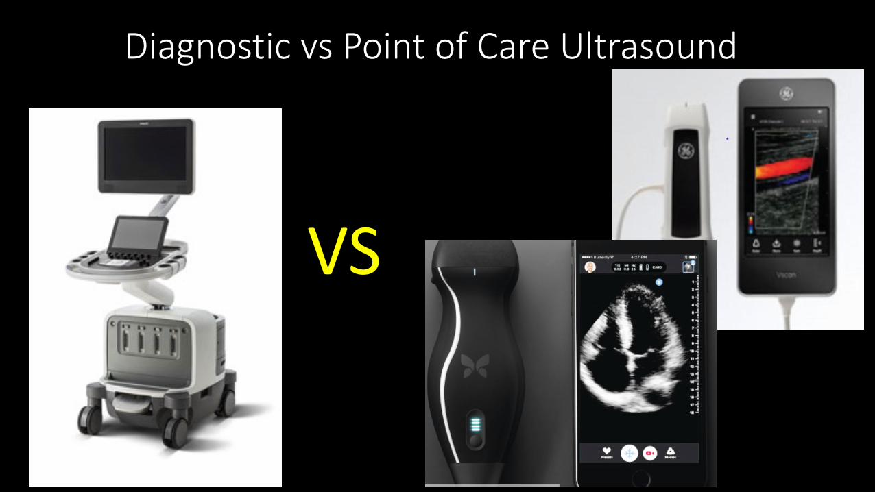
Diagnostic vs Point of Care Ultrasound
VS
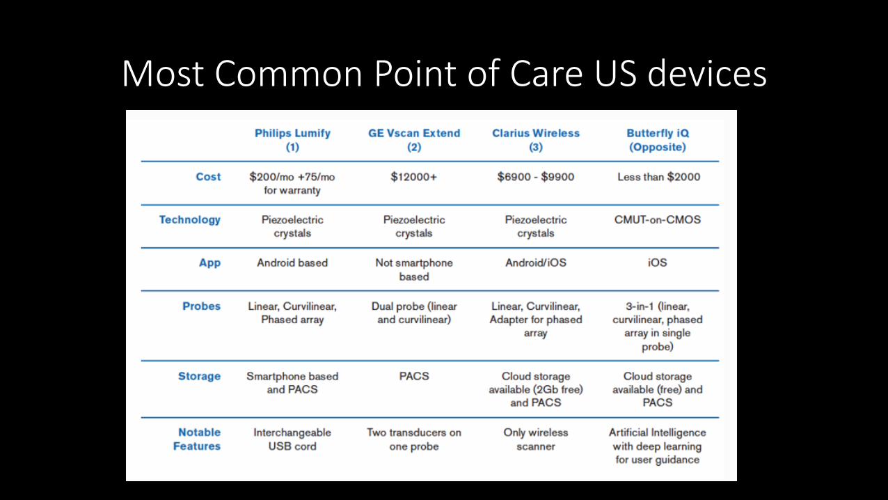
Most Common Point of Care US devices
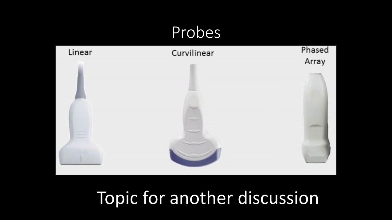
Probes
Topic for another discussion

Case #1
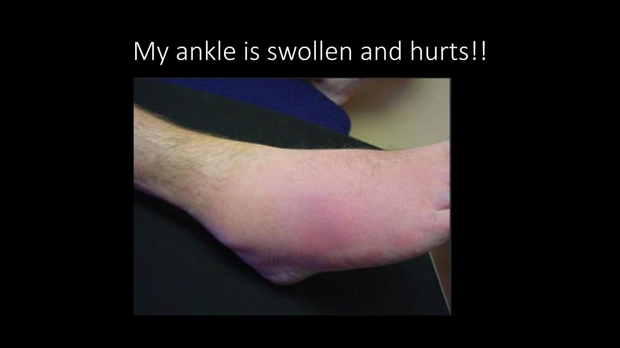
My ankle is swollen and hurts!!
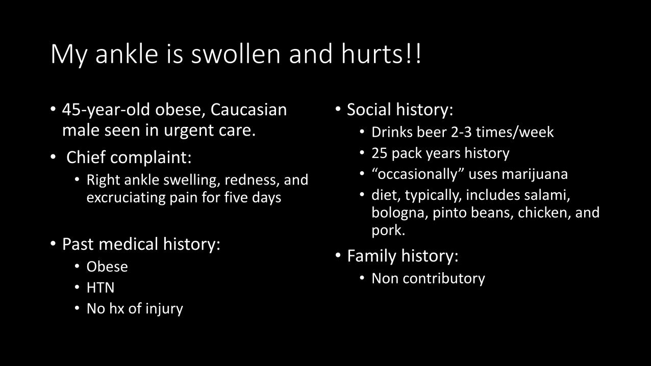
My ankle is swollen and hurts!!
• 45-year-old obese, Caucasian male seen in urgent care.
• Chief complaint: • Right ankle swelling, redness, and
excruciating pain for five days
• Past medical history:• Obese• HTN• No hx of injury
• Social history:• Drinks beer 2-3 times/week• 25 pack years history • “occasionally” uses marijuana• diet, typically, includes salami,
bologna, pinto beans, chicken, and pork.
• Family history:• Non contributory

My ankle is swollen and hurts!!
• Vitals
T max: 99.6HR: 80/minRR: 20/minO2 Sat: 98% on room airBP: 152/90
• LabsWBC 16,000Uric Acid 7.4BUN 15Creat 1.1ALT 32AST 45Blood CX Negative
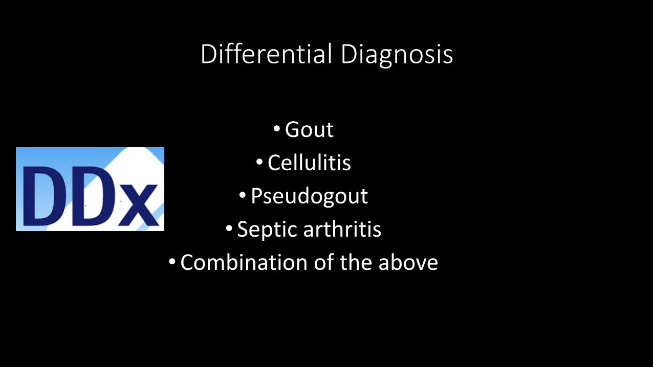
Differential Diagnosis
• Gout • Cellulitis
• Pseudogout• Septic arthritis
• Combination of the above
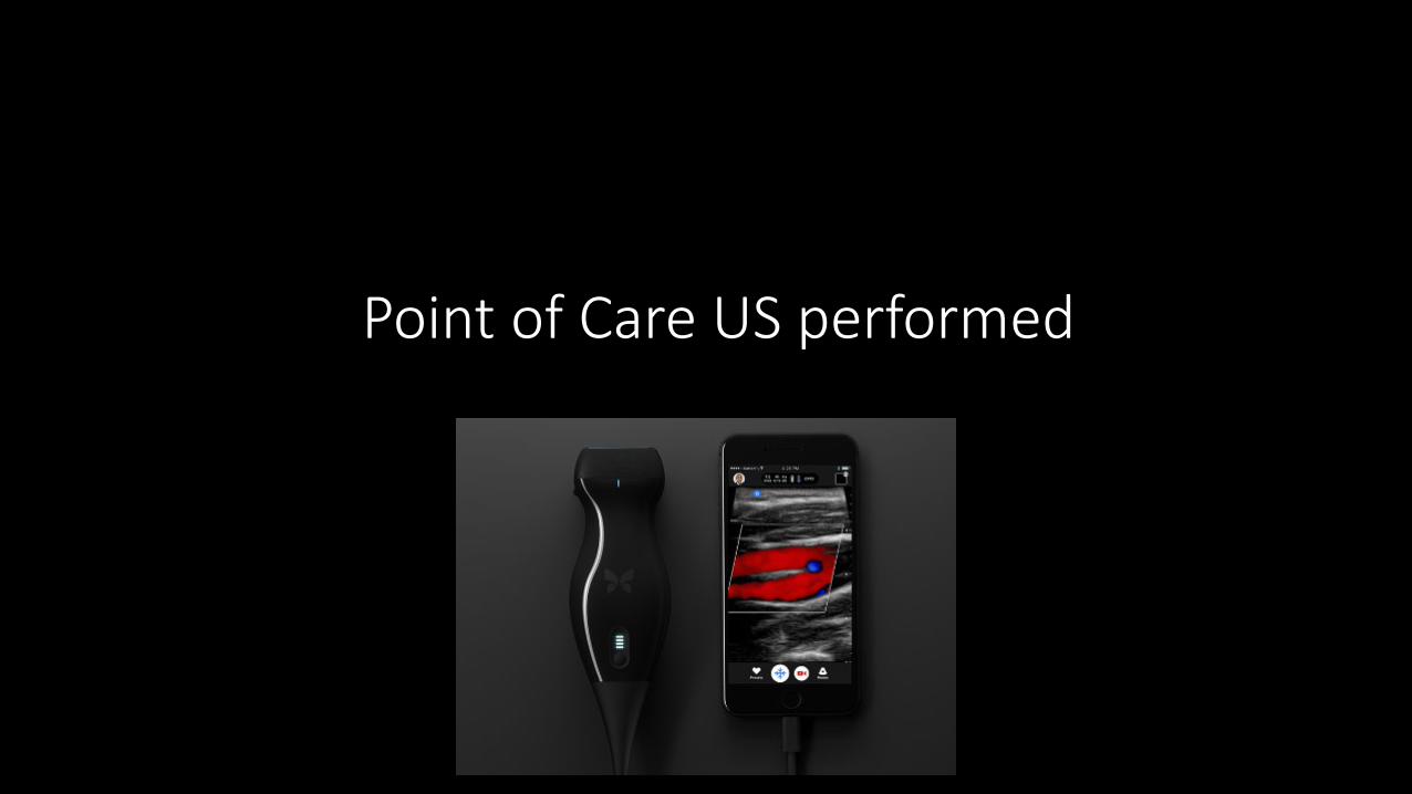
Point of Care US performed
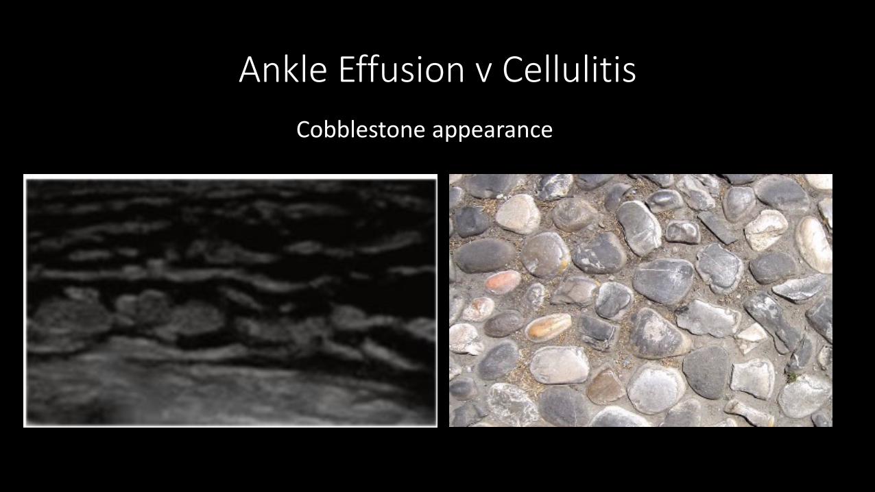
Ankle Effusion v CellulitisCobblestone appearance

Ankle Effusion v Cellulitis
RT Ankle Longitudinal RT Ankle Longitudinal w Doppler color
Ankle Effusion v Cellulitis
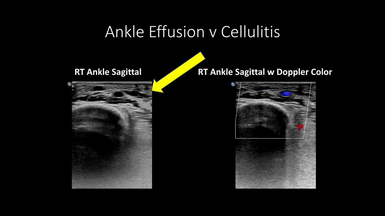
RT Ankle Sagittal RT Ankle Sagittal w Doppler Color
Ankle Effusion v Cellulitis
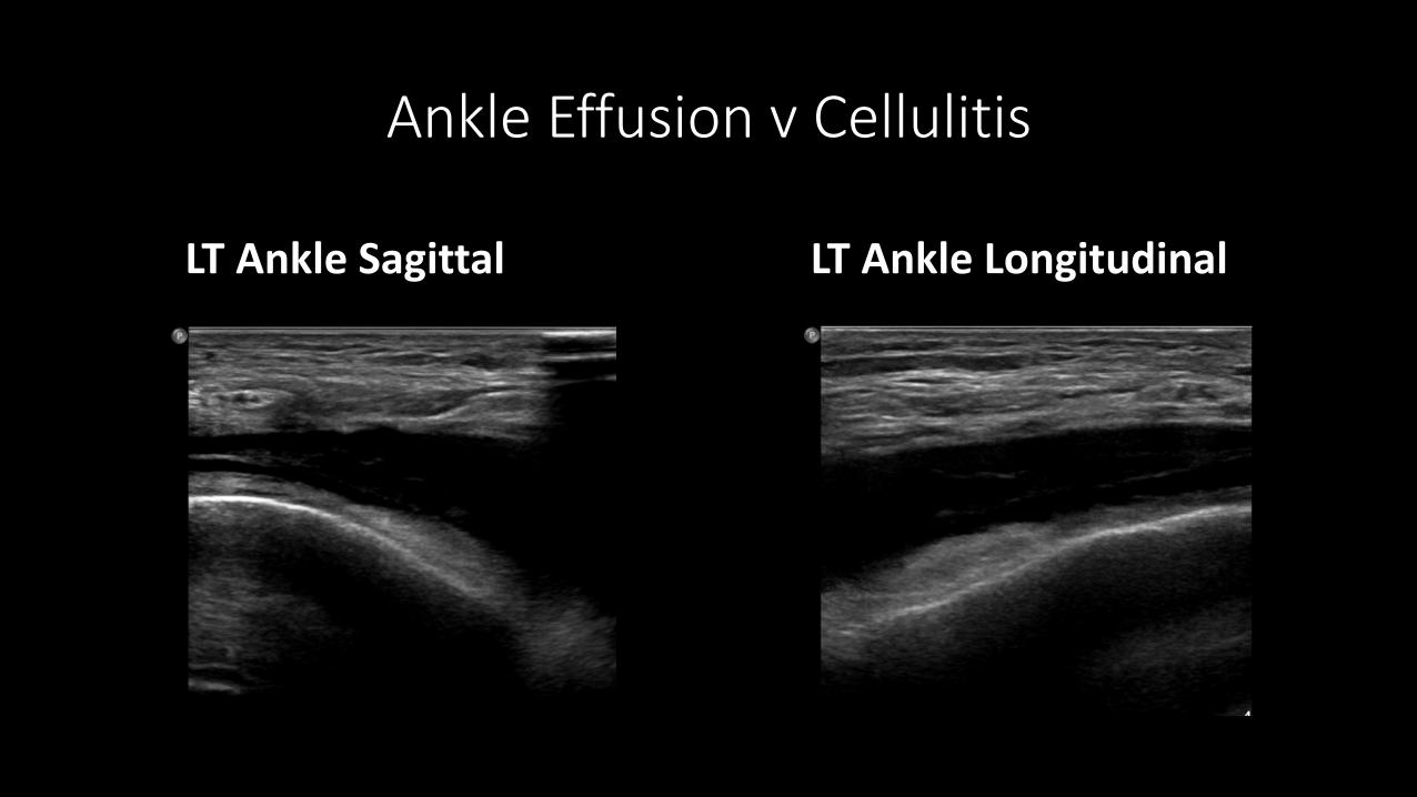
LT Ankle Sagittal LT Ankle Longitudinal
Ankle Effusion v Cellulitis
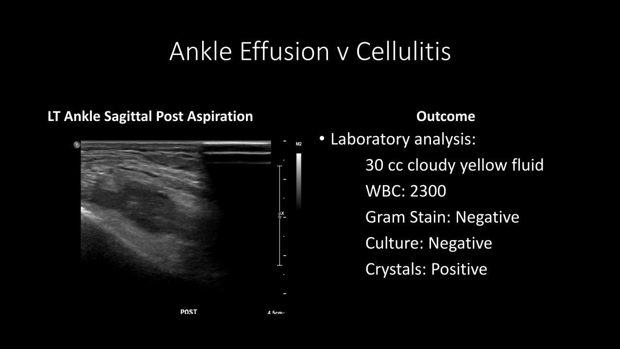
LT Ankle Sagittal Post Aspiration Outcome• Laboratory analysis:
30 cc cloudy yellow fluidWBC: 2300Gram Stain: NegativeCulture: NegativeCrystals: Positive

Ankle Effusion v Cellulitis
Approach to Septic Arthritis. American Family Physician. Sept 15, 2011 issue
Diagnosis
•Gout
CASE #2
I’m having trouble breathing
I’m having trouble breathing
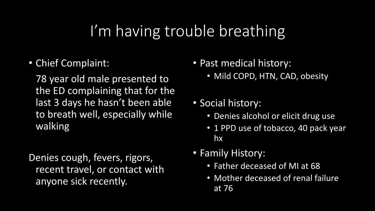
• Chief Complaint:78 year old male presented to the ED complaining that for the last 3 days he hasn’t been able to breath well, especially while walking
Denies cough, fevers, rigors, recent travel, or contact with anyone sick recently.
• Past medical history:• Mild COPD, HTN, CAD, obesity
• Social history:• Denies alcohol or elicit drug use• 1 PPD use of tobacco, 40 pack year
hx
• Family History: • Father deceased of MI at 68• Mother deceased of renal failure
at 76
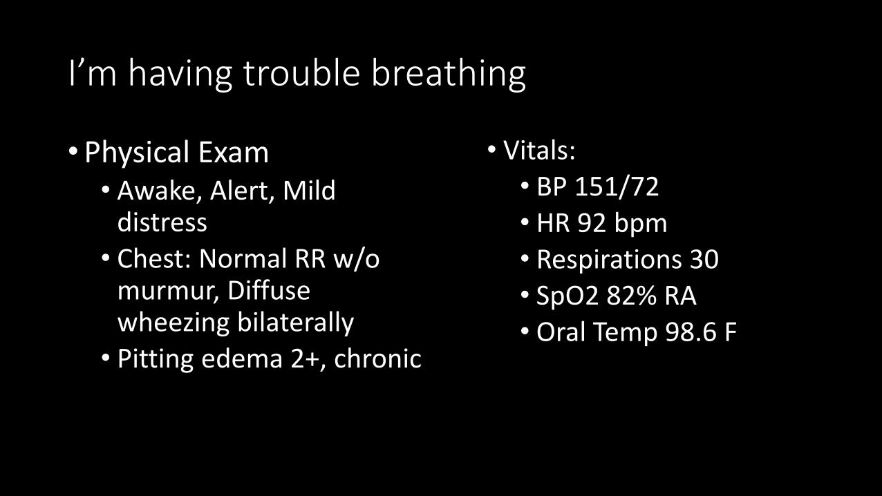
I’m having trouble breathing
• Physical Exam• Awake, Alert, Mild
distress• Chest: Normal RR w/o
murmur, Diffuse wheezing bilaterally
• Pitting edema 2+, chronic
• Vitals:• BP 151/72• HR 92 bpm• Respirations 30• SpO2 82% RA• Oral Temp 98.6 F
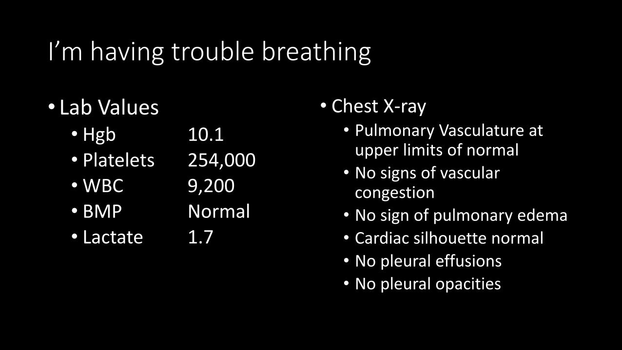
I’m having trouble breathing
• Lab Values• Hgb 10.1• Platelets 254,000• WBC 9,200• BMP Normal• Lactate 1.7
• Chest X-ray• Pulmonary Vasculature at
upper limits of normal• No signs of vascular
congestion• No sign of pulmonary edema• Cardiac silhouette normal• No pleural effusions• No pleural opacities
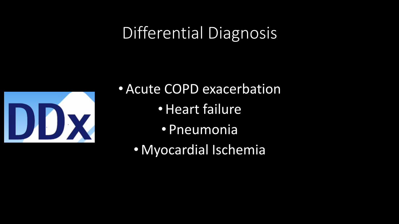
Differential Diagnosis
• Acute COPD exacerbation• Heart failure• Pneumonia
• Myocardial Ischemia
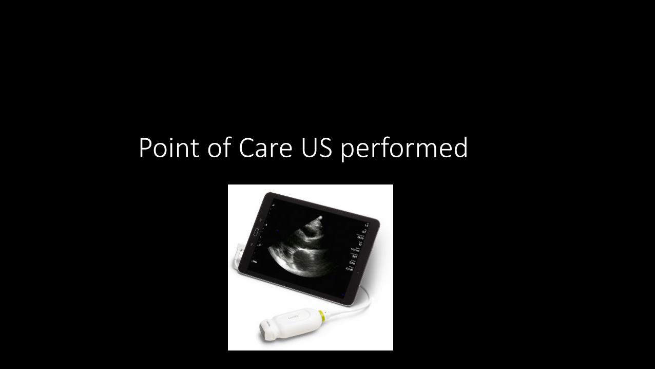
Point of Care US performed

Journal of the American Academy of PAs31(8):48-52, August 2018
Where should I scan?

Journal of the American Academy of PAs31(8):48-52, August 2018
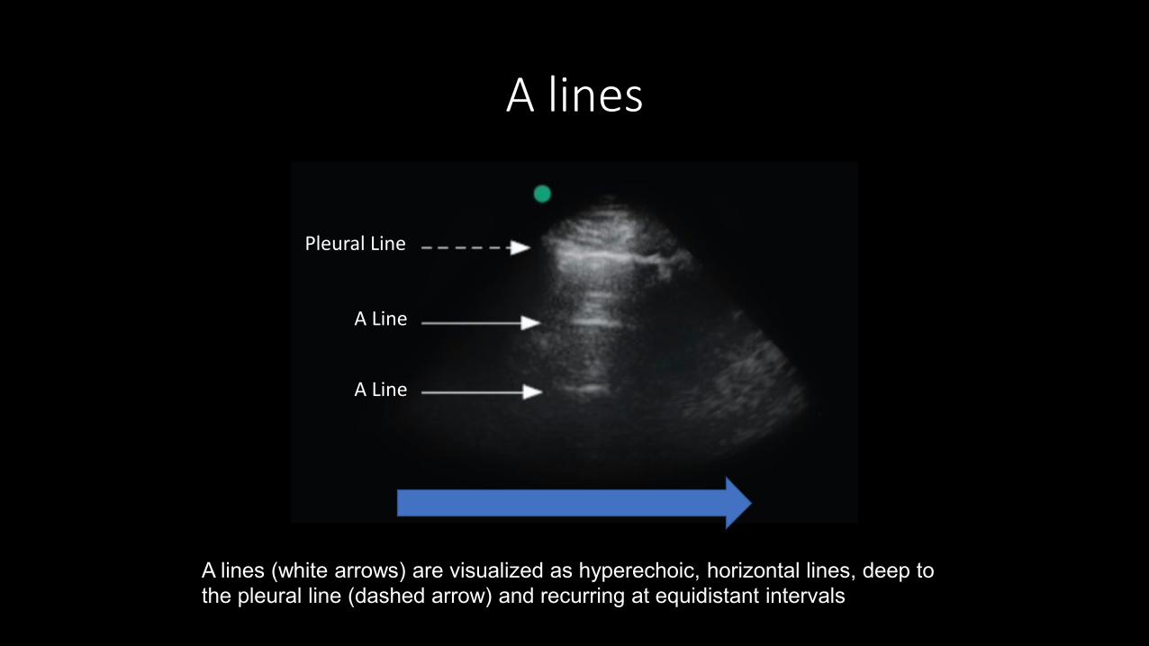
A lines (white arrows) are visualized as hyperechoic, horizontal lines, deep to the pleural line (dashed arrow) and recurring at equidistant intervals
A lines
Pleural Line
A Line
A Line

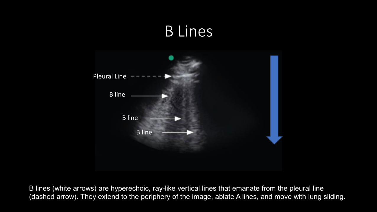
B lines (white arrows) are hyperechoic, ray-like vertical lines that emanate from the pleural line (dashed arrow). They extend to the periphery of the image, ablate A lines, and move with lung sliding.
B Lines
Pleural Line
B line
B line
B line
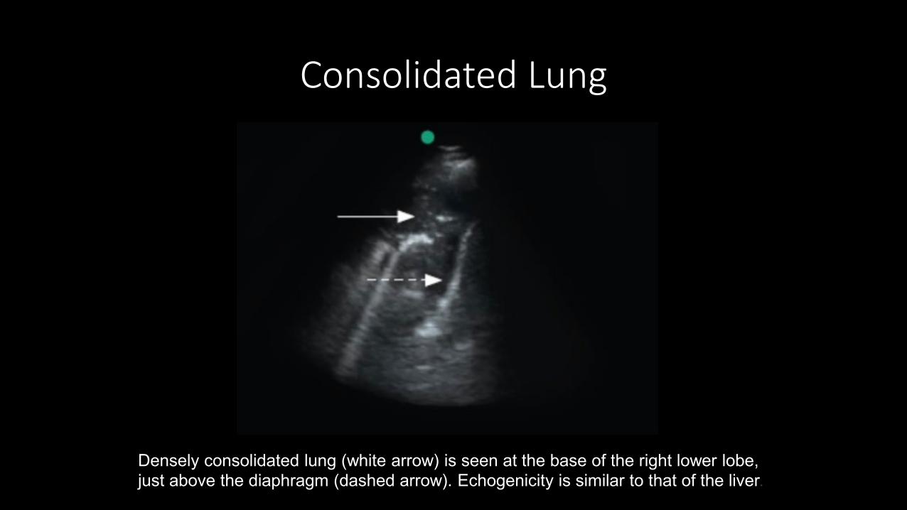
Densely consolidated lung (white arrow) is seen at the base of the right lower lobe, just above the diaphragm (dashed arrow). Echogenicity is similar to that of the liver.
Consolidated Lung
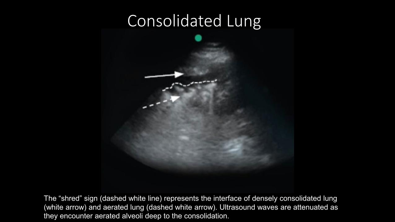
The “shred” sign (dashed white line) represents the interface of densely consolidated lung (white arrow) and aerated lung (dashed white arrow). Ultrasound waves are attenuated as they encounter aerated alveoli deep to the consolidation.
Consolidated Lung
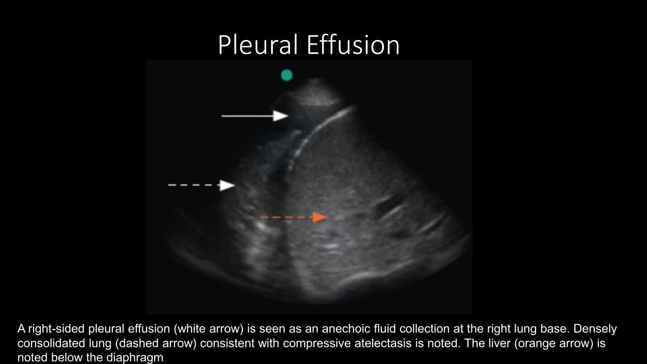
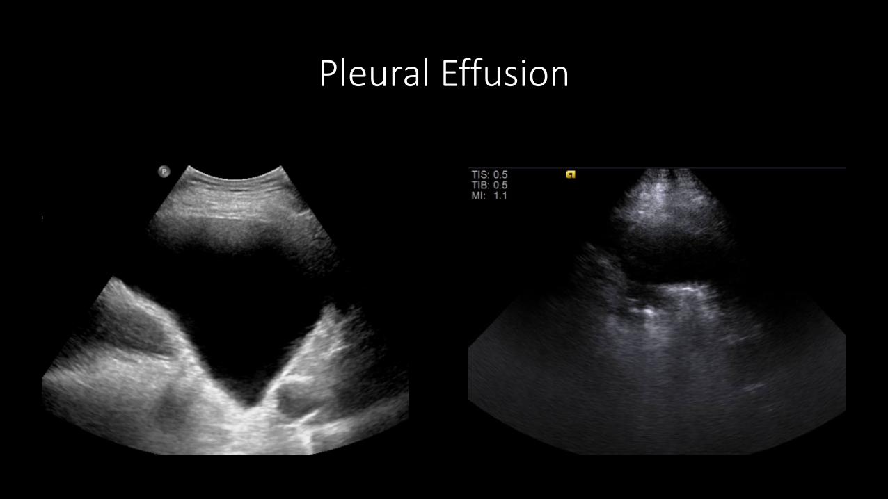
A right-sided pleural effusion (white arrow) is seen as an anechoic fluid collection at the right lung base. Densely consolidated lung (dashed arrow) consistent with compressive atelectasis is noted. The liver (orange arrow) is noted below the diaphragm.
Pleural Effusion
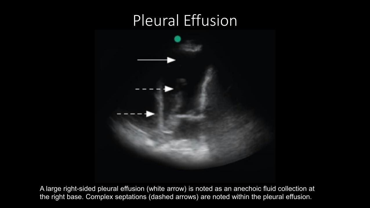
A large right-sided pleural effusion (white arrow) is noted as an anechoic fluid collection at the right base. Complex septations (dashed arrows) are noted within the pleural effusion.
Pleural Effusion
Pleural Effusion
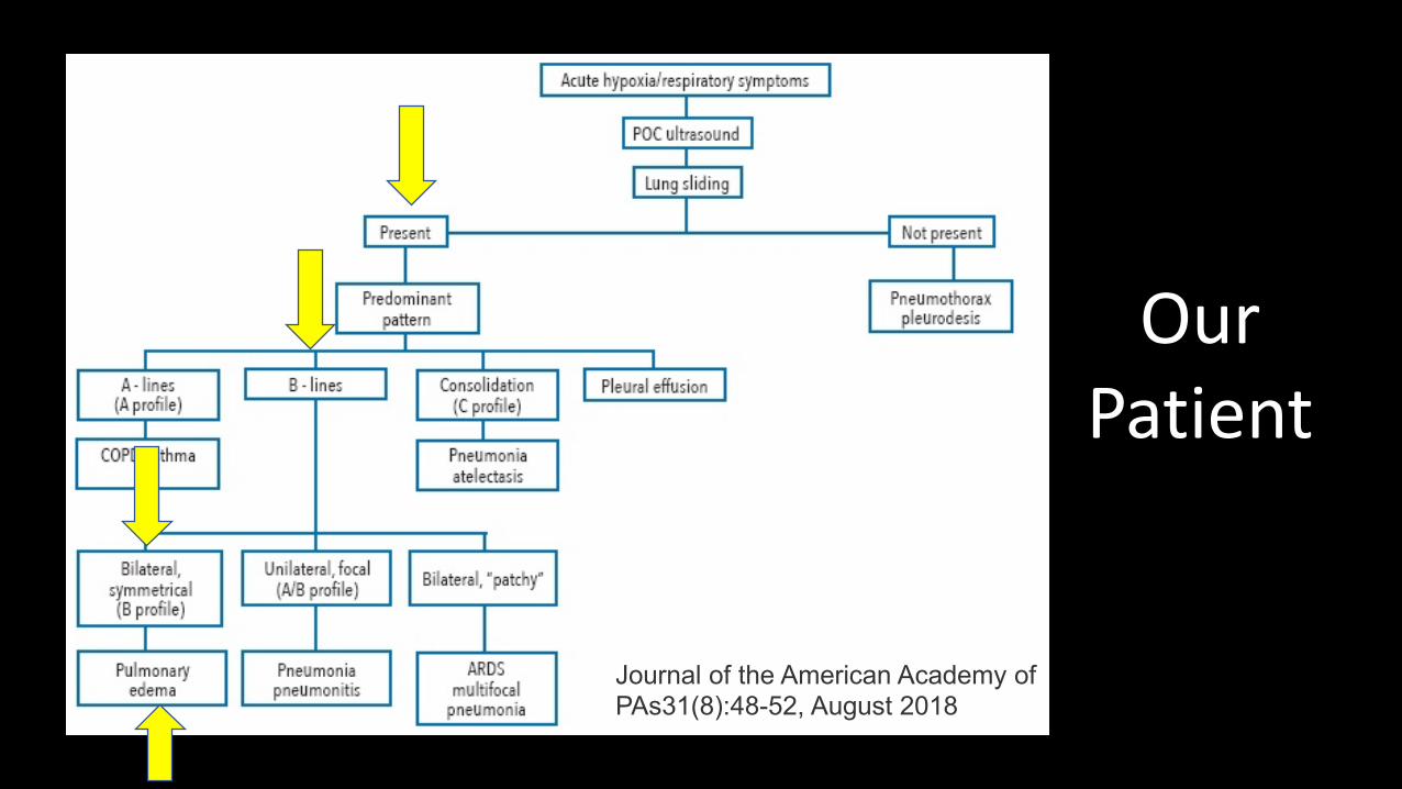
Journal of the American Academy of PAs31(8):48-52, August 2018
Our Patient
B lines (white arrows) are hyperechoic, ray-like vertical lines that emanate from the pleural line (dashed arrow). They extend to the periphery of the image, ablate A lines, and move with lung sliding.
B Lines
Pleural Line
B line
B line
B line
Diagnosis
•Acute Pulmonary Edema
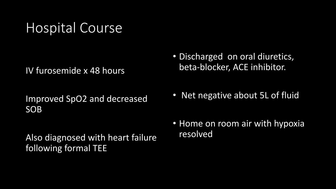
Hospital Course
IV furosemide x 48 hours
Improved SpO2 and decreased SOB
Also diagnosed with heart failure following formal TEE
• Discharged on oral diuretics, beta-blocker, ACE inhibitor.
• Net negative about 5L of fluid
• Home on room air with hypoxia resolved

How can I learn more?
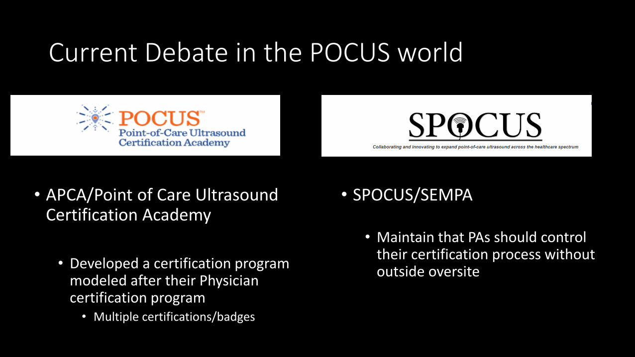
Current Debate in the POCUS world
• APCA/Point of Care Ultrasound Certification Academy
• Developed a certification program modeled after their Physician certification program
• Multiple certifications/badges
• SPOCUS/SEMPA
• Maintain that PAs should control their certification process without outside oversite
Full of Hot Air