pkcα regulates β1 integrin-dependent cell motility through...
TRANSCRIPT

The EMBO Journal Vol.18 No.14 pp.3909–3923, 1999
PKCα regulates β1 integrin-dependent cell motilitythrough association and control of integrin traffic
Tony Ng1, David Shima2, Anthony Squire3,Philippe I.H.Bastiaens3, Steve Gschmeissner4,Martin J.Humphries5 and Peter J.Parker1,6
1Protein Phosphorylation Laboratory, 2Cell Biology Laboratory,3Cell Biophysics Laboratory and 4Electron Microscopy Unit, ImperialCancer Research Fund, 44 Lincoln’s Inn Fields, London WC2A 3PXand 5Wellcome Trust Centre for Cell-Matrix Research, School ofBiological Sciences, University of Manchester, Oxford Road,Manchester M13 9PT, UK
6Corresponding authore-mail: [email protected]
Protein kinase C (PKC) has been implicated in integrin-mediated spreading and migration. In mammary epi-thelial cells there is a partial co-localization betweenβ1 integrin and PKCα. This reflects complexes betweenthese proteins as demonstrated by fluorescense reso-nance energy transfer (FRET) monitored by fluores-cence lifetime imaging microscopy and also bycoprecipitation. Constitutive complexes are observedfor the intact PKCα and also form with the regulatorydomain in an activation-dependent manner. Expressionof PKCα causes upregulation of β1 integrin on the cellsurface, whereas stimulation of PKC induces internaliz-ation of β1 integrin. The integrin initially traffics toan endosomal compartment in a Ca2�/PI 3-kinase/dynamin I-dependent manner and subsequently entersan endocytic recycling pathway. This induction ofendocytosis by PKCα is a function of activity and isnot observed for the regulatory domain. PKCα, butnot PKCα regulatory domain expression stimulatesmigration on β1 integrin substrates. This PKCα-enhanced migratory response is inhibited by blockadeof endocytosis.Keywords: endocytosis/FRET/GFP/integrin/migration/protein kinase C
Introduction
Various integrin receptors for extracellular matrix (ECM)have been implicated in providing proliferative and cellsurvival signals (Meredith et al., 1993; Boudreau et al.,1995; Crouch et al., 1996; Frisch et al., 1996a,b; Malikand Parsons, 1996; Udagawa and McIntyre, 1996; Waryet al., 1996; Ng et al., 1997; Bourdoulous et al., 1998) aswell as mediating cell migration towards specific ECMproteins and soluble stimulants/growth factors (Klemkeet al., 1994; Yun et al., 1996; Hauzenberger et al., 1997;Rabinovitz and Mercurio, 1997; Schneller et al., 1997;Meng and Lowell, 1998; Rigot et al., 1998; Saka et al.,1998; Werr et al., 1998). Largely through pharmacologicalstudies, protein kinase C (PKC) has been implicated as akey molecule involved in integrin-mediated cell spreading
© European Molecular Biology Organization 3909
and migration (Woods and Couchman, 1992; Klemkeet al., 1994; Yebra et al., 1995; Timar et al., 1996). Forexample, the enhanced motility of T-cells due to β1 inte-grin crosslinking can be blocked by a prior incubation withthe PKC effector site inhibitor calphostin C (Hauzenbergeret al., 1997). Similarly, incubation of colon carcinomacells with the phorbol ester tetradecanoyl phorbol acetate(TPA) for 5 h at a concentration that activates but doesnot cause PKC down-regulation increases their haptotacticresponse to fibronectin, collagen, laminin and vitronectin(Rigot et al., 1998).
The extent of ligand–receptor interaction and the out-side-in signal that ensues are generally dependent uponthe ectodomain conformation (affinity) and clustering/oligomerization (avidity) of the receptor as well as thesubsequent processing of the ligand-bound receptor by thecell (Bazzoni and Hemler, 1998). β1 integrin seems toexist in a dynamic conformational equilibrium betweenhigh and low affinity states for ligand binding; both statesdisplay bivalent cation dependence and can be modulatedby various inhibitory and stimulatory antibodies (reviewedby Humphries, 1996). Inhibitory antibodies appear tofunction as allosteric inhibitors of ligand engagement(Fogerty et al., 1990; Mould et al., 1996). An example ofa stimulatory antibody directed against the integrin β1subunit is the mAb 12G10 which recognizes a conforma-tional epitope within the putative βA domain expressedonly in the ligand-competent and ligand-occupied receptorconformers (Mould et al., 1995, 1998). Stimulatory anti-bodies like 12G10 therefore appear to function by stabiliz-ing signalling conformations of the integrin. To someextent, inhibitory antibodies, which recognize ligandattenuated epitopes, and stimulatory antibodies, whichrecognize ligand-stabilized epitopes, are the converse ofeach other. The notion that the ectodomain conformationof β1 integrins can be modified by changes to theintracellular portions of the receptors (so-called ‘inside-out’ signalling) (Crommie and Hemler, 1998; Mastrangeloet al., 1999) is supported by the finding that transfectantsexpressing a splice variant of β1 integrin known as β1B,which lacks both NPXY motifs in two distal cytoplasmicsites (cyto-2 and cyto-3) (Retta et al., 1998), or a dominantnegative β1 cytoplasmic domain–IL2R chimera(Mastrangelo et al., 1999), have substantially reducedactivated integrin on the cell surface, as recognized bythe mAb 12G10, while the total surface β1 integrinexpression remains the same. Truncation of specific res-idues in the cytoplasmic domain of the αv subunit alsohas an impact on the conformation and ligand binding ofαvβ3 integrin (Filardo and Cheresh, 1994).
Little is known about the life cycle of β1 integrin beyondactivation and ligand binding. By immunofluorescence, thebulk of ECM-interacting integrins (α6β4, β1, αIIbβ3 andαVβ3) at steady state appear to reside in an intracellular
T.Ng et al.
vesicular compartment which is at present uncharacterized(Lawson and Maxfield, 1995; Rabinovitz and Mercurio,1997; Miranti et al., 1998). The distribution and redistribu-tion of integrins through various processes such as surfacediffusion, internalization and recycling to the leading front,as well as ripping release from the cell rear have beenthought to be involved in the propagation of directionalcell movement (Lawson and Maxfield, 1995; reviewed byLauffenburger and Horwitz, 1996; Sanchez-Madrid anddel Pozo, 1999). Nevertheless, the control of the variousstages of the integrin receptor life cycle with respect toits activation, internalization and subsequent intracellulartrafficking is poorly understood.
The activities of various protein serine/threonine kinasessuch as protein kinase A, β-adrenergic receptor kinasesand PKC have previously been shown to influence theendocytosis, trafficking and hence desensitization/resensit-ization of receptors (Hausdorff et al., 1990; Inglese et al.,1993; Lefkowitz, 1993; Shih and Malbon, 1996). In thepresent study we have found PKCα to be an importantcytoplasmic component that interacts physically withactivated β1 integrin and regulates its exocytosis to theplasma membrane. It is also established that PKCα controlsinternalization through a Ca2�- and phosphatidylinositide3-kinase (PI3K)-dependent, dynamin I-controlled endo-cytic pathway. Furthermore, PKCα-induced upregulationof integrin-dependent cell migration was found to beblocked under conditions that prevented internalization ofthe receptor complex, identifying a critical role for PKCαin dynamic control of integrin function.
Results
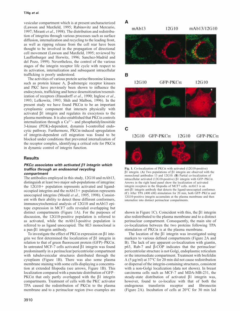
PKCα associates with activated β1 integrin whichtraffics through an endosomal recyclingcompartmentThe antibodies employed in this study, 12G10 and mAb13,distinguish at least two major sub-populations of integrins:the 12G10� population represents activated and ligand-occupied integrins and the mAb13� population representsunoccupied integrins (Mould et al., 1995, 1998). Consist-ent with their ability to detect these different conformers,immunocytochemical analysis of 12G10 and mAb13 epi-tope expression in MCF7 cells revealed overlapping butdistinct compartments (Figure 1A). For the purposes ofdiscussion, the 12G10-positive population is referred toas activated, while the mAb13-positive population isreferred to as ligand unoccupied. The 8E3 monoclonal isa pan-β1 integrin antibody.
To investigate the effect of PKCα expression on β1 inte-grin we first determined the localization of β1 integrin inrelation to that of green fluorescent protein (GFP)–PKCα.In untreated MCF-7 cells activated β1 integrin was foundpredominantly in a perinuclear/pericentriolar compartmentwith tubulovesicular structures distributed through thecytoplasm (Figure 1B). There was also some plasmamembrane staining with some cells displaying a concentra-tion at extended filopodia (see arrows, Figure 1B). Thislocalization compared with a punctate distribution of GFP–PKCα that only partly overlapped with the β1 integrincompartments. Treatment of cells with the PKC activatorTPA caused the redistribution of PKCα to the plasmamembrane and to a perinuclear region (two examples are
3910
Fig. 1. Co-localization of PKCα with activated (12G10-positive)β1 integrin. (A) Two populations of β1 integrin are observed with themonoclonal antibodies 13 and 12G10. (B) Partial co-localization ofintracellular activated (12G10-positive) β1 integrin with GFP–PKCα.Arrows in the right hand panel show the localization of activatedintegrin receptors in the filopodia of MCF7 cells. mAb13 is ananti-β1 integrin antibody that detects the ligand-unoccupied conformer.(C) After TPA (400 nM) stimulation for 20 min, both GFP–PKCα and12G10-positive integrin accumulate at the plasma membrane and theninternalize into distinct perinuclear compartments.
shown in Figure 1C). Coincident with this, the β1 integrinalso redistributed to the plasma membrane and to a distinctperinuclear compartment. Consequently, the main site ofco-localization between the two proteins following TPAstimulation of PKCα is at the plasma membrane.
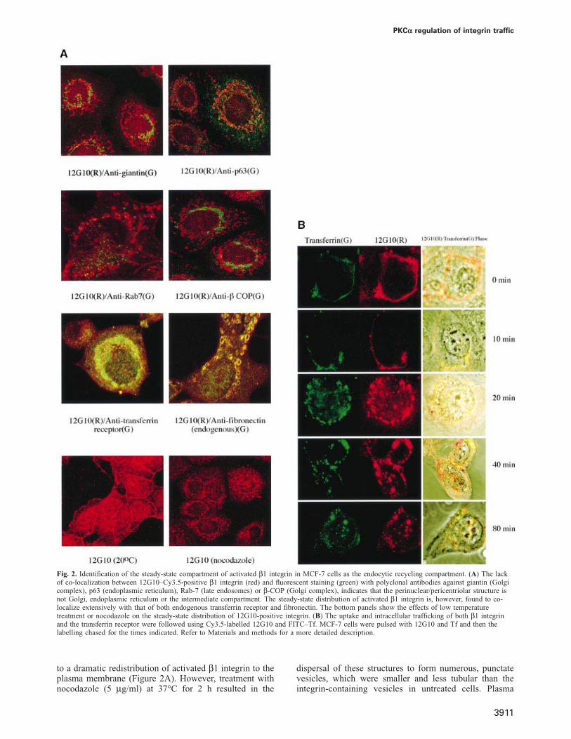
The location of the β1 integrin was investigated usingmarkers to various defined compartments (Figure 2A andB). The lack of any apparent co-localization with giantin,p63, Rab-7 and β-COP indicates that the perinuclear/pericentriolar structure is not Golgi, endoplasmic reticulumor the intermediate compartment. Treatment with brefeldinA (5 μg/ml) at 37°C for 20 min did not cause redistributionor dispersal of the integrin-containing structures, consistentwith a non-Golgi localization (data not shown). In breastcarcinoma cells such as MCF-7 and MDA-MB-231, thesteady-state distribution of activated β1 integrin was,however, found to co-localize with that of both theendogenous transferrin receptor and fibronectin(Figure 2A). Incubation of cells at 20°C for 30 min led
PKCα regulation of integrin traffic
Fig. 2. Identification of the steady-state compartment of activated β1 integrin in MCF-7 cells as the endocytic recycling compartment. (A) The lackof co-localization between 12G10–Cy3.5-positive β1 integrin (red) and fluorescent staining (green) with polyclonal antibodies against giantin (Golgicomplex), p63 (endoplasmic reticulum), Rab-7 (late endosomes) or β-COP (Golgi complex), indicates that the perinuclear/pericentriolar structure isnot Golgi, endoplasmic reticulum or the intermediate compartment. The steady-state distribution of activated β1 integrin is, however, found to co-localize extensively with that of both endogenous transferrin receptor and fibronectin. The bottom panels show the effects of low temperaturetreatment or nocodazole on the steady-state distribution of 12G10-positive integrin. (B) The uptake and intracellular trafficking of both β1 integrinand the transferrin receptor were followed using Cy3.5-labelled 12G10 and FITC–Tf. MCF-7 cells were pulsed with 12G10 and Tf and then thelabelling chased for the times indicated. Refer to Materials and methods for a more detailed description.
to a dramatic redistribution of activated β1 integrin to theplasma membrane (Figure 2A). However, treatment withnocodazole (5 μg/ml) at 37°C for 2 h resulted in the
3911
dispersal of these structures to form numerous, punctatevesicles, which were smaller and less tubular than theintegrin-containing vesicles in untreated cells. Plasma
T.Ng et al.
membrane staining by 12G10 was also reduced in noco-dazole-treated cells.
This behaviour of β1 integrin is reminiscent of thatdescribed for recycling receptors (Gruenberg and Maxfield,1995). To assess this directly, we compared the localizationof β1 integrin with that of the transferrin receptor, whichis known to reside within the pericentriolar, endocyticrecycling compartment. Localization of these proteins wasdetected in parallel through the uptake and intracellulartrafficking of Cy3.5-labelled 12G10 and FITC-transferrin(Tf), following pulse–chase labelling of MCF-7 cells.During the initial 10 min of chase at 37°C, both 12G10–Cy3.5 and FITC–Tf were found predominantly at theplasma membrane and juxta-membrane vesicular struc-tures (Figure 2B). After 20 min, these labelled structureswere found to concentrate in a perinuclear location. Thenear-complete co-localization of both 12G10–Cy3.5 andFITC–Tf throughout the entire time course of pulse–chase suggests that both activated integrin and transferrinreceptors traffic through a similar endocytic, recyclingcompartment.
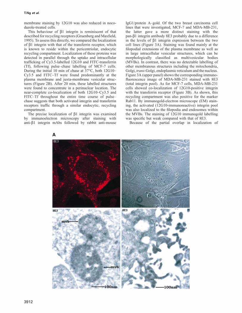
The precise localization of β1 integrin was examinedby immunoelectron microscopy after staining withanti-β1 integrin mAbs followed by rabbit anti-mouse
3912
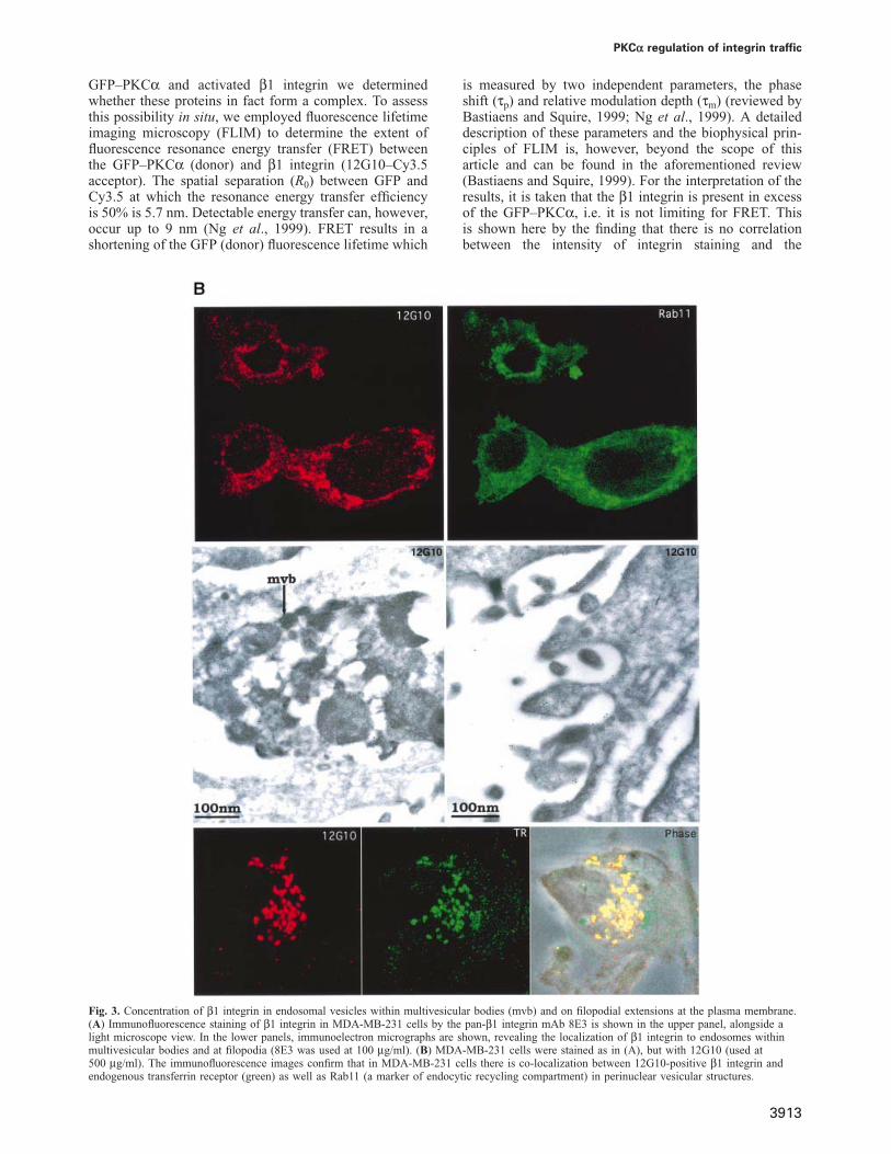
IgG1/protein A–gold. Of the two breast carcinoma celllines that were investigated, MCF-7 and MDA-MB-231,the latter gave a more distinct staining with thepan-β1 integrin antibody 8E3 probably due to a differencein the levels of β1 integrin expression between the twocell lines (Figure 3A). Staining was found mainly at thefilopodial extensions of the plasma membrane as well asin large intracellular vesicular structures, which can bemorphologically classified as multivesicular bodies(MVBs). In contrast, there was no detectable labelling ofother membranous structures including the mitochondria,Golgi, trans-Golgi, endoplasmic reticulum and the nucleus.Figure 3A (upper panel) shows the corresponding immuno-fluorescence image of MDA-MB-231 stained with 8E3(total integrin pool). As for MCF-7 cells, MDA-MB-231cells showed co-localization of 12G10-positive integrinwith the transferrin receptor (Figure 3B). As shown, thisrecycling compartment was also positive for the markerRab11. By immunogold-electron microscope (EM) stain-ing, the activated (12G10-immunoreactive) integrin poolwas also localized to the filopodia and endosomes withinthe MVBs. The staining of 12G10 immunogold labellingwas specific but weak compared with that of 8E3.
Because of the partial overlap in localization of
PKCα regulation of integrin traffic
GFP–PKCα and activated β1 integrin we determinedwhether these proteins in fact form a complex. To assessthis possibility in situ, we employed fluorescence lifetimeimaging microscopy (FLIM) to determine the extent offluorescence resonance energy transfer (FRET) betweenthe GFP–PKCα (donor) and β1 integrin (12G10–Cy3.5acceptor). The spatial separation (R0) between GFP andCy3.5 at which the resonance energy transfer efficiencyis 50% is 5.7 nm. Detectable energy transfer can, however,occur up to 9 nm (Ng et al., 1999). FRET results in ashortening of the GFP (donor) fluorescence lifetime which
Fig. 3. Concentration of β1 integrin in endosomal vesicles within multivesicular bodies (mvb) and on filopodial extensions at the plasma membrane.(A) Immunofluorescence staining of β1 integrin in MDA-MB-231 cells by the pan-β1 integrin mAb 8E3 is shown in the upper panel, alongside alight microscope view. In the lower panels, immunoelectron micrographs are shown, revealing the localization of β1 integrin to endosomes withinmultivesicular bodies and at filopodia (8E3 was used at 100 μg/ml). (B) MDA-MB-231 cells were stained as in (A), but with 12G10 (used at500 μg/ml). The immunofluorescence images confirm that in MDA-MB-231 cells there is co-localization between 12G10-positive β1 integrin andendogenous transferrin receptor (green) as well as Rab11 (a marker of endocytic recycling compartment) in perinuclear vesicular structures.
3913
is measured by two independent parameters, the phaseshift (τp) and relative modulation depth (τm) (reviewed byBastiaens and Squire, 1999; Ng et al., 1999). A detaileddescription of these parameters and the biophysical prin-ciples of FLIM is, however, beyond the scope of thisarticle and can be found in the aforementioned review(Bastiaens and Squire, 1999). For the interpretation of theresults, it is taken that the β1 integrin is present in excessof the GFP–PKCα, i.e. it is not limiting for FRET. Thisis shown here by the finding that there is no correlationbetween the intensity of integrin staining and the
T.Ng et al.
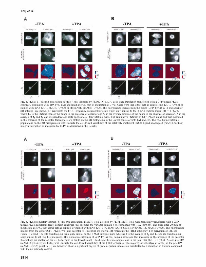
Fig. 4. PKCα–β1 integrin association in MCF7 cells detected by FLIM. (A) MCF7 cells were transiently transfected with a GFP-tagged PKCαconstruct, stimulated with TPA (400 nM) and fixed after 20 min of incubation at 37°C. Cells were then either left as controls (no 12G10–Cy3.5) orstained with mAb 12G10 (12G10–Cy3.5) or (B) mAb13 (mAb13–Cy3.5). The fluorescence images from the donor (GFP–PKCα WT) and acceptor(β1 integrin) are shown. Eff represents the FRET efficiency pseudocolour scale which only applies to the �mAb lifetime maps (Eff � 1–τda/τd
where τda is the lifetime map of the donor in the presence of acceptor and τd is the average lifetime of the donor in the absence of acceptor). τ is theaverage of τp and τm and its pseudocolour scale applies to all four lifetime maps. The cumulative lifetimes of GFP–PKCα alone and that measuredin the presence of the acceptor fluorophore are plotted on the 2D histograms in the lowest panels of both (A) and (B). The two distinct lifetimepopulations on the 2D histograms in (B) illustrate the cell-to-cell variability of the relatively inefficient PKCα–ligand-unoccupied (mAb13-positive)integrin interaction as measured by FLIM as described in the Results.
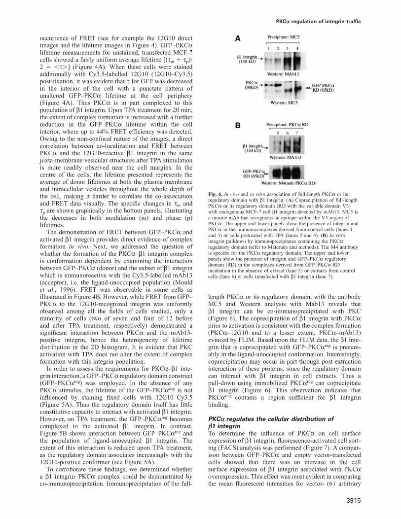
Fig. 5. PKCα regulatory domain–β1 integrin association in MCF7 cells detected by FLIM. MCF7 cells were transiently transfected with a GFP-tagged PKCα regulatory (reg.) domain construct (this includes the variable domain V3), stimulated with TPA (400 nM) and fixed after 20 min ofincubation at 37°C, then either left as controls or stained with mAb 12G10 (A, mAb 12G10–Cy3.5) or mAb13 (B, mAb13-Cy3.5). The fluorescenceimages from the donor (GFP–PKCα WT) and acceptor (β1 integrin) are shown. Eff represents the FRET efficiency. For derivation of Eff, seeFigure 4 legend. The Eff pseudocolour scale only applies to the �MAb lifetime maps whereas τ is the average of τp and τm and its pseudocolourscale applies to all four lifetime maps. The cumulative lifetimes of GFP–PKCα reg. domain alone and that measured in the presence of the acceptorfluorophore are plotted on the 2D histograms in the lowest panel. The distinct lifetime populations in the post-TPA (12G10–Cy3.5) (A) and pre-TPA(mAb13-Cy3.5) (B) 2D histograms illustrate the cell-to-cell variability of the FRET efficiency. The majority of cells (five of seven) in the pre-TPA(mAb13–Cy3.5) panel in (B) do, however, show a significant degree of protein–protein interaction manifested by a reduction in lifetime comparedwith the no antibody control.
3914
PKCα regulation of integrin traffic
occurrence of FRET (see for example the 12G10 directimages and the lifetime images in Figure 4). GFP–PKCαlifetime measurements for unstained, transfected MCF-7cells showed a fairly uniform average lifetime [(τm � τp)/2 � �τ�] (Figure 4A). When these cells were stainedadditionally with Cy3.5-labelled 12G10 (12G10–Cy3.5)post-fixation, it was evident that τ for GFP was decreasedin the interior of the cell with a punctate pattern ofunaltered GFP–PKCα lifetime at the cell periphery(Figure 4A). Thus PKCα is in part complexed to thispopulation of β1 integrin. Upon TPA treatment for 20 min,the extent of complex formation is increased with a furtherreduction in the GFP–PKCα lifetime within the cellinterior, where up to 44% FRET efficiency was detected.Owing to the non-confocal nature of the images, a directcorrelation between co-localization and FRET betweenPKCα and the 12G10-reactive β1 integrin in the samejuxta-membrane vesicular structures after TPA stimulationis more readily observed near the cell margins. In thecentre of the cells, the lifetime presented represents theaverage of donor lifetimes at both the plasma membraneand intracellular vesicles throughout the whole depth ofthe cell, making it harder to correlate the co-associationand FRET data visually. The specific changes in τm andτp are shown graphically in the bottom panels, illustratingthe decreases in both modulation (m) and phase (p)lifetimes.
The demonstration of FRET between GFP–PKCα andactivated β1 integrin provides direct evidence of complexformation in vivo. Next, we addressed the question ofwhether the formation of the PKCα–β1 integrin complexis conformation dependent by examining the interactionbetween GFP–PKCα (donor) and the subset of β1 integrinwhich is immunoreactive with the Cy3.5-labelled mAb13(acceptor), i.e. the ligand-unoccupied population (Mouldet al., 1996). FRET was observable in some cells asillustrated in Figure 4B. However, while FRET from GFP–PKCα to the 12G10-recognized integrin was uniformlyobserved among all the fields of cells studied, only aminority of cells (two of seven and four of 12 beforeand after TPA treatment, respectively) demonstrated asignificant interaction between PKCα and the mAb13-positive integrin, hence the heterogeneity of lifetimedistribution in the 2D histogram. It is evident that PKCactivation with TPA does not alter the extent of complexformation with this integrin population.
In order to assess the requirements for PKCα–β1 inte-grin interaction, a GFP–PKCα regulatory domain construct(GFP–PKCαreg) was employed. In the absence of anyPKCα stimulus, the lifetime of the GFP–PKCαreg is notinfluenced by staining fixed cells with 12G10–Cy3.5(Figure 5A). Thus the regulatory domain itself has littleconstitutive capacity to interact with activated β1 integrin.However, on TPA treatment, the GFP–PKCαreg becomescomplexed to the activated β1 integrin. In contrast,Figure 5B shows interaction between GFP–PKCαreg andthe population of ligand-unoccupied β1 integrin. Theextent of this interaction is reduced upon TPA treatment,as the regulatory domain associates increasingly with the12G10-positive conformer (see Figure 5A).
To corroborate these findings, we determined whethera β1 integrin–PKCα complex could be demonstrated byco-immunoprecipitation. Immunoprecipitation of the full-
3915
Fig. 6. In vivo and in vitro association of full-length PKCα or itsregulatory domain with β1 integrin. (A) Coprecipitation of full-lengthPKCα or its regulatory domain (RD with the variable domain V3)with endogenous MCF-7 cell β1 integrin detected by mAb13. MC5 isa murine mAb that recognizes an epitope within the V3 region ofPKCα. The upper and lower panels show the presence of integrin andPKCα in the immunocomplexes derived from control cells (lanes 1and 3) or cells pretreated with TPA (lanes 2 and 4). (B) In vitrointegrin pulldown by immunoprecipitates containing the PKCαregulatory domain (refer to Materials and methods). The M4 antibodyis specific for the PKCα regulatory domain. The upper and lowerpanels show the presence of integrin and GFP–PKCα regulatorydomain (RD) in the complexes derived from GFP–PKCα RDincubation in the absence of extract (lane 5) or extracts from controlcells (lane 6) or cells transfected with β1 integrin (lane 7).
length PKCα or its regulatory domain, with the antibodyMC5 and Western analysis with Mab13 reveals thatβ1 integrin can be co-immunoprecipitated with PKC(Figure 6). The coprecipitation of β1 integrin with PKCαprior to activation is consistent with the complex formation(PKCα–12G10 and to a lesser extent, PKCα–mAb13)evinced by FLIM. Based upon the FLIM data, the β1 inte-grin that is coprecipitated with GFP–PKCαreg is presum-ably in the ligand-unoccupied conformation. Interestingly,coprecipitation may occur in part through post-extractioninteraction of these proteins, since the regulatory domaincan interact with β1 integrin in cell extracts. Thus apull-down using immobilized PKCαreg can coprecipitateβ1 integrin (Figure 6). This observation indicates thatPKCαreg contains a region sufficient for β1 integrinbinding.
PKCα regulates the cellular distribution ofβ1 integrinTo determine the influence of PKCα on cell surfaceexpression of β1 integrin, fluorescence-activated cell sort-ing (FACS) analysis was performed (Figure 7). A compar-ison between GFP–PKCα and empty vector-transfectedcells showed that there was an increase in the cellsurface expression of β1 integrin associated with PKCαoverexpression. This effect was most evident in comparingthe mean fluorescent intensities for vector- (61 arbitrary
T.Ng et al.
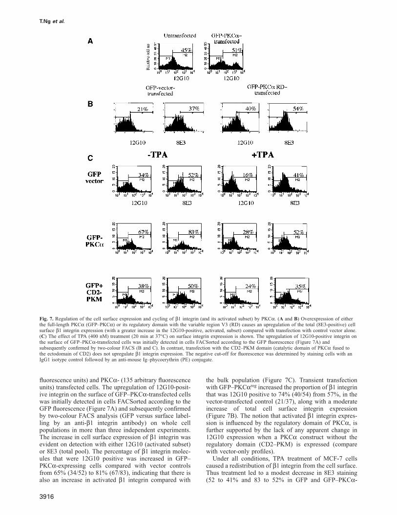
Fig. 7. Regulation of the cell surface expression and cycling of β1 integrin (and its activated subset) by PKCα. (A and B) Overexpression of eitherthe full-length PKCα (GFP–PKCα) or its regulatory domain with the variable region V3 (RD) causes an upregulation of the total (8E3-positive) cellsurface β1 integrin expression (with a greater increase in the 12G10-positive, activated, subset) compared with transfection with control vector alone.(C) The effect of TPA (400 nM) treatment (20 min at 37°C) on surface integrin expression is shown. The upregulation of 12G10-positive integrin onthe surface of GFP–PKCα-transfected cells was initially detected in cells FACSorted according to the GFP fluorescence (Figure 7A) andsubsequently confirmed by two-colour FACS (B and C). In contrast, transfection with the CD2–PKM domain (catalytic domain of PKCα fused tothe ectodomain of CD2) does not upregulate β1 integrin expression. The negative cut-off for fluorescence was determined by staining cells with anIgG1 isotype control followed by an anti-mouse Ig–phycoerythrin (PE) conjugate.
fluorescence units) and PKCα- (135 arbitrary fluorescenceunits) transfected cells. The upregulation of 12G10-posit-ive integrin on the surface of GFP–PKCα-transfected cellswas initially detected in cells FACSorted according to theGFP fluorescence (Figure 7A) and subsequently confirmedby two-colour FACS analysis (GFP versus surface label-ling by an anti-β1 integrin antibody) on whole cellpopulations in more than three independent experiments.The increase in cell surface expression of β1 integrin wasevident on detection with either 12G10 (activated subset)or 8E3 (total pool). The percentage of β1 integrin molec-ules that were 12G10 positive was increased in GFP–PKCα-expressing cells compared with vector controlsfrom 65% (34/52) to 81% (67/83), indicating that there isalso an increase in activated β1 integrin compared with
3916
the bulk population (Figure 7C). Transient transfectionwith GFP–PKCαreg increased the proportion of β1 integrinthat was 12G10 positive to 74% (40/54) from 57%, in thevector-transfected control (21/37), along with a moderateincrease of total cell surface integrin expression(Figure 7B). The notion that activated β1 integrin expres-sion is influenced by the regulatory domain of PKCα, isfurther supported by the lack of any apparent change in12G10 expression when a PKCα construct without theregulatory domain (CD2–PKM) is expressed (comparewith vector-only profiles).
Under all conditions, TPA treatment of MCF-7 cellscaused a redistribution of β1 integrin from the cell surface.Thus treatment led to a modest decrease in 8E3 staining(52 to 41% and 83 to 52% in GFP and GFP–PKCα-
PKCα regulation of integrin traffic
3917
T.Ng et al.
expressing cells, respectively) and a greater concomitanteffect on 12G10-positive β1 integrin (2- and 2.4-folddecreases in GFP and GFP–PKCα-positive cells, respect-ively). The greater change in the 12G10-positive β1 inte-grin subset relative to the total pool (8E3-positive)indicates that the active conformer is removed morereadily from the cell surface.
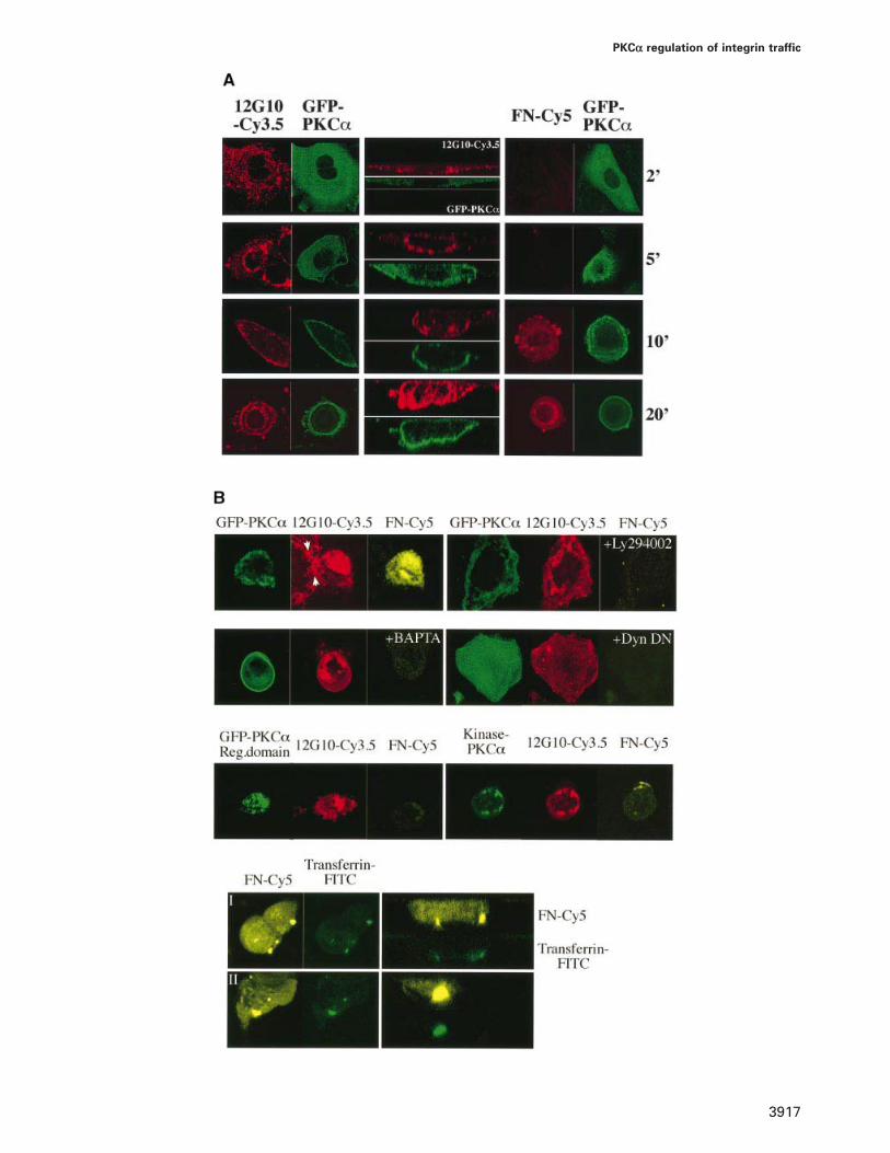
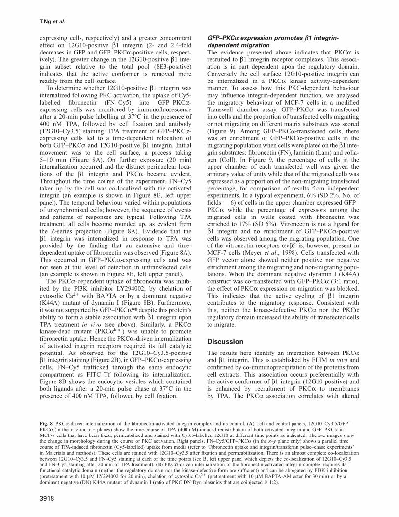
To determine whether 12G10-positive β1 integrin wasinternalized following PKC activation, the uptake of Cy5-labelled fibronectin (FN–Cy5) into GFP–PKCα-expressing cells was monitored by immunofluorescenceafter a 20-min pulse labelling at 37°C in the presence of400 nM TPA, followed by cell fixation and antibody(12G10–Cy3.5) staining. TPA treatment of GFP–PKCα-expressing cells led to a time-dependent relocation ofboth GFP–PKCα and 12G10-positive β1 integrin. Initialmovement was to the cell surface, a process taking5–10 min (Figure 8A). On further exposure (20 min)internalization occurred and the distinct perinuclear loca-tions of the β1 integrin and PKCα became evident.Throughout the time course of the experiment, FN–Cy5taken up by the cell was co-localized with the activatedintegrin (an example is shown in Figure 8B, left upperpanel). The temporal behaviour varied within populationsof unsynchronized cells; however, the sequence of eventsand patterns of responses are typical. Following TPAtreatment, all cells become rounded up, as evident fromthe Z-series projection (Figure 8A). Evidence that theβ1 integrin was internalized in response to TPA wasprovided by the finding that an extensive and time-dependent uptake of fibronectin was observed (Figure 8A).This occurred in GFP–PKCα-expressing cells and wasnot seen at this level of detection in untransfected cells(an example is shown in Figure 8B, left upper panel).
The PKCα-dependent uptake of fibronectin was inhib-ited by the PI3K inhibitor LY294002, by chelation ofcytosolic Ca2� with BAPTA or by a dominant negative(K44A) mutant of dynamin I (Figure 8B). Furthermore,it was not supported by GFP–PKCαreg despite this protein’sability to form a stable association with β1 integrin uponTPA treatment in vivo (see above). Similarly, a PKCαkinase-dead mutant (PKCαkin–) was unable to promotefibronectin uptake. Hence the PKCα-driven internalizationof activated integrin receptors required its full catalyticpotential. As observed for the 12G10–Cy3.5-positiveβ1 integrin staining (Figure 2B), in GFP–PKCα-expressingcells, FN–Cy5 trafficked through the same endocyticcompartment as FITC–Tf following its internalization.Figure 8B shows the endocytic vesicles which containedboth ligands after a 20-min pulse–chase at 37°C in thepresence of 400 nM TPA, followed by cell fixation.
Fig. 8. PKCα-driven internalization of the fibronectin-activated integrin complex and its control. (A) Left and central panels, 12G10–Cy3.5/GFP–PKCα (in the x–y and x–z planes) show the time-course of TPA (400 nM)-induced redistribution of both activated integrin and GFP–PKCα inMCF-7 cells that have been fixed, permeabilized and stained with Cy3.5-labelled 12G10 at different time points as indicated. The x–z images showthe change in morphology during the course of PKC activation. Right panels, FN–Cy5/GFP–PKCα (in the x–y plane only) shows a parallel timecourse of TPA-induced fibronectin (Cy5-labelled) uptake from media (refer to ‘Fibronectin uptake and integrin/transferrin pulse–chase experiments’in Materials and methods). These cells are stained with 12G10–Cy3.5 after fixation and permeabilization. There is an almost complete co-localizationbetween 12G10–Cy3.5 and FN–Cy5 staining at each of the time points (see B, left upper panel which depicts the co-localization of 12G10–Cy3.5and FN–Cy5 staining after 20 min of TPA treatment). (B) PKCα-driven internalization of the fibronectin-activated integrin complex requires itsfunctional catalytic domain (neither the regulatory domain nor the kinase-defective form are sufficient) and can be abrogated by PI3K inhibition(pretreatment with 10 μM LY294002 for 20 min), chelation of cytosolic Ca2� (pretreatment with 10 μM BAPTA-AM ester for 30 min) or by adominant negative (DN) K44A mutant of dynamin I (ratio of PKC:DN Dyn plasmids that are coinjected is 1:2).
3918
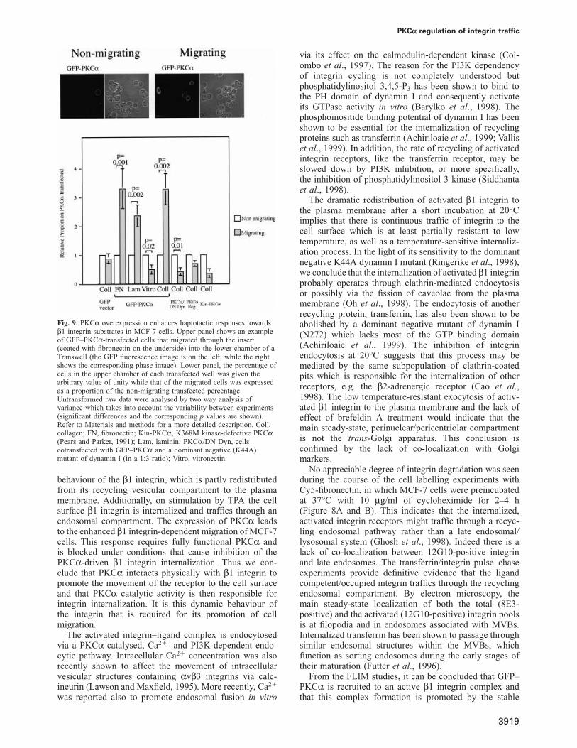
GFP–PKCα expression promotes β1 integrin-dependent migrationThe evidence presented above indicates that PKCα isrecruited to β1 integrin receptor complexes. This associ-ation is in part dependent upon the regulatory domain.Conversely the cell surface 12G10-positive integrin canbe internalized in a PKCα kinase activity-dependentmanner. To assess how this PKC-dependent behaviourmay influence integrin-dependent function, we analysedthe migratory behaviour of MCF-7 cells in a modifiedTranswell chamber assay. GFP–PKCα was transfectedinto cells and the proportion of transfected cells migratingor not migrating on different matrix substrates was scored(Figure 9). Among GFP–PKCα-transfected cells, therewas an enrichment of GFP–PKCα-positive cells in themigrating population when cells were plated on the β1 inte-grin substrates: fibronectin (FN), laminin (Lam) and colla-gen (Coll). In Figure 9, the percentage of cells in theupper chamber of each transfected well was given thearbitrary value of unity while that of the migrated cells wasexpressed as a proportion of the non-migrating transfectedpercentage, for comparison of results from independentexperiments. In a typical experiment, 6% (SD 2%, No. offields � 6) of cells in the upper chamber expressed GFP–PKCα while the percentage of expressors among themigrated cells in wells coated with fibronectin wasenriched to 17% (SD 6%). Vitronectin is not a ligand forβ1 integrin and no enrichment of GFP–PKCα-positivecells was observed among the migrating population. Oneof the vitronectin receptors αvβ5 is, however, present inMCF-7 cells (Meyer et al., 1998). Cells transfected withGFP vector alone showed neither positive nor negativeenrichment among the migrating and non-migrating popu-lations. When the dominant negative dynamin I (K44A)construct was co-transfected with GFP–PKCα (3:1 ratio),the effect of PKCα expression on migration was blocked.This indicates that the active cycling of β1 integrincontributes to the migratory response. Consistent withthis, neither the kinase-defective PKCα nor the PKCαregulatory domain increased the ability of transfected cellsto migrate.
Discussion
The results here identify an interaction between PKCαand β1 integrin. This is established by FLIM in vivo andconfirmed by co-immunoprecipitation of the proteins fromcell extracts. This association occurs preferentially withthe active conformer of β1 integrin (12G10 positive) andis enhanced by recruitment of PKCα to membranesby TPA. The PKCα association correlates with altered
PKCα regulation of integrin traffic
Fig. 9. PKCα overexpression enhances haptotactic responses towardsβ1 integrin substrates in MCF-7 cells. Upper panel shows an exampleof GFP–PKCα-transfected cells that migrated through the insert(coated with fibronectin on the underside) into the lower chamber of aTranswell (the GFP fluorescence image is on the left, while the rightshows the corresponding phase image). Lower panel, the percentage ofcells in the upper chamber of each transfected well was given thearbitrary value of unity while that of the migrated cells was expressedas a proportion of the non-migrating transfected percentage.Untransformed raw data were analysed by two way analysis ofvariance which takes into account the variability between experiments(significant differences and the corresponding p values are shown).Refer to Materials and methods for a more detailed description. Coll,collagen; FN, fibronectin; Kin-PKCα, K368M kinase-defective PKCα(Pears and Parker, 1991); Lam, laminin; PKCα/DN Dyn, cellscotransfected with GFP–PKCα and a dominant negative (K44A)mutant of dynamin I (in a 1:3 ratio); Vitro, vitronectin.
behaviour of the β1 integrin, which is partly redistributedfrom its recycling vesicular compartment to the plasmamembrane. Additionally, on stimulation by TPA the cellsurface β1 integrin is internalized and traffics through anendosomal compartment. The expression of PKCα leadsto the enhanced β1 integrin-dependent migration of MCF-7cells. This response requires fully functional PKCα andis blocked under conditions that cause inhibition of thePKCα-driven β1 integrin internalization. Thus we con-clude that PKCα interacts physically with β1 integrin topromote the movement of the receptor to the cell surfaceand that PKCα catalytic activity is then responsible forintegrin internalization. It is this dynamic behaviour ofthe integrin that is required for its promotion of cellmigration.
The activated integrin–ligand complex is endocytosedvia a PKCα-catalysed, Ca2�- and PI3K-dependent endo-cytic pathway. Intracellular Ca2� concentration was alsorecently shown to affect the movement of intracellularvesicular structures containing αvβ3 integrins via calc-ineurin (Lawson and Maxfield, 1995). More recently, Ca2�
was reported also to promote endosomal fusion in vitro
3919
via its effect on the calmodulin-dependent kinase (Col-ombo et al., 1997). The reason for the PI3K dependencyof integrin cycling is not completely understood butphosphatidylinositol 3,4,5-P3 has been shown to bind tothe PH domain of dynamin I and consequently activateits GTPase activity in vitro (Barylko et al., 1998). Thephosphoinositide binding potential of dynamin I has beenshown to be essential for the internalization of recyclingproteins such as transferrin (Achiriloaie et al., 1999; Valliset al., 1999). In addition, the rate of recycling of activatedintegrin receptors, like the transferrin receptor, may beslowed down by PI3K inhibition, or more specifically,the inhibition of phosphatidylinositol 3-kinase (Siddhantaet al., 1998).
The dramatic redistribution of activated β1 integrin tothe plasma membrane after a short incubation at 20°Cimplies that there is continuous traffic of integrin to thecell surface which is at least partially resistant to lowtemperature, as well as a temperature-sensitive internaliz-ation process. In the light of its sensitivity to the dominantnegative K44A dynamin I mutant (Ringerike et al., 1998),we conclude that the internalization of activated β1 integrinprobably operates through clathrin-mediated endocytosisor possibly via the fission of caveolae from the plasmamembrane (Oh et al., 1998). The endocytosis of anotherrecycling protein, transferrin, has also been shown to beabolished by a dominant negative mutant of dynamin I(N272) which lacks most of the GTP binding domain(Achiriloaie et al., 1999). The inhibition of integrinendocytosis at 20°C suggests that this process may bemediated by the same subpopulation of clathrin-coatedpits which is responsible for the internalization of otherreceptors, e.g. the β2-adrenergic receptor (Cao et al.,1998). The low temperature-resistant exocytosis of activ-ated β1 integrin to the plasma membrane and the lack ofeffect of brefeldin A treatment would indicate that themain steady-state, perinuclear/pericentriolar compartmentis not the trans-Golgi apparatus. This conclusion isconfirmed by the lack of co-localization with Golgimarkers.
No appreciable degree of integrin degradation was seenduring the course of the cell labelling experiments withCy5-fibronectin, in which MCF-7 cells were preincubatedat 37°C with 10 μg/ml of cycloheximide for 2–4 h(Figure 8A and B). This indicates that the internalized,activated integrin receptors might traffic through a recyc-ling endosomal pathway rather than a late endosomal/lysosomal system (Ghosh et al., 1998). Indeed there is alack of co-localization between 12G10-positive integrinand late endosomes. The transferrin/integrin pulse–chaseexperiments provide definitive evidence that the ligandcompetent/occupied integrin traffics through the recyclingendosomal compartment. By electron microscopy, themain steady-state localization of both the total (8E3-positive) and the activated (12G10-positive) integrin poolsis at filopodia and in endosomes associated with MVBs.Internalized transferrin has been shown to passage throughsimilar endosomal structures within the MVBs, whichfunction as sorting endosomes during the early stages oftheir maturation (Futter et al., 1996).
From the FLIM studies, it can be concluded that GFP–PKCα is recruited to an active β1 integrin complex andthat this complex formation is promoted by the stable
T.Ng et al.
membrane association of GFP–PKCα, driven by its activ-ator TPA. In the absence of TPA, GFP–PKCα can stillcomplex activated β1 integrin, unlike the GFP–PKCαreg
protein, which preferentially associates with the ligand-unoccupied integrin conformer. This difference betweenintact GFP–PKCα and GFP–PKCαreg indicates that eitherthe kinase domain contributes to the PKCα–activatedintegrin complex formation or the regulatory domainconformation is distinct in the intact protein. However, inthe presence of TPA in vivo, the regulatory domain issufficient for complex formation with the activated integrinto occur.
The enhanced surface expression of the 12G10-immuno-reactive β1 integrin in PKCα-overexpressing cells is atleast in part due to an overall upregulation of integrinexocytosis, as shown by the pan-β1 integrin antibody8E3. The ligand-competent/occupied integrin conformeris, however, shown to turn over more quickly, with respectto both the exocytosis and internalization processes. Thisis akin to the finding that the high affinity epidermalgrowth factor (EGF) receptor is internalized more quicklycompared with the bulk population and that the eliminationof a major PKC phosphorylation site (T654) of thisreceptor significantly reduces its rate of internalization(Felder et al., 1992). Previous studies with phorbol estershave demonstrated an upregulation of cell surface TS2/16-immunoreactive β1 integrin, which can be abrogatedby a prior incubation with nocodazole (Chun et al.,1997). Nocodazole is shown here to disperse the integrin-containing vesicular structures as well as prevent thesteady-state accumulation of activated integrin at theplasma membrane. The effect of microtubule depolymer-ization may be to destabilize/disperse the Rab11-con-taining recycling endosomal structures as shownpreviously (Casanova et al., 1999). Intracellular vesiclescontaining β2 integrins can also be stimulated to transloc-ate to the cell surface by phorbol ester treatment in U937cells (Kiley and Parker, 1997; Kiley et al., 1997). Themechanism by which PKC brings about an upregulationof integrin exocytosis may be related to its ability toactivate ARF-dependent PLD (Geny and Cockcroft, 1992;Whatmore et al., 1996). Consistent with the effects seenhere, it has been established that the regulatory domainof PKCα is sufficient for allosteric activation of PLD(Singer et al., 1996).
Stimulation of the conventional and novel PKC isotypesin colon carcinoma cells increases the cell migratoryresponse to fibronectin, collagen, laminin and vitronectinindiscriminately (Rigot et al., 1998). In contrast, transienttransfection experiments in this study demonstrate anenhanced haptotactic response specifically towards β1 inte-grin substrates in PKCα-overexpressing MCF-7 cells. Infact, PKCα overexpression in MCF-7 cells confers adramatic increase in random motility. UntransfectedMCF-7 cells fail to migrate across the filter in a controlchamber [where the undersurface of the chamber is coatedwith bovine serum albumin (BSA) alone] after a 36 hincubation. However, PKCα-transfected cells migrateacross the BSA-coated filters and the migrating cells arefound to be consistently (100%) GFP–PKCα positive(T.Ng and P.J.Parker, unpublished results). Morpholo-gically, the loss of activated integrin from the site ofattachment to cell substratum in TPA-stimulated, PKCα-
3920
transfected MCF-7 cells is seen to be followed by distinctcell shape changes (such as cell rounding). This, coupledwith the observation that activated integrin-containingstructures are found to extend into the filopodia (Figure 2;Rabinovitz and Mercurio, 1997; Miranti et al., 1998),provide further evidence that the active cycling of β1 inte-grin may contribute to the migratory response. The struc-tural determinants in β1 integrin that mediate the PKCα-enhanced haptotactic responses have yet to be defined butmay reside in the NPXY-containing cytoplasmic domains(Filardo et al., 1995; Saka et al., 1998; Sakai et al., 1998).
A further physiological implication of these studiesrelates to the general observation that the signallingfunction of a growing number of surface receptors suchas the β2-AR (Daaka et al., 1998) and insulin-like growthfactor-1 (IGF-1) receptor (Lin et al., 1998) are affectedby the ability of these receptors to internalize and sub-sequently traffic intracellularly. For instance, the endo-cytosis of both β2-AR and IGF1 receptor is clathrinmediated and inhibitable by the K44A mutant of dynamin I.A consequence of the blockade of receptor internalizationis the impairment of either receptor to induce mitogen-activated protein kinase (MAPK) activation. A similareffect was obtained using a dominant negative mutant ofβ-arrestin 1 (S412D) to inhibit IGF-1 receptor endocytosis(Lin et al., 1998). Given the very same sensitivity of theintegrin internalization machinery to the dominant negativeeffect of K44A dynamin I, the ‘outside-in’ signallingresponses elicited by integrin heterodimers may similarlybe affected by conditions which can disrupt the normalcycling of these receptors.
Materials and methods
Cell culture and transfectionHuman breast carcinoma cells (MCF-7 and MDA-MB-231 cells) werecultured in Dulbecco’s modified Eagle’s medium (DMEM) containing10% fetal calf serum at 37°C, in a 10% CO2 atmosphere. In addition,the MCF-7 cultures were grown in media supplemented with insulin(10 μg/ml). Cells were transfected using Lipofectant (Life Technologies).Transfected cells were stimulated as indicated in the text and figurelegends, 48 h after transfection.
Plasmid constructsMyc- and GFP-tagged PKCα constructs were obtained by subcloningthe PKCα coding sequence into the EcoRI sites of a modified pCDNA3vector (Clontech) and a pZeoSV-derived vector (Invitrogen), permittingfusion of a myc coding sequence (ATGGAGCAGAAGCTCATATCGG-AGGAGGACCTAGGGCCCGAATTC) and the coding sequence of GFP,respectively, at the N-terminus of PKCα. In order to fuse PKCα in-frame with the myc and GFP tags, the p-Babe vector (Alvaro et al.,1997) containing human PKCα was used as a template in a PCRusing 5� primer 5�-GGGAATTCCAGCAAGCTTGGTTGGGGGGGGG-ACC-3� (for insertion in pCDNA3) or 5�-GGGAATTCCAGCAA-GCTTGTTGGGGGGGGGACC-3� (for insertion in pZeoSV) and a 3�primer 5�-CATATCGCAGGTGTCACATTTCATCCCTTGATGGAT-3�corresponding to the 391–420 bp of the PKCα coding sequence. The455/4 bp PKCα N-terminal amplified sequences were digested attheir unique BanI restriction site. The resulting 265/4 bp sequencescorresponding to the N-terminal sequence of PKCα were then ligatedto the C-terminal part of PKCα obtained from a combined BanI andEcoRI digestion of the original p-Babe vector. The resulting ligationproduct containing the full-length sequence of PKCα was digested withEcoRI and ligated to the EcoRI site of the corresponding vectors. Theintegrity of the PCR amplified N-terminal sequence of PKCα wasverified by sequencing. The construction of the CD2–PKM plasmid hasbeen described previously (Garcia-Paramio et al., 1998). The GFP-tagged PKCα regulatory domain (with variable domain V3) constructwas kindly provided by D.Joubert (INSERM U469, Montpellier, France).
PKCα regulation of integrin traffic
The dominant negative (K44A) mutant of dynamin I and the full-lengthchicken β1 integrin plasmids were generous gifts of S.Schmidt (Scripps,La Jolla, CA) and Dr A.Horwitz (University of Illinois, Urbana, IL),respectively.
Antibodies and direct conjugation to fluorophores or proteinG–SepharoseMC5 is a murine mAb that recognizes the V3 region of PKCα (Younget al., 1988). The monoclonal antibody M4 (a gift of S.Jaken, LakePlacid, NY), is directed against the PKCα regulatory domain and wasused as ascites fluid. The characterization of the mouse anti-humanintegrin mAb 12G10, which recognizes an activation epitope within theputative A domain region has been described previously (Fogerty et al.,1990; Mould et al., 1995, 1998). The rat anti-human β1 antibody mAb13recognizes the ligand-unoccupied forms of the integrin and blocks ligandbinding (Humphries, 1996; Mould et al., 1996). 8E3 is a pan-β1 integrinmurine mAb. Direct conjugation of proteins/antibodies to the fluoro-phores Cy3 and Cy3.5/Cy5 (Amersham Life Science) was performed atpH 9 as described previously (Bastiaens and Jovin, 1998). Anti-Rab 7polyclonal antiserum was a gift from M.Zerial (EMBL, Heidelberg).Rabbit anti-Rab11 was kindly provided by Dr S.Tooze (ICRF, London).Goat anti-transferrin receptor and anti-fibronectin polyclonal antibodieswere from Santa Cruz Biotechnology. mAb MC5 was coupled to proteinG–Sepharose (Pharmacia) at pH 9 (in 0.2 M sodium borate buffer) usingdimethyl pimelimidate according to the manufacturer’s protocol.
Immunoprecipitation, Western blotting and in vitro integrinpull-downMyc-tagged PKCα or GFP–PKCαreg was immunoprecipitated from thewhole cell lysates of MCF-7 cells using 10–20 μg of protein G-coupledMC5 as described previously (Kanner et al., 1989), except that 1%(w/v) n-octyl β-D-glucopyranoside was used instead of NP-40. Theimmunoprecipitates were denatured in sample buffer (Laemmli, 1970),separated on an 8% polyacrylamide gel under reducing conditions andtransferred electrophoretically to PVDF membrane. Blots were thenprobed with the rat anti-integrin mAb13 (2–4 μg/ml) followed by adonkey anti-rat–HRP conjugate (Amersham) in the presence of 0.5 μg/ml mouse IgG to block immunoreactivity with the precipitating antibody.The lower part of the same membrane was probed with MC5 or M3(used at 2 μg/ml and 1/1000 dilution of ascites, respectively) to detectPKCα or its regulatory domain. Incubation with primary antibodies wasperformed overnight at 4°C. Detection was with ECL (Amersham)according to recommended procedures.
For in vitro pull-down assays, immunoprecipitates containing GFP–PKCαreg protein were washed thoroughly with modified RIPA buffer(Kanner et al., 1989), then left to tumble overnight at 4°C in the samebuffer (negative control) or in freshly lysed detergent extracts of MCF-7cells alone or cells which had been transiently transfected with a full-length chicken β1 integrin construct (Reszka et al., 1992). The GFP–PKCαreg protein complexes were then washed three times with RIPAbuffer, once with Tris–saline buffer (pH 7.4) and subsequently resus-pended in Laemmli sample buffer.
Fibronectin uptake and integrin/transferrin pulse–chaseexperimentsFor fibronectin uptake studies, MCF-7 cells transiently transfected withvarious PKCα constructs were pretreated with cycloheximide (10 μg/ml) for 2–4 h to inhibit the synthesis of new proteins, including integrinreceptors. Cells were then incubated at 4°C with media containing 50 μg/ml Cy5-labelled fibronectin, with or without 50 μg/ml FITC-labelledtransferrin (Sigma) for 30 min, washed with phosphate-buffered saline(PBS), then stimulated with TPA (400 nM) at 37°C for various lengthsof time before paraformaldehyde fixation, permeabilization and stainingwith 12G10–Cy3.5. The pulse–chase experiments were performed aspreviously described (Mayor et al., 1993). Briefly, untransfected MCF-7cells on coverslips were serum starved for 30 min, pulsed for 5 minwith Cy3.5-labelled 12G10 and FITC–Tf at 37°C, then the excess ligand/antibody was washed off with medium at 4°C. The pulsed cells werechased for various periods of time (up to 80 min) at 37°C, fixed in 2%(w/v) paraformaldehyde for 15 min at room temperature, then examinedby confocal microscopy.
Immunocytochemical staining and confocal microscopyImmunocytochemical staining was performed as described elsewhere(Kiley et al., 1997) except for the following modifications. Cells werepermeabilized with 0.2% (v/v) Triton X-100/PBS following fixation in4% (w/v) paraformaldehyde. Primary antibodies were diluted 1:200 in
3921
PBS containing 1% BSA, except for the fluorophore-conjugated antibod-ies which were used at 1:20–1:50. The secondary conjugates used wereCy5-conjugated donkey anti-goat IgG (1:200) and Texas Red/Cy3-conjugated donkey anti-rabbit IgG (1:400) (Jackson ImmunoResearchLaboratories, West Grove, PA). Confocal images were acquired on aconfocal laser scanning microscope (model LSM 510, Carl Zeiss Inc.)equipped with a 63�/1.4Plan-APOCHROMAT oil immersion objective.Each image represents a 2-dimensional projection of sections in thez-series, taken across the depth of the cell at 0.2 μm intervals.
Immunogold electron microscopyCells were scraped from culture plates and fixed in 4% (w/v) paraform-aldehyde for 1 h, followed by 2% (w/v) paraformaldehyde overnight at4°C. Cells were processed and ultrathin sections were collected asdescribed (Gorlich et al., 1995). Sections were incubated in the followingorder: anti-β1 integrin mAb (12G10/8E3, at 500 or 100 μg/ml respect-ively), rabbit-anti mouse IgG1 (1/100), protein A–gold 10 nM particles(1/100). Antibody reagents were diluted in PBS containing 1% (w/v)BSA. After antibody labelling, sections were examined using a Jeol1010 microscope.
FLIM measurementsA detailed description of the FLIM apparatus used for FRET determina-tion in this work can be found elsewhere (Squire and Bastiaens, 1999).The lifetime instrument performs phase and modulation based imagingfluorimetry by microscopy. All images were taken using a Zeiss Plan-APOCHROMAT 100�/1.4NA phase 3 oil objective and the homodynephase sensitive images recorded at a modulation frequency of80.218 MHz. For experiments involving the anti-integrin IgG–Cy3.5donor/acceptor FRET pair, the donor (GFP–PKCα or GFP–PKCαreg)was excited using the 488 nm line of a Argon/Krypton laser andthe resultant fluorescence separated using a combination of dichroicbeamsplitter (Q 505 LP; Chroma Technology Corp.) and narrow bandemitter filter (BP514/10; Lys & Optik). Acceptor images (anti-integrinmAb–Cy3.5) were recorded using a 100 W mercury arc lamp (ZeissAttoarc) as a source of sample illumination combined with a high QCy3 filter set (exciter: HQ 535/50, dichroic:Q 565 LP, emitter: HQ 610/75 LP; Chroma Technology Corp.).
Flow cytometric analysis and FACSortingTransiently transfected MCF-7 cells were detached from plates usingEDTA then, without fixation, stained with either an anti-β1 integrinmAb or mouse IgG isotype control, followed by an anti-mouse IgG–RPE conjugate. Cells were analysed on the same day by a FACScan, aspreviously described (Ng et al., 1995). All antibody incubation andwashing steps were carried out at 4°C. Cell purification by FACSortingaccording to GFP fluorescence was performed using Becton DickinsonFACS Vantage.
Transwell chamber haptotactic migration assays withimmunofluorescence microscopyMCF-7 cells were transiently transfected with a vector control or variousPKCα constructs. After 20 h, cells were detached from culture plateswith trypsin, washed three times with serum-free medium supplementedwith glutamine and 0.5% (w/v) BSA (migration buffer), then replatedat 106 cells/ml on to the inserts of 8 μm pore size Transwell chambers(Costar, Cambridge, MA) according to the manufacturer’s instructions.The underside of the inserts was precoated by filling the lower chamberwith either 0.1% (w/v) BSA or different integrin substrates (vitronectin5 μg/ml; fibronectin, laminin and collagen, 10 μg/ml) and incubationovernight at 4°C. After 20 h, cells were trypsinized from both the topand underside of the inserts and then washed three times in migrationbuffer. The cell suspensions obtained, along with the fluid from eitherthe upper or lower chambers, were centrifuged and cytospins recoveredon coverslips before mounting on a glass slide. The percentage of GFP-,GFP–PKCα- or GFP–PKCαreg-transfected cells that had migratedthrough the insert was compared with that of the non-migrating populationthat remained in the upper chamber. For the untagged kinase-defectivePKCα, immunofluorescence staining with a PKCα C-terminus-specificpolyclonal antibody was performed to ascertain the percentage oftransfected cells in each chamber. The cells in six or more low powerfields per slide were counted with the aid of a fluorescence microscope.Data from three independent experiments were pooled and analysed bytwo way analysis of variance using the statistical software MINITAB.
T.Ng et al.
Acknowledgements
We wish to thank Drs Fiona Watt, Birgit Leitinger and Nancy Hogg fortheir valuable comments. We also thank Dr M.Zerial (EMBL, Heidelberg,Germany) for the anti-Rab7 polyclonal antiserum, Dr S.Tooze (ICRF,London,UK) for the anti-Rab11 serum, Dr D.Joubert (INSERM U469,Montpellier, France) for the GFP-tagged PKCα regulatory domainconstruct, Dr S.L.Schmid (Scripps, La Jolla, CA) for the dominantnegative (K44A) mutant of dynamin I, as well as Dr A.Horwitz(University of Illinois, Urbana, IL) for the full-length chicken β1 integrinplasmid. M.J.H. is supported by grants from the Wellcome Trust.
References
Achiriloaie,M., Barylko,B. and Albanesi,J.P. (1999) Essential role of thedynamin pleckstrin homology domain in receptor-mediatedendocytosis. Mol. Cell. Biol., 19, 1410–1415.
Alvaro,V., Prevostel,C., Joubert,D., Slosberg,E. and Weinstein,B.I. (1997)Ectopic expression of a mutant form of PKCα originally found inhuman tumors: aberrant subcellular translocation and effects on growthcontrol. Oncogene, 14, 677–685.
Barylko,B., Binns,D., Lin,K.M., Atkinson,M.A.L., Jameson,D.M.,Yin,H.L. and Albanesi,J.P. (1998) Synergistic activation of dynaminGTPase by Grb2 and phosphoinositides. J. Biol. Chem., 273, 3791–3797.
Bastiaens,P.I.H. and Jovin,T.M. (1998) Fluorescence resonance energytransfer microscopy. In Celis,J.E. (ed.), Cell Biology: A LaboratoryHandbook. Academic Press, New York, NY, pp. 136–146.
Bastiaens,P.I.H. and Squire,A. (1999) Fluorescence lifetime imagingmicroscopy: spatial resolution of biochemical processes in the cell.Trends Cell Biol., 9, 48–52.
Bazzoni,G. and Hemler,M.E. (1998) Are changes in integrin affinity andconformation overemphasized? Trends Biochem. Sci., 23, 30–34.
Boudreau,N., Sympson,C.J., Werb,Z. and Bissell,M.J. (1995) Suppressionof ICE and apoptosis in mammary epithelial cells by extracellularmatrix. Science, 267, 891–893.
Bourdoulous,S., Orend,G., MacKenna,D.A., Pasqualini,R. andRuoslahti,E. (1998) Fibronectin matrix regulates activation of Rhoand cdc42 GTPases and cell cycle progression. J. Cell Biol., 143,267–276.
Cao,T.T., Mays,R.W. and von Zastrow,M. (1998) Regulated endocytosisof G-protein-coupled receptors by a biochemically and functionallydistinct subpopulation of clathrin-coated pits. J. Biol. Chem., 273,24592–24602.
Casanova,J.E., Wang,X. and Kumar,R. (1999) Association of Rab25 abdRab11a with the apical recycling system of polarized Madin-DarbyCanine kidney cells. Mol. Biol. Cell, 10, 47–61.
Chun,J., Auer,K.A. and Jacobson,B.S. (1997) Arachidonate initiatedprotein kinase C activation regulates HeLa cell spreading on a gelatinsubstrate by inducing F-actin formation and exocytotic upregulationof β1 integrin. J. Cell Physiol., 173, 361–370.
Colombo,M.I., Beron,W. and Stahl,P.D. (1997) Calmodulin regulatesendosome fusion. J. Biol. Chem., 272, 7707–7712.
Crommie,D. and Hemler,M.E. (1998) B1 integrin cytoplasmic domainregulates the constitutive conformation detected by MAb 15/7, butnot the ligand-induced conformation. J. Cell. Biochem., 71, 63–73.
Crouch,D.H., Fincham,V.J. and Frame,M.C. (1996) Targeted proteolysisof the focal adhesion kinase pp125FAK during c-Myc-inducedapoptosis is suppressed by integrin signalling. Oncogene, 12, 2689–2696.
Daaka,Y., Luttrell,L.M., Ahn,S., Rocca,G.J.D., Ferguson,S.S.G.,Caron,M.G. and Lefkowitz,R.J. (1998) Essential role for G protein-coupled receptor endocytosis in the activation of mitogen-activatedprotein kinase. J. Biol. Chem., 273, 685–688.
Felder,S., LaVin,J., Ullrich,A. and Schlessinger,J. (1992) Kinetics ofbinding, endocytosis and recycling of EGF receptor mutants. J. CellBiol., 117, 203–212.
Filardo,E.J. and Cheresh,D.A. (1994) A β turn in the cytoplasmic tailof the integrin αv subunit influences conformation and ligand bindingof αvβ3. J. Biol. Chem., 269, 4641–4647.
Filardo,E.J., Brooks,P.C., Deming,S.L., Damsky,C. and Cheresh,D.A.(1995) Requirement of the NPXY motif in the integrin β 3 subunitcytoplasmic tail for melanoma cell migration in vitro and in vivo.J. Cell Biol., 130, 441–450.
Fogerty,F.J., Akiyama,S.K., Yamada,K.M. and Mosher,D.F. (1990)Inhibition of binding of fibronectin to matrix assembly sites by anti-integrin (α 5 β 1) antibodies. J. Cell Biol., 111, 699–708.
3922
Frisch,S.M., Vuori,K., Kelaita,D. and Sicks,S. (1996a) A role for Jun-N-terminal kinase in anoikis; suppression by bcl-2 and crmA. J. CellBiol., 135, 1377–1382.
Frisch,S.M., Vuori,K., Ruoslahti,E. and Chan-Hui,P.Y. (1996b) Controlof adhesion-dependent cell survival by focal adhesion kinase. J. CellBiol., 134, 793–799.
Futter,C.E., Pearse,A., Hewlett,L.J. and Hopkins,C.R. (1996)Multivesicular endosomes containing internalized EGF–EGF receptorcomplexes mature and then fuse directly with lysosomes. J. Cell Biol.,132, 1011–1023.
Garcia-Paramio,P., Cabrerizo,Y., Bornancin,F. and Parker,P.J. (1998) Thebroad specificity of dominant inhibitory protein kinase C mutantsinfers a common step in phosphorylation. Biochem. J., 333, 631–636.
Geny,B. and Cockcroft,S. (1992) Synergistic activation of phospholipaseD by protein kinase C- and G-protein-mediated pathways instreptolysin O-permeabilized HL60 cells. Biochem. J., 284, 531–538.
Ghosh,R.N., Mallet,W.G., Soe,T.T., McGraw,T.E. and Maxfield,F.R.(1998) An endocytosed TGN38 chimeric protein is delivered to theTGN after trafficking through the endocytic recycling compartmentin CHO cells. J. Cell Biol., 142, 923–936.
Gorlich,D., Vogel,F., Mills,A.D., Hartmann,E. and Laskey,R.A. (1995)Distinct functions for the two importin subunits in nuclear proteinimport. Nature, 377, 246–248.
Gruenberg,J. and Maxfield,F.R. (1995) Membrane transport in theendocytic pathway. Curr. Opin. Cell Biol., 7, 552–563.
Hausdorff,W.P., Caron,M.G. and Lefkowitz,R.J. (1990) Turning off thesignal: desensitization of β-adrenergic receptor function. FASEB J., 4,2881–2889.
Hauzenberger,D., Klominek,J., Holgersson,J., Bergstrom,S.E. andSundqvist,K.G. (1997) Triggering of motile behavior in T lymphocytesvia cross-linking of α4 β1 and αL β2. J. Immunol., 158, 76–84.
Humphries,M.J. (1996) Integrin activation: the link between ligandbinding and signal transduction. Curr. Opin. Cell Biol., 8, 632–640.
Inglese,J., Freedman,N.J., Koch,W.J. and Lefkowitz,R.J. (1993) Structureand mechanism of the G protein-coupled receptor kinases. J. Biol.Chem., 268, 23735–23738.
Kanner,S.B., Reynolds,A.B. and Parsons,J.T. (1989) Immunoaffinitypurification of tyrosine-phosphorylated cellular proteins. J. Immunol.Methods, 120, 115–124.
Kiley,S.C. and Parker,P.J. (1997) Defective microtubule reorganisationin phorbol ester-resistant U937 variants—reconstitution of the normal-cell phenotype with nocodazole. Cell Growth Differ., 8, 231–242.
Kiley,S.C., Adams,P.D. and Parker,P.J. (1997) Cloning andcharacterisation of phorbol ester differentiation-resistant U937 cellvariants. Cell Growth Differ., 8, 221–230.
Klemke,R.L., Yebra,M., Bayna,E.M. and Cheresh,D.A. (1994) Receptortyrosine kinase signaling required for integrin αVβ5-directed cellmotility but not adhesion on vitronectin. J. Cell Biol., 127, 859–866.
Laemmli,U.K. (1970) Cleavage of structural proteins during the assemblyof the head of bacteriophage T4. Nature, 227, 680–685.
Lauffenburger,D.A. and Horwitz,A.F. (1996) Cell migration: a physicallyintegrated molecular process. Cell, 84, 359–369.
Lawson,M.A. and Maxfield,F.R. (1995) Ca2�- and calcineurin-dependentrecycling of an integrin to the front of the migrating neutrophils.Nature, 377, 75–79.
Lefkowitz,R.J. (1993) G protein-coupled receptor kinases. Cell, 74,409–412.
Lin,F.T., Daaka,Y. and Lefkowitz,R.J. (1998) β-arrestins regulatemitogenic signaling and clathrin-mediated endocytosis of the insulin-like growth factor 1 receptor. J. Biol. Chem., 273, 31640–31643.
Malik,R.K. and Parsons,J.T. (1996) Integrin-dependent activation of thep70 ribosomal S6 kinase signaling pathway. J. Biol. Chem., 271,29785–29791.
Mastrangelo,A.M., Homan,S.M., Humphries,M.J. and LaFlamme,S.E.(1999) Amino acid motifs required for isolated β cytoplasmic domainsto regulate ‘in trans’ β1 integrin conformation and function in cellattachment. J. Cell Sci., 112, 217–229.
Mayor,S., Presley,J.F. and Maxfield,F.R. (1993) Sorting of membranecomponents from endosomes and subsequent recycling to the cellsurface occurs by a bulk flow process. J. Cell Biol., 121, 1257–1269.
Meng,F. and Lowell,C.A. (1998) A β1 integrin signaling pathwayinvolving Src-family kinases, Cbl and PI-3 kinase is required formacrophage spreading and migration. EMBO J., 17, 4391–4403.
Meredith,J.E., Fazeli,B. and Schwartz,M.A. (1993) The extracellularmatrix as a cell survival factor. Mol. Biol. Cell, 4, 953–961.
Meyer,T., Marshall,J.F. and Hart,I.R. (1998) Expression of αv integrins
PKCα regulation of integrin traffic
and vitronectin receptor identity in breast cancer cells. Br. J. Cancer,77, 530–536.
Miranti,C.K., Leng,L., Maschberger,P., Brugge,J.S. and Shattil,S.J.(1998) Identification of a novel integrin signaling pathway involvingthe kinase Syk and the guanine nucleotide exchange factor Vav1.Curr. Biol., 8, 1289–1299.
Mould,A.P., Garratt,A.N., Askari,J.A., Akiyama,S.K. andHumphries,M.J. (1995) Identification of a novel anti-integrinmonoclonal antibody that recognises a ligand-induced binding siteepitope on the β1 subunit. FEBS Lett., 363, 118–122.
Mould,A.P., Akiyama,S.K. and Humphries,M.J. (1996) The inhibitoryanti-β1 integrin monoclonal antibody 13 recognizes an epitope that isattenuated by ligand occupancy. Evidence for allosteric inhibition ofintegrin function. J. Biol. Chem., 271, 20365–20374.
Mould,A.P., Garratt,A.N., Puzon-McLaughlin,W., Takada,Y. andHumphries,M.J. (1998) Regulation of integrin function: evidence thatbivalent-cation-induced conformational changes lead to the unmaskingof ligand-binding sites within integrin α5 β1. Biochem. J., 331,821–828.
Ng,T.T.C., Guntermann,C., Nye,K.E., Parkin,J.M., Anderson,J.,Norman,J.E. and Morrow,W.J.W. (1995) Adhesion co-receptorexpression and intracellular signalling in HIV disease: implicationsfor immunotherapy. AIDS, 9, 337–343.
Ng,T.T.C., Kanner,S.B., Humphries,M.J., Wickremasinghe,R.G.,Nye,K.E., Anderson,J., Khoo,S.H. and Morrow,W.J.W. (1997) Theintegrin-triggered rescue of T lymphocyte apoptosis is blocked inHIV-1-infected individuals. J. Immunol., 158, 2984–2999.
Ng,T. et al. (1999) Imaging protein kinase C α activation in cells.Science, 283, 2085–2089.
Oh,P., McIntosh,D.P. and Schnitzer,J.E. (1998) Dynamin at the neck ofcaveolae mediates their budding to form transport vesicles by GTP-driven fission from the plasma membrane of endothelium. J. CellBiol., 141, 101–114.
Pears,C. and Parker,P.J. (1991) Down-regulation of a kinase defectivePKC-a. FEBS Lett., 284, 120–122.
Rabinovitz,I. and Mercurio,A.M. (1997) The integrin α6β4 functions incarcinoma cell migration on laminin-1 by mediating the formationand stabilization of actin-containing motility structures. J. Cell Biol.,139, 1873–1884.
Reszka,A.A., Hayashi,Y. and Horwitz,A.F. (1992) Identification of aminoacid sequences in the integrin β1 cytoplasmic domain implicated incytoskeletal association. J. Cell Biol., 117, 1321–1330.
Retta,S.F., Balzac,F., Ferraris,P., Belkins,A.M., Fassler,R.,Humphries,M.J., De Leo,G., Silengo,L. and Tarone,G. (1998) β1-cytoplasmic subdomains involved in dominant negative function. Mol.Biol. Cell, 9, 715–731.
Rigot,V., Lehmann,M., Andre,F., Daemi,N., Marvaldi,J. and Luis,J.(1998) Integrin ligation and PKC activation are required for migrationof colon carcinoma cells. J. Cell Sci., 111, 3119–3127.
Ringerike,T., Stang,E., Johannessen,L.E., Sandnes,D., Levy,F.O. andMadshus,I.H. (1998) High-affinity binding of epidermal growth factor(EGF) to EGF receptor is disrupted by overexpression of mutantdynamin (K44A). J. Biol. Chem., 273, 16639–16642.
Saka,T., Peyruchaud,O., Fassler,R. and Mosher,D.F. (1998) Restorationof β1A integrins is required for lysophosphatidic acid-inducedmigration of β 1-null mouse fibroblastic cells. J. Biol. Chem., 273,19378–19382.
Sakai,T., Zhang,Q., Fassler,R. and Mosher,D.F. (1998) Modulation ofβ1A integrin functions by tyrosine residues in the β1 cytoplasmicdomain. J. Cell Biol., 141, 527–538.
Sanchez-Madrid,F. and del Pozo,M.A. (1999) Leukocyte polarization incell migration and immune interactions. EMBO J., 18, 501–511.
Schneller,M., Vuori,K. and Ruoslahti,E. (1997) αVβ3 integrin associateswith activated insulin and PDGF β receptors and potentiates thebiological activity of PDGF. EMBO J., 16, 5600–5607.
Shih,M. and Malbon,C.C. (1996) Protein kinase C deficiency blocksrecovery from agonist-induced desensitization. J. Biol. Chem., 271,21478–21483.
Siddhanta,U., McIlroy,J., Shah,A., Zhang,Y. and Backer,J.M. (1998)Distinct roles for the p110α and hVPS34 phosphatidylinositol 3�-kinases in vesicular trafficking, regulation of the actin cytoskeletonand mitogenesis. J. Cell Biol., 143, 1647–1659.
Singer,W.D., Brown,H.A., Jiang,X. and Sternweis,P.C. (1996) Regulationof phospholipase D by protein kinase C is synergistic with ADP-ribosylation factor and independent of protein kinase activity. J. Biol.Chem., 271, 4504–4510.
Squire,A. and Bastiaens,P.I.H. (1999) Three dimensional image
3923
restoration in fluorescence lifetime imaging microscopy. J. Microsc.,193, 36–49.
Timar,J., Trikha,M., Szekeres,K., Bazaz,R., Tovari,J., Silletti,S., Raz,A.and Honn,K.V. (1996) Autocrine motility factor signals integrin-mediated metastatic melanoma cell adhesion and invasion. CancerRes., 56, 1902–1908.
Udagawa,T. and McIntyre,B.W. (1996) ADP-ribosylation of the G proteinRho inhibits integrin regulation of tumor cell growth. J. Biol. Chem.,271, 12542–12548.
Vallis,Y., Wigge,P., Marks,B., Evans,P.R. and McMahon,H.T. (1999)Importance of the pleckstrin homology domain of dynamin in clathrin-mediated endocytosis. Curr. Biol., 9, 257–260.
Wary,K.K., Mainiero,F., Isakoff,S.J., Marcantonio,E.E. andGiancotti,F.G. (1996) The adaptor protein Shc couples a class ofintegrins to the control of cell cycle progression. Cell, 87, 733–743.
Werr,J., Xie,X., Hedqvist,P., Ruoslahti,E. and Lindbom,L. (1998) β1integrins are critically involved in neutrophil locomotion inextravascular tissue in vivo. J. Exp. Med., 187, 2091–2096.
Whatmore,J., Morgan,C.P., Cunningham,E., Collison,K.S., Willison,K.R.and Cockcroft,S. (1996) ADP-ribosylation factor 1-regulatedphospholipase D activity is localized at the plasma membrane andintracellular organelles in HL60 cells. Biochem. J., 320, 785–794.
Woods,A. and Couchman,J.R. (1992) Protein kinase C involvement infocal adhesion formation. J. Cell Sci., 101, 277–290.
Yebra,M., Filardo,E.J., Bayna,E.M., Kawahara,E., Becker,J.C. andCheresh,D.A. (1995) Induction of carcinoma cell migration onvitronectin by NF-kB-dependent gene expression. Mol. Biol. Cell, 6,841–850.
Young,S., Rothbard,J. and Parker,P. (1988) A monoclonal antibodyrecognising the site of limited proteolysis of protein kinase C. Eur. J.Biochem., 173, 247–252.
Yun,Z., Menter,D.G. and Nicolson,G.L. (1996) Involvement of integrinαVβ3 in cell adhesion, motility and liver metastasis of murineRAW117 large cell lymphoma. Cancer Res., 56, 3103–3111.
Received April 15, 1999; revised and accepted May 21, 1999