physiotherapeutic treatment for axillary cord formation ...physiotherapeutic treatment for axillary...
TRANSCRIPT
Physiotherapeutic treatment for axillary cord formation following breast cancer surgery
Elisabeth Josenhans Physiotherapist Kunhardtstr 4 20249 Hamburg Tel. +49(0)40/4802703 www.praxisjosenhans.de [email protected]
Physiotherapeutic treatment for axillary cord formation
following breast cancer surgery
Science prize 2007of the ZVK
(Deutscher Verband für Physiotherapie - Zentralverband der Physiotherapeuten / Krankengymnasten e.V. German central association of physiotherapists)
Elisabeth Josenhans
Physiotherapist
Physiotherapeutic treatment for axillary cord formation following breast cancer surgery
Elisabeth Josenhans Physiotherapist Kunhardtstr 4 20249 Hamburg Tel. +49(0)40/4802703 www.praxisjosenhans.de [email protected]
Introduction According to the Deutsche Krebsgesellschaft (German cancer association), breast cancer is diagnosed in approximately 55,100 German women each year, and is the most frequently occurring malignant tumor in women (Deutsche Krebsgesellschaft e.V. 2006). In most cases, surgical treatment is required for this diagnosis. The hospital stay is usually short and therefore physiotherapeutic measures are limited to the demonstration of exercises for improving arm mobility. These exercises are to be continued by the patient at home. In the event of lymphedema, treatment with manual lymph drainage (MLD) is performed after discharge from hospital. Following breast cancer surgery with the removal of axillary lymph nodes, adhesion formation can result in one or more painful cords in the arm originating from the axilla. A literature review reveals that a physiotherapeutic treatment concept specifically designed for cord formation has not yet been established. Physicians at Hamburg breast centers who were asked about the cause of such cord formation could not provide any clear, unanimous statement regarding it. Hypotheses were made concerning scar cords, fascial adhesion of axillary tissue, inflamed lymph cords and re-formation of tissue. The physicians advised the affected patients to perform exercises and to wait until the problem resolved itself. Patients who were severely affected were offered surgical cord removal. It was stated that cord formation occurred in approximately 5% to 10% of all patients following breast cancer surgery. The physicians consulted were not aware of any scientific studies of this phenomenon. Based on this estimate and on the figures provided by the Deutsche Krebsgesellschaft, in Germany each year approximately 2,755 to 5,500 women are affected by axillary cord formation following surgery for breast cancer. Other physiotherapists were consulted, who stated that such cords had been seen, but that no specific treatment for them was known. All colleagues agreed that due to the risk of lymphedema, according to their understanding, no manual treatment should be employed. In the Internet, on the numerous webpages related to breast cancer, there were only isolated references to cord formation. Internet searches using the terms “Strang” (cord), “Geigensaitenphänomen” (fiddle-string phenomenon) and “fibrosierte Lymphbahnen” (lymph vessel fibrosis) in conjunction with breast cancer surgery yielded no information about treatment possibilities. For several years, patients with breast cancer surgery have been treated in my physiotherapeutic practice. Among these were patients with an axillary cord which, upon elevation or abduction of the shoulder, was stretched visibly and/or palpably in a very painful manner. This hindered the free mobility of the arm. In some patients the cord could be seen extending past the elbow to the lower arm. This complication hinders both daily mobility and subsequent treatment by impairing the positioning of patients for radiation treatment, where the arm must be positioned above the head. Patients described how they had attempted unsuccessfully over a period of weeks or months to improve the painful restriction of arm mobility by means of active home exercises. Some of these patients even reported that the cord had thickened due to the exercises. At the urgent request of a patient with very restricted shoulder mobility and severe suffering, the painful cord was treated with manual releasing techniques, with the result that the cord was remedied after a few treatment sessions. After checking with the physicians involved, additional patients were treated specifically for cord formation by means of manual techniques. The results were encouraging. In every case, even when months had passed since the operation, it was possible to eliminate or greatly reduce the tension of the cord.

Physiotherapeutic treatment for axillary cord formation following breast cancer surgery
Elisabeth Josenhans Physiotherapist Kunhardtstr 4 20249 Hamburg Tel. +49(0)40/4802703 www.praxisjosenhans.de [email protected]
In the context of the problems associated with a life-threatening cancer, physicians view the impairment of shoulder functioning as an insignificant, marginal problem that does not require treatment. However, as a result of their sudden illness due to cancer, the patients are in an exceptional psychological state. Dealing with the illness and subsequent treatments such as chemotherapy and radiation is a severe burden for the patients. If painful restriction of shoulder mobility occurs due to cord formation, everyday movements become difficult or impossible, which can lead to additional psychological stress. Therefore the reduction of shoulder mobility is not regarded as an insignificant problem by patients. Numerous observations during the treatments showed that patients react very positively to the immediate effect of the treatment in reducing pain and improving shoulder mobility. Patients report that due to the physiotherapeutic treatment, they feel that at least in one area progress has been made. With each treatment session, the arm becomes more mobile and the pain more bearable. Many patients report that in addition, they feel that the manipulation and treatment of the area of the operation has a healing effect. Particularly in the case of patients who must deal with a total mastectomy, the treatment and manipulation of the area of the operation may lead to a better acceptance of the altered body structure. The physiotherapeutic treatment performed on 123 breast cancer patients after surgery which resulted in axillary cord formation is explained in this paper. Objective of the study The objective of this work is to show that by means of a new treatment approach, cord formation following breast cancer surgery can be remedied or reduced in a few treatment sessions, with minimal side effects. Study design The target group consisted of patients following breast cancer surgery who had - a visible or palpable cord and/or - mobility restrictions of shoulder elevation and/or - pain during shoulder elevation. The mobility restrictions or pain during shoulder movement had to be related to the surgery; patients with such symptoms resulting from other conditions were not included in the study. The analysis did not include patients - who had a mobility restriction solely due to a problem with the shoulder joint or - who discontinued the treatment. The treatment was discontinued by fewer than 10% of the patients. Reasons for discontinuation were due to rehabilitation measures, metastasis, additional required surgery, thrombosis in the port, or erysipelas of the arm after the incision. No patients discontinued treatment due to complications arising from the treatment or as a result of non-acceptance of the treatment. During the study from December 2003 to September 2006, 123 patients received physiotherapeutic treatment. Documentation was carried out at the beginning and end of the series of treatments, with the aid of a standardized form. In particular, the following parameters were recorded: - extent of shoulder mobility - pain experienced during the shoulder movement
Physiotherapeutic treatment for axillary cord formation following breast cancer surgery
Elisabeth Josenhans Physiotherapist Kunhardtstr 4 20249 Hamburg Tel. +49(0)40/4802703 www.praxisjosenhans.de [email protected]
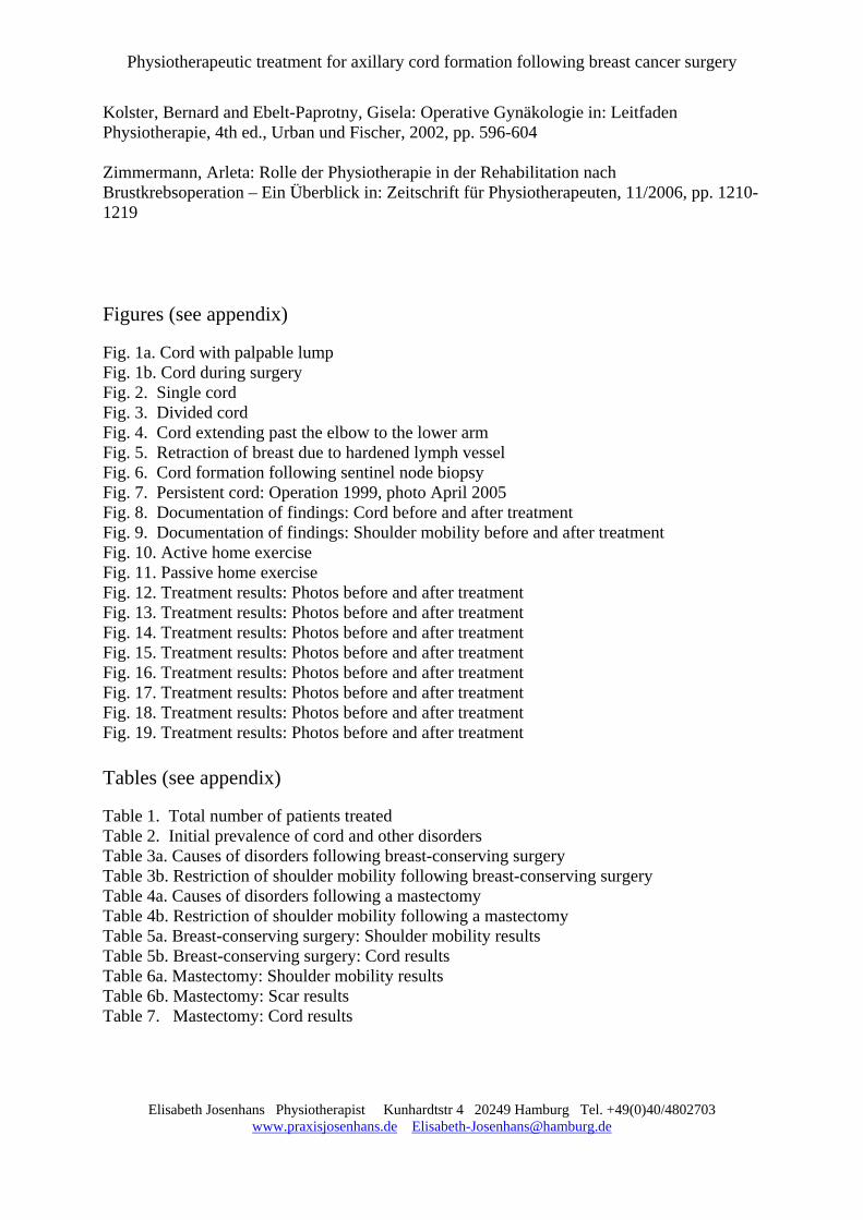
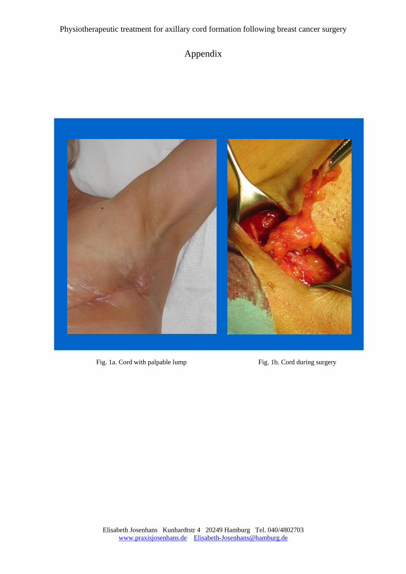
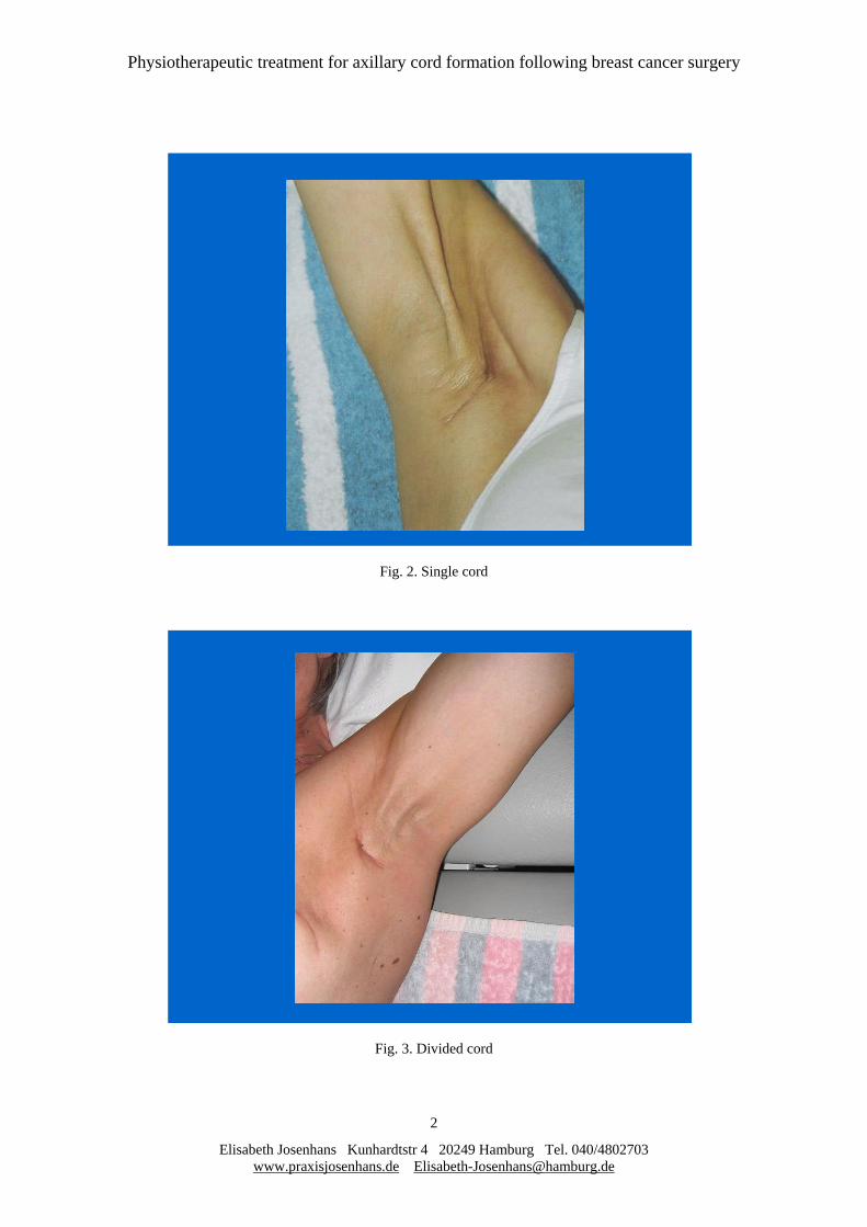
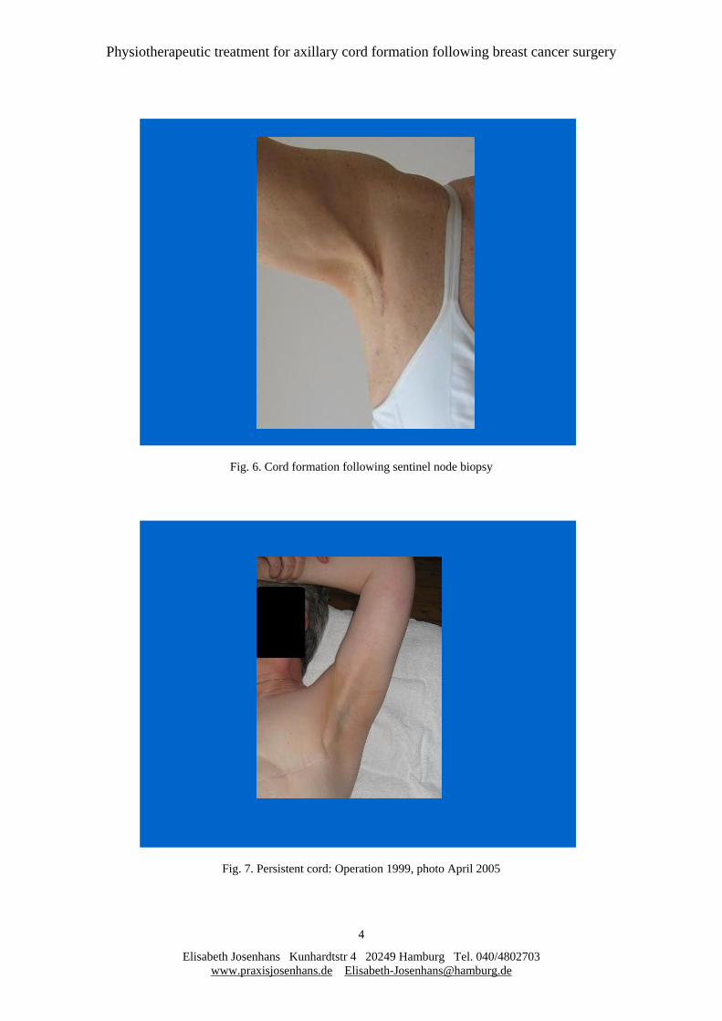
- cord formation - scar adhesions - effect of the treatment on the cord - duration of the entire series of treatments and - complications in the form of lymph drainage disorders. All of the patients received treatment using manual techniques as described in the “Treatment concept” section. There was no control group. The patients were classified into two groups: 1. patients with breast-conserving surgery, and 2. patients with total mastectomies, since the more extensive scars resulted in additional problems due to pulling of the scar. The documentation was designed to determine the effect of the treatment on - the cord - the mobility of the shoulder and - pulling of the scar. The study was also designed to investigate complications resulting from treatment and the duration of the treatment. Cause, symptoms and types of cord In June, 2005, additional surgery had to be performed on a patient who was undergoing physiotherapeutic treatment, since a lump could be felt on a very pronounced cord (Fig. 1a). A metastatic formation was feared. During surgery, the cord was clearly identified (see Fig. 1b) and removed. The pathologist found that the cord was a lymph vessel that had undergone fibrosis, which was surrounded by fat and granulation tissue. The lump was found to be a cystic enlargement of this lymph vessel. The discussion with the surgeon resulted in the following opinion concerning the origin of the cord: The removal of axillary lymph nodes causes the severing of the lymph vessels supplying the arm and breast area. The lymph vessels thus rendered nonfunctional undergo fibrosis and can become attached to the axilla or chest via scar tissue. When the patient’s arm is raised, the hardened lymph vessel which is attached in this way is pulled tight and can become apparent beneath the skin. The inflexible lymph vessel that is pulled tight can result in significant pain, causing the patient to avoid such movement and to protect the arm. The protective posture leads to a reduction of shoulder flexion and abduction. The other shoulder movements always retain complete, pain-free mobility, unless an additional shoulder problem exists (differential diagnosis: shoulder joint capsular pattern). Types of cord The following types of cord can be observed: - A single cord can appear in the axilla (see Fig. 2). - The lymph vessels appear divided (see Fig. 3). - The cord is visible beyond the elbow (see Fig. 4). - In the soft breast tissue there is a retraction due to hardened lymph vessels (see Fig. 5). - A cord can develop in the chest below the breast. - The removal of only a single lymph node in a sentinel node biopsy can result in cord formation (see Fig. 6). - The cord can still be present several years after the surgery (see Fig. 7).
Physiotherapeutic treatment for axillary cord formation following breast cancer surgery
Elisabeth Josenhans Physiotherapist Kunhardtstr 4 20249 Hamburg Tel. +49(0)40/4802703 www.praxisjosenhans.de [email protected]
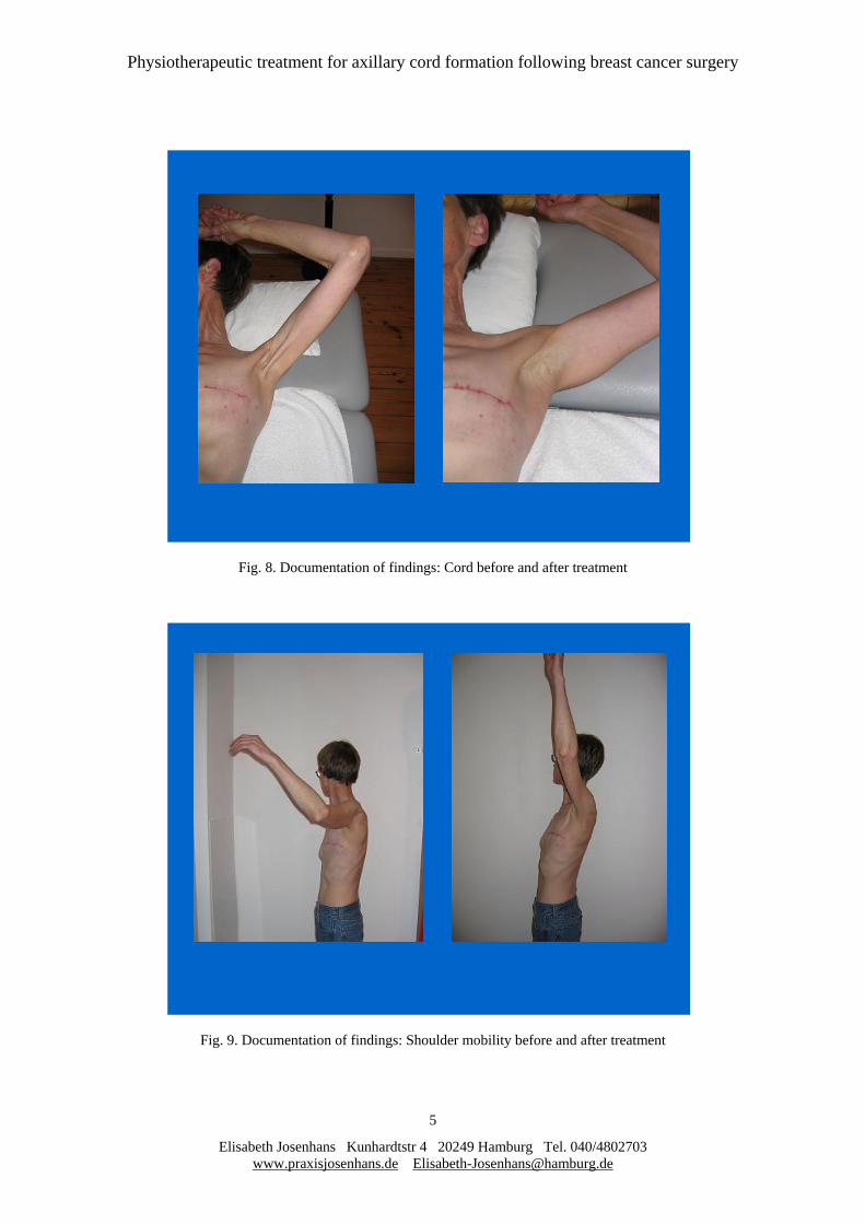
The cord was more clearly apparent in the case of thin patients than in obese patients, since fat tissue conceals the cord. Research method Documentation was carried out for all breast cancer patients with painful and/or restricted arm elevation and/or cord formation by means of a standardized form, and the visible cords were documented photographically. The parameters documented included: Medical history Type of surgery (breast-conserving, mastectomy, reconstruction) Extent of lymph node removal (sentinel node, number, infection, level) Shoulder mobility (flexion, abduction; photo if appropriate) Occurrence of pain (cord, shoulder mobility) Arm circumference in the case of swelling Status of the cord (extent, thickness, pain; photo if appropriate) Status of the scar (extent, attached areas, painful areas, redness, swelling) Neurological changes (sensitivity impairments, scapula alata) Seroma formation (number of punctures if any) Other treatments (chemotherapy, radiation) The following parameters were documented at the conclusion of treatment: Shoulder mobility (see above) Occurrence of pain (see above) Arm circumference in the case of swelling Status of the cord (see above) Status of the scar (see above) Number of treatment sessions Special features of the case/complications during treatment Shoulder mobility was measured by means of the neutral-null method. If other restrictions were found in addition to those affecting flexion and abduction, these were also documented. The level of pain was recorded by means of a scale from “0” (no pain) to “+++” (severe pain). The position and extent of the cord were described, and the thickness was recorded by means of a scale from “+” (small cord) to “+++” (thick, pronounced cord). A drawing was made of the scar, points of attachment were shaded on the drawing, and pain data were recorded on the drawing using the symbols “+” (slight pain) to “+++” (severe pain). Redness and swelling were described. In the case of visible findings, photographic documentation was performed (see Figs. 8 and 9). Treatment concept At the beginning of each treatment session, the breast and axilla area and the entire arm on the affected side were palpated, and the patient was asked to report problems. The treatment was carried out in accordance with the pain data provided by the patient and the findings from palpating. Patients were treated particularly cautiously in the early stage shortly after surgery.

Physiotherapeutic treatment for axillary cord formation following breast cancer surgery
Elisabeth Josenhans Physiotherapist Kunhardtstr 4 20249 Hamburg Tel. +49(0)40/4802703 www.praxisjosenhans.de [email protected]
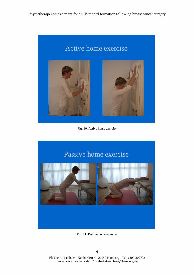
The affected arm was placed in an abducted position, raised as high as possible without causing pain. The technique primarily applied involved release manipulations in the connective tissue (approx. 2/3 of the respective treatment session). The place where the cord is “anchored” in the axilla (see Fig. 2) is usually clearly evident. The places where the cord is attached can be precisely distinguished by touch. The manipulations were applied at the cord attachment deep in the tissue, and the mobility of the tissue layers was restored. The cord itself was stretched and mobilized by means of hooking manipulations, with little skin displacement. The treatment followed the course of the cord along the arm, as far as the hand if necessary. Manipulations were never performed for a long period in one place, to avoid causing lymphedema or hyperemia / reddening of the skin. During the treatment session, the arm was repeatedly raised in a higher abducted position. An improvement of mobility of 20 to 40 degrees was often achieved during a single treatment session. In some cases, manual fixation of the cord in the arm accompanied by simultaneous stretching resulted in an audible tear. This was completely pain-free, and immediately resulted in increased mobility of the shoulder. In the case of adhesion of the scars in deeper web layers, scar massage, scar mobilization manipulations and scar stretching were employed. Depending upon the findings, treatments were complemented with myofascial release, muscle stretching (biceps and pectoralis, trapezius, subscapularis, and latissimus muscles), stretch positions, hold-relax techniques, treatment of the shoulder joint using manual techniques, scapular mobilization, and PNF arm and scapular patterns. The patients were shown home exercises to be performed so as to retain the increased mobility resulting from the treatment. Experience showed that shortly after surgery, guided active and passive flexion motions were better tolerated than free active movements (see Figs. 10 and 11). Later the series of exercises was expanded to include the turned stretched position, pectoralis stretching e.g. in a door frame, active theraband exercises with a flexion/external rotation PNF pattern, and exercises for straightening the vertebral column. The patients were instructed to observe the arm and scar area carefully following treatment and to report any reddening of the scar area or swelling prior to the next treatment. If there appeared to be a tendency toward swelling, parallel manual lymph drainage treatment was provided. Relative contraindications - Lymphedema: For patients with existing lymphedema, treatment of the cord was carried out cautiously in conjunction with lymph drainage. - Radiation: Patients who received physiotherapeutic treatment in parallel with radiation were treated only in the area not subjected to radiation. Contraindications - Reddening of the scars: In this case treatment of the scar area was avoided. - Radiation: Treatment using manual techniques was not carried out in the area subjected to radiation until two weeks after the radiation treatment. - Metastasis in the axilla: In this case treatment using manual techniques was not carried out at the axilla. Extent of the treatments Patients were treated two to three times per week for half an hour each time.
Physiotherapeutic treatment for axillary cord formation following breast cancer surgery
Elisabeth Josenhans Physiotherapist Kunhardtstr 4 20249 Hamburg Tel. +49(0)40/4802703 www.praxisjosenhans.de [email protected]
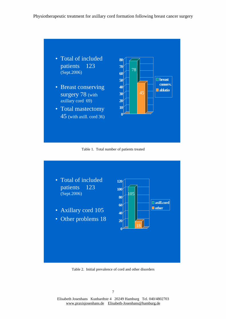
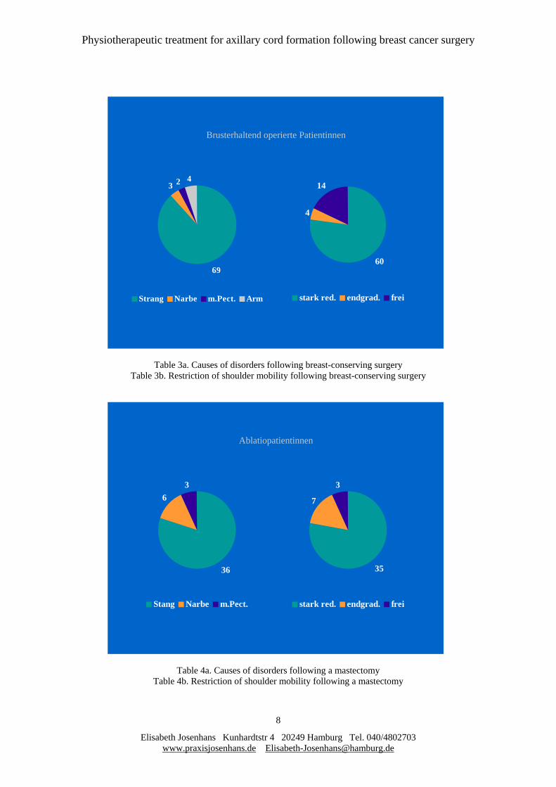
Patients with severely restricted shoulder mobility who were soon to receive radiation treatment were also treated up to five times in the week immediately preceding the beginning of radiation. Initial findings Of the 123 patients included in the study, - 78 (63%) had received breast-conserving surgery, and - 45 (37%) had total mastectomies (see Table 1). Axillary cord formation occurred in 105 (85%) of the patients (see Table 2). Treatment was begun by - 50% of the patients within four weeks after the surgery, - 33% of the patients one to three months after the surgery, and - 17% of the patients one to eleven years after the surgery. Of the 78 patients who had received breast-conserving surgery, - 69 (88%) had a cord, - 3 (4%) had only scar problems, - 2 (3%) had painful changes in the pectoral muscle due to radiation, and - 4 (5%) had diffuse arm problems (see Table 3a). Of these patients, - 60 (77%) had significantly restricted shoulder mobility, - 4 (5%) had a slightly restricted range of shoulder motion, and - 14 (18%) had no restriction of shoulder mobility (see Table 3b). Of the 45 patients who had a mastectomy, - 36 (80%) had a cord and problems with pulling of the scar, - 6 (13%) had only problems with pulling of the scar, and - 3 (7%) had painful changes in the pectoral muscle following radiation (see Table 4a). Of these patients, - 35 (78%) had significantly restricted shoulder mobility, - 7 (16%) had a slightly restricted range of shoulder motion, and - 3 (6%) had no restriction of shoulder mobility (see Table 4b). Final findings / Results Patients with breast-conserving surgery Shoulder mobility - in 73 (94%) of the patients was completely free and was the same for both shoulders at the conclusion of treatment, and - in 5 (6%) of the patients remained slightly restricted (see Table 5a). The cord - in 66 (96%) of the patients was no longer visible or palpable, and - in 3 (4%) of the patients was still visible or palpable, but was greatly reduced (see Table 5b).
Physiotherapeutic treatment for axillary cord formation following breast cancer surgery
Elisabeth Josenhans Physiotherapist Kunhardtstr 4 20249 Hamburg Tel. +49(0)40/4802703 www.praxisjosenhans.de [email protected]
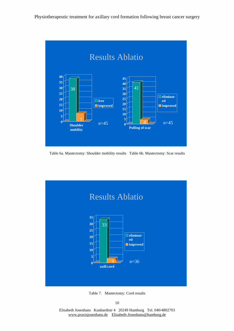
Patients with a mastectomy Shoulder mobility - in 38 (84%) of the patients was completely free and was the same for both shoulders at the conclusion of treatment, and - in 7 (16%) of the patients was still restricted at the end of the range of motion (see Table 6a). Pulling of the scar - in 39 (93%) of the patients was eliminated, and - in 3 (7%) of the patients was still noticeable, but had improved (see Table 6b). The cord - in 33 (92%) of the patients was no longer visible or palpable, and - in 3 (8%) of the patients was still visible or palpable, but was greatly reduced (see Table 7). Overall evaluation The ultimate objective of the treatment was unrestricted, pain-free shoulder mobility. This goal was achieved in 90% of the 123 patients who were treated. The cord disappeared in 94% of the total of 105 patients with cord formation; in 6% of the patients the cord was still visible following treatment (for example, see Figs. 12 to 17). Figures 18 and 19 illustrate a case where the treatment goal of pain-free flexion that is the same for both shoulders was achieved; however, here the cord still remains visible. The patients with breast-conserving surgery received an average of 9 treatment sessions. For patients with mastectomies, on average 12 treatment sessions were required. Complications No serious complications were observed. A few patients experienced slight local swellings following the treatment. The subsequent treatment was then carried out even more gently. No persistent swelling or lymphedema occurred. If the scar became red, this area was omitted from the following treatments until the redness had disappeared. Discussion In the medical literature there are references to physiotherapeutic measures following breast cancer surgery (Feige et al. 2001, Bastert 2003). Reference is made to active and passive exercises for the restoration of shoulder mobility. Cord-like lymph vessels that have undergone sclerosis are mentioned in the very comprehensive work of Hussein et al. (2005) concerning physical-therapeutic measures following breast cancer surgery which emphasizes the importance of physiotherapy. The authors recommend relaxation exercises, stretching techniques, posture training, and light massage in the painful area, but not in the axilla. In the case of scar problems, manual displacement techniques over a large area are recommended, however it is stated that that no lasting reduction of pain is possible. No treatment suggestions are given for cord formation. The physiotherapeutic literature contains references to cord formation, including references to the “fiddle-string” phenomenon and to sclerosis of the lymph vessels (Friebel et al. 2006, Henscher 2004, Kolster and Ebelt-Paprotny 2002, Zimmermann 2006).
Physiotherapeutic treatment for axillary cord formation following breast cancer surgery
Elisabeth Josenhans Physiotherapist Kunhardtstr 4 20249 Hamburg Tel. +49(0)40/4802703 www.praxisjosenhans.de [email protected]
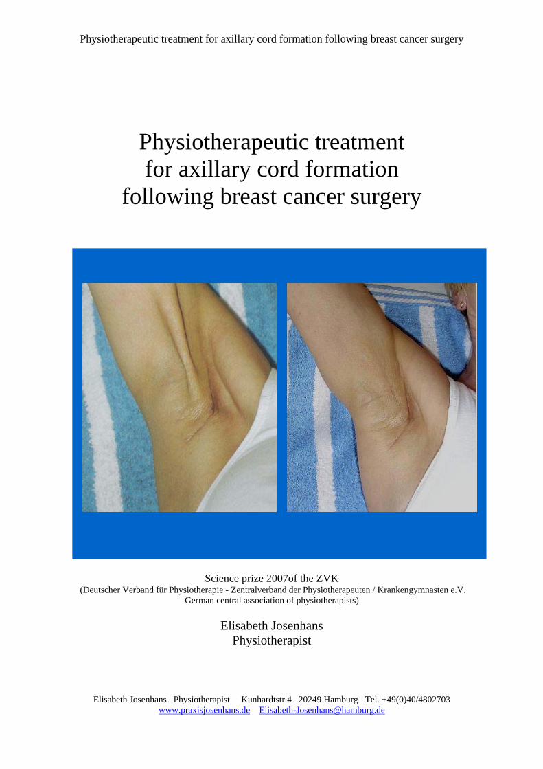
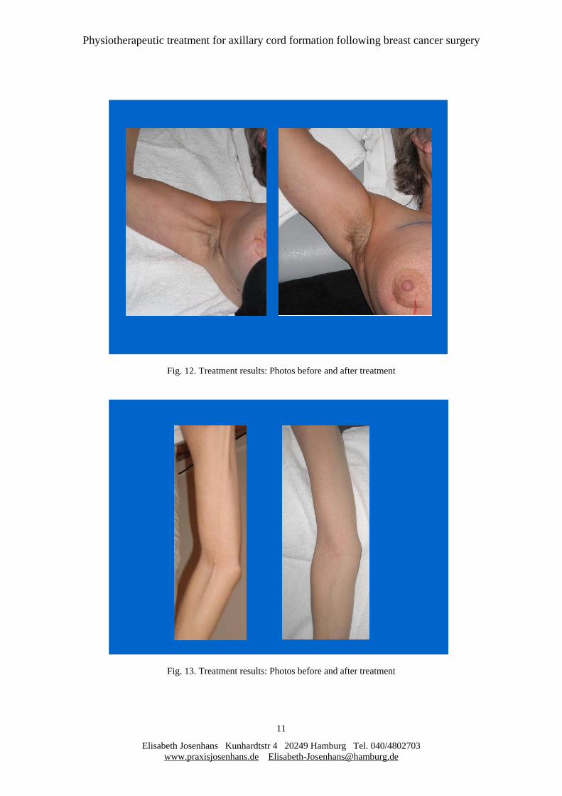
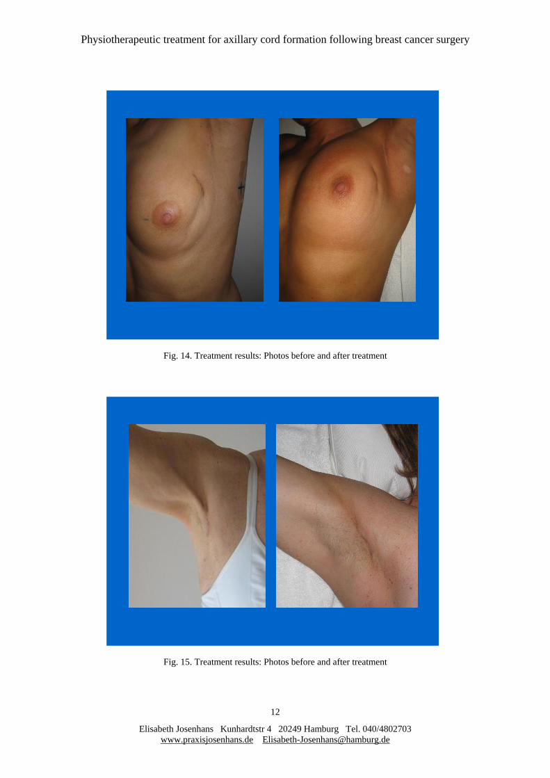
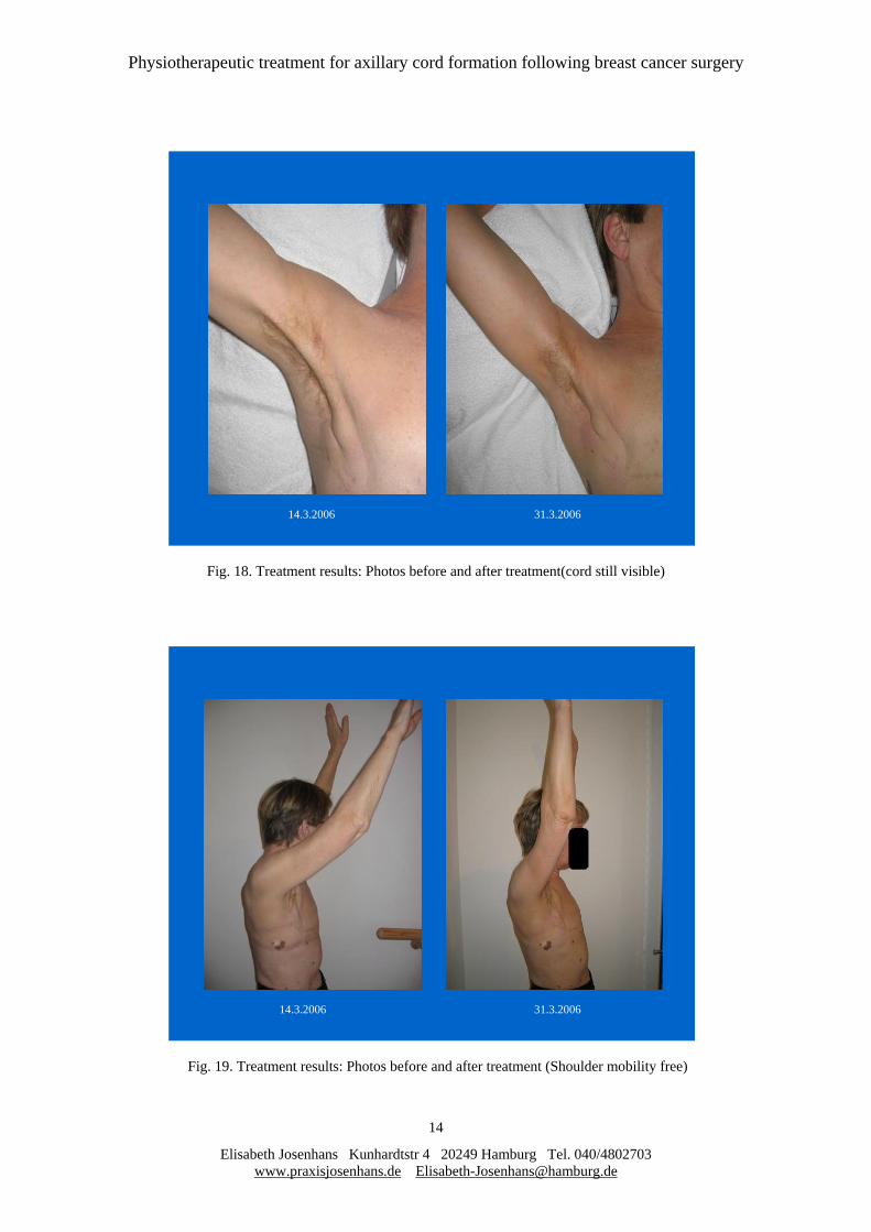
Physiotherapeutic treatments that are recommended for the acute phase are for prophylaxis of pneumonia, thrombosis and edema. Treatments recommended for the later phase are stretch positions, shoulder joint and scapula mobilization and active exercises. It is stated that massage is contraindicated, as it may give rise to lymphedema. Marnitz therapy is permitted. No treatment suggestions are given for cord formation. According to physiotherapeutic principles, attached tissues that become tight when stretched and cause pain should first be detached from the relevant point of attachment before being subjected to stretching. Physiotherapists proceed in this way for every other part of the body – why not use this method for the problem of cord formation? It is necessary to question the principle previously applied, that following breast cancer surgery accompanied by the removal of lymph nodes, patients should be provided only with active therapy and home exercises. This principle was probably justified in the era of radical removal of the lymph nodes. Less radical operating procedures in the axilla, such as sentinel node biopsy, considerably reduce the risk of lymphedema, so that manual releasing techniques can be applied to the arm on the side of the operation, even at the axilla. If these techniques are employed with the proper care, the problems associated with the cord can be eliminated without side effects. Kneading massage techniques should of course continue to be avoided, so as not to cause irritation of the tissue. Photographic documentation with striking illustrations of conditions before and after treatment convinced many physicians at Hamburg breast centers, as well as radiologists, oncologists and office-based gynecologists, of the effectiveness of the treatment. As a result, patients with cord formation were specifically directed to receive physiotherapeutic treatment; the large number of such cases made the seriousness of the situation evident. The question arises as to why this problem and the frequency of painful cord formation in the large number of patients with breast cancer surgery has not yet been scientifically investigated and treatments established. In light of the experience of the three past years the author affirms that the above-described treatment using manual techniques is superior to the conventional exercise treatment. This study demonstrates that in the treated patients, the treatment using manual releasing techniques resulted in a rapid restoration of pain-free functioning of the shoulder joint, without causing lymphedema. The assumption that the cord would disappear by itself cannot be confirmed. There were many examples of patients with a cord and related problems who came for physiotherapeutic treatment several years after surgery. In all cases, an improvement of the condition was achieved. One patient with surgery in the year 2002 suffered from a pronounced cord and painful limitation of shoulder mobility. Following a series of six treatment sessions, pain-free mobility was restored so that the mobility of both shoulders was the same, and the cord was reduced although still visible (see Figs. 18 and 19). With the required care, continuously focusing on the tissue so as to have an understanding of the changes taking place, treatment could be carried out with no complications. The duration of the treatment was short, allowing e.g. important subsequent treatments such as radiation to be carried out on time. The costs for the treatment described here are low, a significant point in times of restrictive health policies.
Physiotherapeutic treatment for axillary cord formation following breast cancer surgery
Elisabeth Josenhans Physiotherapist Kunhardtstr 4 20249 Hamburg Tel. +49(0)40/4802703 www.praxisjosenhans.de [email protected]
Conclusions Every patient with the above-described symptoms should be able to find information about axillary cord formation and its treatment via - information at the clinic - references to the problem in patient brochures - references to the problem in books about breast cancer and - Internet Every physiotherapist who works with breast cancer patients should be aware of treatment possibilities for rapid improvement of the symptoms resulting from cord formation. Physicians who treat breast cancer patients must be informed of the enhanced possibilities of physiotherapeutic treatment following breast cancer surgery. The objective of this paper is to report that a successful treatment concept for cord formation following breast cancer surgery has been developed and tested. Since it was not possible to carry out a randomized study with controls in an individual therapeutic practice, it would be desirable for scientific investigations to be performed, in order to determine: 1. whether the cord disappears by itself, and if so, the time period involved, 2. the percentage of patients with cord formation for whom treatment consisting solely of exercises is successful, and the time frame involved, and 3. the percentage of patients with cord formation for whom the above-described treatment using manual techniques is successful, and the time frame involved.
References Bastert, G.: Malignome der Mamma Spezielle gynäkologische Onkologie 2, 4th ed., Urban und Fischer, 2003, p. 167 Deutsche Krebsgesellschaft e.V.: Breast cancer webpage, December 2006, http://www.krebsgesellschaft.de/brustkrebs_mammakarzinom,6067.html Feige, Axel et al.: Frauenheilkunde, 2nd ed., Urban und Fischer, 2001, p. 545 Friebel, S., Waldemann-Rex, S. and Reuss, C.: Gynäkologie und Geburtshilfe, Gelbe Reihe, Krankheitslehre für Physiotherapeuten und Masseure, Urban und Fischer, 2006, pp. 83-89 Henscher, Ulla: Physiotherapie nach Brustoperation in: Hüter-Becker, A. (ed.): Physiotherapie in der Gynäkologie, Georg Thieme Verlag, 2004, pp. 193-211 Hussein, Dr. M., Baumeister, Dr. R.G.H. and Schwoerer, Dr. M.: Physikalisch-therapeutische Maßnahmen nach Brustkrebsoperation incl. Behandlung von Lymphödemen mit Hinweisen zur Heil-und Hilfsmittelverordnung in: Manual Empfehlungen zur Diagnostik, Therapie und Nachsorge Manmmakarzinome, W. Zuckschwerdt Verlag München, 10th ed., 2005, pp. 173-182
Physiotherapeutic treatment for axillary cord formation following breast cancer surgery
Elisabeth Josenhans Physiotherapist Kunhardtstr 4 20249 Hamburg Tel. +49(0)40/4802703 www.praxisjosenhans.de [email protected]
Kolster, Bernard and Ebelt-Paprotny, Gisela: Operative Gynäkologie in: Leitfaden Physiotherapie, 4th ed., Urban und Fischer, 2002, pp. 596-604 Zimmermann, Arleta: Rolle der Physiotherapie in der Rehabilitation nach Brustkrebsoperation – Ein Überblick in: Zeitschrift für Physiotherapeuten, 11/2006, pp. 1210-1219 Figures (see appendix) Fig. 1a. Cord with palpable lump Fig. 1b. Cord during surgery Fig. 2. Single cord Fig. 3. Divided cord Fig. 4. Cord extending past the elbow to the lower arm Fig. 5. Retraction of breast due to hardened lymph vessel Fig. 6. Cord formation following sentinel node biopsy Fig. 7. Persistent cord: Operation 1999, photo April 2005 Fig. 8. Documentation of findings: Cord before and after treatment Fig. 9. Documentation of findings: Shoulder mobility before and after treatment Fig. 10. Active home exercise Fig. 11. Passive home exercise Fig. 12. Treatment results: Photos before and after treatment Fig. 13. Treatment results: Photos before and after treatment Fig. 14. Treatment results: Photos before and after treatment Fig. 15. Treatment results: Photos before and after treatment Fig. 16. Treatment results: Photos before and after treatment Fig. 17. Treatment results: Photos before and after treatment Fig. 18. Treatment results: Photos before and after treatment Fig. 19. Treatment results: Photos before and after treatment Tables (see appendix) Table 1. Total number of patients treated Table 2. Initial prevalence of cord and other disorders Table 3a. Causes of disorders following breast-conserving surgery Table 3b. Restriction of shoulder mobility following breast-conserving surgery Table 4a. Causes of disorders following a mastectomy Table 4b. Restriction of shoulder mobility following a mastectomy Table 5a. Breast-conserving surgery: Shoulder mobility results Table 5b. Breast-conserving surgery: Cord results Table 6a. Mastectomy: Shoulder mobility results Table 6b. Mastectomy: Scar results Table 7. Mastectomy: Cord results
Physiotherapeutic treatment for axillary cord formation following breast cancer surgery
Elisabeth Josenhans Kunhardtstr 4 20249 Hamburg Tel. 040/4802703 www.praxisjosenhans.de [email protected]
Appendix
Fig. 1a. Cord with palpable lump Fig. 1b. Cord during surgery
Physiotherapeutic treatment for axillary cord formation following breast cancer surgery
Elisabeth Josenhans Kunhardtstr 4 20249 Hamburg Tel. 040/4802703 www.praxisjosenhans.de [email protected]
2
Fig. 2. Single cord
Fig. 3. Divided cord
Physiotherapeutic treatment for axillary cord formation following breast cancer surgery
Elisabeth Josenhans Kunhardtstr 4 20249 Hamburg Tel. 040/4802703 www.praxisjosenhans.de [email protected]
3
Fig. 4. Cord extending past the elbow to the lower arm
Fig. 5. Retraction of breast due to hardened lymph vessel
Physiotherapeutic treatment for axillary cord formation following breast cancer surgery
Elisabeth Josenhans Kunhardtstr 4 20249 Hamburg Tel. 040/4802703 www.praxisjosenhans.de [email protected]
4
Fig. 6. Cord formation following sentinel node biopsy
Fig. 7. Persistent cord: Operation 1999, photo April 2005
Physiotherapeutic treatment for axillary cord formation following breast cancer surgery
Elisabeth Josenhans Kunhardtstr 4 20249 Hamburg Tel. 040/4802703 www.praxisjosenhans.de [email protected]
5
Fig. 8. Documentation of findings: Cord before and after treatment
Fig. 9. Documentation of findings: Shoulder mobility before and after treatment
Physiotherapeutic treatment for axillary cord formation following breast cancer surgery
Elisabeth Josenhans Kunhardtstr 4 20249 Hamburg Tel. 040/4802703 www.praxisjosenhans.de [email protected]
6
Fig. 10. Active home exercise
Fig. 11. Passive home exercise
Active home exercise
Passive home exercise
Physiotherapeutic treatment for axillary cord formation following breast cancer surgery
Elisabeth Josenhans Kunhardtstr 4 20249 Hamburg Tel. 040/4802703 www.praxisjosenhans.de [email protected]
7
Table 1. Total number of patients treated
Table 2. Initial prevalence of cord and other disorders
• Total of includedpatients 123(Sept.2006)
• Breast conservingsurgery 78 (withaxillary cord 69)
• Total mastectomy45 (with axill. cord 36) 0
1020
30405060
7080
breastconserv.ablatio45
78
0
20
40
60
80
100
120
axill.cordother
• Total of includedpatients 123(Sept.2006)
• Axillary cord 105• Other problems 18
18
105
Physiotherapeutic treatment for axillary cord formation following breast cancer surgery
Elisabeth Josenhans Kunhardtstr 4 20249 Hamburg Tel. 040/4802703 www.praxisjosenhans.de [email protected]
8
Table 3a. Causes of disorders following breast-conserving surgery
Table 3b. Restriction of shoulder mobility following breast-conserving surgery
Table 4a. Causes of disorders following a mastectomy
Table 4b. Restriction of shoulder mobility following a mastectomy
Brusterhaltend operierte Patientinnen
69
3 2 4
Strang Narbe m.Pect. Arm
60
4
14
stark red. endgrad. frei
Ablatiopatientinnen
36
63
Stang Narbe m.Pect.
35
7
3
stark red. endgrad. frei

Physiotherapeutic treatment for axillary cord formation following breast cancer surgery
Elisabeth Josenhans Kunhardtstr 4 20249 Hamburg Tel. 040/4802703 www.praxisjosenhans.de [email protected]
9
Table 5a. Breast-conserving surgery: Shoulder mobility results
Table 5b. Breast-conserving surgery: Cord results
Results breast-conserving
0
10
20
30
40
50
60
70
axillary cord
eliminat-edimproved
01020304050607080
Shouldermobility
freeimproved
n=695
66
3n=78
73
Physiotherapeutic treatment for axillary cord formation following breast cancer surgery
Elisabeth Josenhans Kunhardtstr 4 20249 Hamburg Tel. 040/4802703 www.praxisjosenhans.de [email protected]
10
Table 6a. Mastectomy: Shoulder mobility results Table 6b. Mastectomy: Scar results
Table 7. Mastectomy: Cord results
Results Ablatio
05
10152025303540
Shouldermobility
freeimproved
05
1015202530354045
Pulling of scar
eliminat-edimproved
38
7
41
4n=45 n=45
Results Ablatio
0
5
10
15
20
25
30
35
axill.cord
eliminat-edimproved
33
3 n=36
Physiotherapeutic treatment for axillary cord formation following breast cancer surgery
Elisabeth Josenhans Kunhardtstr 4 20249 Hamburg Tel. 040/4802703 www.praxisjosenhans.de [email protected]
11
Fig. 12. Treatment results: Photos before and after treatment
Fig. 13. Treatment results: Photos before and after treatment
Physiotherapeutic treatment for axillary cord formation following breast cancer surgery
Elisabeth Josenhans Kunhardtstr 4 20249 Hamburg Tel. 040/4802703 www.praxisjosenhans.de [email protected]
12
Fig. 14. Treatment results: Photos before and after treatment
Fig. 15. Treatment results: Photos before and after treatment
Physiotherapeutic treatment for axillary cord formation following breast cancer surgery
Elisabeth Josenhans Kunhardtstr 4 20249 Hamburg Tel. 040/4802703 www.praxisjosenhans.de [email protected]
13
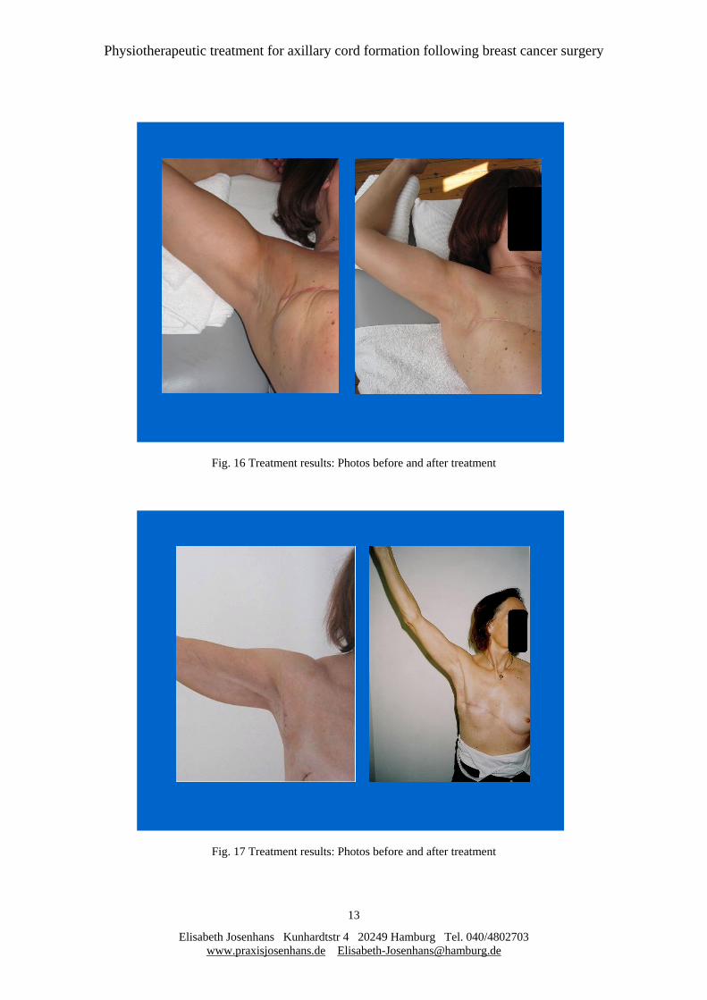
Fig. 16 Treatment results: Photos before and after treatment
Fig. 17 Treatment results: Photos before and after treatment
Physiotherapeutic treatment for axillary cord formation following breast cancer surgery
Elisabeth Josenhans Kunhardtstr 4 20249 Hamburg Tel. 040/4802703 www.praxisjosenhans.de [email protected]
14
Fig. 18. Treatment results: Photos before and after treatment(cord still visible)
Fig. 19. Treatment results: Photos before and after treatment (Shoulder mobility free)
14.3.2006 31.3.2006
14.3.2006 31.3.2006