physicochemical properties and biocompatibility of white dextrin modified injectable...
TRANSCRIPT
Physicochemical Properties and Biocompatibility ofWhite Dextrin Modified Injectable Calcium–MagnesiumPhosphate Cement
Fangping Chen and Changsheng Liu*
The State Key Laboratory of Bioreactor Engineering, East China University of Science and Technology,Shanghai, 200237, People’s Republic of China
Key Laboratory for Ultrafine Materials of Ministry of Education, School of Materials Science andEngineering, East China University of Science and Technology, Shanghai, 200237, People’s Republic ofChina
Jie Wei
Key Laboratory for Ultrafine Materials of Ministry of Education, School of Materials Science andEngineering, East China University of Science and Technology, Shanghai, 200237, People’s Republic ofChina
Engineering Research Center for Biomedical Materials of Ministry of Education, East China Universityof Science and Technology, Shanghai, 200237, People’s Republic of China
Xiaolong Chen
Engineering Research Center for Biomedical Materials of Ministry of Education, East China Universityof Science and Technology, Shanghai, 200237, People’s Republic of China
This investigation was supported by the National Science Foundation of China (NO.31100678 and No. 50732002), Natural Science Foundation of Shanghai (No.11ZR1408200), Program for
Changjiang Scholars, and Innovative Research Team in University and Program of Shanghai Leading Academic Discipline Project (No. B502).*[email protected]
© 2011 The American Ceramic Society and Wiley Periodicals, Inc.
Int. J. Appl. Ceram. Technol., 1–12 (2011)DOI:10.1111/j.1744-7402.2011.00705.x
The objective of this study was to develop anti-washout and fast-setting calcium–magnesium phosphate cement (as-
CMP) by introducing white dextrin (WD) and magnesium phosphate cement (MPC) to CPC. The results showed that WDimparted anti-washout to the as-CMP paste, whereas MPC simultaneously improved the paste anti-washout and fast-setting.The synergistic effect of MPC and WD obviously shortened the setting time of as-CMP. The WD did not affect the phaseevolution and microstructure of the paste. Furthermore, all samples were biocompatible and as-CMP exhibited the best cell
metabolic activity, cell attachment, and proliferation, which would provide basic data for the clinical application.
Introduction
Attributable to the unique combination of self-set-ting under ambient conditions, conformity to the shapeof bone defects, osteoconductivity, and biocompatibil-ity, calcium phosphate cement (CPC) has received par-ticular attention over the past few decades. However,the current CPC exposed some limitations of its rela-tively long setting time and low biodegradation rate.1,2
In addition, most CPCs tend to crumble upon earlycontact with blood or other fluids before completion ofthe setting reaction, which further prolongs the settingtime. Therefore, it is necessary to develop injectablecement with good resistance to water and fast setting tosatisfy the need of the clinics.
To improve the anti-washout property of injectablecement, preventing liquid penetration into the cementpaste is an effective strategy. To attain this goal, gellingagents had been utilized to impart viscosity to cement.Recently, modified starch, hydroxypropyl-methylcellu-lose, carboxymethylcellulose, lactic acid, glycerol, citricacid, polyacrylic acid, and chitosan had been tried toprepare the anti-washout type cement.3–6 In terms ofimproving the cohesion of the cement, these additivesworked, but some of them suppressed the hydroxyapa-tite (HAP) formation from the dissolved cement andshowed an inflammatory response.
Dextrin is a biocompatible natural polymer, andused clinically as a peritoneal dialysis solution in end-stage renal failure patients7 and as a carrier for the anti-cancer agent, 5-fluorouracil.8 In addition, dextrin isused in postage stamps, envelopes, and wallpapersextensively, as it readily formed an adhesive paste whenmixed with water. White dextrin (WD), as one of thedextrins, is derived from starch by partial thermal deg-radation under acidic conditions.9 The differencebetween WD and other dextrins is that WD can forma viscous paste in cold water. In addition, WD is read-ily degraded by amylases to maltose and isomaltose.10
Therefore, the white dextrin, incorporated reasonably
into injectable CPC, may contribute to better anti-washout property of the paste.
When the paste comes into contact with the physi-ological fluid or when bleeding occurs, the paste tendsto crumbledue to the long setting time and difficulty inachieving complete homeostasis.11,12 Consequently,speeding up the setting process and shortening the set-ting time play an important role in developing theanti-washout type cement. Magnesium phosphatecement (MPC), consisting of magnesium oxide (MgO)and calcium biphosphate (Ca (H2PO4)2), is of specialinterest due to its rapid setting and high initialmechanical strength in comparison with the phosphatecement. The major components of MPC could react inaqueous environment to form magnesium phosphate(Mg3(PO4)2) and calcium triphosphate (Ca3(PO4)2) asfinal products. Previous study in our laboratory indi-cated that MPC was set in 7–15 min and had a com-pressive strength of 35 MPa after setting for 30 min.13
Studies also showed MPC had excellent biocompatibil-ity.14–16 Although the introduction of MPC powdersinto CPC is not critical to acquire the noncrumblingproperty, MPC is used in an attempt to speed up thehydration and to enhance the anti-washout ability.
In this study, anti-washout type fast-setting cal-cium–magnesium phosphate cement (as-CMP) wasdeveloped by introducing WD and MPC into CPC.Moreover, the anti-washout property, setting time,compressive strength, degradability, and phase composi-tion were investigated. In addition, the in vitro biocom-patibility of the as-CMP was explored.
Materials and Methods
Materials and Preparation
All calcium phosphates were prepared in our labo-ratory, and the preparation methods can be obtainedfrom the relevant literature.17 Briefly, tetracalciumphosphate (TTCP) was synthesized by a solid-to-solid
2 International Journal of Applied Ceramic Technology—Chen, et al. 2011
reaction between calcium phosphate and calciumcarbonate at 1500°C for 8 h. Dicalcium phosphatedihydrate (DCPD) was prepared from (NH4)2HPO4
and Ca (NO3)2 in an acidic environment, and dical-cium phosphate anhydrous (DCPA) was obtained byremoving the crystallization water in DCPD at 120°C.The CPC powder was composed of TTCP and DCPAin an equivalent molar ratio. The c-CPC paste con-sisted of CPC powders and deionized water at the fixedratio of 2 g/mL.
The MPC powders were formed by mixing MgOwith Ca(H2PO4)2 in a molar ratio of 2:1. The MgO wasprepared by heating magnesium carbonate basic penta-hydrate [(MgCO3)4·Mg(OH)2·5H2O] in a furnace at1500°C for 6 h. The resultant powder was initiallycooled to room temperature, and then ground in a plan-etary mill for 5 min, followed by sieving through 200meshes. The obtained MPC powders were kept in a des-iccator for further experiment. The calcium–magnesiumphosphate cement (CMP) powders were prepared bymixing CPC with MPC powders in an equivalent weightratio. The c-CMP paste was prepared by mixing CMPpowders with deionized water at the ratio of 2 g/mL.
White dextrin (Shanghai No. 4 Reagent & H.VChemical Co. Ltd., China) solution, with 4 wt% WDin the deionized water, was selected as the liquid for as-CMP. The as-CMP paste was formed by mixing theCMP powders homogeneously with the WD solutionwith a spatula at 2 g/mL. Except the WD powders allthe other chemicals used were purchased from Sinop-ham Chemical Reagent Co., Ltd. Shanghai, China.
Anti-Washout Evaluation
After immersing the c-CPC, c-CMP, and as-CMPpastes into the deionized water, shaking was exerted onthe samples using a bumping table at 100 rpm at 37°C. All the pastes were taken out after being shaken for10, 15, 30, 45, and 60 min, respectively. The massesof the pastes before and after immersion were mea-sured. As an index of the anti-washout property, thewashout mass loss rate of the paste was calculatedaccording to Eq. (1). Each test was repeated three timesand the average value was calculated.
Washout mass loss% ¼Mass lost during immersion=Total mass
before immersion
ð1Þ
Setting Time and Compressive Strength
The prepared c-CPC, c-CMP, and as-CMP werefilled into the glass tubes (Φ6 9 10 mm). The top andbottom surfaces of the tube were tightly covered withtwo sheets of plastic film held by a ‘C’ clamp. Thepastes were stored at 37°C in a 100% relative humiditychamber to set. The preset pastes were measured atintervals of 0.5 min by using a vicat apparatus accord-ing to the ASTM Test Method C 187-98, called vicatmethod to determine the setting time. The vicat appa-ratus consists of a frame bearing a movable rod (300 gin mass) of 1 mm diameter fitted with a stainless steelneedle at the end. The time when the needle of thevicat apparatus could only penetrate less than 1 mminto the cement was taken as the setting time. Eachexperiment was repeated five times and the averagevalue was calculated.
The hardened cement (Φ6 9 10 mm3), removedfrom the mold after setting for 24 h, was uniformlypolished on both sides. The compressive strength of thecement was measured at a loading rate of 1 mm/minusing a universal testing machine (AG-2000A, Shima-dzu Autograph, Shimadzu, Kyoto, Japan). Threereplicates were carried out for each group, and theresults were expressed as means ± SD.
In Vitro Degradation
The in vitro degradation was performed by immers-ing the samples in the simulated body fluid (SBF) overa period of 30 days. The degradation rate of the cementwas determined by its weight loss rate in the SBF.
The SBF was prepared by dissolving reagent-gradeNaCl, NaHCO3, KCl, K2HPO4·3H2O, MgCl2·6H2O,CaCl2, and Na2SO4 in deionized water in turn. Thesolution was then buffered to pH 7.4 at 37°C withTris and HCl. The ion concentration and pH of theSBF are similar to those in human blood plasma.18
Before immersion, the 24-h-set pastes were driedat 60°C for 24 h. The initial weight of each samplewas accurately measured and recorded as W0. The sam-ples were immersed in SBF at a weight-to-volume rateof 0.2 g/mL in plastic bottles. The plastic bottles werecontinuously shaken in a water bath at 100 rpm at 37°Cfor 30 days. The solution was changed every day withfreshly made solutions. When the solution was changed ,the pastes were gently rinsed with deionized water anddried at 60°C for 2 h, The final weight of each sample
www.ceramics.org/ACT Fast-Setting Calcium–Magnesium Phosphate Cement 3
was measured and recorded as Wt. The weight loss rateof each sample was calculated according to Eq. (2).Each test was repeated three times and the averagevalue was calculated.
Weight loss rate ¼ ðW 0 �W tÞ=W 0 � 100%: ð2Þ
As-CMP samples were incubated in SBF for7 days without changing the solution, and changes inthe concentration of Ca, Mg, and P ions in the SBFwere monitored by inductively coupled plasma-atomicemission spectroscopy (ICP-AES) at 24, 48, 120, and168 h. The pH of the SBF after soaking in as-CMPwas measured using an electrolyte-type pH meter for7 days without changing the solution.
Phase Analysis and Microstructure Observation
The precured c-CPC, c-CMP, and as-CMP pasteswere immersed in SBF at 37°C at a weight-to-volumerate of 0.2 g/mL.18 After immersion for 24 h, all sam-ples were gently rinsed with deionized water to removeSBF, and immersed in liquid nitrogen for 15 min tostop the reaction, then dried by freezing overnight.When the samples were ground to fine powders, thephase compositions were analyzed with X-ray diffrac-tion (XRD, D/max 2550 VB/PC, Rigaku, Tokyo,Japan). Microstructure of the precipitate was observedusing scanning electronic microscope (SEM, H-800,Hitachi, Tokyo, Japan) coated with gold at a high mag-nification with an acceleration voltage of 15 kV.
Biocompatibility In Vitro
The MG-63 cells were cultured in cell cultureflasks using L-Dulbecco’s modified Eagle’s medium(L-DMEM), supplemented with 10% fetal bovineserum (FBS), 1% L-glutamine, 100 U/mL penicillin,and 100 lg/mL streptomycin. Cell culture was main-tained in a gas-jacket incubator equilibrated with 5%CO2 at 37°C and the culture medium was refreshedevery 2 days. When the cells had grown to confluence,1 mL 0.25% trypsin and 0.03% EDTA were added todigest. The cells were trypsinised and resuspended infresh media before being seeded on the top of disks.
The c-CPC and as-CMP disks, with the size of10 mm in diameter and 3 mm in thickness, werelocated into 96 well tissue culture plates.
Cell metabolic viability was measured by a 3-(4,5-Dimethylthiazol-2-yl) -2,5-diphenyltetrazolium bromide(MTT) assay method. After the c-CPC and as-CMPsamples were sonicated in ethanol and sterilized byethylene oxide gas, MG-63 cells with a density of3 9 103 cells/mL were seeded evenly into the wellswith a pipette, followed by incubation in anL-DMEM medium at 37°C and 100% humidity with5% CO2 for 1, 3, and 5 days, with the medium beingreplaced every second day. The culture medium wasthen removed and 20 lL MTT solution (5 mg/mL)was added to the wells. The cell-seeded disks were fur-ther cultured with 5% CO2 at 37°C for 4 h, allowingthe yellow dye to be transformed into blue crystals bymitochondrial dehydrogenases. Subsequently, the intra-cellular formazan was solubilized by adding 200 lL ofDMSO to each well. The optical density (OD) at590 nm wavelength was measured with UV-spectro-photometer (Cary 500, Varian Shanghai, China). Eachsample was performed in triplicate. Results arereported as O.D. units.
For cell attachment, the samples were sonicated inethanol and sterilized by ethylene oxide gas. MG-63cells with a density of 5 9 103 cells per disk wereseeded evenly into the wells with a pipette. The cell-seed disks were maintained at 37°C under 5% CO2
condition for 3 h, and then the culture medium wasremoved. Thereafter, the residual cultured medium andunattached cells were removed by washing with PBSthree times. After the attached cells on the disks weredigested by trypsin, the adherent cells were countedusing a hemacytometer, and the cell attachment effi-ciency was determined by counting the number of cellsremaining in the wells.
For cell growth experiments, the cells were seededat a density of 2.5 9 103 cells for each disk on differ-ent samples, the same procedure was used to measurethe cell number at each predetermined intervals of 1, 3,and 5 days. The morphologies of the cells, cultured onthe samples for 5 days were observed using a scanningelectronic microscope (SEM, Hitachi H-800, Hitachi).The cell/sample constructs were washed twice with PBSsolution and fixed with 4% formalin in PBS (pH 7.4)for 30 min. They were subsequently washed twice withPBS and dehydrated in a series of graded ethanol (50,60, 70, 80, 90, and 100% v/v) for 3 min each. Finally,specimens were air-dried in a desiccator overnight forSEM observation.
4 International Journal of Applied Ceramic Technology—Chen, et al. 2011
Statistical Analysis
Statistical analysis was conducted using one-wayANOVA with post hoc tests. The results were expressedas the mean ± SD. A value of P < 0.05 was consideredto be statistically significant.
Results
Anti-Washout Property
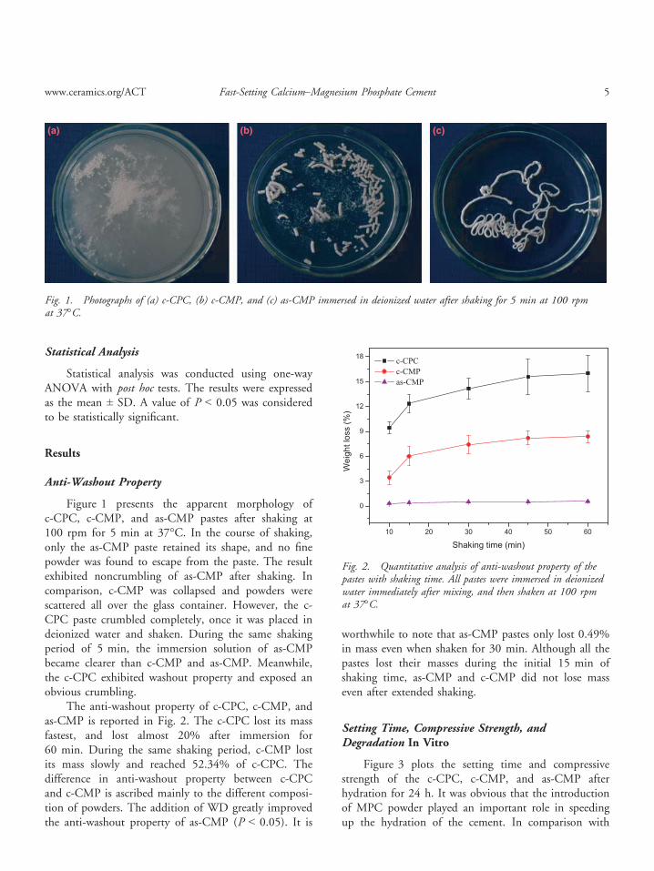
Figure 1 presents the apparent morphology ofc-CPC, c-CMP, and as-CMP pastes after shaking at100 rpm for 5 min at 37°C. In the course of shaking,only the as-CMP paste retained its shape, and no finepowder was found to escape from the paste. The resultexhibited noncrumbling of as-CMP after shaking. Incomparison, c-CMP was collapsed and powders werescattered all over the glass container. However, the c-CPC paste crumbled completely, once it was placed indeionized water and shaken. During the same shakingperiod of 5 min, the immersion solution of as-CMPbecame clearer than c-CMP and as-CMP. Meanwhile,the c-CPC exhibited washout property and exposed anobvious crumbling.
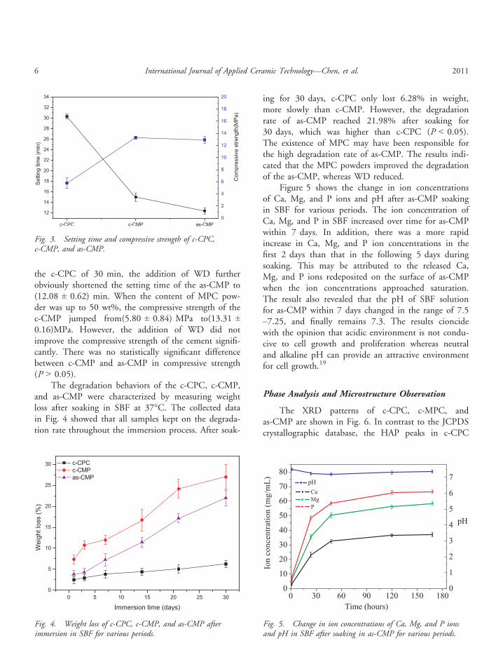
The anti-washout property of c-CPC, c-CMP, andas-CMP is reported in Fig. 2. The c-CPC lost its massfastest, and lost almost 20% after immersion for60 min. During the same shaking period, c-CMP lostits mass slowly and reached 52.34% of c-CPC. Thedifference in anti-washout property between c-CPCand c-CMP is ascribed mainly to the different composi-tion of powders. The addition of WD greatly improvedthe anti-washout property of as-CMP (P < 0.05). It is
worthwhile to note that as-CMP pastes only lost 0.49%in mass even when shaken for 30 min. Although all thepastes lost their masses during the initial 15 min ofshaking time, as-CMP and c-CMP did not lose masseven after extended shaking.
Setting Time, Compressive Strength, andDegradation In Vitro
Figure 3 plots the setting time and compressivestrength of the c-CPC, c-CMP, and as-CMP afterhydration for 24 h. It was obvious that the introductionof MPC powder played an important role in speedingup the hydration of the cement. In comparison with
(a) (b) (c)
Fig. 1. Photographs of (a) c-CPC, (b) c-CMP, and (c) as-CMP immersed in deionized water after shaking for 5 min at 100 rpmat 37°C.
Fig. 2. Quantitative analysis of anti-washout property of thepastes with shaking time. All pastes were immersed in deionizedwater immediately after mixing, and then shaken at 100 rpmat 37°C.
www.ceramics.org/ACT Fast-Setting Calcium–Magnesium Phosphate Cement 5
the c-CPC of 30 min, the addition of WD furtherobviously shortened the setting time of the as-CMP to(12.08 ± 0.62) min. When the content of MPC pow-der was up to 50 wt%, the compressive strength of thec-CMP jumped from(5.80 ± 0.84) MPa to(13.31 ±0.16)MPa. However, the addition of WD did notimprove the compressive strength of the cement signifi-cantly. There was no statistically significant differencebetween c-CMP and as-CMP in compressive strength(P > 0.05).
The degradation behaviors of the c-CPC, c-CMP,and as-CMP were characterized by measuring weightloss after soaking in SBF at 37°C. The collected datain Fig. 4 showed that all samples kept on the degrada-tion rate throughout the immersion process. After soak-
ing for 30 days, c-CPC only lost 6.28% in weight,more slowly than c-CMP. However, the degradationrate of as-CMP reached 21.98% after soaking for30 days, which was higher than c-CPC (P < 0.05).The existence of MPC may have been responsible forthe high degradation rate of as-CMP. The results indi-cated that the MPC powders improved the degradationof the as-CMP, whereas WD reduced.
Figure 5 shows the change in ion concentrationsof Ca, Mg, and P ions and pH after as-CMP soakingin SBF for various periods. The ion concentration ofCa, Mg, and P in SBF increased over time for as-CMPwithin 7 days. In addition, there was a more rapidincrease in Ca, Mg, and P ion concentrations in thefirst 2 days than that in the following 5 days duringsoaking. This may be attributed to the released Ca,Mg, and P ions redeposited on the surface of as-CMPwhen the ion concentrations approached saturation.The result also revealed that the pH of SBF solutionfor as-CMP within 7 days changed in the range of 7.5–7.25, and finally remains 7.3. The results cioncidewith the opinion that acidic environment is not condu-cive to cell growth and proliferation whereas neutraland alkaline pH can provide an attractive environmentfor cell growth.19
Phase Analysis and Microstructure Observation
The XRD patterns of c-CPC, c-MPC, andas-CMP are shown in Fig. 6. In contrast to the JCPDScrystallographic database, the HAP peaks in c-CPC
Fig. 4. Weight loss of c-CPC, c-CMP, and as-CMP afterimmersion in SBF for various periods.
Fig. 5. Change in ion concentrations of Ca, Mg, and P ionsand pH in SBF after soaking in as-CMP for various periods.
Fig. 3. Setting time and compressive strength of c-CPC,c-CMP, and as-CMP.
6 International Journal of Applied Ceramic Technology—Chen, et al. 2011
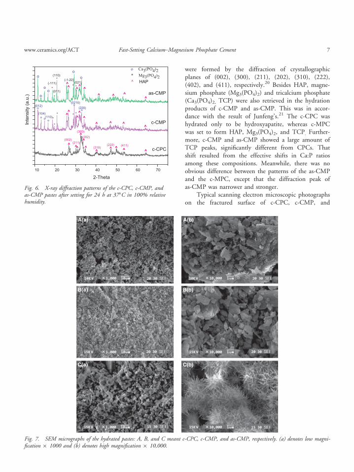
were formed by the diffraction of crystallographicplanes of (002), (300), (211), (202), (310), (222),(402), and (411), respectively.20 Besides HAP, magne-sium phosphate (Mg3(PO4)2) and tricalcium phosphate(Ca3(PO4)2, TCP) were also retrieved in the hydrationproducts of c-CMP and as-CMP. This was in accor-dance with the result of Junfeng’s.21 The c-CPC washydrated only to be hydroxyapatite, whereas c-MPCwas set to form HAP, Mg3(PO4)2, and TCP. Further-more, c-CMP and as-CMP showed a large amount ofTCP peaks, significantly different from CPCs. Thatshift resulted from the effective shifts in Ca:P ratiosamong these compositions. Meanwhile, there was noobvious difference between the patterns of the as-CMPand the c-MPC, except that the diffraction peak ofas-CMP was narrower and stronger.
Typical scanning electron microscopic photographson the fractured surface of c-CPC, c-CMP, and
Fig. 6. X-ray diffraction patterns of the c-CPC, c-CMP, andas-CMP pastes after setting for 24 h at 37°C in 100% relativehumidity.
A(a)
B(a)
C(a)
B(b)
C(b)
A(b)
Fig. 7. SEM micrographs of the hydrated pastes: A, B, and C meant c-CPC, c-CMP, and as-CMP, respectively. (a) denotes low magni-fication 9 1000 and (b) denotes high magnification 9 10,000.
www.ceramics.org/ACT Fast-Setting Calcium–Magnesium Phosphate Cement 7
as-CMP are shown in Fig. 7. After soaking for 24 h, itwas obviously observed that many crystalline particlesbridged over all the cements. The hydrated c-CPC wasmore loose in structure compared with that of c-CMPand as-CMP. At a high magnification of 10,0009, itwas clear to observe the morphology of all hydratedcements. Relative small needle-like crystals, which aretypical of hydroxyapatite, were covered on the surfaceof c-CPC, whereas platelet-like crystals were depositedon the surface of c-CMP. Interestingly, the needle-likecrystals and large platelet-like crystals were found tocoexist in as-CMP, and most of the platelet-like crystalswere covered with numerous but small needle-like crys-tals, which had a length of approximately 1–2 lm anda diameter of about 0.1 lm.
Biocompatibility In Vitro
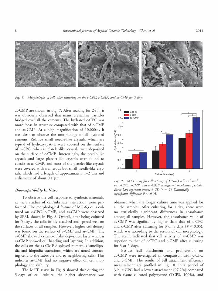
To observe the cell response to synthetic materials,in vitro studies of cell/substrate interaction were per-formed. The morphological feature of MG-63 cells cul-tured on c-CPC, c-CMP, and as-CMP were observedby SEM, shown in Fig. 8. Overall, after being culturedfor 5 days, the cells firmly attached and spread well onthe surfaces of all samples. However, higher cell densitywas found on the surface of c-CMP and as-CMP. Thec-CMP showed extensive flaky deposition layer whereasas-CMP showed cell banding and layering. In addition,the cells on the as-CMP displayed numerous lamellipo-dia and filopodia extensions, which are noted contact-ing cells to the substrate and to neighboring cells. Thisindicates as-CMP had no negative effect on cell mor-phology and viability.
The MTT assays in Fig. 9 showed that during the5 days of cell culture, the higher absorbance was
obtained when the longer culture time was applied forall the samples. After culturing for 1 day, there wereno statistically significant differences in absorbanceamong all samples. However, the absorbance value ofas-CMP was significantly higher than that of c-CPCand c-CMP after culturing for 3 or 5 days (P < 0.05),which was according to the results of cell morphology.The result indicated that cell activity of as-CMP wassuperior to that of c-CPC and c-CMP after culturingfor 3 or 5 days.
Besides, cell attachment and proliferation onas-CMP were investigated in comparison with c-CPCand c-CMP. The results of cell attachment efficiencymeasurement are profiled in Fig. 10. In a period of3 h, c-CPC had a lower attachment (97.2%) comparedwith tissue cultured polystyrene (TCPS, 100%), and
(a) (b) (c)
Fig. 8. Morphologies of cells after culturing on the c-CPC, c-CMP, and as-CMP for 5 days.
Fig. 9. MTT assay for cell activity of MG-63 cells culturedon c-CPC, c-CMP, and as-CMP at different incubation periods.Error bars represent means ± SD (n = 5). Statisticallysignificant difference P < 0.05.
8 International Journal of Applied Ceramic Technology—Chen, et al. 2011
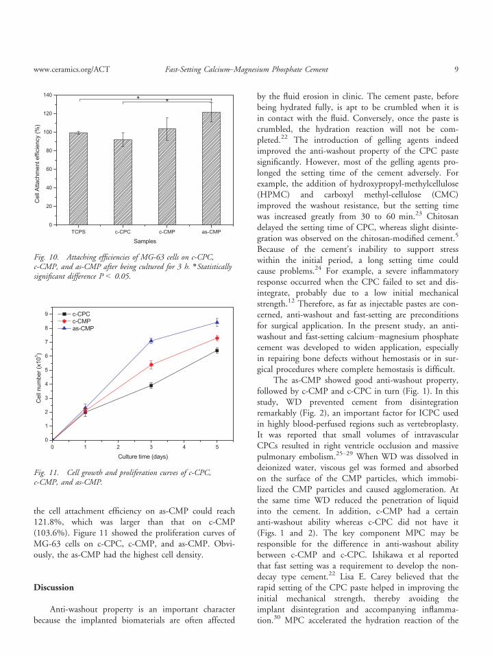
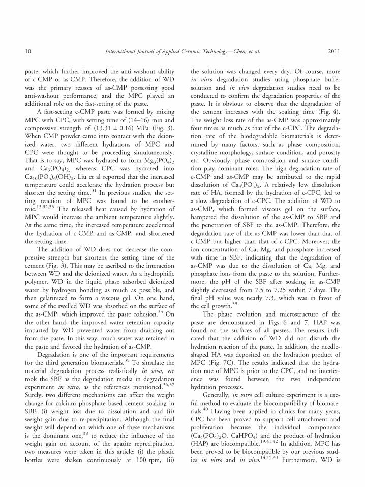
the cell attachment efficiency on as-CMP could reach121.8%, which was larger than that on c-CMP(103.6%). Figure 11 showed the proliferation curves ofMG-63 cells on c-CPC, c-CMP, and as-CMP. Obvi-ously, the as-CMP had the highest cell density.
Discussion
Anti-washout property is an important characterbecause the implanted biomaterials are often affected
by the fluid erosion in clinic. The cement paste, beforebeing hydrated fully, is apt to be crumbled when it isin contact with the fluid. Conversely, once the paste iscrumbled, the hydration reaction will not be com-pleted.22 The introduction of gelling agents indeedimproved the anti-washout property of the CPC pastesignificantly. However, most of the gelling agents pro-longed the setting time of the cement adversely. Forexample, the addition of hydroxypropyl-methylcellulose(HPMC) and carboxyl methyl-cellulose (CMC)improved the washout resistance, but the setting timewas increased greatly from 30 to 60 min.23 Chitosandelayed the setting time of CPC, whereas slight disinte-gration was observed on the chitosan-modified cement.5
Because of the cement’s inability to support stresswithin the initial period, a long setting time couldcause problems.24 For example, a severe inflammatoryresponse occurred when the CPC failed to set and dis-integrate, probably due to a low initial mechanicalstrength.12 Therefore, as far as injectable pastes are con-cerned, anti-washout and fast-setting are preconditionsfor surgical application. In the present study, an anti-washout and fast-setting calcium–magnesium phosphatecement was developed to widen application, especiallyin repairing bone defects without hemostasis or in sur-gical procedures where complete hemostasis is difficult.
The as-CMP showed good anti-washout property,followed by c-CMP and c-CPC in turn (Fig. 1). In thisstudy, WD prevented cement from disintegrationremarkably (Fig. 2), an important factor for ICPC usedin highly blood-perfused regions such as vertebroplasty.It was reported that small volumes of intravascularCPCs resulted in right ventricle occlusion and massivepulmonary embolism.25–29 When WD was dissolved indeionized water, viscous gel was formed and absorbedon the surface of the CMP particles, which immobi-lized the CMP particles and caused agglomeration. Atthe same time WD reduced the penetration of liquidinto the cement. In addition, c-CMP had a certainanti-washout ability whereas c-CPC did not have it(Figs. 1 and 2). The key component MPC may beresponsible for the difference in anti-washout abilitybetween c-CMP and c-CPC. Ishikawa et al reportedthat fast setting was a requirement to develop the non-decay type cement.22 Lisa E. Carey believed that therapid setting of the CPC paste helped in improving theinitial mechanical strength, thereby avoiding theimplant disintegration and accompanying inflamma-tion.30 MPC accelerated the hydration reaction of the
Fig. 10. Attaching efficiencies of MG-63 cells on c-CPC,c-CMP, and as-CMP after being cultured for 3 h. *Statisticallysignificant difference P < 0.05.
Fig. 11. Cell growth and proliferation curves of c-CPC,c-CMP, and as-CMP.
www.ceramics.org/ACT Fast-Setting Calcium–Magnesium Phosphate Cement 9
paste, which further improved the anti-washout abilityof c-CMP or as-CMP. Therefore, the addition of WDwas the primary reason of as-CMP possessing goodanti-washout performance, and the MPC played anadditional role on the fast-setting of the paste.
A fast-setting c-CMP paste was formed by mixingMPC with CPC, with setting time of (14–16) min andcompressive strength of (13.31 ± 0.16) MPa (Fig. 3).When CMP powder came into contact with the deion-ized water, two different hydrations of MPC andCPC were thought to be proceeding simultaneously.That is to say, MPC was hydrated to form Mg3(PO4)2and Ca3(PO4)2, whereas CPC was hydrated intoCa10(PO4)6(OH)2. Liu et al reported that the increasedtemperature could accelerate the hydration process butshorten the setting time.31 In previous studies, the set-ting reaction of MPC was found to be exother-mic.13,32,33 The released heat caused by hydration ofMPC would increase the ambient temperature slightly.At the same time, the increased temperature acceleratedthe hydration of c-CMP and as-CMP, and shortenedthe setting time.
The addition of WD does not decrease the com-pressive strength but shortens the setting time of thecement (Fig. 3). This may be ascribed to the interactionbetween WD and the deionized water. As a hydrophilicpolymer, WD in the liquid phase adsorbed deionizedwater by hydrogen bonding as much as possible, andthen gelatinized to form a viscous gel. On one hand,some of the swelled WD was absorbed on the surface ofthe as-CMP, which improved the paste cohesion.34 Onthe other hand, the improved water retention capacityimparted by WD prevented water from draining outfrom the paste. In this way, much water was retained inthe paste and favored the hydration of as-CMP.
Degradation is one of the important requirementsfor the third generation biomaterials.35 To simulate thematerial degradation process realistically in vivo, wetook the SBF as the degradation media in degradationexperiment in vitro, as the references mentioned.36,37
Surely, two different mechanisms can affect the weightchange for calcium phosphate based cement soaking inSBF: (i) weight loss due to dissolution and and (ii)weight gain due to re-precipitation. Although the finalweight will depend on which one of these mechanismsis the dominant one,38 to reduce the influence of theweight gain on account of the apatite reprecipitation,two measures were taken in this article: (i) the plasticbottles were shaken continuously at 100 rpm, (ii)
the solution was changed every day. Of course, morein vitro degradation studies using phosphate buffersolution and in vivo degradation studies need to beconducted to confirm the degradation properties of thepaste. It is obvious to observe that the degradation ofthe cement increases with the soaking time (Fig. 4).The weight loss rate of the as-CMP was approximatelyfour times as much as that of the c-CPC. The degrada-tion rate of the biodegradable biomaterials is deter-mined by many factors, such as phase composition,crystalline morphology, surface condition, and porosityetc. Obviously, phase composition and surface condi-tion play dominant roles. The high degradation rate ofc-CMP and as-CMP may be attributed to the rapiddissolution of Ca3(PO4)2. A relatively low dissolutionrate of HA, formed by the hydration of c-CPC, led toa slow degradation of c-CPC. The addition of WD toas-CMP, which formed viscous gel on the surface,hampered the dissolution of the as-CMP to SBF andthe penetration of SBF to the as-CMP. Therefore, thedegradation rate of the as-CMP was lower than that ofc-CMP but higher than that of c-CPC. Moreover, theion concentration of Ca, Mg, and phosphate increasedwith time in SBF, indicating that the degradation ofas-CMP was due to the dissolution of Ca, Mg, andphosphate ions from the paste to the solution. Further-more, the pH of the SBF after soaking in as-CMPslightly decreased from 7.5 to 7.25 within 7 days. Thefinal pH value was nearly 7.3, which was in favor ofthe cell growth.39
The phase evolution and microstructure of thepaste are demonstrated in Figs. 6 and 7. HAP wasfound on the surfaces of all pastes. The results indi-cated that the addition of WD did not disturb thehydration reaction of the paste. In addition, the needle-shaped HA was deposited on the hydration product ofMPC (Fig. 7C). The results indicated that the hydra-tion rate of MPC is prior to the CPC, and no interfer-ence was found between the two independenthydration processes.
Generally, in vitro cell culture experiment is a use-ful method to evaluate the biocompatibility of biomate-rials.40 Having been applied in clinics for many years,CPC has been proved to support cell attachment andproliferation because the individual components(Ca4(PO4)2O, CaHPO4) and the product of hydration(HAP) are biocompatible.19,41,42 In addition, MPC hasbeen proved to be biocompatible by our previous stud-ies in vitro and in vivo.14,15,43 Furthermore, WD is
10 International Journal of Applied Ceramic Technology—Chen, et al. 2011
known to be nontoxic as a derivative of dextrin, whichhas been widely used in dialysis and in the drug deliv-ery field. Therefore, from the composition point ofview, it is sure that c-CPC, c-CMP, and as-CMP arenontoxic to the osteoblast-like cell. In addition, MTTassay indicated that the MG-63 cell metabolic activityof as-CMP was superior to that of c-CPC and c-CMPafter culturing for 3 or 5 days, which might have some-thing to do with different surface conditions. With theaddition of the WD and the increase of hydrophilicity,the as-CMP was envisioned to have better cell biocom-patibility compared to c-CPC and c-CMP. To inspectthis possibility, cell attachment, and proliferation onas-CMP were investigated in comparison with c-CPCand c-CMP, as shown in Figs. 10 and 11. These resultswere basically coincident with the fact that the cellswere liable to attach on hydrophilic surfaces. Besides,the improving of anti-washout character of as-CMPalso benefited the affinity between cells and theas-CMP. The SEM observation further proved that thecells were attached in higher density on the as-CMP.Similar results were obtained as shown in Fig. 9 for themetabolic activity of MG-63 cells measured by MTTassay, and as-CMP has higher cell density. It suggeststhat the introduction of WD was beneficial for theattachment of MG-63 cells.
Conclusions
The anti-washout and fast-setting calcium–magne-sium phosphate cement was developed by adding WDand MPC into CPC. The as-CMP showed the bestanti-washout property, followed by c-CMP and c-CPCin turn. WD imparted anti-washout to the as-CMP,whereas MPC simultaneously improved the anti-wash-out and fast-setting of the paste. The synergistic effectof MPC and WD shortened the setting time of as-CMPobviously to 12 min in comparison with the c-CPC of30 min. As-CMP had a high compressive strength of13.31 MPa. In addition, the MPC powders improvedthe degradation of the as-CMP whereas WD reduced it.Furthermore, the addition of WD did not disturb thehydration reaction of the as-CMP. Two different hydra-tions of MPC and CPC proceeded simultaneously, andthe hydration of MPC is prior to that of CPC. Becauseof the hydrophilic surface imparted by WD, the as-CMP had better cell biocompatibility, cell attachment,and proliferation compared to c-CPC and c-CMP. On
the basis of the above results, we conclude that the anti-washout and fast-setting as-CMP will have promisingprospects in clinical application.
References
1. H. H. K. Xu, S. Takagi, J. B. Quinn, and L. C. Chow, “Fast-SettingCalcium Phosphate Scaffolds with Tailored Macropore Formation Ratesfor Bone Regeneration,” J. Biomed. Mater. Res., 68A 725–734 (2004).
2. M. Bohner, F. A. Theiss, D. Pelt, W. Hirsiger, R. Houriet, and G. Rizzoli,“Compositional Changes of a Dicalcium Phosphate Dihydrate CementAfter Implantation in Sheep,” Biomaterials, 24 3463–3474 (2003).
3. X. P. Wang, L. Chen, H. Xiang, and J. D. Ye, “Influence of Anti-WashoutAgents on the Rheological Properties and Injectability of a Calcium Phos-phate Cement,” J. Biomed. Mater. Res, 81B 410–418 (2007).
4. A. Cherng, S. Takagi, and L. C. Chow, “Effects of Hydroxypropylmethyl-Cellulose and Other Gelling Agents on the Handling Properties ofCalcium Phosphate Cement,” J. Biomed. Mater. Res, 35 273–277 (1997).
5. L. Leroux, Z. Hatim, M. Freche, and J. L. Lacout, “Effects of VariousAdjuvants (Lactic Acid, Glycerol, and Chitosan) on the Injectability of aCalcium Phosphate Cement,” Bone, 25 31S–34S (1999).
6. T. Masaaki, Y. Miyamoto, K. Ishikawa, T. Taketomo, T. Yuasa, and N.Masaru, “Initial Histological Evaluation of Anti-Washout Type Fast-Set-ting Calcium Phosphate Cement Following Subcutaneous Implantation,”Biomaterials, 19 2057–2063 (1998).
7. J. E. Frampton and G. L. Plosker, “Icodextrin: A Review of its Use inPeritoneal Dialysis,” Drugs, 63 2079–2105 (2003).
8. K. B. Hosieet al.,“A Pilot Study of Adjuvant Intraperitoneal 5-FluorouracilUsing 4% Icodextrin as a Novel Carrier Solution,” Eur. J. Surg. Oncol., 29254–260 (2003).
9. L. Salah, “Controlling Degradation of Low-Molecular-Weight NaturalPolymer”Dextrin”Using Gamma Irradiation,” Int. J. Biol. Macromol., 4457–63 (2009).
10. H. D. Hreczuk, D. Chicco, L. German, and R. Duncan, “Dextrins asPotential Carriers for Drug Targeting: Tailored Rates of Dextrin Degrada-tion by Introduction of Pendant Groups,” Int. J. Pharm., 230 57–66(2001).
11. H. H. K. Xu, L. E. Carey, C. G. Simon, S. Takagi, and L. C. Chow,“Premixed Calcium Phosphate Cements: Synthesis, Physical Properties,and Cell Cytotoxicity,” Dent. Mater., 23 433–441 (2007).
12. Y. Ueyama, K. Ishikawa, T. Mano, T. Koyama, H. Nagatsuka, and T.Matsumura, “Initial Tissue Response to Anti-Washout Apatite Cement inthe Rat Palatal Region: Comparison with Conventional Apatite Cement,”J. Biomed. Mater. Res., 55 652–660 (2001).
13. C. S. Liu, “Inorganic Bone Adhesion Agent and Its Use in Human HardTissue Repair”, U.S. Patent No.7094286B2 (2006).
14. Z. Z. Wu, J. Zhang, T. Y. Chen, L. J. Li, C. S. Liu, and Z. W. Chen,“Experimental Study of a New Type of Cement on Tibia Plateau FracturesTreatment,” Clin. Medi. J China, 12 261–264 (2005).
15. Z. Z. Wu, J. Zhang, T. Y. Chen, L. J. Li, C. S. Liuand, and H. Guo,“Experimental Study on Magnesium Phosphate Cement in Fracture Treat-ment,” Chin. J. Repair Reconstr. Surg., 20 912–915 (2006).
16. F. Wu, J. Wei, H. Guo, F. Chen, H. Hong, and C. S. Liu, “Self-SettingBioactive Calcium Magnesium Phosphate Cement with High Strength andDegradability for Bone Regeneration,” Acta Biomaterialia, 4 1873–1884(2008).
17. C. S. Liu, H. F. Shao, F. Y. Chen, and H. Y. Zheng, “Rheological Proper-ties of Concentrated Aqueous Injectable Calcium Phosphate CementSlurry,” Biomaterials, 27 5003–5013 (2006).
18. T. Kokubo and H. Takadama, “How Useful is SBF in Predicting In VivoBone Bioactivity,” Biomaterials, 27 2907–2915 (2006).
19. C. Knabe, G. Berger, R. Gildenhaar, J. Meyer, C. R. Howlett, andB. Markovic, “Effect of Rapidly Resorbable Calcium Phosphates and a Cal-cium Phosphate Bone Cement on the Expression of Bone-Related Genesand Proteins In Vitro,” J. Biomed. Mater. Res., 69A 145–154 (2004).
www.ceramics.org/ACT Fast-Setting Calcium–Magnesium Phosphate Cement 11
20. L. Hong, Z. Z Guo, B. Xue, Y. M. Zhang, and W. Y. Huang, “CollagenModulating Crystallization of Apatite in a Biomimetic Gel System,”Ceram. Int., 37 [7] 2305–2310 (2011).
21. J. Jiaet al.,“Development of Magnesium Calcium Phosphate Biocement forBone Regeneration,” J. R. Soc. Interface, 7 (49) 1171–1180 (2010).
22. K. Ishikawa, Y. Miyamoto, M. Kon, M. Nagayama, and K. Asaoka,“Non-Decay Type Fast-Setting Calcium Phosphate Cement: Composite ofFSCPC with Sodium Alginate,” Biomaterial, 16 527–532 (1995).
23. A. Cherng, S. Takagi, and L. C. Chow, “Effects of Hydroxypropyl-Meth-ylcellulose and Other Gelling Agents on the Handling Properties of Cal-cium Phosphate Cement,” J. Biomed. Mater. Res., 35 273–277 (1997).
24. Y. Miyamoto, K. Ishikawa, M. Takechi, T. Toh, T. Yuasa, and M. Nagay-ama, “Histological and Compositional Evaluations of Three Types of Cal-cium Phosphate Cements When Implanted in Subcutaneous TissueImmediately After Mixing,” J. Biomed. Mater. Res., 48 36–42 (1999).
25. M. Bohner, U. Gbureck, and J. E. Barralet, “Technological Issues for theDevelopment of More Efficient Calcium Phosphate Bone Cements: ACritical Assessment,” Biomaterials, 26 6423–6429 (2005).
26. L. C. Chow, “Calcium Phosphate Cements: Chemistry, Properties, andApplications,” Mater. Res. Soc. Symp. – Proc., 599 27–37 (2000).
27. M. H. Alkhraisat, F. T. Marino, J. R. Retama, L. B. Jerez, and E. Lopez,“Betatricalcium Phosphate Release from Brushite Cement Surface,” J. Bio-med. Mater. Res., 84A 710–717 (2008).
28. M. Bohner, N. Doebelin, and G. Baroud, “Theoretical and ExperimentalApproach to Test the Cohesion of Calcium Phosphate Pastes,” Eur. CellsMater., 12 26–35 (2006).
29. M. HAlkhraisat, C. Rueda, F. T. Marino, J. Torres, L. B. Jerez, and U.Gbureck, “The Effect of Hyaluronic Acid on Brushite Cement Cohesion,”Acta Biomaterialia, 6 257–265 (2010).
30. E. C. Lisa, H. H. K. Xu, G. Carl, J. Simon, T. Shozo, and L. C. Chow,“Premixed Rapid-Setting Calcium Phosphate Composites for BoneRepair,” Biomaterials, 26 5002–5014 (2005).
31. C. S. Liu, W. Gai, S. H. Pan, and Z. S. Liu, “The Exothermal Behaviorin the Hydration Process of Calcium Phosphate Cement,” Biomaterials, 242995–3003 (2003).
32. E. Soudee and J. Pera, “Mechanism of Setting Reaction in Magnesiaphos-phate Cements,” Cem. Concr. Res., 30 315–321 (2000).
33. E. Soudee and J. Pera, “Influence of Magnesia Surface on the Setting Timeof Magnesia-Phosphate Cement,” Cem. Concr. Res., 32 153–157 (2002).
34. K. H. Khayat, “Viscosity-Enhancing Admixtures for Cement–Based Mate-rials— An Overview,” Cem. Concr. Res., 20 171–188 (1998).
35. L. L. Hench and J. M. Polak, “Third-Generation Biomedical Materials,”Science, 295 1014–1017 (2002).
36. R. W. Tan et al.,“In Vitro and In Vivo Degradation of an Injectable BoneRepair Composite,” Polym. Degrad. Stabil., 95 1736–1742 (2010).
37. V. Cauda, A. Schlossbauer, and T. Bein, “Bio-Degradation Study ofColloidal Mesoporous Silica Nanoparticles: Effect of Surface Functionaliza-tion with Organo-Silanes and Poly(Ethylene Glycol),” Micropor. Mesopor.Mat., 132 60–71 (2010).
38. A. Bandyopadhyay, S. Bernard, W. C Xue, and S. Bose, “Calcium Phos-phate Based Resorbable Ceramics: Influence of MgO, ZnO and SiO2 Do-pants,” J. Am. Ceram. Soc., 89 [9] 2675–2688 (2006).
39. E. W. H. Bodde, O. C. Boerman, F. G. M. Russel, A. G. Mikos, P. H.M. Spauwen, and J. A. Jansen, “The Kinetic and Biological Activity ofDifferent Loaded rhBMP-2 Calcium Phosphate Biocement Implants inRats,” J. Biomed. Mater. Res. A, 87 780–791 (2008).
40. W. Y. Zhao, J. Y. Wang, W. Y. Zhai, Z. Wang, and J. Chang, “The Self-Setting Properties and In Vitro Bioactivity of Tricalcium Silicate,” Bioma-terials, 26 6113–6121 (2005).
41. A. Ehara, K. Ogata, S. Imazato, S. Ebisu, T. Nakano, and Y. Umakoshi,“Effects of a–TCP and b-TCP on MC3T3-E1 Proliferation, Differentia-tion and Mineralization,” Biomaterials, 24 831–836 (2003).
42. T. Yuasa, Y. Miyamoto, K. Ishikawa, M. Takechi, Y. Momota, andS. Tatehara, “Effects of Apatite Cements on Proliferation and Differentia-tion of Human Osteoblasts In Vitro,” Biomaterials, 25 1159–1166 (2004).
43. Y. L. Yu, J. Wang, C. S. Liu, B. W. Zhang, H. H. Cheng, and H. Guo,“Evaluation of Inherent Toxicology and Biocompatibility of MagnesiumPhosphate Bone Cement,” Colloids Surf B: Biointerfaces, 76 496–504(2010).
12 International Journal of Applied Ceramic Technology—Chen, et al. 2011