photosynthetic microorganisms as epibionts and euendoliths...
TRANSCRIPT

Fottea 10(1): 129–140, 2010 129
Photosynthetic microorganisms as epibionts and euendoliths on biotic substrates in a thermal spring with ferric–iron deposits
Canella Radea, Ioanna LouvRou, Adriani Pantazidou & Athena economou–amiLLi*
Department of Ecology and Systematics, Faculty of Biology, University of Athens, Panepistimiopolis, Athens 15784, Greece; *e–mail: [email protected], tel.: +30–210–7274325, fax: +30–210–7274885
Abstract: Rust–coloured shells of the aquatic gastropod Ventrosia ventrosa, a new record for eastern Greece, indicating presence of iron (EDAX analysis) were studied for detection of iron–encrusted photosynthetic epibionts in a Greek brackish–water thermal spring (38 °C). Microscopic analyses (LM, SEM) revealed the presence of a biofilm consisted of mostly facultative micro–epibionts, i.e. a) 5 periphytic taxa of coccal and filamentous cyanobacteria, including a taxonomically and ecologically interesting morphospecies, Xenococcus cf. pyriformis, dominated exclusively on the shell surface, and b) pennate diatoms with higher species richness (18 periphytic taxa of the genera Amphora, Brachysira, Cymbella, Diatoma, Encyonema, Navicula, Nitzschia, Pleurosigma, Synedra, Ulnaria; 5 taxa as new records for Greece), most of them emerging only after acid treatment of whole gastropod shells. The abundant diatoms thriving directly or nearby the iron–coatings (Cocconeis placentula var. euglypta and Achnanthes brevipes sensu lato) exhibited different modes of attachment (‘adnate’ and ‘pendunculate’, respectively). Two euendolithic cyanobacteria (Hyella sp. and Leptolyngbya terebrans; the former with special taxonomic interest) were also found perforating the delicate gastropod shells, with no distinct differentiation in the extent of infestation between live and dead gastropod shells. Moreover, the possible impact of these encrusted photosynthetic assemblages on V. ventrosa was investigated; statistical analysis showed that a) there is no ‘drag effect’, induced by the epibionts, influencing the gastropod growth (i.e. shell length), b) shell size enlargement provides a favourable space and promotes the intense fouling by both micro–epibionts and macro–epibionts (egg–capsules), and c) the detachment prevention of egg–capsules is attributed to the biofilm development.
Key words: Epibiosis, Ventrosia ventrosa (Gastropoda, Hydrobiidae), iron–encrusted cyanobacteria and diatoms, euendoliths, thermal springs, Greek Island (Kythnos)
Introduction
Adhesion of microorganisms to biotic substrates (aquatic plants including algae or aquatic animals) forming a biofilm is a well–known phenomenon that has been documented repeatedly mainly in the sea (e.g. in Greece; BeLegRatis et al. 1999; BeLegRatis & economou–amiLLi 2001; Pantazidou et al. 2001; LouvRou 2007). In the case of two or more organisms being in a spatially close facultative association the phenomenon is known as epibiosis; in the description of such microbe–metazoan interactions, the metazoan is generally called a ‘basibiont’, and microbes are called ‘epibionts’ (WahL 1989). Microbial epibionts, although generally ignored in the description of marine organisms, may have profound effects on the basibiont by causing a multitude of beneficial or detrimental effects and
must be taken into account when host ecology is studied (WahL & hay 1995; WahL et al. 1997; giLLan & cadée 2000) and especially when the hosts are small (WahL 1989).
A special mode of colonization is the active penetration in a variety of carbonate substrates both of inorganic and organic origin (including calcareous parts of certain invertebrates) mainly by euendolithic cyanobacteria, and less frequently by chlorophytes or by the stage Conchocelis of rhodophytes (dReW 1949; LaBoReL & Le camPion–aLsumaRd 1979; Le camPion–aLsumaRd 1979; Golubić et al. 1981; anagnostidis & Pantazidou 1985; 1988a,b,c; Pantazidou et al. 2006; LouvRou 2007; tRiBoLLet 2008a,b), whereas active penetration of diatoms into carbonate substrates has not been recorded.
Epibiotic microbial communities are sometimes associated with ferric–iron deposits, as

130 Radea et al.: Microorganisms as epibionts
in the case of communities living on the bivalve Montacuta ferruginosa (giLLan et al. 1998, 2000; giLLan & de RiddeR 2001) or on the mud snail Hydrobia ulvae (giLLan & cadée 2000). In iron–enriched natural environments like thermal springs and vents, the cellular survival would have been achieved through iron detoxification mechanisms, and the iron–scavenging mechanisms exhibited by marine and freshwater cyanobacteria (e.g. strains of Synechococcus, WeBB et al. 1994; Katoh et al. 2000, 2001) may have been specially developed there (BRoWn et al. 2005).
The aim of this paper was to study the phototrophic microbial communities thriving as epibionts on the typically delicate, horny–coloured shells (FaLnioWsKy 1987) of the gastropod Ventrosia ventrosa (montagu 1803). This species was the only mollusc found in abundance with extensive iron coatings (rust–coloured) on its shells in a Greek thermal spring, rich in FeO2 and chemically reminding brackish waters, on Kythnos Island. Apart from identification of the iron–encrusted photosynthetic microorganisms, research was focused on the mode of epibiotic adhesion, the possible facultative or obligate nature of the epibionts, and the existence and boring patterns of the phototrophic euendoliths. Also, the impact of this biofilm on the growth of V. ventrosa was detected, i.e. the possible ‘drag effect’ induced by the epibionts influencing its shell length, the shell size enlargement as favourable space for intense fouling by both micro–epibionts and macro–epibionts (egg–capsules), and the detachment prevention of egg–capsules by the biofilm development and the resultant modified substrate structure.
Material and methods
There are two neighbouring iron thermal water springs on Kythnos, an island belonging to the Cyclades group of the Aegean, i.e. the ‘Kakkavou’ (52 °C) outflow and the ‘Agioi Anargyroi’ (38 °C) outflow (Latitude: 37° 26’ 23” N, Longitude: 24° 25’ 32” E). The latter cooler spring was selected for this study. Lithology and chemical analyses show the brackish nature of these thermal waters and presence of iron (LamBRaKis & KaLLeRgis 2005).
Alive and dead specimens of V. ventrosa were collected at the thermal outflow of ‘Agioi Anargyroi’ in July 2008 mainly close to the vent and along the stream (appr. 180 m from the sea; 34–38 °C) (Figs 1, 2). Samples were also collected far from the vent at the stream outflow (T=33 °C) and served as reference
to determine the gastropod distribution limits. The bottom at each sampling site was swept with a fine–meshed hand–net (600 μm mesh) for shell gastropod collection, and samples were preserved in ethanol solution (80%). The gastropods were examined under a stereo–microscope (Stemi 2000–C, Zeiss, Germany), and specimens with a number of whorls >3 were dissected and identified to species level. Shell length was measured to the nearest 0.25 mm. The length values were not distributed normally and, therefore, non–parametric tests were employed. Statistical analyses (i.e. Mann–Whitney U–test, and Spearman
Figs 1–2. General view of the outflows of the thermal spring ‘Agioi Anargyroi’ on Kythnos Island: (1) Macroscopic view of the vegetation, and of the iron deposits seen as brown patches; (2) A population of the gastropod Ventrosia ventrosa with rust–coloured shells; note the various degrees of iron deposition, and the egg–capsules on the shells.
rank correlation coefficient; see zaR 1984) were done by means of the software package ‘Statistica 7’.For direct study of the epibiotic assemblages, whole shells or fragments of V. ventrosa were observed firstly by light microscopy (Stemi 2000–C and Photomicroscope III, Zeiss), and then by scanning electron microscopy (Jeol JSM–35 operating at 25 kV) after being dehydrated in a gradient of alcohol dilution series (10–100%) and finally in pure aceton, critical point dried and spray coated in gold–palladium. For detailed microscopic study of the epibiotic photosynthetic assemblages whole shells, shell fragments or scratches of the ferric– and non ferric– iron coatings of V. ventrosa were either dissolved
2
1

using Pereny’s solution (10% HNO3, 0.5% Cr2O3, 95% C2H5OH in proportion 4:3:3) for extraction of cyanobacteria, or treated with acids (KMNO4 and HCl, after simonsen 1962) for cleaning the diatom frustules. For dissolving calcium carbonates of the shell surface and releasing the euendolithic algae, fixed samples of V. ventrosa were treated also with Pereny’s solution and observed by light microscopy. For observation of the tunnels (borings and boreholes) and for identification of the boring patterns of the euendoliths, shells were boiled for 2–5 min three–times in a 2.5% solution of sodium hypochlorite (NaOCl) in order to destroy the organic material, which was removed by rinsing several times with distilled water; the specimens were then allowed to dry, sputter coated in gold palladium and observed by scanning electron microscopy.
For energy–dispersive X–rays analyses (EDAX) to detect elements on the shell surface, shells were air–dried, mounted and observed under a Jeol JSM–5600 microscope coupled to an energy dispersive X–ray microanalysis detector Oxford LinkTM IsisTM 300 and software Oxford SEMQuantTM. The analysis was done at 20 kV with a beam current of 0.5 nA, lifetime 50 sec and beam diameter <2 μm.
Results and discussion
The photosynthetic biofilmThe iron–coated shells of V. ventrosa were covered by a biofilm mainly consisted of cyanobacteria and diatoms, the latter with higher species richness; also, certain shells were found poorly infested by euendolithic cyanobacteria (Table 1).
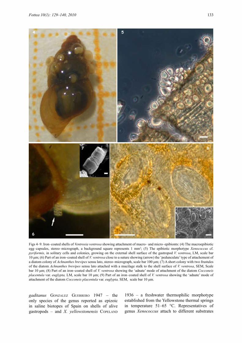
The shell surface of most specimens of V. ventrosa (77.4%) was found to be characteristically rust–coloured (Figs 4, 6, 8) indicating presence of iron, and shell coatings were more developed in adults and especially on corroded shells than in juveniles or non corroded shells. EDAX analysis indicated that the most abundant elements of the coatings were O, Si and Fe; the other elements detected were C, Na, Cl (Fig. 3). Comparatively,
EDAX analysis directly on the concave raphe–valve of diatoms showed the same more or less distribution of elements; whereas on the convex rapheless–valve the proportion was greater in Si and lower in Ca and Fe, as expected possibly due to the lesser accumulation of elements on the convex areas of the valves. The biogenic iron precipitates are expected to affect the distribution of both epibionts and euendoliths through shading or reduced space available for colonization. Similarly, biogenic carbonate precipitates affected diatom colonization on Corallina elongata (Pantazidou et al. 2001); also Potamogeton precipitates affected the attached epiphytes (cattaneo 1978).
The ascertained species composition seems to be in accordance with the biological composition normally found in similar thermal aquatic environments. It is noted that the photosynthetic microbial assemblages of a typical thermal aquatic environment mainly consist of thermophilic prokaryotes, and the group of cyanobacteria have long been considered (vouK 1923; coPeLand 1936; anagnostidis 1959) to dominate in thermal springs (e.g. anagnostidis 1961; BRocK & BRocK 1966); whereas, associate microorganisms belonging to genera of procaryotes other than cyanobacteria (e.g. Archaea, sulphate–reducing bacteria, iron–oxidazing bacteria, etc.) and /or algae (e.g. diatoms, green algae) are also found as a respond to specific ecological niches especially in temperatures lower than 40 °C (e.g. moLisch 1926; anagnostidis 1968; KuLLBeRg 1968; oWen et al. 2008). Diatoms, in particular, give more information on the thermal habitat in many cases due to their qualitative richness and well–defined ecological requirements (e.g. in Greece; economou–amiLLi 1976; anagnostidis & economou–amiLLi 1978; Pantazidou et al. 2001; LouvRou 2007). The only availabe references about euendolithic cyanobacteria from geothermal marine environment or thermal springs are those of thomas & gonzaLves (1965), Pantazidou et al. (2001) and LouvRou (2007), the last two from exoskeletal parts of invertebrates in hydrothermal areas of Milos Island, Greece.
The only available reference for epibiotic microorganisms (i.e. Protozoa) on the shell of V. ventrosa, as syn. Hydrobia stagnalis (BasteR 1765), is that of hoFKeR (1930) from brackish waters. Iron–encrusted biofilms with diatoms, iron bacteria, and cyanobacteria covering the shell of another related mud snail, the brackish–marine Hydrobia ulvae (Pennant 1777), were studied by Fig. 3. EDAX spectrum of the iron–coatings of Ventrosia
ventrosa.
Fottea 10(1): 129–140, 2010 131
giLLan & cadeé (2000).
Epibiotic CyanobacteriaThe most frequent of the cyanobacteria was a mophotype of Xenococcus (Fig. 5). This morphotype was observed exclusively as epibiont on shells of V. ventrosa in approximately 1/4 of the individuals examined, and in areas nearby or upon the fine iron coatings but never directly on heavy iron precipitates; whereas it was absent in the surrounding periphytic samples (to be published elsewhere). Its diacretic features, based
on morphometric parameters and developmental stages only in wild material, are closer to the type of Xenococcus pyriformis setcheLL et gaRdneR in gaRdneR 1918. However, ecology differs. In the present study we refer to it as Xenococcus cf. pyriformis. Further taxonomic investigation on this morphotype, e.g. molecular classification by 16S rRNA–based methods on fresh and cultured material, would be of interest.
It is noted that Xenococcus thuRet in BoRnet et thuRet 1880 is a little known genus with 25 described species including Xenococcus
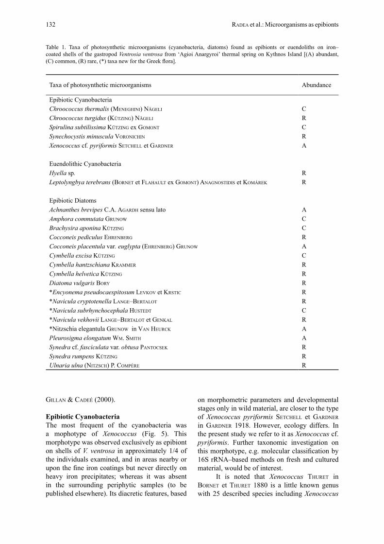
Table 1. Taxa of photosynthetic microorganisms (cyanobacteria, diatoms) found as epibionts or euendoliths on iron–coated shells of the gastropod Ventrosia ventrosa from ‘Agioi Anargyroi’ thermal spring on Kythnos Island [(A) abundant, (C) common, (R) rare, (*) taxa new for the Greek flora].
Taxa of photosynthetic microorganisms Abundance
Epibiotic CyanobacteriaChroococcus thermalis (meneghini) nägeLi CChroococcus turgidus (Kützing) nägeLi RSpirulina subtilissima Kützing ex gomont CSynechocystis minuscula voRonichin RXenococcus cf. pyriformis setcheLL et gaRdneR A
Euendolithic CyanobacteriaHyella sp. RLeptolyngbya terebrans (BoRnet et FLahauLt ex gomont) anagnostidis et KomáReK R
Epibiotic DiatomsAchnanthes brevipes c.a. agaRdh sensu lato AAmphora commutata gRunoW CBrachysira aponina Kützing CCocconeis pediculus ehRenBeRg RCocconeis placentula var. euglypta (ehRenBeRg) gRunoW ACymbella excisa Kützing CCymbella hantzschiana KRammeR RCymbella helvetica Kützing RDiatoma vulgaris BoRy R*Encyonema pseudocaespitosum LevKov et KRstic R*Navicula cryptotenella Lange–BeRtaLot R*Navicula subrhynchocephala hustedt C*Navicula vekhovii Lange–BeRtaLot et genKaL R*Nitzschia elegantula gRunoW in van heuRcK APleurosigma elongatum Wm. smith ASynedra cf. fasciculata var. obtusa PantocseK RSynedra rumpens Kützing RUlnaria ulna (nitzsch) P. comPèRe R
132 Radea et al.: Microorganisms as epibionts
gaditanus gonzaLez gueRReRo 1947 – the only species of the genus reported as epizoic in saline biotopes of Spain on shells of alive gastropods – and X. yellowstonensis coPeLand
1936 – a freshwater thermophilic morphotype established from the Yellowstone thermal springs in temperature 51–65 °C. Representatives of genus Xenococcus attach to different substrates
Figs 4–9. Iron–coated shells of Ventrosia ventrosa showing attachment of macro– and micro–epibionts: (4) The macroepibiotic egg–capsules, stereo–micrograph, a background square represents 1 mm2; (5) The epibiotic morphotype Xenococcus cf. pyriformis, in solitary cells and colonies, growing on the external shell surface of the gastropod V. ventrosa, LM, scale bar 10 μm; (6) Part of an iron–coated shell of V. ventrosa close to a suture showing (arrow) the ‘pedunculate’ type of attachement of a diatom colony of Achnanthes brevipes sensu lato, stereo–micrograph, scale bar 100 μm; (7) A short colony with two frustules of the diatom Achnanthes brevipes sensu lato attached with a mucilage stalk to the shell surface of V. ventrosa, SEM, Scale bar 10 μm; (8) Part of an iron–coated shell of V. ventrosa showing the ‘adnate’ mode of attachment of the diatom Cocconeis placentula var. euglypta, LM, scale bar 10 μm; (9) Part of an iron–coated shell of V. ventrosa showing the ‘adnate’ mode of attachment of the diatom Cocconeis placentula var. euglypta, SEM, scale bar 10 μm.
Fottea 10(1): 129–140, 2010 133

(stones, algae, aquatic plants); several species are known from mountain streams, others from the littoral zone of the sea (KomáReK & anagnostidis 1998). The type species Xenococcus pyriformis is known as epiphytic on marine filamentous and parenchymatous algae first from the Pacific coast of North America and later from various coasts of North and South America too, whereas from Europe it is known from the Black and Baltic seas (Kaas 1985); species of Xenococcus as associates to ‘sulphureta’ biocommunities from Greece are referred by anagnostidis (1968).
The Xenococcus morphotype was found associated with the chroococcalean taxa Chroococcus turgidus, Chroococcus thermalis, Synechocystis minuscula, and the filamentous species Spirulina subtilissima (Table 1). All these are known as periphytic taxa from hydrothermal environments, also from Greek thermal springs in temperatures 34–59 °C (anagnostidis 1961).
Epibiotic DiatomsDirect examination of iron–coatings on the shells of V. ventrosa both with light and scanning electron microscope revealed the presence of the following two taxa of diatoms as true epibionts (Table 1):
The most frequent epibiotic diatom was Cocconeis placentula var. euglypta, with raphe valves closely appressed to the substrate (‘adnate’ mode of attachment, sensu Round et al. 1990) and seemed to prevail all over the shell surface (Figs 8, 9). This diatom, and the genus as a group, is considered as a thermophilic species preferring temperatures over 22 °C (squiRes et al. 1979; vinson & RushFoRth 1989). It was found in Greece also as epibiont on the shells of another gastropod of Hydrobiidae, Potamopyrgus antipodarum (J.E. gRay 1843) in Lake Trichonis (Radea et al. 2008). This and other taxa of Cocconeis closely appressed to the substrate and living over the shell surface of other invertebrates (tube of polychaetes, and the bivalve Cerastoderma glauca BRuguieRi) were also found in hydrothermal or adjacent areas of Milos Island (Pantazidou et al. 2001; LouvRou 2007).
Another obvious presence was a number of morphotypes identified as Achnanthes brevipes sensu lato. Achnanthes species are mostly recorded from cold waters with decreasing abundance at temperatures above 14 °C (vinson & RushFoRth 1989). In our material these morphotypes appeared in short colonies suspended in the water (Figs 6,
7) and connected to the shell surface by mucilage stalks (‘pedunculate’ mode of attachment, sensu Round et al. 1990). The preference for suspending at the edges of the substrates can be attributed to the prevailing better illumination conditions and favourable nutrient renewal at these areas (e.g. cattaneo 1978; BeLegRatis & economou–amiLLi 2001). It is noted that, in flat biotic and artificial substrates, the type of colonizing process with the first colonists appearing on the margins and with the gradual filling–in of the flat areas is referred as ‘edge effect’ (cattaneo 1978; KoRte & BLinn 1983; hamiLton & duthie 1984).
A number of other diatoms (16 taxa of the genera Amphora, Brachysira, Cymbella, Diatoma, Encyonema, Navicula, Nitzschia, Pleurosigma, Synedra, Ulnaria) were mainly revealed only after treatment of the whole gastropod shells with acids (Table 1). Most of them are typical brackish and /or fresh–brackish waters species or species thriving in biotopes with high mineral content (e.g. Lange–BeRtaLot & KRammeR 1987; Lange–BeRtaLot 2001; KRammeR 2002). Due to their ecological requirements, they are expected to survive or flourish in such thermal spring outflows, i.e. in a stable ecosystem not only concerning temperature changes but also the relative stability of the high mineral content reminding brackish waters. Five of these taxa are reported for first time in Greece; Encyonema pseudocaespitosum was recently established from Lake Ohrid (LevKov et al. 2007). It is noted that Amphora commutata is known from Greece only from a lagoon on Milos Island (LouvRou 2007). Of additional taxonomic interest and further study are the morhotypes identified as Achnanthes brevipes sensu lato and Synedra cf. fasciculata var. obtusa.
The above diatoms are considered as facultative epibiotic species. They might be colonizers coming from neighbouring periphytic algal asssemblages and/or might represent ‘trapped’ epibiotic species concealed within the iron coatings in overlapping layers. The first view is supported by their finding at least of the abundant ones on filamentous algae (e.g. Cladophora) or other abiotic substrates (unpubl. data). Similarly, the shells of another mud snail, P. antipodarum, a freshwater–brackish species from the Greek Lake Trichonis, with no specific shell coloration or coatings, were also found to be covered by diatoms common to the surrounding lake system (Radea et al. 2008). The latter view of the ‘trapped’ cells is supported by their high
134 Radea et al.: Microorganisms as epibionts

abundance after cleaning of the epibiotic coatings with acids. Similar observations of ‘trapped’ epibiotic diatoms into calcareous precipitates were made on other invertebrates (LouvRou 2007) or after detaching the calcareous parts of the coralline alga Corallina elongata eLLis et soLandeR where epiphytic diatoms were revealed in overlapping layers (Pantazidou et al. 2001). As in the case of Corallina, the concealed space should be taken into account in quantitative analyses of epibiotic diatoms in the iron–coated shells of V. ventrosa.
Euendolithic CyanobacteriaTwo euendolitic cyanobacteria were found to poorly infest certain shells of V. ventrosa, i.e. Hyella sp. and Leptolyngbya terebrans (Table 1). These taxa are widely distributed within biogenic and non–biogenic calcareous substrates from marine, brackish and fresh waters; also in Greece they were found on members of Hydrobiidae from a saline lagoon (see extended literature in Pantazidou et al. 2006).
The morphotype identified as Hyella sp. is of special taxonomic interest needing further investigation, due to the similarities with the unsure record of Tryponema indicum thomas et gonzaLves 1965, a species needing revision according to KomáReK & anagnostidis (1998) and the only record of euendolithic cyanobacteria from
thermal springs (India). This species and the genus Tryponema itself need revision due to the similar morphology and the exclusively euendolithic mode of life with the related euendolithic genus Hyella BoRnet et FLahauLt (1888, 1889).
Presence of euendoliths and their boring patterns were ascertained with light microscopy and also scanning electron microscopy. Observations of the borings of Hyella sp. and Leptolyngbya terebrans (Figs 10, 11) reflect some aspects of their filamentous morphology (branching, size). It is noted that the internal surfaces of the excavated borings in plain or vertical view display the internal shell of the gastropod (Fig. 11, large arrow); similar mucilagineous and amorphous structures preserved as a thin lining at boreholes of euendolithic cyanobacteria, including Leptolyngbya terebrans, on marine clams were attributed by chacón et al. (2006) to remains of extracellular polymeric substances (EPS). Despite the present poor infestation of the gastropod shells, the role of euendolithic cyanobacteria for increasing the space available for colonization should be taken into account in biofouling, since it is known from other euendoliths that the original borings merge and allow adequate space for secondary colonizers (cyanobacteria, diatoms) to settle in the new cavities (Le camPion–aLsumaRd & Golubić 1985; Pantazidou et al. 2001).
Figs 10–11. Borings of euendoliths on the shell surface of Ventrosia ventrosa: (10) Borings of Hyella sp. reflecting the pseudofilamentous morphology, oriented parallel to the external shell surface; (11) Borings of Hyella sp. reflecting the pseudofilamentous branching and additionally the excavated boreholes on the external shell surface (the internal shell structure of the gastropod is discernible at large arrow). In the insert, fine borings (small arrow) of the filamentous euendolith Leptolyngbya terebrans. SEM. Scale bar 10 μm.
Fottea 10(1): 129–140, 2010 135

The low species richness and abundance of euendoliths and their borings can be attributed to the delicate and small shells of V. ventrosa which are prone to abrasion keeping the community in a state of ‘permanent initial colonization’ (compare chacón et al. 2006), whereas the effect of iron precipitates on the installation and proliferation of euendoliths cannot be excluded. The former view is corroborated by previous findings where the richness and penetration pattern of endolithic colonization was attributed to the nature of the substrate (in those cases harder shells where dissolution effects are minimal) and it was comparable to that in calcareous rocks (e.g. Le camPion–aLsumaRd & Golubić 1985; Le camPion–aLsumaRd et al. 1995, 1996; PeRRy 2000; Pantazidou et al. 2006).
In addition, distinct difference in infestation was not detected between live and dead shells in the material studied. However, great differentiation in species richness and succession of the euendolithic community in the skeletal material of live and dead host invertebrates has previously been reported (e.g. LaBoReL & Le camPion–aLsumaRd 1979; Le camPion–aLsumaRd et al. 1995; tRiBoLLet & PayRi 2001; Pantazidou et al. 2006; tRiBoLLet 2008a,b).
The basibiont as host for epibiosisThe collected gastropods were exclusively individuals of the mud snail V. ventrosa (montagu 1803) (Fig. 4); no other gastropod species was found in the thermal spring under study. It is noted that the geothermal fauna of aquatic invertebrates has generally low species diversity but sometimes greater densities than those of cooler habitats (James 1985). As for the distribution limits of this gastropod, it is noted that at sampling sites close to the vent the abundance of V. ventrosa was extremely high covering large areas of the stream bottom with shells of all ages, the oldest being the majority; whereas along the stream outflow the mud snails progressively diminish in number, and close to the sea only a few immature snails (with 2–3 whorls per shell) were recorded. According to BaRnes (1994) great densities of this species (~28 050 ind.m–2) are frequently observed at sites with suitable environmental conditions. The presence of V. ventrosa close to the venting sites of ‘Agioi Anargyroi’ thermal spring (33–38 °C), a biotope with thermal and chemical constancy reminding brackish waters, is not surprising since this species is a short–lived
mud snail, thriving in non tidal brackish waters in landlocked coastal lagoons (FaLnioWsKi 1987; BaRnes 1999), it is a rather eurythermic and the least specialized species among mud snails with metabolism not changing within a wide range (10–30 °C) of water temperatures (FaLnioWsKi 1987). The present record of V. ventrosa from Kythnos Island (Cyclades, Aegean Sea) is the first one from eastern Greece (BanK 2010), and the closer finding is that from Izmir bay, western coast of Turkey (ÇinaR et al. 2006).
The morphometric characters of the collected V. ventrosa were analyzed in a total of 106 individuals (Table 2). The measured length values were compared (Mann–Whitney U–test) and it was found that there is no significant difference between the iron–coated shells bearing epibiotic assemblages and the uncoated shells with a rather indistinct biofilm (Ζ=1.434804, n1=24, n2=82, p=0.151344). This observation implies a minimal effect of the epibiotic assemblages on the gastropod growth, as opposed to the increased ‘drag effect’ induced by epibionts and on the growth and possibly reproduction of other gastropods (e.g. the prosobranch gastropod Littorina littorea L. reported by WahL 1996, 1997) due to the resulted adverse conditions (higher energy expenditure for attachment and locomotion and a reduced allocation of resources).
Nearly all gastropod shells bear more than two capsules with one egg–embryo per capsule (Fig. 4), and only one capsule was recorded containing two egg–embryos, in accordance with previous observations (Lassen 1979). Egg–capsules are deposited by neighbouring conspecifics and could be considered as a kind of temporary macro–epibionts (Fig. 4). Egg–capsules were also measured in a number of shells of V. ventrosa (Table 1). Statistical analysis showed that the number of egg–capsules on iron–coated shells was significantly higher comparing with that on uncoated shells (Mann–Whitney U–test, Z=2.298262, n1=24, n2=82, p=0.021548). As for the iron–coated shells, a highly significant correlation was also detected between shell length and number of egg–capsules (Spearman Rank Correlation Coefficient R=0.262, N=82, p=0.017). On the contrary, the same correlation was found to be not significant (Spearman Rank Correlation Coefficient R=0.173, N=24, p=0.419) in the case of uncoated shells.
The above data indicate a tendency for egg–capsules deposition on the iron–coated shells
136 Radea et al.: Microorganisms as epibionts
where the encrusted epibiotic organisms and the modified substrate may prevent their detachment. On the other hand, the positive relation between number of epibiotic egg–capsules and length of iron–coated shells can be attributed to the larger and more favourable space for egg–capsules fixation, and it is in accordance with other findings (BecKett et al. 1996; cReed 2000) where shell size does play an important role for species abundance and richness of the epibionts, the larger shells being more intensely fouled comparing to the smaller ones.
Similar implications for a possible impact of epibionts on the egg fixation of a related species of Hydrobiidae, i.e. Hydrobia ulvae, were made by giLLan & cadée (2000). Additionally, BReWeR (1984) found that planulae of Cyanea (Cnidaria: Scyphozoa) do not usually attach to clean natural shells in the laboratory or to fresh shells in the field, where wettabilities are very low (equivalent to or less than that of glass), but high incidence of planulae attachment in the field was observed in aged shells possessing organic and bacterial films which show increased hydrophobicity. Generally, the larvae settlement of many marine invertebrates on solid surfaces is influenced by the chemistry, wettability and positively by the roughness of the substrate surface, and these features depend greatly in case a natural biofilm is attached (e.g. BReWeR 1984; WahL 1989; RodRiguez et al. 1993; maRsden & LaRsKy 2000; haRdeR et al. 2002) and especially in flowing waters (KoBaK 2005).
AcknowledgementsThe authors are grateful to Prof. A. Kelepertzis for providing facilities in the Geology Department of the Athens University and Dr. E. Michaelidis for his assistance in EDAX analysis, and also to the Biology student G. Phillipaeos for his help during field work.
This study was funded in part by the Special Account for Research Grants of the National and Kapodistrian University of Athens (NKUA, grant 70/4/5717).References
anagnostidis, K. (1959): Auffindung des Mastigocladus–Thermen–Typus in Griechen-land. – Biološki Glasnik 12: 86–89.
anagnostidis, K. (1961): Untersuchungen über die Cyanophyceen einiger Thermen in Griechen-land. – Inst. Syst. Bot. & Pflanzengeogr. Univ. Thessaloniki, 7: 1–322, Tab. 1–38, Figs 1–285 (in Greek).
anagnostidis, K. (1968): Untersuchungen über die Salz– und Süsswasser– Thiobiocönosen (Sulphuretum) Griechenlands. Taxonomische, floristische, ökologische, phytocönologische, phytogeographische Studien. – Wiss. Jahrb. physiko–mathem. Fak. Univ. Thessaloniki 10: 406–866, Tabl. 1–80, Fig. 1–110 (in Greek).
anagnostidis, K. & economou–amiLLi, A. (1978): Microorganisms from the volcano of Nea Kammeni Island (Santorini). In dumas, c. (ed.): Thera and the Aegean World, 1. – pp. 707–723, London.
anagnostidis, K. & Pantazidou, A. (1985): Cyanosaccus aegaeus n. sp., a new marine endolithic cyanophyte from the Aegean Sea, Hellas (Greece). – Arch. Hydrobiol., Suppl. 71, Algological Studies 38/39: 105–114.
anagnostidis, K. & Pantazidou, A. (1988a): Cyanosaccus atticus, a new marine euendolithic chroococcoid cyanophyte in relation to the epilithic Podocapsa Erceg. – Arch. Hydrobiol., Suppl. 78, Algological Studies 48: 279–302.
anagnostidis, K. & Pantazidou, A. (1988b): Hyella kalligrammos sp. nov., Hyella maxima (Geitl.) comb. nov., and other freshwater morphotypes of the genus Hyella Born. et Flah. (Chroococcales, Cyanophyceae). – Arch. Hydrobiol., Suppl. 80, Algological Studies 50–53: 227–247.
anagnostidis, K. & Pantazidou, A. (1988c): Endolithic cyanophytes from the saline thermal springs of
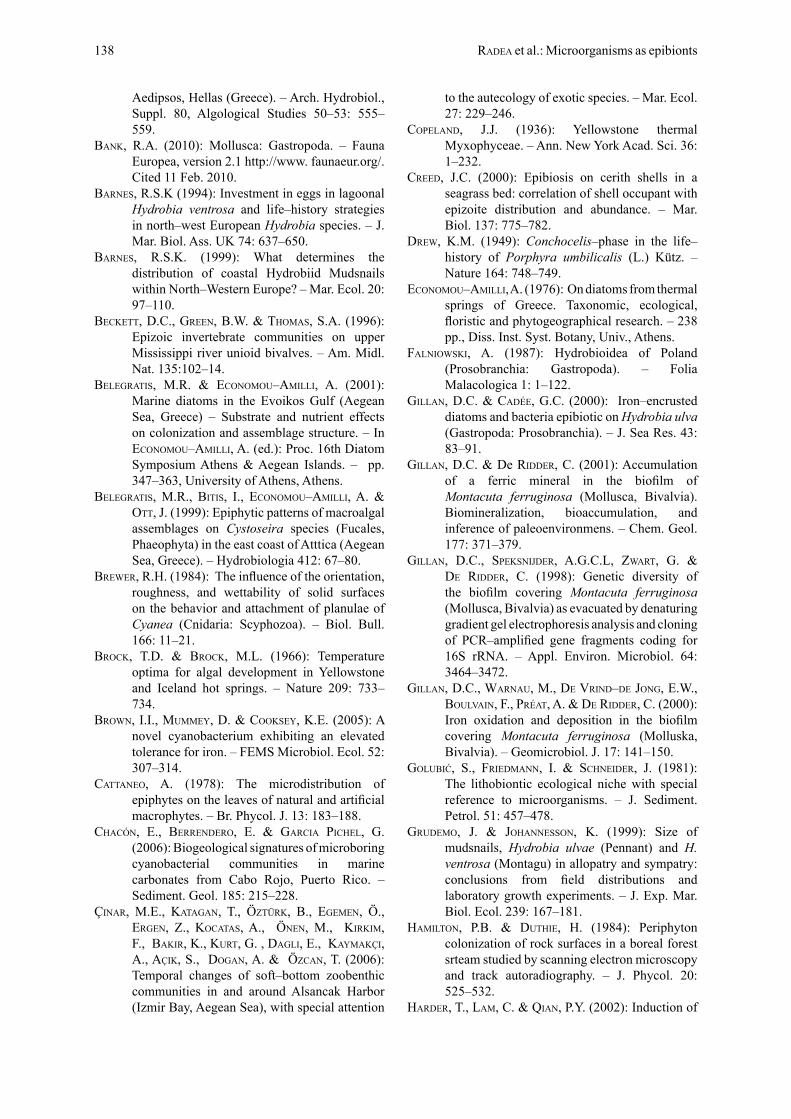
Table 2. Shell length and egg–capsules measured in a number of iron–coated and uncoated shells of V. ventrosa from ‘Agioi Anargyroi’ thermal spring on Kythnos Island.
V. ventrosa Iron–coated shells Uncoated shellsNumber (%) 82 (77.4%) 24 (22.6%)Maximum shell length (mm) * 4.75 (6 whorls) 3 (5 whorls)Mean shell length (mm) ** 2.66±0.6 2.47±0.43Shells bearing egg–capsules (number, %) 44 (53.6%) 10 (41.7%)Egg–capsules per shell (mean number ±SE) 1.43±1.94 0.46±0.78
The maximum ( *) and mean (**) shell length values are lying in the range estimated for this species by various authors in Europe: 3.5–6.2mm (FaLnioWsKi 1987, BaRnes 1994, PRoBst et al. 2000), and 2.2–3.6mm (gRudemo & Johannesson 1999), respectively.
Fottea 10(1): 129–140, 2010 137
Aedipsos, Hellas (Greece). – Arch. Hydrobiol., Suppl. 80, Algological Studies 50–53: 555–559.
BanK, R.A. (2010): Mollusca: Gastropoda. – Fauna Europea, version 2.1 http://www. faunaeur.org/. Cited 11 Feb. 2010.
BaRnes, R.S.K (1994): Investment in eggs in lagoonal Hydrobia ventrosa and life–history strategies in north–west European Hydrobia species. – J. Mar. Biol. Ass. UK 74: 637–650.
BaRnes, R.S.K. (1999): What determines the distribution of coastal Hydrobiid Mudsnails within North–Western Europe? – Mar. Ecol. 20: 97–110.
BecKett, D.C., gReen, B.W. & thomas, S.A. (1996): Epizoic invertebrate communities on upper Mississippi river unioid bivalves. – Am. Midl. Nat. 135:102–14.
BeLegRatis, M.R. & economou–amiLLi, A. (2001): Marine diatoms in the Evoikos Gulf (Aegean Sea, Greece) – Substrate and nutrient effects on colonization and assemblage structure. – In economou–amiLLi, a. (ed.): Proc. 16th Diatom Symposium Athens & Aegean Islands. – pp. 347–363, University of Athens, Athens.
BeLegRatis, M.R., Bitis, I., economou–amiLLi, A. & ott, J. (1999): Epiphytic patterns of macroalgal assemblages on Cystoseira species (Fucales, Phaeophyta) in the east coast of Atttica (Aegean Sea, Greece). – Hydrobiologia 412: 67–80.
BReWeR, R.H. (1984): The influence of the orientation, roughness, and wettability of solid surfaces on the behavior and attachment of planulae of Cyanea (Cnidaria: Scyphozoa). – Biol. Bull. 166: 11–21.
BRocK, T.D. & BRocK, M.L. (1966): Temperature optima for algal development in Yellowstone and Iceland hot springs. – Nature 209: 733–734.
BRoWn, I.I., mummey, D. & cooKsey, K.E. (2005): A novel cyanobacterium exhibiting an elevated tolerance for iron. – FEMS Microbiol. Ecol. 52: 307–314.
cattaneo, A. (1978): The microdistribution of epiphytes on the leaves of natural and artificial macrophytes. – Br. Phycol. J. 13: 183–188.
chacón, E., BeRRendeRo, E. & gaRcia PicheL, G. (2006): Biogeological signatures of microboring cyanobacterial communities in marine carbonates from Cabo Rojo, Puerto Rico. – Sediment. Geol. 185: 215–228.
ÇinaR, M.E., Katagan, T., ÖztüRK, B., egemen, Ö., eRgen, Z., Kocatas, A., Önen, M., KiRKim, F., BaKiR, K., KuRt, G. , dagLi, E., KaymaKÇi, A., aÇiK, S., dogan, A. & Özcan, T. (2006): Temporal changes of soft–bottom zoobenthic communities in and around Alsancak Harbor (Izmir Bay, Aegean Sea), with special attention
to the autecology of exotic species. – Mar. Ecol. 27: 229–246.
coPeLand, J.J. (1936): Yellowstone thermal Myxophyceae. – Ann. New York Acad. Sci. 36: 1–232.
cReed, J.C. (2000): Epibiosis on cerith shells in a seagrass bed: correlation of shell occupant with epizoite distribution and abundance. – Mar. Biol. 137: 775–782.
dReW, K.M. (1949): Conchocelis–phase in the life–history of Porphyra umbilicalis (L.) Kütz. – Nature 164: 748–749.
economou–amiLLi, A. (1976): On diatoms from thermal springs of Greece. Taxonomic, ecological, floristic and phytogeographical research. – 238 pp., Diss. Inst. Syst. Botany, Univ., Athens.
FaLnioWsKi, A. (1987): Hydrobioidea of Poland (Prosobranchia: Gastropoda). – Folia Malacologica 1: 1–122.
giLLan, D.C. & cadée, G.C. (2000): Iron–encrusted diatoms and bacteria epibiotic on Hydrobia ulva (Gastropoda: Prosobranchia). – J. Sea Res. 43: 83–91.
giLLan, D.C. & De RiddeR, C. (2001): Accumulation of a ferric mineral in the biofilm of Montacuta ferruginosa (Mollusca, Bivalvia). Biomineralization, bioaccumulation, and inference of paleoenvironmens. – Chem. Geol. 177: 371–379.
giLLan, D.C., sPeKsniJdeR, A.G.C.L, zWaRt, G. & de RiddeR, C. (1998): Genetic diversity of the biofilm covering Montacuta ferruginosa (Mollusca, Bivalvia) as evacuated by denaturing gradient gel electrophoresis analysis and cloning of PCR–amplified gene fragments coding for 16S rRNA. – Appl. Environ. Microbiol. 64: 3464–3472.
giLLan, D.C., WaRnau, M., de vRind–de Jong, E.W., BouLvain, F., PRéat, A. & de RiddeR, C. (2000): Iron oxidation and deposition in the biofilm covering Montacuta ferruginosa (Molluska, Bivalvia). – Geomicrobiol. J. 17: 141–150.
Golubić, S., FRiedmann, I. & schneideR, J. (1981): The lithobiontic ecological niche with special reference to microorganisms. – J. Sediment. Petrol. 51: 457–478.
gRudemo, J. & Johannesson, K. (1999): Size of mudsnails, Hydrobia ulvae (Pennant) and H. ventrosa (Montagu) in allopatry and sympatry: conclusions from field distributions and laboratory growth experiments. – J. Exp. Mar. Biol. Ecol. 239: 167–181.
hamiLton, P.B. & duthie, H. (1984): Periphyton colonization of rock surfaces in a boreal forest srteam studied by scanning electron microscopy and track autoradiography. – J. Phycol. 20: 525–532.
haRdeR, T., Lam, C. & qian, P.Y. (2002): Induction of
138 Radea et al.: Microorganisms as epibionts

larval settlement of the polychaete Hydroides elegans (Haswell) by marine biofilms: an investigation of monospecific diatom films as settlement cues. – Mar. Ecol. Prog. Ser. 229: 105–112.
hoFKeR, J. (1930): Faunistische Beobachtungen in der Zuidersee während der Trockenlegung. I. Über eine auf den Schalen von Hydrobia stagnalis epontisch lebende Mikrofauna in der Zuidersee. – Zeitschr. Ökol. Morphol. 18: 189–194.
James, M. (1985): Changes in the faunal composition of two thermal streams near Taupo, New Zealand. – N. Zeal. J. Mar. Freshw. Res. 19: 439–443.
Kaas, H. (1985): Algal studies oft the Danish Wadden Sea. III. Blue–green algae in tidal flat sediments (sand flats and lower salt marsh) at Rejsby; taxonomy and ecology. – Opera Bot. 79: 38–61.
Katoh, H., gRossman, A.R., hagino, N. & ogaWa, T. (2000): A gene of Synechocystis sp. strain PCC6803 encoding a novel iron transporter. – J. Bacteriol. 182: 6523–6524.
Katoh, H., hagino, N., gRossman, A.R. & ogaWa, T. (2001): Genes essential to iron transport in the cyanobacterium Synechocystis sp. strain PCC6803. – J. Bacteriol. 183: 2779–2784.
KoBaK, J. (2005): Recruitment and distribution of Dreissena polymorpha (Bivalvia) on substrates of different shape and orientation. – Int. Rev. Hydrobiol. 90: 159–170.
KomáReK, J. & anagnostidis, K. (1998): Cyanoprokaryota, I. Chroococcales. – In ettL, h. gäRtneR, g., heynig, h., & moLLenhaueR, d. (eds): Süsswasserflora von Mitteleuropa, 19/1. – 548 pp., G. Fischer Verl., Stuttgart.
KoRte, V.L. & BLinn, D.W. (1983): Diatom colonization on artificial substrata in pool and riffle zones studied by light and scanning electron microscopy. – J. Phycol. 19: 332–341.
KRammeR, K. (2002): Diatoms of Europe. Diatoms of the European Inland Waters and Comparable Habitats, Vol. 3. Cymbella. – 584 pp., A.R.G. Gantner Verlag K.G, Ruggell.
KuLLBeRg, R.G. (1968): Algal diversity in several thermal spring effluents. – Ecology 49: 751–755.
LaBoReL, J. & Le camPion–aLsumaRd, T. (1979): Infestation massive du squelette de coraux vivants par des Rhodophycées de type Conchocelis. – C. R. Acad. Sci. Paris 288: 1575–1577.
LamBRaKis, N. & KaLLeRgis, G. (2005): Contribution to the study of Greek thermal springs: hydrogeological and hydrochemical characteristics and origin of thermal waters. – Hydrogeol. J. 13: 506–521.
Lange–BeRtaLot, H. & KRammeR, K. (1987): Bacillariaceae, Epithemiaceae, Surirellaceae.
Neue und wenig bekannte Taxa, neue Kombinationen und Synonyme sowie Bemerkungen und Ergänzungen zu den Naviculaceae. – Biblioth. Diatomol. 15: 1–289.
Lange–BeRtaLot, H. (ed.) (2001): Diatoms of Europe. Diatoms of the European Inland Waters and Comparable Habitats. – Vol. 2. Navicula sensu stricto. 10 Genera Separated from Navicula sensu lato. Frustulia. – 526 pp., A.R.G. Gantner Verlag K.G, Ruggell.
Lassen, H.H. (1979): Reproductive effort in Danish mudsnails (Hydrobiidae). – Oecologia 40: 365–369.
Le camPion–aLsumaRd, T. (1979): Les cyanophycées endolithes marines: systematiques, ultrastructure, écologie et biodestruction. – Oceanol. Acta 2 : 143–156.
Le camPion–aLsumaRd, T. & Golubić, S. (1985): Hyella caespitosa and Hyella balani Lehman (Pleurocapsales, Cyanophyta): a comparative study. – Algological Studies 38/39: 119–148.
Le camPion–aLsumaRd, T., Golubić, S. & hutchings, P.A. (1995): Microbial endoliths in skeletons of live and dead corals: Porites lobata (Moorea, French Polynesia). – Mar. Ecol. Prog. Ser. 117: 149–157.
Le camPion–aLsumaRd, T., Golubić, S. & Pantazidou, A. (1996): On the endolithic genus Solentia Ercegović (Cyanophyta/Cyanobacteria). – Algological Studies 83: 107–127.
LevKov, Z., KRstic, S., metzeLtin, D. & naKov, T. (2007): Diatoms of Lakes Prespa and Ohrid. About 500 taxa from ancient lake system. – In Lange–BeRtaLot, h. (ed.): Iconographia Diatomologica, 16. – 611 pp., Koeltz Scientific Books, Koenigstein.
LouvRou, I. (2007): Periphyton and its colonization in marine hydrothermal regions of island Milos (Greece). – 447 pp., Ph.D. Thesis, Univ. Athens, Athens.
maRsden, J.E. & LansKy, D.M. (2000): Substrate selection by settling zebra mussels, Dreissena polymorpha, relative to material, texture, orientation, and sunlight. – Can. J. Zool. 78: 787–793.
moLisch, H. (1926): Pflanzenbiologie in Japan auf Grund eigener Beobachtungen. Die Eisenorganismen in Japan. Die Lebewelt in den heissen Quellen Japans. – pp. 1–103, Jena.
oWen, R.B., Renaut, R.W. & Jones, B. (2008): Geothermal diatoms: a comparative study of floras in hot spring systems of Iceland, New Zealand, and Kenya. – Hydrobiologia 610: 175–192.
Pantazidou, A., BeLegRatis, M.R., LouvRou, I. & economou–amiLLi, A. (2001): Euendolithic algae and patterns of diatom colonization on biotic carbonate substrates. – In economou–
Fottea 10(1): 129–140, 2010 139

amiLLi, a. (ed.): Proc. 16th Diatom Symposium Athens & Aegean Islands. – pp. 381–392, University of Athens, Athens.
Pantazidou, A., LouvRou, I. & economou–amiLLi, A. (2006): Euendolithic shell–boring cyanobacteria and chlorophytes from the saline lagoon Ahivadolimni on Milos Island, Greece. – Eur. J. Phycol. 41: 189–200.
PeRRy, C.T. (2000): Factors controlling sediment preservation on a north Jamaican fringing reef: a process–eased approach to microfacies analysis. – J. Sediment. Res. 70: 633–648.
PRoBst, S., KuBe, J. & BicK, A. (2000): Effects of winter severity on life history patterns and population dynamics of Hydrobia ventrosa (Gastropoda: Prosobranchia). – Arch. Hydrobiol. 148: 383–396.
Radea, C., LouvRou, I. & economou–amiLLi, A. (2008): First record of the New Zealand mud snail Potamopyrgus antipodarum J.E. Gray 1843 (Mollusca: Hydrobiidae) in Greece – Notes on its population structure and associated microalgae. – Aquatic Invasions 3: 341–344.
RodRiguez, S.R., oJedaL, P.F., niBaLdo, C. & inestRosa, N.C. (1993): Settlement of benthic marine invertebrates. – Mar. Ecol. Prog. Ser. 97: 193–207.
Round, F.E., cRaWFoRd, R.M. & mann, D.G. (1990): The Diatoms. Biology and Morphology of the Genera. – 747 pp., Cambridge University Press, Cambridge.
simonsen, R. (1962): Vegetation and Vegetationsbedingungen in der westlichen Ostsee (Kieler Bucht). – Kiel. Meeresforsch. 20: 157–168.
squiRes, L.E., RushFoRth, S.R. & BRotheRson, J.D. (1979): Algal response to a thermal effluent: Study of a power station on the Provo River, Utah, USA. – Hydrobiologia 63: 17–32.
thomas, J. & gonzaLves, E.A. (1965): Thermal algae of Western India. V. Algae of the hot springs at Tuwa. – Hydrobiologia 26: 41–54.
tRiBoLLet, A. (2008a): The boring microflora in modern coral reef ecosystems: a review of its roles. – In WisshaK, m. & taPaniLa, L. (eds): Current Developments in Bioerosion. – pp. 67–94, Springer, Berlin, Heidelberg.
tRiBoLLet, A. (2008b): Dissolution of dead corals by euendolithic microorganisms across the northern Great Barrier Reef (Australia). – Microb. Ecol. 55: 569–580.
tRiBoLLet, A. & PayRi, c. (2001): Bioerosion of the crustose coralline alga Hydrolithon onkodes by microborers in the coral reefs of Moorea, French Polynesia. – Oceanol. Acta 24: 329–342.
vinson, D.K. & RushFoRth, S.R. (1989): Diatoms species composition along a thermal gradient in the Portneuf River, Idaho, USA. – Hydrobiologia
185: 41–54.vouK, V. (1923): Die Probleme der Biologie der
Thermen. – Internat. Rev. Ges. Hydrobiol. u. Hydrogr. 11: 89–99.
WahL, M. (1989): Marine epibiosis. I. Fouling and antifouling: some basic aspects. – Mar. Ecol. Prog. Ser. 58: 175–189.
WahL, M. (1996): Fouled snails in flow: potential of epibionts on Littorina littorea to increase drag and reduce snail growth rates. – Mar. Ecol. Prog. Ser. 138: 157–168.
WahL, M. (1997): Increase drag reduces growth of snails: comparisons of flume and in situ experiments. – Mar. Ecol. Prog. Ser. 151: 291–293.
WahL, M. & hay, M.E. (1995): Associational resistance and shared doom: effects of epibiosis on herbivory. – Oecologia 102: 329–340.
WahL, M., hay, M.E. & endeRLein, P. (1997): Effects of epibiosis on consumer–prey interactions. – Hydrobiologia 355: 49–59.
WeBB, R. tRoyan, T. sheRman, D. & sheRman, L.A. (1994): MapA, an iron–regulated, cytoplasmic membrane protein in the cyanobacterium Synechococcus sp. strain PCC7942. – J. Bacteriol. 179: 4906–4913.
zaR, J.H. (1984): Biostatistical analysis. – 718 pp., 2nd edition, Prentice Hall, New Jersey.
© Czech Phycological SocietyReceived May 20, 2009Accepted August 12, 2009
140 Radea et al.: Microorganisms as epibionts