phic31 integrase vector system user manual
TRANSCRIPT
(ver. 6 - 0403913)
System Biosciences (SBI) 265 North Whisman Rd. Mountain View, CA 94043
Tel: 888.266.5066 (Toll Free in US) 650.968.2200
Fax: 650.968.2277 E-mail: [email protected] Web: www.systembio.com
(ver. 6 - 0403913)
User Manual
Please see product certificatefor proper storage conditions
A limited-use label license covers this product. By use of this product, you accept the terms and conditions outlined in the Licensing and Warranty Statement contained in this user manual.
phiC31 Integrase Vector SystemCatalog#s FC200PA-1, FC500A-1FC501A-1, FC550A-1, FC551A-1,
FC600A-1
PhiC31 Cloning and Expression System Cat.#FC200Pa-1, FC5/6xxA
888-266-5066 (Toll Free) 650-968-2200 (outside US) Page 1
ContentsI. Introduction .............................................................................. 2
A. Background of ϕC31 Integrase Technology ....................... 2
B. ϕC31 Donor Cloning Vector Maps & Details ...................... 3
C. Additional Materials Required ............................................. 4
II. Validation Data for ϕC31 Integrase Cloning System ............... 6
A. Colony Counting Assay ....................................................... 6
B. ϕC31 Donor Cloning Vector Integration by GFP and Puromycin Marker Expression 6
III. Protocol for Transfection and Generation of Stable Cell Lines using ϕC31 Cloning and Integrase Vectors 7
A. General Comments .............................................................. 7
B. Cloning of Inserts into ϕC31 Donor Vectors ........................ 7
C. Stable Cell Line Generation ................................................ 9
D. Verification of Insert Integration at Specific Genomic Loci using the Plasmid Rescue Assay 10
IV. References ............................................................................. 11
V. Technical Support .................................................................. 12
VI. Licensing and Warranty Statement........................................ 12
System Biosciences (SBI) User Manual
Page 2 ver. 6 - 040313 www.systembio.com
I. Introduction
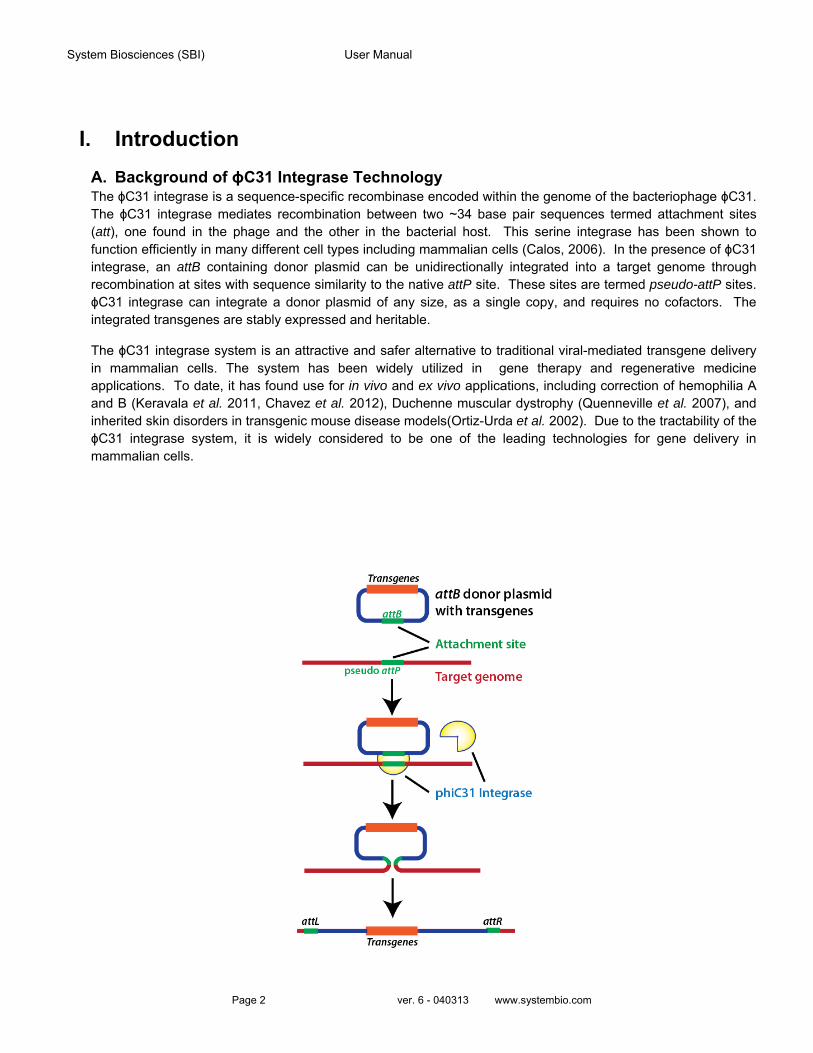
A. Background of ϕC31 Integrase Technology The ϕC31 integrase is a sequence-specific recombinase encoded within the genome of the bacteriophage ϕC31. The ϕC31 integrase mediates recombination between two ~34 base pair sequences termed attachment sites (att), one found in the phage and the other in the bacterial host. This serine integrase has been shown to function efficiently in many different cell types including mammalian cells (Calos, 2006). In the presence of ϕC31 integrase, an attB containing donor plasmid can be unidirectionally integrated into a target genome through recombination at sites with sequence similarity to the native attP site. These sites are termed pseudo-attP sites. ϕC31 integrase can integrate a donor plasmid of any size, as a single copy, and requires no cofactors. The integrated transgenes are stably expressed and heritable.
The ϕC31 integrase system is an attractive and safer alternative to traditional viral-mediated transgene delivery in mammalian cells. The system has been widely utilized in gene therapy and regenerative medicine applications. To date, it has found use for in vivo and ex vivo applications, including correction of hemophilia A and B (Keravala et al. 2011, Chavez et al. 2012), Duchenne muscular dystrophy (Quenneville et al. 2007), and inherited skin disorders in transgenic mouse disease models(Ortiz-Urda et al. 2002). Due to the tractability of the ϕC31 integrase system, it is widely considered to be one of the leading technologies for gene delivery in mammalian cells.
PhiC31 Cloning and Expression System Cat.#FC200Pa-1, FC5/6xxA
888-266-5066 (Toll Free) 650-968-2200 (outside US) Page 3
Fig. 1. Schematic of the ϕC31 Integrase mediated recombination of donor plasmid sequence into pseudo-attP sites in host genome
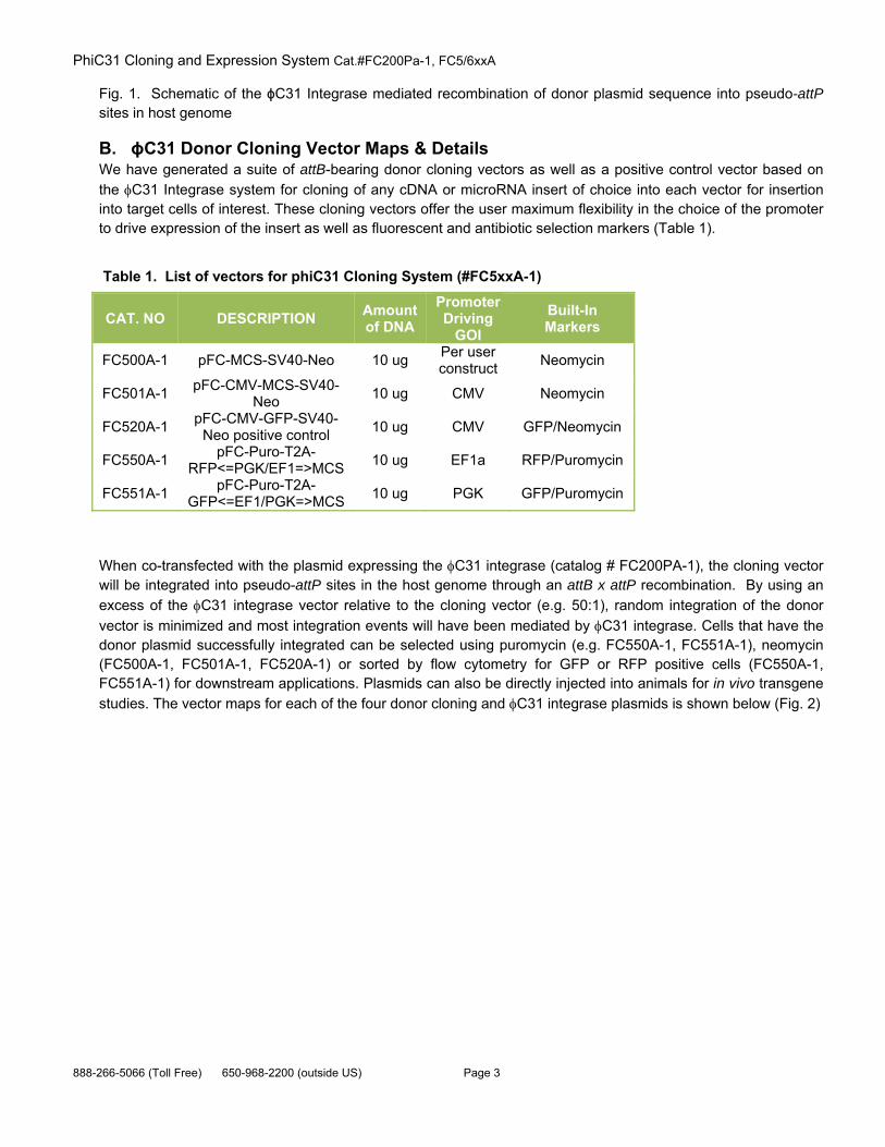
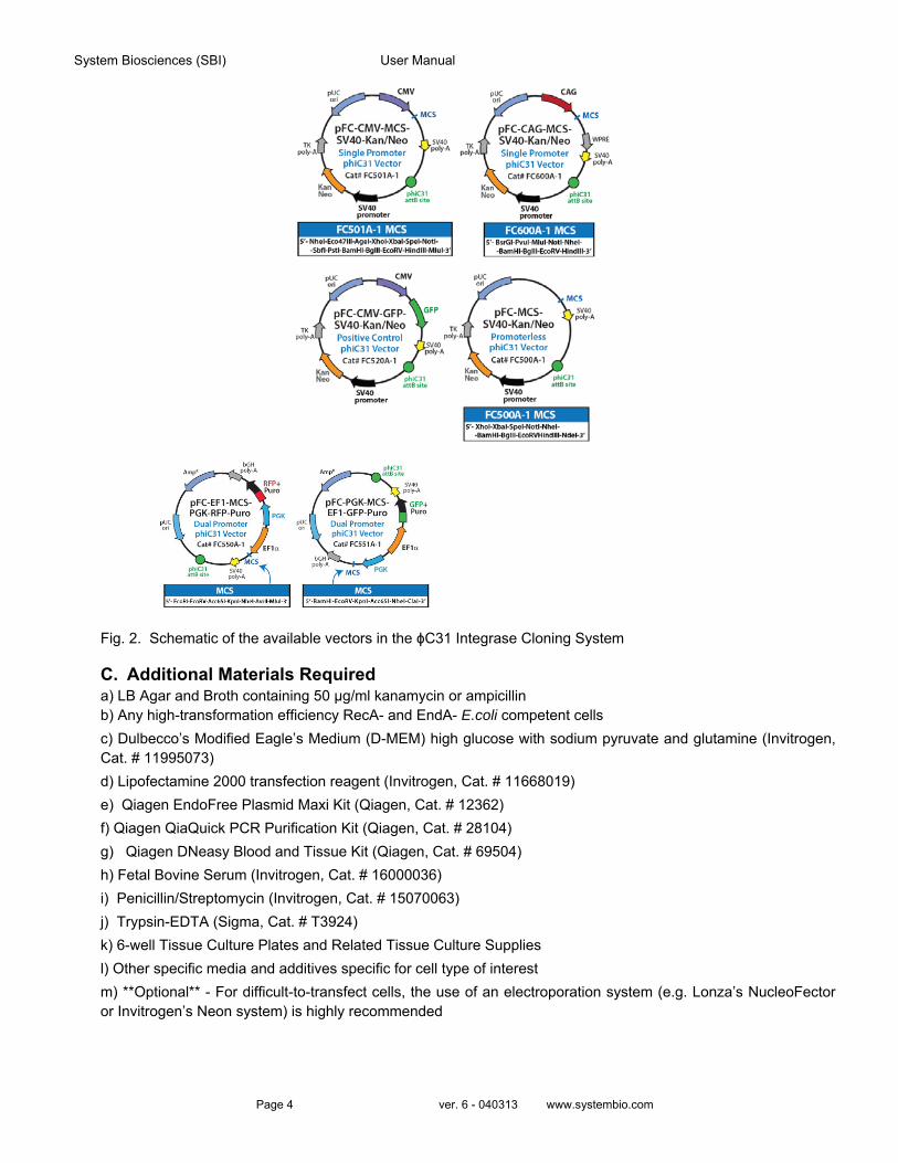
B. ϕC31 Donor Cloning Vector Maps & Details We have generated a suite of attB-bearing donor cloning vectors as well as a positive control vector based on
the C31 Integrase system for cloning of any cDNA or microRNA insert of choice into each vector for insertion into target cells of interest. These cloning vectors offer the user maximum flexibility in the choice of the promoter to drive expression of the insert as well as fluorescent and antibiotic selection markers (Table 1).
Table 1. List of vectors for phiC31 Cloning System (#FC5xxA-1)
When co-transfected with the plasmid expressing the C31 integrase (catalog # FC200PA-1), the cloning vector will be integrated into pseudo-attP sites in the host genome through an attB x attP recombination. By using an
excess of the C31 integrase vector relative to the cloning vector (e.g. 50:1), random integration of the donor
vector is minimized and most integration events will have been mediated by C31 integrase. Cells that have the donor plasmid successfully integrated can be selected using puromycin (e.g. FC550A-1, FC551A-1), neomycin (FC500A-1, FC501A-1, FC520A-1) or sorted by flow cytometry for GFP or RFP positive cells (FC550A-1, FC551A-1) for downstream applications. Plasmids can also be directly injected into animals for in vivo transgene
studies. The vector maps for each of the four donor cloning and C31 integrase plasmids is shown below (Fig. 2)
CAT. NO DESCRIPTION Amount of DNA
Promoter Driving
GOI
Built-In Markers
FC500A-1 pFC-MCS-SV40-Neo 10 ug Per user construct
Neomycin
FC501A-1 pFC-CMV-MCS-SV40-
Neo 10 ug CMV Neomycin
FC520A-1 pFC-CMV-GFP-SV40-
Neo positive control 10 ug CMV GFP/Neomycin
FC550A-1 pFC-Puro-T2A-
RFP<=PGK/EF1=>MCS 10 ug EF1a RFP/Puromycin
FC551A-1 pFC-Puro-T2A-
GFP<=EF1/PGK=>MCS 10 ug PGK GFP/Puromycin
System Biosciences (SBI) User Manual
Page 4 ver. 6 - 040313 www.systembio.com
Fig. 2. Schematic of the available vectors in the ϕC31 Integrase Cloning System
C. Additional Materials Required a) LB Agar and Broth containing 50 µg/ml kanamycin or ampicillin b) Any high-transformation efficiency RecA- and EndA- E.coli competent cells
c) Dulbecco’s Modified Eagle’s Medium (D-MEM) high glucose with sodium pyruvate and glutamine (Invitrogen, Cat. # 11995073)
d) Lipofectamine 2000 transfection reagent (Invitrogen, Cat. # 11668019)
e) Qiagen EndoFree Plasmid Maxi Kit (Qiagen, Cat. # 12362)
f) Qiagen QiaQuick PCR Purification Kit (Qiagen, Cat. # 28104)
g) Qiagen DNeasy Blood and Tissue Kit (Qiagen, Cat. # 69504)
h) Fetal Bovine Serum (Invitrogen, Cat. # 16000036)
i) Penicillin/Streptomycin (Invitrogen, Cat. # 15070063)
j) Trypsin-EDTA (Sigma, Cat. # T3924)
k) 6-well Tissue Culture Plates and Related Tissue Culture Supplies
l) Other specific media and additives specific for cell type of interest
m) **Optional** - For difficult-to-transfect cells, the use of an electroporation system (e.g. Lonza’s NucleoFector or Invitrogen’s Neon system) is highly recommended

PhiC31 Cloning and Expression System Cat.#FC200Pa-1, FC5/6xxA
888-266-5066 (Toll Free) 650-968-2200 (outside US) Page 5
System Biosciences (SBI) User Manual
Page 6 ver. 6 - 040313 www.systembio.com
II. Validation Data for ϕC31 Integrase Cloning System
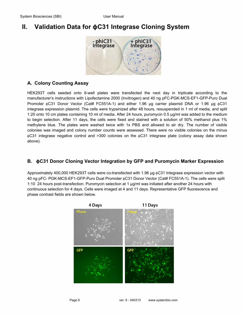
A. Colony Counting Assay
HEK293T cells seeded onto 6-well plates were transfected the next day in triplicate according to the manufacturer’s instructions with Lipofectamine 2000 (Invitrogen) and 40 ng pFC-PGK-MCS-EF1-GFP-Puro Dual
Promoter C31 Donor Vector (Cat# FC551A-1) and either 1.96 µg carrier plasmid DNA or 1.96 µg C31 integrase expression plasmid. The cells were trypsinized after 48 hours, resuspended in 1 ml of media, and split 1:20 onto 10 cm plates containing 10 ml of media. After 24 hours, puromycin 0.5 µg/ml was added to the medium to begin selection. After 11 days, the cells were fixed and stained with a solution of 50% methanol plus 1% methylene blue. The plates were washed twice with 1x PBS and allowed to air dry. The number of visible colonies was imaged and colony number counts were assessed. There were no visible colonies on the minus
C31 integrase negative control and >300 colonies on the C31 integrase plate (colony assay data shown above).
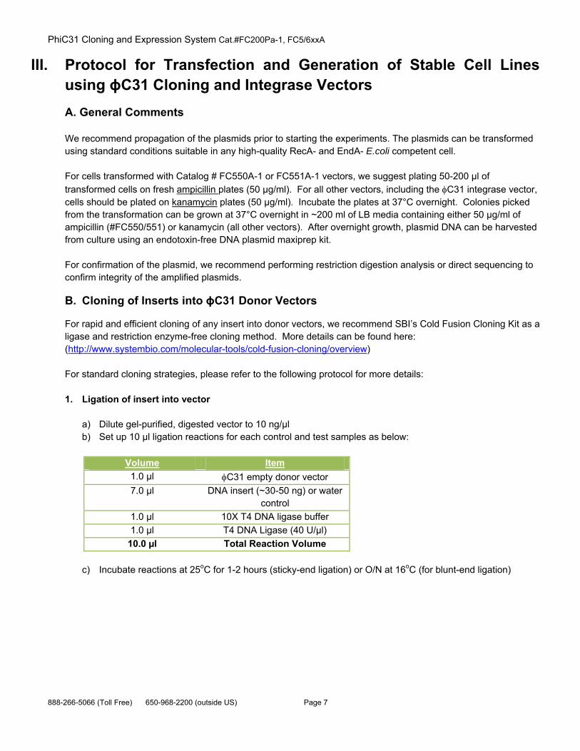
B. ϕC31 Donor Cloning Vector Integration by GFP and Puromycin Marker Expression
Approximately 400,000 HEK293T cells were co-transfected with 1.96 µg C31 Integrase expression vector with
40 ng pFC- PGK-MCS-EF1-GFP-Puro Dual Promoter C31 Donor Vector (Cat# FC551A-1). The cells were split 1:10 24 hours post-transfection. Puromycin selection at 1 µg/ml was initiated after another 24 hours with continuous selection for 4 days. Cells were imaged at 4 and 11 days. Representative GFP fluorescence and phase contrast fields are shown below.
PhiC31 Cloning and Expression System Cat.#FC200Pa-1, FC5/6xxA
888-266-5066 (Toll Free) 650-968-2200 (outside US) Page 7
III. Protocol for Transfection and Generation of Stable Cell Lines using ϕC31 Cloning and Integrase Vectors
A. General Comments
We recommend propagation of the plasmids prior to starting the experiments. The plasmids can be transformed using standard conditions suitable in any high-quality RecA- and EndA- E.coli competent cell.
For cells transformed with Catalog # FC550A-1 or FC551A-1 vectors, we suggest plating 50-200 µl of
transformed cells on fresh ampicillin plates (50 µg/ml). For all other vectors, including the C31 integrase vector, cells should be plated on kanamycin plates (50 µg/ml). Incubate the plates at 37°C overnight. Colonies picked from the transformation can be grown at 37°C overnight in ~200 ml of LB media containing either 50 µg/ml of ampicillin (#FC550/551) or kanamycin (all other vectors). After overnight growth, plasmid DNA can be harvested from culture using an endotoxin-free DNA plasmid maxiprep kit.
For confirmation of the plasmid, we recommend performing restriction digestion analysis or direct sequencing to confirm integrity of the amplified plasmids.
B. Cloning of Inserts into ϕC31 Donor Vectors
For rapid and efficient cloning of any insert into donor vectors, we recommend SBI’s Cold Fusion Cloning Kit as a ligase and restriction enzyme-free cloning method. More details can be found here: (http://www.systembio.com/molecular-tools/cold-fusion-cloning/overview)
For standard cloning strategies, please refer to the following protocol for more details:
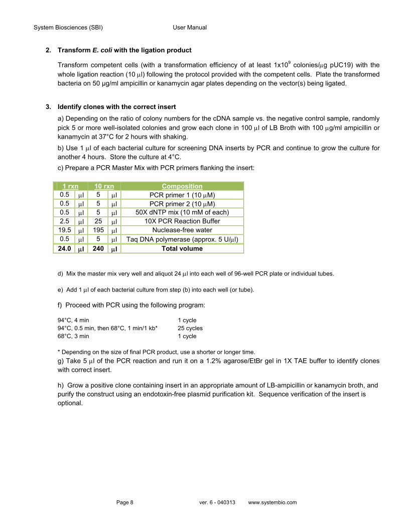
1. Ligation of insert into vector
a) Dilute gel-purified, digested vector to 10 ng/µl b) Set up 10 µl ligation reactions for each control and test samples as below:
Volume Item
1.0 µl C31 empty donor vector
7.0 µl DNA insert (~30-50 ng) or water control
1.0 µl 10X T4 DNA ligase buffer
1.0 µl T4 DNA Ligase (40 U/µl)
10.0 µl Total Reaction Volume
c) Incubate reactions at 25oC for 1-2 hours (sticky-end ligation) or O/N at 16oC (for blunt-end ligation)
System Biosciences (SBI) User Manual
Page 8 ver. 6 - 040313 www.systembio.com
2. Transform E. coli with the ligation product
Transform competent cells (with a transformation efficiency of at least 1x10⁹ colonies/g pUC19) with the
whole ligation reaction (10 l) following the protocol provided with the competent cells. Plate the transformed bacteria on 50 µg/ml ampicillin or kanamycin agar plates depending on the vector(s) being ligated.
3. Identify clones with the correct insert
a) Depending on the ratio of colony numbers for the cDNA sample vs. the negative control sample, randomly pick 5 or more well-isolated colonies and grow each clone in 100 l of LB Broth with 100 g/ml ampicillin or kanamycin at 37°C for 2 hours with shaking.
b) Use 1 l of each bacterial culture for screening DNA inserts by PCR and continue to grow the culture for another 4 hours. Store the culture at 4°C.
c) Prepare a PCR Master Mix with PCR primers flanking the insert:
1 rxn 10 rxn Composition
0.5 l 5 l PCR primer 1 (10 M) 0.5 l 5 l PCR primer 2 (10 M) 0.5 l 5 l 50X dNTP mix (10 mM of each) 2.5 l 25 l 10X PCR Reaction Buffer 19.5 l 195 l Nuclease-free water 0.5 l 5 l Taq DNA polymerase (approx. 5 U/l) 24.0 l 240 l Total volume
d) Mix the master mix very well and aliquot 24 l into each well of 96-well PCR plate or individual tubes.
e) Add 1 l of each bacterial culture from step (b) into each well (or tube).
f) Proceed with PCR using the following program:
94°C, 4 min 1 cycle 94°C, 0.5 min, then 68°C, 1 min/1 kb* 25 cycles 68°C, 3 min 1 cycle * Depending on the size of final PCR product, use a shorter or longer time. g) Take 5 l of the PCR reaction and run it on a 1.2% agarose/EtBr gel in 1X TAE buffer to identify clones with correct insert.
h) Grow a positive clone containing insert in an appropriate amount of LB-ampicillin or kanamycin broth, and purify the construct using an endotoxin-free plasmid purification kit. Sequence verification of the insert is optional.
PhiC31 Cloning and Expression System Cat.#FC200Pa-1, FC5/6xxA
888-266-5066 (Toll Free) 650-968-2200 (outside US) Page 9
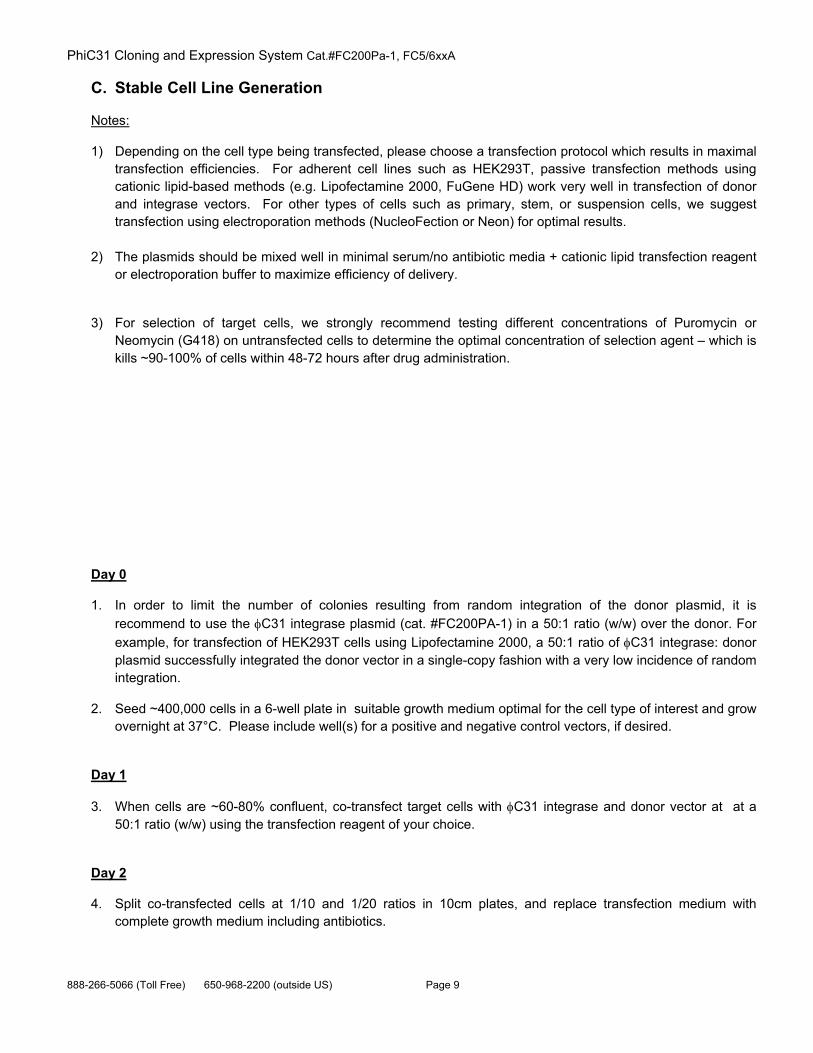
C. Stable Cell Line Generation
Notes:
1) Depending on the cell type being transfected, please choose a transfection protocol which results in maximal transfection efficiencies. For adherent cell lines such as HEK293T, passive transfection methods using cationic lipid-based methods (e.g. Lipofectamine 2000, FuGene HD) work very well in transfection of donor and integrase vectors. For other types of cells such as primary, stem, or suspension cells, we suggest transfection using electroporation methods (NucleoFection or Neon) for optimal results.
2) The plasmids should be mixed well in minimal serum/no antibiotic media + cationic lipid transfection reagent
or electroporation buffer to maximize efficiency of delivery.
3) For selection of target cells, we strongly recommend testing different concentrations of Puromycin or Neomycin (G418) on untransfected cells to determine the optimal concentration of selection agent – which is kills ~90-100% of cells within 48-72 hours after drug administration.
Day 0
1. In order to limit the number of colonies resulting from random integration of the donor plasmid, it is recommend to use the C31 integrase plasmid (cat. #FC200PA-1) in a 50:1 ratio (w/w) over the donor. For
example, for transfection of HEK293T cells using Lipofectamine 2000, a 50:1 ratio of C31 integrase: donor plasmid successfully integrated the donor vector in a single-copy fashion with a very low incidence of random integration.
2. Seed ~400,000 cells in a 6-well plate in suitable growth medium optimal for the cell type of interest and grow overnight at 37°C. Please include well(s) for a positive and negative control vectors, if desired.
Day 1
3. When cells are ~60-80% confluent, co-transfect target cells with C31 integrase and donor vector at at a 50:1 ratio (w/w) using the transfection reagent of your choice.
Day 2
4. Split co-transfected cells at 1/10 and 1/20 ratios in 10cm plates, and replace transfection medium with complete growth medium including antibiotics.
System Biosciences (SBI) User Manual
Page 10 ver. 6 - 040313 www.systembio.com
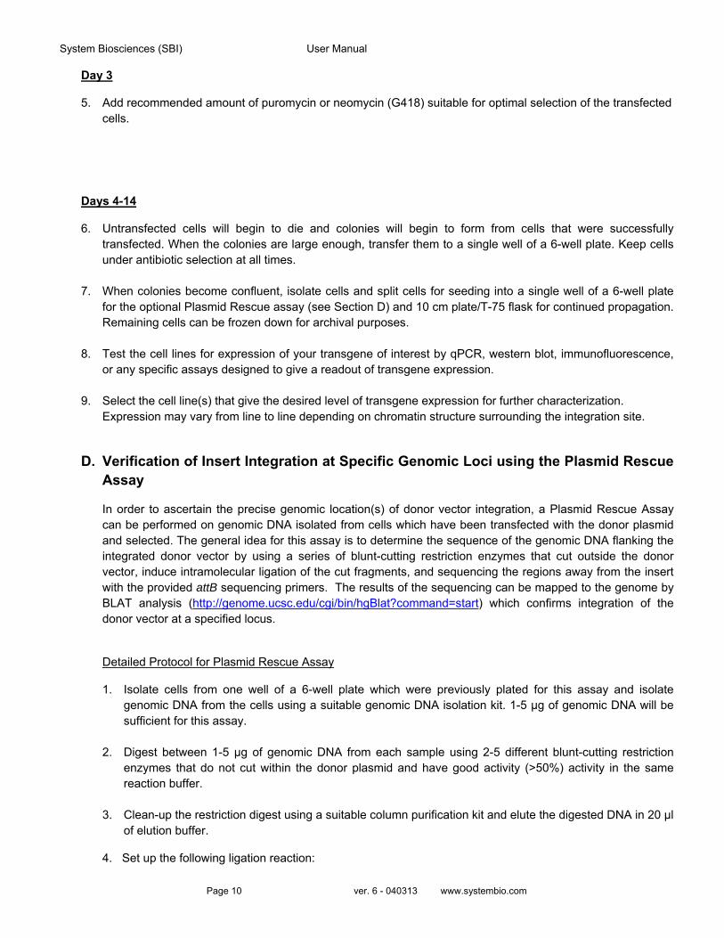
Day 3
5. Add recommended amount of puromycin or neomycin (G418) suitable for optimal selection of the transfected cells.
Days 4-14
6. Untransfected cells will begin to die and colonies will begin to form from cells that were successfully transfected. When the colonies are large enough, transfer them to a single well of a 6-well plate. Keep cells under antibiotic selection at all times.
7. When colonies become confluent, isolate cells and split cells for seeding into a single well of a 6-well plate for the optional Plasmid Rescue assay (see Section D) and 10 cm plate/T-75 flask for continued propagation. Remaining cells can be frozen down for archival purposes.
8. Test the cell lines for expression of your transgene of interest by qPCR, western blot, immunofluorescence, or any specific assays designed to give a readout of transgene expression.
9. Select the cell line(s) that give the desired level of transgene expression for further characterization. Expression may vary from line to line depending on chromatin structure surrounding the integration site.
D. Verification of Insert Integration at Specific Genomic Loci using the Plasmid Rescue Assay
In order to ascertain the precise genomic location(s) of donor vector integration, a Plasmid Rescue Assay can be performed on genomic DNA isolated from cells which have been transfected with the donor plasmid and selected. The general idea for this assay is to determine the sequence of the genomic DNA flanking the integrated donor vector by using a series of blunt-cutting restriction enzymes that cut outside the donor vector, induce intramolecular ligation of the cut fragments, and sequencing the regions away from the insert with the provided attB sequencing primers. The results of the sequencing can be mapped to the genome by BLAT analysis (http://genome.ucsc.edu/cgi/bin/hgBlat?command=start) which confirms integration of the donor vector at a specified locus.
Detailed Protocol for Plasmid Rescue Assay
1. Isolate cells from one well of a 6-well plate which were previously plated for this assay and isolate genomic DNA from the cells using a suitable genomic DNA isolation kit. 1-5 µg of genomic DNA will be sufficient for this assay.
2. Digest between 1-5 µg of genomic DNA from each sample using 2-5 different blunt-cutting restriction enzymes that do not cut within the donor plasmid and have good activity (>50%) activity in the same reaction buffer.
3. Clean-up the restriction digest using a suitable column purification kit and elute the digested DNA in 20 µl
of elution buffer.
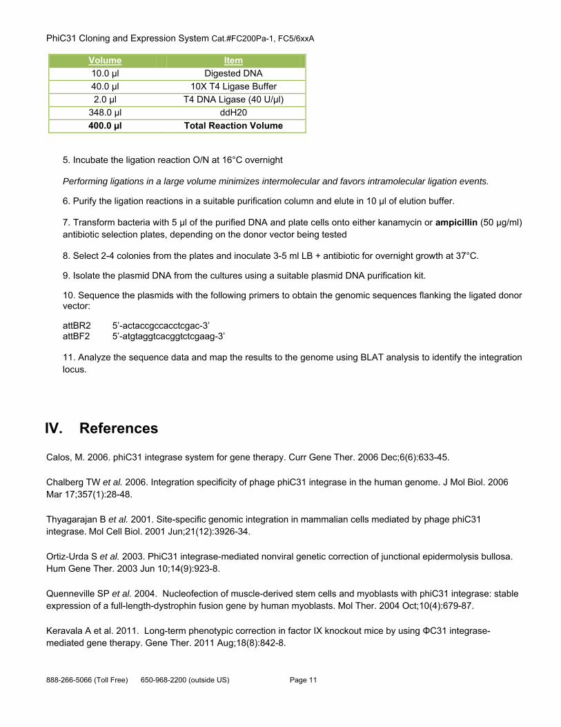
4. Set up the following ligation reaction:
PhiC31 Cloning and Expression System Cat.#FC200Pa-1, FC5/6xxA
888-266-5066 (Toll Free) 650-968-2200 (outside US) Page 11
Volume Item
10.0 µl Digested DNA
40.0 µl 10X T4 Ligase Buffer
2.0 µl T4 DNA Ligase (40 U/µl)
348.0 µl ddH20
400.0 µl Total Reaction Volume
5. Incubate the ligation reaction O/N at 16°C overnight
Performing ligations in a large volume minimizes intermolecular and favors intramolecular ligation events.
6. Purify the ligation reactions in a suitable purification column and elute in 10 µl of elution buffer.
7. Transform bacteria with 5 µl of the purified DNA and plate cells onto either kanamycin or ampicillin (50 µg/ml) antibiotic selection plates, depending on the donor vector being tested
8. Select 2-4 colonies from the plates and inoculate 3-5 ml LB + antibiotic for overnight growth at 37°C.
9. Isolate the plasmid DNA from the cultures using a suitable plasmid DNA purification kit.
10. Sequence the plasmids with the following primers to obtain the genomic sequences flanking the ligated donor vector:
attBR2 5’-actaccgccacctcgac-3’ attBF2 5’-atgtaggtcacggtctcgaag-3’ 11. Analyze the sequence data and map the results to the genome using BLAT analysis to identify the integration locus.
IV. References
Calos, M. 2006. phiC31 integrase system for gene therapy. Curr Gene Ther. 2006 Dec;6(6):633-45.
Chalberg TW et al. 2006. Integration specificity of phage phiC31 integrase in the human genome. J Mol Biol. 2006 Mar 17;357(1):28-48.
Thyagarajan B et al. 2001. Site-specific genomic integration in mammalian cells mediated by phage phiC31 integrase. Mol Cell Biol. 2001 Jun;21(12):3926-34.
Ortiz-Urda S et al. 2003. PhiC31 integrase-mediated nonviral genetic correction of junctional epidermolysis bullosa. Hum Gene Ther. 2003 Jun 10;14(9):923-8.
Quenneville SP et al. 2004. Nucleofection of muscle-derived stem cells and myoblasts with phiC31 integrase: stable expression of a full-length-dystrophin fusion gene by human myoblasts. Mol Ther. 2004 Oct;10(4):679-87.
Keravala A et al. 2011. Long-term phenotypic correction in factor IX knockout mice by using ΦC31 integrase-mediated gene therapy. Gene Ther. 2011 Aug;18(8):842-8.
System Biosciences (SBI) User Manual
Page 12 ver. 6 - 040313 www.systembio.com
Chavez CL et al. 2012. Long-term expression of human coagulation factor VIII in a tolerant mouse model using the φC31 integrase system. Hum Gene Ther. 2012 Apr;23(4):390-8.
V. Technical Support For more information about SBI products or to download manuals in PDF format, please visit our website:
http://www.systembio.com
For additional information or technical assistance, please call or email us at:
650-968-2200
VI. Licensing and Warranty Statement
Limited Use License Use of the PhiC31 Expression System (i.e., the “Product”) is subject to the following terms and conditions. If the terms and conditions are not acceptable, return all components of the Product to System Biosciences (SBI) within 7 calendar days. Purchase and use of any part of the Product constitutes acceptance of the above terms.
The purchaser of the Product is granted a limited license to use the Product under the following terms and conditions:
The Product shall be used by the purchaser for internal research purposes only. The Product is expressly not designed, intended, or warranted for use in humans or for therapeutic or diagnostic use.
The Product may not be resold, modified for resale, or used to manufacture commercial products without prior written consent of SBI.
This Product should be used in accordance with the NIH guidelines developed for stem cell research.
SBI has pending patent applications related to the Product. For information concerning licenses for commercial use, contact SBI.
Purchase of the product does not grant any rights or license for use other than those explicitly listed in this Licensing and Warranty Statement. Use of the Product for any use other than described expressly herein may be covered by patents or subject to rights other than those mentioned. SBI disclaims any and all responsibility for injury or damage which may be caused by the failure of the buyer or any other person to use the Product in accordance with the terms and conditions outlined herein.
Limited Warranty SBI warrants that the Product meets the specifications described in this manual. If it is proven to the satisfaction of SBI that the Product fails to meet these specifications, SBI will replace the Product or provide the purchaser with a refund. This limited warranty shall not extend to anyone other than the original purchaser of the Product. Notice of nonconforming products must be made to SBI within 30 days of receipt of the Product.
SBI’s liability is expressly limited to replacement of Product or a refund limited to the actual purchase price. SBI’s liability does not extend to any damages arising from use or improper use of the Product, or losses associated with the use of additional materials or reagents. This limited warranty is the sole and exclusive warranty. SBI does not provide any other warranties of any kind, expressed or implied, including the merchantability or fitness of the Product for a particular purpose.

PhiC31 Cloning and Expression System Cat.#FC200Pa-1, FC5/6xxA
888-266-5066 (Toll Free) 650-968-2200 (outside US) Page 13
SBI is committed to providing our customers with high-quality products. If you should have any questions or concerns about any SBI products, please contact us at (888) 266-5066.
© 2013 System Biosciences (SBI).