pharmacological alterations of neuroplasticity in the ... · pharmacological alterations of...
TRANSCRIPT
PHARMACOLOGICAL ALTERATIONS OF NEUROPLASTICITY IN THE
HUMAN MOTOR CORTEX INDUCED BY DOPAMINERGIC AND
CHOLINERGIC AGENTS
Dissertation
zur Erlangung des mathematisch-naturwissenschaftlichen Doktorgrades
„Doctor rerum naturalium“
Der Georg-August-Universität Göttingen
vorgelegt von
Nivethida Thirugnanasambandam
aus Pondicherry, Indien
Göttingen, 2010
ii
Doctoral Thesis Committee:
Prof. Dr.med. Michael A Nitsche (Supervisor, First Referee)
Abteilung Klinische Neurophysiologie
Universitätsmedizin Göttingen
Robert-Koch-Straße 40
37075 Göttingen
Prof. Dr. Eberhard Fuchs (Reviewer)
Deutsches Primatenzentrum
Labor für Klinische Neurobiologie
Kellnerweg 4
37077 Göttingen
Prof. Dr. Birgit Kröner-Herwig
Georg August University Göttingen
Clinical Psychology and Psychotherapy
Goßlerstr. 14
37073 Göttingen
Date of Submission: 13th December 2010
Date of oral examination: 17th
January 2011
iii
Statement of Originality
I hereby declare that this thesis is my own work and has been written independently with no
other sources and aids than quoted in the text, references and acknowledgements.
Göttingen, 13th
December 2010
Nivethida Thirugnanasambandam

iv
This doctoral thesis is dedicated with love and gratitude to my father,
K.P.Thirugnanasambandam, to my mother, Parvady Thirugnanasambandam and
to my sisters, Manasadevi and Mirunalini for their incredible support which lies
behind every success in my life…
v
TABLE OF CONTENTS
Chapter 1 – Introduction 1
1.1. Plasticity in the central nervous system ……………………………… 3
1.2. Neuroplasticity in humans ……………………………………………... 5
1.2.1. Motor system as a model for neuroplasticity in humans………….. 6
1.3. Non-invasive brain stimulation techniques ……………………………... 6
1.3.1. Transcranial magnetic stimulation ……………………………... 8
1.3.2. Transcranial direct current stimulation …………………………… 8
1.3.3. Paired associative stimulation …………………………………….. 10
1.4. Neuromodulators ……………………………………………………... 12
1.4.1. Cholinergic system ……………………………………………... 13
1.4.2. Dopaminergic system …………………………………………….. 14
1.5. Aim of the thesis ……………………………………………………... 17
Chapter 2 – Original articles and manuscripts 18
2.1 Nicotinergic impact on focal and non-focal plasticity in non-smoking humans .. 19
2.2 Dose-dependent non-linear effect of l-dopa on focal paired associative
stimulation induced plasticity in humans ……………………………... 53
Chapter 3 – Discussion 78
3.1 Summary of findings ……………………………………………………. 78
3.2 Conclusions …………………………………………………………….. 79
3.3 Future prospects …………………………………………………….. 80
References …………………………………………………………….. ……… 82
Acknowledgements …………………………………………………………….. 93
Curriculum Vitae …………………………………………………………….. 94
1
Chapter 1 Introduction
Neuroplasticity is a marveling feature exhibited by neuronal networks of the simplest
to the most complex brains in nature. By dynamically re-organizing the structure of neuronal
networks or function or both, the brain is constantly responding to environmental stimuli and
injuries (Boller, 2004). Long term- potentiation (LTP) and depression (LTD) are the most
well-studied forms of neuroplasticity and are proposed to underlie the complex processes of
learning and memory formation. In recent decades, non-invasive brain stimulation techniques
have gained importance in the exploration of neuroplasticity in humans. Repetitive
transcranial magnetic stimulation (rTMS), transcranial direct current stimulation (tDCS) and
paired associative stimulation (PAS) are the most commonly used techniques (Huang et al.,
2005; Nitsche and Paulus 2000; 2001; Stefan et al., 2000). These techniques are able to
induce long-term plastic changes in the intact human brain thereby enabling us to study this
phenomenon in humans (Cooke and Bliss, 2006).
Neuromodulators are substances that, depending on concentration, activation of
subreceptors, and other factors, affect plasticity in neuronal networks. Acetylcholine,
dopamine, serotonin and noradrenaline are the most important neuromodulators in the human
central nervous system. Several studies have looked into their impact on neuroplasticity
mainly in animals (Hasselmo and Barkai, 1995; Kung et al., 2007; Otani et al., 1998; 2003).
Very few studies so far have studied the impact of neuromodulators on plasticity in humans
(Kuo et al., 2007; 2008; Nitsche et al., 2009; Monte-Silva et al., 2009; 2010). In the present
projects, we were interested to explore certain aspects of the impact of cholinergic and
dopaminergic activation on neuroplasticity in humans. In the cholinergic system, we explored
the specific impact of the nicotinic sub-receptor on neuroplasticity. In the dopaminergic
system, we explored the dose-dependent effect of dopaminergic activation on plasticity.

2
The first chapter introduces basic concepts necessary for the further understanding of
the studies included in the thesis. The second chapter consists of the papers presenting the
research results. The concluding chapter summarizes the results of the studies and offers an
outlook to future research in the field.
3
1.1 Plasticity in the central nervous system
Neuroplasticity is the phenomenon by which functional or structural reorganization of
neuronal connectivity takes place depending on the amount and pattern of neuronal activity
(Citri and Malenka, 2008). Long term potentiation (LTP) and long term depression (LTD) are
the most extensively studied neuroplastic mechanisms considered to be the physiological
basis of learning and memory formation. These processes are most detailed studied at
glutamatergic synapses, especially in the region of the hippocampus, but also in other cortical
and subcortical areas (Malenka and Bear, 2004). However, plasticity is not limited to the
glutamatergic system, also the most important inhibitory cortical network, the GABAergic
system, has neuroplastic properties (Kano, 1995). Both LTP and LTD at glutamatergic
synapses are mediated by NMDA (N-methyl D-aspartate) receptors, which have calcium
channel properties (Bliss and Collingridge, 1993; Malenka and Bear, 2004). Consequently,
one of the major determinants of plasticity at the synapse is the postsynaptic intracellular
calcium concentration (Lisman, 2003). It is known that moderate increase in intracellular
calcium triggers signal transduction molecules like protein phosphatases that inactivate or
remove AMPA receptors from the subsynaptic membrane, resulting in LTD (Cummings et al.,
1996). Conversely, a large increase in intracellular calcium concentration beyond a critical
threshold triggers other signaling molecules like the calcium-calmodulin kinase and other
protein kinases, which enhance the insertion of AMPA receptors onto the postsynaptic
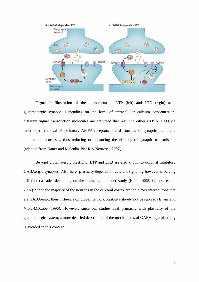
membrane inducing LTP (Malenka and Nicoll, 1993; Malenka and Bear, 2004) (see figure 1).
4
Figure 1: Illustration of the phenomena of LTP (left) and LTD (right) at a
glutamatergic synapse. Depending on the level of intracellular calcium concentration,
different signal transduction molecules are activated that result in either LTP or LTD via
insertion or removal of excitatory AMPA receptors to and from the subsynaptic membrane
and related processes, thus reducing or enhancing the efficacy of synaptic transmission
(adapted from Kauer and Malenka, Nat Rev Neurosci, 2007).
Beyond glutamatergic plasticity, LTP and LTD are also known to occur at inhibitory
GABAergic synapses. Also here, plasticity depends on calcium signaling however involving
different cascades depending on the brain region under study (Kano, 1995; Gaiarsa et al.,
2002). Since the majority of the neurons in the cerebral cortex are inhibitory interneurons that
are GABAergic, their influence on global network plasticity should not be ignored (Evans and
Viola-McCabe, 1996). However, since our studies deal primarily with plasticity of the
glutamatergic system, a more detailed description of the mechanisms of GABAergic plasticity
is avoided in this context.
5
1.2 Neuroplasticity in humans
Apart from animal experiments, studies conducted in recent years on healthy humans
and patients have shown that neuroplastic alterations play a prominent role in adaptive
processes of the human cerebral cortex. Functional MRI and other imaging studies in stroke
patients have revealed changes in the motor/sensory maps following injury that recover to
different extents depending on rehabilitation therapy (Hodics et al., 2006; Levy et al., 2001)
and such changes correlate to functional recovery (Johansen-Berg et al., 2010; Seitz, 2010).
Functionally beneficial plasticity also has been observed in blind individuals who show higher
degrees of tactile discrimination abilities than normal-sighted individuals (Van Boven et al.,
2000). This is accompanied by larger representations of the finger tips in the somatosensory
maps of these subjects and is more pronounced in Braille readers (Pascual-Leone and Torres,
1993). Not only under pathological states, but also in physiological conditions, such kinds of
plasticity have been observed, for example in musicians (Pantev et al., 2003) and following
motor practice (Ziemann et al., 2001). In addition to these behaviorally induced neuroplastic
processes in humans, it has been shown in recent years that cortical plasticity can be induced
by non-invasive brain stimulation protocols, namely transcranial magnetic stimulation,
transcranial direct current stimulation and paired associative stimulation (rTMS, Huang et al.,
2005; tDCS, Nitsche and Paulus, 2000; 2001; PAS, Stefan et al., 2000). These techniques
induce LTP- or LTD- like plasticity that can be monitored by changes of cortical excitability
using TMS. Although they all increase or decrease cortical excitability, the underlying
physiological mechanisms of plasticity induction and the foci differ to some extent. This will
be discussed later in detail.
6
1.2.1 Motor system as a model for neuroplasticity in humans
Most of the studies on system level plasticity in humans, especially those using non-
invasive brain stimulation techniques for plasticity induction, use the motor cortex as model
system. The main reasons for this are that the physiology of the human motor cortex is
relatively well explored, it is situated at the cerebral surface and thus is accessible for non-
invasive brain stimulation, and an objective output parameter for probing cortical excitability
is available, namely the motor evoked potential (MEP) amplitude, which can be measured
non-invasively by transcranial magnetic stimulation (TMS). For obtaining MEPs, small hand
muscles are most often used because of the superficial position of their motor cortex
representation, low thresholds for stimulation and relatively large representations. In all
studies mentioned in the thesis, MEPs have been obtained from the first dorsal interosseous or
the abductor digiti minimi muscles. The MEP amplitude obtained by single pulse TMS is a
measure of corticospinal excitability (Rothwell, 1993) that reflects the synaptic strength and
the balance of excitatory and inhibitory inputs at the synapses of corticospinal neurons
(Ziemann et al., 2003).
1.3 Non-invasive brain stimulation techniques
Since the 1980‟s several techniques have been introduced to stimulate or modulate
cortical neurons non-invasively. The first attempt of non-invasively stimulating neurons of the
intact brain was made by Merton and Morton (1980) by transcranial electrical stimulation
(TES). This technique uses high voltage currents that penetrate the skull and induce action
potentials in cortical neurons. Since this high intensity electrical stimulation also activates
pain receptors on the scalp and induces contraction of head muscles, which is inconvenient
and painful to the subjects, this technique went less popular and is nowadays seldom used to
7
stimulate awake human subjects. In 1985, Barker and colleagues developed an effective
alternative to this technique – transcranial magnetic stimulation (TMS, Barker et al., 1985).
This technique involves application of brief magnetic pulses to the brain through the scalp
non-invasively. The magnetic field penetrates the skull painlessly unlike the high voltage
electric current used in TES. Through electromagnetic induction, a secondary electric field is
induced in the brain tissue that is sufficiently strong to induce neuronal action potentials.
Single pulse TMS applied over the motor cortex induces motor evoked potentials, which are a
convenient measure of corticospinal excitability (Rothwell, 1993), while TMS applied over
the visual cortex is able to elicit phosphenes, which are subjectively perceived light flashes
caused by activation of visual cortical neurones (Hallett, 2007). In recent years, sophisticated
TMS protocols have been developed which are able to probe the functions of cortical
subsystems. For example, it is possible to monitor intracortical inhibition and facilitation via
paired pulse TMS protocols (Kujirai et al., 1993).
Apart from monitoring cortical excitability, specific TMS protocols have been shown
to be suited to modify the same, and thus to induce neuroplasticity. When TMS is applied
repetitively (rTMS), it induces enduring excitability changes, the direction depending on the
frequency of stimulation (Fitzgerald et al., 2006; Huang et al., 2005; Pascual-Leone et al.,
1994). Beyond high and low frequency rTMS, other non-invasive plasticity induction
protocols have been developed in the last years. Stefan et al. in 2000 introduced a variant of
TMS combined with peripheral nerve stimulation which induces an associative kind of
plasticity called paired associative stimulation (PAS). Nitsche and Paulus (2000) introduced
transcranial direct current stimulation (tDCS) which induces changes in cortical excitability
using subthreshold electrical stimulation.
8
1.3.1 Transcranial Magnetic Stimulation
The basic principle of TMS is electromagnetic induction of current in brain tissue. A
rapidly changing magnetic field in the TMS coil painlessly penetrates the skull and induces an
electric field in the brain tissue underlying the coil oriented in the opposite direction. The
intensity of the current – if sufficiently large - depolarizes the underlying neurons (Barker et
al., 1980). The focality of stimulation depends on the coil geometry, orientation and pulse
configuration. The figure of eight coil is most commonly used for stimulation of small hand
muscles (Ueno et al., 1988). Such a coil is capable of stimulating superficial brain regions
with adequate focality. TMS is thought to activate corticospinal neurons transsynaptically (Di
Lazzaro et al., 1998). Considering the orientation of the pyramidal neurons in the primary
motor cortex, TMS applied in the postero-anterior direction results in excitation of the output
neurons that can be recorded as MEPs from the peripheral muscles using surface
electromyography (EMG) (Hallett, 2007). In the present thesis, single pulse TMS is used as a
technique to elicit MEPs and not as an intervention. However, as stated earlier, repetitive
TMS (rTMS) can induce plastic changes in the cortical neurons depending mainly on the
frequency of stimulation. This is analogous to high or low frequency stimulation induced-
plasticity in animal slices preparations (Huang et al., 2005).
1.3.2 Transcranial direct current stimulation
This technique differs from rTMS in that it induces changes in cortical excitability by
application of subthreshold currents, which do not elicit action potentials themselves. The
application of a weak electrical field is able to modulate the resting membrane potential of the
affected cortical neurons depending on electrode polarity. Anodal stimulation induces
subthreshold depolarization and cathodal stimulation hyperpolarization of the neurons.
Consequently, the depolarized neurons exhibit higher excitability whereas those that are
9
hyperpolarized show lower excitability. The concept of neuronal excitability alteration
induced by weak electric field was first demonstrated by Bindman et al. (1964), and Purpura
and McMurtry (1965) in animal slice preparations and in vivo animal experiments. In these
experiments, anodal polarization resulted in higher frequency of neuronal spiking compared
to the baseline whereas cathodal polarization reduced neuronal firing. Interestingly, the
excitabilility and activity changes persisted for hours even after the electric fields were
switched off, if stimulation duration exceeded some minutes (Bindman et al. 1964), and these
neuroplastic excitability alterations were protein synthesis-dependent (Gartside 1968). Nitsche
and Paulus (2000) could show that non-invasive transcranial application of weak direct
currents to the human motor cortex, termed transcranial direct current stimulation (tDCS),
induces similar excitability alterations in the motor cortex of awake humans. Whereas
application for some seconds induces excitability modifications restricted to the time during
stimulation, tDCS for some minutes induces neuroplastic after-effects lasting for up to one
hour or longer (Nitsche and Paulus, 2000, 2001; Nitsche et al., 2003b). Similar to the animal
experiments, anodal stimulation results in an excitability enhancement of the human motor
cortex and cathodal stimulation diminishes neuronal excitability. Following sufficiently long
stimulation duration, the respective excitability changes outlast the duration of stimulation.
The neuroplastic excitability changes are both NMDA- and calcium channel-dependent
(Nitsche et al., 2003a), similar to glutamatergic LTP and LTD elicited in animal experiments.
On the contrary, the after-effects of tDCS are not prominently affected by GABAergic drugs
(Nitsche et al., 2004). Since tDCS targets the neurons under the electrode area non-selectively
(Purpura and McMurtry 1965), it is considered to induce relatively non-focal plasticity.
tDCS is applied to the human cortex via constant current stimulators. The anode and
cathode, both of which are saline-soaked sponge electrodes, are placed on the scalp of the
subject with rubber bands and are connected to the stimulator. For all the experiments where
10
tDCS was used in the current thesis, one electrode was placed over the motor cortex and the
other electrode was placed over the contralateral supra-orbital region. The current intensity
used was 1mA. Stimulation with such intensity for a duration of 9-13 minutes results in motor
cortical excitability changes that outlast the stimulation duration for about one hour (Nitsche
and Paulus 2001; Nitsche et al., 2003b).
1.3.3 Paired Associative Stimulation
PAS is a variant of TMS that induces an associative kind of plasticity. The technique
is similar to the spike-timing dependent plasticity phenomenon at the cellular level (Dan and
Poo, 2006) which is considered to be the underlying mechanism for several forms of learning
and memory processes (Letzkus et al., 2007). Pairs of stimuli are applied – one stimulus is
given to a peripheral nerve of the upper extremity and the other to the motor cortex. The
stimulus applied to the peripheral nerve (usually at the wrist) travels via the spinal cord to the
somatosensory cortex and reaches the motor cortex approximately 25 milliseconds after
application via somatosensory-motor cortical connections. When the second stimulus is
applied to the motor cortex at approximately the time when the peripheral nerve stimulus
reaches the motor cortex, due to the synchronous activation of the somatosensory-motor
cortical connections, facilitatory plasticity is induced (Stefan et al., 2000), which most likely
reflects the kind of associative plasticity first described by Hebb (1949). When the two stimuli
are asynchronous, inhibitory plasticity is induced (Wolters et al., 2003). Therefore, the
interstimulus interval is the critical factor that determines the direction of plasticity. PAS
induces long-lasting after-effects (approximately upto 60 minutes following the stimulation)
which are, similar to tDCS, dependent on NMDA receptors and calcium channel activity
(Stefan et al., 2002; Wolters et al., 2003). Therefore PAS is thought to induce LTP-like or
LTD-like plasticity at glutamatergic synapses. Unlike tDCS, it is proposed that PAS induces
11
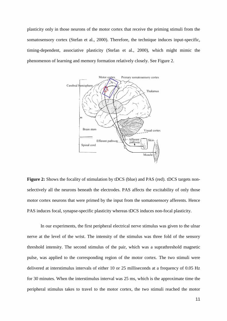
plasticity only in those neurons of the motor cortex that receive the priming stimuli from the
somatosensory cortex (Stefan et al., 2000). Therefore, the technique induces input-specific,
timing-dependent, associative plasticity (Stefan et al., 2000), which might mimic the
phenomenon of learning and memory formation relatively closely. See Figure 2.
Figure 2: Shows the focality of stimulation by tDCS (blue) and PAS (red). tDCS targets non-
selectively all the neurons beneath the electrodes. PAS affects the excitability of only those
motor cortex neurons that were primed by the input from the somatosensory afferents. Hence
PAS induces focal, synapse-specific plasticity whereas tDCS induces non-focal plasticity.
In our experiments, the first peripheral electrical nerve stimulus was given to the ulnar
nerve at the level of the wrist. The intensity of the stimulus was three fold of the sensory
threshold intensity. The second stimulus of the pair, which was a suprathreshold magnetic
pulse, was applied to the corresponding region of the motor cortex. The two stimuli were
delivered at interstimulus intervals of either 10 or 25 milliseconds at a frequency of 0.05 Hz
for 30 minutes. When the interstimulus interval was 25 ms, which is the approximate time the
peripheral stimulus takes to travel to the motor cortex, the two stimuli reached the motor
12
cortex approximately synchronously, which induces facilitatory plasticity (Stefan et al.,
2000). When the interstimulus interval was 10 ms, the motor cortex received the second pulse
much before the arrival of the first stimulus. Such asynchronous activation of the
somatosensory-motor connections results in inhibitory plasticity (Wolters et al., 2003).
1.4 Neuromodulators
Neuromodulators are substances that influence plasticity but are not essential for the
occurrence of plasticity (Malenka and Bear, 2004). Such substances act primarily on the
drivers of plasticity, namely the NMDA receptors in case of plasticity of the glutamatergic
system, usually in a non-linear fashion (Gu, 2002). Some of the major neuromodulators in the
human central nervous system are dopamine, acetylcholine, serotonin and noradrenaline. The
effects of these neuromodulators may be exerted either directly or indirectly on NMDA
activity. These substances are known to alter plasticity by either altering the excitability levels
of neurons, by enhancing the signal-to-noise ratio of neuronal responses or by altering the
threshold of activity-dependent changes at the synapses (Gu, 2002). In any case, their effects
depend on several factors of which the most important are (i) the type of subreceptors
activated (ii) the dosage of the substance (iii) background cortical activity in the specific brain
region. Because neuromodulators influence plasticity in neural networks considerably and
neuroplasticity is assumed to be an important physiological basis of learning and memory
formation, it might be speculated that neuromodulators exert their effects on cognition via
their impact on neuroplastic processes. Indeed it has been shown that altered neuromodulator
levels in healthy individuals influence cognitive performance, learning and memory
formation. For example, dopamine improves motor and verbal memory in healthy subjects
(Floel et al., 2005; Knecht et al., 2004). Altered neuromodulator levels are also observed in
pathological conditions accompanied by learning and memory deficits, such as in patients
13
suffering from Lewy body dementia (dopamine), Alzheimer‟s disease (acetylcholine), and
depression (serotonin), amongst others.
Alterations of neuromodulator levels can be induced in healthy humans by
pharmacological interventions. This enables us to study the physiological effects of
neuromodulators on plasticity and thus explore their impact on cortical functions. In our
studies, we explored two important aspects of the effects of neuromodulatory activity on
plasticity, namely (a) activity of subreceptors (nicotinic receptor activation in the cholinergic
system), and (b) dose-dependency of the effects on plasticity (dopaminergic system).
1.4.1 Cholinergic system
The cholinergic system comprises two types of receptors, namely the muscarinic and
nicotinic receptors. The muscarinic receptors are G-protein coupled (Ishii and Kurachi, 2006),
whereas the nicotinic receptors are ligand-gated cation channels (Gotti et al., 2009). Some
subtypes of the nicotinic receptors are calcium channels that play an important role in
glutamatergic plasticity (Shen and Yakel, 2009). Acetylcholine is a non-specific agonist of
both receptor types. Muscarine and nicotine are agonists specific to the respective receptor
types. Cholinergic activation is known to improve attention, arousal, learning and working
memory from many animal and human studies (Gold, 2003; Sarter et al., 2003). Such an
impact of cholinergic activation could be secondary to its effects on neuroplasticity. Effects of
acetylcholine on plasticity have been explored in several animal studies (Blitzer et al., 1990;
Hasselmo and Barkai, 1995). However, the role of cholinergic activation on neuroplasticity at
the system level in humans has been sparsely studied. Kuo et al. (2007) explored the impact
of rivastigmine, a cholinesterase inhibitor that is therapeutically used in patients suffering
from Alzheimer´s disease (Onor et al., 2007), on neuroplasticity induced by non-invasive
brain stimulation protocols. The drug increases the availability of acetylcholine at the
14
synapses, thereby activating non-specifically both muscarinic and nicotinic receptors. The
participants were administered rivastigmine along with excitability - enhancing or excitability
– diminishing, focal (PAS) or non-focal (tDCS) brain stimulation. Rivastigmine enhanced
focal facilitatory plasticity induced by PAS-25 and suppressed non-focal facilitatory plasticity
induced by anodal tDCS. Non-focal inhibitory plasticity following cathodal tDCS and focal
inhibitory plasticity were preserved or prolonged. Therefore it was concluded that cholinergic
activation resulted in a focusing effect on facilitatory plasticity probably due to enhanced
signal-to-noise ratio in the system. This might at least partially account for the improvement
of learning and memory following cholinergic activation (Hasselmo and Barkai, 1995). In this
thesis, we aimed to study the nicotinergic impact on neuroplasticity to explore further the
contribution of these receptors to the plasticity alteration induced by cholinergic activity
modulation. So far, nicotine has been shown to affect plasticity primarily in animal
experiments (Radcliffe and Dani, 1998). Studies exploring nicotinic impact on human
neuroplasticity are very sparse. The only available study by Swayne and colleagues (2009)
revealed that nicotine exposure enhances and prolongs the facilitatory after-effects of
intermittent theta burst stimulation in healthy subjects (Huang et al., 2005). Indirect hints for
plasticity-improving effects of nicotine stem from animal and human experiments
demonstrating a beneficial effect of nicotine on cognition, including learning and memory
formation (Froeliger et al. 2009; Hahn and Stolerman 2002; Heishman et al., 2010; Kastner et
al., 2010; Kumari et al. 2003). However, so far a systematic exploration of the nicotinic
impact on plasticity, especially considering the likely focusing effect of the drug on
facilitatory plasticity, is missing.
1.4.2 Dopaminergic system
The dopamine receptors are classified into D1-like (D1 and D5) and D2-like (D2,D3
and D4) receptor families. They are located at post-synaptic and also as autoreceptors at the
15
pre-synaptic sites of neuronal connections (Missale et al., 1998; Vallone et al., 2000).
Depending on the subtype and location of these receptors, the functional effects of
dopaminergic activation differs largely. The dopaminergic impact on neuroplasticity in
addition to the receptor subtype and location also depends on several other factors, the major
ones being the level of cortical activity, concentration level and the specific brain region
involved (Seamans and Yang, 2004).
Several studies have explored the impact of dopamine on cortical activity and
plasticity, both in animals and humans. With regard to animal experiments, the dopaminergic
impact on neuroplasticity has been studied in several brain regions – striatum (Kung et al.,
2007), hippocampus (O‟Çarroll et al., 2006) and prefrontal cortex (Otani et al., 1998; 2003). It
has been shown that dopamine exerts heterogeneous effects on plasticity depending on
subreceptor specificity and dopamine concentration. D1 activation results in enhancement of
both LTP and LTD mediated by higher NMDA activity (Bailey et al., 2000; Huang et al.,
2004) whereas D2 activation has variable effects due to suppressed NMDA and GABA
activities (Chen et al., 1996; Otani et al., 1998; Tseng and O‟Donnel, 2004). The balance of
NMDA and GABA activity, which is influenced by dopamine, determines the resultant
plasticity at the synapse. Considering the effect of concentration of dopamine on plasticity,
low and high levels impair neuroplasticity whereas moderate concentrations enhance it
(Seamans and Yang, 2004). In the cognitive domain, presumably at least in part caused by the
effect of dopamine on plasticity, it has been shown to improve working memory performance,
acquisition, stabilization and retrieval of long term memory in animal experiments (Brozoski
et al., 1979; Seamans et al., 1998). The heterogeneity of the effects of dopamine on
neuroplasticity is thought to be reflected by its heterogeneous effects on cognition (Goldman-
Rakic et al., 2000; Seamans and Yang, 2004; Zahrt et al., 1997).
16
The effects of dopamine on plasticity were also explored in a couple of studies in
humans. Kuo et al. (2008) demonstrated that 100 mg l-dopa enhances focal (PAS-induced)
facilitatory plasticity and suppresses non-focal (tDCS-induced) facilitatory plasticity in
healthy human subjects, that is, dopamine had a focusing effect on facilitatory plasticity.
Moreover, Monte-Silva et al. (2010) demonstrated an inverted-U shaped dose-response curve
of l-dopa on tDCS-induced non-focal plasticity in healthy human subjects. Ueki and
colleagues (2006) showed that in Parkinson‟s disease patients, who were off-medication and
thus in a hypo-dopaminergic state, PAS-induced plasticity could not be induced, but was
restored by administration of l-dopa. Functionally, these effects of dopamine on plasticity
might at least partly explain the positive effects of dopaminergic activation on learning and
memory formation in humans. Here 100mg l-dopa has been shown to be effective in
improving verbal memory in healthy human subjects (Floel et al., 2005a; Knecht et al., 2004)
and in stroke patients (Floel et al., 2005b). Since PAS induces a kind of plasticity which might
reflect the neurophysiological basis of learning and memory processes relatively closely, it
would be interesting to explore the dosage-dependent effect of dopaminergic activation on
this kind of plasticity. Such studies however have so far not been performed. Moreover, since
in some studies dopamine also failed to induce positive effects on cognitive performance
(Ghilardi et al., 2007; Gotham et al., 1988; Shohamy et al., 2006), non-linear dose-dependent
effect on PAS-induced plasticity, if observed, might partly account for the heterogeneous
dopaminergic effects on cognition.
17
1.5 Aim of the thesis
In this dissertation, we aimed to explore specific aspects of the impact of two main
neuromodulatory systems of the human central nervous system on plasticity, namely the
cholinergic and dopaminergic systems. The first study included in the dissertation deals with
the cholinergic system and aims at defining the specific contribution of the nicotinic receptors
to the global cholinergic effects on plasticity. Considering the calcium channel properties of
some of the nicotinic subreceptors, and the importance of calcium-dependent mechanisms for
neuroplasticity, we expected a prominent contribution of nicotinic receptors to the global
cholinergic effects on plasticity explored in a preceding study (Kuo et al., 2007). Moreover,
since global cholinergic activation resulted in a focusing effect on facilitatory plasticity, we
aimed to determine if this effect is caused by the nicotinic subreceptor.
In the second study we aimed to improve our understanding of the dopaminergic
impact on plasticity. Specifically we were interested to learn if associative plasticity induced
by PAS, which is thought to resemble Hebbian plasticity to a certain degree, is affected by
modulation of dopaminergic activity in a non-linear dosage-dependent way. The results of
previous studies using other plasticity-probing protocols (Monte-Silva et al. 2010) are in favor
for such an effect, which however has not been described for associative plasticity before. If
such an effect exists, it might help to understand the partly heterogeneous effects of
dopaminergic activation on learning and memory formation, which are thought to be closely
related to Hebbian plasticity.
Hereby, in the present thesis we explore two important factors that determine the
impact of neuromodulators on plasticity in the human motor cortex, namely subreceptor
specificity and dosage-dependency.
18
Chapter 2 Original articles and manuscripts
In this chapter, the manuscripts of two studies incorporated in the thesis are included.
The first study focuses on the nicotinergic impact on focal and non-focal plasticity in healthy
non-smoking human subjects. The second study explores the dose-dependent effect of l-dopa
on focal, associative plasticity in healthy human subjects.
Thirugnanasambandam N, Grundey J, Adam K, Drees A, Skwirba AC, Lang N,
Paulus W, Nitsche MA. Nicotinergic impact on focal and non-focal neuroplasticity induced
by non-invasive brain stimulation in non-smoking humans. Accepted,
Neuropsychopharmacology.
Thirugnanasambandam N, Grundey J, Paulus W, Nitsche MA. Dose-dependent non-
linear effect of L-DOPA on paired associative stimulation-induced neuroplasticity in humans.
Submitted

19
2.1 Nicotinergic impact on focal and non-focal neuroplasticity
induced by non-invasive brain stimulation in non-smoking humans
Authors: Nivethida Thirugnanasambandam MBBS,M.Tech1, Jessica Grundey MD
1, Kim
Adam1, Anne Drees
1, Angela C. Skwirba
1, Nicolas Lang MD
2, Walter Paulus MD
1, Michael
A. Nitsche MD 1*
Author affiliation: 1Department of Clinical Neurophysiology, Georg-August-University
Goettingen, Robert-Koch-Strasse 40, 37075 Goettingen, Germany; 2 Department of
Neurology, Christian-Albrechts University, Kiel, Germany.
ABSTRACT
Nicotine improves cognitive performance and modulates neuroplasticity in brain networks.
The neurophysiological mechanisms underlying nicotine-induced behavioral changes have
been sparsely studied, especially in humans. Global cholinergic activation focuses plasticity in
humans. However, the specific contribution of nicotinic receptors to these effects is unclear.
Henceforth, we explored the impact of nicotine on non-focal neuroplasticity induced by
transcranial direct current stimulation (tDCS) and focal, synapse-specific plasticity induced by
paired associative stimulation (PAS) in healthy non-smoking individuals. Forty eight subjects
participated in the study. Each subject received placebo and nicotine patches combined with
one of the stimulation protocols to the primary motor cortex in different sessions. Transcranial
magnetic stimulation (TMS) - elicited motor evoked potential (MEP) amplitudes were
recorded as a measure of corticospinal excitability until the evening of the second day
following the stimulation. Nicotine abolished or reduced both PAS- and tDCS-induced
inhibitory neuroplasticity. Non-focal facilitatory plasticity was also abolished, whereas focal
facilitatory plasticity was slightly prolonged by nicotine. Thus, nicotinergic influence on
facilitatory, but not inhibitory plasticity mimics that of global cholinergic enhancement.
20
Therefore, activating nicotinic receptors has clearly discernable effects from global
cholinergic activation. These nicotine-generated plasticity alterations might be important for
the effects of the drug on cognitive function.
INTRODUCTION
Smoking tobacco is the single largest preventable cause of mortality and morbidity (Peto et al.
1992). Nicotine is the primary constituent of tobacco that is responsible for its addictive
properties. Nicotine is the classical agonist at nicotinic acetylcholine receptors (nAchRs)
which are ligand-gated cation channels. Studies in animals and humans have shown that
nicotine improves attention as well as working and long-term memory (Froeliger et al. 2009;
Hahn and Stolerman 2002; Kumari et al. 2003). While many studies focused on the
behavioral effects of nicotine in healthy humans and patients (Jacobsen et al. 2004; Sacco et
al. 2005), very few have investigated the nicotinergic impact on cortical excitability and
plasticity, which are the likely neurophysiological basis for the cognitive effects of the
substance. For global cholinergic enhancement, it was shown that cholinesterase-inhibitors
reduce intracortical inhibition, increase facilitation, and enhance focal, but diminish non-focal
facilitatory plasticity in healthy humans (Korchounov et al. 2005; Kuo et al. 2007). A study
on tobacco smokers, who are under chronic nicotine exposure, revealed enhanced motor
cortex inhibition and reduced facilitation (Lang et al. 2008). Nicotine also enhances and
prolongs the facilitatory after-effects of intermittent theta burst stimulation in human motor
cortex (Swayne et al. 2009). Thus, global cholinergic and nicotinergic activation might have
at least partially dissimilar effects on cortical excitability.
Transcranial direct current stimulation (tDCS) and paired associative stimulation (PAS) are
non-invasive brain stimulation techniques that induce neuroplastic cortical excitability
alterations (Nitsche and Paulus 2000, 2001; Nitsche et al. 2003a; Stefan et al. 2000; Wolters
et al. 2003). Both techniques induce NMDA- and calcium-dependent changes of cortical
21
excitability (Nitsche et al. 2003b; Stefan et al. 2002; Wolters et al. 2003). tDCS modulates
spontaneous neuronal activity and excitability by either depolarizing or hyperpolarizing
neurons. Anodal tDCS induces depolarization that enhances neuronal excitability whereas
cathodal tDCS hyperpolarizes neurons, decreasing their excitability levels (Nitsche and
Paulus 2000, 2001; Nitsche et al. 2003a). Since tDCS affects all neurons beneath the
electrodes, it is thought to induce relatively non-focal plasticity. PAS, on the other hand,
induces focal, synapse-specific, timing-dependent, associative neuroplasticity in the targeted
neurons. Here an electrical pulse to a mixed peripheral nerve at an intensity which activated
somatosensory fibres is followed by a suprathreshold magnetic pulse applied to the
corresponding area of the primary motor cortex. Depending on the interstimulus interval,
there occurs synchronous or asynchronous activation of somatosensory-motor cortical
connections that enhance or reduce excitability respectively (Stefan et al. 2000).
In the current study we aimed to identify the specific contribution of nicotinic receptors to the
cholinergic effect on focal and non-focal neuroplasticity by exploring the effects of nicotine
on tDCS- and PAS-generated plasticity in healthy non-smoking humans to improve our
comprehension of the cognition-enhancing and addictive properties of this substance. As in
the foregoing studies, the motor cortex was taken as a model system in this single blinded,
placebo-controlled, partial crossover study because it allows a convenient monitoring of
excitability alterations by measuring motor evoked potential (MEP) amplitudes via
transcranial magnetic stimulation (TMS).
22
MATERIALS AND METHODS
Subjects
Forty eight healthy human volunteers participated in the study. All of them were complete
non-smokers, that is, none of them had smoked tobacco for at least 3 years prior to the study.
They did not suffer from any chronic or acute medical illness or any history of
neurological/psychiatric diseases, and did not take any chronic or acute medication. This
information was obtained by a detailed free personal interview with the subjects. Pregnancy,
family history of epilepsy, presence of any metallic implant or cardiac pacemaker was ruled
out. All of them were right-handed according to the Edinburgh handedness inventory
(Oldfield, 1971). The selection of subjects was not based on their results from previous
plasticity experiments in our laboratory; most of them were naïve to the experimental
procedure. Table 1 shows the demographic characteristics of the different groups. All subjects
gave written informed consent before participating in the study. The experiments were
approved by the local Ethics Committee and conformed to the Declaration of Helsinki.
Allocation of the subjects to the respective experimental conditions as well as order of
sessions was randomized (See table 1).
Assessing motor cortex excitability
Single transcranial magnetic (TMS) pulses were delivered from a Magstim 200 stimulator
(Magstim Company, Whitland, Dyfed, UK) at a frequency of 0.25 Hz with a figure-of-eight
shaped coil (diameter of one winding, 70mm; peak magnetic field, 2.2 T). The coil was held
tangentially to the scalp at an angle of 45º to the sagittal plane with the coil handle pointing
laterally and posterior. This induced a postero-anterior current flow in the brain at an angle
that optimally activates the corticospinal system monosynaptically (Di Lazzaro et al., 1998).
Motor evoked potentials (MEPs) were recorded using a surface electromyogram (EMG) set-
up. Surface EMG electrodes (Ag-AgCl) were placed over the abductor digiti minimi muscle
23
(ADM) in a belly-tendon montage. Signals were amplified, band-pass filtered (2Hz - 2KHz),
digitized (5KHz) and stored in a laboratory computer for offline analysis using Signal
software and CED 1401 (Cambridge Electronic Design, Cambridge, UK). MEPs were elicited
using single pulse TMS over the motor cortex representation of the ADM. The position of the
coil on the scalp, where the stimulus elicited consistently the largest MEP amplitudes for
slightly suprathreshold intensity was marked as the motor „hotspot‟. Stimulus intensity was
then adjusted in order to obtain peak to peak MEP amplitudes of approximately 1mV
(SI1mV). This TMS intensity was kept constant throughout the experiment. The mean MEP
amplitude was calculated from at least 20 pulses for baseline, and post-intervention
excitability monitoring. The change of the mean MEP amplitude over time reflects alterations
of motor cortex excitability.
Transcranial direct current stimulation
Twenty four subjects participated in the tDCS experiments. tDCS was administered by a
battery-driven constant current stimulator (Schneider Electronic, Gleichen, Germany) through
rubber electrodes covered by saline soaked sponges (35 square cm). One electrode was placed
over the motor cortex representation of the right ADM as determined by single pulse TMS
and the other electrode over the contralateral supra-orbital region. All subjects received 1mA
of either anodal (for 13 min) or cathodal stimulation (for 9 min), combined with nicotine or
placebo medication in different experimental sessions. Therefore, twelve subjects received
anodal tDCS with nicotine or placebo patches and the remaining twelve received cathodal
tDCS with nicotine or placebo patches. This stimulation intensity and duration (13 min anodal
tDCS and 9 min cathodal tDCS) generates after-effects on cortical excitability lasting for
approximately 60 min after stimulation (Nitsche and Paulus 2001; Nitsche et al. 2003a). The
two consecutive experimental sessions per subject were separated by at least one week
interval.

24
Paired associative stimulation
Twenty four subjects participated in the PAS experiment. Here a peripheral electrical pulse
over the right ulnar nerve at wrist level was followed by a TMS pulse over the motor cortex
representation of the ADM at inter-stimulus intervals (ISI) of either 10 (PAS 10) or 25
milliseconds (PAS 25). The peripheral pulse was delivered from a Digitimer D185 multipulse
stimulator (Digitimer, Welwyn Garden City, UK) at an intensity of 300% of the sensory
perceptual threshold. The suprathreshold magnetic pulse was delivered from a Magstim 200
stimulator with an intensity which elicited MEP amplitudes of approximately SI1mV. The
paired pulses were repeated 90 times at a frequency of 0.05 Hz. This protocol induces long-
lasting excitability changes in the motor cortex depending on the ISI duration. An ISI of 10
ms induces excitability diminution whereas an ISI of 25 ms induces facilitation (Stefan et al.
2000; Wolters et al. 2003). The subjects were instructed to count the number of pulses they
received at their wrist throughout the whole stimulation duration in order to guarantee
sufficient attention to the procedure, which has been shown to be crucial to obtain the
intended effects (Stefan et al., 2004).
Pharmacological intervention
Each subject participated in two sessions in randomized order. 30cm2 nicotine transdermal
patches, each containing nicotine 0.83mg/cm2 releasing 15mg over 16 hours or placebo
patches were administered to all subjects in combination with one of the stimulation protocols
- anodal tDCS, cathodal tDCS, PAS-10 or PAS-25. By this dosage of nicotine,
physiologically and behaviorally relevant plasma levels are accomplished (Tønnesen et al.
1991). Subjects received the patch 6 hours before the start of the stimulation. This was the
approximate time for the plasma level of nicotine to reach its maximum following application
of the patch (Nørregaard et al. 1992). The patch was retained until the end of the last after-
25
measurement of the experiment on the evening of the second day. In order to counteract
possible systemic side effects of nicotine, the subjects were instructed to take 20mg
domperidone, a peripheral acting dopamine D2-receptor antagonist with antiemetic effects, in
case of need.
Course of the experiment
The subjects received either a placebo or nicotine patch, which was adhered to the left upper
arm and remained there until the end of the last after-measurement on the following evening.
They were given 20mg domperidone and asked to take it orally in case of any side effects.
Unpublished results from our group show that domperidone alone does not have any
significant effect on motor cortical excitability. Six hours later, subjects were seated
comfortably in a reclined position on a dentist´s chair with proper arm and head rests and
asked to relax completely. The EMG electrodes were placed at the right ADM as described
above. The motor „hotspot‟ was determined over the left motor cortex and marked with a
water-proof skin marker, and the TMS intensity needed to induce MEP amplitudes of 1mV
(SI1mV) size was determined. Twenty MEPs were recorded at this stimulus intensity and the
mean MEP amplitude was calculated as the baseline. One of the stimulation protocols, either
tDCS or PAS, was administered. At least 20 MEPs were recorded immediately following the
stimulation (0min) and at time points of 5, 10, 15, 20, 25, 30, 60, 90, and 120 min. For the
nicotine patch sessions, the after-measurements were also conducted in the evening of the
stimulation day and in the morning and evening of the day following the plasticity induction
procedure (See figure 1). We used a waterproof pen to mark the ADM electrodes and TMS
coil positions and ensured that these were positioned over the same spot during the whole
course of the experiment.
26
Data analysis and statistics
The individual means of the 20 MEP amplitudes recorded at each time point were calculated
for all subjects. The post-intervention mean MEP amplitudes from each subject were then
normalized to the respective individual mean baseline MEP amplitude. The normalized mean
MEP amplitudes from all subjects were pooled together and the grand average across subjects
for each time bin was calculated.
A repeated measures ANOVA was performed on the normalized data using MEP amplitude
as the dependent factor including all time points up to 120 min after stimulation. Drug
(Nicotine versus Placebo) and time points were included as within-subjects factors.
Stimulation (anodal tDCS/ cathodal tDCS/ PAS-10/ PAS-25) served as between-subjects
factors. The Mauchly test was performed to test for sphericity, and the Greenhouse-Geisser
correction applied when necessary. Conditional on significant results of the ANOVA, we
performed post-hoc comparisons using Student‟s t-tests (paired, two-tailed, p<0.05, not
adjusted for multiple comparisons) where we compared (i) the mean MEP amplitudes at the
time points after plasticity induction versus baseline and (ii) the mean MEP amplitudes
following nicotine versus placebo at one time point within a stimulation condition. Moreover,
we compared absolute baseline MEP values between the stimulation and drug conditions via
Student´s t-tests to exclude a priori differences. Chi square test was performed to look for
significant differences in gender distribution between the groups. For comparison of the age
of subjects between the groups, Student‟s t-tests (two - tailed, unpaired, p<0.05) were
performed.
RESULTS
All subjects tolerated the experimental procedure well. None of them complained of any side
effects of either nicotine or the stimulation. Especially the participants did not complain of
any sedative effects of the patch. During the experiment, they were completely alert and
27
relaxed. Since no systemic side effects of nicotine were perceived and none of the participants
needed to take domperidone, the subjects were blinded effectively. Gender distribution did
not differ significantly between the various groups tested by chi square test (p = 0.083). There
were significant differences in the mean age between some of the groups as tested by
Student‟s unpaired t-tests. However, the maximum difference of mean age between groups
was 2.65 years. Absolute baseline MEP amplitudes did not differ significantly between
groups (Student‟s t-test, two-tailed, paired, p>0.05 for all cases) or medication conditions
(Student‟s t-test, unpaired, two-tailed, p = 0.66).
The ANOVA revealed a significant main effect of the between-subjects factor stimulation
(F(3,44) = 18.137; p < 0.001), in accordance with different effects of inhibitory and
facilitatory tDCS and PAS on MEP amplitudes. The main effects of either nicotine (F(1,44) =
0.093; p = 0.762) or time (F(10,440) = 1.654; p = 0.089) were not significant. However, the
interactions between nicotine X stimulation (F(3,44) = 5.498; p = 0.003); time X stimulation
(F(30,440) = 3.070; p < 0.001) were significant, showing that nicotine had different effects on
the above-mentioned stimulation protocols, and that the time course of the effects of these
stimulation protocols was not identical. The three-way interaction nicotine X time X
stimulation (F(30,440) = 1.848; p = 0.005) was also significant. Thus, application of nicotine
patch significantly influenced the after-effects of the different stimulation protocols
differently over time.
Nicotinergic impact on tDCS-induced plasticity
As shown by the post hoc t-tests, in the control condition without nicotine, MEPs were
significantly enhanced following anodal tDCS for up to 90 minutes. Cathodal tDCS
diminished excitability levels significantly also for up to 90 minutes after stimulation. Under
nicotine, both anodal and cathodal tDCS-induced after-effects were abolished. The post-hoc
28
test revealed that for anodal tDCS, post-tDCS MEP amplitudes under nicotine were not
different from baseline values, but differed significantly from those under placebo medication
(table S1). A trendwise reversal of the effects of anodal tDCS from facilitation to inhibition
under nicotine did not reach statistical significance (p > 0.073). The excitability diminution
induced by cathodal tDCS, as compared to baseline excitability, was also abolished under
nicotine; however relative to the placebo medication condition nicotine induced only a
trendwise change (Figure 2 A, B, table S1).
Effect of nicotine on PAS-induced plasticity
As shown by the post hoc tests, PAS induced a significant facilitation of MEP amplitudes
following PAS-25 and excitability diminution following PAS-10, lasting for up to 90 or 120
minutes after stimulation (table S1) under placebo medication. Under nicotine, the facilitatory
effects of PAS-25 remained significant as compared to the respective baseline MEP for up to
90 min after PAS. MEP amplitudes under nicotine did not differ versus the respective placebo
medication condition for up to 90 min after PAS-25, but were significantly larger as compared
to placebo 120 min after PAS-25, being in favor for a prolonged excitability enhancement
accomplished by PAS-25 under nicotine. The missing difference of PAS-25 under nicotine
relative to baseline excitability 120 min after placticity induction might be caused by the
relatively large variability of the MEPs at this time point (see Table S1). The inhibitory effect
of PAS-10 was abolished under nicotine. Consequently, the post hoc tests revealed no
significant differences of the respective MEP amplitudes relative to baseline, but significant
differences of the respective MEP amplitudes relative to those under placebo medication
(figure 3A, B).
29
DISCUSSION
The present study shows that in healthy non-smoking individuals nicotine prominently affects
neuroplasticity. Our data illustrate that (i) nicotine exposure slightly prolongs or at least
preserves the synapse-specific cortical excitability enhancement induced by PAS-25, but
abolishes the PAS-10 induced depression of cortical excitability (ii) the non-focal excitability
enhancing after-effect of anodal tDCS and the excitability diminution caused by cathodal
tDCS are both abolished (Figure 4). There is also a trendwise reversal of anodal tDCS -
induced facilitation to inhibition under nicotine. Hence, we observe a focusing effect of
nicotine on facilitatory neuroplasticity and an abolishment of inhibitory plasticity, the latter
irrespective of the focality of stimulation.
Some of our observations match with those of previous studies. The enhancement or
preservation of facilitatory plasticity induced by PAS-25 by nicotine is similar to the effect it
had on intermittent theta burst stimulation (Swayne et al., 2009). The effect of nicotine on
facilitatory plasticity is also comparable to the effect of the cholinesterase inhibitor
rivastigmine on identical plasticity induction protocols, however its effects on inhibitory
plasticity are at variance to those under global cholinergic enhancement under rivastigmine
(Kuo et al., 2007).
Proposed mechanism of action
The focusing effect of nicotine on facilitatory plasticity, i.e. consolidating/preserving focal,
PAS-induced, but diminishing non-focal tDCS-generated plasticity, is quite similar to that of
global cholinergic enhancement via rivastigmine described in a previous study (Kuo et al.,
2007). A likely explanation for this effect is the different impact of cholinergic activation on
recurrent activation of afferent input to cortical neurons. It has been shown that excitatory
glutamatergic synaptic transmission is suppressed by presynaptic inhibition at intrinsic,
30
recurrent synapses, but not at afferent fibre synapses via cholinergic activation (Hasselmo &
Bower, 1992; Hasselmo et al., 1995; Vogt & Regehr, 2001). Since PAS induces plasticity by
a combination of afferent somatosensory input with a motor cortical stimulus, while tDCS is
thought to affect primarily excitability of cortical interneurons (Nitsche et al., 2005), it might
be speculated that these specific aspects of the stimulation techniques caused the differences
of the effects also in the present experiments. In accordance, in animal experiments an
inhibitory effect of the activation of nicotinergic subreceptors on feedforward interneurons
has has been shown to prevent LTP induction by inhibiting pyramidal neurons, whereas
spike-timing dependent LTP was enhanced (Rosza et al., 2008; Yamazaki et al., 2005),
although the latter effect was not shown in all studies (Couey et al., 2007). Due to the results
of the present experiment, this effect of cholinergic activation on facilitatory plasticity in
humans can likely be attributed to an impact of nicotinic receptors. Another possible
explanation of the results is based on the enhancement of intracellular calcium concentration
by nicotinic receptors, e.g. the alpha-7 subreceptor. Intracellular calcium is a key determinant
of plasticity induction, and the after-effects of tDCS and PAS are calcium-dependent (Nitsche
et al., 2003b; Stefan et al., 2002). The amount of intracellular calcium determines if
inhibitory, facilitatory, or no plasticity is induced. A slight enhancement of intracellular
calcium induces LTD, a large enhancement LTP (Lisman, 2001). Medium and very large
concentrations of intracellular calcium induce no or convert plasticity, the latter possibly due
to an activation of hyperpolarizing calcium channels (Misonou et al., 2004). Since tDCS
induces facilitatory plasticity by tonic depolarization of neurons for some minutes, which
might activate voltage-dependent calcium channels, whereas PAS is a phasic stimulation
technique, which induces only short lasting depolarization, the amount of intracellular
calcium increase caused by tDCS might be larger than that caused by PAS. Therefore, a
further calcium enhancement via nicotinic receptor activation might enhance the calcium level
above the concentration inducing LTP-like plasticity, and therefore result in an abolishment of
31
plasticity in case of tDCS, but not PAS. This effect on different kinds of facilitatory plasticity
of nicotine might enhance the signal-to-noise ratio (Hasselmo and Barkai, 1995), which
would facilitate the representation of meaningful, synchronous inputs and suppress non-
meaningful inputs. Indeed, it has been recently suggested that nicotine improves memory
performance via calcium-dependent mechanisms in animals (Biala & Kruk, 2009).
In contrast, nicotine abolished all kinds of inhibitory plasticity irrespective of the specific
stimulation protocol. One possible explanation might be that the calcium-enhancing properties
of nicotinic receptor activation here resulted in an intracellular calcium level too large to
induce LTD-like plasticity. This mechanism of action would also explain the different effect
of global cholinergic activation by rivastigmine on inhibitory plasticity, as described in a
previous study, where this substance prolonged tDCS- and PAS-generated inhibitory
plasticity (Kuo et al., 2007), because muscarinic receptors inhibit voltage-gated calcium
channels (Brown, 2010), and thus might counteract the effects of nicotinic receptors in this
case. The effects of nicotine on inhibitory plasticity obtained in the present study are not in
accordance with some animal experiments, where nicotinic activation has been shown to be
important for LTD induction (Partridge et al., 2002; Fujii & Sumikawa, 2001), however, the
effect of nicotine on LTD seems to depend on the general proneness of the system to
inhibitory plasticity (Alzoubi et al., 2007, 2008), which likely differs between animal
preparations and in vivo studies in humans.
It should be mentioned that these mechanistic explanations of the results are hypothetical
presently. Alternative explanations, such as the modification of NMDA receptor-dependent
plasticity by nicotine-dependent alteration of GABAergic activity (Couey et al. 2007), cannot
be ruled out. These hypotheses should be tested more directly in future studies, e.g. by
altering the activity of nicotinic subreceptors, or calcium channels, in combination with
nicotine exposure.
32
General remarks
The results of the present study demonstrate that nicotine clearly influences neuroplasticity in
non-smoking individuals. Nicotine focuses facilitatory plasticity whereas it abolishes
inhibitory plasticity. The effects differ from those of non-specific cholinergic activation. The
focusing effect of nicotine on facilitatory plasticity might help to explain how this drug
improves attention, working memory and long-term memory in animals and humans via
enhancing the signal to noise ratio of plasticity. Also the abolition of inhibitory plasticity by
nicotine might affect cognitive processes. First, it might shift the net balance of plasticity
more into the direction of facilitatory plasticity, and therefore indirectly enhance cognitive
performance further. Second, inhibitory plasticity, especially long-term depression (LTD) has
been shown to be directly involved in certain forms of learning and memory formation.
Collingridge and colleagues (2010) describe a role of LTD in hippocampal-based learning and
memory formation, and recognition memory in perirhinal cortex. Since our findings show that
nicotine abolishes LTD-like plasticity it could be speculated that nicotine might worsen LTD-
dependent forms of learning and memory. However, an impairment of cognitive functions by
nicotine has been rarely described (Toledano et al., 2010), thus further behavioral studies are
needed to explore the cognitive effects of nicotine more systematically.
Some limiting aspects of this study should be mentioned. Blinding could have been somewhat
compromised considering the fact that the experimenters were not blinded to the intervention.
However, the data were collected by more than one investigator without notable difference in
the results, which probably indicates low experimenter bias, and experimenters were not
informed about hypotheses about expected outcomes of the experiments, which should have
limited expectancy effects. Although the subjects did not complain of any sedation due to
nicotine, the degree of alertness was not explicitly assessed and hence its effect on the
measurements cannot be ruled out completely. We did not measure plasma concentrations of
nicotine, thus it could be argued that inter-individual differences of the bioavailability of the
33
substance had an impact on the results. However, since we studied a fairly homogenous group
of participants, and we induced plasticity during steady state drug concentration, we do not
think that variability of plasma concentration of nicotine can explain the results. We studied
the effect of only a single dosage of nicotine in the present experiments. Thus it cannot be
ruled out that the effect of nicotine on plasticity differs dose-dependently, as shown for other
neuromodulators, like dopamine (Monte-Silva et al. 2009, 2010). Moreover, since nicotine
receptors are rapidly modified by chronic exposure, this study cannot discern between
primary effects of nicotine on nicotinic receptors and secondary effects caused by receptor
desensitization or upregulation. Moreover, it should not be taken for granted that the effects
obtained on motor cortex plasticity, as in the current study, translate exactly to other cortices,
where nicotinic receptor density, and subreceptor composition might differ (McGehee and
Role, 1995; Gotti et el., 2009) Furthermore the results of a single dosage nicotine application
in non-smokers, as performed here, might differ from the effects of nicotine in smokers who
are chronically exposed to it. Future studies should address these aspects in larger detail.
Conclusion
The results of this study deliver clear evidence for an important role of nicotine in the
formation of neuroplasticity, the likely basis of learning and memory formation, in humans.
Via its focusing effect on facilitatory plasticity, nicotine might be an attractive candidate to
enhance these processes in neuropsychiatric diseases accompanied by cognitive decline. The
abolition of inhibitory plasticity by nicotine also could have a significant impact on some
forms of learning and memory, and also affect addictive behavior to some extent. Moreover,
its effect on plasticity might be an important mechanism for starting nicotine consumption,
addiction and the high probability of relapse in smokers. Interestingly the effects of nicotine
on plasticity share some of those of dopamine (Kuo et al., 2008), which might be an
explanation for the frequent nicotine consumption in schizophrenia, in which dopaminergic
34
malfunctioning is an important pathologic mechanism. Clearly, more studies are needed to
explore the exact role of nicotine in healthy humans and in those suffering from
neuropsychiatric diseases to a larger degree. Moreover the results of this study are important
in another aspect. Non-invasive brain stimulation techniques are increasingly used as
scientific and therapeutic tools. The results of the present study show that the activity of the
nicotinergic system might critically affect the effects of brain stimulation. This potentially
important confounding factor should thus be taken into account in future studies using brain
stimulation.
FUNDING:
The study was supported by the Deutsche Forschungsgemeinschaft (DFG grant NI683/4-1
„Towards risk prediction of nicotine dependency by exploring individual limits of cortical
neuroplasticity in humans‟; NI 683/4-2 “Impact of the nicotinergic alpha7 receptor on cortical
plasticity in smokers and nonsmokers”) within the DFG priority program „Nicotine:
Molecular and Physiological Effects in Central Nervous System‟.
REFERENCES
Alzoubi KH, Aleisa AM, Alkadhi KA. (2007). Adult-onset hypothyroidism facilitates and
enhances LTD: reversal by chronic nicotine treatment. Neurobiol Dis. 26(1):264-72.
Alzoubi KH, Aleisa AM, Alkadhi KA. (2008). Effect of chronic stress or nicotine on
hypothyroidism-induced enhancement of LTD: electrophysiological and molecular studies.
Neurobiol Dis. 32(1):81-7.
Biała G, Kruk M. (2009). Influence of bupropion and calcium channel antagonists on the
nicotine-induced memory-related response of mice in the elevated plus maze. Pharmacol Rep.
61(2):236-44.
35
Brown DA. (2010). Muscarinic acetylcholine receptors (mAChRs) in the nervous system:
some functions and mechanisms. J Mol Neurosci. 41:340-346.
Couey JJ, Meredith RM, Spijker S, Poorthuis RB, Smit AB, Brussaard AB, Mansvelder HD.
(2007). Distributed network actions by nicotine increase the threshold for spike-timing-
dependent plasticity in prefrontal cortex. Neuron. 54(1):73-87.
Di Lazzaro V, Oliviero A, Profice P, Saturno E, Pilato F, Insola A, Mazzone P, Tonali P,
Rothwell JC. (1998). Comparison of descending volleys evoked by transcranial magnetic and
electric stimulation in conscious humans. Electroencephalogr Clin Neurophysiol. 109(5):397-
401.
Froeliger B, Gilbert DG, McClernon FJ. (2009). Effects of nicotine on novelty detection and
memory recognition performance: double-blind, placebo-controlled studies of smokers and
nonsmokers. Psychopharmacology (Berl). 205(4):625-33.
Fujii S, Sumikawa K. (2001). Nicotine accelerates reversal of long-term potentiation and
enhances long-term depression in the rat hippocampal CA1 region. Brain Res. 894(2):340-6.
Gotti C, Clementi F, Fornari A, Gaimarri A, Guiducci S, Manfredi I, Moretti M, Pedrazzi P,
Pucci L, Zoli M. (2009). Structural and functional diversity of native brain neuronal nicotinic
receptors. Biochem Pharmacol. 78(7):703-11.
Hahn B, Stolerman IP. (2002). Nicotine-induced attentional enhancement in rats: effects of
chronic exposure to nicotine. Neuropsychopharmacology. 27(5):712-722.
Hasselmo ME, Barkai E. (1995). Cholinergic modulation of activitydependent synaptic
plasticity in the piriform cortex and associative memory function in a network biophysical
simulation. J Neurosci. 15:6592– 6604.
36
Hasselmo ME, Bower JM. (1992). Cholinergic suppression specific to intrinsic not afferent
fiber synapses in rat piriform (olfactory) cortex. J Neurophysiol 67:1222–1229.
Hasselmo ME, Schnell E, Barkai E. (1995). Dynamics of learning and recall at excitatory
recurrent synapses and cholinergic modulation in rat hippocampal region CA3. J Neurosci
15:5249 –5262.
Jacobsen LK, D'Souza DC, Mencl WE, Pugh KR, Skudlarski P, Krystal JH. (2004). Nicotine
effects on brain function and functional connectivity in schizophrenia. Biol Psychiatry.
55(8):850-858.
Korchounov A, Ilic TV, Schwinge T, Ziemann U. (2005). Modification of motor cortical
excitability by an acetylcholinesterase inhibitor. Exp Brain Res. 164(3):399-405.
Kumari V, Gray JA, ffytche DH, Mitterschiffthaler MT, Das M, Zachariah E, Vythelingum
GN, Williams SC, Simmons A, Sharma T. (2003). Cognitive effects of nicotine in humans: an
fMRI study. Neuroimage. 19(3):1002-1013.
Kuo MF, Grosch J, Fregni F, Paulus W, Nitsche MA. (2007). Focusing effect of acetylcholine
on neuroplasticity in the human motor cortex. J Neurosci. 27(52):14442-7.
Kuo MF, Paulus W, Nitsche MA. (2008). Boosting focally-induced brain plasticity by
dopamine. Cereb Cortex. 18(3):648-651.
Lang N, Hasan A, Sueske E, Paulus W, Nitsche MA. (2008). Cortical hypoexcitability in
chronic smokers? A transcranial magnetic stimulation study. Neuropsychopharmacology.
33(10):2517-2523.
Lisman JE. (2001). Three Ca2+ levels affect plasticity differently: the LTP zone, the LTD
zone and no man's land. J Physiol. 532(Pt 2):285.

37
McGehee DS, Role LW. (1995). Physiological diversity of nicotinic acetylcholine receptors
expressed by vertebrate neurons. Annu Rev Physiol. 57:521-46.
McKay BE, Placzek AN, Dani JA. (2007). Regulation of synaptic transmission and plasticity
by neuronal nicotinic acetylcholine receptors. Biochem Pharmacol. 74(8):1120-1133.
Misonou H, Mohapatra DP, Park EW, Leung V, Zhen D, Misonou K, Anderson AE, Trimmer
JS. (2004). Regulation of ion channel localization and phosphorylation by neuronal activity.
Nat Neurosci. 7(7):711-718.
Monte-Silva K, Kuo MF, Thirugnanasambandam N, Liebetanz D, Paulus W, Nitsche MA.
(2009). Dose-dependent inverted U-shaped effect of dopamine (D2-like) receptor activation
on focal and nonfocal plasticity in humans. J Neurosci. 29(19):6124-6131.
Nitsche MA, Paulus W. (2000). Excitability changes induced in the human motor cortex by
weak transcranial direct current stimulation. J Physiol. 527 (Pt 3):633-639.
Nitsche MA, Paulus W. (2001). Sustained excitability elevations induced by transcranial DC
motor cortex stimulation in humans. Neurology. 57(10):1899-901.
Nitsche MA, Fricke K, Henschke U, Schlitterlau A, Liebetanz D, Lang N, Henning S, Tergau
F, Paulus W. (2003a). Pharmacological modulation of cortical excitability shifts induced by
transcranial direct current stimulation in humans. J Physiol. 553(Pt 1):293-301.
Nitsche MA, Nitsche MS, Klein CC, Tergau F, Rothwell JC, Paulus W. (2003b). Level of
action of cathodal DC polarisation induced inhibition of the human motor cortex. Clin
Neurophysiol. 114(4):600-604.
Nitsche MA, Seeber A, Frommann K, Klein CC, Rochford C, Nitsche MS, Fricke K,
Liebetanz D, Lang N, Antal A, Paulus W, Tergau F. (2005). Modulating parameters of
38
excitability during and after transcranial direct current stimulation of the human motor cortex.
J Physiol. 568(Pt 1):291-303.
Nørregaard J, Tønnesen P, Simonsen K, Säwe U. (1992). Long-term nicotine substitution
after application of a 16-hour nicotine patch in smoking cessation. Eur J Clin Pharmacol.
43(1): 57-60
Partridge JG, Apparsundaram S, Gerhardt GA, Ronesi J, Lovinger DM. (2002). Nicotinic
acetylcholine receptors interact with dopamine in induction of striatal long-term depression. J
Neurosci. 22(7):2541-9.
Peto R, Lopez AD, Boreham J, Thun M, Heath C Jr. (1992). Mortality from tobacco in
developed countries: indirect estimation from national vital statistics. Lancet.
339(8804):1268-1278.
Rózsa B, Katona G, Kaszás A, Szipöcs R, Vizi ES. (2008). Dendritic nicotinic receptors
modulate backpropagating action potentials and long-term plasticity of interneurons. Eur J
Neurosci. 27(2):364-77.
Sacco KA, Termine A, Seyal A, Dudas MM, Vessicchio JC, Krishnan-Sarin S, Jatlow PI,
Wexler BE, George TP. (2005). Effects of cigarette smoking on spatial working memory and
attentional deficits in schizophrenia: involvement of nicotinic receptor mechanisms. Arch Gen
Psychiatry. 62(6):649-659.
Stefan K, Kunesch E, Cohen LG, Benecke R, Classen J. (2000). Induction of plasticity in the
human motor cortex by paired associative stimulation. Brain. 123 (Pt 3):572-584.
39
Stefan K, Kunesch E, Benecke R, Cohen LG, Classen J. (2002). Mechanisms of enhancement
of human motor cortex excitability induced by interventional paired associative stimulation. J
Physiol. 543(Pt 2):699-708.
Swayne OB, Teo JT, Greenwood RJ, Rothwell JC. (2009). The facilitatory effects of
intermittent theta burst stimulation on corticospinal excitability are enhanced by nicotine. Clin
Neurophysiol. 120(8):1610-5.
Toledano A, Alvarez MI, Toledano-Díaz A. (2010). Diversity and variability of the effects of
nicotine on different cortical regions of the brain - therapeutic and toxicological implications.
Cent Nerv Syst Agents Med Chem. 10(3):180-206.
Tønnesen P, Nørregaard J, Simonsen K, Säwe U. (1991). A double-blind trial of a 16-hour
transdermal nicotine patch in smoking cessation. N Engl J Med. 325(5):311-5.
Vogt KE, Regehr WG. (2001). Cholinergic modulation of excitatory synaptic transmission in
the CA3 area of the hippocampus. J Neurosci 21:75– 83.
Wolters A, Sandbrink F, Schlottmann A, Kunesch E, Stefan K, Cohen LG, Benecke R,
Classen J. (2003). A temporally asymmetric Hebbian rule governing plasticity in the human
motor cortex. J Neurophysiol. 89(5):2339-2345.
Wonnacott S. (1997). Presynaptic nicotinic Ach receptors. Trends Neurosci. 20(2):92-98.
Yamazaki Y, Jia Y, Hamaue N, Sumikawa K. (2005). Nicotine-induced switch in the
nicotinic cholinergic mechanisms of facilitation of long-term potentiation induction. Eur J
Neurosci. 22(4):845-60.
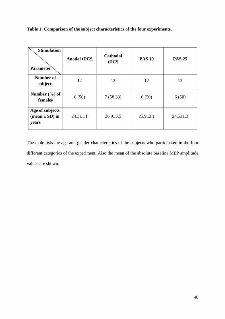
40
Table 1: Comparison of the subject characteristics of the four experiments.
Stimulation
Parameter
Anodal tDCS Cathodal
tDCS PAS 10 PAS 25
Number of
subjects 12 12 12 12
Number (%) of
females 6 (50) 7 (58.33) 6 (50) 6 (50)
Age of subjects
(mean ± SD) in
years
24.3±1.1 26.9±3.5 25.9±2.1 24.5±1.3
The table lists the age and gender characteristics of the subjects who participated in the four
different categories of the experiment. Also the mean of the absolute baseline MEP amplitude
values are shown.
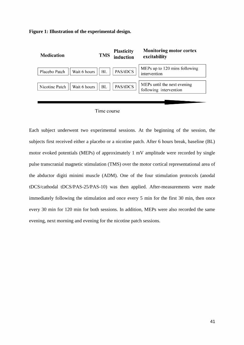
41
Figure 1: Illustration of the experimental design.
Each subject underwent two experimental sessions. At the beginning of the session, the
subjects first received either a placebo or a nicotine patch. After 6 hours break, baseline (BL)
motor evoked potentials (MEPs) of approximately 1 mV amplitude were recorded by single
pulse transcranial magnetic stimulation (TMS) over the motor cortical representational area of
the abductor digiti minimi muscle (ADM). One of the four stimulation protocols (anodal
tDCS/cathodal tDCS/PAS-25/PAS-10) was then applied. After-measurements were made
immediately following the stimulation and once every 5 min for the first 30 min, then once
every 30 min for 120 min for both sessions. In addition, MEPs were also recorded the same
evening, next morning and evening for the nicotine patch sessions.
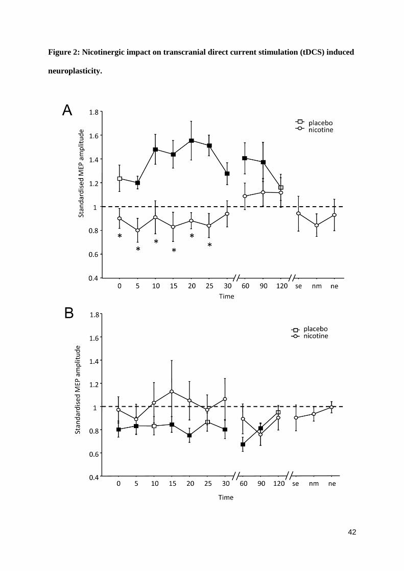
42
Figure 2: Nicotinergic impact on transcranial direct current stimulation (tDCS) induced
neuroplasticity.
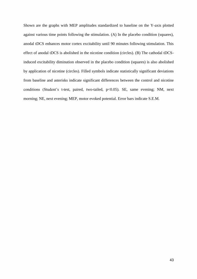
43
Shown are the graphs with MEP amplitudes standardized to baseline on the Y-axis plotted
against various time points following the stimulation. (A) In the placebo condition (squares),
anodal tDCS enhances motor cortex excitability until 90 minutes following stimulation. This
effect of anodal tDCS is abolished in the nicotine condition (circles). (B) The cathodal tDCS-
induced excitability diminution observed in the placebo condition (squares) is also abolished
by application of nicotine (circles). Filled symbols indicate statistically significant deviations
from baseline and asterisks indicate significant differences between the control and nicotine
conditions (Student‟s t-test, paired, two-tailed, p<0.05). SE, same evening; NM, next
morning; NE, next evening; MEP, motor evoked potential. Error bars indicate S.E.M.
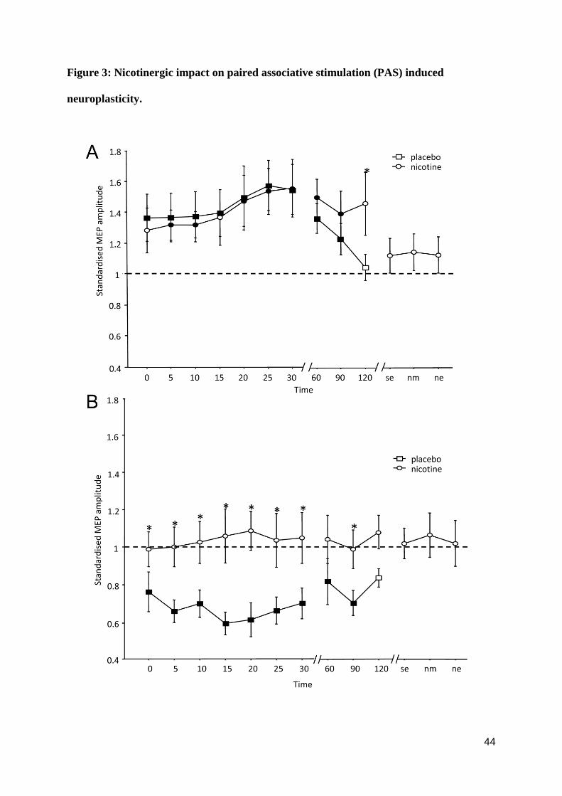
44
Figure 3: Nicotinergic impact on paired associative stimulation (PAS) induced
neuroplasticity.
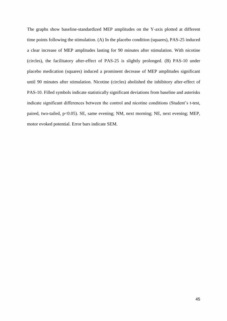
45
The graphs show baseline-standardized MEP amplitudes on the Y-axis plotted at different
time points following the stimulation. (A) In the placebo condition (squares), PAS-25 induced
a clear increase of MEP amplitudes lasting for 90 minutes after stimulation. With nicotine
(circles), the facilitatory after-effect of PAS-25 is slightly prolonged. (B) PAS-10 under
placebo medication (squares) induced a prominent decrease of MEP amplitudes significant
until 90 minutes after stimulation. Nicotine (circles) abolished the inhibitory after-effect of
PAS-10. Filled symbols indicate statistically significant deviations from baseline and asterisks
indicate significant differences between the control and nicotine conditions (Student‟s t-test,
paired, two-tailed, p<0.05). SE, same evening; NM, next morning; NE, next evening; MEP,
motor evoked potential. Error bars indicate SEM.
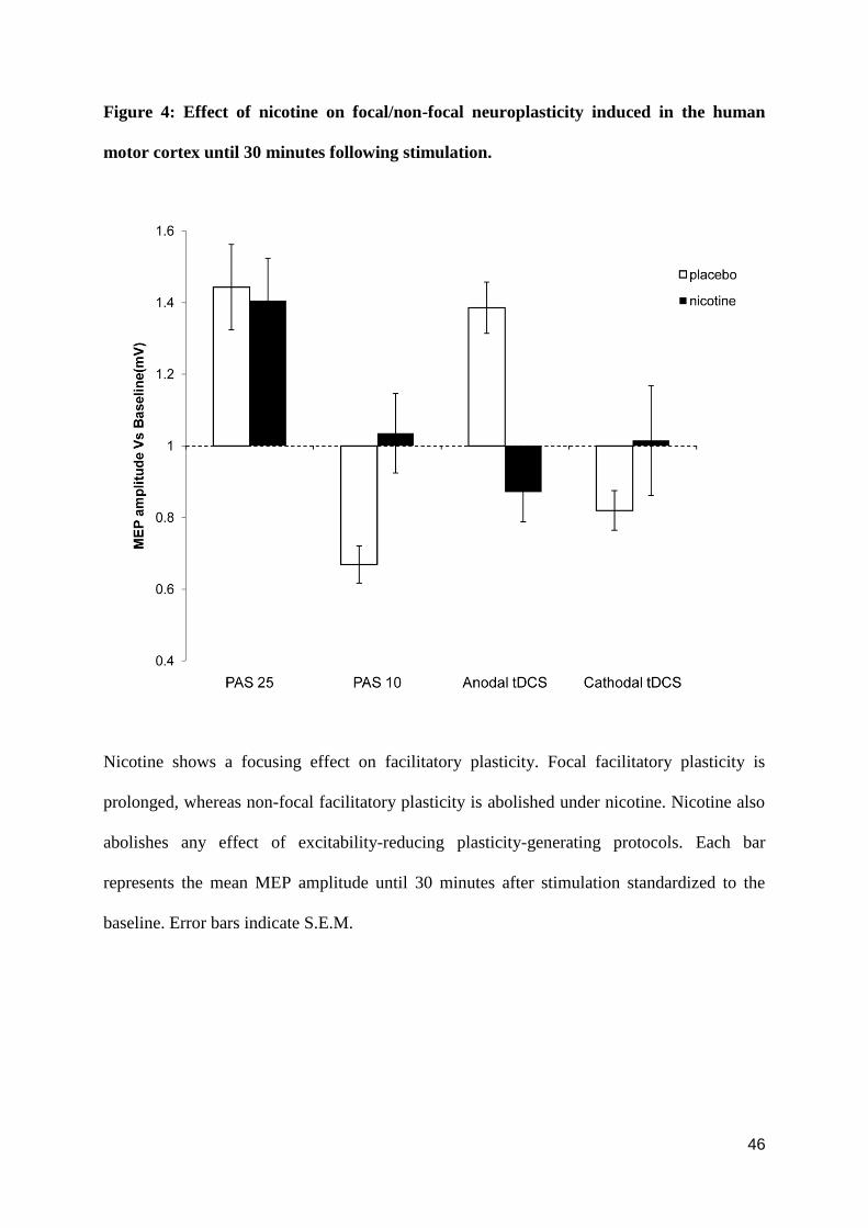
46
Figure 4: Effect of nicotine on focal/non-focal neuroplasticity induced in the human
motor cortex until 30 minutes following stimulation.
Nicotine shows a focusing effect on facilitatory plasticity. Focal facilitatory plasticity is
prolonged, whereas non-focal facilitatory plasticity is abolished under nicotine. Nicotine also
abolishes any effect of excitability-reducing plasticity-generating protocols. Each bar
represents the mean MEP amplitude until 30 minutes after stimulation standardized to the
baseline. Error bars indicate S.E.M.
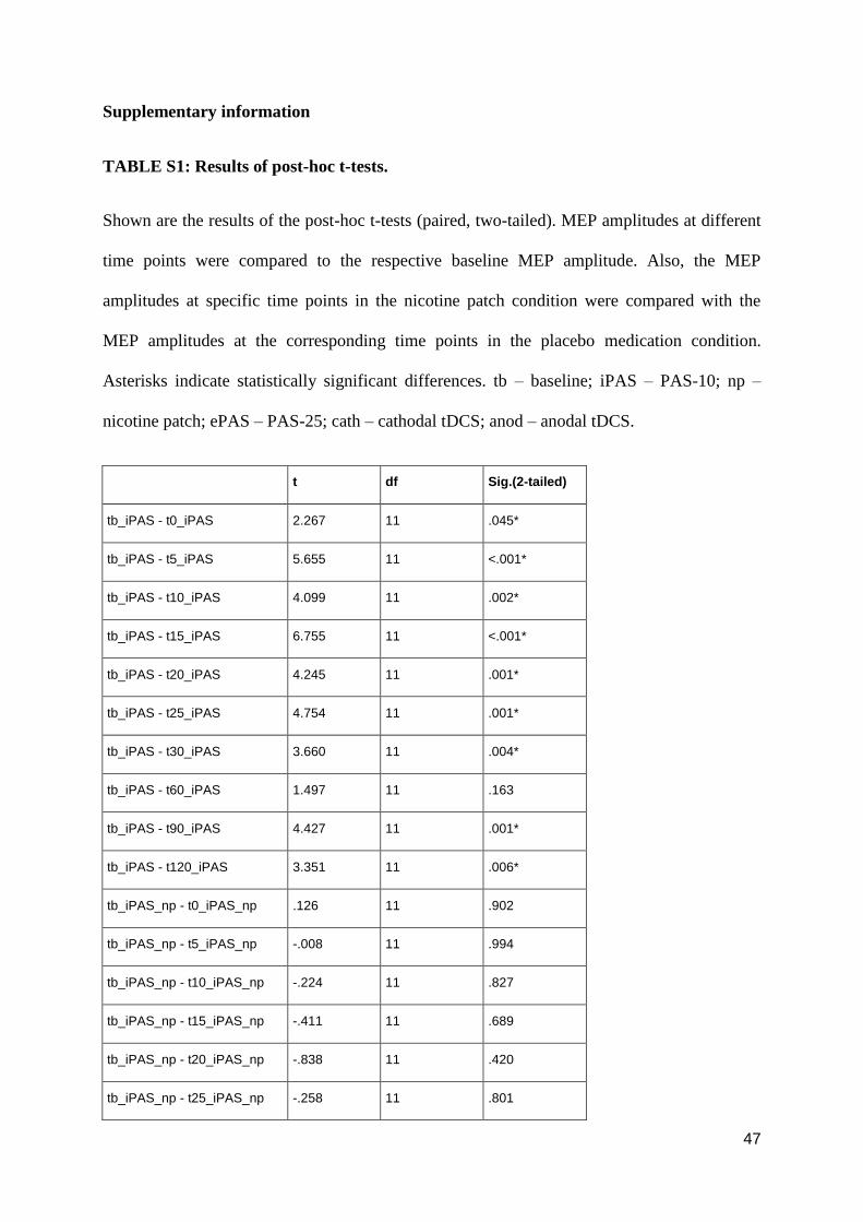
47
Supplementary information
TABLE S1: Results of post-hoc t-tests.
Shown are the results of the post-hoc t-tests (paired, two-tailed). MEP amplitudes at different
time points were compared to the respective baseline MEP amplitude. Also, the MEP
amplitudes at specific time points in the nicotine patch condition were compared with the
MEP amplitudes at the corresponding time points in the placebo medication condition.
Asterisks indicate statistically significant differences. tb – baseline; iPAS – PAS-10; np –
nicotine patch; ePAS – PAS-25; cath – cathodal tDCS; anod – anodal tDCS.
t df Sig.(2-tailed)
tb_iPAS - t0_iPAS 2.267 11 .045*
tb_iPAS - t5_iPAS 5.655 11 <.001*
tb_iPAS - t10_iPAS 4.099 11 .002*
tb_iPAS - t15_iPAS 6.755 11 <.001*
tb_iPAS - t20_iPAS 4.245 11 .001*
tb_iPAS - t25_iPAS 4.754 11 .001*
tb_iPAS - t30_iPAS 3.660 11 .004*
tb_iPAS - t60_iPAS 1.497 11 .163
tb_iPAS - t90_iPAS 4.427 11 .001*
tb_iPAS - t120_iPAS 3.351 11 .006*
tb_iPAS_np - t0_iPAS_np .126 11 .902
tb_iPAS_np - t5_iPAS_np -.008 11 .994
tb_iPAS_np - t10_iPAS_np -.224 11 .827
tb_iPAS_np - t15_iPAS_np -.411 11 .689
tb_iPAS_np - t20_iPAS_np -.838 11 .420
tb_iPAS_np - t25_iPAS_np -.258 11 .801
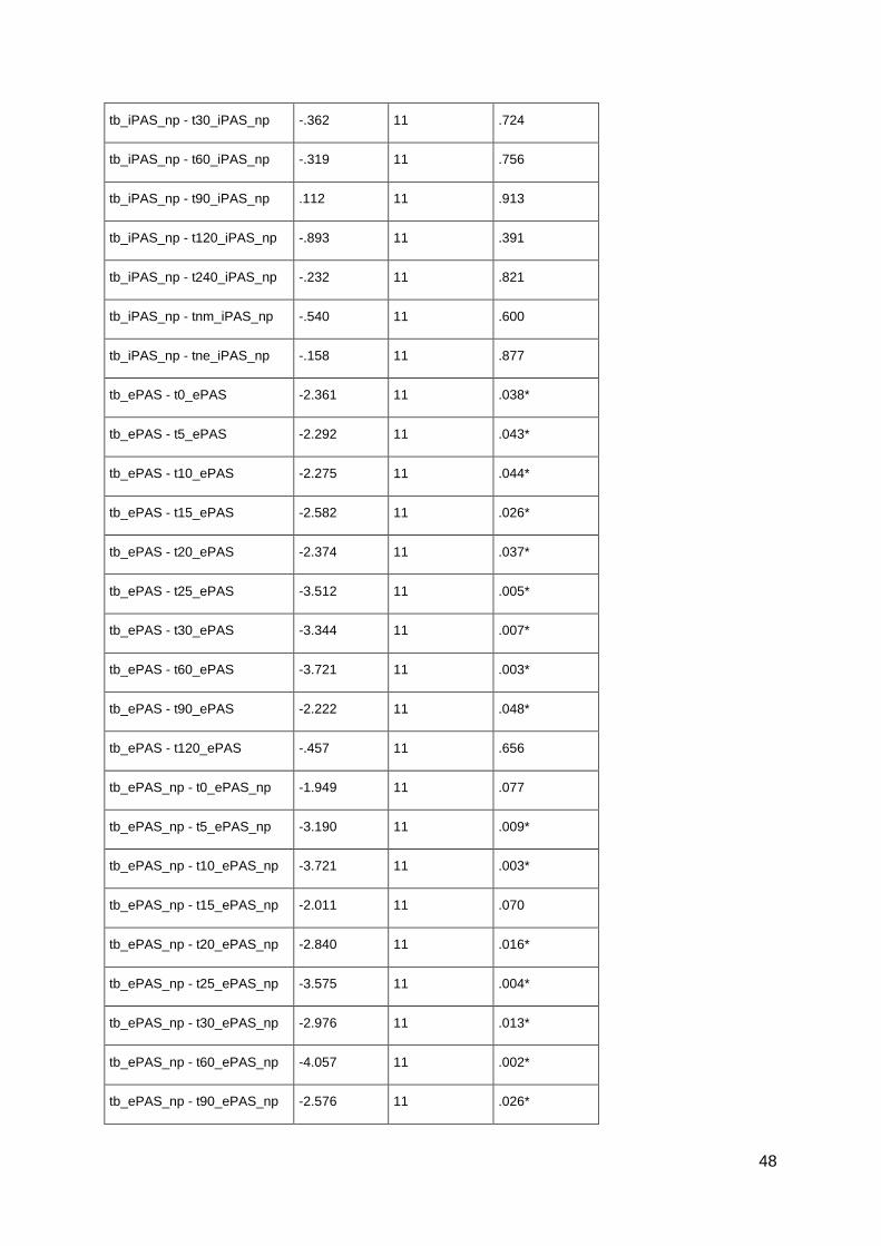
48
tb_iPAS_np - t30_iPAS_np -.362 11 .724
tb_iPAS_np - t60_iPAS_np -.319 11 .756
tb_iPAS_np - t90_iPAS_np .112 11 .913
tb_iPAS_np - t120_iPAS_np -.893 11 .391
tb_iPAS_np - t240_iPAS_np -.232 11 .821
tb_iPAS_np - tnm_iPAS_np -.540 11 .600
tb_iPAS_np - tne_iPAS_np -.158 11 .877
tb_ePAS - t0_ePAS -2.361 11 .038*
tb_ePAS - t5_ePAS -2.292 11 .043*
tb_ePAS - t10_ePAS -2.275 11 .044*
tb_ePAS - t15_ePAS -2.582 11 .026*
tb_ePAS - t20_ePAS -2.374 11 .037*
tb_ePAS - t25_ePAS -3.512 11 .005*
tb_ePAS - t30_ePAS -3.344 11 .007*
tb_ePAS - t60_ePAS -3.721 11 .003*
tb_ePAS - t90_ePAS -2.222 11 .048*
tb_ePAS - t120_ePAS -.457 11 .656
tb_ePAS_np - t0_ePAS_np -1.949 11 .077
tb_ePAS_np - t5_ePAS_np -3.190 11 .009*
tb_ePAS_np - t10_ePAS_np -3.721 11 .003*
tb_ePAS_np - t15_ePAS_np -2.011 11 .070
tb_ePAS_np - t20_ePAS_np -2.840 11 .016*
tb_ePAS_np - t25_ePAS_np -3.575 11 .004*
tb_ePAS_np - t30_ePAS_np -2.976 11 .013*
tb_ePAS_np - t60_ePAS_np -4.057 11 .002*
tb_ePAS_np - t90_ePAS_np -2.576 11 .026*
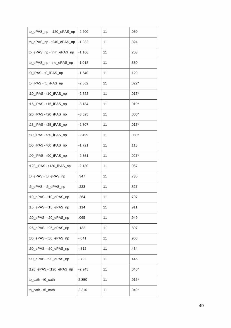
49
tb_ePAS_np - t120_ePAS_np -2.200 11 .050
tb_ePAS_np - t240_ePAS_np -1.032 11 .324
tb_ePAS_np - tnm_ePAS_np -1.166 11 .268
tb_ePAS_np - tne_ePAS_np -1.018 11 .330
t0_iPAS - t0_iPAS_np -1.640 11 .129
t5_iPAS - t5_iPAS_np -2.662 11 .022*
t10_iPAS - t10_iPAS_np -2.823 11 .017*
t15_iPAS - t15_iPAS_np -3.134 11 .010*
t20_iPAS - t20_iPAS_np -3.525 11 .005*
t25_iPAS - t25_iPAS_np -2.807 11 .017*
t30_iPAS - t30_iPAS_np -2.499 11 .030*
t60_iPAS - t60_iPAS_np -1.721 11 .113
t90_iPAS - t90_iPAS_np -2.551 11 .027*
t120_iPAS - t120_iPAS_np -2.130 11 .057
t0_ePAS - t0_ePAS_np .347 11 .735
t5_ePAS - t5_ePAS_np .223 11 .827
t10_ePAS - t10_ePAS_np .264 11 .797
t15_ePAS - t15_ePAS_np .114 11 .911
t20_ePAS - t20_ePAS_np .065 11 .949
t25_ePAS - t25_ePAS_np .132 11 .897
t30_ePAS - t30_ePAS_np -.041 11 .968
t60_ePAS - t60_ePAS_np -.812 11 .434
t90_ePAS - t90_ePAS_np -.792 11 .445
t120_ePAS - t120_ePAS_np -2.245 11 .046*
tb_cath - t0_cath 2.850 11 .016*
tb_cath - t5_cath 2.210 11 .049*

50
tb_cath - t10_cath 2.113 11 .058
tb_cath - t15_cath 2.255 11 .045*
tb_cath - t20_cath 4.090 11 .002*
tb_cath - t25_cath 1.695 11 .118
tb_cath - t30_cath 2.507 11 .029*
tb_cath - t60_cath 5.315 11 <.001*
tb_cath - t90_cath 4.127 11 .002*
tb_cath - t120_cath 1.031 11 .325
tb_cath_np - t0_cath_np -.625 11 .545
tb_cath_np - t5_cath_np .545 11 .597
tb_cath_np - t10_cath_np -.004 11 .997
tb_cath_np - t15_cath_np -.163 11 .874
tb_cath_np - t20_cath_np .271 11 .791
tb_cath_np - t25_cath_np .613 11 .553
tb_cath_np - t30_cath_np -.093 11 .927
tb_cath_np - t60_cath_np .691 11 .504
tb_cath_np - t90_cath_np 2.021 11 .068
tb_cath_np - t120_cath_np 1.708 11 .116
tb_cath_np - t240_cath_np .889 11 .393
tb_cath_np - tnm_cath_np .254 11 .804
tb_cath_np - tne_cath_np .543 11 .598
tb_anod - t0_anod -2.120 11 .058
tb_anod - t5_anod -3.771 11 .003*
tb_anod - t10_anod -3.788 11 .003*
tb_anod - t15_anod -3.763 11 .003*
tb_anod - t20_anod -3.391 11 .006*
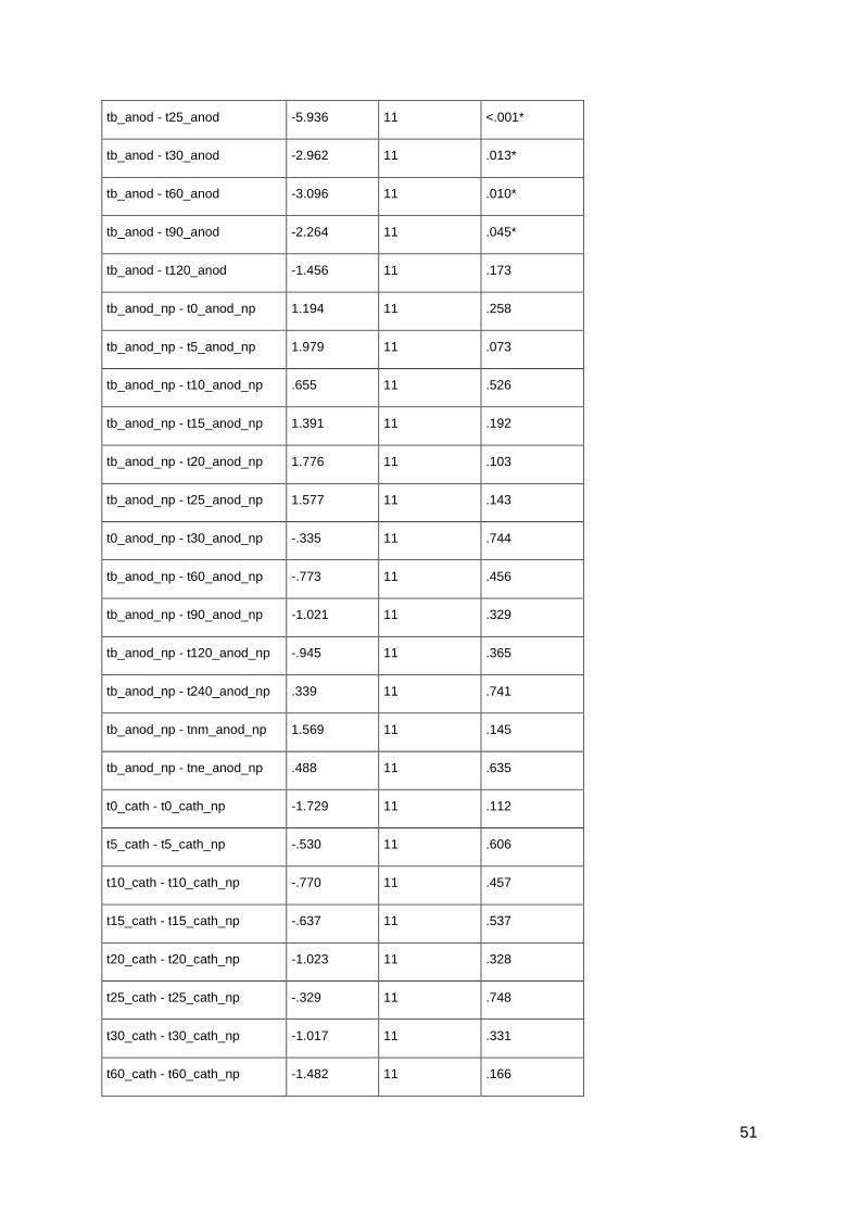
51
tb_anod - t25_anod -5.936 11 <.001*
tb_anod - t30_anod -2.962 11 .013*
tb_anod - t60_anod -3.096 11 .010*
tb_anod - t90_anod -2.264 11 .045*
tb_anod - t120_anod -1.456 11 .173
tb_anod_np - t0_anod_np 1.194 11 .258
tb_anod_np - t5_anod_np 1.979 11 .073
tb_anod_np - t10_anod_np .655 11 .526
tb_anod_np - t15_anod_np 1.391 11 .192
tb_anod_np - t20_anod_np 1.776 11 .103
tb_anod_np - t25_anod_np 1.577 11 .143
t0_anod_np - t30_anod_np -.335 11 .744
tb_anod_np - t60_anod_np -.773 11 .456
tb_anod_np - t90_anod_np -1.021 11 .329
tb_anod_np - t120_anod_np -.945 11 .365
tb_anod_np - t240_anod_np .339 11 .741
tb_anod_np - tnm_anod_np 1.569 11 .145
tb_anod_np - tne_anod_np .488 11 .635
t0_cath - t0_cath_np -1.729 11 .112
t5_cath - t5_cath_np -.530 11 .606
t10_cath - t10_cath_np -.770 11 .457
t15_cath - t15_cath_np -.637 11 .537
t20_cath - t20_cath_np -1.023 11 .328
t25_cath - t25_cath_np -.329 11 .748
t30_cath - t30_cath_np -1.017 11 .331
t60_cath - t60_cath_np -1.482 11 .166
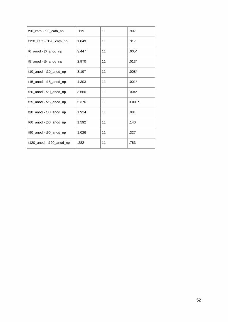
52
t90_cath - t90_cath_np .119 11 .907
t120_cath - t120_cath_np 1.049 11 .317
t0_anod - t0_anod_np 3.447 11 .005*
t5_anod - t5_anod_np 2.970 11 .013*
t10_anod - t10_anod_np 3.197 11 .008*
t15_anod - t15_anod_np 4.303 11 .001*
t20_anod - t20_anod_np 3.666 11 .004*
t25_anod - t25_anod_np 5.376 11 <.001*
t30_anod - t30_anod_np 1.924 11 .081
t60_anod - t60_anod_np 1.592 11 .140
t90_anod - t90_anod_np 1.026 11 .327
t120_anod - t120_anod_np .282 11 .783
53
2.2 Dose-dependent non-linear effect of L-DOPA on paired
associative stimulation-induced neuroplasticity in humans.
Authors: Nivethida Thirugnanasambandama, Jessica Grundey
a, Walter Paulus
a, Michael A.
Nitschea*
Affiliation: a Department of Clinical Neurophysiology, Georg-August-University Goettingen,
Robert-Koch-Strasse 40, 37075 Goettingen, Germany
ABSTRACT
Dopamine is one of the major neuromodulators in the central nervous system, which is
involved in learning and memory processes. A non-linear, inverted U-shaped dose-response
curve of its effects on cognition has been observed in animal studies. The basis for this non-
linear effect might be a similar effect of dopamine on neuroplasticity. Whereas it has been
shown that dopamine affects paired associative stimulation (PAS) –induced plasticity, which
might reflect learning-related processes to a larger degree than other non-invasive plasticity
induction protocols in the human motor cortex in principle, its dose-dependency has not been
explored so far. We studied the effect of different dosages of the dopamine precursor l-dopa
on motor cortex plasticity induced by facilitatory and inhibitory PAS of the motor cortex in
twelve healthy humans. They received 25, 100 or 200 mg l-dopa or placebo medication
combined with either excitability-enhancing or -diminishing PAS. Cortical excitability level
was monitored before and for up to two days after plasticity induction by assessment of
transcranial magnetic stimulation (TMS)-induced motor evoked potentials (MEP). Low dose
l-dopa abolished the after-effects of PAS while medium dose l-dopa prolonged facilitatory
plasticity. High dose l-dopa reversed the excitability enhancement accomplished by
facilitatory PAS to diminution. Thus the results show a clear non-linear effect of l-dopa
54
dosage on associative plasticity, different from that on non-focal plasticity. This might help to
explain dopaminergic effect on cognition and could be relevant for understanding the
pathophysiology and treatment of neuropsychiatric diseases accompanied by alterations of the
dopaminergic system.
INTRODUCTION
Dopamine is a neuromodulator that influences the functions of other neurotransmitters and
ion channels directly or indirectly (Abekawa et al., 2000; Briand et al., 2007; Grobin and
Deutch, 1998; Rétaux et al., 1991). These effects are heterogeneous, depending on neuronal
activity, dosage, sub-receptor specificity and brain region, amongst others (Seamans and
Yang, 2004). Dopamine is involved in various neuropsychiatric diseases at least partly
associated with cognitive impairment, such as Parkinson‟s disease, restless legs syndrome,
Lewy body dementia, and schizophrenia (Iversen and Iversen, 2007; Molloy et al., 2005;
Poewe et al., 2010; Trenkwalder and Paulus, 2010). In the cognitive domain, dopamine has
been shown to improve attention (Nieoullon, 2002), working memory (Durstewitz and
Seamans, 2008; Zahrt et al., 1997), learning and memory formation (Knecht et al., 2004;
Molina-Luna et al., 2009; Shohamy et al., 2005). The likely neurophysiological foundation of
the learning- and memory-modulating effects of dopamine is its effect on neuroplasticity,
namely long term- potentiation (LTP), and -depression (LTD). Dopamine influences
neuroplasticity in animals (Calabresi et al., 2007; Kung et al., 2007; Seamans and Yang,
2004) and humans (Kuo et al., 2008; Lang et al., 2008). Associative or spike-timing
dependent plasticity (STDP) is a strong candidate mechanism for memory formation (Hebb,
1949; Letzkus et al., 2007). Associative, timing-dependent plasticity can be induced in
humans by paired associative stimulation (PAS, Stefan et al., 2000) where combined
somatosensory stimulation of a peripheral nerve and the motor cortex via transcranial
magnetic stimulation (TMS) is performed. Interstimulus intervals which result in synchronous
55
activation of motor cortical neurons by somatosensory afferents and motor cortex TMS
enhance cortical excitability, whereas asynchronous stimulation diminishes it (Stefan et al.,
2000). These excitability alterations last for about an hour and are both NMDA receptor- and
calcium-dependent (Stefan et al., 2002; Wolters et al., 2003).
Interestingly, PAS-induced plasticity is absent in a hypo-dopaminergic state in patients
suffering from Parkinson´s disease off medication, but restituted by dopaminergic agents in
them (Ueki et al., 2006). Likewise, dopamine is known to enhance PAS-induced facilitatory
plasticity in healthy humans at a dosage of 100mg of the dopamine precursor levodopa (Kuo
et al., 2008). This relates well with the positive effect of this drug on learning and memory
formation (Floel et al., 2005a; Knecht et al., 2004; Shohamy et al., 2005). However, l-dopa
did not improve performance in all studies testing its effects on cognition (Ghilardi et al.,
2007; Gotham et al., 1988; Shohamy et al., 2006). This could be partly due to a non-linear
dosage-dependent effect of l-dopa on cognition, and underlying neuroplastic phenomena.
Animal studies have revealed an inverted U-shaped dose-response curve of dopamine on
cognition (Granon et al., 2000; Seamans and Yang, 2004; Williams and Goldman-Rakic,
1995; Zahrt et al., 1997). However, only dosage-dependency of dopaminergic medication on
non-focal plasticity (Monte-Silva et al., 2010) but not associative plasticity has been explored
directly so far.
In the current study, we aimed to explore such a non-linear effect of l-dopa on associative
plasticity in humans, using the human motor cortex as a model system.
MATERIALS AND METHODS
Subjects
Twelve healthy human volunteers (six females; age, 29.67 ± 8.04 years) participated in the
study. All of them were right-handed according to the Edinburgh handedness inventory
56
(Oldfield, 1971). The eligible age of subjects to participate in the study was between 18 and
65 years. The subjects did not have any metallic implant in the body or any history of a
neurological/psychiatric or chronic or acute medical disease. They took no acute/chronic
medication during or up to two weeks before participating in the study. Pregnancy was ruled
out in female subjects. All participants signed an informed consent form before participating
in the experiment. The experiment conforms to the guidelines stated in the Declaration of
Helsinki and was approved by the local Ethics Committee.
Monitoring motor cortex excitability
Motor cortex excitability was monitored by the peak to peak amplitudes of motor evoked
potentials (MEP) generated by transcranial magnetic stimulation (TMS). Single magnetic
pulses were delivered from a Magstim 200 stimulator (Magstim Company, Whitland, Dyfed,
UK) at a frequency of 0.25 Hz. A figure-of-eight coil (diameter of one winding 70mm; peak
magnetic field 2.2T) was held tangentially on the scalp at an angle of 45º to the mid-sagittal
plane with the handle pointing laterally and posteriorly to deliver the pulses. This coil
position induces a postero-anterior directed current flow in the brain. The exact point for TMS
was the motor cortex representation of the right abductor digiti minimi (ADM) muscle. This
„motor hot spot‟, as determined by TMS, was defined as the point where a magnetic stimulus
of constant, slightly suprathreshold intensity elicited consistently MEPs of the highest
amplitude. Surface electromyography (EMG) electrodes (Ag-AgCl) were placed over the
right ADM in a belly-tendon montage for recording the MEPs. The signals from the EMG
electrodes were amplified (gain = 1000), band-pass filtered (2Hz – 2KHz), digitized at a
frequency of 5KHz and stored in a laboratory computer for later offline analysis by Signal
software and CED 1401 hardware (Cambridge Electronic Design, Cambridge, UK). The
intensity of the magnetic stimulus required to elicit approximately 1mV MEP amplitudes
(SI1mV) was determined. At this intensity, 25-30 MEPs were recorded before and at several
57
time points after the intervention. The change in the mean MEP amplitude over time was
considered as measure of cortical excitability alterations caused by the intervention. Table 1
shows the mean values of the basic neurophysiological parameters measured during the
experiment.
Pharmacological intervention
The subjects received low dose (25mg), medium dose (100mg) or high dose (200mg) l-dopa
in combination with the dopamine decarboxylase inhibitor benserazide (one-fourth the dose of
l-dopa) or a placebo medication at each experimental session. One hour before the intake of
this medication, the subjects received a 20mg oral tablet of domperidone in order to
counteract the systemic side effects of l-dopa. For the high dose sessions, the subjects were
asked to take 20mg domperidone orally three times daily for two days before the experiment.
The rationale for using the above-mentioned dosages of l-dopa is that these had prominent
non-linear effects on another plasticity induction protocol in a recently published study
(Monte-Silva et al., 2010).
Paired Associative Stimulation (PAS)
For PAS, an electrical pulse was delivered to the ulnar nerve at the wrist followed by a
magnetic pulse to the motor hot spot of the ADM. The intensity of the electrical pulse was
three times the sensory perceptual threshold, delivered from a Digitimer D185 multipulse
stimulator (Digitimer, Welwyn Garden City, UK). The magnetic pulse had an intensity which
resulted in MEP amplitudes of about 1mV (SI1mV). Both stimuli were separated by an
interval of either 10 or 25 milliseconds, with the peripheral nerve pulse always followed by
the TMS stimulus. These paired pulses were administered 90 times at a frequency of 0.05 Hz
for 30 minutes over the motor hot spot of the ADM. Here, the inter-stimulus interval (ISI)
determines the direction of plasticity that is induced. When the ISI is 10 milliseconds (PAS-
58
10), excitability diminution occurs, whereas an ISI of 25 milliseconds (PAS-25) induces
excitability enhancement (Stefan et al., 2000; 2002; Wolters et al., 2003). The reason for these
different effects is that in PAS –10 the somatosensory stimulus reaches the primary motor
cortex some milliseconds before the TMS stimulus (asynchronous stimulation), whereas in
case of PAS-25, both stimuli reach the motor cortex simultaneously and such synchronous
activation results in facilitation at the synapse.
Course of the experiment
The study design was single-blinded, complete crossover, and placebo-controlled. Between
the experimental sessions (eight sessions per subject), an interval of at least one week was
essential to avoid interference effects.
The participants received domperidone or equivalent placebo medication before the start of
the neurophysiological part of experiments, as outlined above. They were seated comfortably
on a reclining chair with head and arm rests, and asked to relax completely. EMG electrodes
were placed at the right ADM, and the motor cortex hotspot was determined. Both the
position of the EMG electrodes and the motor hotspot were marked with a permanent skin
marker in order to ensure their constant positioning throughout an experimental session.
SI1mV was determined and at least 25 MEPs were recorded as baseline 1 at this stimulus
intensity. Immediately after the baseline measurement, the participants received
low/medium/high dose l-dopa or placebo medication. The combination of drug dose and PAS
was given in a randomized order for all the subjects. Baseline 2 (25 MEPs) was obtained after
one hour, because at this time l-dopa has reached its maximal plasma concentration
(Crevoisier et al., 1987) and have prominent effects on brain function (Floel et al., 2005a; Kuo
et al., 2008), to reveal an influence of the medication on cortical excitability. In case of any
drug-induced MEP amplitude changes, another set of MEPs was recorded at the adjusted
59
SI1mV (baseline 3). Subsequently, either PAS-10 or PAS-25 was administered as described
above. Following PAS, 25-30 MEPs were recorded at 0, 5, 10, 15, 20, 25, 30, 60, 90 and 120
minutes for all sessions. Since the after-effects of PAS have not been reported to last for more
than 90 minutes, we recorded MEPs in the placebo condition only until 120 mins after
cessation of stimulation. Further after-measurements were conducted the evening of the same
day (se), next morning (nm), next afternoon (na), next evening (ne) and on the third day
morning (3m) for all sessions except the placebo sessions (see Figure 1).
Data analysis and statistics
Individual mean MEP amplitudes for each subject for baseline 1, 2, 3 and each time point
following intervention were calculated. The post-intervention MEP amplitudes were
normalized to the mean baseline 3. In most of the individual measurements l-dopa did not
alter baseline MEP amplitudes. In these cases, there was no need to change the S1mV and
baseline 3 was identical to baseline 2. The normalized MEP amplitudes from all subjects were
pooled together session-wise by calculating the grand average across subjects for each
condition and time point.
A repeated-measures ANOVA was performed for the normalized data. MEP amplitude served
as dependent variable. We included only the data until 120 minutes after PAS in the analysis,
which were obtained from all sessions. PAS (PAS-10/ PAS-25) and drug (low dose/ medium
dose/ high dose/ placebo) served as within-subjects factors. Mauchly‟s sphericity test was
performed and Greenhouse-Geisser correction applied when necessary. If the ANOVA
yielded significant results, we performed post-hoc comparisons using Student‟s t-test (paired,
two-tailed, p<0.05, not adjusted for multiple comparisons). Here we compared (i) the mean
MEP amplitudes at all time points after PAS versus baseline 3 and (ii) the means at a specific
time point for the various drug conditions against the placebo medication condition. Baseline

60
MEP amplitudes of all drug/PAS combinations were compared by Student´s t-tests to exclude
a priori differences between conditions, and baseline 1 and 2 MEP amplitudes to test for any
influence of the drug alone on cortical excitability.
RESULTS
All except one male subject tolerated the experimental procedures well. This subject
developed nausea and vomiting approximately 90 minutes after the intake of 200 mg l-dopa.
We had to exclude the data of this participant from the analysis because of artifacts of MEP
measures caused by insufficient relaxation. Mean absolute baseline MEP amplitudes and
percentage of maximal stimulator output to achieve baseline amplitudes of about 1 mV did
not differ significantly between sessions (Student‟s t-test, paired, two-tailed, p>0.05).
Baseline MEP amplitudes were not affected by any of the drug dosages significantly
(Student‟s paired t-test, two-tailed, p>0.05). There was no significant difference of baseline 3
between the different sessions (Student‟s paired t-test, two-tailed, p>0.05). Also, baseline 1
and baseline 3 did not differ significantly (Student‟s paired t-test, two-tailed, p>0.05) See
Table 1.
The repeated-measures ANOVA resulted in significant main effects of drug dosage (F(3,30) =
2.990; p = 0.047) and PAS (F(1,10) = 11.261; p = 0.007). There was no significant effect of
time though (F(10,100) = 0.948; p = 0.426). Two-way interactions of drug dosage X PAS
(F(3,30) = 12.182; p <0.001) and PAS X time (F(10,100) = 2.761; p = 0.005) were significant.
There was no significant interaction between drug dosage and time (F(30,300) = 1.176; p =
0.247). The three-way interaction of drug dosage X PAS X time was significant (F(30,300) =
2.245; p<0.001).
61
Dose-dependent effect of l-dopa on PAS-induced neuroplasticity
In the placebo medication condition, PAS-10 diminished excitability and PAS-25 enhanced it
until at least 30 minutes following the stimulation. Under low dose (25mg) l-dopa, the
excitability diminution induced by PAS-10 as well as the excitability enhancement induced by
PAS-25 (Figure 2A) were diminished, that is, following both PAS-10 and PAS-25, there was
no significant difference in the MEP amplitudes compared to baseline values. Furthermore,
MEP amplitudes in the low dose condition differed significantly from those in the placebo
condition at the initial time points after PAS-25 and at later time points after PAS-10.
Following medium dose (100mg) l-dopa, the excitability changes induced by both PAS-10
and PAS-25 were preserved (Figure 2B). We observed that the excitability enhancement
caused by PAS-25 was no longer significant between 60 and 120 min after PAS, but then
recovered later and remained significantly enhanced until the afternoon of the next day after
PAS, and thus for about 24h. More prominently enhanced and prolonged facilitation with 100
mg l-dopa was observed in a previous study by Kuo et al. (2008). The MEP amplitudes at
identical time points did not differ significantly between the medium dose and placebo
conditions. After intake of high dose (200mg) l-dopa, the excitability diminution induced by
PAS-10 lasted longer compared to that under placebo medication, whereas the facilitatory
after-effects of PAS-25 were converted into inhibition (Figure 2C). Such inhibition was
significant compared to the baseline until 20 minutes following the stimulation. Differences
between the MEP amplitudes in the high dose and placebo conditions were significant only
for PAS-25.
DISCUSSION
The results of the present study reveal a non-linear effect of l-dopa dosage on associative
plasticity in the human motor cortex. Whereas low dose l-dopa reduces or abolishes the after-
62
effects of both facilitatory as well as inhibitory PAS protocols, medium dosage of the drug
prolongs PAS-25-generated facilitation and preserves inhibition resulting from PAS-10. In
contrast, high dose l-dopa reverses PAS-25-induced facilitation into inhibition, and induces a
trendwise prolongation of the inhibition induced by PAS-10.
These results are similar to those of the tDCS study (Monte-Silva et al., 2010) only for the
low dose condition, but not for the medium and high doses. This is discussed in further detail
below.
Proposed mechanisms of action
For the medium l-dopa medication, the results are in principal accordance with those of a
former study with regard to the prolongation of the facilitatory after-effects of PAS. However,
there are minor differences in the magnitude of the effects, most probably caused by
interindividual variability due to the different groups of participants. The prolongation of
facilitatory plasticity by l-dopa might be primarily caused by enhanced D1 receptor activation,
because D2-like receptor block does not abolish this kind of plasticity (Nitsche et al., 2009).
Specifically the NMDA receptor-enhancing function of moderate D1 activation (Seamans and
Yang, 2004) is a likely candidate mechanism, since PAS is known to induce NMDA receptor-
dependent plasticity (Stefan et al., 2002). In accordance to both, D1-, and D2-like activation
(Monte-Silva et al., 2009; Nitsche et al., 2009), inhibitory plasticity was not enhanced or
diminished by the medium l-dopa dosage. This does however not mean that dopamine does
not affect inhibitory associative plasticity per se, as it was abolished by D2 receptor block
(Nitsche et al., 2009). It is more likely that a balanced activation of both receptors is needed
for this kind of plasticity.
The plasticity-diminishing or –abolishing effects of low dosage l-dopa medication is in
accordance with a similar effect of low dosage ropinirole, a combined D2/D3 dopaminergic

63
agonist, on PAS-induced facilitatory plasticity (Monte-Silva et al., 2009). It is thus most
probably caused by a minor activation of these receptors. The proposed mechanism of action
is a preferential activation of presynaptic autoreceptors by low dose dopaminergic activation,
which reduces dopamine release into the synaptic cleft (Schmitz et al., 2003; Yamada et al.,
1980). This mechanism of action might also explain the plasticity-abolishing effect of low-
dose l-dopa on inhibitory plasticity. However, this kind of plasticity was not affected by low
dosage ropinirole. The reason for this might be that ropinirole as a dopaminergic agonist
activates also postsynaptic D2 receptors independent from dopamine excretion, which is not
the case for l-dopa. This minor activation of postsynaptic D2 receptors might have been
sufficient to preserve inhibitory plasticity in case of low dose ropinirole application.
For high-dosage l-dopa, the conversion of the facilitatory after-effects of PAS-25 into
inhibition is different to that of high-dose ropinirole, which abolished this kind of plasticity,
when it was administered in high dosages, but did not convert it into inhibition. Thus the D1
receptor might have contributed to this effect. Here it is of importance that a high grade of D1
receptor activation in difference to low or medium D1 activation inhibits NMDA receptors
(Seamans and Yang, 2004). Such an inhibition might reduce NMDA receptor activation to a
level inducing LTD-like plasticity. The reason for this is that a low enhancement of
intracellular calcium concentration induces LTD, while a larger enhancement generates LTP
(Lisman, 2001), and NMDA receptor activity controls the amount of calcium influx.
Alternatively it might be speculated that NMDA receptors are not inhibited by the level of
dopaminergic activation accomplished by 200 mg l-dopa, but that a major enhancement of
NMDA receptor activity will result in an intracellular calcium concentration sufficiently large
to activate hyperpolarizing potassium channels (Misonou et al., 2004), which will convert
facilitatory plasticity into inhibition. For the only trendwise effect of high-dosage l-dopa
64
medication on PAS-10-generated inhibition, this might hint for a larger range of dopaminergic
activation compatible with inhibitory plasticity.
The results of the present study not only show some similarities with, but also differences
from a recently conducted study, where the effects of identical l-dopa dosages on motor
cortex plasticity induced by transcranial direct current stimulation (tDCS) were explored
(Monte-Silva et al., 2010). tDCS induces plasticity by a tonic modulation of resting
membrane potentials (Nitsche and Paulus, 2000; 2001; Nitsche et al., 2003; Nitsche et al.,
2008). The after-effects of tDCS, like those of PAS, depend on NMDA receptor and calcium
channel activity (Nitsche et al., 2003; 2004). However, in difference to PAS, plasticity
induction by tDCS is thought not to be restricted to specific synaptic subgroups because of the
relatively large electrodes which deliver the direct currents, and for the induction of after-
effects, a tonic stimulation of some minutes is needed. For low dosage of l-dopa, also
plasticity induced by tDCS was prevented. Since the induction of after-effects of tDCS,
similar to those accomplished by PAS, need dopaminergic activity (Nitsche et al., 2006), this
result is compatible with a primarily presynaptic effect of low dose l-dopa. However, for
medium l-dopa medication, tDCS-induced facilitatory after-effects were converted into
inhibition, thus mimicking the high-dosage l-dopa effects on PAS-25-induced plasticity. This
pattern of results can be explained by a larger calcium increase induced by the less selective
and tonic stimulation induced by tDCS, as compared to the more specific and phasic PAS
stimulation procedure. Thus facilitatory tDCS in concert with medium l-dopa medication
might have enhanced intracellular calcium sufficiently to activate hyperpolarizing potassium
channels. For high-dose l-dopa medication, facilitatory as well as inhibitory plasticity were
abolished, which could be explained by an NMDA receptor-inactivating effect of large D1
activation.
65
Taken together, a complex picture of dopaminergic effects on plasticity emerges. The results
of the present and other studies suggest that the effect of l-dopa on plasticity depends on its
dosage, sub-receptor specificity and the type of plasticity induced. Interestingly, a focusing
effect of l-dopa on facilitatory plasticity as revealed by decreased non-focal tDCS – induced
facilitation and preserved focal PAS – induced facilitation seems to be restricted to medium
enhancement of dopaminergic activation.
General Remarks
In the present study, we aimed to explore the impact of l-dopa on focal associative plasticity
as induced by PAS. Previous studies showed an improvement of cognitive performance in
humans under 100mg (medium dose) l-dopa administration (Floel et al., 2005a; Knecht et al.,
2004). However, some studies revealed heterogeneous effects of the drug on cognition (Cools
et al., 2001; Gotham et al., 1988; Kulisevsky et al., 2000). Based on our results, it can be
speculated that the non-uniformity of the effects of l-dopa on cognition might be partly
explained by its non-linear dose-dependent effects on plasticity. This might be relevant
because dopamine levels are altered in many neuropsychiatric conditions where cognitive
performance is impaired (Floel et al., 2005b; Liepert et al., 2008). Alterations of plasticity in
these patients might correlate with impaired cognition and restoring plasticity might probably
help to regain cognitive performance. Some limitations of the present study should be
mentioned. Since we did not measure the plasma levels of dopamine, we could not control for
inter-individual variability caused by differences in the bioavailability of the drug. However,
we would not expect a large interindividual variability, because we studied a fairly
homogenous group of subjects. Blinding might have been somewhat compromised by
different durations of the after-measures, but with the multitude of sessions (8 per subject) and
blinded PAS protocols we do not think that an expectancy effect could have been relevant.
We only studied young healthy subjects in our current study and it is important to be aware
66
that these results might not directly translate to elderly subjects or patient populations, in
which the activity of the dopaminergic system might differ. Further studies are needed to
explore the effects in these subject groups. Moreover, apart from other confounding factors
like age (Floel et al., 2008), tobacco smoking (Lang et al., 2008), genetics (Cheeran et al.,
2008), altered dopamine levels in patients should be borne in mind when brain stimulation
protocols are applied for diagnostic or therapeutic purposes.
ACKNOWLEDGEMENTS
This project was supported by the DFG, grant NI 683/6-1.
REFERENCES
Abekawa T, Ohmori T, Ito K, Koyama T (2000) D1 dopamine receptor activation reduces
extracellular glutamate and GABA concentrations in the medial prefrontal cortex. Brain Res
867(1-2):250-4.
Briand LA, Gritton H, Howe WM, Young DA, Sarter M (2007) Modulators in concert for
cognition: modulator interactions in the prefrontal cortex. Prog Neurobiol 83(2):69-91.
Calabresi P, Picconi B, Tozzi A, Di Filippo (2007) Dopamine-mediated regulation of
corticostriatal synaptic plasticity. Trends Neurosci 30(5):211-219.
Cheeran B, Talelli P, Mori F, Koch G, Suppa A, Edwards M, Houlden H, Bhatia
K,Greenwood R, Rothwell JC (2008) A common polymorphism in the brain-derived
neurotrophic factor gene (BDNF) modulates human cortical plasticity and the response to
rTMS. J Physiol 586:5717-5725.
67
Cools R, Barker RA, Sahakian BJ, Robbins TW (2001) Enhanced or impaired cognitive
function in Parkinson's disease as a function of dopaminergic medication and task demands.
Cereb Cortex 11(12):1136-43.
Crevoisier C, Hoevels B, Zürcher G, Da Prada M (1987) Bioavailability of L-dopa after
Madopar HBS administration in healthy volunteers. Eur Neurol 27 Suppl 1:36-46.
Durstewitz D, Seamans JK (2008) The dual-state theory of prefrontal cortex dopamine
function with relevance to catechol-o-methyltransferase genotypes and schizophrenia. Biol
Psychiatry 64(9):739-49.
Floel A, Breitenstein C, Hummel F, Celnik P, Gingert C, Sawaki L, Knecht S, Cohen LG
(2005a) Dopaminergic influences on formation of a motor memory. Ann Neurol 58:121-130.
Floel A, Hummel F, Breitenstein C, Knecht S, Cohen LG (2005b) Dopaminergic effects on
encoding of a motor memory in chronic stroke. Neurology 65(3):472-4.
Floel A, Vomhof P, Lorenzen A, Roesser N, Breitenstein C, Knecht S (2008) Levodopa
improves skilled hand functions in the elderly. Eur J Neurosci 27(5):1301-1307.
Ghilardi MF, Feigin AS, Battaglia F, Silvestri G, Mattis P, Eidelberg D, Di Rocco A (2007)
L-Dopa infusion does not improve explicit sequence learning in Parkinson's disease.
Parkinsonism Relat Disord 13(3):146-51.
Gotham AM, Brown RG, Marsden CD (1988) 'Frontal' cognitive function in patients with
Parkinson's disease 'on' and 'off' levodopa. Brain 111 ( Pt 2):299-321.
Granon S, Passetti F, Thomas KL, Dalley JW, Everitt BJ, Robbins TW (2000) Enhanced and
impaired attentional performance after infusion of D1 dopaminergic receptor agents into rat
prefrontal cortex. J Neurosci 20(3):1208-15.
68
Grobin AC, Deutch AY (1998) Dopaminergic regulation of extracellular gamma-
aminobutyric acid levels in the prefrontal cortex of the rat. J Pharmacol Exp Ther 285(1):350-
357.
Hebb DO. (1949) The organization of behavior. New York: Wiley
Iversen SD, Iversen LL (2007) Dopamine: 50 years in perspective. Trends Neurosci
30(5):188-193.
Knecht S, Breitenstein C, Bushuven S, Wailke S, Kamping S, Floel A, Zwitserlood P,
Ringelstein EB (2004) Levodopa: faster and better word learning in normal humans. Ann
Neurol 56:20–26.
Kulisevsky J (2000) Role of dopamine in learning and memory: implications for the treatment
of cognitive dysfunction in patients with Parkinson's disease. Drugs Aging 16(5):365-79.
Kung VW, Hassam R, Morton AJ, Jones S (2007) Dopamine-dependent long term
potentiation in the dorsal striatum is reduced in the R6/2 mouse model of Huntington's
disease. Neuroscience 146(4):1571-1580.
Kuo MF, Paulus W, Nitsche MA (2008) Boosting focally-induced brain plasticity by
dopamine. Cereb Cortex 18(3):648-651.
Lang N, Hasan A, Sueske E, Paulus W, Nitsche MA (2008) Cortical hypoexcitability in
chronic smokers? A transcranial magnetic stimulation study. Neuropsychopharmacology
33(10):2517-2523.
Lang N, Speck S, Harms J, Rothkegel H, Paulus W, Sommer M (2008) Dopaminergic
Potentiation of rTMS-Induced Motor Cortex Inhibition. Biol Psychiatry 63(2):231-233.
69
Letzkus JJ, Kampa BM, Stuart GJ. (2007) Does spike timing-dependent synaptic plasticity
underlie memory formation? Clin Exp Pharmacol Physiol 34(10):1070-6.
Liepert J (2008) Pharmacotherapy in restorative neurology. Curr Opin Neurol 21(6):639-643.
Lisman JE (2001) Three Ca2+ levels affect plasticity differently: the LTP zone, the LTD zone
and no man's land. J Physiol 532(Pt 2):285.
Misonou H, Mohapatra DP, Park EW, Leung V, Zhen D, Misonou K, Anderson AE, Trimmer
JS (2004) Regulation of ion channel localization and phosphorylation by neuronal activity.
Nat Neurosci 7(7):711-718.
Molina-Luna K, Pekanovic A, Röhrich S, Hertler B, Schubring-Giese M, Rioult-Pedotti MS,
Luft AR (2009) Dopamine in motor cortex is necessary for skill learning and synaptic
plasticity. PLoS One 4(9):e7082.
Molloy S, McKeith IG, O'Brien JT, Burn DJ (2005) The role of levodopa in the management
of dementia with Lewy bodies. J Neurol Neurosurg Psychiatry 76(9):1200-3.
Monte-Silva K, Kuo MF, Thirugnanasambandam N, Liebetanz D, Paulus W, Nitsche MA
(2009) Dose-dependent inverted U-shaped effect of dopamine (D2-like) receptor activation on
focal and nonfocal plasticity in humans. J Neurosci 29(19):6124-31.
Monte-Silva KK, Liebetanz D, Grundey J, Paulus W, Nitsche MA (2010) Dosage-dependent
non-linear effect of l-dopa on human motor cortex plasticity. J Physiol 588(Pt 18):3415-24.
Nieoullon A. (2002) Dopamine and the regulation of cognition and attention. Prog Neurobiol
67(1):53-83.
70
Nitsche MA, Kuo MF, Grosch J, Bergner C, Monte-Silva K, Paulus W (2009) D1-receptor
impact on neuroplasticity in humans. J Neurosci 29:2648-2653.
Nitsche MA, Cohen LG, Wassermann EM, Priori A, Lang N, Antal A, Paulus W, Hummel F,
Boggio PS, Fregni F, Pascual-Leone A (2008) Transcranial direct current stimulation: State of
the art 2008. Brain Stimul 1(3):206-23.
Nitsche MA, Fricke K, Henschke U, Schlitterlau A, Liebetanz D, Lang N, Henning S, Tergau
F, Paulus W (2003) Pharmacological modulation of cortical excitability shifts induced by
transcranial direct current stimulation in humans. J Physiol 553(Pt 1):293-301.
Nitsche MA, Jaussi W, Liebetanz D, Lang N, Tergau F, Paulus W (2004) Consolidation of
human motor cortical neuroplasticity by D-cycloserine. Neuropsychopharmacology
29(8):1573-1578.
Nitsche MA, Lampe C, Antal A, Liebetanz D, Lang N, Tergau F, Paulus W (2006)
Dopaminergic modulation of long-lasting direct current induced cortical excitability changes
in the human motor cortex. Eur J Neurosci 23:1651-1657.
Nitsche MA, Nitsche MS, Klein CC, Tergau F, Rothwell JC, Paulus W (2003) Level of action
of cathodal DC polarisation induced inhibition of the human motor cortex. Clin Neurophysiol
114(4):600-604.
Nitsche MA, Paulus W (2000) Excitability changes induced in the human motor cortex by
weak transcranial direct current stimulation. J Physiol 527 (Pt 3):633-639.
Nitsche MA, Paulus W (2001) Sustained excitability elevations induced by transcranial DC
motor cortex stimulation in humans. Neurology 57(10):1899-901.
71
Poewe W, Antonini A, Zijlmans JC, Burkhard PR, Vingerhoets F (2010) Levodopa in the
treatment of Parkinson's disease: an old drug still going strong. Clin Interv Aging 5:229-38.
Rétaux S, Besson MJ, Penit-Soria J (1991) Synergism between D1 and D2 dopamine
receptors in the inhibition of the evoked release of [3H]GABA in the rat prefrontal cortex.
Neuroscience 43(2-3):323-329.
Rioult-Pedotti MS, Friedman D, Donoghue JP (2000) Learning-induced LTP in neocortex.
Science 290:533-536.
Rioult-Pedotti MS, Friedman D, Hess G, Donoghue JP (1998) Strengthening of horizontal
cortical connections following skill learning. Nat Neurosci 1(3):230-4.
Schmitz Y, Benoit-Marand M, Gonon F, Sulzer D (2003) Presynaptic regulation of
dopaminergic neurotransmission. J Neurochem 87(2):273-89.
Seamans JK, Yang CR (2004) The principal features and mechanisms of dopamine
modulation in the prefrontal cortex. Prog Neurobiol 74:1-58.
Shohamy D, Myers CE, Geghman KD, Sage J, Gluck MA (2006) L-dopa impairs learning,
but spares generalization, in Parkinson's disease. Neuropsychologia 44(5):774-84.
Shohamy D, Myers CE, Grossman S, Sage J, Gluck MA (2005) The role of dopamine in
cognitive sequence learning: evidence from Parkinson's disease. Behav Brain Res 156(2):191-
9.
Stefan K, Kunesch E, Benecke R, Cohen LG, Classen J (2002) Mechanisms of enhancement
of human motor cortex excitability induced by interventional paired associative stimulation. J
Physiol 543(Pt 2):699-708.
72
Stefan K, Kunesch E, Cohen LG, Benecke R, Classen J (2000) Induction of plasticity in the
human motor cortex by paired associative stimulation. Brain 123 (Pt 3):572-584.
Trenkwalder C, Paulus W (2010) Restless legs syndrome: pathophysiology, clinical
presentation and management. Nat Rev Neurol 6(6):337-46.
Ueki Y, Mima T, Kotb MA, Sawada H, Saiki H, Ikeda A, Begum T, Reza F, Nagamine T,
Fukuyama H (2006) Altered plasticity of the human motor cortex in Parkinson's disease. Ann
Neurol 59(1):60-71.
Williams GV, Goldman-Rakic PS (1995) Modulation of memory fields by dopamine D1
receptors in prefrontal cortex. Nature 376(6541): 572-575.
Wolters A, Sandbrink F, Schlottmann A, Kunesch E, Stefan K, Cohen LG, Benecke R,
Classen J (2003) A temporally asymmetric Hebbian rule governing plasticity in the human
motor cortex. J Neurophysiol 89(5):2339-2345.
Yamada K, Furukawa T (1980) Direct evidence for involvement of dopaminergic inhibition
and cholinergic activation in yawning. Psychopharmacology (Berl) 67(1):39-43.
Zahrt J, Taylor JR, Mathew RG, Arnsten AF (1997) Supranormal stimulation of D1 dopamine
receptors in the rodent prefrontal cortex impairs spatial working memory performance. J
Neurosci 17(21):8528-35.
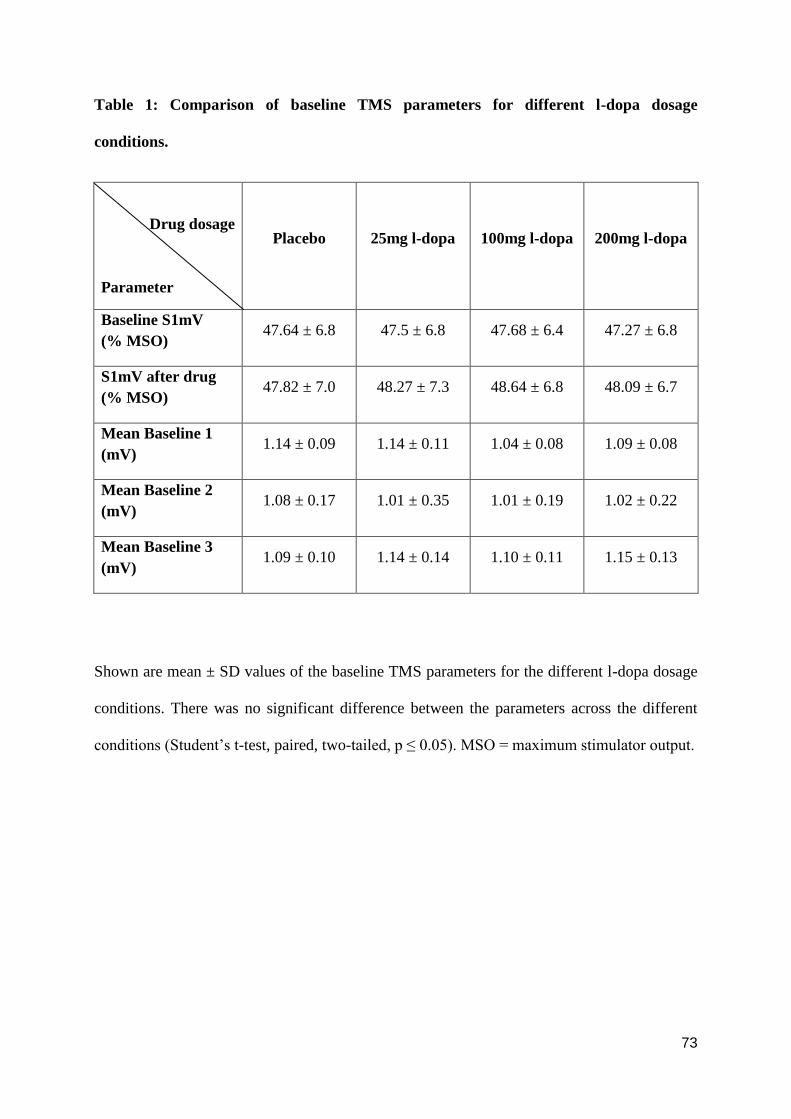
73
Table 1: Comparison of baseline TMS parameters for different l-dopa dosage
conditions.
Drug dosage
Parameter
Placebo 25mg l-dopa 100mg l-dopa 200mg l-dopa
Baseline S1mV
(% MSO) 47.64 ± 6.8 47.5 ± 6.8 47.68 ± 6.4 47.27 ± 6.8
S1mV after drug
(% MSO) 47.82 ± 7.0 48.27 ± 7.3 48.64 ± 6.8 48.09 ± 6.7
Mean Baseline 1
(mV) 1.14 ± 0.09 1.14 ± 0.11 1.04 ± 0.08 1.09 ± 0.08
Mean Baseline 2
(mV) 1.08 ± 0.17 1.01 ± 0.35 1.01 ± 0.19 1.02 ± 0.22
Mean Baseline 3
(mV) 1.09 ± 0.10 1.14 ± 0.14 1.10 ± 0.11 1.15 ± 0.13
Shown are mean ± SD values of the baseline TMS parameters for the different l-dopa dosage
conditions. There was no significant difference between the parameters across the different
conditions (Student‟s t-test, paired, two-tailed, p ≤ 0.05). MSO = maximum stimulator output.
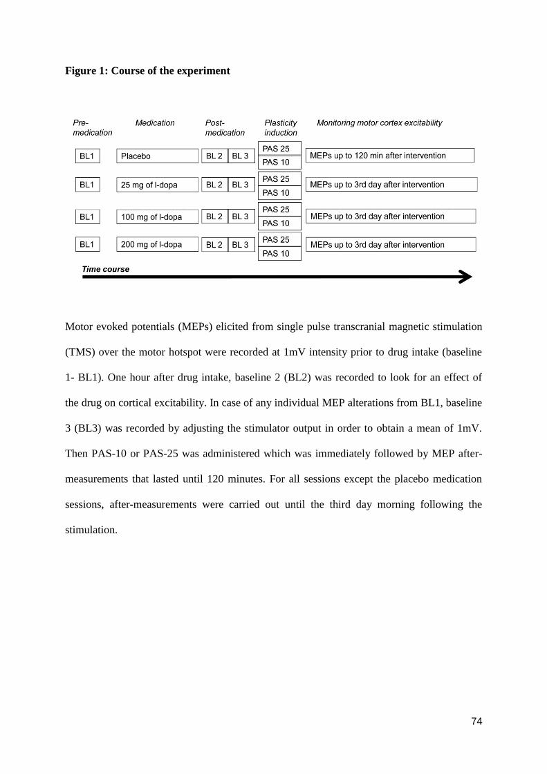
74
Figure 1: Course of the experiment
Motor evoked potentials (MEPs) elicited from single pulse transcranial magnetic stimulation
(TMS) over the motor hotspot were recorded at 1mV intensity prior to drug intake (baseline
1- BL1). One hour after drug intake, baseline 2 (BL2) was recorded to look for an effect of
the drug on cortical excitability. In case of any individual MEP alterations from BL1, baseline
3 (BL3) was recorded by adjusting the stimulator output in order to obtain a mean of 1mV.
Then PAS-10 or PAS-25 was administered which was immediately followed by MEP after-
measurements that lasted until 120 minutes. For all sessions except the placebo medication
sessions, after-measurements were carried out until the third day morning following the
stimulation.
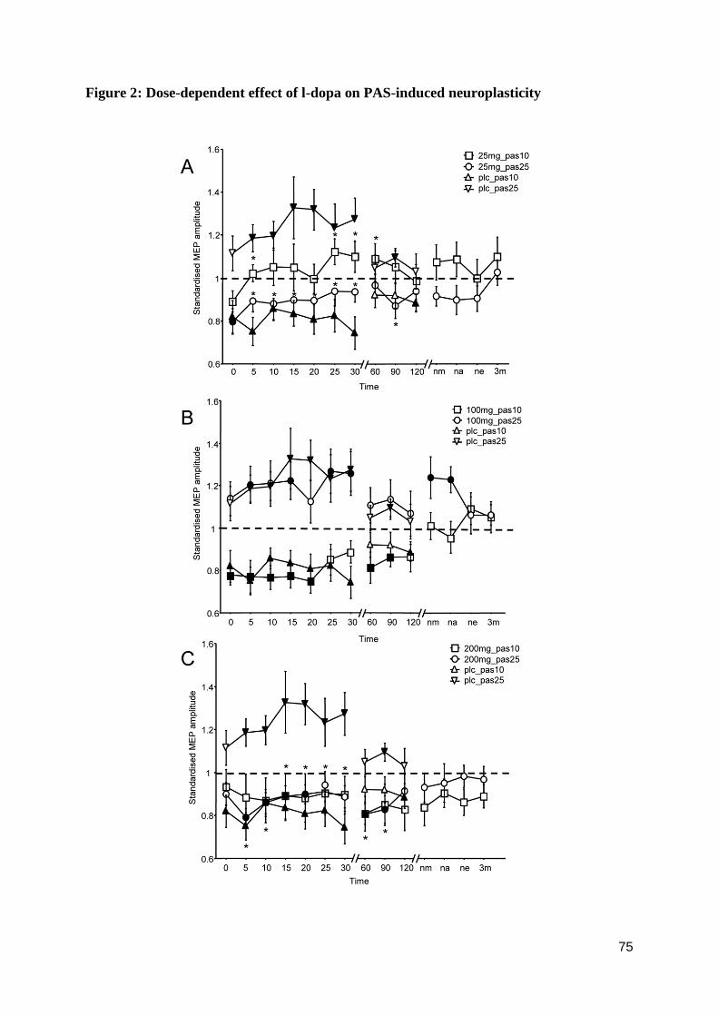
75
Figure 2: Dose-dependent effect of l-dopa on PAS-induced neuroplasticity
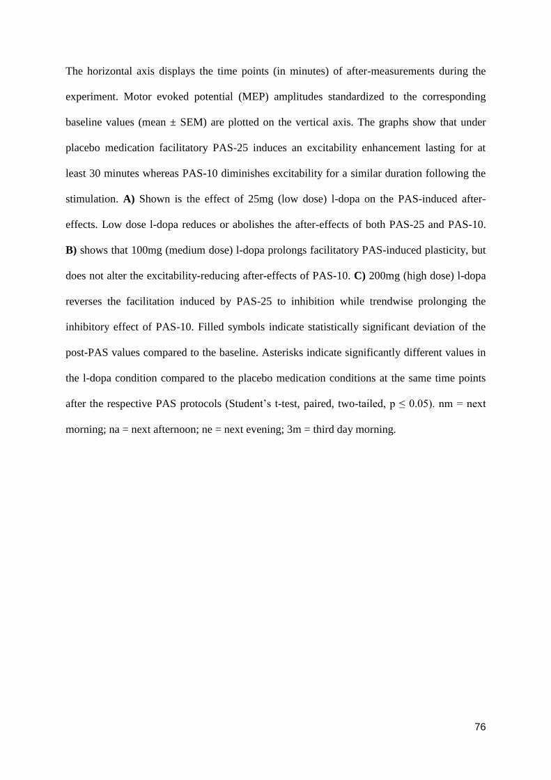
76
The horizontal axis displays the time points (in minutes) of after-measurements during the
experiment. Motor evoked potential (MEP) amplitudes standardized to the corresponding
baseline values (mean ± SEM) are plotted on the vertical axis. The graphs show that under
placebo medication facilitatory PAS-25 induces an excitability enhancement lasting for at
least 30 minutes whereas PAS-10 diminishes excitability for a similar duration following the
stimulation. A) Shown is the effect of 25mg (low dose) l-dopa on the PAS-induced after-
effects. Low dose l-dopa reduces or abolishes the after-effects of both PAS-25 and PAS-10.
B) shows that 100mg (medium dose) l-dopa prolongs facilitatory PAS-induced plasticity, but
does not alter the excitability-reducing after-effects of PAS-10. C) 200mg (high dose) l-dopa
reverses the facilitation induced by PAS-25 to inhibition while trendwise prolonging the
inhibitory effect of PAS-10. Filled symbols indicate statistically significant deviation of the
post-PAS values compared to the baseline. Asterisks indicate significantly different values in
the l-dopa condition compared to the placebo medication conditions at the same time points
after the respective PAS protocols (Student‟s t-test, paired, two-tailed, p ≤ 0.05). nm = next
morning; na = next afternoon; ne = next evening; 3m = third day morning.
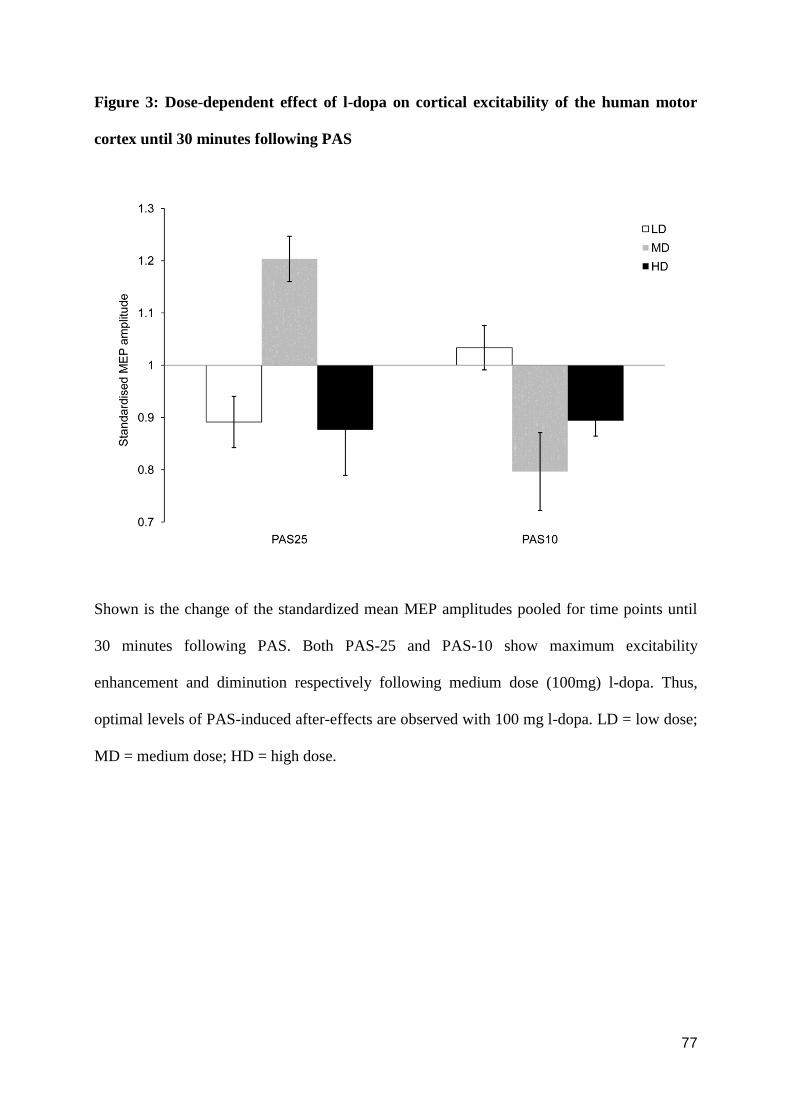
77
Figure 3: Dose-dependent effect of l-dopa on cortical excitability of the human motor
cortex until 30 minutes following PAS
Shown is the change of the standardized mean MEP amplitudes pooled for time points until
30 minutes following PAS. Both PAS-25 and PAS-10 show maximum excitability
enhancement and diminution respectively following medium dose (100mg) l-dopa. Thus,
optimal levels of PAS-induced after-effects are observed with 100 mg l-dopa. LD = low dose;
MD = medium dose; HD = high dose.
78
Chapter 3 Discussion
3.1 Summary of findings
In the aforementioned studies, we explored specific aspects of the cholinergic and
dopaminergic impact on stimulation-induced plasticity in healthy humans. The results show
(a) a prominent impact of the nicotinic subreceptor on plasticity, and (b) a dose-dependent
effect of dopaminergic activation on associative plasticity.
The first study on nicotinergic modulation of plasticity shows clearly that nicotine
exposure in non-smoking healthy individuals has a focusing effect on facilitatory plasticity. It
abolished non-focal faciltitatory plasticity induced anodal tDCS, but prolonged PAS-induced
facilitation. Nicotine exposure, on the other hand, abolished both kinds of inhibitory plasticity
that were explored in the current study, i.e. focal and non-focal plasticity. Thus, although the
nicotinergic impact on facilitatory plasticity resembles that of non-specific cholinergic
activation, its effect on inhibitory plasticity differs clearly. These effects of nicotine on
plasticity might contribute to the effect of the substance on cognition and substance addiction.
However, direct evidence for this association has not been obtained so far and should be
explored in future studies.
In the second study we explored the dose-dependency of dopaminergic activation on
associative neuroplasticity in healthy humans. The results showed that with low dose l-dopa,
both focal facilitatory and inhibitory plasticity are abolished. With medium dose l-dopa focal
facilitatory and inhibitory plasticity were preserved/prolonged. Administration of high dose l-
dopa reversed the focal facilitatory after-effects to inhibition while prolonging inhibitory
plasticity. Thus the results of this study show a clear dose-dependent non-linear effect of
dopamine on associative plasticity. Interestingly, especially the effects of medium dosed l-
79
dopa on PAS differ clearly from those obtained by tDCS, favoring a focusing effect of this
dosage with regard to facilitatory plasticity. Future studies have to show if these effects on
plasticity correlate with learning and memory performance, and thus might explain the
partially seemingly contradictory effects of l-dopa on these functions.
3.2 Conclusions
From the results of these studies, we conclude that –
(i) Neuromodulators clearly influence plasticity induced by non-invasive brain stimulation
protocols.
(ii) Subreceptor specificity and concentration levels of neuromodulators are important factors
that determine the impact of neuromodulators on plasticity.
(iii) Nicotinergic impact on facilitatory neuroplasticity is similar to the impact of non-specific
cholinergic activation, that is, both show a focusing effect with regard to facilitatory
plasticity.
(iv) Nicotinergic activation abolishes all kinds of inhibitory plasticity, different from the
effect of non-specific cholinergic activation.
(v) L-dopa only at medium dosage (100mg) produces a focusing effect on facilitatory
plasticity.
(vi) Future studies have to show if these effects of nicotinic and dopaminergic activation
correlate with the impact of the respective substances on learning and memory formation.
(vii) Interactions of drug- or disease-related alterations of neuromodulator activity with the
after-effects of brain stimulation protocols should be considered carefully before applying
80
plasticity-altering protocols to neuropsychiatric patients for therapeutic purposes, because
the effects of brain stimulation might critically depend on neuromodulator level.
3.3 Future prospects
Our study exploring the impact of nicotinic receptor activation on neuroplasticity in
humans gives clear evidence for the relevance of this receptor with regard to this mechanism.
Future studies should explore the mechanisms of action of these effects in larger detail, e.g.
by pharmacological interventions, especially exploring the contribution of nicotinic
subreceptors and calcium channels, as well as using TMS to probe the impact of nicotine on
excitatory and inhibitory cortical systems, e.g. by specific TMS protocols. Furthermore it will
be interesting to explore a dosage-dependency of nicotinic receptor activation on plasticity,
which might be non-linear, similar to the effects of dopaminergic activation. Although more is
known about the impact of dopamine on plasticity in humans, especially with regard to
subreceptors and dosage-dependency, also here we need to explore the mechanisms of action
into larger detail, as mentioned above. Beyond experiments in humans, cellular, slice and in
vivo animal experiments will help to understand the mechanisms of action of both
neuromodulators on plasticity into larger detail, because they enable the exploration of
biological effects on a level not accessible for experiments in humans.
Both, dopamine and nicotine, have been shown to alter not only neuroplasticity, but
also learning and memory formation. Since neuroplasticity is thought to be an important
neurophysiological basis for cognition, it is attractive to speculate that alterations of plasticity
induced by these substances are the basis for the respective cognitive effects. Such a causal
relationship has not been established at present, but will be an important aspect of future
research.
81
Pathological alterations of neuromodulators are thought to be an important feature of
various neuropsychiatric diseases, e.g. Alzheimer´s disease, Lewy body dementia,
Schizophrenia, and others. These might affect neuroplasticity in specific ways associated with
clinical symptoms, e.g. loss of memory and learning abilities in Alzheimers disease caused by
reduced plasticity due to decline of the cholinergic system. It will be interesting to explore in
future studies if therapeutic medication in these patients improves symptoms at least partly
due to a re-establishment of plasticity. If this would be the case, plasticity induction via non-
invasive brain stimulation might turn out as a biomarker for the clinical efficacy of the
respective substances.
Taken together, we are still at the beginning of our understanding of the
neurophysiological and functional effects of neuromodulators in the human central nervous
system. However, the central involvement of these substances in various brain functions in
health and disease makes studies in this area important and promising for both, improving our
understanding of brain function, but also in developing new therapeutic strategies to treat
patients suffering from diseases involving pathological alterations of neuromodulatory
activity.
82
References
Bailey CH, Giustetto M, Huang YY, Hawkins RD, Kandel ER (2000) Is heterosynaptic
modulation essential for stabilizing Hebbian plasticity and memory? Nat. Rev. Neurosci
1:11–20.
Barker AT, Jalinous R, Freeston IL. Non-invasive magnetic stimulation of human motor
cortex. Lancet 1985; 1: 1106-7.
Bindman LJ, Lippold OCJ, Redfearn JWT (1964) The action of brief polarizing currents on
the cerebral cortex of the rat (1) during current flow and (2) in the production of long-
lasting after-effects. J. Physiol 172: 369–382.
Bliss TV, Collingridge GL. (1993) A synaptic model of memory: long-term potentiation in
the hippocampus. Nature 361(6407):31-9.
Blitzer RD, Gil O, Landau EM (1990) Cholinergic stimulation enhances long-term
potentiation in the CA1 region of rat hippocampus. Neurosci Lett 119:207-210.
Boller F (2004) Rational basis of rehabilitation following cerebral lesions: a review of the
concept of cerebral plasticity. Funct Neurol 19:65-72.
Brozoski TJ, Brown RM, Rosvold HE, Goldman PS (1979) Cognitive deficit caused by
regional depletion of dopamine in prefrontal cortex of rhesus monkey. Science
205:929–932.
Bütefisch CM, Khurana V, Kopylev L, Cohen LG. (2004) Enhancing encoding of a motor
memory in the primary motor cortex by cortical stimulation. J Neurophysiol.
91(5):2110-6.
83
Bütefisch CM, Davis BC, Wise SP, Sawaki L, Kopylev L, Classen J, Cohen LG (2000)
Mechanisms of use-dependent plasticity in the human motor cortex. Proc Natl Acad Sci
U S A. 97:3661-3665.
Buonomano DV, Merzenich MM (1998) Cortical plasticity: from synapses to maps. Annu
Rev Neurosci 21:149-86.
Cheeran B, Talelli P, Mori F, Koch G, Suppa A, Edwards M, Houlden H, Bhatia K,
Greenwood R, Rothwell JC (2008) A common polymorphism in the brain-derived
neurotrophic factor gene (BDNF) modulates human cortical plasticity and the response
to rTMS. J Physiol. 586:5717-5725.
Chen Z, Ito K, Fujii S, Miura M, Furuse H, Sasaki H, Kaneko K, Kato H, Hiyakawa H (1996)
Roles of dopamine receptors in long-term depression: enhancement via D1 receptors
and inhibition via D2 receptors. Receptors Channels 4:1–8.
Citri A, Malenka RC (2008) Synaptic plasticity: multiple forms, functions, and mechanisms.
Neuropsychopharmacology 33:18-41.
Cooke SF, Bliss TV (2006) Plasticity in the human central nervous system. Brain 129:1659-
1673.
Cummings JA, Mulkey RM, Nicoll RA, Malenka RC. (1996) Ca2+ signaling requirements for
long-term depression in the hippocampus. Neuron. 16(4):825-33.
Dan Y, Poo MM (2004) Spike timing-dependent plasticity of neural circuits. Neuron
44:23-30.
84
Di Lazzaro V, Oliviero A, Profice P, Saturno E, Pilato F, Insola A, et al. Comparison of
descending volleys evoked by transcranial magnetic and electric stimulation in
conscious humans. Electroencephalogr Clin Neurophysiol 1998; 109: 397-401.
Evans MS, Viola-McCabe KE (1996) Midazolam inhibits long-term potentiation through
modulation of GABAA receptors. Neuropharmacology. 35(3):347-57.
Floel A, Breitenstein C, Hummel F, Celnik P, Gingert C, Sawaki L, Knecht S, Cohen LG
(2005a) Dopaminergic influences on formation of a motor memory. Ann Neurol.
58:121-130.
Floel A, Hummel F, Breitenstein C, Knecht S, Cohen LG (2005b) Dopaminergic effects on
encoding of a motor memory in chronic stroke. Neurology 65(3):472-4.
Fitzgerald PB, Fountain S, Daskalakis ZJ. (2006) A comprehensive review of the effects of
rTMS on motor cortical excitability and inhibition. Clin Neurophysiol; 117: 2584-96.
Froeliger B, Gilbert DG, McClernon FJ. (2009). Effects of nicotine on novelty detection and
memory recognition performance: double-blind, placebo-controlled studies of smokers
and nonsmokers. Psychopharmacology (Berl). 205(4):625-33.
Gaiarsa JL, Caillard O, Ben-Ari Y. (2002) Long-term plasticity at GABAergic and
glycinergic synapses: mechanisms and functional significance. Trends Neurosci.
25(11):564-70.
Gartside IB. ( 1968) Mechanisms of sustained increases of firing rate of neurones in the rat
cerebral cortex after polarization: role of protein synthesis. Nature. 220(5165):383-4.
85
Ghilardi MF, Feigin AS, Battaglia F, Silvestri G, Mattis P, Eidelberg D, Di Rocco A (2007)
L-Dopa infusion does not improve explicit sequence learning in Parkinson's disease.
Parkinsonism Relat Disord 13(3):146-51.
Gold PE (2003) Acetylcholine modulation of neural systems involved in learning and
memory. Neurobiol Learn Mem 80:194-210.
Goldman-Rakic PS, Muly EC 3rd, Williams GV (2000) D(1) receptors in prefrontal cells and
circuits. Brain Res Rev 31: 295-301.
Gotham AM, Brown RG, Marsden CD (1988) 'Frontal' cognitive function in patients with
Parkinson's disease 'on' and 'off' levodopa. Brain 111 ( Pt 2):299-321.
Gotti C, Clementi F, Fornari A, Gaimarri A, Guiducci S, Manfredi I, Moretti M, Pedrazzi P,
Pucci L, Zoli M. (2009). Structural and functional diversity of native brain neuronal
nicotinic receptors. Biochem Pharmacol. 78(7):703-11.
Gu Q (2002) Neuromodulatory transmitter systems in the cortex and their role in cortical
plasticity. Neuroscience 111:815-835.
Hahn B, Stolerman IP. (2002). Nicotine-induced attentional enhancement in rats: effects of
chronic exposure to nicotine. Neuropsychopharmacology. 27(5):712-722.
Hallett M. (2007) Transcranial magnetic stimulation: a primer. Neuron. 55(2):187-99.
Hasselmo ME, Barkai E. (1995). Cholinergic modulation of activitydependent synaptic
plasticity in the piriform cortex and associative memory function in a network
biophysical simulation. J Neurosci. 15:6592– 6604.
Hebb DO. (1949) The organization of behavior. New York: Wiley
86
Heishman SJ, Kleykamp BA, Singleton EG. (2010) Meta-analysis of the acute effects of
nicotine and smoking on human performance. Psychopharmacology (Berl). 210(4):453-
69.
Hodics T, Cohen LG, Cramer SC. (2006) Functional imaging of intervention effects in stroke
motor rehabilitation. Arch Phys Med Rehabil. 87(12 Suppl 2):S36-42.
Huang YZ, Edwards MJ, Rounis E, Bhatia KP, Rothwell JC. Theta burst stimulation of the
human motor cortex. Neuron 2005; 45: 201-6.
Huang YY, Simpson E, Kellendonk C, Kandel ER (2004) Genetic evidence for the
bidirectional modulation of synaptic plasticity in the prefrontal cortex by D1 receptors.
Proc. Natl. Acad. Sci. USA 101:3236– 3241.
Ishii M, Kurachi Y. (2006) Muscarinic acetylcholine receptors. Curr Pharm Des.12(28):3573-
81.
Johansen-Berg H, Scholz J, Stagg CJ. (2010) Relevance of structural brain connectivity to
learning and recovery from stroke. Front Syst Neurosci. 4:146.
Kano M. (1995) Plasticity of inhibitory synapses in the brain: a possible memory mechanism
that has been overlooked. Neurosci Res. 21(3):177-82.
Kauer JA, Malenka RC. (2007) Synaptic plasticity and addiction. Nat Rev Neurosci.
8(11):844-58.
Knecht S, Breitenstein C, Bushuven S, Wailke S, Kamping S, Floel A, Zwitserlood P,
Ringelstein EB (2004) Levodopa: faster and better word learning in normal humans.
Ann Neurol 56:20–26.
87
Kujirai T, Caramia MD, Rothwell JC, Day BL, Thompson PD, Ferbert A, et al.
(1993)Corticocortical inhibition in human motor cortex. J Physiol 471: 501-19.
Kumari V, Gray JA, ffytche DH, Mitterschiffthaler MT, Das M, Zachariah E, Vythelingum
GN, Williams SC, Simmons A, Sharma T. (2003). Cognitive effects of nicotine in
humans: an fMRI study. Neuroimage. 19(3):1002-1013.
Kung VW, Hassam R, Morton AJ, Jones S (2007) Dopamine-dependent long term
potentiation in the dorsal striatum is reduced in the R6/2 mouse model of Huntington's
disease. Neuroscience 146(4):1571-1580.
Kuo MF, Grosch J, Fregni F, Paulus W, Nitsche MA. (2007) Focusing effect of acetylcholine
on neuroplasticity in the human motor cortex. J Neurosci. 27(52):14442-7.
Kuo MF, Paulus W, Nitsche MA. (2006) Sex differences in cortical neuroplasticity in
humans. Neuroreport. 17(16):1703-7.
Kuo MF, Paulus W, Nitsche MA (2008) Boosting focally-induced brain plasticity by
dopamine. Cereb Cortex 18(3):648-651.
Letzkus JJ, Kampa BM, Stuart GJ. (2007) Does spike timing-dependent synaptic plasticity
underlie memory formation? Clin Exp Pharmacol Physiol 34(10):1070-6.
Levy CE, Nichols DS, Schmalbrock PM, Keller P, Chakeres DW. (2001) Functional MRI
evidence of cortical reorganization in upper-limb stroke hemiplegia treated with
constraint-induced movement therapy. Am J Phys Med Rehabil. 80(1):4-12
Liepert J, Miltner WH, Bauder H, Sommer M, Dettmers C, Taub E, Weiller C. (1998) Motor
cortex plasticity during constraint-induced movement therapy in stroke patients.
Neurosci Lett. 250(1):5-8.
88
Lisman J (2003) Long-term potentiation: outstanding questions and attempted synthesis.
Philos Trans R Soc Lond B Biol Sci. 358:829-42.
Malenka RC, Bear MF (2004) LTP and LTD: an embarrassment of riches. Neuron 44:5-21.
Malenka RC, Nicoll RA. (1993) NMDA-receptor-dependent synaptic plasticity: multiple
forms and mechanisms. Trends Neurosci. 16(12):521-7.
Merton PA, Morton HB (1980) Srimulation of the cerebral cortex in the intact human subject.
Nature 285: 227.
Missale C, Nash SR, Robinson SW, Jaber M, Caron MG (1998) Dopamine receptors: from
structure to function. Physiol Rev 78:189–225.
Monte-Silva K, Kuo MF, Thirugnanasambandam N, Liebetanz D, Paulus W, Nitsche MA.
(2009). Dose-dependent inverted U-shaped effect of dopamine (D2-like) receptor
activation on focal and nonfocal plasticity in humans. J Neurosci. 29(19):6124-6131.
Monte-Silva KK, Liebetanz D, Grundey J, Paulus W, Nitsche MA (2010) Dosage-dependent
non-linear effect of l-dopa on human motor cortex plasticity. J Physiol 588(Pt 18):3415-
24.
Nitsche MA, Paulus W (2000) Excitability changes induced in the human motor cortex by
weak transcranial direct current stimulation. J. Physiol 527:633–639.
Nitsche MA, Paulus W (2001) Sustained excitability elevations induced by transcranial DC
motor cortex stimulation in humans. Neurology 57:1899-1901.
Nitsche MA, Fricke K, Henschke U, Schlitterlau A, Liebetanz D, Lang N, Henning S, Tergau
F, Paulus W (2003a) Pharmacological modulation of cortical excitability shifts induced
by transcranial direct current stimulation in humans. J Physiol 553: 293-301.
89
Nitsche MA, Nitsche MS, Klein CC, Tergau F, Rothwell JC, Paulus W. (2003b). Level of
action of cathodal DC polarisation induced inhibition of the human motor cortex. Clin
Neurophysiol. 114(4):600-604.
Nitsche MA, Liebetanz D, Schlitterlau A, Henschke U, Fricke K, Frommann K, Lang N,
Henning S, Paulus W, Tergau F. (2004) GABAergic modulation of DC stimulation-
induced motor cortex excitability shifts in humans. Eur J Neurosci. 19(10):2720-6.
Nitsche MA, Kuo MF, Grosch J, Bergner C, Monte-Silva K, Paulus W (2009) D1-receptor
impact on neuroplasticity in humans. J Neurosci 29:2648-2653.
Nosten-Bertrand M, Errington ML, Murphy KP, Tokugawa Y, Barboni E, Kozlova E,
Michalovich D, Morris RG, Silver J, Stewart CL, Bliss TV, Morris RJ (1996) Normal
spatial learning despite regional inhibition of LTP in mice lacking Thy-1. Nature 379:
826–9.
O'Carroll CM, Martin SJ, Sandin J, Frenguelli B, Morris RG (2006) Dopaminergic
modulation of the persistence of one-trial hippocampus-dependent memory. Learn
Mem. 13:760-769.
Onor ML, Trevisiol M, Aguglia E. (2007) Rivastigmine in the treatment of Alzheimer's
disease: an update. Clin Interv Aging 2(1):17-32.
Otani S, Blond O, Desce JM, Crepel F (1998) Dopamine facilitates long term depression of
glutamatergic transmission in rat prefrontal cortex. Neuroscience 85:669–676.
Otani S, Daniel H, Roisin MP, Crepel F (2003) Dopaminergic modulation of long-term
synaptic plasticity in rat prefrontal neurons. Cereb Cortex 13:1251-1256.
Pantev C, Ross B, Fujioka T, Trainor LJ, Schulte M, Schulz M. (2003) Music and learning-
induced cortical plasticity. Ann N Y Acad Sci. 999:438-50.
90
Pascual-Leone A, Grafman J, Hallett M. (1994) Modulation of cortical motor output maps
during development of implicit and explicit knowledge. Science. 263(5151):1287-9.
Pascual-Leone A, Torres F. (1993) Plasticity of the sensorimotor cortex representation of the
reading finger in Braille readers. Brain. 116 ( Pt 1):39-52.
Purpura DP, McMurtry JG (1965) Intracellular activities and evoked potential changes during
polarization of motor cortex. J. Neurophysiol 28: 166–185.
Radcliffe KA, Dani JA. (1998) Nicotinic stimulation produces multiple forms of increased
glutamatergic synaptic transmission. J Neurosci. 18(18):7075-83.
Rioult-Pedotti MS, Friedman D, Donoghue JP (2000) Learning-induced LTP in neocortex.
Science 290: 533-536.
Rothwell JC. (1993) Evoked potentials, magnetic stimulation studies, and event-related
potentials. Curr Opin Neurol. 6(5):715-23.
Sarter M, Hasselmo ME, Bruno JP, Givens B (2005) Unraveling the attentional functions of
cortical cholinergic inputs: interactions between signal-driven and cognitive modulation
of signal detection. Brain Res Brain Res Rev 48:98-111.
Seamans JK, Yang CR (2004) The principal features and mechanisms of dopamine
modulation in the prefrontal cortex. Prog Neurobio l74:1-58.
Seamans JK, Floresco SB, Phillips AG (1998) D1 receptor modulation of hippocampal–
prefrontal cortical circuits integrating spatial memory with executive functions in the
rat. J. Neurosci. 18:1613–1621.
Seitz RJ. (2010) How imaging will guide rehabilitation. Curr Opin Neurol. 23(1):79-86.
91
Shen JX, Yakel JL.(2009) Nicotinic acetylcholine receptor-mediated calcium signaling in the
nervous system. Acta Pharmacol Sin. 30(6):673-80.
Shohamy D, Myers CE, Geghman KD, Sage J, Gluck MA (2006) L-dopa impairs learning,
but spares generalization, in Parkinson's disease. Neuropsychologia 44(5):774-84.
Stefan K, Kunesch E, Cohen LG, Benecke R, Classen J (2000) Induction of plasticity in the
human motor cortex by paired associative stimulation. Brain 123:572-584.
Stefan K, Kunesch E, Benecke R, Cohen LG, Classen J. (2002) Mechanisms of enhancement of
human motor cortex excitability induced by interventional paired associative stimulation. J
Physiol. 543(Pt 2):699-708.
Swayne OB, Teo JT, Greenwood RJ, Rothwell JC. (2009) The facilitatory effects of
intermittent theta burst stimulation on corticospinal excitability are enhanced by
nicotine. Clin Neurophysiol. 120(8):1610-5.
Tseng KY, O'Donnell P (2004) Dopamine-glutamate interactions controlling prefrontal
cortical pyramidal cell excitability involve multiple signaling mechanisms J Neurosci.
24:5131-5139.
Ueki Y, Mima T, Kotb MA, Sawada H, Saiki H, Ikeda A, Begum T, Reza F, Nagamine T,
Fukuyama H (2006) Altered plasticity of the human motor cortex in Parkinson's disease.
Ann Neurol. 59:60-71.
Ueno S, Tashiro T, Harada K. Localized stimulation of neural tissues in the brain by means of
a paired configuration of time-varying magnetic fields. J Appl Phys 1988; 64: 5862-
5864.
Urban NN, Henze DA, Lewis DA, Barrionuevo G (1996) Properties of LTP induction in the
CA3 region of the primate hippocampus. Learn Mem 3: 86–95.
92
Vallone D, Picetti R, Borrelli E (2000) Structure and function of dopamine receptors.
Neurosci Biobehav Rev 24:125-132.
Van Boven RW, Hamilton RH, Kauffman T, Keenan JP, Pascual-Leone A. (2000) Tactile
spatial resolution in blind braille readers. Neurology. 54(12):2230-6.
Wolters A, Sandbrink F, Schlottmann A, Kunesch E, Stefan K, Cohen LG, Benecke R,
Classen1 J (2003) A temporally ssymmetric hebbian rule governing plasticity in the
human motor cortex. J Neurophysiol 89: 2339–2345.
Zahrt J, Taylor JR, Mathew RG, Arnsten AF (1997) Supranormal stimulation of D1 dopamine
receptors in the rodent prefrontal cortex impairs spatial working memory performance. J
Neurosci. 17: 8528-8535.
Chen R, Tam A, Bütefisch C, Corwell B, Ziemann U, Rothwell JC, Cohen LG. (1998)
Intracortical inhibition and facilitation in different representations of the human motor
cortex. J Neurophysiol. 80(6):2870-81.
Ziemann U, Muellbacher W, Hallett M, Cohen LG. (2001) Modulation of practice-dependent
plasticity in human motor cortex. Brain. 124(Pt 6):1171-81.
Ziemann U (2003) Pharmacology of TMS. Suppl Clin Neurophysiol 56:226-231.
93
Acknowledgements
I owe my heartfelt thanks and sincere gratitude to Prof. Nitsche, who offered me his
constant support, guidance and encouragement as a mentor and supervisor. But for him, this
thesis would not have been possible. His enthusiastic spirit and healthy attitude towards
research has been a great inspiration to me always. I am extremely grateful to him for sharing
with me his knowledge and expertise in the field of brain stimulation. I am greatly indebted to
Prof. Paulus, for providing me the opportunity to work in his department for my PhD and for
having supported me in several ways. I also wish to thank the other members of my thesis
committee, Prof. Fuchs and Prof. Kröner-Herwig for their valuable advice and support.
My special thanks go to Prof. Antal for her support and guidance in many ways. I am
very grateful to Dr. Klara Boros who acquainted me with the stimulation techniques. I would
like to specially thank the support of my colleague, Nicole Neef, who often shared my
happiness and also tolerated my bad moods. My sincere thanks go to our Secretary, Marion
Kurze for helping me out with all the bureaucracy always with a smiling face and to Manuel
Hewitt who never hesitated to help me with troubleshooting technical problems. I would like
to acknowledge all my colleagues at the department – Vera Moliadze, Csaba Poreisz, Holger
Rothkegel, Leila Chaieb, Min-Fang Kuo and others for lending help in various situations and
sharing a pleasant working environment. I extend my thanks to all the volunteers who
participated in my experiments.
The list is incomplete without the names of my friends who made Göttingen a home
away from home – Pitchaiah Cherukuri, Mayur Vadhvani and Aniket Ghosh. I would like to
say thank you to a very special friend, Raghavendran Lakshmi Narayanan for being there with
me through the toughest of times. Last but never the least, thanks to all my teachers in the past
without whom I could not have been the person I am today.
94
Curriculum Vitae
Last name: Thirugnanasambandam
First name: Nivethida
Date of birth: 20/05/1981
Nationality: Indian
Email: [email protected]
EDUCATION
Aug 2007 – now PhD Systems Neuroscience
(Thesis title: Pharmacological alterations of neuroplasticity in
the human motor cortex induced by dopaminergic and
cholinergic agents. Supervisor: Prof.Dr. Michael Nitsche)
July 2005 – June 2007 M.Tech (Master of Technology) in Biomedical engineering,
School of Biosciences and Bioengineering, Indian Institute of
Technology Bombay, Mumbai, India
July 1998 – Dec 2003 M.B.,B.S. (Bachelor of Medicine, Bachelor of Surgery)
Jawaharlal Institute of Post-graduate Medical Education and
Research (JIPMER), Pondicherry, India
LIST OF PUBLICATIONS
Sparing R, Dafotakis M, Meister IG, Thirugnanasambandam N, Fink GR. Enhancing
language performance with non-invasive brain stimulation-A transcranial direct
current stimulation study in healthy humans. (2008) Neuropsychologia. 46(1):261-8.
Meister IG, Weier K, Staedtgen M, Buelte D, Thirugnanasambandam N, Sparing R.
95
Covert word reading induces a late response in the hand motor system of the
language dominant hemisphere. (2009) Neuroscience. 161(1):67-72.
Monte-Silva K, Kuo MF, Thirugnanasambandam N, Liebetanz D, Paulus W, Nitsche
MA. Dose-dependent inverted U-shaped effect of dopamine (D2-like) receptor
activation on focal and non-focal plasticity in humans. (2009) J Neurosci.
29(19):6124-31.
Nitsche MA, Jakoubkova M, Thirugnanasambandam N, Schmalfuss L, Hullemann S,
Sonka K, Paulus W, Trenkwalder C, Happe S. Contribution of the premotor cortex to
consolidation of motor sequence learning in humans during sleep. (2010) J.
Neurophysiol.
Antal A, Chaieb L, Moliadze V, Monte-Silva K, Poreisz C, Thirugnanasambandam N,
Nitsche MA, Shoukier M, Ludwid H, Paulus W. Brain-derived neurotrophic factor
(BDNF) gene polymorphisms shape cortical plasticity in humans. (2010) Brain
Stimulation.
Thirugnanasambandam N, Grundey J, Adam K, Drees A, Skwirba AC, Lang N, Paulus
W, Nitsche MA. Nicotinergic impact on focal and non-focal plasticity induced by
non-invasive brain stimulation in non-smoking humans. (accepted,
Neuropsychopharmacology).
Fricke C, Seeber AA, Thirugnanasambandam N, Nitsche MA, Paulus W, Rothwell JC.
Time course of the induction of homeostatic plasticity generated by repeated
transcranial direct current stimulation (tDCS) of the human motor cortex (Under
revision – J Neurophysiol.)
Thirugnanasambandam N, Paulus W, Nitsche MA. Dose-dependent effect of l-dopa on
96
focal neuroplasticity induced by paired associative stimulation in humans.
(Submitted)
Thirugnanasambandam N, Sparing R, Dafotakis M, Meister IG, Paulus W, Nitsche MA,
and Fink GR. Isometric contraction interferes with transcranial direct current
stimulation (tDCS) induced plasticity – evidence of state-dependent neuromodulation
in human motor cortex. (Submitted)
Grundey J, Thirugnanasambandam N, Adam K, Drees A, Skwirba AC, Lang N, Paulus
W, Nitsche MA. Nicotinergic impact on focal and non-focal plasticity induced by
non-invasive brain stimulation in smokers. (manuscript under preparation)
ORAL PRESENTATIONS
NWG (German Neuroscience Society) Symposium on Transcranial magnetic and
electrical stimulation, Goettingen (February 2010)
7th
ECNS/ISNIP Conference, Istanbul (September 2010) Symposium on AC/DC
induced neuroplasticity from the perspective of electrophysiological, pharmacological
and functional imaging methods.
SCHOLARSHIPS
DAAD Scholarship for the Masters' sandwich program for the year 2006-07
Göttingen, 13th
December 2010