pharmacol rev 60:358–403, 2008 printed in u.s.a...
TRANSCRIPT

Antipsychotic Drugs: Comparison in Animal Modelsof Efficacy, Neurotransmitter Regulation,
and NeuroprotectionJEFFREY A. LIEBERMAN, FRANK P. BYMASTER, HERBERT Y. MELTZER, ARIEL Y. DEUTCH, GARY E. DUNCAN,
CHRISTINE E. MARX, JUNE R. APRILLE, DONARD S. DWYER, XIN-MIN LI, SAHEBARAO P. MAHADIK, RONALD S. DUMAN,JOSEPH H. PORTER, JOSEPHINE S. MODICA-NAPOLITANO, SAMUEL S. NEWTON, AND JOHN G. CSERNANSKY
Department of Psychiatry, Columbia University College of Physicians and Surgeons and the New York State Psychiatric Institute, NewYork, New York (J.A.L.); Department of Psychiatry, Indiana University School of Medicine, Indianapolis, Indiana (F.P.B.); Division of
Psychopharmacology, Vanderbilt University Medical Center, Psychiatric Hospital at Vanderbilt, Nashville, Tennessee (H.Y.M);Departments of Psychiatry and Pharmacology, Vanderbilt University Medical Center, Nashville, Tennessee (A.Y.D.); Departments of
Psychiatry & Biology, University of North Carolina System–Chapel Hill, Chapel Hill, North Carolina (G.E.D.); Department of Psychiatryand Behavioral Sciences, Duke University Medical Center and Durham Veterans Affairs Medical Center, Durham, North Carolina
(C.E.M.); Department of Biology, Washington and Lee University, Lexington, Virginia (J.R.A.); Louisiana State University Health SciencesCenter–Shreveport, Shreveport, Louisiana (D.S.D); Department of Psychiatry and International Medical Graduate Program, University of
Manitoba, Winnipeg, Manitoba, Canada (X.-M.L.); Department of Psychiatry and Health Behavior, Medical College of Georgia andMedical Research, Veterans Affairs Medical Center, Augusta, Georgia (S.P.M.); Department of Psychiatry, Yale University School of
Medicine, New Haven, Connecticut (R.S.D., S.S.N.); Department of Psychology, Virginia Commonwealth University, Richmond, Virginia(J.H.P.); Department of Biology, Merrimack College, North Andover, Massachusetts (J.S.M.-N.); and Department of Psychiatry and
Behavioral Sciences, Northwestern University Feinberg School of Medicine, Chicago, Illinois (J.G.C.)
Abstract . . . . . . . . . . . . . . . . . . . . . . . . . . . . . . . . . . . . . . . . . . . . . . . . . . . . . . . . . . . . . . . . . . . . . . . . . . . . . . . 359I. Introduction. . . . . . . . . . . . . . . . . . . . . . . . . . . . . . . . . . . . . . . . . . . . . . . . . . . . . . . . . . . . . . . . . . . . . . . . . . . . 359
II. Pathophysiology of schizophrenia. . . . . . . . . . . . . . . . . . . . . . . . . . . . . . . . . . . . . . . . . . . . . . . . . . . . . . . . . 360A. Neurotransmitter dysregulation. . . . . . . . . . . . . . . . . . . . . . . . . . . . . . . . . . . . . . . . . . . . . . . . . . . . . . . 361
1. Dopamine. . . . . . . . . . . . . . . . . . . . . . . . . . . . . . . . . . . . . . . . . . . . . . . . . . . . . . . . . . . . . . . . . . . . . . . . 3632. GABA . . . . . . . . . . . . . . . . . . . . . . . . . . . . . . . . . . . . . . . . . . . . . . . . . . . . . . . . . . . . . . . . . . . . . . . . . . . 3633. Glutamate . . . . . . . . . . . . . . . . . . . . . . . . . . . . . . . . . . . . . . . . . . . . . . . . . . . . . . . . . . . . . . . . . . . . . . . 3644. Other—serotonin, acetylcholine, norepinephrine . . . . . . . . . . . . . . . . . . . . . . . . . . . . . . . . . . . . . 3675. Intracellular signaling cascades . . . . . . . . . . . . . . . . . . . . . . . . . . . . . . . . . . . . . . . . . . . . . . . . . . . . 367
B. Neuroanatomical pathology. . . . . . . . . . . . . . . . . . . . . . . . . . . . . . . . . . . . . . . . . . . . . . . . . . . . . . . . . . . 369C. Apoptosis and N-methyl-D-aspartate antagonist-induced neurodegeneration . . . . . . . . . . . . . . . 370D. Altered levels of neuroactive steroids . . . . . . . . . . . . . . . . . . . . . . . . . . . . . . . . . . . . . . . . . . . . . . . . . . 371E. Decreased mitochondrial function . . . . . . . . . . . . . . . . . . . . . . . . . . . . . . . . . . . . . . . . . . . . . . . . . . . . . 371F. Dysfunction of glucose metabolism . . . . . . . . . . . . . . . . . . . . . . . . . . . . . . . . . . . . . . . . . . . . . . . . . . . . 372G. Elevated levels of oxidative stress . . . . . . . . . . . . . . . . . . . . . . . . . . . . . . . . . . . . . . . . . . . . . . . . . . . . . 373H. Reduced neurotrophic factor expression . . . . . . . . . . . . . . . . . . . . . . . . . . . . . . . . . . . . . . . . . . . . . . . . 373
III. Comparison of antipsychotic drugs in animal models of antipsychotic efficacy,neurotransmitter regulation, and neuroprotection . . . . . . . . . . . . . . . . . . . . . . . . . . . . . . . . . . . . . . . . . . 374A. Traditional animal models of antipsychotic activity . . . . . . . . . . . . . . . . . . . . . . . . . . . . . . . . . . . . . 374
1. Dopamine stimulant-induced hyperactivity . . . . . . . . . . . . . . . . . . . . . . . . . . . . . . . . . . . . . . . . . . 3742. Conditioned avoidance responding. . . . . . . . . . . . . . . . . . . . . . . . . . . . . . . . . . . . . . . . . . . . . . . . . . 3743. Forelimb and hind limb retraction time (paw test) . . . . . . . . . . . . . . . . . . . . . . . . . . . . . . . . . . . 3744. Drug discrimination. . . . . . . . . . . . . . . . . . . . . . . . . . . . . . . . . . . . . . . . . . . . . . . . . . . . . . . . . . . . . . . 3745. Electrophysiology and brain activation patterns . . . . . . . . . . . . . . . . . . . . . . . . . . . . . . . . . . . . . 375
B. Neurotransmitter regulation via antipsychotic drugs . . . . . . . . . . . . . . . . . . . . . . . . . . . . . . . . . . . . 3751. Dopamine and antipsychotic drugs . . . . . . . . . . . . . . . . . . . . . . . . . . . . . . . . . . . . . . . . . . . . . . . . . 3752. GABA and antipsychotic drugs. . . . . . . . . . . . . . . . . . . . . . . . . . . . . . . . . . . . . . . . . . . . . . . . . . . . . 3763. Glutamate and antipsychotic drugs . . . . . . . . . . . . . . . . . . . . . . . . . . . . . . . . . . . . . . . . . . . . . . . . . 377
Address correspondence to: Dr. Jeffrey Lieberman, Department of Psychiatry, Columbia University College of Physicians and Surgeonsand the New York State Psychiatric Institute, 1051 Riverside Dr., Unit 4, New York, NY 10032. E-mail: [email protected]
C.E.M. is a coapplicant on a pending U.S. patent application for the use of neurosteroids to treat central nervous system disorders.This article is available online at http://pharmrev.aspetjournals.org.doi:10.1124/pr.107.00107.
0031-6997/08/6003-358–403$20.00PHARMACOLOGICAL REVIEWS Vol. 60, No. 3U.S. Government work not protected by U.S. copyright 7107/3400706Pharmacol Rev 60:358–403, 2008 Printed in U.S.A.
358
by guest on July 27, 2018D
ownloaded from
/content/suppl/2008/11/10/60.3.358.DC1.html Supplemental Material can be found at:
/content/60/4/582.full.pdfAn erratum has been published:

a. N-Methyl-D-aspartate antagonists in animal models . . . . . . . . . . . . . . . . . . . . . . . . . . . . . . . . 3784. Other—peptides and antipsychotic drugs. . . . . . . . . . . . . . . . . . . . . . . . . . . . . . . . . . . . . . . . . . . . 3795. Intracellular signaling cascades and antipsychotic drugs. . . . . . . . . . . . . . . . . . . . . . . . . . . . . . 3796. Effects of antipsychotic drugs on monoamine and amino acid neurotransmitter efflux . . . 380
a. Dopamine and norepinephrine extracellular concentrations . . . . . . . . . . . . . . . . . . . . . . . . . . 380b. Serotonin extracellular concentrations . . . . . . . . . . . . . . . . . . . . . . . . . . . . . . . . . . . . . . . . . . . . . 381c. Acetylcholine extracellular concentrations. . . . . . . . . . . . . . . . . . . . . . . . . . . . . . . . . . . . . . . . . . 381d. Glutamate and GABA extracellular concentrations. . . . . . . . . . . . . . . . . . . . . . . . . . . . . . . . . . 381
C. Neuroanatomical plasticity after treatment with antipsychotic drugs . . . . . . . . . . . . . . . . . . . . . 381D. Apoptosis and N-methyl-D-aspartic acid antagonist-induced neurodegeneration . . . . . . . . . . . . 382E. Second-generation antipsychotic drugs increase neuroactive steroids in animal models . . . . . 384F. Effects of antipsychotic drugs on mitochondria and oxidative phosphorylation . . . . . . . . . . . . . 384
1. Impaired mitochondrial function and risk for tardive dyskinesia. . . . . . . . . . . . . . . . . . . . . . . 3842. Antipsychotic drugs differentially inhibit complex I activity . . . . . . . . . . . . . . . . . . . . . . . . . . . 3853. Compensatory changes in mitochondrial function with antipsychotic drug treatment . . . . 385
G. Glucose transport and mechanism of neuroprotection . . . . . . . . . . . . . . . . . . . . . . . . . . . . . . . . . . . 3851. Antipsychotic drugs inhibit glucose transport . . . . . . . . . . . . . . . . . . . . . . . . . . . . . . . . . . . . . . . . 3862. Second-generation antipsychotic drugs promote neurite outgrowth and cell survival—role of
Akt. . . . . . . . . . . . . . . . . . . . . . . . . . . . . . . . . . . . . . . . . . . . . . . . . . . . . . . . . . . . . . . . . . . . . . . . . . . . . . 386H. Second-generation antipsychotic drugs demonstrate antioxidant properties . . . . . . . . . . . . . . . . 387I. Regulation of neurogenesis and neurotrophic factor expression . . . . . . . . . . . . . . . . . . . . . . . . . . . 387
1. Regulation of neurotrophic factor expression . . . . . . . . . . . . . . . . . . . . . . . . . . . . . . . . . . . . . . . . 3872. Regulation of neurogenesis and cell proliferation. . . . . . . . . . . . . . . . . . . . . . . . . . . . . . . . . . . . . 390
IV. Conclusions . . . . . . . . . . . . . . . . . . . . . . . . . . . . . . . . . . . . . . . . . . . . . . . . . . . . . . . . . . . . . . . . . . . . . . . . . . . . 391Acknowledgments. . . . . . . . . . . . . . . . . . . . . . . . . . . . . . . . . . . . . . . . . . . . . . . . . . . . . . . . . . . . . . . . . . . . . . . 393References . . . . . . . . . . . . . . . . . . . . . . . . . . . . . . . . . . . . . . . . . . . . . . . . . . . . . . . . . . . . . . . . . . . . . . . . . . . . . 393
Abstract——Various lines of evidence indicate thepresence of progressive pathophysiological processesoccurring within the brains of patients with schizophre-nia. By modulating chemical neurotransmission, anti-psychotic drugs may influence a variety of functionsregulating neuronal resilience and viability and havethe potential for neuroprotection. This article reviewsthe current literature describing preclinical and clinicalstudies that evaluate the efficacy of antipsychotic drugs,their mechanism of action and the potential of first- and
second-generation antipsychotic drugs to exert effectson cellular processes that may be neuroprotective inschizophrenia. The evidence to date suggests that al-though all antipsychotic drugs have the ability to reducepsychotic symptoms via D2 receptor antagonism, someantipsychotics may differ in other pharmacologicalproperties and their capacities to mitigate and possiblyreverse cellular processes that may underlie the patho-physiology of schizophrenia.
I. Introduction
Our understanding of the pathophysiology of schizo-phrenia has increased as knowledge of the molecular,cellular, and systems biology of brain function has ad-vanced. Beginning with the dopamine (DA1) hypothesis
1Abbreviations: DA, dopamine; 1H-MRS, proton magnetic resonancespectroscopy; 5-HT, serotonin, 5-hydroxytryptamine; A10, ventral teg-mental area; A9, nigrostriatal; AC, adenylyl cyclase; ACh, acetylcho-line; ALLO, allopregnanolone, 3�-hydroxy-5�-pregnan-20-one; AMPA,�-amino3-hydroxy-5-methyl-4-isoxazole propionic acid; APD, antipsy-chotic drug; BrdU, bromodeoxyuridine; ChAT, choline acetyltrans-ferase; CREB, cAMP-response element-binding protein; CSF, cerebro-spinal fluid; DAAO, D-amino acid oxidase; DARPP-32, dopamine- andan adenosine 3�,5�-monophosphate-regulated phospho-protein of 32kDa; DD, drug discrimination; DHEA, dehydroepiandrosterone; DOPA,3,4-dihydroxyphenylalanine; EAAT, excitatory amino acid transporter;EPO, erythropoietin; EPOr, erythropoietin receptor; EPS, extrapyrami-dal symptoms; EPSPs, excitatory postsynaptic potentials; ERK, extra-
cellular-regulated kinase; FGA, first-generation antipsychotic drug;FRT, forelimb retraction time; GAD, glutamic acid decarboxylase;GAT, GABA transporter; GLUT, glucose transporter; GPCR, G-protein-coupled receptor; GSK-3, glycogen synthase kinase-3; HRT,hind limb retraction time; IP3, inositol triphosphate; LY294002,2-(4-morpholinyl)-8-phenyl-1(4H)-benzopyran-4-one hydrochloride;M100907, [R-(�)-(2,3-dimethoxyphenyl)-1-[2-(4-fluorophenylethyl)]-4-piperidine-methanol]; MAP, mitogen-activated protein; MCT,monocarboxylate transporter; mGlu, metabotropic glutamate recep-tors; MK-801, dizocilpine, 5H-dibenzo[a,d]cyclohepten-5,10-imine(dizocilpine maleate); MPP�, 1-methyl-4-phenylpyridinium ion;NAAG, N-acetylaspartylglutamate; NGF, nerve growth factor,BDNF, brain-derived neurotrophic factor, FGF, fibroblast growthfactor; NMDA, N-methyl-D-aspartic acid; NRG1, neuroregulin-1;NT-3, neurotrophin-3; PCP, phencyclidine; PD98059, 2�-amino-3�-methoxyflavone; PFC, prefrontal cortex; PK, protein kinase; PLC,phospholipase C; PPI, prepulse inhibition; PV, parvalbumin; RGS,regulators of G-protein signaling; SGA, second-generation antipsy-chotic drug; SOD, superoxide dismutase; SR46349B, eplivanserin;SST, somatostatin; TD, tardive dyskinesia.
ANTIPSYCHOTIC DRUGS IN NEUROPROTECTION 359

of schizophrenia, we now have more sophisticated andpowerful ways of modeling the pathophysiology ofschizophrenia. With this enhanced capacity to conceptu-alize the disease, we have acquired the ability to exam-ine the actions of therapeutic agents at a variety oflevels and to discern any differences that may existamong them. Ultimately, in controlled clinical trials, theclinical relevance of such differences can be tested.
Although some debate exists as to whether schizo-phrenia is wholly neurodevelopmental in nature, thereis evidence supporting a progressive and possibly neu-rodegenerative process as well. “Neuroprotection” refersto therapies that help to maintain the structural integ-rity and normal functioning of the central nervous sys-tem in response to a pathological process and conse-quent neurobiological stress. Therapies that may beneuroprotective will probably encompass the mitigationand/or possible reversal of a broad range of anatomical,physiological, and molecular processes thought to under-lie the pathophysiology of schizophrenia.
There is increasing interest in understanding not onlythe manner through which antipsychotic drugs (APDs)are believed to play an important role in modulatingdysfunction in chemical neurotransmission to controlthe symptoms of schizophrenia but also their potentialrole for neuroprotection. The first-generation antipsy-chotic drugs (FGAs) treat some of the symptoms ofschizophrenia including delusions and hallucinationsbut, depending on their potency and the dose used, canhave substantial side effects, including effects on theextrapyramidal system in the form of extrapyramidalsigns (EPS) and tardive dyskinesia (TD) and hyperpro-lactinemia. The second-generation antipsychotic drugs(SGAs) also reduce the positive symptoms of schizophre-nia, but with less EPS and TD and, in general, reducedhyperprolactinemia as well. However, most SGAs tendto cause weight gain and disturbances in glucose andlipid metabolism.
Research also suggests that some of the SGAs mayhave additional therapeutic properties including cogni-tive enhancement, reduction of negative symptoms, en-hanced relapse prevention, and prevention of diseaseprogression and clinical deterioration, although theseeffects have not been consistently or definitively demon-strated. Presumably, the differential therapeutic effectsof SGAs are due to some distinct pharmacological prop-erties. Heretofore, theories of the mechanism of action ofAPDs have focused on drug effects on dopamine recep-tors and to a lesser extent on other neuroreceptors in-cluding those for serotonin (5-HT1A,2A,2C,3,6,7) and nor-epinephrine (�1,2) (Miyamoto et al., 2005).
Recently, a growing body of evidence derived fromnontraditional assays and paradigms used to studyAPDs has demonstrated that specific SGAs induce ef-fects in a range of cellular and molecular assays thatsuggest unique therapeutic targets (not shared by allSGAs and FGAs) beyond the antagonism of DA neuro-
transmission. These include in vitro and whole animalstudies, which show that some SGAs may increase orpreserve neurotrophic factor levels, neurogenesis, neu-ronal plasticity, mitochondrial biogenesis, cell energet-ics, and antioxidant defense enzymes. Furthermore,some SGAs may uniquely protect against N-methyl-D-aspartic acid (NMDA) antagonist-induced neurotoxicityand the consequent behavioral effects. Recent findings ofthe ability of specific SGAs to ameliorate the loss of graymatter in patients in the early stages of schizophreniafurther support the hypothesis of unique pharmacologi-cal properties and therapeutic benefits (Lieberman etal., 2005b; van Haren et al., 2007).
These putative properties of select SGAs have becomemore relevant in light of the increasing acceptance bythe field of a progressive pathophysiological process andpossibly neurodegenerative process coincident with (orshortly before) the onset of the illness that may underliethe clinical deterioration that occurs in many patientswith schizophrenia (Wyatt, 1991; DeLisi et al., 1997;Csernansky and Bardgett, 1998; Woods, 1998; Lieber-man, 1999). In this article we will critically review stud-ies of the effects of FGAs and SGAs on a number ofprocesses pertinent to the neurobiology and pharmaco-therapy of schizophrenia.
II. Pathophysiology of Schizophrenia
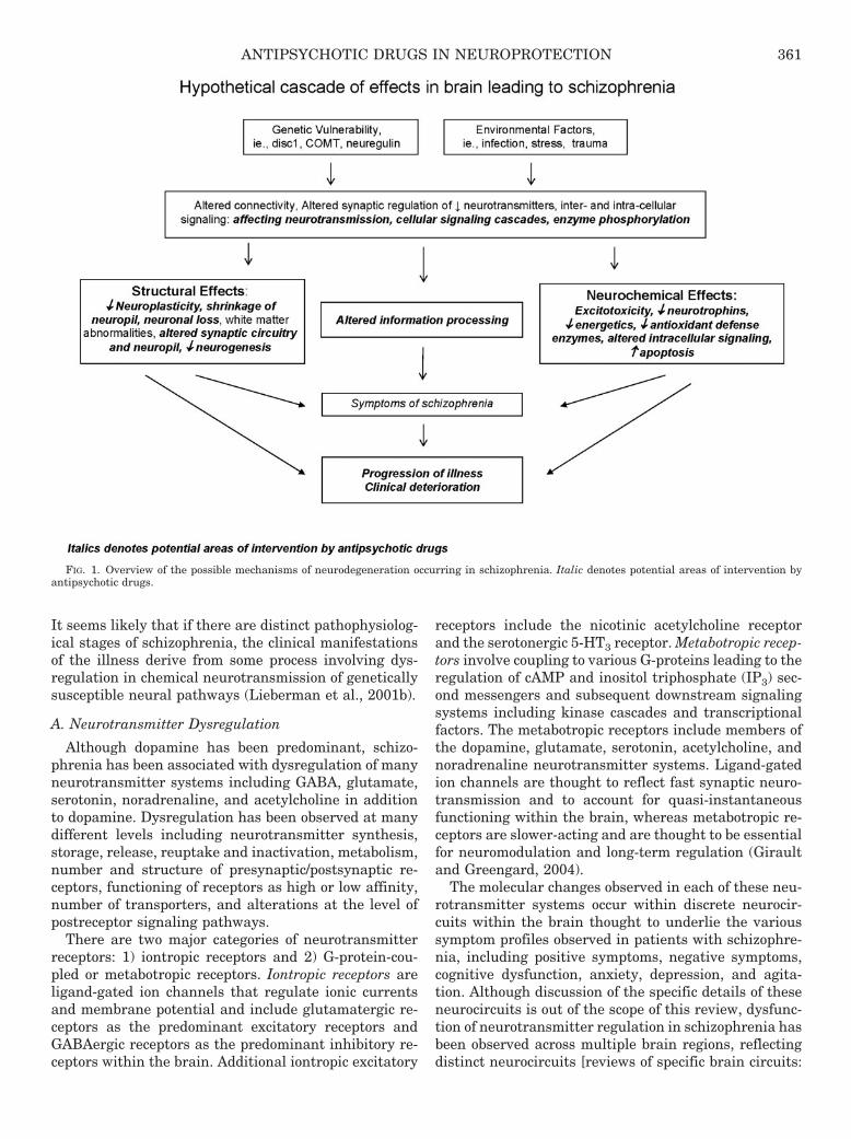
Schizophrenia has been characterized as both a neu-rodegenerative and neurodevelopmental disorder. Kra-epelin proposed in the early 1900s that schizophreniawas a degenerative disease in which a patient’s deteri-oration occurred after the onset of the illness marked bymental symptoms after what seemed to be a relativelynormal childhood. However, more recent research hasemphasized the role of genes and their effects, alongwith environmental factors, on neurodevelopment asproducing the diathesis from which schizophreniaarises. Numerous genetic association and linkage stud-ies have implicated genetic variants within many com-ponents of each neurotransmitter system in the patho-physiology of schizophrenia, although not withoutcontroversy (for review, see Riley and Kendler, 2006;Catapano and Manji, 2007; Eisener et al., 2007; Lang etal., 2007), leading to compensatory changes and alter-ations in brain development. However, it has been pro-posed that distinct pathological processes may underliethe various clinical stages of the illness with neurode-velopmental mechanisms underlying the premorbidphase of the illness and a progressive pathophysiologicalprocess beginning with neurochemical dysregulationthat can lead to neurodegeneration occurring after theformal onset of the illness and possibly beginning in itsprodromal stage (for review, see Wyatt, 1991; DeLisi etal., 1997; Csernansky and Bardgett, 1998; Woods, 1998;Lieberman, 1999; Lieberman et al., 2001b, 2006) (Fig. 1).
360 LIEBERMAN ET AL.
It seems likely that if there are distinct pathophysiolog-ical stages of schizophrenia, the clinical manifestationsof the illness derive from some process involving dys-regulation in chemical neurotransmission of geneticallysusceptible neural pathways (Lieberman et al., 2001b).
A. Neurotransmitter Dysregulation
Although dopamine has been predominant, schizo-phrenia has been associated with dysregulation of manyneurotransmitter systems including GABA, glutamate,serotonin, noradrenaline, and acetylcholine in additionto dopamine. Dysregulation has been observed at manydifferent levels including neurotransmitter synthesis,storage, release, reuptake and inactivation, metabolism,number and structure of presynaptic/postsynaptic re-ceptors, functioning of receptors as high or low affinity,number of transporters, and alterations at the level ofpostreceptor signaling pathways.
There are two major categories of neurotransmitterreceptors: 1) iontropic receptors and 2) G-protein-cou-pled or metabotropic receptors. Iontropic receptors areligand-gated ion channels that regulate ionic currentsand membrane potential and include glutamatergic re-ceptors as the predominant excitatory receptors andGABAergic receptors as the predominant inhibitory re-ceptors within the brain. Additional iontropic excitatory
receptors include the nicotinic acetylcholine receptorand the serotonergic 5-HT3 receptor. Metabotropic recep-tors involve coupling to various G-proteins leading to theregulation of cAMP and inositol triphosphate (IP3) sec-ond messengers and subsequent downstream signalingsystems including kinase cascades and transcriptionalfactors. The metabotropic receptors include members ofthe dopamine, glutamate, serotonin, acetylcholine, andnoradrenaline neurotransmitter systems. Ligand-gatedion channels are thought to reflect fast synaptic neuro-transmission and to account for quasi-instantaneousfunctioning within the brain, whereas metabotropic re-ceptors are slower-acting and are thought to be essentialfor neuromodulation and long-term regulation (Giraultand Greengard, 2004).
The molecular changes observed in each of these neu-rotransmitter systems occur within discrete neurocir-cuits within the brain thought to underlie the varioussymptom profiles observed in patients with schizophre-nia, including positive symptoms, negative symptoms,cognitive dysfunction, anxiety, depression, and agita-tion. Although discussion of the specific details of theseneurocircuits is out of the scope of this review, dysfunc-tion of neurotransmitter regulation in schizophrenia hasbeen observed across multiple brain regions, reflectingdistinct neurocircuits [reviews of specific brain circuits:
FIG. 1. Overview of the possible mechanisms of neurodegeneration occurring in schizophrenia. Italic denotes potential areas of intervention byantipsychotic drugs.
ANTIPSYCHOTIC DRUGS IN NEUROPROTECTION 361

basal ganglia-thalamo-cortical loops (Alexander et al.,1986), amygdalo-entorrhinal inputs to hippocampus(Benes and Berretta, 2000), and basal ganglia and cer-ebellar loops (Middleton and Strick, 2000); reviews ofspecific brain regions: basal ganglia (Tisch et al., 2004)and thalamus (Clinton and Meador-Woodruff, 2004)].
In addition, altered regulation at the level of mole-cules and neurocircuits is thought to underlie alter-ations in complex brain processes. In this regard, schizo-phrenia has been conceptualized as a diseasecharacterized by abnormal information processing thatoccurs within subcortical and cortical regions, includingsensory gating deficits at the level of the thalamus andaltered desynchronization of modal or supramodal cor-
tical associative functions (for review, see Braus et al.,2002). Related to the “abnormal information processing”concept, schizophrenia has been associated with abnor-malities in neural oscillations, an “emergent property” ofneural networks arising from temporal synchrony be-tween synaptic transmission and the firing of distinctneuronal populations (Ford et al., 2007).
We will focus primarily on the dopaminergic,GABAergic, and glutamatergic neurotransmitter sys-tems, although references to other systems will be made.As a review, several of the molecules involved in signal-ing cascades associated with neurotransmitter-receptorinteractions and that of other molecules are summarizedin Fig. 2.
-OH
H2O2
O2-
Beta-carotene
Vit C, E Glutathione
Oxidative Stress
Neurogenesis Mitochondrial
Pathway
Signaling
P
P
SOD, CAT,GSH-Py
Antioxidant Def Systems
Diverse functions
Neurotransmitters
GPCR GPCR
RGSs
Gi,GsAC
PKA
cAMPATP
DARPP-32
CREB
ERK Gene
Regulation
GRKs B-arrestin 2
GSK-3β
DA
“PKB”
Akt PP2A
5-HT
PKC
GABA
Cl-
NMDA
AMPA
Kainate
Na+
T
Ca2+
Ca2+
Na+
EPSP
BZ
NS
P
IPSP
EPSP
↑Ca2+
b1,2
5-HT3
nAChR
M2,4
Integrin
ERK
JNK
P38
c-fos
c-jun
Fos
Jun
AP1
Trk
Akt
PIP3
PI3K
P GSK-3β Bax
Proapoptotic
Apoptosis
Cell Proliferation
Cell Survival
transcription
Mitochondria
↑Bax/↓Bcl-2↑Bax/↓Bcl-XL
Cyt c
Apoptosome
Caspase-3
Cellular Degradation, DNA
Fragmentation
Nucleus
Caspases
↑Bax/↓Bcl-2
SOD1
Proapoptotic
P53
translation
MAPK
Homer 1b/c
PP
↑Ca2=
P DAG IP3
Gq
PI3K
Akt
mTor
PLC
Glutamate
T
Iontropic Receptors
Glutamate
mGlu1,5
T
T
DA1,5
5-HT1,4-7
GABAB
mGlu2-4,6-8
DA2,3,4
GABA
M1,3,5
ACh ACh
5-HT
T
T
NE
T
T
TT
T
Reelin
GABAA
Ca2+
Ca2+Gly/Ser
KYN Na+
NAAG
Mg2+
re
Metabotropic Receptors Glutamate
αα2
5-HT2
PV CR
SST
T
ACh
ER Stress, Death Receptor Pathways
Neurotrophinse.g. BDNF 5-HT
FIG. 2. A summary of the intracellular signaling cascades that occur within neurons and glia within the brain. Schizophrenia has been associatedwith dysregulation at a number of loci along these signaling pathways, and antipsychotic drugs may act to reverse some of the pathological changesthat have been observed. This slide provides a summary of signaling cascades that occur within neurons and glial cells in the brain that may contributeto schizophrenia, although not all of the cascades shown will be found in a given cell or pathway. The left side summarizes the excitatory and inhibitoryiontropic receptors. The top illustrates components of the two key signaling cascades associated with G-protein-coupled metabotropic receptorsincluding adenylyl cyclase and phospholipase C activation. The bottom demonstrates the apoptosis cascade and specific neurotrophic factor-receptorinteractions. The right side summarizes some of the key molecules involved in oxidative stress. AP-1, activator protein-1 complex; BZ, benzodiazepines;CAT, catalase; Cl-, chloride; CR, calretinin; Cyt c, cytochrome c; DAG, diacylglycerol; ER, endoplasmic reticulum; GRK, G-protein-coupled receptorkinases; GSH-Px, glutathione peroxidase; H2O2, hydrogen peroxide; IPSP, inhibitory postsynaptic potential; KYN, kynurenic acid; M, muscarinicacetylcholine receptors; MAPK, mitogen-activated protein kinase; mTOR, mammalian target of rapamycin; nAChR, nicotinic acetylcholine receptor;NE, norepinephrine; NS, neuroactive steroids; O2-, superoxide radical; -OH, hydroxyl anion; P, phosphorylation; PI3K, phosphatidylinositol 3-kinase;PIP3, phosphatidylinositol triphosphate; PP2A, protein phosphatase 2A; T, transporter protein; Vit, vitamin.
362 LIEBERMAN ET AL.

1. Dopamine. One of the most popular theories un-derlying the pathophysiology of schizophrenia involvesincreased dopaminergic activity within the mesolimbicdopamine system thought to underlie the positive orpsychotic symptoms of schizophrenia and decreased do-paminergic activity within the mesocortical dopaminesystem thought to reflect negative symptoms and cogni-tive dysfunction also seen in schizophrenia (for reviews,see Abi-Dargham and Moore, 2003; Guillin et al., 2007;Meisenzahl et al., 2007). In line with this theory, allcurrently available APDs reduce psychotic symptomsvia blockade of dopamine neurotransmission within thestriatal complex of the mesolimbic pathways.
Dopamine is synthesized from tyrosine to DOPA viatyrosine hydroxylase and then to dopamine via DOPAdecarboxylase, reactions occurring within two major cellgroups, substantia nigra pars compacta projecting to thestriatum and the ventral tegmental area projecting tothe ventral striatum and cerebral cortex. There are fivemetabotropic dopamine receptors divided into two majorclasses: D1-like receptors (D1 and D5) and D2-like recep-tors (D2–4). Two enzymes are responsible for the cata-bolic inactivation of dopamine, different isozymes ofmonoamine oxidase (MAO-A and MAO-B) and catechol-O-methyltransferase. In addition, dopamine releasedfrom presynaptic terminals is recaptured into presynap-tic terminals via the dopamine transporter.
All antipsychotics to date act as antagonists (or par-tial agonists) at the D2 receptor, and most show a dose-dependent threshold of D2 receptor occupancy for theirtherapeutic effects (Kapur and Mamo, 2003). Althoughindividual studies report contradictory findings, a meta-analysis of 13 in vivo studies demonstrated a 12% in-crease in D2 receptor binding in drug-naive and in drug-free patients with schizophrenia, providing limitedsupport for D2 receptor up-regulation and supersensitiv-ity in schizophrenia (Laruelle, 1998). Seeman et al.(2006) have suggested that elevations in the high-affin-ity state of dopamine D2 receptors (D2
High receptors)may reflect a common point of convergence among thevarious pathways for eliciting psychosis. They indicatedthat many of the causes of psychosis in adult humanssuch as drugs, steroids, ethanol, and brain lesions leadto dopamine supersensitivity in rats and to an increasein the high-affinity state of dopamine D2
High receptors instriata (Seeman et al., 2006). Other proposed links be-tween dopamine D2 neurotransmission and schizophre-nia include polymorphisms in the D2 receptor gene (forreview, see Lang et al., 2007) and alterations in thecomponents of the post-dopamine D2 receptor signalingcascade discussed in section II.A.5. However, becauseAPDs can up-regulate D2 receptors, the possibility of atreatment effect and drug artifact must be considered.
Whereas the D2 receptor plays a predominant role inthe current treatment of psychotic symptoms, other com-ponents of the dopamine neurotransmitter system havebeen implicated in schizophrenia. These include the D1
receptor (Abi-Dargham and Moore, 2003; Goldman-Ra-kic et al., 2004), D3 receptor (Micheli and Heidbreder,2006), D4 receptor (Kramer et al., 2007), catechol-O-methyltransferase (Kramer et al., 2007; Lewandowski,2007), dopamine transporter (Schmitt et al., 2006; Ma-teos et al., 2007), dopamine receptor-interacting pro-teins calcyon and neuronal Ca2� sensor 1 (Bergson etal., 2003), and dopamine receptor-adenosine receptorinteractions (Fuxe et al., 2007).
2. GABA. GABAergic synapses are the key inhibi-tory synapses within the brain, and decreased GABAer-gic neurotransmission has been implicated in the patho-physiology of schizophrenia (for review, see Benes andBerretta, 2001; Blum and Mann, 2002; Wassef et al.,2003; Lewis et al., 2004; Guidotti et al., 2005). It hasbeen proposed that deficits in GABAergic neurotrans-mission may result in an imbalance between excitatoryand inhibitory neurotransmission, favoring excitationand possible excitotoxicity. Olney et al. (1999) suggestedthat a developmental deficit of inhibitory GABA inter-neurons may set the stage for ongoing neurodegenera-tion through the uncontrolled activation of glutamater-gic neurons. In addition, GABAergic interneurons playan important role in regulating pyramidal neuron firingrates (McBain and Fisahn, 2001), and, as a result, re-duced GABAergic function would alter the synchronousfiring patterns of cortical neurons, which may underlieinformation-processing deficits known to be present inpatients with schizophrenia (Hajos, 2006).
GABA is synthesized from glutamate via two molecu-lar forms of glutamic acid decarboxylase (GAD67 andGAD65). GABAergic neurons (or interneurons) coexpressspecific proteins and can be classified by location withinspecific neuronal circuits based on the expression ofthese proteins—reelin, parvalbumin (PV), and calreti-nin. Reelin is an extracellular matrix protein constitu-tively released from GABAergic terminals that binds tointegrin receptors to regulate synaptic plasticity (e.g.,long-term potentiation) and protein synthesis withinneuronal dendrites and spines. PV and calretinin arecalcium-binding proteins that probably contribute to in-tracellular Ca2� signaling cascades. Three GABA recep-tors have been identified thus far: GABAA and GABACreceptors are iontropic receptors, whereas the GABABreceptor is metabotropic and coupled to a GTP-bindingprotein. The GABAA receptor is a heteropentamericstructure consisting of various subtypes composed of atleast 16 different GABAA receptor subunits—six �, four�, three �, one �, one �, and one � (Mohler et al., 1995;Whiting, 2003). The � subunits of the GABAA receptorconfer different affinities for GABA, and these subunitsshow a very specialized regional cellular and subcellulardistribution. In addition, a subset of GABAA receptorscontain a binding site for benzodiazepines. The benzodi-azepine binding site on the GABAA receptor is alloster-ically coupled to the GABA binding site, resulting inincreased receptor occupancy at low GABA concentra-
ANTIPSYCHOTIC DRUGS IN NEUROPROTECTION 363

tions that increases the frequency of channel openings(Pritchett et al., 1989). GABA neurotransmission is ter-minated via reuptake by GABA transporter proteins.
GABAergic dysfunction in schizophrenia has beencharacterized as a reduction in the availability of GABAand related proteins presynaptically and compensatoryup-regulation of GABA receptors postsynaptically. Moststudies have reported low GABA levels in at least somebrain regions in patients with schizophrenia, althoughthere is no clear consensus on the specific brain lociaffected with the exception of the amygdala. At thepresynaptic level, down-regulation of mRNA and/or pro-tein for GAD67, reelin, and PV has been observed inpostmortem brain tissues of patients with schizophre-nia. Lower levels of the GABA transporter 1 (GAT1)have also been observed, which may reflect a compensa-tory change in response to low GABA levels.
Within the cerebral cortex and hippocampus, there isevidence for fewer GABAergic interneurons, althoughthis reduction is localized primarily to cortical layer II.Several authors have suggested that the loss of thissubset of GABAergic neurons is probably not sufficientto support the reductions observed in GAD67, reelin, andGAT1 (for review, see Guidotti et al., 2005), implyingthat other mechanisms such as promoter-related down-regulation of gene expression must be involved. Recentwork has demonstrated an increase in DNA-methyl-transferase-1 expression within select GABAergic inter-neurons in postmortem schizophrenia brains that couldunderlie down-regulation of gene expression (Veldic etal., 2004, 2005). Other work focusing on the chandelierclass of GABAergic neurons that form distinctive verti-cal arrays called “cartridges” of synaptic terminals alongthe axon initial segments of pyramidal neurons found nodifferences in the relative density, laminar distribution,or size of parvalbumin-containing neurons (Lewis,2000). However, the density of GAT1-immunoreactivechandelier neuronal axon cartridges was decreased by40% in subjects with schizophrenia compared withhealthy control subjects and subjects with other psychi-atric disorders (Lewis, 2000).
At the postsynaptic level, the majority of data supportincreased expression of GABAA receptors in schizophre-nia. The numbers of GABAA receptors labeled by[3H]muscimol (which labels all GABA receptors) in theprefrontal cortex (Hanada et al., 1987; Benes et al.,1996b), superior temporal gyrus (Deng and Huang,2006), and hippocampus (Benes et al., 1996a; Benes,1997) are increased in postmortem brain tissue of pa-tients with schizophrenia. In contrast, the numbers ofGABAA receptors with benzodiazepine-binding sites la-beled by [3H]flunitrazepam are reduced or unchanged inprefrontal cortex (Pandey et al., 1997) and hippocampus(Squires et al., 1993; Benes et al., 1996a) of schizophre-nia brains. Subsequent work has demonstrated up-reg-ulation of mRNAs and proteins for �1 and �5 subunitswithin the prefrontal cortex. The �5 subunit confers a 3-
to 10-fold higher affinity for GABA than that observedfor the �1-containing receptor, suggesting increases inGABAA receptors with a higher affinity for GABA.
The data implicating the GABAB receptor in thepathophysiology of schizophrenia are more limited.There is evidence for a reduction in GABAB receptorimmunoreactivity in the entorhinal cortex and inferiortemporal cortex of the brain in schizophrenia (Mizukamiet al., 2002). In addition, baclofen, a GABAB agonist, canreverse spontaneous gating deficits in animal models ofschizophrenia (Bortolato et al., 2007).
Somatostatin (SST) is a neuropeptide present in asubpopulation of GABA neurons, and a reduction in thedensity of neurons positive for SST as well as expressionof SST mRNA per neuron is seen in dorsolateral prefron-tal cortex in schizophrenia (Morris et al., 2008). There isevidence that neuroregulin-1 (NRG1) may regulateGABAergic neurotransmission via binding to presynap-tic ErbB4 receptors (Woo et al., 2007). NRG1 is a regu-lator of neural development, and NRG1 and ErbB4 havebeen identified as susceptibility genes for schizophrenia(Britsch, 2007).
Preliminary data using real-time quantitative poly-merase chain reaction demonstrated that several ofthese molecular changes (i.e., decreased transcripts forSST, PV, GAD67, GAT1, and the �1 and � subunits ofGABAA receptors) are observed within four cortical ar-eas (dorsolateral prefrontal cortex, anterior cingulatecortex, and primary motor and visual cortices). Thisfinding suggests that a conserved set of molecular alter-ations in GABA neurotransmission may contribute tothe pathophysiology of schizophrenia (Hashimoto et al.,2008).
3. Glutamate. Glutamatergic synapses are the keyexcitatory synapses within the brain, and mechanismsof both hyperglutamatergic and hypoglutamatergicfunctioning have been implicated in the pathophysiologyof schizophrenia (for review, see Olney et al., 1999; Deut-sch et al., 2001; Coyle, 2006). It has been proposed thatNMDA receptor hypofunction may lead to excessivestimulation of other iontropic receptors, causing a cas-cade of excitotoxic events including oxidative stress andapoptosis (for review, see Deutsch et al., 2001). Dysregu-lation of glutamateric functioning has been observedacross many components of the glutamate neurotrans-mission system.
Glutamatergic receptors include both iontropic andmetabotropic receptor subtypes. The iontropic receptorsinclude NMDA, �-amino-3-hydroxy-5-methyl-4-isox-azole propionic acid (AMPA), and kainate receptors.Binding of glutamate to these receptors causes Ca2� andNa� entry into neurons, resulting in excitatory postsyn-aptic potentials and membrane depolarization. In addi-tion, increased intracellular Ca2� levels activate a num-ber of signaling cascades (Berridge, 1998). The NMDAreceptor forms a channel allowing for ion influx,whereas the AMPA and kainate receptors open voltage-
364 LIEBERMAN ET AL.

sensitive ion channels on the cell membrane. The NMDAreceptor is voltage-gated and is blocked by magnesiumand modulated by two coagonists, glycine and D-serine,as well as by several intracellular and extracellular me-diators (for review, see Millan, 2005). The NMDA recep-tor is a heteromeric assembly of an obligatory NR1 sub-unit (eight distinct isoforms) and a combination ofNR2A, NR2B, NR2C, NR2D, NR3A, and NR3B subunits(Dingledine et al., 1999; Millan, 2005). The properties ofthe NMDA receptor depend on the composition of sub-units. In the human cortex, NR1, NR2A, and NR2B arethe predominant subunits found (Cull-Candy et al.,2001). NR2 is the binding site for glutamate and othermediators, and NR1 is the binding site for glycine andD-serine (glycine modulatory site) (Johnson and Ascher,1987). Eight metabotropic glutamate receptors, termedmGlu1–8, have been cloned and are classified into threegroups based on sequence, identity, and transductionmechanisms: group I, mGlu1,5, are coupled to Gq protein,leading to an increase in PLC; group II, mGlu2,3, arecoupled to Gi and Go, leading to a decrease in AC; andgroup III, mGlu4,6,7,8, are coupled to Gi and Go, leadingto a decrease in AC. Glutamate neurotransmission isterminated via excitatory amino acid transporters(EAATs) expressed on astrocytes, Bergmann glia, andneurons throughout the brain, and several EAAT-inter-acting proteins can regulate EAAT activity (for review,see Huerta et al., 2006).
The NMDA receptor hypofunction hypothesis ofschizophrenia is based on the observation that phencyc-lidine (PCP), an NMDA antagonist, can induce a spec-trum of behavioral effects in humans that resemble thepositive, negative, and cognitive symptoms seen inschizophrenia (Deutsch et al., 1989; Javitt and Zukin,1991; Coyle, 1996; Tamminga, 1998). All NMDA antag-onists [including ketamine and MK-801 (dizocilpine)]tested in humans can trigger a florid psychotic responsesimilar to that with PCP (for review, see Olney et al.,1999). In addition, ketamine can precipitate psychosesin patients with schizophrenia (Lahti et al., 1995, 2001;Malhotra et al., 1996).
There are numerous indications that NMDA receptorfunctioning is reduced in patients with schizophrenia(for review, see Millan, 2005). Endogenous antagonistsof the NMDA receptor, kynurenic acid and N-acetyl-aspartyl-glutamate (NAAG), are elevated within the ce-rebrospinal fluid and/or brain of patients with schizo-phrenia (Tsai et al., 1995; Coyle, 1996; Schwarcz et al.,2001; Erhardt et al., 2007). NAAG is also a potent selec-tive agonist of the mGluR3 metabotropic receptor, whichinhibits glutamate release (Wroblewska et al., 1997),further limiting NMDA receptor function. Indices of ox-idative stress are elevated in schizophrenia, which couldlead to reduced activation of the NMDA receptor viaoxidation of the redox-sensitivity site (Smythies, 1999).Of particular note, the levels of glutathione, an endoge-nous redox regulator, are reduced in the cerebrospinal
fluid and prefrontal cortex of patients with schizophre-nia (Do et al., 2000), and expression of two genes respon-sible for glutathione synthesis is decreased in fibroblastsof subjects with schizophrenia compared with that incontrol subjects (Tosic et al., 2004). Phosphorylation ofthe NR1 or NR2 subunits by protein kinases can dra-matically affect NMDA receptor activity (Dingledine etal., 1999; Yamakura and Shimoji, 1999; Cull-Candy etal., 2001), and there is evidence for decreased phosphor-ylation of the NMDA receptor type 1 subunit at serine897, a target of protein kinase A, in the brains of pa-tients with schizophrenia (Emamian et al., 2004).
NMDA receptor activity requires the binding of coago-nists glycine or D-serine, and alterations in glycine andD-serine metabolism have been reported in schizophre-nia (for review, see Boks et al., 2007). Low glycine levelsand low glycine/serine ratios but elevated levels of serinewere observed in medication-free patients with schizo-phrenia compared with those in healthy control subjects(Sumiyoshi et al., 2004). Likewise, Neeman et al. (2005)reported lower glycine levels and glycine/serine ratios inchronically ill patients with schizophrenia treated withFGAs or SGAs (Neeman et al., 2005). Of interest, in bothstudies, low glycine levels correlated with greater nega-tive symptomatology (Sumiyoshi et al., 2004; Neeman etal., 2005). Increased binding to the glycine binding sitehas been reported in several cortical regions in schizo-phrenia (Ishimaru et al., 1992). Grimwood et al. (1999)reported an increase in the number of glycine bindingsites per NMDA receptor subunits in patients withschizophrenia. Burnet et al. (2008) reported a reductionin sodium-coupled neutral amino acid transporter 2, apossible transporter of glycine, within the dorsolateralprefrontal cortex and cerebellum of patients with schizo-phrenia, although no change was observed for the gly-cine transporter GlyT1 mRNA or protein. Low levels ofD-serine have been observed in patients with schizophre-nia (Hashimoto et al., 2003, 2005; Yamada et al., 2005),along with select increases in postmortem tissue in theactivity and/or expression of mRNA for D-amino acidoxidase (DAAO), the enzyme that degrades D-serine(Verrall et al., 2007; Madeira et al., 2008). The D-serinetransporter in neurons and glia, Asc-1 protein, wasfound to be reduced within the dorsolateral prefrontalcortex and cerebellum of subjects with schizophrenia(Burnet et al., 2008). There is evidence for increasedlevels of serine racemase, the enzyme that synthesizesD-serine from L-serine, within the dorsolateral prefron-tal cortex of patients with schizophrenia (Verrall et al.,2007). Glycine, D-serine, other glycine modulatory siteagonists, and glycine transport inhibitors show benefitin treating symptoms in schizophrenia and in animalmodels of schizophrenia (for review, see Boks et al.,2007, Shim et al., 2008).
Alterations in NMDA subunit receptor mRNA expres-sion have been observed in the brains of patients withschizophrenia (for review, see Millan, 2005). However,
ANTIPSYCHOTIC DRUGS IN NEUROPROTECTION 365

there is considerable inconsistency in the observationsthat have been made, possibly reflecting variations intreatment, disease status, outcome measurements, age,and brain region examined. The majority of findingssuggest a reduction in mRNA for the NR1 subunitwithin the thalamus, hippocampus, and cortex, whichwould be associated with reduced NMDA receptor func-tion. Alterations have also been observed in NMDA re-ceptor binding and in expression of NR2A, NR2B, NR2C,and NR2D subunits. Of interest, an increase in thelevels of NR1 subunits was observed in the substantianigra in schizophrenia (Mueller et al., 2004). Corticaland subcortical glutamatergic pathways send glutama-tergic afferents to the substantia nigra and ventroteg-mental area. The increase in NR1 subunit expressionwithin the substantia nigra could reflect increased ac-tivity at NMDA receptors on subcortical dopaminergiccell bodies that may contribute to the dopaminergic hy-persensitivity/hyperactivity seen in schizophrenia (forreview, see Millan, 2005).
In animal testing, administration of NMDA antago-nists results in a number of behavioral, metabolic, andelectrophysiological changes thought to model varioussymptoms occurring in patients with schizophrenia (forreview, see Morris et al., 2005; Rujescu et al., 2006;Mouri et al., 2007). In addition, administration ofNMDA antagonists has been linked to neurodegenera-tive changes associated with excitotoxicity (Olney et al.,1999; Deutsch et al., 2001) and apoptosis (Griffiths et al.,2000; Wang et al., 2000, 2003). Excitotoxicity is thoughtto reflect excessive synaptic release of glutamate, over-stimulation of glutamatergic iontropic receptors leadingto dysregulation of Ca2� homeostasis and subsequentcell damage (Arundine and Tymianski, 2003). Indeed,postmortem studies have revealed a number of patho-logical changes occurring within the brains of patientswith schizophrenia as reviewed in section II.B.
In rodents, blocking of NMDA receptors is associatedwith increased release of glutamate within the cerebralcortex (Moghaddam et al., 1997; Adams and Moghad-dam, 1998) and nucleus accumbens (Razoux et al.,2007). However, elevations in glutamate within the pre-frontal cortex of rodents occurs during short-term ad-ministration of NMDA antagonists, whereas long-termadministration over 7 consecutive days actually resultsin a trend for lower basal levels and lower dialysatelevels of glutamate upon challenge (Zuo et al., 2006).Thus, excitotoxic events associated with NMDA antago-nists may be reflected by initial increases in glutama-tergic neurotransmission that are followed subsequentlyand chronically by lower levels.
Studies measuring glutamate levels within patientswith schizophrenia compared with healthy control sub-jects have shown variable results. In cerebrospinal fluid(CSF), a reduction in glutamate has been reported (Kimet al., 1980), although a number of other studies havereported no change (Perry, 1982; Gattaz et al., 1985;
Tsai et al., 1995; Korpi et al., 1987; Deutsch et al., 1989;Faustman et al., 1999) in patients with schizophreniacompared with control subjects. In one of these studies,cluster analysis had revealed one subgroup of patientswith schizophrenia characterized by low CSF glutamate,enlarged ventricles, and higher thought disorder,whereas another was characterized by high CSF gluta-mate, normal brain structure, and less thought disorder(Tsai et al., 1995). In another study, ratings of positivesymptoms were inversely correlated with glutamateconcentrations (Faustman et al., 1999). These two stud-ies suggest that lower glutamate levels may be associ-ated with greater severity of positive symptoms andpossibly also degenerative changes within the brain. Inpostmortem brain tissue, Perry (1982) reported nochange in glutamate levels relative to those of controls,whereas Tsai et al. (1995) reported a reduction. In blood,no difference in glutamate levels (Alfredsson and Wiesel,1989), increased levels of glutamate (Macciardi et al.,1990; van der Heijden et al., 2004), and reduced levels ofglutamate (Palomino et al., 2007) have been reported.
Studies using short-echo proton magnetic resonancespectroscopy (1H-MRS) to examine brain glutamate/glu-tamine levels in vivo revealed significantly higher levelsof glutamine in the left anterior cingulate cortex andthalamus of neuroleptic-naive patients experiencingtheir first episode of schizophrenia compared with thosein healthy control subjects (Theberge et al., 2002). Withuse of this “in vivo” approach, significantly lower levelsof glutamine and glutamate were found in the left ante-rior cingulate cortex of patients with chronic schizophre-nia than in healthy volunteers, whereas glutamine lev-els in the left thalamus were higher (Theberge et al.,2002). Another study using 3-T 1H-MRS reported signif-icant elevations of glutamate/glutamine levels in themedial prefrontal cortex of nonpsychotic adolescents athigh genetic risk for schizophrenia compared with thosein low-risk offspring. These subsequent studies providetentative support for the proposition that higher levelsof glutamate may be present during the early stages ofthe illness followed by lower levels subsequently. How-ever, many different factors could affect the measure-ment of glutamate and other excitatory amino acids inschizophrenia notwithstanding the type of assessment(i.e., CSF, postmortem tissue, blood, or 3-T 1H-MRS) andbrain region, including the likelihood of compensatorychanges in glutamate and related neurotransmitter sys-tems over time, effects of medication, response to treat-ment, active psychosis, subtypes of schizophrenia, andpatients’ current symptom profile.
There is some evidence for regionally selective in-creases in the density of kainate and AMPA bindingsites in the postmortem brains of patients with schizo-phrenia (Nishikawa et al., 1983; Toru et al., 1988; Nogaet al., 1997), although not all studies have shown in-creased binding (Kurumaji et al., 1992; Healy et al.,1998). In addition, there is evidence for decreased ex-
366 LIEBERMAN ET AL.

pression of the neuronal transporter (EAAT3) in schizo-phrenia (McCullumsmith and Meador-Woodruff, 2002),but increased levels of expression of the glial EAATtransporter in medication-free patients (Matute et al.,2005). Increased expression of EAAT-interacting pro-teins has been observed within the thalamus (Huerta etal., 2006). There is evidence for dysfunction of the astro-cytic neuropeptidase glutamate carboxypeptidase II, thedipeptidase that hydrolyzes NAAG into glutamate andN-acetylaspartate, which could contribute to NMDA re-ceptor hypoactivity (Carlsson and Carlsson, 1990; Olneyand Farber, 1995; Coyle, 1996). These collective findingssuggest that glutamate signaling is impaired in schizo-phrenia, although the mechanisms of regulation arecomplex.
Several authors have proposed a model of the neuro-anatomical circuitry within the cerebral cortex that maybe altered in the brains of patients with schizophrenia(for review, see Olney et al., 1999): Stimulation ofNMDA receptors on the GABAergic inhibitory interneu-rons within the cortex leads to the release of GABA,which acts upon GABA-gated chloride ion channels(GABAA receptor complex) to inhibit glutamatergic neu-rons and the release of glutamate. Blockade of NMDAreceptors would therefore decrease GABAergic inhibi-tory tone and result in heightened activity of glutama-tergic neurons within the cortex and at their terminalfields. In rat, administration of dizocilpine, a selectiveNMDA antagonist, can decrease the amplitude and fre-quency of excitatory postsynaptic currents in GABAergicinterneurons and inhibitory postsynaptic currents in py-ramidal neurons and from the rat cerebral cortex (Li etal., 2002), a finding consistent with reduced GABAergicinhibitory tone.
NMDA antagonists can also up-regulate dopamineneurotransmission. In addition, blocking of NMDA re-ceptors is associated with dopamine release within thecerebral cortex in rodents (Moghaddam et al., 1997; Ad-ams and Moghaddam, 1998). Increased mesolimbic do-paminergic responsivity and stress- and psychostimu-lant-induced hyperlocomotion have been observed aftersubchronic PCP administration (Jentsch et al., 1998). Ithas been suggested that NMDA receptor hypofunctionmay actually precede the dopaminergic alterations ob-served in schizophrenia.
4. Other—Serotonin, Acetylcholine, Norepinephrine.Whereas dopamine, GABA, and glutamate are three keyneurotransmitter systems implicated in the pathophys-iology of schizophrenia, alterations in other neurotrans-mitter systems have been suggested and include seroto-nin (Abi-Dargham, 2007), acetylcholine (Sarter et al.,2005) [muscarinic receptors (Raedler et al., 2007; Lang-mead et al., 2008) and nicotinic receptors (Woodruff-Pakand Gould, 2002; Levin and Rezvani, 2007)], norepi-nephrine (Friedman et al., 1999; Yamamoto andHornykiewicz, 2004), and numerous neuropeptides[neuropeptide Y (Eaton et al., 2007), tachykinins (Chahl,
2006), neurotensin (Caceda et al., 2006), and orexins/hypocretins (Deutch and Bubser, 2007)].
5. Intracellular Signaling Cascades. As mentionedin section II.A, metabotropic receptors involve couplingto various G-proteins, leading to the regulation of cAMPand IP3 second messengers and subsequent downstreamsignaling systems including kinase cascades and tran-scriptional factors. One key regulatory aspect of thekinase signaling cascades is protein phosphorylation,with protein kinases resulting in phosphorylation of pro-teins, which alters their regulation and downstreameffects, and protein phosphatases reversing the phos-phorylation reactions providing for finely tuned regula-tion. As a model, we will briefly review the intracellularsignaling underlying the actions of dopamine (for re-view, see Girault and Greengard, 2004; Beaulieu et al.,2005, 2007). However, many metabotropic receptors andeven iontropic receptors can interact with these andother effector molecules. There is evidence for dysregu-lation within these signaling cascades in patients withschizophrenia.
As reviewed by Beaulieu et al. (2007), the stimulationof dopamine receptors leads to a conformation change inthe receptor and activation of G-proteins that eitheractivate or inhibit adenylyl cyclase, thereby modulatingthe activity of cAMP-dependent protein kinase (PK) A.The D1 class receptors activate adenylyl cyclase,whereas the D2 class receptors inhibit adenylyl cyclase.PKA phosphorylates a number of downstream proteintargets including DARPP-32, cAMP-response element-binding protein (CREB), and extracellular signal-regu-lated kinase (ERK). This initial wave of responses re-flects G-protein-mediated signaling and is thought to berelatively rapid and transient in nature. After stimula-tion, dopamine receptors are phosphorylated by G-pro-tein-coupled receptor kinases and �-arrestins are re-cruited, leading to termination of G-protein-dependentsignaling and internalization of the receptor (desensiti-zation). In addition, the D2 class receptors are associatedwith cAMP-independent signaling involving formationof a signaling complex comprising �-arrestin 2, proteinphosphatase 2A, and Akt (protein kinase B). The forma-tion of this signaling complex leads to inactivation of Aktby protein phosphatase 2A, and subsequent activation ofglycogen synthase kinase-3 (GSK-3)-mediated signaling.This second wave reflects �-arrestin 2-mediated signal-ing and is thought to be a more progressive and longer-lasting response. These signaling cascades control pro-tein phosphorylation, resulting in the regulation ofligand- and voltage-gated ion channels, as well as pro-duction of transcription factors that regulate the subse-quent expression of specific genes.
In addition to adenylyl cyclase regulation, severalneurotransmitter receptors interact with G-proteins toregulate PLC and subsequent signaling via IP3 and in-tracellular Ca2� release and diacylglycerol. IP3 interactswith receptors on the endoplasmic reticulum, leading to
ANTIPSYCHOTIC DRUGS IN NEUROPROTECTION 367
increased Ca2� levels within the cytosol and increasedCa2� signaling. Diacylglycerol activates protein kinaseC, leading to the phosphorylation of a number of pro-teins. Whereas dopamine receptors and the GABAB re-ceptor (Bowery, 2006) regulate adenylyl cyclase, selectmetabotropic receptors within the other neurotransmit-ter systems interact with both signaling cascades: glu-tamatergic receptors (AC: mGlu2–4,6–8; PLC: mGlu1,5)(Gerber et al., 2007, Moghaddam, 2004), muscarinic ace-tylcholine (ACh) receptors (AC: M2,4; PLC: M1,3,5) (Rae-dler et al., 2007; Langmead et al., 2008), 5-HT receptors(AC: 5-HT1A,B,D,4,5A,B,6,7; PLC: 5-HT2A,B,C) (Barnes andSharp, 1999; Hoyer et al., 2002), and adrenergic recep-tors (PLC: �1,2; AC: �1,2) (Ramos and Arnsten, 2007).
Regulators of G-protein signaling (RGS4) (28 RGSproteins) function as GTPase-activator proteins for het-eromeric G-protein � (G�) subunits and accelerate thehydrolysis of G�-bound GTP, shortening the duration ofintracellular G-protein-coupled receptor signaling andthereby modulating the intracellular effects of G-pro-tein-coupled neurotransmitters (for review, see Lang etal., 2007). RGS4 mRNA levels were significantly lowerin postmortem samples of the dorsolateral prefrontalcortex of subjects with schizophrenia compared withthose of matched control subjects (Mirnics et al., 2001b).RGS9-2 expression was reduced in schizophrenia hip-pocampi compared with control tissue and in amphet-amine-sensitized rat striatum as an animal model ofschizophrenia (Seeman et al., 2007).
DARPP-32 is a key regulator of kinase phosphatasesignaling cascades and is modulated by dopaminergic,serotonergic, and glutamateric neurotransmission(Svenningsson et al., 2003). DARPP-32 can be phosphor-ylated at four distinct sites, the location of phosphoryla-tion influencing its function as an amplifier or inhibitorof PKA (or PKG)-mediated signaling. A significant re-duction in DARPP-32 expression has been observedpostmortem in the dorsolateral prefrontal cortex of pa-tients with schizophrenia (Albert et al., 2002).
Akt is a serine/threonine protein kinase regulated byboth G-protein-coupled receptors (GPCRs) and a numberof neurotrophic receptors. Akt is involved in a range ofdiverse cellular processes including neuronal cell prolif-eration, survival, apoptosis, differentiation, neurotro-phin secretion, and synaptic plasticity (Dudek et al.,1997; Lawlor and Alessi, 2001; Ciani et al., 2002; Brazilet al., 2004; Sweatt, 2004). Akt is modulated by phos-phorylation at different residues after dopamine recep-tor activation or NMDA receptor potentiation (for re-view, see Lei et al., 2008). GSK-3� is constitutivelyactive and is involved in a number of diverse functionsincluding glycogen synthesis, cell growth and differenti-ation, amyloid � metabolism, and phosphorylation of tau(Gould and Manji, 2005).
GSK-3 is a central component of the developmentallyimportant wingless signaling and insulin signalingpathways, and both pathways have been implicated in
schizophrenia (for review, see Lovestone et al., 2007).Akt-GSK-3� signaling has also been implicated in PCP-induced neurodegeneration (Lei et al., 2008). Of interest,heightened GSK-3� activity is proapoptotic via activa-tion of the Bcl-2 family member BAX. Akt is the princi-pal kinase to phosphorylate and inhibit GSK-3� activity,a regulatory pathway that may facilitate neuronal sur-vival. A decrease in AKT1 protein levels and decreasedphosphorylation of GSK-3� at Ser-9 were observed inperipheral lymphocytes and postmortem brain tissuefrom patients with schizophrenia, suggestive of a pro-apoptotic state (Emamian et al., 2004). Emamian et al.(2004) and others (Bajestan et al., 2006; Kalkman, 2006)have implicated the Akt1 gene as a potential suscepti-bility gene for schizophrenia. Likewise, Zhao et al.(2006) reported decreases in Akt content and activity inthe dorsolateral prefrontal cortex that were accompa-nied by an elevated content of GSK-3� and GSK-3� butwithout changes in phospho-Ser(21/9) GSK-3�/� levelsin postmortem tissue of medicated patients with schizo-phrenia (relative to those of control patients).
In contrast, others have observed a reduction inGSK-3� protein levels and GSK-3 activity in frontalcortex (Kozlovsky et al., 2000, 2001) and decreasedGSK-3� mRNA in postmortem dorsolateral prefrontalcortex of patients with schizophrenia compared withthat of patients with bipolar and unipolar disorders andhealthy control subjects (Kozlovsky et al., 2004). Reduc-tions in GSK-3� may result in an imbalance in the rateand timing of apoptosis during neurodevelopment (Ko-zlovsky et al., 2004).
Mitogen-activated protein (MAP) kinases are a familyof serine/threonine kinases that regulate neuronal sur-vival, differentiation, and plasticity and are activatedafter ligand binding to NMDA, muscarinic, acetylcho-line, serotonin, and dopamine receptors (for review, seeSchaeffer and Weber, 1999; Einat et al., 2003; Kyosseva,2004). MAP kinases include ERK1 and ERK2, c-JunNH2-terminal kinase/stress-activated protein kinase,and p38 MAP kinase. When activated, the MAP kinasesare translocated to the nucleus and activate transcrip-tion of immediate early genes c-fos and c-jun, leading toincreased translation of the Fos and Jun families ofproteins, which heterodimerize to form the activatorprotein-1 complex that controls subsequent transcrip-tion of neuronal genes encoding neuropeptides and neu-rotransmitter receptors (for review, see Kyosseva, 2004).Increased expression of several intermediates of theERK cascade and downstream transcript targets wasobserved in the cerebellar vermis of patients with schizo-phrenia (Kyosseva et al., 1999). In addition, selectiveincreases in ERK2, c-fos, and c-jun protein and mRNAlevels were observed within the thalamus of patientswith schizophrenia relative to levels in control subjects(Kyosseva, 2004). Finally, given the pervasiveness ofCa2� signaling motifs, it has been argued that many ofthe changes observed in schizophrenia may be associ-
368 LIEBERMAN ET AL.

ated with dysfunction in calcium signaling (Lidow,2003).
In summary, schizophrenia has been associated withdysfunction in many neurotransmitter systems and atmany different levels. The current view emphasizesNMDA receptor hypofunction as an underlying mecha-nism that may lead to both reduced GABAergic tone andincreased dopaminergic tone. However, this basic tenetrests upon a plethora of molecular changes that havebeen observed across many brain pathways, for whichthere exists an intricate balance of interactions amongseveral of the neurotransmitter systems. In addition,differences in gene expression and the experience ofenvironmental “insults” may underlie the variabilitythat is seen in the risk of developing this mental illness.
B. Neuroanatomical Pathology
Numerous studies have documented the presence ofstructural changes in the brains of patients with schizo-phrenia including loss of cortical volume (gray matterand white matter), increased ventricular volume, in-creased neuronal density, reduction of neuropil, damageto myelinated fiber tracts (white matter), and alter-ations in the number and distribution of supporting glia.Collectively, these changes reflect alterations in thestructure and connections of neurons, a finding thatunderscores disruption in the communication betweenbrain regions.
A large number of studies have suggested that there isa loss of cortical volume in schizophrenia, particularly inprefrontal and temporal cortical areas (for review, seeHarrison, 1999; Convit et al., 2001; Narr et al., 2005,Steen et al., 2006). Despite a decrease in the volume ofthe prefrontal cortex (PFC) in schizophrenia, a signifi-cant decrease in neuronal number has not been found,giving rise to the “reduced neuropil hypothesis”(Selemon and Goldman-Rakic, 1999). Postmortem stud-ies suggest that decreases in axon terminals, pretermi-nals (presynaptic elements), and dendrites, albeit tovarying degrees, contribute to the loss of cortical volume(Harrison, 1999; Glantz and Lewis, 2000; Mirnics et al.,2001a). However, these observations do not preclude theloss of selective groups of neurons, and several studieshave described reduced numbers of neurons in severalcortical and subcortical regions and within specific neu-rochemically defined neuronal cell groups (for review,see Perez-Neri et al., 2006). Longitudinal studies havesuggested that there is progressive volume loss in first-episode schizophrenia (Steen et al., 2006) in severalcortical regions /DeLisi et al., 1997), total cerebral graymatter (Cahn et al., 2002), frontal cortex (Gur et al.,1998), and superior temporal gyrus (Kasai et al., 2003).As a corollary, there is evidence for increased ventriclevolume in patients with schizophrenia during the courseof the disease and/or during a psychotic episode (DeLisiet al., 1997; Nair et al., 1997; Rapoport et al., 1997;
Davis et al., 1998; Lieberman et al., 2001b; Mathalon etal., 2001).
Although the majority of studies have reported de-creases in cortical gray matter volume, an increase inthe volume of the caudate nucleus has been observed inpatients with schizophrenia. Caudate hypertrophy, ear-lier thought to be a pathological feature of schizophrenia(Heckers et al., 1991; Swayze et al., 1992), has morerecently been shown to be a side effect of antipsychotictreatment (Chakos et al., 1994; Hokama et al., 1995).
Several lines of evidence suggest a compromise in theintegrity of white matter tracts providing anatomicaland functional connections between brain regions (forreview, see Davis et al., 2003, Walterfang et al., 2006).Decreased global white matter volume has been ob-served in patients with schizophrenia (Cannon et al.,1998; Wright et al., 2000), with reductions revealed incomparison with both unaffected siblings and healthycontrol subjects (Cannon et al., 1998), an important find-ing as white matter volumes also decrease with age inhealthy individuals (Bartzokis et al., 2001). Volume re-ductions have also been observed specifically within thewhite matter of the PFC (Breier et al., 1992; Buchananet al., 1998; Sanfilipo et al., 2000; Sigmundsson et al.,2001), frontal cortex (Ho et al., 2003), temporal cortex(Okugawa et al., 2002; Mitelman et al., 2003), and pari-etal and occipital cortices (Milev et al., 2003; Mitelmanet al., 2003). In some studies, a reduction in white mat-ter volume has been associated with negative symptoms(Sanfilipo et al., 2000; Sigmundsson et al., 2001; Ho etal., 2003). Numerous studies provide evidence of focaldamage occurring and accumulating along white mattertracts within the brains of patients with schizophrenia,including white matter hyperintensities, reductions inmyelin or axonal membrane integrity, and decreasedanisotrophy (or decreased coherence) within white mat-ter (for details, see Davis et al., 2003; Walterfang et al.,2006).
Again, although the majority of studies indicate de-creases in white matter volume in patients with schizo-phrenia, at least one study has reported an increase inwhite matter in the temporal lobes in childhood-onsetschizophrenia (Taylor et al., 2005). Recently, Federspielet al. (2006) found evidence for both reduced and ele-vated anisotrophy (connectivity in white matter bun-dles) in patients with schizophrenia compared with thatin control subjects. Increases in white matter volumehave been observed during exacerbation of psychosiswith decreases occurring upon symptom remission(Christensen et al., 2004), and increased anisotrophyhas been reported in hallucinating patients comparedwith control subjects and patients without hallucina-tions (Hubl et al., 2004). These disparate findings as awhole may point to dynamic changes taking place withinthe brain wherein increases in white matter volumemight reflect active processes of disease (i.e., swelling ofmyelin, necrosis and apoptosis of oligodendroglia, or re-
ANTIPSYCHOTIC DRUGS IN NEUROPROTECTION 369
modeling of connections associated with psychosis) orpossibly compensatory, restorative changes, whereasloss of white matter might reflect a more refractorystate.
At the cellular level, morphological abnormalities anddensity changes have been observed in neurons and inglia, including the oligodendroglia, which provide andmaintain the myelin sheath surrounding neuronal ax-ons (for review, see Walterfang et al., 2006). Among themost intriguing of the pathological features of schizo-phrenia is a decrease in dendritic spine density in PFCneurons. Postmortem studies have shown a decrease inbasal dendritic spine density of layer III and V pyrami-dal cells in the PFC (Garey et al., 1998; Glantz andLewis, 2000; Kalus et al., 2000; Broadbelt et al., 2002;Black et al., 2004; Kolluri et al., 2005). Because thedendritic spines of pyramidal cells receive inputs fromDA axons (Sesack et al., 2003) and DA receptors areexpressed on spines, it is possible that changes in DAtransmission may lead to structural changes in the den-drites of PFC pyramidal cells. Specifically, because DAaxons synapse predominantly on spine necks, with anexcitatory input synapsing with spine heads (Sesack etal., 2003), a loss of cortical DA would be predicted todecrease the capacity of pyramidal cells to gate excita-tory input onto dendritic spines, which in turn wouldlead to hyperexcitability of the cell and a (slow) excito-toxic process. This seems to be the case in striatal me-dium spiny neurons, which share with cortical pyrami-dal cells the triadic arrangement of DA axonsterminating on the spine neck and a corticostriatal glu-tamatergic axon that synapses onto the spine head.Thus, medium spiny neurons in the striatum of animalswith lesions of the nigrostriatal DA neurons or humanswith Parkinson’s disease have a decrease in overall den-dritic length and spine density (Zaja-Milatovic et al.,2005).
Following this reasoning, Wang and Deutch (2008)recently examined the effects of lesions disrupting theDA innervation of the PFC on pyramidal cells. Theyfound that layer V pyramidal cells had a decrease intotal dendritic length, dendritic spine density, and den-dritic complexity (branching). Thus, DA denervation ofthe PFC resulted in dystrophic changes of pyramidal celldendrites in the PFC, recapitulating a key pathologicalfeature of schizophrenia (Glantz and Lewis, 2000).
In summary, many studies have shown neuropatho-logical changes within the brains of patients with schizo-phrenia. Altered brain structure and function are evi-dent during the first episode of schizophrenia, and thereis evidence (at least for some patients) of progressive lossof tissue volume and cellular elements over time. Sev-eral of the changes seem to reflect active states of psy-chosis, illustrating the dynamic state of morphologicalchanges occurring within the brain and the potential forcompensatory changes to occur at least early in thestages of the illness.
C. Apoptosis and N-Methyl-D-aspartate Antagonist-Induced Neurodegeneration
As noted in the preceding section, the cortical neuro-pathology observed in schizophrenia predominantly in-cludes neuronal atrophy, decreased neuropil, and alter-ations in neuronal density suggesting that theconnections between neurons, synaptic circuitry, is al-tered. Dysregulation of neuronal apoptosis has been im-plicated in the pathophysiology of schizophrenia, andmost recently sublethal apoptotic activity has been pro-posed, resulting in the loss of synapses without celldeath (for review, see Jarskog et al., 2005; Glantz et al.,2006).
Apoptosis or programmed cell death is a process nor-mally associated with the elimination of redundant neu-rons during neurodevelopment (Johnson et al., 1995).Apoptosis involves the regulation of a complex molecularcascade controlling the activation of a family of cysteineproteases known as caspase proteins (for review, seeGlantz et al., 2006). Caspases are responsible for break-ing down important structural and functional proteins,leading to cellular degradation and eventually death.Apoptosis results from a cascade of gene activation andinvolves genes that both promote (i.e., Bax) (Schlesingeret al., 1997; Gross et al., 1998) and oppose the process(i.e., Bcl-2) (Craig, 1995; Schlesinger et al., 1997; Adamsand Cory, 1998).
Although widespread neuronal loss is not observedwithin the brains of patients with schizophrenia, theanterior cingulate cortex is one area in which layer-specific reductions in subtypes of neurons have beenidentified (Benes et al., 1991, 2001). Using the Klenowmethod to identify apoptotic-positive neurons, subjectswith chronic schizophrenia actually demonstrated a de-crease in a distinct subset of Klenow-positive neuronscompared with that in matched control subjects andsubjects with bipolar disorder (Benes et al., 2003). Beneset al. suggested that the reduction in apoptotic-positiveneurons represented either a compensatory down-reg-ulation to promote cell survival or a failure to mountan appropriate apoptotic response to an oxidativechallenge.
Caspase activity has also been localized to dendrites,dendritic spines, and axonal terminals (Yan et al., 2001),and synaptic apoptotic activity has been implicated inadaptive plasticity and neurodegenerative disorders(Mattson and Duan, 1999). Two reports have describedalterations in apoptotic regulatory proteins in patientswith schizophrenia. In one study, a 50% increase in theBax/Bcl-2 ratio was observed in the temporal cortex ofpatients with schizophrenia compared with the ratio inmatched control subjects (Jarskog et al., 2004). An ele-vated ratio of proapoptotic (i.e., Bax) to antiapoptotic(e.g., Bcl-2) protein levels may up-regulate cytochrome crelease from mitochondria and subsequent caspase acti-vation [for review (Glantz et al., 2006). In a second
370 LIEBERMAN ET AL.

study, Bcl-2 levels were reported to be 30% lower in thetemporal cortex in patients with schizophrenia than incontrol subjects (Jarskog et al., 2000). Bcl-2 levels canexert neuroprotective and neurotrophic effects, and thelower levels suggest less neuroprotection.
A vast array of stimuli can activate apoptosis in neu-rons (Sastry and Rao, 2000). Many of these stimuli havebeen implicated in the pathophysiology of schizophreniaincluding glutamate excitotoxicity, increased calciumflux, mitochondria dysfunction, oxidative stress, and de-creased neurotrophic levels. Given the importance ofNMDA receptor hypofunction to schizophrenia, it is im-portant to note that the administration of NMDA antag-onists is associated with apoptotic neurodegeneration.Early work identified vacuolated neurons as injured ordying neurons within posterior cingulate and retrosple-nial cortices after the administration of NMDA antago-nists, with additional regions being affected, dependingon dose and duration of exposure (Farber et al., 1995).Subsequent work demonstrated NMDA antagonist-in-duced apoptotic neurons via electron microscopy or ter-minal dUTP nick-end labeling (Johnson et al., 1998) orsilver staining (Griffiths et al., 2000). The mechanism ofNMDA antagonist (PCP)-induced apoptosis was shownto involve increased expression of Bax and decreasedexpression of Bcl-XL, with a decrease in the Bcl-XL/Baxratio that could be prevented by the addition of super-oxide dismutase or catalase (Wang et al., 2000). Addi-tional studies have supported and extended these initialfindings (Wang et al., 2003, 2004a, 2005a, 2008; Wangand Johnson, 2005). Recent studies have demon-strated a role for caspase-3 (Wang and Johnson, 2007)and Akt-GSK-3� signaling (Lei et al., 2008) in PCP-induced neurodegeneration.
In summary, schizophrenia is not associated withwidespread neuronal cell loss but rather with a selectivereduction in the number of specific cell types, as well aschanges in the morphology of neurons including reduc-tions in dendritic length and spine density. Apoptoticmechanisms may underlie both the loss of specificgroups of neurons and changes in neuronal morphology.Of interest, in some instances, there is evidence forreduced apoptotic activity. Given that schizophrenia re-flects impaired information processing, an inability toreduce neuronal number and/or connections seen nor-mally in development may be as relevant to schizophre-nia as a reduction in dendritic processes and spine den-sity or loss of specific cell groups.
D. Altered Levels of Neuroactive Steroids
Neuroactive steroids are endogenous neuromodula-tors synthesized either within the brain (neurosteroids)or in the periphery by the adrenal glands and gonads.Neuroactive steroids can alter neuronal excitability vianongenomic effects by acting at inhibitory GABAA recep-tors and/or excitatory NMDA receptors, among others(for review, see Paul and Purdy, 1992; Belelli and Lam-
bert, 2005). There is also evidence for a potential role ofthese neurosteroids in controlling GABA and glutamaterelease. Neuroactive steroids/neurosteroids have alsobeen implicated in neuroprotection, myelination, andmodulation of the stress response (for review, see Marxet al., 2006b).
A number of neuroactive steroids are present in hu-man postmortem brain at physiologically relevant nano-molar concentrations (Marx et al., 2006b) and serve asallosteric modulators of the GABAA receptor. Allopreg-nanolone (ALLO) potentiates the GABAA receptor re-sponse more potently than benzodiazepines or barbitu-rates (Majewska et al., 1986; Morrow et al., 1987, 1990).ALLO levels are lower in postmortem brain tissue fromparietal cortex in subjects with schizophrenia, suggest-ing that an ALLO deficit is potentially present in thisdisorder (Marx et al., 2006b).
Pregnenolone sulfate and dehydroepiandrosterone(DHEA) are positive modulators of NMDA receptors(Wu et al., 1991; Irwin et al., 1994; Bergeron et al., 1996;Debonnel et al., 1996; Compagnone and Mellon, 1998)and negative modulators of GABAA receptors (Majewskaet al., 1988, 1990; Imamura and Prasad, 1998; Park-Chung et al., 1999). Pregnenolone and DHEA levels areelevated postmortem in subjects with schizophrenia inthe posterior cingulate and parietal cortex comparedwith levels in control subjects (Marx et al., 2006b).
A number of studies have reported altered levels ofneuroactive steroids in patients with schizophrenia (forreview, see Shulman and Tibbo, 2005), although varia-tions in the results observed clearly exist. Recent find-ings have described significant elevations of plasma lev-els of DHEA in patients with schizophrenia comparedwith control subjects regardless of gender (di Michele etal., 2005). There is evidence that DHEA can improvesome of the symptoms of schizophrenia (Strauss et al.,1952; Strous et al., 2003).
In summary, given the complexity of the regulation ofneuroactive steroids and neurosteroids and the range ofchanges observed in patients with schizophrenia, it isdifficult to assimilate all of the current information intoa single, explanatory model. Given that both preg-nenolone sulfate and DHEA are positive modulators ofexcitatory NMDA receptors and allopregnanolone is apositive modulator of inhibitory GABAA receptors, theneuroactive steroid milieu in subjects with schizophre-nia may reflect a net increase in neuronal excitation(Marx et al., 2006b). Alternatively, the NMDA receptorhypofunction theory of schizophrenia suggests that ele-vations in pregnenolone and DHEA may be beneficial(Shulman and Tibbo, 2005), which is consistent withsome of these data.
E. Decreased Mitochondrial Function
Mitochondrial insufficiency during brain developmenthas been suggested as a cause of reduced synaptic plas-ticity that eventually contributes to the pathogenesis of
ANTIPSYCHOTIC DRUGS IN NEUROPROTECTION 371

schizophrenia (Ben-Shachar and Laifenfeld, 2004). Im-pairment of oxidative energy metabolism has beenshown to potentiate NMDA receptor-mediated excitotox-icity, and it has been proposed that decreases in ATPsynthesis can impair the function of the Na�/K�-AT-Pase pump (Simpson and Isacson, 1993; Weller andPaul, 1993; Greene and Greenamyre, 1995), thereby de-creasing plasma membrane potential, relieving the volt-age-dependent Mg2� blockade of NMDA-receptor, andresulting in hypersensitivity to glutamate (Greene andGreenamyre, 1996). Several independent lines of evi-dence support a role for mitochondrial insufficiency inthe pathogenesis of schizophrenia.
Brain imaging studies have revealed decreased en-ergy metabolism in the frontal lobes of patients withschizophrenia compared with that in healthy controlsubjects (for review, see Ben-Shachar and Laifenfeld,2004). Analysis of mitochondrial respiratory enzymes inpostmortem brain samples indicates a decrease in theactivity of respiratory complex IV (cytochrome c oxidase)in the frontal cortex and temporal cortex and a decreasein activity of respiratory enzyme complexes I and III inthe temporal cortex and basal ganglia of patients withschizophrenia compared with that in healthy controlsubjects (Maurer et al., 2001). A reduction in the numberof mitochondria throughout the neuropil in both thecaudate and putamen of postmortem samples of patientswith schizophrenia versus control subjects has also beenobserved (Kung and Roberts, 1999).
More recently, altered gene expression of mitochon-drial proteins has been demonstrated in patients withschizophrenia, including a reduction in mRNA and pro-tein levels of the 24- and 51-kDa subunits of complex I inthe prefrontal cortex, consistent with diminished respi-ratory capacity (Karry et al., 2004). More extensive pro-teomic analysis has revealed that nearly half of all pro-tein differences detected between patients withschizophrenia and healthy control subjects are associ-ated with mitochondrial function and oxidative stress(Prabakaran et al., 2004). Large-scale DNA microarrayanalysis of postmortem brains of patients with schizo-phrenia has demonstrated a global down-regulation ofmitochondrial genes, although medication effects couldnot be ruled out (Iwamoto et al., 2005).
In summary, mitochondrial functioning is essentialfor normal brain development and the maintenance ofnormal brain function. Evidence has shown impairedmitochondrial functioning within the brains of patientswith schizophrenia. Possible links between impaired mi-tochondrial functioning and glutamate-induced neuro-toxicity have been proposed.
F. Dysfunction of Glucose Metabolism
The notion of a possible relationship between glu-cose metabolism and psychiatric illness is more than100 years old and was first articulated by Maudsleywho observed that diabetes and insanity are often
coexpressed in families (as quoted by Mukherjee et al.,1989). Insulin-shock therapy was subsequently shownto be successful in treating some patients with long-standing psychosis (Sakel, 1994). Since these earlyobservations, evidence has accumulated to support arelationship between glucose metabolism dysfunctionand schizophrenia.
Ben-Shachar (2002), Karry et al. (2004), and Maureret al. (2001) have reported generalized mitochondrial(energy) dysfunction in schizophrenia, whereas Blass(2002) has emphasized a more selective defect in glucosemetabolism as a contributory factor in psychosis. Thesefindings are consistent with the hypofrontality or de-creased cerebral blood flow and glucose metabolic rate inthe frontal cortex of patients with untreated schizophre-nia detected in most brain imaging studies (Ingvar andFranzen, 1974; Wolkin et al., 1985; Weinberger et al.,1986; Buchsbaum et al., 1990; Andreasen et al., 1992),but not all (Mathew et al., 1982; Gur et al., 1987). More-over, lower rates of glucose metabolism (especially inprefrontal areas) are correlated with negative symptoms(Andreasen et al., 1992) and poorer cognitive perfor-mance (Weinberger et al., 1986; Buchsbaum et al., 1990)in patients with schizophrenia.
Glucose is the required energetic fuel for the mamma-lian brain, with glucose transporter (GLUT) proteinsdelivering glucose from the circulation to the brain:GLUT1 found in the microvascular endothelial cells ofthe blood-brain barrier and glia and GLUT3 found inneurons (for review, see Simpson et al., 2007). Lactate isthe glycolytic product of glucose metabolism and istransported into and out of neural cells by monocarboxy-late transporters (MCTs): MCT1 in the blood-brain bar-rier and astrocytes and MCT2 in neurons. McDermottand de Silva (2005) postulated that impaired neuronalglucose uptake via GLUT 1 and GLUT 3 may explain theimaging, postmortem, and pharmacological findings inschizophrenia. They have suggested that reduced glu-cose availability in situations of high demand may pro-duce acute symptoms of misperceptions, misinterpreta-tions, anxiety, and irritability that are features similarto those seen in prodromal and first-onset schizophre-nia. In addition, reduced glucose uptake would reducethe production of glutamate, resulting in a state func-tionally similar to those produced by NMDA antago-nists. It is also possible that abnormalities in insulinsignaling may contribute to deficiencies in glucose me-tabolism in neurons and limit optimal brain develop-ment and brain function (Bondy and Cheng, 2002).
In summary, decreased cerebral blood flow and glu-cose metabolic rate in the frontal cortex of patients withuntreated schizophrenia have been detected in mostbrain imaging studies. Lower rates of glucose metabo-lism have been correlated with negative symptoms andpoorer cognitive performance. And it has been postu-lated that reduced availability of glucose via fewer glu-cose transporters or decreased functional capacity could
372 LIEBERMAN ET AL.

explain the imaging, postmortem and pharmacologicalfindings reported in schizophrenia, although supportingdata for this theory are needed. Dwyer et al. (2003b)have suggested that it may be possible to improve func-tional activity in patients with schizophrenia by enhanc-ing glucose metabolism and related signaling pathways(e.g., insulin-like growth factor and Akt/protein ki-nase B) in the brain with small-molecule drugs. Girgiset al. (2008) suggested that this may be a mechanismby which clozapine-like SGAs exert their therapeuticeffects.
G. Elevated Levels of Oxidative Stress
Oxidative stress occurs when there is dysequilibriumbetween prooxidant and antioxidant processes in favorof the former and generally occurs as a consequence ofincreased production of free radicals when the antioxi-dant defense system is inefficient or as a combination ofboth events. Free radicals (superoxide radical, hydrogenperoxide, and hydroxyl anion) are formed during manybiochemical reactions involving oxygen including themitochondrial respiratory process. These reactive oxy-gen species are generally kept in check by an antioxi-dant defense system comprising a series of enzymatic[superoxide dismutase (SOD), catalase, and glutathioneperoxidase] and nonenzymatic [glutathione, �-tocoph-erol (vitamin E), ascorbic acid (vitamin C), and �-caro-tene] components (for review, see Reddy and Yao, 1996).Oxidative stress can initiate a number of pathophysio-logical processes, leading to cellular toxicity and is amechanism that is common to many neurodegenerativediseases (Reddy and Yao, 1996).
A number of studies provided evidence for an eleva-tion in oxidative stress and a reduction in antioxidantcapacity in patients with schizophrenia. Markers of lipidperoxidation, thiobarbituric acid reactive species andmalondialdehyde, are elevated in patients with schizo-phrenia (Dietrich-Muszalska et al., 2005; Zhang et al.,2006). An increase in the oxidative metabolites of bili-rubin (i.e., biopyrrins) has been observed in urine ofpatients with schizophrenia (Miyaoka et al., 2005). Post-mortem studies have provided evidence for oxidativeDNA damage in the hippocampus of elderly patientswith schizophrenia (Nishioka and Arnold, 2004) and forelevated levels of nitric oxide in brains of patients withschizophrenia (Yao et al., 2004). In addition, there isevidence for lower levels of antioxidants and/or antioxi-dant activity in patients with schizophrenia (Ranjekaret al., 2003; Dietrich-Muszalska et al., 2005; Yao et al.,2006; Zhang et al., 2006).
Because lower antioxidant enzyme activity has beenobserved in patients with schizophrenia, it has beenproposed that oxidative stress-mediated cell damagemay underlie development of schizophrenia (Ranjekar etal., 2003). However, some studies have actually reportedincreased levels of antioxidants and/or antioxidant en-zymes in patients with schizophrenia (Zhang et al.,
2003; Michel et al., 2004). This latter finding mightsuggest the presence of a compensatory process designedto maintain homeostasis (Michel et al., 2004), wherebyup-regulation of some antioxidants such as SOD mayoccur in response to and coincide with elevations inoxidative stress.
In summary, markers of oxidative stress are ele-vated in schizophrenia, and there is evidence for bothup- and down-regulation of antioxidants and/or anti-oxidant enzymes. Similar to changes occurring in neu-rotransmitter regulation and neuronal morphology,the brain may try to compensate for ongoing neurode-generative changes (i.e., increased oxidative stress).The increases in oxidative stress parameters may re-flect changes co-occurring in other systems such asfree radical production that occurs in mitochondriaduring oxidative phosphorylation.
H. Reduced Neurotrophic Factor Expression
Neurotrophic factors are critical for normal brain de-velopment and maintenance throughout the life of theorganism (Levi-Montalcini, 1987; Barde, 1994; Dono,2003; Rosenstein and Krum, 2004). The neurotrophicfactors include nerve growth factor (NGF), brain-derivedneurotrophic factor (BDNF), basic fibroblast growth fac-tor (FGF), neurotrophin-3 (NT-3), epidermal growth fac-tor, and vascular endothelial growth factor. Their rolehas generally been to enhance neuroplasticity (i.e., reg-ulation of apoptosis and increased cell survival) and topromote regrowth (dendritic sprouting and synaptogen-esis) and new growth (neurogenesis) (Thoenen, 1995;Cameron et al., 1998; Sofroniew et al., 2001; Sun et al.,2003; Radecki et al., 2005). Because abnormal neurode-velopment and a variety of pathophysiological processesafter the onset of symptoms are considered to contributeto the complex neuropathophysiology of schizophrenia,neurotrophic factors may play a pivotal role in improvedneuroplasticity and thereby long-term clinical outcome.
Altered expression of neurotrophic factors has beenimplicated in the neuropathophysiology of schizophreniaand bipolar disorders (Shoval and Weizman, 2005; Buck-ley et al., 2007a). In postmortem brain tissue from pa-tients with schizophrenia, a significant increase inBDNF concentrations and a decrease in NT-3 concentra-tions were observed in cortical areas and a significantdecrease of BDNF levels was observed in the hippocam-pus (Durany et al., 2001). Significant reductions inBDNF levels have been reported in the serum of patientswith chronic schizophrenia (Toyooka et al., 2002; Tan etal., 2005) and in drug-naive, first-episode psychotic pa-tients (Buckley et al., 2007b). Pillai et al. (2007) alsoreported significant reductions of BDNF levels in theplasma and cerebrospinal fluid CSF of drug-naive, first-episode patients. Likewise, significant reductions inplasma NGF have been observed in never-medicated,first-episode psychotic patients and in chronically med-icated patients with schizophrenia (Parikh et al., 2003).
ANTIPSYCHOTIC DRUGS IN NEUROPROTECTION 373

In summary, neurotrophic factors play an importantrole in neuroplasticity, promotion of regrowth and newgrowth, and the general resilience of cells. The majorityof findings have demonstrated a reduction in neurotro-phic factors BDNF, NT-3, and NGF. These observationssuggest that the brains of patients with schizophreniamay be disadvantaged in their ability to maintain ade-quate connections between neurons, to effectively con-trol programmed cell death and cell proliferation, and toadapt to changes in their environment and defendagainst various physiological insults.
III. Comparison of Antipsychotic Drugs inAnimal Models of Antipsychotic Efficacy,
Neurotransmitter Regulation, andNeuroprotection
A. Traditional Animal Models of Antipsychotic Activity
All marketed APDs to date fundamentally share somedegree of interaction with DA D2 receptors acting eitheras DA antagonists or as weak partial agonists (as in thecase of aripiprazole). The clinical effects of APDs arewell correlated with DA D2 receptor affinity in bindingassays (Seeman et al., 1976; Creese et al., 1996). How-ever, most SGAs have been shown to have lower affinityfor DA D2 receptors in binding assays than FGAs and tohave high affinity for 5-HT2A receptors relative to DA D2receptor affinity (Meltzer et al., 1989; Bymaster et al.,1996; Schotte et al., 1996). Both lower affinity for DA D2receptors and higher affinity for 5-HT2A receptors havebeen proposed to contribute to the novel profile of theSGAs (Meltzer et al., 1989; Kapur and Seeman, 2001).Particular SGAs also may have affinity for a number ofother neuronal receptors possibly including �-adrener-gic, histamine H1, serotonergic receptors other than5-HT2A, and muscarinic receptors, and this may affecttheir efficacy and side effect profile (Bymaster et al.,1996; Schotte et al., 1996). Thus, in vitro binding assayshave demonstrated that SGAs have a different and morevariable receptor binding profile than FGAs.
Traditional animal models of schizophrenia have fo-cused on finding drugs that selectively block limbic DAD2 receptors. Models include dopaminomimetic-inducedmotor hyperactivity, conditioned avoidance behavior,electrophysiological, brain regional selectivity, neuro-chemical, and neuroendocrine paradigms. Drug-inducedcatalepsy (freezing behaviors), which is the animal ho-molog of EPS, is often evaluated to determine the poten-tial for this adverse event. We highlight the comparativeeffects of FGAs and SGAs in these models.
1. Dopamine Stimulant-Induced Hyperactivity. Awidely used screening test for in vivo DA D2 antagonistactivity is blockade of DA stimulant-induced (i.e., d-amphetamine) hyperactivity, which proceeds to stereo-typy at higher doses. The hyperactivity is considered tobe due predominantly to activation of limbic (i.e., nu-cleus accumbens) DA D2 receptors, whereas the stereo-
typy is caused by stimulation of DA D2 receptors in thedorsal striatal brain regions (Ellenbroek, 1993). Thus,the greater potency of a drug to block agonist-inducedhyperactivity versus agonist-induced stereotypy is con-sidered an indication of antipsychotic efficacy with re-duced propensity to induce EPS. In this model, SGAsmore potently block hyperactivity than stereotypy, con-sistent with the low level of EPS observed with SGAs(for review, see Arnt and Skarsfeldt, 1998).
2. Conditioned Avoidance Responding. Dopamine D2antagonists inhibit conditioned avoidance responding atdoses lower than those required to inhibit escape re-sponding, and the doses of the various drugs are corre-lated with their antipsychotic activity (Arnt, 1982).However, most FGAs produce escape failures indicativeof motoric effects only slightly above the doses requiredto block conditioned avoidance responses. In contrast,SGAs effectively inhibit conditioned avoidance respond-ing at doses that do not cause significant escape failures,suggesting reduced propensity to produce EPS (Moore etal., 1992, 1993; Seeger et al., 1995). Consistent with thisobservation, SGAs compared with FGAs are more potentin blocking agonist-induced hyperactivity or conditionedavoidance responding than in causing catalepsy (Mooreet al., 1992; Arnt and Skarsfeldt, 1998). Furthermore,long-term treatment of rats with SGAs did not producechronic jaw movements thought to model tardive dyski-nesia, unlike treatment with FGAs (Gao et al., 1998;Rosengarten and Quartermain, 2002).
3. Forelimb and Hind Limb Retraction Time (PawTest). The paw test is a paradigm established in ratsthat distinguishes between EPS and the therapeuticeffects of APDs based on forelimb (FRT) and hind limbretraction time (HRT) (Ellenbroek et al., 1987; Ellen-broek, 1993). After receiving an injection of an APD, therats are placed on a platform that has four holes (twoholes for the forelimbs and two holes for the hind limbs).The retraction times for the forelimbs and hind limbsseem to be predictive of the EPS liability and therapeu-tic efficacy (respectively) of APDs. FGAs such as halo-peridol and chlorpromazine increase HRT and FRT atequipotent doses, whereas SGAs such as clozapine andolanzapine are more potent in increasing HRT thanFRT. Thus, it seems that the FRT is predictive of EPSeffects, whereas the HRT is predictive of treatment effi-cacy. The paw test has been extensively characterized,and more than 25 APDs have been shown to reliablyincrease HRT. In a recent review, Geyer and Ellenbroek(2003) concluded that the paw test has a high degree ofpredictive validity and is effective in assessing antipsy-chotic effects and EPS liability.
4. Drug Discrimination. Drug discrimination (DD)has been used both to classify drugs in terms of theirsubjective effects and to identify in vivo pharmacologicalproperties and mechanisms of drug action. DD withclozapine as the training drug has proven to be useful asa preclinical screen in the development of putative APDs
374 LIEBERMAN ET AL.

(Millan et al., 1999b; Tang et al., 1997). The majority ofclozapine DD studies have been conducted with ratstrained to discriminate 5.0 mg/kg clozapine from vehicle(Goudie and Taylor, 1998; Goudie and Smith, 1999;Kelley and Porter, 1997; Wiley and Porter, 1992); how-ever, recent studies suggest that a lower training dose ofclozapine may provide a more sensitive preclinical assayfor screening SGAs from FGAs (Goudie and Taylor,1998; Goudie et al., 1998; Porter et al., 2000; Prus et al.,2004, 2005). For example, Porter et al. (2000) trainedrats to discriminate 1.25 mg/kg clozapine from vehicleand found that the SGAs olanzapine, risperidone, andsertindole fully substituted for clozapine. These SGAshad previously been shown not to substitute for cloza-pine when the clozapine training dose was 5.0 mg/kg(Goudie and Taylor, 1998). More SGAs seem to be “cloz-apine-like” when a lower training dose is used, althoughit should be noted that quetiapine produces only partialsubstitution for clozapine at 1.25 mg/kg (Porter et al.,2000), but substitutes fully for clozapine when a 5.0mg/kg training dose is used (Goudie and Taylor, 1998).Thus, the clozapine DD model can provide valuable in-formation about the similarities and differences amongFGAs and SGAs that is useful for the development ofnew drugs.
5. Electrophysiology and Brain Activation Patterns.Electrophysiological studies have shown that long-termtreatment with FGAs reduces the number of spontane-ously firing ventral tegmental area (A10) DA neuronsthat project to limbic and cortical areas and of nigrostri-atal (A9) DA neurons that project to the striatum(Chiodo and Bunney, 1983; White and Wang, 1983). Incontrast, SGAs reduce the number of spontaneously fir-ing DA neurons in the A10 area but not the A9 area,consistent with their antipsychotic activity and reducedEPS potential (Goldstein et al., 1993; Skarsfeldt, 1995;Stockton and Rasmussen, 1996). Activation of the im-mediate early gene c-fos and its protein product Fos hasbeen shown to be associated with increases in neuronalactivity including those induced by APDs (Robertson etal., 1994; Deutch and Duman, 1996; Deutch et al., 1996;Robertson and Fibiger, 1996). However, the pattern ofFos expression differs, depending on the type of drug.Fos expression is increased by FGAs and SGAs in thenucleus accumbens, but expression is increased togreater degree in the striatum by FGAs than SGAs.Furthermore, SGAs uniquely increase Fos expression inthe prefrontal cortex.
In summary, numerous differences have been ob-served between SGAs and FGAs in traditional animalmodels of antipsychotic activity. These differences prob-ably reflect the more variable receptor binding profile ofSGAs compared with FGAs and other potential as yetunidentified mechanisms of action such as effects oninsulin signaling and glucose metabolism.
B. Neurotransmitter Regulation via AntipsychoticDrugs
1. Dopamine and Antipsychotic Drugs. APDs act asantagonists at the dopamine D2 receptor, reducing thehypothesized overactivity in dopamine neurotransmis-sion and, consequently, the positive symptoms of schizo-phrenia. Whereas the FGAs are effective in alleviatingthe positive symptoms of schizophrenia, they are lesseffective or ineffective in reducing negative symptomsand cognitive deficits, and they have been associatedwith a number of side effects including extrapyramidalsymptoms (i.e., dystonia, akathisia, parkinsonism, andtardive dyskinesia), hyperprolactemia, and weight andmetabolic effects (Lieberman et al., 2005a).
The SGAs are also effective in reducing the positivesymptoms of schizophrenia, and a meta-analysis ofFGAs and SGAs by Davis et al. (2003) suggested thatsome SGAs (amisulpride, clozapine, olanzapine and ris-peridone) may be clinically superior to FGAs. However,not all studies or reviews support this position (Lieber-man et al., 2005a; Jones et al., 2006; Lewis and Lieber-man, 2008).
The proposal of clinical superiority of SGAs over theFGAs are based on a number of observations (for review,see Abi-Dargham and Laruelle, 2005) as follows:
• Improvements are seen in symptom domains otherthan psychosis—negative symptoms, depression,and anxiety.
• A lower incidence of extrapyramidal symptoms hasbeen observed in patients treated with SGAs thanwith FGAs.
• Most SGAs share with clozapine a high 5-HT2A/D2affinity ratio, which is thought to provide protectionagainst EPS and superiority in terms of negativesymptoms; 5-HT2A and D2 receptor antagonismmay act synergistically to increase prefrontal DA,an effect not observed with selective D2 or 5-HT2Areceptor antagonists administered alone.
• SGAs show greater selectivity for the mesolimbicDA system more than for the nigrostriatal DA sys-tem: 1) SGAs show a dose-related selectivity foraffecting the firing of A10 versus A9 neurons andfor inducing gene expression in the nucleus accum-bens versus the corpus striatum, 2) imaging studieshave shown that SGAs provide higher blockade ofD2 receptors in temporal cortex compared with thestriatum, whereas FGAs provide a similar level ofD2 receptor occupancy in both, and 3) several imag-ing studies have reported higher extrastriatal occu-pancies compared with striatal occupancies foramisulpride, clozapine, olanzapine, quetiapine, ris-peridone, and sertindole.
• Imagining studies show lower occupancies of stri-atal D2 receptors at therapeutic doses of SGAs thatFGAs and suggest that clinical results obtainedafter moderate occupancies (50–75%) are better
ANTIPSYCHOTIC DRUGS IN NEUROPROTECTION 375

than those obtained after high occupancies (75–100%).
• SGAs are typically associated with a faster disso-ciation from D2 receptors in rodents than FGAs, afinding that may allow for a physiological synapticsurge of DA to stimulate D2 receptors, supportingthe proposal of moderate D2 receptor blockade.
Although the initial treatment of patients with schizo-phrenia may serve to decrease dopamine hyperactivity,the long-term therapeutic use of APDs is known to elicitdopamine supersensitivity (up-regulation of D2
High re-ceptors) (for review, see Seeman et al., 2006). However,not all antipsychotic drugs produce the same level ofdopamine supersensitivity or elevation of D2
High recep-tors. In this regard, clozapine and quetiapine induce thelowest elevation of D2
High receptors, in contrast to theelevation elicited by haloperidol, risperidone, ziprasi-done, and olanzapine. These differences probably reflectthe important differences in how tightly an antipsy-chotic drug binds to the dopamine D2 receptor and therate of dissociation in vitro or in vivo (i.e., the fast-off-D2principle) (Seeman and Tallerico, 1999).
Recently a meta-analysis of the single photon emis-sion computed tomography and positron emission to-mography in vivo receptor imaging literature was pub-lished (Stone et al., 2008). Single photon emissioncomputed tomography and positron emission tomogra-phy enable in vivo imaging of regional antipsychoticmedication binding to receptor subtypes in living pa-tients with schizophrenia. SPECT The results of this15-study meta-analysis revealed that both FGAs andSGAs produce high temporal cortex D2/D3 receptor oc-cupancy, whereas only FGAs produce high striatalD2/D3 receptor occupancy. The extrapyramidal side ef-fects were related primarily to striatal D2/D3 receptoroccupancy. The clinically effective dose correlated withdoses inducing maximal dopamine D2/D3 receptor occu-pancy in both the striatum and temporal cortex, a strongcorrelation occurring in the temporal cortex. It was con-cluded that cortical dopamine D2/D3 receptor occupancyis involved in antipsychotic efficacy, striatal D2/D3 occu-pancy having a likely therapeutic role and also inducingEPS.
2. GABA and Antipsychotic Drugs. A variety of dif-ferent responses on GABAA receptor binding have beenobserved in rodents treated with APDs or APDs com-bined with benzodiazepines. Early studies showed thathaloperidol is associated with increases in the density ofGABA receptors (Frey et al., 1987; Gale, 1980; Huffmanand Ticku, 1983; See et al., 1989). In rats treated withAPDs for 28 days, [3H]muscimol binding was shown tobe decreased in the hippocampus and temporal regionsafter 28 days of treatment with clozapine or olanzapinebut not after treatment with haloperidol or chlorprom-azine (Farnbach-Pralong et al., 1998), a result that waspossibly suggestive of increased GABAergic neurotrans-
mission with SGAs. In contrast, Skilbeck et al. (2008)reported that after 7 days of administration of haloper-idol or olanzapine, [3H]muscimol binding density wasincreased most prominently in the PFC after treatmentwith either drug, although larger and more prolongedeffects were induced by olanzapine in subcortical re-gions. After 28 days, no changes were observed in[3H]muscimol binding in any region, although [3H]fluni-trazepam binding density (Bmax) was increased for bothantipsychotic treatments in the PFC. They argued that asubset of GABAA receptors sensitive to benzodiazepinesare regulated differently from other GABAA receptorsubtypes after antipsychotic drug administration in atime- and region-dependent manner (Skilbeck et al.,2008). It is possible that differences in dosing or brainregion examined may explain some of the differencesobserved between these studies.
In rats treated with APDs for 6 months, Zink et al.(2004b) reported increased expression of GAD67 in theinfralimbic cortex and anterior cingulate cortex and dif-ferential effects of haloperidol and clozapine on[3H]muscimol binding to GABAA receptors within corti-cal, limbic, and subcortical areas. Haloperidol stronglyincreased GABAA receptor binding in the striatum andnucleus accumbens with reduced binding in the parietaland temporal cortex. In contrast, clozapine had onlysmall effects in the basal ganglia and failed to elicitmajor changes in these parts of the association cortex.However, clozapine led to an increase in GABAA recep-tor binding in limbic areas including the infralimbiccortex and anterior cingulate cortex, whereas haloperi-dol had a similar effect in the anterior cingulate cortexbut a smaller one in the infralimbic cortex. As discussedby Zink et al. (2004b), the increased GABAA receptorbinding in the basal ganglia seen with haloperidol andsuggestive of reduced GABAergic tone may explain thegreater association of haloperidol treatment with extra-pyramidal symptoms. In contrast, the increased GABAAreceptor binding in the limbic cortical regions seen to thegreatest extent with clozapine may reflect the positiveeffects of clozapine on negative symptoms and cognitiveabilities.
Benzodiazepines can be also be used in conjunctionwith atypical antipsychotics to treat schizophrenia, andbenzodiazepines can alter GABAA receptor density inrat brain (Wu et al., 1994; Hutchinson et al., 1996, Tokiet al., 1996). McLeod et al. (2008) assessed the effects oftreatment with diazepam, haloperidol, or the combina-tion of diazepam and haloperidol on GABAA bindingsites and found regionally selective increases in GABAbinding sites with diazepam or the combination of diaz-epam and haloperidol. However, treatment with halo-peridol alone decreased GABA binding sites in the thal-amus and increased these sites in the hypothalamus. Bycontrast, treatment with diazepam, haloperidol, and acombination of the two drugs resulted in widespreaddecreases in the number of benzodiazepine binding sites
376 LIEBERMAN ET AL.
in the rat central nervous system, the notable exceptionbeing increased numbers of benzodiazepine bindingsites in frontal cortex of rats that received diazepam(McLeod et al., 2008).
Long-term treatment with clozapine or haloperidolhas also been shown to affect GABA transporter expres-sion (Zink et al., 2004a). In adult male rats, clozapine orhaloperidol treatment resulted in up-regulation of GAT1mRNA expression, whereas vesicular GABA transporterexpression declined in cortical and limbic brain regions,and haloperidol showed a greater effect than clozapine.GAT3 mRNA expression was suppressed in parietal andtemporal cortices.
3. Glutamate and Antipsychotic Drugs. APDs canplay a role in facilitating glutamatergic neurotransmis-sion and lessening NMDA receptor hypofunction. In thisregard, there is evidence that long-term treatment withhaloperidol, clozapine, or raclopride can significantly re-duce levels of the NMDA receptor endogenous antago-nist kynurenic acid in the striatum, hippocampus, andfrontal cortex of rat brain (Ceresoli-Borroni et al., 2006).APDs (e.g., haloperidol and clozapine) can increasephosphorylation of the NR1 subunit of the NMDA recep-tor (Leveque et al., 2000). In striatal culture, activationof the cAMP pathway led to the phosphorylation of897Ser-NR1 in a PKA-dependent manner. Thus, D2 an-tagonists probably activate the NMDA receptor viaPKA-mediated phosphorylation of 897Ser-NR1. Haloper-idol has been shown to increase NR1 phosphorylationlevels at S897 in vivo in the striatum as well as in aneuronal culture system (Leveque et al., 2000). APDscan also reduce oxidative stress in a number of neuro-toxic models (see section III.H), resulting in a possiblereduction in the oxidative inhibition of the NMDA re-ceptor. Of particular note, Steullet et al. (2006) demon-strated that decreasing glutathione levels in slices of rathippocampus results in hypofunction of NMDA recep-tors (for review, see Steullet et al., 2006). In PC12 cells,SGAs olanzapine and quetiapine were able to restorereductions in glutathione peroxidase activity observedafter exposure to �-amyloid peptide (A�25–35) (Wang etal., 2005b). A number of studies have shown facilitatoryeffects of clozapine on glutamate neurotransmission andNMDA functioning primarily in cortex, whereas halo-peridol seems to enhance glutamate levels and activityat NMDA receptors primarily in the striatum (for re-view, see Millan, 2005).
There is also evidence that haloperidol may inhibitNMDA receptor function. Long-term treatment withhaloperidol but not with clozapine decreased NMDANR1 subunit expression within the primate dorsolateralprefrontal cortex, a region involved in cognition andnegative symptoms (O’Connor et al., 2006). Similar find-ings were observed in rat prefrontal cortex wherein pro-longed treatment with haloperidol but not with olanza-pine reduced the synaptic level of the obligatory NMDAsubunit NR1 and the regulatory NMDA subunit NR2A
and its scaffolding protein PSD95 and reduced traffick-ing of GluR1 to the membrane (Fumagalli et al., 2008).In addition, haloperidol altered the total and phosphor-ylated levels of calcium calmodulin kinase type II atsynaptic sites and its interaction with regulatory NMDAsubunit NR2B (Fumagalli et al., 2008).
APDs as a whole do not seem to regulate D-serine orits metabolic enzymes or glycine. Administration of hal-operidol to rats did not significantly affect serine race-mase or degrading enzyme DAAO (Verrall et al., 2007).Previous work had shown a 2-fold elevation in DAAOactivity in patients with schizophrenia, the levels ofDAAO activity being the highest in patients with priorantipsychotic drug use (Madeira et al., 2008). However,long-term administration of haloperidol or clozapine for21 days to mice did not alter DAAO activity, suggestingthat antipsychotic drug administration was not respon-sible for the higher levels of DAAO activity seen inpatients with schizophrenia (Madeira et al., 2008). Ofinterest, in chronically ill patients who were treatedwith FGAs or SGAs, the glycine/serine ratio was signif-icantly higher in patients treated with clozapine than inthose treated with FGAs or other SGAs and not differentfrom that in healthy subjects (Neeman et al., 2005).Glycine agonists and transporter inhibitors have beenshown to potentiate the ability of FGAs and most SGAsbut not of clozapine to improve negative and cognitivesymptoms. This finding may reflect the fact that cloza-pine itself can enhance activity at NMDA receptors (forreview, see Millan, 2005).
Several of the early studies assessing changes in glu-tamate levels in the CSF, postmortem brain tissue, orblood in patients with schizophrenia indicated increasedglutamate levels with antipsychotic drug therapy (Gat-taz et al., 1985; Tsai et al., 1995; Tortorella et al., 2001;van der Heijden et al., 2004), although not all (Korpi etal., 1987; Alfredsson and Wiesel, 1989; Faustman et al.,1999). Some studies have shown increases in glutamateand/or aspartate levels in patients switched from FGAsto SGAs (Evins et al., 1997; Goff et al., 2002), although inat least one study a reduction was observed. In thislatter study, serum levels of aspartate, glutamate, andother amino acids were elevated in patients with neuro-leptic-resistant schizophrenia before clozapine treat-ment, and 12 weeks after clozapine treatment a signifi-cant reduction in serum levels of glutamate wasobserved (Tortorella et al., 2001). However, in the studyof Tortorella et al. (2001), glutamate levels were alreadyelevated and increased further after exposure to SGAs.In patients diagnosed with schizophrenia, bipolar disor-der, or nonspecified psychosis at their first psychoticepisode, the observed decrease in plasma glutamate lev-els was restored after treatment with APDs or APDscombined with lithium or other mood stabilizers (Palo-mino et al., 2007). Of interest, although the majority ofpatients were treated with atypical antipsychotics, 7 to11% were treated with typical antipsychotics and
ANTIPSYCHOTIC DRUGS IN NEUROPROTECTION 377

showed similar increases in plasma glutamate levels(Palomino et al., 2007).
The mechanism of how APDs elevate glutamate levelsis not known, although a reduction in glutamate trans-porter expression has been demonstrated in astrocyticcultures treated with clozapine (Vallejo-Illarramendi etal., 2005) and in rat brain after treatment with clozapineor haloperidol (Schneider et al., 1998; Melone et al.,2001; Schmitt et al., 2003). Interestingly, although bothclozapine and haloperidol decreased glutamate trans-porter GLT-1 in the striatum, haloperidol resulted insignificantly greater reductions than those with cloza-pine, a finding that may have relevance in neuroleptic-induced movement disorders (Schneider et al., 1998).APDs, and the SGAs in particular, can also up-regulatethe levels of neurotrophin BDNF (see section III.I), andthe time course of the medication-induced restoration ofplasma glutamate levels in patients who experiencedtheir first psychotic episode seemed to parallel that ofBDNF (Palomino et al., 2007).
a. N-Methyl-D-aspartate Antagonists in Animal Mod-els. NMDA antagonists induce a number of changes inanimal models that resemble schizophrenia. In this sec-tion, we review data from animal models assessing theeffects of SGAs and FGAs on regulation of NMDA an-tagonists in behavioral and electrophysiological studies.
i. Behavioral activation. In rodents, NMDA antago-nists induce behavioral activation that is characterizedby increased locomotor activity, ataxia, and stereotypichead weaving. Both SGAs and FGAs can block the be-havioral activation induced by NMDA antagonists, butthe SGAs are more selective. Corbett et al. (1995) foundthat clozapine and olanzapine were substantially morepotent in blocking MK-801-induced behavioral activa-tion than apomorphine-induced climbing. In contrast,haloperidol was more potent in blocking effects of apo-morphine than those of MK-801, and risperidone wasalmost equipotent in blocking effects of both drugs. Clo-zapine and olanzapine block the effects of PCP at doseshaving no effect on baseline locomotor activity, whereashaloperidol is effective only at doses that suppress nor-mal activity (Gleason and Shannon, 1997). The differen-tial effects of FGAs and SGAs on NMDA antagonist-induced behavioral activation may be due to the 5-HT2receptor-blocking properties of the SGAs, as similar ef-fects are induced by selective 5-HT2 antagonists (Glea-son and Shannon, 1997). The PCP-induced locomotorhyperactivity was reversed after short-term administra-tion of olanzapine or clozapine and after long-term 10-month administration of olanzapine but not of haloper-idol (Moy et al., 2004).
ii. Prepulse inhibition. Patients with schizophreniaexhibit deficits in sensorimotor gating as indicated byreduced prepulse inhibition (PPI) of startle responses(Braff et al., 2001). In animal studies using PPI proce-dures, deficits can be induced by DA agonists, 5-HTagonists, and noncompetitive NMDA antagonists. Al-
though both FGAs and SGAs can block the disruptiveeffects of dopaminergic agonists on PPI, most studiesshowed that SGAs but not FGAs block the effects ofNMDA antagonists on PPI (Geyer et al., 2001). Cloza-pine is more effective than haloperidol in blocking theconsequence of this experimentally induced NMDA re-ceptor hypofunction (Keith et al., 1991; Bakshi et al.,1994). Some of the newer SGAs (e.g., olanzapine, quetia-pine, and ziprasidone) are also effective in blocking PPIdeficits induced by NMDA antagonists (Bakshi andGeyer, 1995; Swerdlow et al., 1996; Mansbach et al.,2001). Postnatal administration of PCP produced a def-icit in PPI that was reversed by either pretreatment orpost-treatment with olanzapine (Wang et al., 2001). Fur-thermore, similar to the rodent findings, clozapineblocked PCP-induced disruption of PPI in monkeys,whereas haloperidol did not (Linn et al., 2003).
iii. Social interactions. PCP can also disrupt normalsocial interactions in rats. Pretreatment with clozapineor olanzapine before injection of PCP reduced the dis-ruption in social behavior induced by PCP (Corbett etal., 1995). However, haloperidol and risperidone werenot effective in altering the effects of PCP in the socialinteraction test (Corbett et al., 1995).
iv. Ketamine-induced activation of brain metabo-lism. Subanesthetic doses of NMDA antagonists in-duce robust increases in regional 2-deoxyglucose uptake(Kurumaji et al., 1989; Duncan et al., 1998b) presum-ably by disinhibiting neural circuits (Grunze et al., 1996;Greene, 2001). The striking alterations in brain meta-bolic activity patterns induced by subanesthetic doses ofketamine are almost identical to those induced by theselective NMDA antagonist MK-801 (Duncan et al.,1999) indicating that the neuroanatomically selectiveeffects of ketamine result from reduced NMDA receptorfunction. Pretreatment of rats with clozapine but notwith haloperidol blocked the brain metabolic activationinduced by ketamine (Duncan et al., 1998a). Clozapineand olanzapine but not risperidone or haloperidol alsoblocked ketamine-induced brain metabolic activation(Duncan et al., 2000). Thus, the profile of the differentFGAs and SGAs in the ketamine-induced brain meta-bolic activation model is similar to that reported for thebehavioral models described in section III.B.3.a.i.
v. Electrophysiological responses to N-methyl-D-aspar-tate antagonists. Electrophysiological studies in vitroindicate that SGAs can modulate responses to gluta-mate and reverse the inhibitory effects of PCP on gluta-mate-induced excitation. Haloperidol and clozapinemodulate the glutamate-mediated neurotransmission inthe rat medial prefrontal cortical slice with applicationof both drugs markedly facilitating NMDA-evoked re-sponses (Arvanov et al., 1997). Clozapine, but not halo-peridol, produced bursts of excitatory postsynaptic po-tentials (EPSPs) that were blocked by glutamatereceptor antagonists, suggesting that clozapine in-creased release of excitatory amino acids in the prepa-
378 LIEBERMAN ET AL.

ration (Arvanov et al., 1997). This latter finding is con-sistent with in vivo work showing that short-termclozapine but not haloperidol increases extracellularconcentrations of glutamate in the medial PFC of rats(Daly and Moghaddam, 1993; Yamamoto et al., 1994).
Clozapine also increased the amplitude of EPSPs pro-duced by stimulation of the corpus callosum, whereashaloperidol decreased EPSP amplitude in this test sys-tem (Arvanov et al., 1997). Clozapine preferentially po-tentiated NMDA receptor-mediated transmission andhaloperidol depressed non-NMDA-mediated responses.Robust electrophysiological enhancement of NMDA re-sponses after corpus callosum stimulation was alsofound for olanzapine, risperidone, and quetiapine butnot for chlorpromazine or loxapine (Ninan et al., 2003a).These results suggest that the SGAs, in comparison withthe FGAs, could have very different effects on gluta-mate-mediated transmission in vivo. However, there areno studies available to demonstrate the effects of APDson NMDA receptor function in vivo.
Consistent with the findings of differential effects ofhaloperidol and clozapine on NMDA receptor function,clozapine, but not haloperidol, can prevent PCP-inducedblockade of NMDA responses in vitro (Wang and Liang,1998). Furthermore, subchronic (7-day) treatment ofrats with PCP induces electrophysiological hypersensi-tivity that is blocked by clozapine or olanzapine but nothaloperidol (Ninan et al., 2003b).
In vivo work has shown that administration of theNMDA antagonist PCP can induce marked disruption ofthe activity of pyramidal neurons in the rat PFC, in-creasing the activity of 45% of pyramidal neurons re-corded and decreasing the activity of 33%. PCP admin-istration also markedly reduced cortical synchrony inthe delta frequency range (0.3–4Hz) as assessed by re-cording local field potentials. The subsequent adminis-tration of haloperidol or clozapine reversed PCP effectson pyramidal cell firing and cortical synchronization(Kargieman et al., 2007).
4. Other—Peptides and Antipsychotic Drugs. Admin-istration of MK-801, a noncompetitive NMDA receptorantagonist, increased levels of the neuropeptide-degrad-ing enzymes, prolyl oligopeptidase and thimet oligopep-tidase, in the posterior cingulate/retrospelenial cortices.Clozapine but not haloperidol significantly attenuatedMK-801-induced changes in the levels of the neuropep-tide-degrading enzymes in rat brain (Arif et al., 2007).
5. Intracellular Signaling Cascades and AntipsychoticDrugs. The effects of FGAs and SGAs on neuronal sig-naling systems are complex. FGAs and particularlySGAs may interact with a number of cell surface recep-tors including GPCR and some iontropic receptors eitherdirectly or indirectly through neurotransmitter release(Bymaster et al., 1996). Thus, the drugs may interactwith GPCRs and alter cAMP and IP3 second messen-gers, depending on the receptors involved. In addition,SGAs and FGAs may interact with GPCRs as antago-
nists, partial agonists, or inverse agonists, thereby add-ing to their signaling complexity (Zorn et al., 1994:Weiner et al., 2001; Bymaster et al., 1999; Olianas et al.,1999; Shapiro et al., 2003). The process whereby theinteraction of FGAs and SGAs with cell surface recep-tors or intracellular proteins results in long-term adap-tive processes that produces their antipsychotic effects isunknown. These long-term adaptive processes seem tobe important as antipsychotics take several weeks ormore for full effectiveness (Lieberman et al., 1993). Re-cently, a number of studies have focused on the effects ofFGAs and SGAs on downstream signaling systems in-cluding kinase cascades and transcriptional factors(Pozzi et al., 2003; Browning et al., 2005; Lu and Dwyer,2005).
In vitro and in vivo studies have shown that FGAs andSGAs modulate mitogen-activated protein kinase sig-naling cascades including Akt (protein kinase B) andERK (also called MAP kinase) signal transduction path-ways. In vitro studies in hippocampal neuron cultures at25 days demonstrated that 50 nM haloperidol and ris-peridone significantly increased the levels of ERK phos-phorylation (Yang et al., 2004). Clozapine, quetiapine,and olanzapine significantly enhanced neurite out-growth and Akt phosphorylation induced by NGF inPC12 cells (Lu and Dwyer, 2005). In contrast, the FGAshaloperidol, fluphenazine, and chlorpromazine reducedneurite outgrowth and AKT phosphorylation induced byNGF. In primary cultured rat cortical neurons, haloper-idol induced apoptotic injury and reduced phosphoryla-tion levels of Akt and activated caspase-3 (Ukai et al.,2004).
In vivo studies showed that short-term administrationof haloperidol stimulated the phosphorylation of ERK1/2in mouse dorsal striatum, whereas clozapine reducedERK1/2 phosphorylation (Pozzi et al., 2003). FGAscaused mild activation of ERK in dorsal striatum andsuperficial prefrontal cortex, whereas clozapine had noeffect in the striatum, but more widespread effects incortex (Valjent et al., 2004). In a short-term study 30min after administration of clozapine, there was a selec-tive increase in phosphorylation of prefrontal corticalmitogen-activated protein kinase kinase 1/2 and ERK,which was reversed by administration of a 5-HT2A re-ceptor agonist (Browning et al., 2005). Short-term treat-ment with haloperidol and olanzapine produced a gen-eral reduction in ERK1/2 phosphorylation in ratprefrontal cortex in the nuclear and cytosolic compart-ments, an effect Browning et al. (2005) suggested may bethe result of blockade of dopaminergic and serotonergicreceptors. However, olanzapine treatment for 14 daysresulted in an increase in ERK1/2 phosphorylation inthe nuclear and membrane compartments at varioustime points after sacrifice. Long-term haloperidol ad-ministration did not alter ERK1/2 phosphorylation.Thus, FGAs and SGAs can modulate ERK phosphoryla-tion and activity and thus modulate neuronal vitality,
ANTIPSYCHOTIC DRUGS IN NEUROPROTECTION 379

survival, and plasticity and some, but not all, studiessuggest that there are differential effects between thetwo groups of drugs.
Long-term administration of haloperidol to rats sig-nificantly reduced phosphorylated GSK-3� protein lev-els in frontal cortex, whereas long-term administrationof clozapine caused significant elevation of protein levels(Kozlovsky et al., 2006). In another study, significantincreases in the levels of total protein were identifiedafter administration of clozapine, haloperidol, or risperi-done (Alimohamad et al., 2005). Short-term treatment ofmice with SGAs including risperidone, clozapine, olan-zapine, quetiapine, and ziprasidone rapidly increasedthe level of brain phosphorylated GSK-3 in the cortex,hippocampus, striatum, and cerebellum in a dose-depen-dent manner (Li et al., 2007). Haloperidol and clozapineincreased phosphorylation of GSK-3�/� in rat frontalcortex, whereas clozapine increased phosphorylation ofAkt for 1 h, and the response to haloperidol was tran-sient (Roh et al., 2007). Overall, SGAs increase phos-phorylation of GSK-3�, resulting in inhibition of its ac-tivity, consistent with increased activity of Akt. Theeffects of FGAs on GSK3� are controversial.
Antipsychotic treatment has been suggested to re-quire cAMP-induced PKA activation and subsequentphosphorylation of nuclear proteins. PKA-deficient micedo not demonstrate haloperidol-induced catalepsy andfail to induce Fos in striatal regions (Adams et al., 1997).CREB is a downstream target of PKA, and short-termhaloperidol treatment in rats enhanced phosphorylationof the transcription factor CREB in striatum (Konradi etal., 1993). Short-term administration of haloperidol orolanzapine to mice increased PKA activity in dorsal stri-atum and had no effect in nucleus accumbens, whereasolanzapine decreased PKA activity in medial prefrontalcortex (Turalba et al., 2004). Olanzapine reduced phos-phorylated CREB immunoreactivity in medial prefron-tal cortex, consistent with reduced activity of PKA, buthaloperidol and olanzapine were without effect in nu-cleus accumbens and striatum. A short-term studyshowed that haloperidol stimulated the phosphorylationof CREB in mouse dorsal striatum, but, in contrast,clozapine reduced CREB phosphorylation (Pozzi et al.,2003). Long-term, but not short-term, clozapine in-creased PKA activity in rat cortex, hippocampus, andstriatum, whereas long-term, but not short-term, halo-peridol increased activity of PKA only in striatal areas(Dwivedi et al., 2002.). However, a PKA inhibitor did notblock the dopamine D2 antagonist eticlopride-inducedFos expression (Adams and Keefe, 2001), raising ques-tions about the role of phosphorylation of CREB in theeffects of haloperidol. Long-term treatment of mice withhaloperidol produced increases in the guanosine 5�-O-(3-thio)triphosphate-mediated adenylyl cyclase activity inmouse frontal cortex, whereas olanzapine caused re-duced activity. In striatum, olanzapine treatment signif-icantly reduced the activity, whereas the effect of halo-
peridol treatment was not significantly different fromthe control (Kaplan et al., 1999).
6. Effects of Antipsychotic Drugs on Monoamine andAmino Acid Neurotransmitter Efflux. The differentialrelease of monoamines and amino acids by the SGAs andFGAs has been suggested to account for some of theclinically relevant differences observed between thesetwo classes of drugs, as well as differences among drugswithin the class of SGAs. Because of the likely effects ofneurotransmitters on neuronal activation and metabo-lism, altered efflux of monoamines and amino acids maybe involved in the neuroprotective properties of SGAs.
a. Dopamine and Norepinephrine Extracellular Con-centrations. Decreased dopaminergic activity in thecortex of patients with schizophrenia has been inferredon the basis of cerebrospinal fluid and imaging studies(Weinberger et al., 1988), but no direct evidence for thisinference has been offered. However, altered DA D1 re-ceptor density has been related to impaired workingmemory in patients with schizophrenia (Okubo et al.,1997; Abi-Dargham et al., 2002). Thus, it has been sug-gested that cognitive deficits in schizophrenia may berelated, in part, to diminished cortical dopaminergic ornoradrenergic activity or both (Meltzer and McGurk,1999).
In microdialysis studies, Invernizzi et al. (1990) firstreported the ability of clozapine, the prototypical SGA,to increase the efflux of cortical DA in the rat. Subse-quently, many studies have shown that clozapine pref-erentially increases DA efflux in the cortex comparedwith that observed in the dorsal and ventral striatum(Moghaddam and Bunney, 1990; Kuroki et al., 1999).Other SGAs including olanzapine, quetiapine, risperi-done, ziprasidone, and zotepine have been shown tohave a preferential ability to increase cortical DA efflux(Volonte et al., 1997; Li et al., 1998; Kuroki et al., 1999;Rowley et al., 2000; Zhang et al., 2000), and for cloza-pine, risperidone, olanzapine, the increase in DA releaseoccurs after long- and short-term administration. SGAs,but not FGAs, also increase cortical norepinephrine ef-flux in prefrontal cortex (Li et al., 1998; Rowley et al.,1998; Zhang et al., 2000). The SGAs have also beenshown to increase the efflux of both neurotransmittersin the hippocampus (Chung et al., 2004). Aripiprazole, apartial DA D2/D3 agonist, which is also a serotonergic5-HT2A inverse agonist and 5-HT1A partial agonist, hasbeen shown to increase DA release in the medial pre-frontal cortex and hippocampus after short- and long-term administration (Li et al., 2004). However, onestudy showed that aripiprazole produced no change inprefrontal cortical DA efflux (Jordan et al., 2004). Re-cently, an increase in DA efflux in cortical regions byaripiprazole has been reported in the mouse (Zocchi etal., 2005).
The ability of the SGAs to increase cortical DA releaseis correlated with their affinity for 5-HT2A receptors(Kuroki et al., 1999). However, 5-HT2A inverse agonists
380 LIEBERMAN ET AL.

such as M100907 increase cortical DA efflux only whencombined with low doses of D2 blockers such as haloper-idol (Liegeois et al., 2002). Moreover, M100907 is inef-fective in increasing DA efflux in the cortex in the pres-ence of high-dose (1.0 mg/kg) haloperidol. Other 5-HT2Ainverse agonists such as SR46349B increase DA efflux inthe cortex when combined with haloperidol (Rinaldi-Carmona et al., 1992; Bonaccorso et al., 2002), and �2Areceptor blockade can also potentiate the ability of theD2 antagonist, raclopride, to increase cortical DA release(Hertel et al., 1999).
b. Serotonin Extracellular Concentrations. TheSGAs, with the exception of aripiprazole, all share5-HT2A receptor antagonism but have a varied effect onother 5-HT receptors. Some are 5-HT2C, 5-HT6, and5-HT7 antagonists, as well as 5-HT1A partial agonists.All have been shown to enhance DA efflux by a 5-HT1A-dependent mechanism (Ichikawa et al., 2001). 5-HT1Aagonism may inhibit the firing of 5-HT neurons in theraphe and, thus, decrease 5-HT release in terminal re-gions. Risperidone (1 or 2 mg/kg) and clozapine (20 mg/kg) significantly increased extracellular 5-HT levels inthe medial prefrontal cortex and the nucleus accumbensof rats, respectively (Hertel et al., 1996; Ichikawa et al.,1998). Olanzapine (1 and 10 mg/kg), S(�)-sulpiride (10and 25 mg/kg), haloperidol (0.1 and 1 mg/kg), and theselective 5-HT2A receptor antagonist M100907 (1 mg/kg)had no significant effect on extracellular 5-HT levels ineither region (Ichikawa et al., 1998). Thus, the ability toincrease extracellular 5-HT levels in the medial prefron-tal cortex and the nucleus accumbens by these APDs isnot directly related to their affinity for 5-HT2A receptors,as olanzapine and M100907 had no significant effect onextracellular 5-HT levels.
c. Acetylcholine Extracellular Concentrations. SGAs,with the exception of aripiprazole, have been shown toincrease the efflux of ACh in the rat medial prefrontalcortex and hippocampus (Parada et al., 1997; Ichikawaet al., 2002; Shirazi-Southall et al., 2002; Chung et al.,2004). This effect is blocked by the cholinergic M1 recep-tor antagonist telenzepine (Li et al., 2005), suggestingthat the ACh efflux is due to activation of cholinergiccortical inputs. Given the observed effects of muscarinicagonists on cognitive behaviors in a variety of animalmodels, it is highly likely that the increased efflux ofACh is relevant to the ability of the SGAs to improvecognition.
d. Glutamate and GABA Extracellular Concentrations.There have been numerous in vivo microdialysis studieson the effect of haloperidol and clozapine, as well asother APDs, in modulating the efflux of glutamate oraspartate in the cortex or striatum. The results aremixed, reflecting differences in dosages, duration oftreatment, use of anesthesia, and the difficulty of sepa-rating glutamate efflux that is neuronally based fromthat of the amino acid compartment (Yamamoto et al.,1994). Pietraszek et al. (2002) reported that 6 weeks of
treatment with clozapine, followed by its withdrawal for4 days, lowered both the basal and the stimulated levelsof glutamate and aspartate. In contrast, a 6-week treat-ment with haloperidol, followed by withdrawal, elevatedthe basal but not the veratridine-stimulated extracellu-lar levels of glutamate and aspartate. Haloperidol, butnot clozapine, enhanced the activity of cortical neurons.In a study of the effect of these two APDs on glutamaterelease from nerve terminals isolated from rat prefron-tal cortex, Yang and Wang (2005) found that both halo-peridol and clozapine inhibited glutamate release by ionchannel activities that influence nerve terminal excit-ability. At the present time, the effects of APDs on glu-tamate release cannot be related to their therapeuticactions with any degree of confidence. Short-term sys-temic administration of clozapine markedly and admin-istration of haloperidol to a much lesser extent reducedextracellular GABA levels in prefrontal cortex in awakerats without altering striatal GABA efflux (Bourdelaisand Deutch, 1994). Thus, release of GABA from inter-neurons in the prefrontal cortex is inhibited by bothFGAs and SGAs.
In summary, although variations do exist across stud-ies and in the plethora of findings reported, comparedwith most SGAs, FGAs, as represented by haloperidol,show specific pharmacological differences including 1)the highest occupancy of striatal D2 receptors with aslow dissociation rate from the receptor indicatinggreater overall blockade, 2) regionally selective in-creased efflux in neurotransmitter release of DA andacetylcholine particularly after long-term administra-tion, 3) evidence for decreasing expression of the NR1subunit of the NMDA receptor, which would reduceNMDA receptor function, and 4) greater selectively forincreased glutamatergic neurotransmission within thebasal ganglia. In addition, FGAs and SGAs in severalinstances had distinctly different effects on intracellularsignaling cascades in brain. Of particular note, SGAshad more effects on cortical signaling cascades thanFGAs, consistent with their effects in many systems incortical areas.
C. Neuroanatomical Plasticity after Treatment withAntipsychotic Drugs
In a longitudinal, controlled, double-blind clinicaltrial, Lieberman et al. (2005b) reported significant re-ductions in gray matter volume in first-episode patientswho were treated with haloperidol, but only a slight andnonstatistically significant reduction in patients whowere treated with olanzapine. This finding suggestedthat olanzapine seemed to ameliorate neurodegenera-tive changes in the brain occurring either as a progres-sion of the disease and/or exposure to the FGA haloper-idol. Reductions in gray matter have been reportedpreviously in first-episode schizophrenia patients whoreceived primarily FGAs (DeLisi et al., 1997; Gur et al.,1998; Lieberman et al., 2001a; Cahn et al., 2002). In
ANTIPSYCHOTIC DRUGS IN NEUROPROTECTION 381

addition, van Haren et al. (2007) found that treatmentwith clozapine and olanzapine was associated with lessreduction in gray matter signal intensity than treatmentwith haloperidol in patients with schizophrenia followedlongitudinally for up to 5 years and undergoing serialmagnetic resonance imaging scans.
It is noteworthy that caudate enlargement has beenencountered primarily with FGAs and not with SGAs(Chakos et al., 1994; Corson et al., 1999; Andersson etal., 2002). The functional significance of caudate hyper-trophy is presently unclear, but it may be related to theinduction of EPS by the FGAs (Keshavan et al., 1994). Inrats, striatal enlargement was most evident in the ani-mals that developed high vacuous chewing movements,a potential animal model of TD (Chakos et al., 1998). Inpatients with schizophrenia, larger caudate volumeshave been associated with poor performance on neuro-psychological tests (Hokama et al., 1995), the deficitsyndrome (Buchanan et al., 1993), and greater severityof symptoms (Gur et al., 1998).
Clozapine has been reported to reverse the increasesin basal ganglia volume that are associated with FGAdrug therapy (Chakos et al., 1995; Scheepers et al.,2001a). In treatment-resistant patients who respondedto clozapine treatment, the degree of reduction in the leftcaudate volume was significantly related to the extent ofimprovement in positive and general symptoms (Scheep-ers et al., 2001b). In a study of first-episode patients,caudate volume increases were observed after treatmentwith haloperidol but not after treatment with olanzap-ine (Lieberman et al., 2005b). This finding confirms re-sults of an earlier study suggesting that SGAs do notproduce an increase in basal ganglia volume (Corson etal., 1999). However, a recent study of patients withneuroleptic-naive, first-episode schizophrenia reportedan increase in basal ganglia volume in patients treatedwith risperidone (Massana et al., 2005). In addition,Molina et al. (2005) found increases in gray matter vol-ume in neuroleptic-naive patients treated with risperi-done and neuroleptic-resistant patients treated with clo-zapine (Molina et al., 2005).
In an attempt to define the mechanism underlying theability of SGAs such as olanzapine and clozapine toprevent volume loss in the PFC in schizophrenia, Wangand Deutch (2008) subjected rats to lesions disruptingthe DA innervation of the PFC and then 3 weeks later, atime point when a large decrease in dendritic spinedensity and length was observed, assigned animals ran-domly to receive haloperidol, olanzapine, or vehicle ad-ministration for 3 weeks (Wang and Deutch, 2008). Theanimals were then sacrificed, and layer V pyramidalcells in the prelimbic (area 32) PFC were reconstructedfrom Golgi-impregnated material. Strikingly, olanzap-ine reversed the dystrophic changes in dendrites of layerV pyramidal cells, whereas haloperidol did not provideany benefit. Neither olanzapine nor haloperidol treat-ment of sham-lesioned animals changed the dendritic
trees of pyramidal cells, suggesting that there is not adirect neurotoxic effect of haloperidol.
The ability of the SGA olanzapine to reverse dystro-phic changes in pyramidal neuron dendrites suggeststhat dystrophic changes in dendrites of pyramidal cellsmay be a factor responsible for the decrease in corticalvolume in patients with schizophrenia. One possible rea-son for this is that some SGAs increase DA tone in thePFC, which may slow progressive morphologicalchanges in schizophrenia. Consistent with this specula-tion, SGAs (e.g., clozapine and olanzapine) but not hal-operidol increase extracellular DA concentrations in thePFC (Li et al., 1998). The loss of dendritic spines alsosuggests that the presynaptic elements that normallyform synapses with spines either retract or reroute tosynapse onto a different site, possibly causing thechanges in cortical connectivity seen in schizophrenia. Aretraction of presynaptic elements that pairs with spinesmay account for the decreased expression of a group ofgenes associated with presynaptic elements in schizo-phrenia, as uncovered in gene array studies (Mirnics etal., 2001a).
The data of Wang and Deutch (2008) on dendriticspine recovery in response to treatment with an SGA arealso striking because the response to the SGA was com-plete recovery of dendritic elements rather than someattenuation of the loss of spines or dendritic length. It isnot yet known whether starting an SGA at a time pointlater than 3 weeks after cortical DA denervation willattenuate but not completely reverse the changes inpyramidal cell dendrites. However, the complete rever-sal in animal studies offers the exciting possibility thatintervention with a suitable APD at an appropriate timepoint may not only prevent further volume changes inschizophrenia but also actually reverse dystrophic neu-ronal changes, including those in patients with a longduration of illness.
In summary, recent evidence suggests that SGAs can,to varying degrees, mitigate and in some cases reversesome of the morphological changes observed in patientswith schizophrenia, including gray matter volume re-ductions, caudate hypertrophy, white matter volume in-creases, and decreases in dendritic spine density andlength observed within the prefrontal cortex. The mech-anisms underlying these changes need to be defined butmay reflect important differences between SGAs andFGAs in receptor binding profiles, oxidative stress pa-rameters, regulation of neuromodulators, and neurotro-phic factors.
D. Apoptosis and N-Methyl-D-aspartate Antagonist-Induced Neurodegeneration
In this section, we review data from animal modelsassessing the effects of APDs on the effects of NMDAantagonist-induced glutamate release and neurodegen-eration and in other animal models of apoptosis.
382 LIEBERMAN ET AL.

NMDA antagonists stimulate the release of severalneurotransmitters within the brain. In the rat prefron-tal cortex, short-term administration of NMDA antago-nists stimulates the release of glutamate (Moghaddamet al., 1997; Adams and Moghaddam, 2001; Lorrain etal., 2003), dopamine (Schmidt and Fadayel, 1996;Moghaddam and Adams, 1998; Mathe et al., 1999), andserotonin (Martin et al., 1998; Millan et al., 1999a; Ad-ams and Moghaddam, 2001; Amargos-Bosch et al.,2006). Clozapine and haloperidol both blocked the MK-801-induced increase in glutamate in rat medial pre-frontal cortex, whereas only clozapine was able to blockthe increased efflux of serotonin (Lopez-Gil et al., 2007).Only systemic administration of MK-801 and not localadministration within the prefrontal cortex resulted inan increased efflux of glutamate and serotonin, suggest-ing that NMDA receptor blockade was occurring distalto the prefrontal cortex. In contrast, both systemic andlocal administration of clozapine or haloperidol couldblock the effects of MK-801, indicating that the medialprefrontal cortex was a site of action of these two APDs.Abekawa et al. (2006) also reported that clozapine andhaloperidol dose relatedly attenuated PCP-induced hy-perlocomotion, and concentration dependently blockedPCP-induced short-term increases in glutamate levels inthe medial PFC but with locomotor activity in the saline-treated group reduced to a lesser extent by clozapinethan haloperidol. In contrast, Adams and Moghaddam(2001) reported no effect of haloperidol, clozapine, or the5-HT2A antagonist, M100907, on the hyperglutamater-gic effects of PCP. Overall, these findings suggest thatboth FGAs and SGAs can reverse some of the effects ofNMDA receptor antagonists (i.e., short-term increase inglutamate release).
Early work demonstrated efficacy for both FGAs andSGAs in preventing NMDA antagonist neurotoxicity(Farber et al., 1993). However, a subsequent studyshowed differences among APDs on the basis of theirpotency in blocking MK-801 neurotoxicity, demonstrat-ing that olanzapine, clozapine, and fluperlapine segre-gated into a high-potency group for blocking neurotoxiceffects, followed by the three typical antipsychotics (hal-operidol, loxapine, and thioridazine) as a moderate po-tency group, the antidepressant amoxapine being theleast effective of all (Farber et al., 1996). Fujimura et al.(2000) reported similar findings showing that pretreat-ment (15 min) with clozapine or olanzapine but not withrisperidone or haloperidol blocked the neuronal vacuol-ization produced by dizocilpine and significantly atten-uated the expression of Fos-like protein in the retrosple-nial cortex of rats. These findings suggest that someantipsychotics and in particular olanzapine and cloza-pine may be more effective than other APDs in reducingneurodegenerative changes associated with the admin-istration of an NMDA antagonist.
Similar results have been more recently obtained inneonatal rats administered olanzapine in conjunction
with repeated doses of PCP (Wang et al., 2001). In thisstudy, the characteristics of neuronal loss, as well asWestern blot analysis of Bac and Bcl-2 expression, sug-gest that PCP induced progressive apoptotic neuronaldeath in the cortex and olanzapine ameliorated theseeffects. In addition, in neonatal rats exposed to intra-ventricular kainic acid (Humphrey et al., 2002), admin-istration of olanzapine and the antioxidant, melatonin,but not haloperidol ameliorated apoptotic neuronal lossin the hippocampus (Csernansky et al., 2006).
Long-term administration of methamphetamine trig-gers a mitochondria-dependent induction of apoptoticcascades and altered Bcl-2 expression that results inselected brain lesions. Long-term olanzapine exposurereduces methamphetamine-induced mortality and ty-rosine hydroxylase immunoreactivity and preventsBcl-2 decreases (He et al., 2004). Likewise, quetiapinecounteracts anxiety-like behavioral changes that occurafter long-term methamphetamine exposure (He et al.,2005a).
Okadaic acid is a protein phosphatase-2A inhibitorused to increase tau phosphorylation and induce neuro-nal death in models of Alzheimer’s disease and has beenshown to result in neurodegeneration and a spatialmemory deficit after injections into the hippocampus ofrats (He et al., 2001). Olanzapine pretreatment was ableto block both the okadaic acid-induced hippocampal celldeath and impairment in spatial memory (He et al.,2005b). Repeated stress also leads to a reduction in thehippocampal expression of Bcl-2 in addition to a reduc-tion in BDNF levels, and these effects can be blocked bylong-term olanzapine treatment (Luo et al., 2004).
Not all SGAs have the ability to block apoptotic neu-rodegeneration. For example, the catechol and hydroqui-none metabolites of remoxipride, a substituted benz-amide-type APD, has been shown to induce apoptosis inHL60 cells and human bone marrow progenitor cells(McGuinness et al., 1999).
Recently, olanzapine and quetiapine were observed toblock the activation of caspase-3, an enzyme involved inapoptosis (Wang et al., 2005b). In addition, these drugsblocked overproduction of intracellular free radicals inPC12 cells exposed to A�, which is the major constituentof amyloid plaques found in Alzheimer’s disease andknown to be toxic in a variety of cell cultures (Wang etal., 2005b). In addition, clozapine or olanzapine admin-istration for 28 days has been shown to up-regulateBcl-2 gene expression in the frontal cortex and hip-pocampus (Bai et al., 2004).
In summary, although both FGAs and SGAs can mit-igate increases in glutamate release associated withshort-term administration of NMDA antagonists, SGAsseem to enhance cell survival to a greater extent thanFGAs. Beyond the familiar effects of SGAs on monoam-inergic and cholinergic neurotransmitter receptors andpossible links against excitotoxic damage (Farber et al.,1998; Olney et al., 1999), SGAs in varying degrees may
ANTIPSYCHOTIC DRUGS IN NEUROPROTECTION 383

have properties that promote cell survival via antiapop-totic effects on the molecular cascade associated withapoptosis as shown here. In addition, some SGAs mayfacilitate cell survival via an ability to increase neuro-active steroids, reduce oxidative stress, and to facilitateneurotrophic factor expression as discussed in the nextsection.
E. Second-Generation Antipsychotic Drugs IncreaseNeuroactive Steroids in Animal Models
In animal models, olanzapine (Marx et al., 2000, 2003)and clozapine (Barbaccia et al., 2001; Marx et al., 2003)but not haloperidol dose dependently increase ALLOlevels in rodent cerebral cortex. Olanzapine also in-creases ALLO levels in rodent hippocampus (Marx et al.,2006a). It has been hypothesized that olanzapine- andclozapine-induced elevations in ALLO may represent apotential mechanism contributing to the therapeutic ef-ficacy of these agents (Marx et al., 2003, 2005). Recentreports that ALLO administration potentiates the ac-tions of olanzapine on DA-mediated rodent behaviorsfurther support the possibility that ALLO induction maybe relevant to antipsychotic therapeutic mechanisms(Ugale et al., 2004).
In addition to a potential impact on GABAergic neu-rotransmission, clozapine- and olanzapine-induced ele-vations in ALLO may also result in neuroprotective andneurotrophic effects. For example, ALLO is neuroprotec-tive against kainic acid- and NMDA-induced excitotox-icity (Lockhart et al., 2002; Ciriza et al., 2004) andcontusion injury (Djebaili et al., 2004) and demonstratesanticonvulsant actions in a number of rodent seizuremodels (Belelli et al., 1989; Kokate et al., 1994, 1996;Devaud et al., 1995). ALLO administration doubles thelifespan of Niemann-Pick type C mice and delays theonset of neurological symptoms in this model (Griffin etal., 2004). ALLO also increases proliferation in humanand rodent neuronal stem cells (Wang et al., 2005c).Antipsychotic-induced changes in ALLO may thus playa role in the regulation of a number of functionallyrelevant central nervous system events.
Neuroactive steroids also affect myelination. For ex-ample, ALLO administration in vitro increases myelinbasic protein expression (Ghoumari et al., 2003), and theALLO precursor progesterone, a steroid that can be syn-thesized in the brain and in the periphery, also affectsmyelination (Schumacher et al., 2000; Ibanez et al.,2003). Both clozapine and olanzapine dose dependentlyincrease progesterone in rodents (Barbaccia et al., 2001;Marx et al., 2003, 2006a). If alterations in ALLO andprogesterone also occur in patients with schizophreniaafter antipsychotic drug administration, these changescould be relevant to myelin regulation, a process thatseems to be dysregulated in subjects with schizophrenia(Hakak et al., 2001; Tkachev et al., 2003). Progesteroneis also neuroprotective against cerebral contusion injury(Roof et al., 1994; Djebaili et al., 2004) and ischemia
(Jiang et al., 1996; Gibson et al., 2005; Moralí et al.,2005).
Finally, recent data suggest that clozapine and olan-zapine also elevate the neuroactive steroid pregnenolonein rodent hippocampus (Marx et al., 2006a), a potentialprecursor to many other neuroactive steroids. Preg-nenolone (Flood et al., 1992) and pregnenolone sulfate(Flood et al., 1992, 1995; Vallee et al., 1997, 2003; Akwaet al., 2001) demonstrate pronounced effects on learningand memory in rodents and deficits in this neuroactivesteroid have been linked to depression in humans(George et al., 1994). Clozapine- and olanzapine-inducedelevations in pregnenolone could be relevant to cognitivedeficits and depressive symptoms in patients withschizophrenia.
In subjects with schizophrenia or bipolar disorder,median ALLO levels were 67% higher in the posteriorcingulate in subjects who were receiving clozapine at thetime of death compared with subjects with these disor-ders who were not receiving clozapine at this time point,although these results did not achieve statistical signif-icance (Marx et al., 2006b). Given the small sample sizesin human postmortem tissue studies, however, type IIerror cannot be excluded. Earlier work demonstrated arole for ALLO in antidepressant-like (Khisti andChopde, 2000; Khisti et al., 2000; Pinna et al., 2003) andanxiolytic-like (Crawley et al., 1986; Wieland et al.,1991; Brot et al., 1997) effects in rodent models, and lowALLO levels have been linked to depressive symptomsin humans (Uzunova et al., 1998).
In summary, the SGAs olanzapine and clozapine canincrease ALLO levels in rodent brain, and ALLO hasbeen shown to be neuroprotective in models of excitotox-icity, to increase proliferation of human and rodent neu-ronal stem cells, and to potentially regulate myelination.In fact, both olanzapine and clozapine can elevate otherneurosteroids including progesterone and pregnenolone.Only one study to date has reported a trend for increasesin ALLO levels in patients with schizophrenia who werereceiving clozapine at the time of their death. Additionalstudies are necessary in patients with schizophrenia tounderstand how neurosteroids and their metabolic path-ways may be dysregulated in schizophrenia and the rolethat SGAs can play in restoring neurosteroid levels.
F. Effects of Antipsychotic Drugs on Mitochondria andOxidative Phosphorylation
APDs have a negative impact on energy metabolismvia inhibition of the electron transport system, althoughsome drugs show significantly greater inhibition thanothers. Alterations in energy metabolism have been sug-gested to reflect a greater risk for development of tardivedyskinesia.
1. Impaired Mitochondrial Function and Risk for Tar-dive Dyskinesia. Because schizophrenia has histori-cally been associated with alterations in the dopaminesystem, the development of APDs has been guided by the
384 LIEBERMAN ET AL.
ability of potential new drugs to bind DA receptors.Thus, both FGAs and SGAs ameliorate schizophrenicsymptoms most certainly by acting as DA receptor block-ers. Over time, however, enthusiasm for the FGAs as atreatment for schizophrenia has been tempered by thefact that extended use of FGAs is correlated with theonset of TD. Some evidence suggests that impairment ofmitochondrial function may be related to FGA-inducedextrapyramidal symptoms such as TD, for example, 1)alterations in brain energy metabolism have been dem-onstrated in human patients symptomatic for TD (Goffet al., 1995) and 2) vacuous chewing movements, therodent equivalent of TD, can be induced by long-termtreatment with haloperidol (Andreassen and Jørgensen,2000; Rosengarten and Quartermain, 2002) or the mito-chondrial toxin, 3-nitropropionic acid (Andreassen andJørgensen, 1995).
2. Antipsychotic Drugs Differentially Inhibit ComplexI Activity. In vitro, several FGAs have been shown tointerfere with the enzymes of the electron transportpathway in isolated mitochondrial membranes andbrain homogenates (Burkhardt et al., 1993; Maurer andMoller, 1997; Modica-Napolitano et al., 2003). The site ofthe greatest inhibitory effect of FGAs is respiratory com-plex I, the NADH-oxidizing enzyme first in the electrontransport sequence. Strong inhibition of complex I activ-ity is observed in isolated mitochondria after the admin-istration of the FGAs, chlorpromazine, thioridazine, andhaloperidol. In contrast, risperidone and quetiapinehave only mild inhibitory effects, and olanzapine andclozapine have barely measurable effects on complex Iactivity (Modica-Napolitano et al., 2003). Long-term ad-ministration of haloperidol or fluphenazine results in ageneralized reduction in complex I activity in rat braintissue, whereas long-term administration of clozapinehas no effect on complex I activity (Prince et al., 1997).Altogether, in vivo and in vitro studies consistently dem-onstrate greater potency of FGAs versus SGAs as inhib-itors of complex I activity.
Although separate assays have traditionally beenused for each step in the electron transport pathway, anintegrated assay has been developed to assess the rela-tive ability of FGAs and SGAs to inhibit the entireelectron transport pathway in intact mitochondria. Thisis an in vitro assay that includes the sequential activi-ties of respiratory complexes I, III, and IV, comprisingthe complete sequence for stepwise oxidative transfer ofelectrons from NADH to oxygen. This integrated assaymay be a better indication of any possible rate-limitingeffect of site-specific drug inhibition on respiration over-all (Modica-Napolitano et al., 2003). The drug potencyfor respiratory inhibition measured in the integratedassay was chlorpromazine � risperidone � haloperi-dol � quetiapine. Clozapine and olanzapine did not in-hibit at all, and the insolubility of thioridazine limitedits testing in this assay (see Fig. 3 in Modica-Napolitanoet al., 2003). Except for risperidone and possibly thiorid-
azine, the order of potency for inhibition of electrontransport was well correlated with the relative risk ofthese drugs for causing TD.
3. Compensatory Changes in Mitochondrial Functionwith Antipsychotic Drug Treatment. We were sur-prised to find that some studies suggest that neuro-leptics may reverse mitochondrial insufficiency in thefrontal cortex of patients with schizophrenia. This sug-gestion may seem puzzling given the aforementionedability of the FGAs to inhibit mitochondrial respirationbut can be reconciled by distinguishing separate effectsof drug treatment on mitochondrial biogenesis versus adirect effect on mitochondrial enzyme activity. Ultra-structural studies of postmortem brain samples revealeda reduction in the number of mitochondria in the cau-date and putamen of patients with schizophrenia offdrugs compared with control subjects and near-normalnumbers of mitochondria in the striatum of a subset ofpatients with schizophrenia taking drugs (Kung andRoberts, 1999). In rats, long-term treatment with anti-psychotics (haloperidol, fluphenazine, and clozapine)caused a significant increase in mitochondrial cyto-chrome c oxidase activity in the frontal cortex (Prince etal., 1998). Mitochondrial cytochrome c oxidase activitywas also significantly increased in postmortem samplesfrom several regions of brain tissue from medicated pa-tients with chronic schizophrenia (Prince et al., 2000).These studies suggest that antipsychotic treatment maynormalize mitochondrial density in frontal cortex, pos-sibly contributing to a reversal of schizophrenic symp-toms by restoration of the capacity for oxidative energymetabolism. This finding is a likely corollary of DA re-ceptor blockade, as cells can adapt to altered energydemands by adjusting the number and subcellular local-ization of their mitochondria (Ben-Shachar and Laifen-feld, 2004).
In summary, APDs show differential effects on inhi-bition of the electron transport system, with FGAs con-sistently showing greater inhibition of the electrontransport system compared with SGAs. The greater im-pairment of mitochondrial function by FGAs has beenimplicated in the development of extrapyramidal symp-toms, such as tardive dyskinesia. Interestingly, the neg-ative impact that some of the APDs may have on energymetabolism seems to lead to compensatory changes inactivity of mitochondrial enzymes and the numberand location of mitochondria that may serve at leasttemporally to restore the capacity for oxidative energymetabolism.
G. Glucose Transport and Mechanism ofNeuroprotection
Many FGAs and SGAs inhibit glucose transport, al-beit to varying degrees, but paradoxically SGAs havebeen shown to stimulate neuronal growth and survival.The activation of Akt and ERK signaling pathways may
ANTIPSYCHOTIC DRUGS IN NEUROPROTECTION 385

play an important role in stimulation of neuronal growthand survival.
1. Antipsychotic Drugs Inhibit Glucose Transport.Many APDs produce significant inhibition of glucosetransport in erythrocytes (Baker and Rogers, 1972) andcultured neuronal and muscle cell lines (Dwyer et al.,1999b, 2002). The FGAs, chlorpromazine and fluphena-zine, are potent inhibitors of glucose transport, whereashaloperidol and sulpiride are essentially inactive (Table1). Among the SGAs, risperidone, ziprasidone, and clo-zapine effectively block glucose transport, whereas olan-zapine and quetiapine are somewhat less effective(Ardizzone et al., 2001; Dwyer et al., 2002, 2003b); aripi-prazole has not been evaluated to date. The drugs seemto act by binding directly to the glucose transporter andaffect transport via GLUT1 and GLUT3 isoforms. Withlonger incubation periods, the drugs significantly in-crease the expression of GLUT1 and GLUT3 in cells,perhaps as a consequence of relative glucose deprivation(Dwyer et al., 1999b). Furthermore, olanzapine has beenshown to enhance cellular uptake of glucose (Dwyer etal., 2003a).
2. SGAs Promote Neurite Outgrowth and Cell Sur-vival—Role of Akt. In light of these effects on glucosetransport, the effects of APDs on cell growth are para-doxical and reflect the dual nature of many of thesedrugs. Thus, at low concentrations (10–50 �M), cloza-pine and quetiapine produce positive effects on neuronalgrowth (Bai et al., 2002; Lu and Dwyer, 2005), whereasat slightly higher concentrations, they adversely affectcell viability. Chlorpromazine and fluphenazine are uni-formly toxic for cells over the same concentration range.On the other hand, olanzapine is mitogenic and neuro-protective over a wide range of concentrations (Dwyer etal., 2003a; Lu et al., 2004). Recently, some APDs havebeen reported to protect neuronal cells against a varietyof insults (Bai et al., 2002; Qing et al., 2003; Lu et al.,2004) and to promote neurite outgrowth in the PC12 cellline (Lu and Dwyer, 2005). Other APDs produce theopposite effects: inhibition of cell growth and neuriteextension (Dwyer et al., 2003a; Lu and Dwyer, 2005).
The nature of the cellular response to a particular drugseems to depend on its overall biochemical profile, in-cluding the degree of glucose transport inhibition, affin-ity for calmodulin, mitochondrial toxicity, and ability toactivate signaling pathways (Akt and Src) that providetrophic influences (Table 1).
It is not known how the specific APDs, particularly theSGAs, produce these neurotrophic effects; however,clues are beginning to emerge. Optimization of glucosemetabolism in cells or preferential use of glucose ratherthan glutamine for energy is associated with decreasedsusceptibility to harmful insults (Kan et al., 1994; Goos-sens et al., 1996; Dwyer et al., 1999a; Moley and Mueck-ler, 2000). Moreover, the SGAs, olanzapine, quetiapine,and clozapine, stimulate phosphorylation (activation) ofthe serine/threonine kinases, Akt and ERK (Lu et al.,2004; Lu and Dwyer, 2005). Inhibition of Akt with aselective antagonist abrogates the protective effects ofolanzapine on PC12 cells (Lu et al., 2004). In addition,inhibition of Akt and ERK activation with LY294002and PD98059, respectively, blocks the induction of neu-rite outgrowth by olanzapine, quetiapine, and clozapine(Lu and Dwyer, 2005). In contrast, fluphenazine, chlor-promazine, and selective calmodulin antagonists inhibitactivation of Akt by olanzapine and nerve growth factor(Lu and Dwyer, 2005), suggesting that calmodulin playsa significant role in the trophic effects of this SGA. Thus,with respect to activation of key signaling pathways(Akt and ERK), the SGAs seem to have a more favorableprofile than the FGAs.
The role of Akt in the neuroenhancement produced bySGAs is intriguing for several reasons. First, Akt isdownstream of the insulin receptor and regulates re-cruitment of GLUTs to the cell surface (Hajduch et al.,2001; Lawlor and Alessi, 2001). It also regulates theproduction of glucose-6-phosphate dehydrogenase, therate-limiting step in the pentose phosphate pathway(Hajduch et al., 2001; Lawlor and Alessi, 2001). Thepentose phosphate pathway provides NADPH to defendthe cell against oxidative stress (neuroprotection) andfor the synthesis of fatty acids (in support of neurite
TABLE 1Biological profile of antipsychotic drugs
Inhibition ofGlucose Transporta
CalmodulinAntagonismb
Activationof Akt/Erkc Neuroprotectiond Enhancement of
Neurite Outgrowthe
Chlorpromazine ��� �� � � �Fluphenazine ��� ��� � � �Haloperidol � � � � �Clozapine ��� � � �� ��Olanzapine ��� N.R. �� �� ��Quetiapine � N.R. � � ��Risperidone �� N.R. � � �Ziprasidone �� N.R. � N.E. �
�, intensity of drug activity; �, no effect; N.R., not reported; N.E., not established.Compiled from:a Dwyer et al., 1999b; Ardizzone et al., 2001.b Weiss et al., 1983; Roufogalis et al, 1983.c Lu et al., 2004; Lu and Dwyer, 2005.d Bai et al., 2002; Lu et al., 2004.e Lu and Dwyer, 2005; Dwyer and Dickson, 2007.
386 LIEBERMAN ET AL.

outgrowth), nitric oxide, and neurotransmitters (Baqueret al., 1988; Biagiotti et al., 2001). Second, Akt is broadlyinvolved in the regulation of cell growth, differentiation,and survival (Hajduch et al., 2001; Lawlor and Alessi,2001). Finally, Emamian et al. (2004) recently reportedan association between a particular haplotype of theAkt1 gene and schizophrenia and found reduced levels ofAkt1 in the brain and peripheral blood lymphocytes ofpatients with schizophrenia compared with control sub-jects. Thus, therapeutic strategies with the aim of en-hancing the activity of Akt or its downstream targetsmay provide clinical benefits in schizophrenia.
In summary, APDs are associated with inhibition ofglucose transport, albeit to varying degrees. Somewhatparadoxically, APDs can promote neurite outgrowth andcell survival in cell cultures, a finding that is dependenton drug and dose, degree of glucose transport inhibition,affinity for calmodulin, mitochondrial toxicity, and theability to active the Akt and Src pathways. Overall,SGAs seem to have a more favorable profile than FGAs.
H. Second-Generation Antipsychotic DrugsDemonstrate Antioxidant Properties
The rat pheochromocytoma (PC12) cell line possessesproperties associated with neuroblasts and neurons andis a well established model for studying the cellularbiology of neurons, including the mechanisms involvedin neurotoxicity, neuroprotection, and neuronal repair.Exposure of cells to hydrogen peroxide induces a concen-tration-dependent decrease in cell viability that was at-tenuated by olanzapine treatment (Wei et al., 2003a).�-amyloid(25–35) peptide induces oxidative stress and ap-optosis in PC12 cultures, which are prevented by olan-zapine and quetiapine (Wei et al., 2003b; Wang et al.,2005b). These findings suggest that some SGAs may actas antioxidants and that this mechanism may be thebasis for part of their neuroprotective effects.
SOD1 reduces cellular oxidative stress and neuronaldamage by inactivation of oxygen free radicals (Fridov-ich, 1986). The enzyme occurs in the large pyramidalneurons of the hippocampus and cortex (Delacourte etal., 1988; Ceballos et al., 1991) and its long-term inhibi-tion produces apoptotic cell death of spinal neurons(Rothstein et al., 1994). It has been shown that PC12cultures treated with olanzapine show increased SOD1gene expression (Li et al., 1999). Other SGAs, such asclozapine, quetiapine, and risperidone were found tomodulate the expression of SOD1 in a similar fashion(Bai et al., 2002). Inhibition of the mitochondrial oxida-tive processes by the neurotoxin 1-methyl-4-phenylpyri-dinium ion (MPP�) reduces SOD1 gene expression, thusincreasing cell death, whereas SGAs protect the culturesagainst MPP�-induced cell death (Qing et al., 2003).This effect was observed for the SGAs clozapine, olan-zapine, quetiapine, and risperidone but not for the FGAhaloperidol.
A reduction in oxidative stress has also been observedin patients undergoing APD therapy. Dakhale et al.(2004) reported a significant increase in serum SOD andserum malondialdehyde and a decrease in plasma ascor-bic acid in patients with schizophrenia, and treatmentwith SGAs significantly decreased serum malondialde-hyde and increased plasma ascorbic acid levels. In addi-tion, the levels of oxidative stress may differ in patientsbeing treated with FGAs versus SGAs. Kropp et al.(2005) compared the levels of malondialdehyde in pa-tients taking antipsychotic medications and found sig-nificantly lowers levels of this marker of lipid peroxida-tion in patients being treated with SGAs (clozapine,quetiapine, amisulpride, and risperidone) comparedwith treatment with FGAs (Kropp et al., 2005). How-ever, not all studies have found differences betweenFGAs and SGAs in oxidative stress parameters (Zhanget al., 2006).
In summary, some of the SGAs have been shown toincrease cell viability in a number of experimental con-ditions that are associated with cell death such as expo-sure to hydrogen peroxide, �-amyloid, or a mitochondrialneurotoxin. SGAs also lead to a reduction in measures ofoxidative stress in patients with schizophrenia that tendto be greater than that observed with FGAs.
I. Regulation of Neurogenesis and Neurotrophic FactorExpression
In animal studies, APDs can modulate the levels andexpression of specific neurotrophin factors and modulateneurogenesis and cell proliferation, with a number ofdifferences noted between FGAs and SGAs. There isevidence for regulation of neurotrophin factors in pa-tients treated with antipsychotic agents, although notwithout controversy. Recent work in rodents has alsodemonstrated a role for APDs in stimulating neurogen-esis and cell proliferation.
1. Regulation of Neurotrophic Factor Expression. Inthe rat, differential treatment effects of APDs (primarilyhaloperidol, olanzapine, risperidone, and quetiapine)have resulted in brain region-specific changes in thelevels of BDNF protein, BDNF mRNA, or the BDNFreceptor, TrkB (Table 2) (Angelucci et al., 2000b, 2005;Dawson et al., 2001; Chlan-Fourney et al., 2002; Bai etal., 2003; Fumagalli et al., 2003a; Parikh et al., 2004a).Changes in the levels of NGF (Angelucci et al., 2000a,2005) and basic FGF (Fumagalli et al., 2004) have alsobeen reported in rat brain with APD treatment. Dataindicate that less than 3 days of treatment with either aFGA or SGA had little or no effect on the levels of anyneurotrophic factor, whereas 21 to 28 days of treatmentwith haloperidol generally decreased the levels. In con-trast, SGAs showed either no effect or a slight increasein neurotrophic factor levels.
There has been a systematic investigation over thelast few years of the effects of APDs on the levels of NGF,BDNF, and their respective receptors, TrkA and TrkB,
ANTIPSYCHOTIC DRUGS IN NEUROPROTECTION 387

in several regions of rat brain after 7, 14, 45, 90, and 180days of treatment. The NGF and BDNF levels in rathippocampus, striatum, and sensory motor cortex weresignificantly higher after a 7- or 14-day treatment withhaloperidol, olanzapine, or risperidone compared withvehicle controls (S. Mahadik, unpublished observations).The data from a 45- and 90-day treatment indicated thathaloperidol significantly reduced the NGF levels in ratseptum, hippocampus, nucleus basalis, and cortex, butno change was found with risperidone, olanzapine, andclozapine (Parikh et al., 2004b,c). Likewise, a 45-daytreatment with haloperidol, but not with olanzapine,reduced hippocampal BDNF and its receptor TrkB levels(Parikh et al., 2004a). Furthermore, switching rats after45 days of treatment with haloperidol to risperidone orclozapine for the next 45 days increased the NGF levelsfrom 25 to �70% (Parikh et al., 2004b), and switchingrats after 45 days of treatment with haloperidol to olan-zapine increased BDNF levels comparable with the lev-els observed in 90-day, vehicle-treated controls (Parikhet al., 2004a). Finally, because NGF primarily regulatescholinergic activity, the effects of antipsychotics on NGFlevels paralleled the levels of the cholinergic marker,choline acetyltransferase (ChAT) (Parikh et al.,
2004b,c). The representative immunohistochemical datafrom these studies are shown in Fig. 3.
Recently, Terry et al. (2006) reported that exposure ofrats to oral haloperidol or ziprasidone for 7 or 14 daysresulted in significant increases in NGF and ChAT im-munoreactivity in the hippocampus. At 45 days, NGFand ChAT immunoreactivity had decreased to controllevels in the ziprasidone-treated group but was mark-edly reduced in rats treated with haloperidol. After 90days, NGF and ChAT levels were substantially lowerthan those of controls in both groups. Although exposureto ziprasidone had less deleterious effects on NGF andChAT levels at 45 days, this beneficial effect was notevident at 90 days.
Data from 90 and 180 days of antipsychotic treatmentin rats indicated that rat hippocampal BDNF levelswere not altered by treatment with olanzapine but weresignificantly reduced by treatment with haloperidol (40and 60% after 90 and 180 days, respectively), chlorprom-azine (50% at both time points), and risperidone (20 and40% after 90 and 180 days, respectively) (Pillai et al.,2006). Moreover, hippocampal NGF levels were signifi-cantly reduced (�60%) with haloperidol and chlorprom-azine, but only smaller reductions (20–30%) were found
TABLE 2Regulation of neurotrophic factors by antipsychotic drug treatments in animals and schizophrenic patients
Treatment Factor/Region Effect Reference
AtypicalClozapine (10 mg/kg, 28 days) BDNF/HP Decreased Lipska et al., 2001Clozapine (20 mg/kg, 19 days) BDNF/HP No Effect Chlan-Fourney et al., 2002Clozapine (27 mg/kg, 28 days) BDNF/HP Decreased Bai et al., 2003Olanzapine (2.7 mg/kg, 28 days) BDNF/HP Increased Bai et al., 2003Olanzapine (15 mg/kg, 30 days) BDNF/HP Decreased Angelucci et al., 2005Olanzapine (10 mg/kg, 45 days) BDNF/HP No effect Parikh et al., 2004aRisperidone (2.3 mg/100g food, 19 days) BDNF/HP, FrCtx Decreased Angelucci et al., 2000bRisperidone (1 mg/kg, 19 days) BDNF/HP No effect Chlan-Fourney et al., 2002Risperidone (4 mg/kg, 19 days) BDNF/HP Decreased Chlan-Fourney et al., 2002Olanzapine (15 mg/kg) NGF/HP, OcCtx Increased Angelucci et al., 2005Risperidone (2.3 mg/100g food, 19 days) NGF/HP, Striatum Decreased Angelucci et al., 2000aClozapine (10 mg/kg, 21 days) FGF2/PfrCtx, Striatum Increased Riva et al., 1999; Maragnoli et al., 2004
TypicalHaloperidol (1.15 mg/100g food, 19 days) BDNF/HP, FrCtx Decreased Angelucci et al., 2000bHaloperidol (1 mg/kg, 28 days) BDNF/HP Decreased Lipska et al., 2001Haloperidol (2 mg/kg, 21 days) BDNF/HP, FrCtx Decreased Nibuya et al., 1995Haloperidol (1 mg/kg, 28 days) BDNF/HP Decreased Bai et al., 2003Haloperidol (1 mg/kg, 19 days) BDNF/HP Decreased Chlan-Fourney et al., 2002Haloperidol (2 mg/kg) BDNF/HP Decreased Parikh et al., 2004aHaloperidol (1 mg/kg, 3 days) BDNF/Amy, Ctx Decreased Meredith et al., 2004Ritanserin (2 mg/kg, 19 days) BDNF/HP (CA1) Decreased Chlan-Fourney et al., 2002Eticlopride (3 mg/kg, 3 days) BDNF/Amy, Ctx Decreased Meredith et al., 2004Haloperidol (1.15 mg/100g food, 19 days) NGF/HP, striatum Decreased Angelucci et al., 2000aHaloperidol (1 mg/kg, 21 days) FGF2/striatum No effect Riva et al., 1999
Schizophrenia modelsIbotinic acid lesion BDNF/HP, PfrCtx Decreased Lipska et al., 2001; Ashe et al., 2002MK-801 BDNF/HP Decreased Fumagalli et al., 2003bMK-801 � quetiapine BDNF/HP Normalized Fumagalli et al., 2003bMK-801 � haloperidol BDNF/HP Decreased Fumagalli et al., 2003bStress BDNF/HP Decreased Smith et al. 1995; Luo et al, 2004Stress � olanzapine BDNF/HP Increased Luo et al., 2004
PostmortemSchizophrenic BDNF/Ctx Decreased Durany et al., 2001Schizophrenic BDNF/PfrCtx Decreased Weickert et al., 2003, 2005Schizophrenic BDNF/Serum Decreased Toyooka et al., 2002Schizophrenic BDNF/Serum No effect Shimizu et al., 2003Schizophrenic BDNF/Serum Decreased Tan et al., 2005
HP, hippocampus; OcCtx, occipital cortex; FrCtx, frontal cortex; Amy, amygdala; CTx, cortex; PFRCtx, prefrontal cortex.
388 LIEBERMAN ET AL.

with olanzapine and risperidone. In contrast, levels ofboth striatal BDNF and NGF were very significantlyreduced (75–90%) by treatment with all of the APDsafter 90 and 180 days (90 � 180). Of interest, eventhough treatment with olanzapine or risperidone for 180days very significantly reduced (�80–90% versus con-trols) the levels of striatal BDNF and NGF, these anti-psychotics very effectively restored the levels (60–70% ofcontrols) in animals treated with haloperidol for 90 dayswhen they were switched to olanzapine or risperidonefor the next 90 days (total 180 days). However, themechanisms involved in reducing neurotrophic levelsand those involved in elevating neurotrophic levels aftera switch from haloperidol to olanzapine or risperidoneare not known.
Erythropoietin (EPO) and its receptor, EPOr, arehighly expressed within neuronal, glial, and endothelialcells in the brain, and they have been thought to play arole in neuroprotection (Bernaudin et al., 1999, 2000;Brines et al., 2000; Siren et al., 2001; Marti, 2004). Inthe rat brain, 14 days of exposure to haloperidol in-creased both EPO and EPOr in the hippocampus; how-ever, after 45 days of exposure to haloperidol, the levelswere decreased significantly relative to day 14 (Pillaiand Mahadik, 2006). In contrast, olanzapine treatmentfor 14 and 45 days resulted in elevations in the levels ofEPO and EPOr in both brain regions in the rat, and EPOlevels in the hippocampus were significantly increasedat day 45 compared with those at day 14.
SGAs can also ameliorate the reductions in neurotro-phins observed after various experimental paradigms.For example, immobilization stress decreases BDNFprotein levels and BDNF immunoreactivity in the rathippocampus, and the stress-induced BDNF reductionswere attenuated by long-term administration of quetia-pine (Xu et al., 2002). In addition, quetiapine has beenshown to up-regulate FGF-2 and BDNF expression inrat hippocampus when NMDA receptors were blocked(Fumagalli et al., 2004). Similar effects were observedwith olanzapine, but not with haloperidol, providing afurther link between SGAs and neurotrophic responses(Fumagalli et al., 2003b).
How APDs affect neurotrophin levels is not well un-derstood. For example, DA D2 receptor activation hasbeen shown to up-regulate FGF-2 expression within therat brain (Heckers et al., 1991; Swayze et al., 1992).Likewise, in cell cultures, dopamine and dopamine re-ceptor agonists increased neurotrophic factor expression(Kuppers and Beyer, 2001; Guo et al., 2002; Ohta et al.,2003). These observations suggest that APDs by virtueof their DA D2 receptor antagonism would inhibit neu-rotrophin expression albeit to varying degrees, reflect-ing their binding affinities for the DA D2 receptor, dis-sociation rates, and doses used. There is evidence thathaloperidol can block the expression of BDNF by DA D2
agonists (Okazawa et al., 1992). In addition, high, butnot low, doses of clozapine or risperidone that are knownto markedly block DA D2 receptors also decrease the
CON
CLOZ
HAL RISP
5 6 7 8
CLOZ / HAL HAL / CLOZRISP / HAL HAL / RISP
FIG. 3. Differential effects of 90-day treatment with FGAs [haloperidol (HAL)] and SGAs [risperidone (RISP) and clozapine (CLOZ)] on cerebralcortical NGF and ChAT. Animals received each drug (HAL � 2 mg/kg/day; RISP � 2.5 mg/kg/day; CLOZ � 20 mg/kg/day) in drinking watercontinuously for 90 days. Some animals treated with HAL for 45 days were switched to either RISP (HAL/RISP) or CLOZ (HAL/CLOZ) administration,and some animals with RISP and CLOZ administration for 45 days were switched to HAL treatment (RISP/HAL and CLOZ/HAL, respectively) for thenext 45 days to investigate the restoration or prevention, respectively, of HAL-induced reduction of NGF and ChAT. Plasma levels of drugs weresimilar to plasma drug levels reported in patients with schizophrenia at therapeutic doses, and all the immunohistochemical procedures were doneas described previously (Parikh et al., 2004a). Immunohistograms show NGF (red) in cortical neuronal cell bodies that are surrounded by cholinergicprojections (ChAT, green) of cholinergic neurons from nucleus basalis. CO (vehicle-treated) shows dense localization of NGF and ChAT. HAL showsvery significant reductions in both NGF and ChAT. However, RISP shows a slight reduction, whereas no reduction was found with CLOZ.Furthermore, post-treatment with RISP or CLOZ shows significant restoration (HAL/RISP � HAL/CLOZ) of HAL-induced reduction of NGF andChAT. Likewise, pretreatment with RISP or CLOZ shows significant prevention (RISP/HAL � CLOZ/HAL) of HAL-induced reduction of NGF andChAT. The detailed quantitative data were reported earlier (Parikh et al., 2004a,b).
ANTIPSYCHOTIC DRUGS IN NEUROPROTECTION 389

expression of BDNF in the hippocampus (Chlan-Four-ney et al., 2002). Another factor may involve the inter-action of the SGAs with a number of other neuronalreceptors including 5-HT2 receptors, as the selective5-HT2 receptor antagonist ritanserin increases the ex-pression of hippocampal BDNF (Chlan-Fourney et al.,2002).
A reduction in neurotrophin levels has been observedin patients with schizophrenia, but only a few studies todate have demonstrated a selective role for SGAs inameliorating these changes. For example, plasma NGFlevels in patients with chronic schizophrenia treatedwith atypical antipsychotics were significantly higherthan the levels observed in patients treated with FGAs(Parikh et al., 2003). Another study reported that serumBDNF levels in patients treated with clozapine were notsignificantly higher than values from patients treatedwith typical antipsychotics (Grillo et al., 2007), althoughserum BDNF levels were strongly and positively corre-lated with the dose of clozapine (Grillo et al., 2007).These findings are consistent with an earlier study re-porting reductions in basal NGF levels in neuroleptic-free patients with schizophrenia subsequently treatedwith haloperidol (Aloe et al., 1997). Other studies,though, have not shown increases in neurotrophin levelsafter treatment with SGAs (Pirildar et al., 2004; Tan etal., 2005). Of interest, there is evidence for elevation ofNGF serum concentrations in APD-naive patients withschizophrenia who were and were not substance abus-ers. Furthermore, reductions (or normalization) in NGFlevels has been reported with APD treatment (Jockers-Scherubl et al., 2006). These disparate findings mayreflect to some degree complexities surrounding thepresence of comorbid substance abuse, environmentalfactors such as stress, and the potential for endogenouscompensatory changes within the brain.
2. Regulation of Neurogenesis and Cell Proliferation.One mechanism that has generated a great deal of in-terest is the regulation of cell proliferation and neuro-genesis in the adult brain. Recent studies demonstratethat neural progenitor cells continue to divide and giverise to new neurons in the subgranular zone of thehippocampus and the subventricular zone. Increasedneurogenesis, cell migration to target sites, and differ-entiation into mature neuronal and glial phenotypeshave been reported in the adult rodent brain under avariety of pathophysiological conditions (Dawirs et al.,1998; Gould et al., 1999; Nilsson et al., 1999; Madsen etal., 2000; Malberg et al., 2000; Wakade et al., 2002;Halim et al., 2004; Kodama et al., 2004; Wang et al.,2004b; Kippin et al., 2005). The rate of proliferation andsurvival of new neurons is a dynamic process regulatedby neuronal activity and environmental and endocrinefactors (Duman, 2004). In addition to neurogenesis, pro-liferation of glia, including astrocytes and oligodendro-cytes and endothelial cells, takes place throughout theadult brain.
Recent studies demonstrate that neurogenesis andcell proliferation in the rodent brain are regulated byvarious psychotropic drugs, including APDs (Duman,2004; Kodama et al., 2004), although the findings forFGAs have been controversial. After 4 days of treatmentwith haloperidol (5 mg/kg) followed by labeling of new-born cells for 7 days with bromodeoxyuridine (BrdU),the marker for cell proliferation, an increase in dentategranule cell proliferation was seen in gerbil hippocam-pus (Dawirs et al., 1998). Likewise, 3 days of haloperidol(2 mg/kg) administration followed by labeling of new-born cells with BrdU and subsequently analyzing la-beled cells surviving after 28 days with continuous ex-posure to haloperidol was found to increase neural stemcell proliferation in rat brain (Kippin et al., 2005). Incontrast, a 28-day study of haloperidol (2.0 mg/kg/day)followed by labeling of proliferating cells with BrdU andanalyzing them either immediately or 21 days later withcontinuing drug exposure for determination of cell sur-vival showed no increased proliferation or survival inhippocampal dentate gyrus (Halim et al., 2004). Anotherstudy with 21 days of exposure to haloperidol (2.0 mg/kg/day) followed by labeling of proliferating cells withBrdU and analyzing immediately or analyzing for cellsurvival 14 days later with or without haloperidol ad-ministration also demonstrated no increased prolifera-tion or survival (Wang et al., 2004b). More recently, thetemporal course of cell proliferation was investigatedafter continuous exposure to haloperidol for 7, 14, 21,and 45 days at doses 0.05 and 2.0 mg/kg/day (S. Ma-hadik, unpublished observations). Compared with vehi-cle treatment, haloperidol (2.0 mg/kg/day group) hadincreased the number of proliferating cells at the 7thday, which reached a maximum (2- to 3-fold) at the 14thday. However, cell proliferation returned to control lev-els at the 21st day and was significantly reduced (halo-peridol 0.5 mg/kg � �25% and haloperidol 2 mg/kg ��5% of controls) at the 45th day. Data on the survival ofthe 14th day BrdU-labeled cells for the next 28 days oncontinuous exposure to haloperidol indicated that thesecells do not survive.
The effects of SGAs on cell proliferation in the adultrodent brain have been more consistent. Both olanzap-ine and risperidone significantly increased cell prolifer-ation in the subventricular zone and hippocampus after21 days of treatment (Wakade et al., 2002). In otherstudies, olanzapine treatment for 21 days was also re-ported to cause 2- to 4-fold increases in cell proliferation,depending on the rat brain region examined (Kodama etal., 2004; Wang et al., 2004b; Green et al., 2006). Rep-resentative data on the effects of haloperidol and olan-zapine on cell proliferation are shown in Fig. 4.
The formation of new neurons in the hippocampus issignificant as adult rat brain neurogenesis has beenconsistently observed with both pharmacological andnonpharmacological antidepressant therapies (Madsenet al., 2000; Malberg et al., 2000) and seems to be func-
390 LIEBERMAN ET AL.

tionally important in the behavioral effects of antide-pressant drugs observed in mice (Santarelli et al., 2003).APDs seem to produce similar effects to varying degrees.The increase in cell proliferation in the prefrontal cortexand striatum leads to increased numbers of endothelialcells and oligodendrocytes, as well as a subpopulation ofunidentified cells (Kodama et al., 2004). The increase inoligodendrocytes could contribute to a reversal of whitematter loss that has been reported in patients withschizophrenia (Hakak et al., 2001; Davis et al., 2003). Inaddition, increased striatal and cortical cell proliferationcould contribute in part to increased gray matter andalso reflects the ability of these antipsychotic agents toeither protect against cell loss or to confer beneficialincreases in endothelial cell number.
In summary, a number of studies have demonstrateddifferential effects of APDs on the level and expression ofspecific neurotrophin factors and their receptors withinthe rodent brain, and some evidence exists for regulationin patients with schizophrenia also. In addition, APDs
can stimulate neurogenesis and cell proliferation in theadult rodent brain. However, to date the studies point-ing to APD effects on neurogenesis have been performedpredominantly in nonprimate species, and there are nodata to support survival of newly generated neurons inthe adult primate forebrain (Bhardwaj et al., 2006). Ad-ditional work will be necessary to determine whetherthe regulation of neurogenesis and cell proliferationwith APDs occurs within the adult nonhuman primateforebrain and is a mechanism that exists in humans.
IV. Conclusions
Schizophrenia entails a progressive pathophysiologi-cal process that possibly involves a limited neurodegen-erative component, which causes the clinical deteriora-tion that historically has been the hallmark of theillness. In this article, we reviewed studies of the effectsof APDs, including FGAs and SGAs, on a number ofassays and mechanisms pertinent to the pharmacother-
FIG. 4. Differential temporal effects of haloperidol (HAL) and olanzapine (OLZ) administration on cell proliferation in hippocampus of adult ratbrain. Animals were treated with vehicle, HAL or OLZ, as described in Fig. 4 for 14 and 45 days. All procedures were as described in Wakade et al.(2002). Newly born cells were labeled with bromodeoxyuridine (BrdU) and visualized with DAB immunostaining staining (brown dots), and thenstained cells were counted. A, top shows the representative immunohistograms of control-14 day, HAL-14 day, and olanzapine-14 day; bottom showsthe control-45 day, HAL-45 day, and olanzapine-45 day. Most of the proliferating cells are in the hilus and subgranular zone of the dentate gyrus. Theinset in olanzapine-45 day shows a higher magnification of a group of BrdU-positive cells. B, differential temporal effects on the numbers ofproliferating cells (�, p � 0.001 versus vehicle).
ANTIPSYCHOTIC DRUGS IN NEUROPROTECTION 391
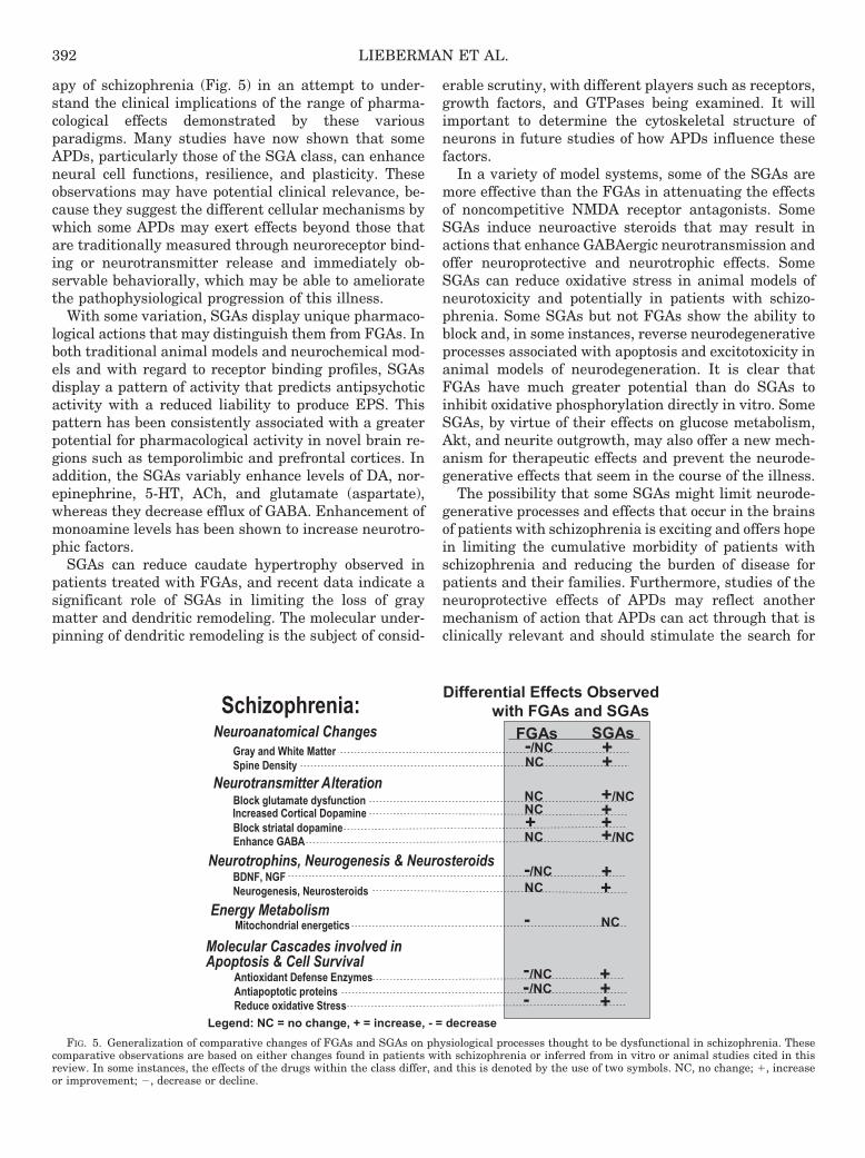
apy of schizophrenia (Fig. 5) in an attempt to under-stand the clinical implications of the range of pharma-cological effects demonstrated by these variousparadigms. Many studies have now shown that someAPDs, particularly those of the SGA class, can enhanceneural cell functions, resilience, and plasticity. Theseobservations may have potential clinical relevance, be-cause they suggest the different cellular mechanisms bywhich some APDs may exert effects beyond those thatare traditionally measured through neuroreceptor bind-ing or neurotransmitter release and immediately ob-servable behaviorally, which may be able to amelioratethe pathophysiological progression of this illness.
With some variation, SGAs display unique pharmaco-logical actions that may distinguish them from FGAs. Inboth traditional animal models and neurochemical mod-els and with regard to receptor binding profiles, SGAsdisplay a pattern of activity that predicts antipsychoticactivity with a reduced liability to produce EPS. Thispattern has been consistently associated with a greaterpotential for pharmacological activity in novel brain re-gions such as temporolimbic and prefrontal cortices. Inaddition, the SGAs variably enhance levels of DA, nor-epinephrine, 5-HT, ACh, and glutamate (aspartate),whereas they decrease efflux of GABA. Enhancement ofmonoamine levels has been shown to increase neurotro-phic factors.
SGAs can reduce caudate hypertrophy observed inpatients treated with FGAs, and recent data indicate asignificant role of SGAs in limiting the loss of graymatter and dendritic remodeling. The molecular under-pinning of dendritic remodeling is the subject of consid-
erable scrutiny, with different players such as receptors,growth factors, and GTPases being examined. It willimportant to determine the cytoskeletal structure ofneurons in future studies of how APDs influence thesefactors.
In a variety of model systems, some of the SGAs aremore effective than the FGAs in attenuating the effectsof noncompetitive NMDA receptor antagonists. SomeSGAs induce neuroactive steroids that may result inactions that enhance GABAergic neurotransmission andoffer neuroprotective and neurotrophic effects. SomeSGAs can reduce oxidative stress in animal models ofneurotoxicity and potentially in patients with schizo-phrenia. Some SGAs but not FGAs show the ability toblock and, in some instances, reverse neurodegenerativeprocesses associated with apoptosis and excitotoxicity inanimal models of neurodegeneration. It is clear thatFGAs have much greater potential than do SGAs toinhibit oxidative phosphorylation directly in vitro. SomeSGAs, by virtue of their effects on glucose metabolism,Akt, and neurite outgrowth, may also offer a new mech-anism for therapeutic effects and prevent the neurode-generative effects that seem in the course of the illness.
The possibility that some SGAs might limit neurode-generative processes and effects that occur in the brainsof patients with schizophrenia is exciting and offers hopein limiting the cumulative morbidity of patients withschizophrenia and reducing the burden of disease forpatients and their families. Furthermore, studies of theneuroprotective effects of APDs may reflect anothermechanism of action that APDs can act through that isclinically relevant and should stimulate the search for
Differential Effects ObservedDifferential Effects Observedwith FGAs and SGAswith FGAs and SGAs
Gray and White MatterSpine Density
Energy Metabolism
Neuroanatomical Changes
Molecular Cascades involved inApoptosis & Cell Survival
Neurotransmitter AlterationBlock glutamate dysfunctionIncreased Cortical DopamineBlock striatal dopamineEnhance GABA
Mitochondrial energetics
Neurotrophins, Neurogenesis & Neurosteroids
Schizophrenia:Schizophrenia:
Antioxidant Defense EnzymesAntiapoptotic proteinsReduce oxidative Stress
SGAsFGAs
BDNF, NGFNeurogenesis, Neurosteroids
++/NC
++
+
+++
+-/NC
-/NC
-
-
NC
NCNC
NC
/NC
NC
NC
-/NC
-
Legend: NC = no change, + = increase, - = decrease
+/NC+ +
FIG. 5. Generalization of comparative changes of FGAs and SGAs on physiological processes thought to be dysfunctional in schizophrenia. Thesecomparative observations are based on either changes found in patients with schizophrenia or inferred from in vitro or animal studies cited in thisreview. In some instances, the effects of the drugs within the class differ, and this is denoted by the use of two symbols. NC, no change; �, increaseor improvement; �, decrease or decline.
392 LIEBERMAN ET AL.

new drugs for schizophrenia with novel mechanismsbeyond the more familiar effects current drug treat-ments have on the monoaminergic neurotransmittersystems.
As pointed out in this critical review of the literatureon the effects of APDs, there is inconsistency in theresults of these different studies and many instances ofinterstudy variability of the biological effects betweenthe FGA and SGA drug classes and also variation withineach of the two drug classes. Research is new and lim-ited in some of the areas reviewed, and technical proce-dures have not been standardized, which contributes tovariability. In addition, many studies use only haloper-idol as the FGA and not all of the SGAs have been fullyevaluated. It is difficult in animal studies to be sure thatclinically equivalent doses of the APDs are adminis-tered. Many studies were short-term in nature, and of-ten subchronic studies were relatively short-term. Thesefactors make extrapolation of these results to the effectsof APDs in humans after weeks or months of treatmentdifficult at best. Furthermore, many of the biologicalprocesses evaluated in cell lines and in animals have notbeen fully characterized in humans, and their clinicalrelevance is not known. Consequently, the impact ofalterations of these processes in patients is unknown.Clearly, more work is needed, but the bulk of the datasupports our tentative conclusion that some APDs,mainly of the SGA group, may have effects that can betherapeutic on many biological processes and neuropro-tective on the pathophysiology of schizophrenia,whereas the FGAs are neutral and in high doses mayeven promote neurodegeneration. Therefore, we believethat there may be clinical benefits in the use of selectiveSGAs versus FGAs for long-term treatment particularlyin the early stages of schizophrenia and related psy-chotic disorders.
Acknowledgments.We acknowledge the many contributions of ourcolleagues to this review, Dr. Sara Kollack-Walker for editorial as-sistance, and Dr. Yvonne Cole for formatting and submitting themanuscript.
REFERENCES
Abekawa T, Ito K, and Koyama T (2006) Role of the simultaneous enhancement ofNMDA and dopamine D1 receptor-mediated neurotransmission in the effects ofclozapine on phencyclidine-induced acute increases in glutamate levels in the ratmedial prefrontal cortex. Naunyn Schmiedebergs Arch Pharmacol 374:177–193.
Abi-Dargham A (2007) Alterations of serotonin transmission in schizophrenia. IntRev Neurobiol 78:133–164.
Abi-Dargham A and Laruelle M (2005) Mechanisms of action of second generationantipsychotic drugs in schizophrenia: insights from brain imaging studies. EurPsychiatry 20:15–27.
Abi-Dargham A, Mawlawi O, Lombardo I, Gil R, Martinez D, Huang Y, Hwang DR,Keilp J, Kochan L, Van Heertum R, et al. (2002) Prefrontal dopamine D1 receptorsand working memory in schizophrenia. J Neurosci 22:3708–3719.
Abi-Dargham A and Moore H (2003) Prefrontal DA transmission at D1 receptors andthe pathology of schizophrenia. Neuroscientist 9:404–416.
Adams AC and Keefe KA (2001) Examination of the involvement of protein kinase Ain D2 dopamine receptor antagonist-induced immediate early gene expression.J Neurochem 77:326–335.
Adams B and Moghaddam B (1998) Corticolimbic dopamine neurotransmission istemporally dissociated from the cognitive and locomotor effects of phencyclidine.J Neurosci 18:5545–5554.
Adams BW and Moghaddam B (2001) Effect of clozapine, haloperidol, or M100907 onphencyclidine-activated glutamate efflux in the prefrontal cortex. Biol Psychiatry50:750–757.
Adams JM and Cory S (1998) The Bcl-2 protein family: arbiters of cell survival.Science 281:1322–1326.
Adams MR, Brandon EP, Chartoff EH, Idzerda RL, Dorsa DM, and McKnight GS(1997) Loss of haloperidol induced gene expression and catalepsy in protein kinaseA-deficient mice. Proc Natl Acad Sci U S A 94:12157–12161.
Akwa Y, Ladurelle N, Covey DF, and Baulieu EE (2001) The synthetic enantiomer ofpregnenolone sulfate is very active on memory in rats and mice, even more so thanits physiological neurosteroid counterpart: distinct mechanisms? Proc Natl AcadSci U S A 98:14033–14037.
Albert KA, Hemmings HC Jr, Adamo AI, Potkin SG, Akbarian S, Sandman CA,Cotman CW, Bunney WE Jr, and Greengard P (2002) Evidence for decreasedDARPP-32 in the prefrontal cortex of patients with schizophrenia. Arch GenPsychiatry 59:705–712.
Alexander GE, DeLong MR, and Strick PL (1986) Parallel organization of function-ally segregated circuits linking basal ganglia and cortex. Annu Rev Neurosci9:357–381.
Alfredsson G and Wiesel FA (1989) Monoamine metabolites and amino acids inserum from schizophrenic patients before and during sulpiride treatment. Psycho-pharmacology (Berl) 99:322–327.
Alimohamad H, Rajakumar N, Seah YH, and Rushlow W (2005) Antipsychotics alterthe protein expression levels of beta-catenin and GSK-3 in the rat medial prefron-tal cortex and striatum. Biol Psychiatry 57:533–542.
Aloe L, Iannitelli A, Bersani G, Alleva E, Angelucci F, Maselli P, and Manni L (1997)Haloperidol administration in humans lowers plasma nerve growth factor level:evidence that sedation induces opposite effects to arousal. Neuropsychobiology36:65–68.
Amargos-Bosch M, Lopez-Gil X, Artigas F, and Adell A (2006) Clozapine and olan-zapine, but not haloperidol, suppress serotonin efflux in the medial prefrontalcortex elicited by phencyclidine and ketamine. Int J Neuropsychopharmacol9:565–573.
Andersson C, Hamer RM, Lawler CP, Mailman RB, and Lieberman JA (2002)Striatal volume changes in the rat following long-term administration of typicaland atypical antipsychotic drugs. Neuropsychopharmacology 27:143–151.
Andreasen NC, Rezai K, Alliger R, Swayze VW 2nd, Flaum M, Kirchner P, Cohen G,and O’Leary DS (1992) Hypofrontality in neuroleptic-naïve patients and in pa-tients with chronic schizophrenia. Assessment with xenon 133 single-photon emis-sion computed tomography and the Tower of London. Arch Gen Psychiatry 49:943–958.
Andreassen OA and Jørgensen HA (1995) The mitochondrial toxin 3-nitropropionicacid induces vacuous chewing movements in rats. Implications for tardive dyski-nesia? Psychopharmacology (Berl) 119:474–476.
Andreassen OA and Jørgensen HA (2000) Neurotoxicity associated with neuroleptic-induced oral dyskinesias in rats: implications for tardive dyskinesia. Prog Neuro-biol 61:525–541.
Angelucci F, Aloe L, Gruber SH, Fiore M, and Mathe AA (2000a) Chronic antipsy-chotic treatment selectively alters nerve growth factor and neuropeptide Y immu-noreactivity and the distribution of choline acetyl transferase in rat brain regions.Int J Neuropsychopharmacol 3:13–25.
Angelucci F, Aloe L, Iannitelli A, Gruber SH, and Mathe AA (2005) Effect of chronicolanzapine treatment on nerve growth factor and brain-derived neurotrophic fac-tor in the rat brain. Eur Neuropsychopharmacol 15:311–317.
Angelucci F, Mathe AA, and Aloe L (2000b) Brain-derived neurotrophic factor andtyrosine kinase receptor TrkB in rat brain are significantly altered after haloper-idol and risperidone administration. J Neurosci Res 60:783–794.
Ardizzone TD, Bradley RJ, Freeman AM 3rd, and Dwyer DS (2001) Inhibition ofglucose transport in PC12 cells by the atypical antipsychotic drugs risperidone andclozapine, and structural analogs of clozapine. Brain Res 923:82–90.
Arif M, Chikuma T, Ahmed MM, Yoshida S, and Kato T (2007) Suppressive effect ofclozapine but not haloperidol on the increases of neuropeptide-degrading enzymesand glial cells in MK-801-treated rat brain regions. Neurosci Res 57:248–258.
Arnt J (1982) Pharmacological specificity of conditioned avoidance response inhibi-tion in rats: inhibition by neuroleptics and correlation to dopamine receptorblockade. Acta Pharmacol Toxicol (Copenh) 51:321–329.
Arnt J and Skarsfeldt T (1998) Do novel antipsychotics have similar pharmacologicalcharacteristics? A review of the evidence. Neuropsychopharmacology 18:63–101.
Arundine M and Tymianski M (2003) Molecular mechanisms of calcium-dependentneurodegeneration in excitotoxicity. Cell Calcium 34:325–337.
Arvanov VL, Liang X, Schwartz J, Grossman S, and Wang RY (1997) Clozapine andhaloperidol modulate N-methyl-D-aspartate- and non-N-methyl-D-aspartate recep-tor-mediated neurotransmission in rat prefrontal cortical neurons in vitro. J Phar-macol Exp Ther 283:226–234.
Ashe PC, Chlan-Fourney J, Juorio AV, and Li XM (2002) Brain-derived neurotrophicfactor (BDNF) mRNA in rats with neonatal ibotenic acid lesions of the ventralhippocampus. Brain Res 956:126–135.
Bai O, Chlan-Fourney J, Bowen R, Keegan D, and Li XM (2003) Expression ofbrain-derived neurotrophic factor mRNA in rat hippocampus after treatment withantipsychotic drugs. J Neurosci Res 71:127–131.
Bai O, Wei Z, Lu W, Bowen R, Keegan D, and Li XM (2002) Protective effects ofatypical antipsychotic drugs on PC12 cells after serum withdrawal. J Neurosci Res69:278–283.
Bai O, Zhang H, and Li XM (2004) Antipsychotic drugs clozapine and olanzapineupregulate bcl-2 mRNA and protein in rat frontal cortex and hippocampus. BrainRes 1010:81–86.
Bajestan SN, Sabouri AH, Nakamura M, Takashima H, Keikhaee MR, Behdani F,Fayyazi MR, Sargolzaee MR, Bajestan MN, Sabouri Z, et al. (2006) Association ofAKT1 haplotype with the risk of schizophrenia in Iranian population. Am J MedGenet B Neuropsychiatr Genet 141B:383–386.
Baker GF and Rogers HJ (1972) Effects of psychotropic drugs on the erythrocytepermeability to glucose and ethylidene glucose. Biochem Pharmacol 21:1871–1878.
Bakshi VP and Geyer MA (1995) Antagonism of phencyclidine-induced deficits in
ANTIPSYCHOTIC DRUGS IN NEUROPROTECTION 393

prepulse inhibition by the putative atypical antipsychotic olanzapine. Psychophar-macology (Berl) 122:198–201.
Bakshi VP, Swerdlow NR, and Geyer MA (1994) Clozapine antagonizes phencyclid-ine-induced deficits in sensorimotor gating of the startle response. J PharmacolExp Ther 271:787–794.
Baquer NZ, Hothersall JS, and McLean P (1988) Function and regulation of thepentose phosphate pathway in brain. Curr Top Cell Regul 29:265–289.
Barbaccia ML, Affricano D, Purdy RH, Maciocco E, Spiga F, and Biggio G (2001)Clozapine, but not haloperidol, increases brain concentrations of neuroactive ste-roids in the rat. Neuropsychopharmacology 25:489–497.
Barde YA (1994) Neurotrophins: a family of proteins supporting the survival ofneurons. Prog Clin Biol Res 390:45–56.
Barnes NM and Sharp T (1999) A review of central 5-HT receptors and theirfunction. Neuropharmacology 38:1083–1152.
Bartzokis G, Beckson M, Lu PH, Nuechterlein KH, Edwards N, and Mintz J (2001)Age-related changes in frontal and temporal lobe volumes in men: a magneticresonance imaging study. Arch Gen Psychiatry 58:461–465.
Beaulieu JM, Gainetdinov RR, and Caron MG (2007) The Akt-GSK-3 signalingcascade in the actions of dopamine. Trends Pharmacol Sci 28:166–172.
Beaulieu JM, Sotnikova TD, Marion S, Lefkowitz RJ, Gainetdinov RR, and CaronMG (2005) An Akt/�-arrestin 2/PP2A signaling complex mediates dopaminergicneurotransmission and behavior. Cell 122:261–273.
Belelli D, Bolger MB, and Gee KW (1989) Anticonvulsant profile of the progesteronemetabolite 5 �-pregnan-3 �-ol-20-one. Eur J Pharmacol 166:325–329.
Belelli D and Lambert JJ (2005) Neurosteroids: endogenous regulators of the GABAAreceptor. Nat Rev Neurosci 6:565–575.
Ben-Shachar D (2002) Mitochondrial dysfunction in schizophrenia: a possible link-age to dopamine. J Neurochem 83:1241–1251.
Ben-Shachar D and Laifenfeld D (2004) Mitochondria, synaptic plasticity, andschizophrenia. Int Rev Neurobiol 59:273–296.
Benes FM (1997) The role of stress and dopamine-GABA interactions in the vulner-ability for schizophrenia. J Psychiatr Res 31:257–275.
Benes FM and Berretta S (2000) Amygdalo-entorhinal inputs to the hippocampalformation in relation to schizophrenia. Ann N Y Acad Sci 911:293–304.
Benes FM and Berretta S (2001) GABAergic interneurons: implications for under-standing schizophrenia and bipolar disorder. Neuropsychopharmacology 25:1–27.
Benes FM, Khan Y, Vincent SL, and Wickramasinghe R (1996a) Differences in thesubregional and cellular distribution of GABAA receptor binding in the hippocam-pal formation of schizophrenic brain. Synapse 22:338–349.
Benes FM, McSparren J, Bird ED, SanGiovanni JP, and Vincent SL (1991) Deficitsin small interneurons in prefrontal and cingulated cortices of schizophrenic andschizoaffective patients. Arch Gen Psychiatry 48:996–1001.
Benes FM, Vincent SL, Marie A, and Khan Y (1996b) Increased GABAA receptorbinding on neurons of prefrontal cortex in schizophrenic subjects. Neuroscience75:1021–1031.
Benes FM, Vincent SL, and Todtenkopf M (2001) The density of pyramidal andnonpyramidal neurons in anterior cingulate cortex of schizophrenic and bipolarsubjects. Biol Psychiatry 50:395–406.
Benes FM, Walsh J, Bhattacharyya S, Sheth A, and Berretta S (2003) DNA frag-mentation decreased in schizophrenia but not bipolar disorder. Arch Gen Psychi-atry 60:359–364.
Bergeron R, de Montigny C, and Debonnel G (1996) Potentiation of neuronal NMDAresponse induced by dehydroepiandrosterone and its suppression by progesterone:effects mediated via sigma receptors. J Neurosci 16:1193–1202.
Bergson C, Levenson R, Goldman-Rakic PS, and Lidow MS (2003) Dopamine recep-tor-interacting proteins: the Ca2� connection in dopamine signaling. Trends Phar-macol Sci 24:486–492.
Bernaudin M, Bellail A, Marti HH, Yvon A, Vivien D, Duchatelle I, Mackenzie ET,and Petit E (2000) Neurons and astrocytes express EPO mRNA: oxygen-sensingmechanisms that involve the redox-state of the brain. Glia 30:271–278.
Bernaudin M, Marti HH, Roussel S, Divoux D, Nouvelot A, MacKenzie ET, and PetitE (1999) A potential role for erythropoietin in focal permanent cerebral ischemiain mice. J Cereb Blood Flow Metab 19:643–651.
Berridge MJ (1998) Neuronal calcium signaling. Neuron 21:13–26.Bhardwaj RD, Curtis MA, Spalding KL, Buchholz BA, Fink D, Bjork-Eriksson T,
Nordborg C, Gage FH, Druid H, Eriksson PS, et al. (2006) Neocortical neurogen-esis in humans is restricted to development. Proc Natl Acad Sci U S A 103:12564–12568.
Biagiotti E, Guidi L, Capellacci S, Ambrogini P, Papa S, Del Grande P, and NinfaliP (2001) Glucose-6-phosphate dehydrogenase supports the functioning of the syn-apses in rat cerebellar cortex. Brain Res 911:152–157.
Black JE, Kodish IM, Grossman AW, Klintsova AY, Orlovskaya D, Vostrikov V,Uranova N, and Greenough WT (2004) Pathology of layer V pyramidal neurons inthe prefrontal cortex of patients with schizophrenia. Am J Psychiatry 161:742–744.
Blass JP (2002) Glucose/mitochondria in neurological conditions. Int Rev Neurobiol51:325–376.
Blum BP and Mann JJ (2002) The GABAergic system in schizophrenia. Int J Neu-ropsychopharmacol 5:159–179.
Boks MP, Rietkerk T, van de Beek MH, Sommer IE, de Koning TJ, and Kahn RS(2007) Reviewing the role of the genes G72 and DAAO in glutamate neurotrans-mission in schizophrenia. Eur Neuropsychopharmacol 17:567–572.
Bonaccorso S, Meltzer HY, Li Z, Dai J, Alboszta AR, and Ichikawa J (2002)SR46349-B, a 5-HT2A/2C receptor antagonist, potentiates haloperidol-induced do-pamine release in rat medial prefrontal cortex and nucleus accumbens. Neuropsy-chopharmacology 27:430–441.
Bondy CA and Cheng CM (2002) Insulin-like growth factor-1 promotes neuronalglucose utilization during brain development and repair processes. Int Rev Neu-robiol 51:189–217.
Bortolato M, Frau R, Orru M, Piras AP, Fa M, Tuveri A, Puligheddu M, Gessa GL,Castelli MP, Mereu G, et al. (2007) Activation of GABAB receptors reverses
spontaneous gating deficits in juvenile DBA/2J mice. Psychopharmacology (Berl)194:361–369.
Bourdelais AJ and Deutch AY (1994) The effects of haloperidol and clozapine onextracellular GABA levels in the prefrontal cortex of the rat: an in vivo microdi-alysis study. Cereb Cortex 4:69–77.
Bowery NG (2006) GABAB receptor: a site of therapeutic benefit. Curr Opin Phar-macol 6:37–43.
Braff DL, Geyer MA, and Swerdlow NR (2001) Human studies of prepulse inhibitionof startle: normal subjects, patient groups, and pharmacological studies. Psycho-pharmacology (Berl) 156:234–258.
Braus DF, Weber-Fahr W, Tost H, Ruf M, and Henn FA (2002) Sensory informationprocessing in neuroleptic-naive first-episode schizophrenia patients: a functionalmagnetic resonance imaging study. Arch Gen Psychiatry 59:696–701.
Brazil DP, Yang ZZ, and Hemmings BA (2004) Advances in protein kinase Bsignaling: AKTion on multiple fronts. Trends Biochem Sci 29:233–242.
Breier A, Buchanan RW, Elkashef A, Munson RC, Kirkpatrick B, and Gellad F(1992) Brain morphology and schizophrenia. A magnetic resonance imaging studyof limbic, prefrontal cortex, and caudate structures. Arch Gen Psychiatry 49:921–926.
Britsch S (2007) The neuregulin-I/ErbB signaling system in development and dis-ease. Adv Anat Embryol Cell Biol 190:1–65.
Brines ML, Ghezzi P, Keenan S, Agnello D, de Lanerolle NC, Cerami C, Itri LM, andCerami A (2000) Erythropoietin crosses the blood-brain barrier to protect againstexperimental brain injury. Proc Natl Acad Sci U S A 97:10526–10531.
Broadbelt K, Byne W, and Jones LB (2002) Evidence for a decrease in basilardendrites of pyramidal cells in schizophrenic medial prefrontal cortex. SchizophrRes 58:75–81.
Brot MD, Akwa Y, Purdy RH, Koob GF, and Britton KT (1997) The anxiolytic-likeeffects of the neurosteroid allopregnanolone: interactions with GABAA receptors.Eur J Pharmacol 325:1–7.
Browning JL, Patel T, Brandt PC, Young KA, Holcomb LA, and Hicks PB (2005)Clozapine and the mitogen-activated protein kinase signal transduction pathway:implications for antipsychotic actions. Biol Psychiatry 57:617–623.
Buchanan RW, Breier A, Kirkpatrick B, Elkashef A, Munson RC, Gellad F, andCarpenter WT Jr (1993) Structural abnormalities in deficit and nondeficit schizo-phrenia. Am J Psychiatry 150:59–65.
Buchanan RW, Vladar K, Barta PE, and Pearlson GD (1998) Structural evaluationof the prefrontal cortex in schizophrenia. Am J Psychiatry 155:1049–1055.
Buchsbaum MS, Nuechterlein KH, Haier RJ, Wu J, Sicotte N, Hazlett E, Asarnow R,Potkin S, and Guich S (1990) Glucose metabolic rate in normals and schizophren-ics during the Continuous Performance Test assessed by positron emission tomog-raphy. Br J Psychiatry 156:216–227.
Buckley PF, Mahadik S, Pillai A, and Terry A (2007a) Neurotrophins and schizo-phrenia. Schizophr Res 84:1–11.
Buckley PF, Pillai A, Evans D, Stirewalt E, and Mahadik S (2007b) Brain derivedneurotrophic factor in first-episode psychosis. Schizophr Res 91:1–5.
Burkhardt C, Kelly JP, Lim YH, Filley CM, and Parker WD Jr (1993) Neurolepticmedications inhibit complex I of the electron transport chain. Ann Neurol 33:512–517.
Burnet PW, Hutchinson L, von Hesling M, Gilbert EJ, Brandon NJ, Rutter AR,Hutson PH, and Harrison PJ (2008) Expression of D-serine and glycine transport-ers in the prefrontal cortex and cerebellum in schizophrenia. Schizophr Res 102:283–294.
Bymaster FP, Calligaro DO, Falcone JF, Marsh RD, Moore NA, Tye NC, Seeman P,and Wong DT (1996) Radioreceptor binding profile of the atypical antipsychoticolanzapine. Neuropsychopharmacology 14:87–96.
Bymaster FP, Nelson DL, DeLapp NW, Falcone JF, Eckols K, Truex LL, ForemanMM, Lucaites VL, and Calligaro DO (1999) Antagonism by olanzapine of dopamineD1, serotonin2, muscarinic, histamine H1 and �1-adrenergic receptors in vitro.Schizophr Res 37:107–122.
Caceda R, Kinkead B, and Nemeroff CB (2006) Neurotensin: role psychiatric andneurological diseases. Peptides 27:2385–2404.
Cahn W, Hulshoff Pol HE, Lems EB, van Haren NE, Schnack HG, van der LindenJA, Schothorst PF, van Engeland H, and Kahn RS (2002) Brain volume changes infirst-episode schizophrenia: a 1-year follow-up study. Arch Gen Psychiatry 59:1002–1010.
Cameron HA, Hazel TG, and McKay RD (1998) Regulation of neurogenesis by growthfactors and neurotransmitters. J Neurobiol 36:287–306.
Cannon TD, van Erp TG, Huttunen M, Lonnqvist J, Salonen O, Valanne L, PoutanenVP, Standertskjold-Nordenstam CG, Gur RE, and Yan M (1998) Regional graymatter, white matter, and cerebrospinal fluid distributions in schizophrenic pa-tients, their siblings, and controls. Arch Gen Psychiatry 55:1084–1091.
Carlsson M and Carlsson A (1990) Interactions between glutamatergic and mono-aminergic systems within the basal ganglia—implications for schizophrenia andParkinson’s disease. Trends Neurosci 13:272–276.
Catapano LA and Manji HK (2007) G protein-coupled receptors in major psychiatricdisorders. Biochem Biophys Acta 1768:976–993.
Ceballos I, Javoy-Agid F, Delacourte A, Defossez A, Lafon M, Hirsch E, Nicole A,Sinet PM, and Agid Y (1991) Neuronal localization of copper-zinc superoxidedismutase protein and mRNA within the human hippocampus from control andAlzheimer’s disease brains. Free Radic Res Commun 12–13:571–580.
Ceresoli-Borroni G, Rassoulpour A, Wu HQ, Guidetti P, and Schwarcz R (2006)Chronic neuroleptic treatment reduces endogenous kynurenic acid levels in ratbrain. J Neural Transm 113:1355–1365.
Chahl LA (2006) Tachykinins and neuropsychiatric disorders. Curr Drug Targets7:993–1003.
Chakos MH, Lieberman JA, Alvir J, Bilder R, and Ashtari M (1995) Caudate nucleivolumes in schizophrenic patients treated with typical antipsychotics or clozapine.Lancet 345:456–457.
Chakos MH, Lieberman JA, Bilder RM, Borenstein M, Lerner G, Bogerts B, Wu H,Kinon B, and Ashtari M (1994) Increase in caudate nuclei volumes of first-episode
394 LIEBERMAN ET AL.

schizophrenic patients taking antipsychotic drugs. Am J Psychiatry 151:1430–1436.
Chakos MH, Shirakawa O, Lieberman J, Lee H, Bilder R, and Tamminga CA (1998)Striatal enlargement in rats chronically treated with neuroleptic. Biol Psychiatry44:675–684.
Chiodo LA and Bunney BS (1983) Typical and atypical neuroleptics: differentialeffects of chronic administration on the activity of A9 and A10 midbrain dopami-nergic neurons. J Neurosci 3:1607–1619.
Chlan-Fourney J, Ashe P, Nylen K, Juorio AV, and Li XM (2002) Differentialregulation of hippocampal BDNF mRNA by typical and atypical antipsychoticadministration. Brain Res 954:11–20.
Christensen J, Holcomb J, and Garver DL (2004) State-related changes in cerebralwhite matter may underlie psychosis exacerbation. Psychiatry Res 130:71–78.
Chung YC, Li Z, Dai J, Meltzer HY, and Ichikawa J (2004) Clozapine increases bothacetylcholine and dopamine release in rat ventral hippocampus: role of 5-HT1Areceptor agonism. Brain Res 1023:54–63.
Ciani E, Virgili M, and Contestabile A (2002) Akt pathway mediates a cGMP-dependent survival role of nitric oxide in cerebellar granule neurones. J Neuro-chem 81:218–228.
Ciriza I, Azcoitia I, and Garcia-Segura LM (2004) Reduced progesterone metabolitesprotect rat hippocampal neurones from kainic acid excitotoxicity in vivo. J Neu-roendocrinol 16:58–63.
Clinton SM and Meador-Woodruff JH (2004) Thalamic dysfunction in schizophrenia:neurochemical, neuropathological, and in vivo imaging abnormalities. SchizophrRes 69:237–253.
Compagnone NA and Mellon SH (1998) Dehydroepiandrosterone: a potential signal-ing molecule for neocortical organization during development. Proc Natl Acad SciU S A 95:4678–4683.
Convit A, Wolf OT, de Leon MJ, Patalinjug M, Kandil E, Caraos C, Scherer A, SaintLouis LA, and Cancro R (2001) Volumetric analysis of the pre-frontal regions:findings in aging and schizophrenia. Psychiatry Res 107:61–73.
Corbett R, Camacho F, Woods AT, Kerman LL, Fishkin RJ, Brooks K, and Dunn RW(1995) Antipsychotic agents antagonize non-competitive N-methyl-D-aspartate an-tagonist-induced behaviors. Psychopharmacology (Berl) 120:67–74.
Corson PW, Nopoulos P, Miller DD, Arndt S, and Andreasen NC (1999) Change inbasal ganglia volume over 2 years in patients with schizophrenia: typical versusatypical neuroleptics. Am J Psychiatry 156:1200–1204.
Coyle JT (1996) The glutamatergic dysfunction hypothesis for schizophrenia. HarvRev Psychiatry 3:241–253.
Coyle JT (2006) Glutamate and schizophrenia: beyond the dopamine hypothesis. CellMol Neurobiol 26:365–384.
Craig RW (1995) The Bcl-2 gene family. Semin Cancer Biol 6:35–43.Crawley JN, Glowa JR, Majewska MD, and Paul SM (1986) Anxiolytic activity of an
endogenous adrenal steroid. Brain Res 398:382–385.Creese I, Burt DR, and Snyder SH (1996) Dopamine receptor binding predicts
clinical and pharmacological potencies of antischizophrenic drugs. J Neuropsychi-atry Clin Neurosci 8:223–226.
Csernansky JG and Bardgett ME (1998) Limbic-cortical neuronal damage and thepathophysiology of schizophrenia. Schizophr Bull 24:231–248.
Csernansky JG, Martin MV, Czeisler B, Meltzer MA, Ali Z, and Dong H (2006)Neuroprotective effects of olanzapine in a rat model of neurodevelopmental injury.Pharmacol Biochem Behav 83:208–213.
Cull-Candy S, Brickley S, and Farrant M (2001) NMDA receptor subunits: diversity,development and disease. Curr Opin Neurobiol 11:327–335.
Dakhale G, Khanzode S, Khanzode S, Saoji A, Khobragade L, and Turankar A (2004)Oxidative damage and schizophrenia: the potential benefit by atypical antipsy-chotics. Neuropsychobiology 49:205–209.
Daly DA and Moghaddam B (1993) Actions of clozapine and haloperidol on theextracellular levels of excitatory amino acids in the prefrontal cortex and striatumof conscious rats. Neurosci Lett 152:61–64.
Davis KL, Buchsbaum MS, Shihabuddin L, Spiegel-Cohen J, Metzger M, Frecska E,Keefe RS, and Powchik P (1998) Ventricular enlargement in poor-outcome schizo-phrenia. Biol Psychiatry 43:783–793.
Davis KL, Stewart DG, Friedman JI, Buchsbaum M, Harvey PD, Hof PR, BuxbaumJ, and Haroutunian V (2003) White matter changes in schizophrenia: evidence formyelin-related dysfunction. Arch Gen Psychiatry 60:443–456.
Dawirs RR, Hildebrandt K, and Teuchert-Noodt G (1998) Adult treatment withhaloperidol increases dentate granule cell proliferation in the gerbil hippocampus.J Neural Transm 105:317–327.
Dawson NM, Hamid EH, Egan MF, and Meredith GE (2001) Changes in the patternof brain-derived neurotrophic factor immunoreactivity in the rat brain after acuteand subchronic haloperidol treatment. Synapse 39:70–81.
Debonnel G, Bergeron R, and de Montigny C (1996) Potentiation by dehydroepi-androsterone of the neuronal response to N-methyl-D-aspartate in the CA3 regionof the rat dorsal hippocampus: an effect mediated via sigma receptors. J Endocri-nol 150 (Suppl):S33–S42.
Delacourte A, Defossez A, Ceballos I, Nicole A, and Sinet PM (1988) Preferentiallocalization of copper zinc superoxide dismutase in the vulnerable cortical neuronsin Alzheimer’s disease. Neurosci Lett 92:247–253.
DeLisi LE, Sakuma M, Tew W, Kushner M, Hoff AL, and Grimson R (1997) Schizo-phrenia as a chronic active brain process: a study of progressive brain structuralchange subsequent to the onset of schizophrenia. Psychiatry Res 74:129–140.
Deng C and Huang XF (2006) Increased density of GABAA receptors in the superiortemporal gyrus in schizophrenia. Exp Brain Res 168:587–590.
Deutch AY and Bubser M (2007) The orexins/hypocretins and schizophrenia. Schizo-phr Bull 33:1277–1283.
Deutch AY and Duman RS (1996) The effects of antipsychotic drugs on Fos proteinexpression in the prefrontal cortex: cellular localization and pharmacological char-acterization. Neuroscience 70:377–389.
Deutch AY, Lewis DA, Whitehead RE, Elsworth JD, Iadarola MJ, Redmond DE Jr,and Roth RH (1996) Effects of D2 dopamine receptor antagonists on Fos protein
expression in the striatal complex and entorhinal cortex of the nonhuman primate.Synapse 23:182–191.
Deutsch SI, Mastropaolo J, Schwartz BL, Rosse RB, and Morihisa JM (1989) A“glutamatergic hypothesis” of schizophrenia: rationale for pharmacotherapy withglycine. Clin Neuropharmacol 12:1–13.
Deutsch SI, Rosse RB, Schwartz BL, and Mastropaolo J (2001) A revised excitotoxichypothesis of schizophrenia: therapeutic implications. Clin Neuropharmacol 24:43–49.
Devaud LL, Purdy RH, and Morrow AL (1995) The neurosteroid, 3�-hydroxy-5�-pregnan-20-one, protects against bicuculline-induced seizures during ethanolwithdrawal in rats. Alcohol Clin Exp Res 19:350–355.
Dietrich-Muszalska A, Olas B, and Rabe-Jablonska J (2005) Oxidative stress inblood platelets from schizophrenic patients. Platelets 16:386–391.
di Michele F, Caltagirone C, Bonaviri G, Romeo E, and Spalletta G (2005) Plasmadehydroepiandrosterone levels are strongly increased in schizophrenia. J Psychi-atr Res 39:267–273.
Dingledine R, Borges K, Bowie D, and Traynelis SF (1999) The glutamate receptorion channels. Pharmacol Rev 51:7–61.
Djebaili M, Hoffman SW, and Stein DG (2004) Allopregnanolone and progesteronedecrease cell death and cognitive deficits after a contusion of the rat pre-frontalcortex. Neuroscience 123:349–359.
Do KQ, Trabesinger AH, Kirsten-Kruger M, Lauer CJ, Dydak U, Hell D, Holsboer F,Boesiger P, and Cuenod M (2000) Schizophrenia: glutathione deficit in cerebrospi-nal fluid and prefrontal cortex in vivo. Eur J Neurosci 12:3721–3728, 2000.
Dono R (2003) Fibroblast growth factors as regulators of central nervous systemdevelopment and function. Am J Physiol Regul Integr Comp Physiol 284:R867–R881.
Dudek H, Datta SR, Franke TF, Birnbaum MJ, Yao R, Cooper GM, Segal RA, KaplanDR, and Greenberg ME (1997) Regulation of neuronal survival by the serine-threonine protein kinase Akt. Science 275:661–665.
Duman RS (2004) Depression: a case of neuronal life and death? Biol Psychiatry56:140–145.
Duncan GE, Leipzig JN, Mailman RB, and Lieberman JA (1998a) Differential effectsof clozapine and haloperidol on ketamine-induced brain metabolic activation.Brain Res 812:65–75.
Duncan GE, Miyamoto S, Leipzig JN, and Lieberman JA (1999) Comparison of brainmetabolic activity patterns induced by ketamine, MK-801 and amphetamine inrats: support for NMDA receptor involvement in responses to subanesthetic doseof ketamine. Brain Res 843:171–183.
Duncan GE, Miyamoto S, Leipzig JN, and Lieberman JA (2000) Comparison of theeffects of clozapine, risperidone, and olanzapine on ketamine-induced alterationsin regional brain metabolism. J Pharmacol Exp Ther 293:8–14.
Duncan GE, Moy SS, Knapp DJ, Mueller RA, and Breese GR (1998b) Metabolicmapping of the rat brain after subanesthetic doses of ketamine: potential rele-vance to schizophrenia. Brain Res 787:181–190.
Durany N, Michel T, Zochling R, Boissl KW, Cruz-Sanchez FF, Riederer P, andThome J (2001) Brain-derived neurotrophic factor and neurotrophin 3 in schizo-phrenic psychoses. Schizophr Res 52:79–86.
Dwivedi Y, Rizavi HS, and Pandey GN (2002) Differential effects of haloperidol andclozapine on [3H]cAMP binding, protein kinase A (PKA) activity, and mRNA andprotein expression of selective regulatory and catalytic subunit isoforms of PKA inrat brain. J Pharmacol Exp Ther 301:197–209.
Dwyer DS, Liu Y, and Bradley RJ (1999a) An ethanol-sensitive variant of the PC12neuronal cell line: sensitivity to alcohol is associated with increased cell adhesionand decreased glucose accumulation. J Cell Physiol 178:93–101.
Dwyer DS, Ardizzone TD, and Bradley RJ (2002) Psychoactive drugs affect glucosetransport and the regulation of glucose metabolism. Int Rev Neurobiol 51:503–530.
Dwyer DS and Dickson A (2007) Neuroprotection and enhancement of neuriteoutgrowth with small molecular weight compounds from screens of chemicallibraries. Int Rev Neurobiol 77:247–289.
Dwyer DS, Lu XH, and Bradley RJ (2003a) Cytotoxicity of conventional and atypicalantipsychotic drugs in relation to glucose metabolism. Brain Res 971:31–39.
Dwyer DS, Lu XH, and Freeman AM (2003b) Neuronal glucose metabolism andschizophrenia: therapeutic prospects? Expert Rev Neurother 3:29–40.
Dwyer DS, Pinkofsky HB, Liu Y, and Bradley RJ (1999b) Antipsychotic drugs affectglucose uptake and the expression of glucose transporters in PC12 cells. ProgNeuropsychopharmacol Biol Psychiatry 23:69–80.
Eaton K, Sallee FR, and Sah R (2007) Relevance of neuropeptide Y (NPY) inpsychiatry. Curr Top Med Chem 7:1645–1659.
Einat H, Yuan P, Gould TD, Li J, Du J, Zhang L, Manji HK, and Chen G (2003) Therole of the extracellular signal-regulated kinase signaling pathway in mood mod-ulation. J Neurosci 23:7311–7316.
Eisener A, Pato MT, Medeiros H, Carvalho C, and Pato CN (2007) Genetics ofschizophrenia: recent advances. Psychopharmacol Bull 40:168–177.
Ellenbroek BA (1993) Treatment of schizophrenia: a clinical and preclinical evalua-tion of neuroleptic drugs. Pharmacol Ther 57:1–78.
Ellenbroek BA, Peeters BW, Honig WM, and Cools AR (1987) The paw test: abehavioural paradigm for differentiating between classical and atypical neurolep-tic drugs. Psychopharmacology (Berl) 93:343–348.
Emamian ES, Hall D, Birnbaum MJ, Karayiorgou M, and Gogos JA (2004) Conver-gent evidence for impaired AKT1-GSK3beta signaling in schizophrenia. Nat Genet36:131–137.
Emamian ES, Karayiorgou M, and Gogos JA (2004) Decreased phosphorylation ofNMDA receptor type 1 at serine 897 in brains of patients with schizophrenia.J Neurosci 24:1561–1564.
Erhardt S, Schwieler L, Nilsson L, Linderholm K, and Engberg G (2007) Thekynurenic acid hypothesis of schizophrenia. Physiol Behav 92:203–209.
Evins AE, Amico ET, Shih V, and Goff DC (1997) Clozapine treatment increasesserum glutamate and aspartate compared to conventional neuroleptics. J NeuralTransm 104:761–766.
ANTIPSYCHOTIC DRUGS IN NEUROPROTECTION 395

Farber NB, Foster J, Duhan NL, and Olney JW (1996) Olanzapine and fluperlapinemimic clozapine in preventing MK-801 neurotoxicity. Schizophr Res 21:33–37.
Farber NB, Hanslick J, Kirby C, McWilliams L, and Olney JW (1998) Serotonergicagents that activate 5HT2A receptors prevent NMDA antagonist neurotoxicity.Neuropsychopharmacology 18:57–62.
Farber NB, Price MT, Labruyere J, Nemnich J, St Peter H, Wozniak DF, and OlneyJW (1993) Antipsychotic drugs block phencyclidine receptor-mediated neurotoxic-ity. Biol Psychiatry 34:119–121.
Farber NB, Wozniak DF, Price MT, Labruyere J, Huss J, St Peter H, and Olney JW(1995) Age-specific neurotoxicity in the rat associated with NMDA receptor block-ade: potential relevance to schizophrenia? Biol Psychiatry 38:788–796.
Farnbach-Pralong D, Bradbury R, Copolov D, and Dean B (1998) Clozapine andolanzapine treatment decreases rat cortical and limbic GABAA receptors. EurJ Pharmacol 349:R7–R8.
Faustman WO, Bardgett M, Faull KF, Pfefferbaum A, and Csernansky JG (1999)Cerebrospinal fluid glutamate inversely correlates with positive symptom severityin unmedicated male schizophrenic/schizoaffective patients. Biol Psychiatry 45:68–75.
Federspiel A, Begre S, Kiefer C, Schroth G, Strik WK, and Dierks T (2006) Alter-ations of white matter connectivity in first episode schizophrenia. Neurobiol Dis22:702–709.
Flood JF, Morley JE, and Roberts E (1992) Memory-enhancing effects in male miceof pregnenolone and steroids metabolically derived from it. Proc Natl Acad SciU S A 89:1567–1571.
Flood JF, Morley JE, and Roberts E (1995) Pregnenolone sulfate enhances post-training memory processes when injected in very low doses into limbic systemstructures: the amygdala is by far the most sensitive. Proc Natl Acad Sci U S A92:10806–10810.
Ford JM, Krystal JH, and Mathalon DH (2007) Neural synchrony in schizophrenia:from networks to new treatments. Schizophr Bull 33:848–852.
Frey JM, Ticku MK, and Huffman RD (1987) GABAergic supersensitivity within thepars reticulata of the rat substantia nigra following chronic haloperidol adminis-tration. Brain Res 425:73–84.
Fridovich I (1986) Superoxide dismutases. Adv Enzymol Relat Areas Mol Biol 58:61–97.
Friedman JI, Adler DN, and Davis KL (1999) The role of norepinephrine in thepathophysiology of cognitive disorders: potential applications to the treatment ofcognitive dysfunction in schizophrenia and Alzheimer’s disease. Biol Psychiatry46:1243–1252.
Fujimura M, Hashimoto K, and Yamagami K (2000) Effects of antipsychotic drugs onneurotoxicity, expression of fos-like protein and c-fos mRNA in the retrosplenialcortex after administration of dizocilpine. Eur J Pharmacol 398:1–10.
Fumagalli F, Bedogni F, Maragnoli ME, Gennarelli M, Perez J, Racagni G, and RivaMA (2003a) Dopaminergic D2 receptor activation modulates FGF-2 gene expres-sion in rat prefrontal cortex and hippocampus. J Neurosci Res 74:74–80.
Fumagalli F, Frasca A, Racagni G, and Riva MA (2008) Dynamic regulation ofglutamatergic post-synaptic activity in rat prefrontal cortex by repeated adminis-tration antipsychotic drugs. Mol Pharmacol 73:1484–1490.
Fumagalli F, Molteni R, Bedogni F, Gennarelli M, Perez J, Racagni G, and Riva MA(2004) Quetiapine regulates FGF-2 and BDNF expression in the hippocampus ofanimals treated with MK-801. Neuroreport 15:2109–2112.
Fumagalli F, Molteni R, Roceri M, Bedogni F, Santero R, Fossati C, Gennarelli M,Racagni G, and Riva MA (2003b) Effect of antipsychotic drugs on brain-derivedneurotrophic factor expression under reduced N-methyl-D-aspartate receptor ac-tivity. J Neurosci Res 72:622–628.
Fuxe K, Ferre S, Genedani S, Franco R, and Agnati LF (2007) Adenosine receptor-dopamine receptor interactions in the basal ganglia and their relevance for brainfunction. Physiol Behav 92:210–217.
Gale K (1980) Chronic blockade of dopamine receptors by antischizophrenic drugsenhances GABA binding in substantia nigra. Nature 283:569–570.
Gao XM, Sakai K, and Tamminga CA (1998) Chronic olanzapine or sertindoletreatment results in reduced oral chewing movements in rats compared to halo-peridol. Neuropsychopharmacology 19:428–433.
Garey LJ, Ong WY, Patel TS, Kanani M, Davis A, Mortimer AM, Barnes TR, andHirsch SR (1998) Reduced dendritic spine density on cerebral cortical pyramidalneurons in schizophrenia. J Neurol Neurosurg Psychiatry 65:446–453.
Gattaz WF, Gasser T, and Beckmann H (1985) Multidimensional analysis of theconcentrations of 17 substances in the CSF of schizophrenics and controls. BiolPsychiatry 20:360–366.
George MS, Guidotti A, Rubinow D, Pan B, Mikalauskas K, and Post RM (1994) CSFneuroactive steroids in affective disorders: pregnenolone, progesterone, and DBI.Biol Psychiatry 35:775–780.
Gerber U, Gee CE, and Benquet P (2007) Metabotropic glutamate receptors: intra-cellular signaling pathways. Curr Opin Pharmacol 7:56–61.
Geyer MA and Ellenbroek B (2003) Animal behavior models of the mechanismsunderlying antipsychotic atypicality. Prog Neuropsychopharmacol Biol Psychiatry27:1071–1079.
Geyer MA, Krebs-Thomson K, Braff DL, and Swerdlow NR (2001) Pharmacologicalstudies of prepulse inhibition models of sensorimotor gating deficits in schizophre-nia: a decade in review. Psychopharmacology (Berl) 156:117–154.
Ghoumari AM, Ibanez C, El-Etr M, Leclerc P, Eychenne B, O’Malley BW, BaulieuEE, and Schumacher M (2003) Progesterone and its metabolites increase myelinbasic protein expression in organotypic slice cultures of rat cerebellum. J Neuro-chem 86:848–859.
Gibson CL, Constantin D, Prior MJ, Bath PM, and Murphy SP (2005) Progesteronesuppresses the inflammatory response and nitric oxide synthase-2 expressionfollowing cerebral ischemia. Exp Neurol 193:522–530.
Girault JA and Greengard P (2004) The neurobiology of dopamine signaling. ArchNeurol 61:641–644.
Girgis RR, Javitch JA, and Lieberman JA (2008) Antipsychotic drug mechanisms:
links between therapeutic effects, metabolic side effects and the insulin signalingpathway. Mol Psychiatry, in press.
Glantz LA, Gilmore JH, Lieberman JA, and Jarskog LF (2006) Apoptotic mecha-nisms and the synaptic pathology of schizophrenia. Schizophr Res 81:47–63.
Glantz LA and Lewis DA (2000) Decreased dendritic spine density on prefrontalcortical pyramidal neurons in schizophrenia. Arch Gen Psychiatry 57:65–73.
Gleason SD and Shannon HE (1997) Blockade of phencyclidine-induced hyperloco-motion by olanzapine, clozapine and serotonin receptor subtype selective antago-nists in mice. Psychopharmacology (Berl) 129:79–84.
Goff DC, Hennen J, Lyoo IK, Tsai G, Wald LL, Evins AE, Yurgelun-Todd DA, andRenshaw PF (2002) Modulation of brain and serum glutamatergic concentrationsfollowing a switch from conventional neuroleptics to olanzapine. Biol Psychiatry51:493–497.
Goff DC, Tsai G, Beal MF, and Coyle JT (1995) Tardive dyskinesia and substrates ofenergy metabolism in CSF. Am J Psychiatry 152:1730–1736.
Goldman-Rakic PS, Castner SA, Svensson TH, Siever LJ, and Williams GV (2004)Targeting the dopamine D1 receptor in schizophrenia: insights for cognitive dys-function. Psychopharmacology (Berl) 174:3–16.
Goldstein JM, Litwin LC, Sutton EB, and Malick JB (1993) Seroquel: electrophysi-ological profile of a potential atypical antipsychotic. Psychopharmacology (Berl)112:293–298.
Goossens V, Grooten J, and Fiers W (1996) The oxidative metabolism of glutamine.A modulator of reactive oxygen intermediate-mediated cytotoxicity of tumor ne-crosis factor in L929 fibrosarcoma cells. J Biol Chem 271:192–196.
Goudie A and Taylor A (1998) Comparative characterisation of the discriminativestimulus properties of clozapine and other antipsychotics in rats. Psychopharma-cology 135:392–400.
Goudie AJ and Smith JA (1999) Discriminative stimulus properties of antipsychot-ics. Pharmacol Biochem Behav 64:193–201.
Goudie AJ, Smith JA, Taylor A, Taylor MA, and Tricklebank MD (1998) Discrimi-native stimulus properties of the atypical neuroleptic clozapine in rats: tests withsubtype selective receptor ligands. Behav Pharmacol 9:699–710.
Gould E, Beylin A, Tanapat P, Reeves A, and Shors TJ (1999) Learning enhancesadult neurogenesis in the hippocampal formation. Nat Neurosci 2:260–265.
Gould TD and Manji HK (2005) Glycogen synthase kinase-3: a putative moleculartarget for lithium mimetic drugs. Neuropsychopharmacology 30:1223–1237.
Green W, Patil P, Marsden CA, Bennett GW, and Wigmore PM (2006) Treatmentwith olanzapine increases cell proliferation in the subventricular zone and pre-frontal cortex. Brain Res 1070:242–245.
Greene JG and Greenamyre JT (1995) Characterization of the excitotoxic potentialof the reversible succinate dehydrogenase inhibitor malonate. J Neurochem 64:430–436.
Greene JG and Greenamyre JT (1996) Bioenergetics and glutamate excitotoxicity.Prog Neurobiol 48:613–634.
Greene R (2001) Circuit analysis of NMDAR hypofunction in the hippocampus, invitro, and psychosis of schizophrenia. Hippocampus 11:569–577.
Griffin LD, Gong W, Verot L, and Mellon SH (2004) Niemann-Pick type C diseaseinvolves disrupted neurosteroidogenesis and responds to allopregnanolone. NatMed 10:704–711.
Griffiths MR, Cooper AJ, Barber DJ, and Mitchell IJ (2000) Pharmacological mech-anisms mediating phencyclidine-induced apoptosis of striatopallidal neurons: theroles of glutamate, dopamine, acetylcholine and corticosteroids. Brain Res 855:1–10.
Grillo RW, Ottoni GL, Leke R, Souza DO, Portela LV, and Lara DR (2007) Reducedserum BDNF levels in schizophrenic patients on clozapine or typical antipsychot-ics. J Psychiatr Res 41:31–35.
Grimwood S, Slater P, Deakin JF, and Hutson PH (1999) NR2-B-containing NMDAreceptors are up-regulated in temporal cortex in schizophrenia. Neuroreport 10:461–465.
Gross A, Jockel J, Wei MC, and Korsmeyer SJ (1998) Enforced dimerization of BAXresults in its translocation, mitochondrial dysfunction and apoptosis. EMBO J17:3878–3885.
Grunze HC, Rainnie DG, Hasselmo ME, Barkai E, Hearn EF, McCarley RW, andGreene RW (1996) NMDA-dependent modulation of CA1 local circuit inhibition.J Neurosci 16:2034–2043.
Guidotti A, Auta J, Davis JM, Dong E, Grayson DR, Veldic M, Zhang X, and CostaE (2005) GABAergic dysfunction in schizophrenia: new treatment strategies on thehorizon. Psychopharmacology (Berl) 180:191–205.
Guillin O, Abi-Dargham A, and Laruelle M (2007) Neurobiology of dopamine inschizophrenia. Int Rev Neurobiol 78:1–39.
Guo H, Tang Z, Yu Y, Xu L, Jin G, and Zhou J (2002) Apomorphine induces trophicfactors that support fetal rat mesencephalic dopaminergic neurons in cultures.Eur J Neurosci 16:1861–1870.
Gur RE, Maany V, Mozley PD, Swanson C, Bilker W, and Gur RC (1998) SubcorticalMRI volumes in neuroleptic-naive and treated patients with schizophrenia. Am JPsychiatry 155:1711–1717.
Gur RE, Resnick SM, Alavi A, Gur RC, Caroff S, Dann R, Silver FL, Saykin AJ,Chawluk JB, and Kushner M (1987) Regional brain function in schizophrenia. I. Apositron emission tomography study. Arch Gen Psychiatry 44:119–125.
Hajduch E, Litherland GJ, and Hundal HS (2001) Protein kinase B (PKB/Akt)—akey regulator of glucose transport? FEBS Lett 492:199–203.
Hajos M (2006) Targeting information-processing deficit in schizophrenia: a novelapproach to psychotherapeutic drug discovery. Trends Pharmacol Sci 27:391–398.
Hakak Y, Walker JR, Li C, Wong WH, Davis KL, Buxbaum JD, Haroutunian V, andFienberg AA (2001) Genome-wide expression analysis reveals dysregulation ofmyelination-related genes in chronic schizophrenia. Proc Natl Acad Sci U S A98:4746–4751.
Halim ND, Weickert CS, McClintock BW, Weinberger DR, and Lipska BK (2004)Effects of chronic haloperidol and clozapine treatment on neurogenesis in the adultrat hippocampus. Neuropsychopharmacology 29:1063–1069.
396 LIEBERMAN ET AL.

Hanada S, Mita T, Nishino N, and Tanaka C (1987) [3H]Muscimol binding sitesincreased in autopsied brains of chronic schizophrenics. Life Sci 40:259–266.
Harrison PJ (1999) The neuropathology of schizophrenia. A critical review of thedata and their interpretation. Brain 122:593–624.
Hashimoto K, Engberg G, Shimizu E, Nordin C, Lindstrom LH, and Iyo M (2005)Reduced D-serine to total serine ratio in the cerebrospinal fluid of drug naïveschizophrenic patients. Prog Neuropsychopharmacol Biol Psychiatry 29:767–769.
Hashimoto K, Fukushima T, Shimizu E, Komatsu N, Watanabe H, Shinoda N,Nakazato M, Kumakiri C, Okada S, Hasegawa H, et al. (2003) Decreased serumlevels of D-serine in patients with schizophrenia: evidence in support of the N-methyl-D-aspartate receptor hypofunction hypothesis of schizophrenia. Arch GenPsychiatry 60:572–576.
Hashimoto T, Bazmi HH, Mirnics K, Wu Q, Sampson AR, and Lewis DA (2008)Conserved regional patterns of GABA-related transcript expression in the neocor-tex of subjects with schizophrenia. Am J Psychiatry 165:479–489.
He J, Xu H, Yang Y, Zhang X, and Li XM (2004) Neuroprotective effects of olanzapineon methamphetamine-induced neurotoxicity are associated with an inhibition ofhyperthermia and prevention of Bcl-2 decrease in rats. Brain Res 1018:186–192.
He J, Xu H, Yang Y, Zhang X, and Li XM (2005a) Chronic administration ofquetiapine alleviates the anxiety-like behavioral changes induced by a neurotoxicregimen of dl-amphetamine in rats. Behav Brain Res 160:178–187.
He J, Yamada K, Zou LB, and Nabeshima T (2001) Spatial memory deficit andneurodegeneration induced by the direct injection of okadaic acid into the hip-pocampus in rats. J Neural Transm 108:1435–1443.
He J, Yang Y, Xu H, Zhang X, and Li XM (2005b) Olanzapine attenuates the okadaicacid-induced spatial memory impairment and hippocampal cell death in rats.Neuropsychopharmacology 30:1511–1520.
Healy DJ, Haroutunian V, Powchik P, Davidson M, Davis KL, Watson SJ, andMeador-Woodruff JH (1998) AMPA receptor binding and subunit mRNA expres-sion in prefrontal cortex and striatum of elderly schizophrenics. Neuropsychop-harmacology 19:278–286.
Heckers S, Heinsen H, Heinsen Y, and Beckmann H (1991) Cortex, white matter,and basal ganglia in schizophrenia: a volumetric postmortem study. Biol Psychi-atry 29:556–566.
Hertel P, Fagerquist MV, and Svensson TH (1999) Enhanced cortical dopamineoutput and antipsychotic-like effects of raclopride by alpha2 adrenoceptor block-ade. Science 286:105–107.
Hertel P, Nomikos GG, Iurlo M, and Svensson TH (1996) Risperidone: regionaleffects in vivo on release and metabolism of dopamine and serotonin in the ratbrain. Psychopharmacology (Berl) 124:74–86.
Ho BC, Andreasen NC, Nopoulos P, Arndt S, Magnotta V, and Flaum M (2003)Progressive structural brain abnormalities and their relationship to clinical out-come: a longitudinal magnetic resonance imaging study early in schizophrenia.Arch Gen Psychiatry 60:585–594.
Hokama H, Shenton ME, Nestor PG, Kikinis R, Levitt JJ, Metcalf D, Wible CG,O’Donnell BF, Jolesz FA, and McCarley RW (1995) Caudate, putamen, and globuspallidus volume in schizophrenia: a quantitative MRI study. Psychiatry Res 61:209–229.
Hoyer D, Hannon JP, and Martin GR (2002) Molecular, pharmacological and func-tional diversity of 5-HT receptors. Pharmacol Biochem Behav 71:533–554.
Hubl D, Koenig T, Strik W, Federspiel A, Kreis R, Boesch C, Maier SE, Schroth G,Lovblad K, and Dierks T (2004) Pathways that make voices: white matter changesin auditory hallucinations. Arch Gen Psychiatry 61:658–668.
Huerta I, McCullumsmith RE, Haroutunian V, Gimenez-Amaya JM, and Meador-Woodruff JH (2006) Expression of excitatory amino acid transporter interactingprotein transcripts in the thalamus in schizophrenia. Synapse 59:394–402.
Huffman RD and Ticku MK (1983) The effects of chronic haloperidol administrationon GABA receptor binding. Pharmacol Biochem Behav 19:199–204.
Humphrey WM, Dong H, Csernansky CA, and Csernansky JG (2002) Immediate anddelayed hippocampal neuronal loss induced by kainic acid during early postnataldevelopment in the rat. Brain Res Dev Brain Res 137:1–12.
Hutchinson MA, Smith PF, and Darlington CL (1996) The behavioural and neuronaleffects of the chronic administration of benzodiazepine anxiolytic and hypnoticdrugs. Prog Neurobiol 49:73–97.
Ibanez C, Shields SA, El-Etr M, Leonelli E, Magnaghi V, Li WW, Sim FJ, BaulieuEE, Melcangi RC, Schumacher M, et al. (2003) Steroids and the reversal ofage-associated changes in myelination and remyelination. Prog Neurobiol 71:49–56.
Ichikawa J, Dai J, O’Laughlin IA, Fowler WL, and Meltzer HY (2002) Atypical, butnot typical, antipsychotic drugs increase cortical acetylcholine release without aneffect in the nucleus accumbens or striatum. Neuropsychopharmacology 26:325–339.
Ichikawa J, Ishii H, Bonaccorso S, Fowler WL, O’Laughlin IA, and Meltzer HY (2001)5-HT2A and D2 receptor blockade increases cortical DA release via 5-HT1A receptoractivation: a possible mechanism of atypical antipsychotic-induced cortical dopa-mine release. J Neurochem 76:1521–1531.
Ichikawa J, Kuroki T, Dai J, and Meltzer HY (1998) Effect of antipsychotic drugs onextracellular serotonin levels in rat medial prefrontal cortex and nucleus accum-bens. Eur J Pharmacol 351:163–171.
Imamura M and Prasad C (1998) Modulation of GABA-gated chloride ion influx inthe brain by dehydroepiandrosterone and its metabolites. Biochem Biophys ResCommun 243:771–775.
Ingvar DH and Franzen G (1974) Abnormalities of cerebral blood flow distribution inpatients with chronic schizophrenia. Acta Psychiatr Scand 50:425–462.
Invernizzi R, Morali F, Pozzi L, and Samanin R (1990) Effects of acute and chronicclozapine on dopamine release and metabolism in the striatum and nucleus ac-cumbens of conscious rats. Br J Pharmacol 100:774–778.
Irwin RP, Lin SZ, Rogawski MA, Purdy RH, and Paul SM (1994) Steroid potentiationand inhibition of N-methyl-D-aspartate receptor-mediated intracellular Ca�� re-sponses: structure-activity studies. J Pharmacol Exp Ther 271:677–682.
Ishimaru M, Kurumaji A, and Toru M (1992) NMDA-associated glycine binding siteincreases in schizophrenic brains. Biol Psychiatry 32:379–381.
Iwamoto K, Bundo M, and Kato T (2005) Altered expression of mitochondria-relatedgenes in postmortem brains of patients with bipolar disorder or schizophrenia, asrevealed by large-scale DNA microarray analysis. Hum Mol Genet 14:241–253.
Jarskog LF, Gilmore JH, Selinger ES, and Lieberman JA (2000) Cortical bcl-2protein expression and apoptotic regulation in schizophrenia. Biol Psychiatry48:641–650.
Jarskog LF, Glantz LA, Gilmore JH, and Lieberman JA (2005) Apoptic mechanismsin the pathophysiology of schizophrenia. Prog Neuropsychopharmacol Biol Psychi-atry 29:846–858.
Jarskog LF, Selinger ES, Lieberman JA, and Gilmore JH (2004) Apoptotic proteinsin the temporal cortex in schizophrenia: high Bax/Bcl-2 ratio without caspase-3activation. Am J Psychiatry 161:109–115.
Javitt DC and Zukin SR (1991) Recent advances in the phencyclidine model ofschizophrenia. Am J Psychiatry 148:1301–1308.
Jentsch JD, Taylor JR, and Roth RH (1998) Subchronic phencyclidine administra-tion increases mesolimbic dopaminergic system responsivity and augments stress-and psychostimulant-induced hyperlocomotion. Neuropsychopharmacology 19:105–113.
Jiang N, Chopp M, Stein D, and Feit H (1996) Progesterone is neuroprotective aftertransient middle cerebral artery occlusion in male rats. Brain Res 735:101–107.
Jockers-Scherubl MC, Rentzsch J, Danker-Hopfe H, Radzei N, Schurer F, Bahri S,and Hellweg R (2006) Adequate antipsychotic treatment normalizes serum nervegrowth factor concentrations in schizophrenia with and without cannabis or addi-tional substance abuse. Neurosci Lett 400:262–266.
Johnson EM Jr, Greenlund LJ, Akins PT, and Hsu CY (1995) Neuronal apoptosis:current understanding of molecular mechanisms and potential role in ischemicbrain injury. J Neurotrauma 12:843–852.
Johnson JW and Ascher P (1987) Glycine potentiates the NMDA response in cul-tured mouse brain neurons. Nature 325:529–531.
Johnson KM, Phillips M, Wang C, and Kevetter GA (1998) Chronic phencyclidineinduces behavioral sensitization and apoptotic cell death in the olfactory andpiriform cortex. J Neurosci Res 52:709–722.
Jones PB, Barnes TR, Davies L, Dunn G, Lloyd H, Hayhurst KP, Murray RM,Markwick A, and Lewis SW (2006) Randomized controlled trial of the effect onquality of life of second- vs first-generation antipsychotic drugs in schizophrenia:Cost Utility of the Latest Antipsychotic Drugs in Schizophrenia Study (CUtLASS1). Arch Gen Psychiatry 63:1079–1087.
Jordan S, Koprivica V, Dunn R, Tottori K, Kikuchi T, and Altar CA (2004) In vivoeffects of aripiprazole on cortical and striatal dopaminergic and serotonergicfunction. Eur J Pharmacol 483:45–53.
Kalkman HO (2006) The role of the phosphatidylinositide 3-kinase-protein kinase Bpathway in schizophrenia. Pharmacol Ther 110:117–134.
Kalus P, Muller TJ, Zuschratter W, and Senitz D (2000) The dendritic architectureof prefrontal pyramidal neurons in schizophrenic patients. Neuroreport 11:3621–3625.
Kan O, Baldwin SA, and Whetton AD (1994) Apoptosis is regulated by the rate ofglucose transport in an interleukin 3 dependent cell line. J Exp Med 180:917–923.
Kaplan GB, Leite-Morris KA, and Keith DJ (1999) Differential effects of treatmentwith typical and atypical antipsychotic drugs on adenylyl cyclase and G proteins.Neurosci Lett 273:147–150.
Kapur S and Mamo D (2003) Half a century of antipsychotics and still a central rolefor dopamine D2 receptors. Prog Neuropsychopharmacol Biol Psychiatry 27:1081–1090.
Kapur S and Seeman P (2001) Does fast dissociation from the dopamine D2 receptorexplain the action of atypical antipsychotics? A new hypothesis. Am J Psychiatry158:360–369.
Kargieman L, Santana N, Mengod G, Celada P, and Artigas F (2007) Antipsychoticdrugs reverse the disruption in prefrontal cortex function produced by NMDAreceptor blockade with phencyclidine. Proc Natl Acad Sci U S A 104:14843–14848.
Karry R, Klein E, and Ben Shachar D (2004) Mitochondrial complex I subunitsexpression is altered in schizophrenia: a postmortem study. Biol Psychiatry 55:676–684.
Kasai K, Shenton ME, Salisbury DF, Hirayasu Y, Lee CU, Ciszewski AA, Yurgelun-Todd D, Kikinis R, Jolesz FA, and McCarley RW (2003) Progressive decrease of leftsuperior temporal gyrus gray matter volume in patients with first-episode schizo-phrenia. Am J Psychiatry 160:156–164.
Keith VA, Mansbach RS, and Geyer MA (1991) Failure of haloperidol to block theeffects of phencyclidine and dizocilpine on prepulse inhibition of startle. BiolPsychiatry 30:557–566.
Kelley BM and Porter JH (1997) The role of muscarinic cholinergic receptors in thediscriminative stimulus properties of clozapine in rats. Pharmacol Biochem Behav57:707–719.
Keshavan MS, Bagwell WW, Haas GL, Sweeney JA, Schooler NR, and Pettegrew JW(1994) Changes in caudate volume with neuroleptic treatment. Lancet 344:1434.
Khisti RT and Chopde CT (2000) Serotonergic agents modulate antidepressant-likeeffect of the neurosteroid 3�-hydroxy-5�-pregnan-20-one in mice. Brain Res 865:291–300.
Khisti RT, Chopde CT, and Jain SP (2000) Antidepressant-like effect of the neuro-steroid 3�-hydroxy-5�-pregnan-20-one in mice forced swim test. Pharmacol Bio-chem Behav 67:137–143.
Kim JS, Kornhuber HH, Schmid-Burgk W, and Holzmuller B (1980) Low cerebro-spinal fluid glutamate in schizophrenic patients and a new hypothesis on schizo-phrenia. Neurosci Lett 20:379–382.
Kippin TE, Kapur S, and van der Kooy D (2005) Dopamine specifically inhibitsforebrain neural stem cell proliferation, suggesting a novel effect of antipsychoticdrugs. J Neurosci 25:5815–5823.
Kodama M, Fujioka T, and Duman RS (2004) Chronic olanzapine or fluoxetineadministration increases cell proliferation in hippocampus and prefrontal cortex ofadult rat. Biol Psychiatry 56:570–580.
ANTIPSYCHOTIC DRUGS IN NEUROPROTECTION 397

Kokate TG, Cohen AL, Karp E, and Rogawski MA (1996) Neuroactive steroidsprotect against pilocarpine- and kainic acid-induced limbic seizures and statusepilepticus in mice. Neuropharmacology 35:1049–1056.
Kokate TG, Svensson BE, and Rogawski MA (1994) Anticonvulsant activity ofneurosteroids: correlation with �-aminobutyric acid-evoked chloride current po-tentiation. J Pharmacol Exp Ther 270:1223–1229.
Kolluri N, Sun Z, Sampson AR, and Lewis DA (2005) Lamina-specific reductions indendritic spine density in the prefrontal cortex of subjects with schizophrenia.Am J Psychiatry 162:1200–1202.
Konradi C, Kobierski LA, Nguyen TV, Heckers S, and Hyman SE (1993) ThecAMP-response-element-binding protein interacts, but Fos protein does not inter-act, with the proenkephalin enhancer in rat striatum. Proc Natl Acad Sci U S A90:7005–7009.
Korpi ER, Kaufmann CA, Marnela KM, and Weinberger DR (1987) Cerebrospinalfluid amino acid concentrations in chronic schizophrenia. Psychiatry Res 20:337–345.
Kozlovsky N, Amar S, Belmaker RH, and Agam G (2006) Psychotropic drugs affectSer9-phosphorylated GSK-3� protein levels in rodent frontal cortex. Int J Neuro-psychopharmacol 9:337–342.
Kozlovsky N, Belmaker RH, and Agam G (2000) Low GSK-3� immunoreactivity inpostmortem frontal cortex of schizophrenic patients. Am J Psychiatry 157:831–833.
Kozlovsky N, Belmaker RH, and Agam G (2001) Low GSK-3 activity in frontal cortexof schizophrenic patients. Schizophr Res 52:101–105.
Kozlovsky N, Shanon-Weickert C, Tomaskovic-Crook E, Kleinman JE, BelmakerRH, and Agam G (2004) Reduced GSK-3� mRNA levels in postmortem dorsolateralprefrontal cortex of schizophrenic patients. J Neural Transm 111:1583–1592.
Kramer UM, Cunillera T, Camara E, Marco-Pallares J, Cucurell D, Nager W, BauerP, Schule R, Schols L, Rodriguez-Fornells A, et al. (2007) The impact of catechol-O-methyltransferase and dopamine D4 receptor genotypes on neurophysiologicalmarkers of performance monitoring. J Neurosci 27:14190–14198.
Kropp S, Kern V, Lange K, Degner D, Hajak G, Kornhuber J, Ruther E, Emrich HM,Schneider U, and Bleich S (2005) Oxidative stress during treatment with first- andsecond-generation antipsychotics. J Neuropsychiatry Clin Neurosci 17:227–231.
Kung L and Roberts RC (1999) Mitochondrial pathology in human schizophrenicstriatum: a postmortem ultrastructural study. Synapse 31:67–75.
Kuppers E and Beyer C (2001) Dopamine regulates brain-derived neurotrophicfactor (BDNF) expression in cultured embryonic mouse striatal cells. Neuroreport12:1175–1179.
Kuroki T, Meltzer HY, and Ichikawa J (1999) Effects of antipsychotic drugs onextracellular dopamine levels in rat medial prefrontal cortex and nucleus accum-bens. J Pharmacol Exp Ther 288:774–781.
Kurumaji A, Ishimaru M, and Toru M (1992) �-[3H]amino-3-hydroxy-5-methylisox-azole-4-propionic acid binding to human cerebral cortical membranes: minimalchanges in postmortem brains of chronic schizophrenics. J Neurochem 59:829–837.
Kurumaji A, Nehls DG, Park CK, and McCulloch J (1989) Effects of NMDA antag-onists, MK-801 and CPP, upon local cerebral glucose use. Brain Res 496:268–284.
Kyosseva SV (2004) Mitogen-activated protein kinase signaling. Int Rev Neurobiol59:201–220.
Kyosseva SV, Elbein AD, Griffin WS, Mrak RE, Lyon M, and Karson CN (1999)Mitogen-activated protein kinases in schizophrenia. Biol Psychiatry 46:689–696.
Lahti AC, Koffel B, LaPorte D, and Tamminga CA (1995) Subanesthetic doses ofketamine stimulate psychosis in schizophrenia. Neuropsychopharmacology 13:9–19.
Lahti AC, Weiler MA, Tamara Michaelidis BA, Parwani A, and Tamminga CA (2001)Effects of ketamine in normal and schizophrenic volunteers. Neuropsychopharma-cology 25:455–467.
Lang UE, Puls I, Muller DJ, Strutz-Seebohm N, and Gallinat J (2007) Molecularmechanisms of schizophrenia. Cell Physiol Biochem 20:687–702.
Langmead CJ, Watson J, and Reavill C (2008) Muscarinic acetylcholine receptors asCNS drug targets. Pharmacol Ther 117:232–243.
Laruelle M (1998) Imaging dopamine transmission in schizophrenia. A review andmeta-analysis. Q J Nucl Med 42:211–221.
Lawlor MA and Alessi DR (2001) PKB/Akt: a key mediator of cell proliferation,survival and insulin responses? J Cell Sci 114:2903–2910.
Lei G, Xia Y, and Johnson KM (2008) The role of Akt-GSK-3� signaling and synapticstrength in phencyclidine-induced neurodegeneration. Neuropsychopharmacology33:1343–1353.
Leveque JC, Macías W, Rajadhyaksha A, Carlson RR, Barczak A, Kang S, Li XM,Coyle JT, Huganir RL, Heckers S, et al. (2000) Intracellular modulation of NMDAreceptor function by antipsychotic drugs. J Neurosci 20:4011–4020.
Levi-Montalcini R (1987) The nerve growth factor 35 years later. Science 237:1154-l162.
Levin ED and Rezvani AH (2007) Nicotinic interactions with antipsychotic drugs,models of schizophrenia and impacts on cognitive function. Biochem Pharmacol74:1182–1191.
Lewandowski KE (2007) Relationship of catechol-O-methyltransferase to schizo-phrenia and its correlates: evidence for associations and complex interactions.Harv Rev Psychiatry 15:233–244.
Lewis DA (2000) GABAergic local circuit neurons and prefrontal cortical dysfunctionin schizophrenia. Brain Res Brain Res Rev 31:270–276.
Lewis DA, Volk DW, and Hashimoto T (2004) Selective alterations in prefrontalcortical GABA neurotransmission in schizophrenia: a novel target for the treat-ment of working memory dysfunction. Psychopharmacology (Berl) 174:143–150.
Lewis S and Lieberman J (2008) CATIE and CUtLASS: can we handle the truth?Br J Psychiatry 192:161–163.
Li Q, Clark S, Lewis DV, and Wilson WA (2002) NMDA receptor antagonistsdisinhibit rat posterior cingulated and retrosplenial cortices: a potential mecha-nism of neurotoxicity. J Neurosci 22:3070–3080.
Li X, Rosborough KM, Friedman AB, Zhu W, and Roth KA (2007) Regulation of
mouse brain glycogen synthase kinase-3 by atypical antipsychotics. Int J Neuro-psychopharmacol 10:7–19.
Li XM, Chlan-Fourney J, Juorio AV, Bennett VL, Shrikhande S, Keegan DL, Qi J,and Boulton AA (1999) Differential effects of olanzapine on the gene expression ofsuperoxide dismutase and the low affinity nerve growth factor receptor. J NeurosciRes 56:72–75.
Li XM, Perry KW, Wong DT, and Bymaster FP (1998) Olanzapine increases in vivodopamine and norepinephrine release in rat prefrontal cortex, nucleus accumbensand striatum. Psychopharmacoloy (Berl) 136:153–161.
Li Z, Huang M, Ichikawa J, Dai J, and Meltzer HY (2005) N-Desmethylclozapine, amajor metabolite of clozapine, increases cortical acetylcholine and dopamine re-lease in vivo via stimulation of M1 muscarinic receptors. Neuropsychopharmacol-ogy 30:1986–1995.
Li Z, Ichikawa J, Dai J, and Meltzer HY (2004) Aripiprazole, a novel antipsychoticdrug, preferentially increases dopamine release in the prefrontal cortex and hip-pocampus in rat brain. Eur J Pharmacol 493:75–83.
Lidow MS (2003) Calcium signaling dysfunction in schizophrenia: a unifying ap-proach. Brain Res Rev 43:70–84.
Lieberman J, Chakos M, Wu H, Alvir J, Hoffman E, Robinson D, and Bilder R(2001a) Longitudinal study of brain morphology in first episode schizophrenia.Biol Psychiatry 49:487–499.
Lieberman J, Jody D, Geisler S, Alvir J, Loebel A, Szymanski S, Woerner M, andBorenstein M (1993) Time course and biologic correlates of treatment response infirst-episode schizophrenia. Arch Gen Psychiatry 50:369–376.
Lieberman JA (1999) Is schizophrenia a neurodegenerative disorder? A clinical andneurobiological perspective. Biol Psychiatry 46:729–739.
Lieberman JA, Perkins D, Belger A, Chakos M, Jarskog F, Boteva K, and Gilmore J(2001b) The early stages of schizophrenia: speculations on pathogenesis, patho-physiology and therapeutic approaches. Biol Psychiatry 50:884–897.
Lieberman JA, Malaspina D, and Jarskog LF (2006) Preventing clinical deteriora-tion in the course of schizophrenia: the potential for neuroprotection. CNS Spectr11 (Suppl):1–13.
Lieberman JA, Stroup TS, McEvoy JP, Swartz MS, Rosenheck RA, Perkins DO,Keefe RS, Davis SM, Davis CE, Lebowitz BD, et al. (2005a) Effectiveness ofantipsychotic drugs in patients with chronic schizophrenia. N Engl J Med 353:1209–1223.
Lieberman JA, Tollefson GD, Charles C, Zipursky R, Sharma T, Kahn RS, Keefe RS,Green AI, Gur RE, McEvoy J, et al. (2005b) Antipsychotic drug effects on brainmorphology in first-episode psychosis. Arch Gen Psychiatry 62:361–370.
Liegeois JF, Ichikawa J, and Meltzer HY (2002) 5-HT2A receptor antagonism poten-tiates haloperidol-induced dopamine release in rat medial prefrontal cortex andinhibits that in the nucleus accumbens in a dose-dependent manner. Brain Res947:157–165.
Linn GS, Negi SS, Gerum SV, and Javitt DC (2003) Reversal of phencyclidine-induced prepulse inhibition deficits by clozapine in monkeys. Psychopharmacology(Berl) 169:234–239.
Lipska BK, Khaing ZZ, Weickert CS, and Weinberger DR (2001) BDNF mRNAexpression in rat hippocampus and prefrontal cortex: effects of neonatal ventralhippocampal damage and antipsychotic drugs. Eur J Neurosci 14:135–144.
Lockhart EM, Warner DS, Pearlstein RD, Penning DH, Mehrabani S, and BoustanyRM (2002) Allopregnanolone attenuates N-methyl-D-aspartate-induced excitotox-icity and apoptosis in the human NT2 cell line in culture. Neurosci Lett 328:33–36.
Lopez-Gil X, Babot Z, Amargos-Bosch M, Sunol C, Artigas F, and Adell A (2007)Clozapine and haloperidol differently suppress the MK-801-increased glutamater-gic and serotonergic transmission in the medial prefrontal cortex of the rat.Neuropsychopharmacology 32:2087–2097.
Lorrain DS, Baccei CS, Bristow LJ, Anderson JJ, and Varney MA (2003) Effects ofketamine and N-methyl-D-aspartate on glutamate and dopamine release in the ratprefrontal cortex: modulation by a group II selective metabotropic glutamatereceptor agonist LY379268. Neuroscience 117:697–706.
Lovestone S, Killick R, Di Forti M, and Murray R (2007) Schizophrenia as a GSK-3dysregulation disorder. Trends Neurosci 30:142–149.
Lu XH, Bradley RJ, and Dwyer DS (2004) Olanzapine produces trophic effects invitro and stimulates phosphorylation of Akt/PKB, ERK1/2 and the mitogen-activated protein kinase p38. Brain Res 1011:58–68.
Lu XH and Dwyer DS (2005) Second generation antipsychotic drugs, olanzapine,quetiapine, and clozapine enhance neurite outgrowth in PC12 cells via PI3K/AKT,ERK, and pertussis toxin-sensitive pathways. J Mol Neurosci 27:43–64.
Luo C, Xu H, and Li XM (2004) Post-stress changes in BDNF and Bcl-2 immunore-activities in hippocampal neurons: effect of chronic administration of olanzapine.Brain Res 1025:194–202.
Macciardi F, Lucca A, Catalano M, Marino C, Zanardi R, and Smeraldi E (1990)Amino acid patterns in schizophrenia: some new findings. Psychiatry Res 32:63–70.
Madeira C, Freitas ME, Vargas-Lopes C, Wolosker H, and Panizzutti R (2008)Increased brain D-amino acid oxidase (DAAO) activity in schizophrenia. SchizophrRes 101:76–83.
Madsen TM, Treschow A, Bengzon J, Bolwig TG, Lindvall O, and Tingstrom A (2000)Increased neurogenesis in a model of electroconvulsive therapy. Biol Psychiatry47:1043–1049.
Majewska MD, Demirgoren S, Spivak CE, and London ED (1990) The neurosteroiddehydroepiandrosterone sulfate is an allosteric antagonist of the GABAA receptor.Brain Res 526:143–146.
Majewska MD, Harrison NL, Schwartz RD, Barker JL, and Paul SM (1986) Steroidhormone metabolites are barbiturate-like modulators of the GABA receptor. Sci-ence 232:1004–1007.
Majewska MD, Mienville JM, and Vicini S (1988) Neurosteroid pregnenolone sulfateantagonizes electrophysiological responses to GABA in neurons. Neurosci Lett90:279–284.
Malberg JE, Eisch AJ, Nestler EJ, and Duman RS (2000) Chronic antidepressant
398 LIEBERMAN ET AL.

treatment increases neurogenesis in adult rat hippocampus. J Neurosci 20:9104–9110.
Malhotra AK, Pinals DA, Weingartner H, Sirocco K, Missar CD, Pickar D, and BreierA (1996) NMDA receptor function and human cognition: the effects of ketamine inhealthy volunteers. Neuropsychopharmacology 14:301–307.
Mansbach RS, Carver J, and Zorn SH (2001) Blockade of drug-induced deficits inprepulse inhibition of acoustic startle by ziprasidone. Pharmacol Biochem Behav69:535–542.
Maragnoli ME, Fumagalli F, Gennarelli M, Racagni G, and Riva MA (2004) Fluox-etine and olanzapine have synergistic effects in the modulation of fibroblastgrowth factor 2 expression within the rat brain. Biol Psychiatry 55:1095–1102.
Marti HH (2004) Erythropoietin and the hypoxic brain. J Exp Biol 207:3233–3242.Martin P, Carlsson ML, and Hjorth S (1998) Systemic PCP treatment elevates brain
extracellular 5-HT: a microdialysis study in awake rats. Neuroreport 9:2985–2988.Marx C, Grobin A, Deutch A, and Lieberman J (2005) Atypical antipsychotics and
stress, in Handbook of Stress and the Brain (Steckler T, Kalin N, and Reul J eds)pp 301–313, Elsevier Science, Amsterdam.
Marx CE, Duncan GE, Gilmore JH, Lieberman JA, and Morrow AL (2000) Olanza-pine increases allopregnanolone in the rat cerebral cortex. Biol Psychiatry 47:1000–1004.
Marx CE, VanDoren MJ, Duncan GE, Lieberman JA, and Morrow AL (2003) Olan-zapine and clozapine increase the GABAergic neuroactive steroid allopreg-nanolone in rodents. Neuropsychopharmacology 28:1–13.
Marx CE, Shampine LJ, Khisti RT, Trost WT, Bradford DW, Grobin AC, MassingMW, Madison RD, Butterfield MI, Lieberman JA, et al. (2006a) Olanzapine andfluoxetine administration and coadministration increase rat hippocampal preg-nenolone, allopregnanolone and peripheral deoxycorticosterone: implications fortherapeutic actions. Pharmacol Biochem Behav 84:609–617.
Marx CE, Stevens RD, Shampine LJ, Uzunova V, Trost WT, Butterfield MI, MassingMW, Hamer RM, Morrow AL, and Lieberman JA (2006b) Neuroactive steroids arealtered in schizophrenia and bipolar disorder: relevance to pathophysiology andtherapeutics. Neuropsychopharmacology 31:1249–1263.
Massana G, Salgado-Pineda P, Junque C, Perez M, Baeza I, Pons A, Massana J,Navarro V, Blanch J, Morer A, et al. (2005) Volume changes in gray matter infirst-episode neuroleptic-naive schizophrenic patients treated with risperidone.J Clin Psychopharmacol 25:111–117.
Mateos JJ, Lomena F, Parellada E, Mireia F, Fernandez-Egea E, Pavia J, Prats A,Pons F, and Bernardo M (2007) Lower striatal dopamine transporter binding inneuroleptic-naïve schizophrenic patients is not related to antipsychotic treatmentbut it suggests an illness trait. Psychopharmacology (Berl) 191:805–811.
Mathalon DH, Sullivan EV, Lim KO, and Pfefferbaum A (2001) Progressive brainvolume changes and the clinical course of schizophrenia in men: a longitudinalmagnetic resonance imaging study. Arch Gen Psychiatry 58:148–157.
Mathe JM, Nomikos GG, Blakeman KH, and Svensson TH (1999) Differentialactions of dizocilpine (MK-801) on the mesolimbic and mesocortical dopaminesystems: role of neuronal activity. Neuropharmacology 38:121–128.
Mathew RJ, Duncan GC, Weinman ML, and Barr DL (1982) Regional cerebral bloodflow in schizophrenia. Arch Gen Psychiatry 39:1121–1124.
Mattson MP and Duan W (1999) “Apoptotic” biochemical cascades in synaptic com-partments: roles in adaptive plasticity and neurodegenerative disorders. J Neuro-sci Res 58:152–166.
Matute C, Melone M, Vallejo-Illarramendi A, and Conti F (2005) Increased expres-sion of the astrocytic glutamate transporter GLT-1 in the prefrontal cortex ofschizophrenics. Glia 49:451–455.
Maurer I and Moller HJ (1997) Inhibition of complex I by neuroleptics in normalhuman brain cortex parallels the extrapyramidal toxicity of neuroleptics. Mol CellBiochem 174:255–259.
Maurer I, Zierz S, and Moller H (2001) Evidence for a mitochondrial oxidativephosphorylation defect in brains for patients with schizophrenia. Schizophr Res48:125–136.
McBain CJ and Fisahn A (2001) Interneurons unbound. Nat Rev Neurosci 2:11–23.McCullumsmith RE and Meador-Woodruff JH (2002) Striatal excitatory amino acid
transporter transcript expression in schizophrenia, bipolar disorder, and majordepressive disorder. Neuropsychopharmacology 26:368–375.
McDermott E and de Silva P (2005) Impaired neuronal glucose uptake in pathogen-esis of schizophrenia—can GLUT 1 and GLUT 3 deficits explain imaging, post-mortem and pharmacological findings? Med Hypotheses 65:1076–1081.
McGuinness SM, Johansson R, Lundstrom J, and Ross D (1999) Induction of apo-ptosis by remoxipride metabolites in HL60 and CD34�/CD19� human bone mar-row progenitor cells: potential relevance to remoxipride-induced aplastic anemia.Chem Biol Interact 121:253–265.
McLeod MC, Sundram S, and Dean B (2008) Treatment with haloperidol anddiazepam alters GABAA receptor density in the rat brain. Prog Neuropsychophar-macol Biol Psychiatry 32:560–567.
Meisenzahl EM, Schmitt GJ, Scheuerecker J, and Moller HJ (2007) The role ofdopamine for the pathophysiology of schizophrenia. Int Rev Psychiatry 19:337–345.
Melone M, Vitellaro-Zuccarello L, Vallejo-Illarramendi A, Perez-Samartin A, MatuteC, Cozzi A, Pellegrini-Giampietro DE, Rothstein JD, and Conti F (2001) Theexpression of glutamate transporter GLT-1 in the rat cerebral cortex is down-regulated by the antipsychotic drug clozapine. Mol Psychiatry 6:380–386.
Meltzer HY, Matsubara S, and Lee JC (1989) Classification of typical and atypicalantipsychotic drugs on the basis of dopamine D-1, D-2 and serotonin2 pKi values.J Pharmacol Exp Ther 251:238–246.
Meltzer HY and McGurk SR (1999) The effects of clozapine, risperidone, and olan-zapine on cognitive function in schizophrenia. Schizophr Bull 25:233–255.
Meredith GE, Switzer RC 3rd, and Napier TC (2004) Short-term, D2 receptorblockade induces synaptic degeneration, reduces levels of tyrosine hydroxylaseand brain-derived neurotrophic factor, and enhances D2-mediated firing in theventral pallidum. Brain Res 995:14–22.
Michel TM, Thome J, Martin D, Nara K, Zwerina S, Tatschner T, Weijers HG, and
Koutsilieri E (2004) Cu, Zn-, and Mn-superoxide dismutase levels in brains ofpatients with schizophrenic psychosis. J Neural Transm 111:1191–1201.
Micheli F and Heidbreder C (2006) Selective dopamine D3 receptor antagonists: areview 2001–2005. Recent Patents CNS Drug Discov 1:271–288.
Middleton FA and Strick PL (2000) Basal ganglia and cerebellar loops: motor andcognitive circuits. Brain Res Brain Res Rev 31:236–250.
Milev P, Ho BC, Arndt S, Nopoulos P, and Andreasen NC (2003) Initial magneticresonance imaging volumetric brain measurements and outcome in schizophrenia:a prospective longitudinal study with 5-year follow-up. Biol Psychiatry 54:608–615.
Millan MJ (2005) N-Methyl-D-aspartate receptors as a target for improved antipsy-chotic agents: novel insights and clinical perspectives. Psychopharmacology (Berl)179:30–53.
Millan MJ, Brocco M, Gobert A, Joly F, Bervoets K, Rivet J, Newman-Tancredi A,Audinot V, and Maurel S (1999a) Contrasting mechanisms of action and sensitiv-ity to antipsychotics of phencyclidine versus amphetamine: importance of nucleusaccumbens 5-HT2A sites for PCP-induced locomotion in the rat. Eur J Neurosci11:4419–4432.
Millan MJ, Schreiber R, Monneyron S, Denorme B, Melon C, Queriaux S, andDekeyne A (1999b) S-16924, a novel, potential antipsychotic with markedserotonin1A agonist properties. IV. A drug discrimination comparison with cloza-pine. J Pharmacol Exp Ther 289:427–436.
Mirnics K, Middleton FA, Lewis DA, and Levitt P (2001a) Analysis of complex braindisorders with gene expression microarrays: schizophrenia as a disease of thesynapse. Trends Neurosci 24:479–486.
Mirnics K, Middleton FA, Stanwood GD, Lewis DA, and Levitt P (2001b) Disease-specific changes in regulator of G-protein signaling 4 (RGS4) expression in schizo-phrenia. Mol Psychiatry 6:293–301.
Mitelman SA, Shihabuddin L, Brickman AM, Hazlett EA, and Buchsbaum MS(2003) MRI assessment of gray and white matter distribution in Brodmann’s areasof the cortex in patients with schizophrenia with good and poor outcomes. Am JPsychiatry 160:2154–2168.
Miyamoto S, Duncan GE, Marx CE, and Lieberman JA (2005) Treatments forschizophrenia: a critical review of pharmacology and mechanisms of action ofantipsychotic drugs. Mol Psychiatry 10:79–104.
Miyaoka T, Yasukawa R, Yasuda H, Shimizu M, Mizuno S, Sukegawa T, Inagaki T,and Horiguchi J (2005) Urinary excretion of biopyrrins, oxidative metabolites ofbilirubin, increases in patients with psychiatric disorders. Eur Neuropsychophar-macol 15:249–252.
Mizukami K, Ishikawa M, Hidaka S, Iwakiri M, Sasaki M, and Iritani S (2002)Immunohistochemical localization of GABAB receptor in the entorhinal cortex andinferior temporal cortex of schizophrenic brain. Prog Neuropsychopharmacol BiolPsychiatry 26:393–396.
Modica-Napolitano JS, Lagace CJ, Brennan WA, and Aprille JR (2003) Differentialeffects of typical and atypical neuroleptics on mitochondrial function in vitro. ArchPharm Res 26:951–959.
Moghaddam B (2004) Targeting metabotropic glutamate receptors for treatment ofthe cognitive symptoms of schizophrenia. Psychopharmacology (Berl) 174:39–44.
Moghaddam B and Adams BW (1998) Reversal of phencyclidine effects by a group IImetabotropic glutamate receptor agonist in rats. Science 281:1349–1352.
Moghaddam B, Adams B, Verma A, and Daly D (1997) Activation of glutamatergicneurotransmission by ketamine: a novel step in the pathway from NMDA receptorblockade to dopaminergic and cognitive disruptions associated with the prefrontalcortex. J Neurosci 17:2921–2927.
Moghaddam B and Bunney BS (1990) Acute effects of typical and atypical antipsy-chotic drugs on the release of dopamine from prefrontal cortex, nucleus accum-bens, and striatum of the rat: an in vivo microdialysis study. J Neurochem 54:1755–1760.
Mohler H, Knoflach F, Paysan J, Motejlek K, Benke D, Luscher B, and Fritschy JM(1995) Heterogeneity of GABAA-receptors: cell-specific expression, pharmacologyand regulation. Neurochem Res 20:631–636.
Moley KH and Mueckler MM (2000) Glucose transport and apoptosis. Apoptosis5:99–105.
Molina V, Reig S, Sanz J, Palomo T, Benito C, Sanchez J, Sarramea F, Pascau J, andDesco M (2005) Increase in gray matter and decrease in white matter volumes inthe cortex during treatment with atypical neuroleptics in schizophrenia. SchizophrRes 80:61–71.
Moore NA, Calligaro DO, Wong DT, Bymaster FP, and Tye NC (1993) The pharma-cology of olanzapine and other new antipsychotic agents. Curr Opin Investig Drugs2:281–293.
Moore NA, Tye NC, Axton MS, and Risius FC (1992) The behavioral pharmacologyof olanzapine, a novel “atypical” antipsychotic agent. J Pharmacol Exp Ther262:545–551.
Moralí G, Letechipía-Vallejo G, Lopez-Loeza E, Montes P, Hernandez-Morales L, andCervantes M (2005) Post-ischemic administration of progesterone in rats exertsneuroprotective effects on the hippocampus. Neurosci Lett 382:286–290.
Morris BJ, Cochran SM, and Pratt JA (2005) PCP: from pharmacology to modellingschizophrenia. Curr Opin Pharmacol 5:101–106.
Morris HM, Hashimoto T, and Lewis DA (2008) Alterations in somatostatin mRNAexpression in the dorsolateral prefrontal cortex of subjects with schizophrenia orschizoaffective disorder. Cereb Cortex 18:1575–1587.
Morrow AL, Pace JR, Purdy RH, and Paul SM (1990) Characterization of steroidinteractions with �-aminobutyric acid receptor-gated chloride ion channels: evi-dence for multiple steroid recognition sites. Mol Pharmacol 37:263–270.
Morrow AL, Suzdak PD, and Paul SM (1987) Steroid hormone metabolites potentiateGABA receptor-mediated chloride ion flux with nanomolar potency. Eur J Phar-macol 142:483–485.
Mouri A, Noda Y, Enomoto T, and Nabeshima T (2007) Phencyclidine animal modelsof schizophrenia: approaches from abnormality of glutamatergic neurotransmis-sion and neurodevelopment. Neurochem Int 51:173–184.
Moy SS, Fernandes A, Qian Y, Rotella DJ, Kostrewa RM, and Breese GR (2004)
ANTIPSYCHOTIC DRUGS IN NEUROPROTECTION 399

Effect of acute and chronic olanzapine treatment on phencyclidine-induced behav-ioral sensitization in rats with neonatal dopamine loss. Pharmacol Biochem Behav78:47–56.
Mueller HT, Haroutunian V, Davis KL, and Meador-Woodruff JH (2004) Expressionof the ionotropic glutamate receptor subunits and NMDA receptor-associatedintracellular proteins in the substantia nigra in schizophrenia. Mol Brain Res121:60–69.
Mukherjee S, Schnur DB, and Reddy R (1989) Family history of type 2 diabetes inschizophrenic patients. Lancet 1:495.
Nair TR, Christensen JD, Kingsbury SJ, Kumar NG, Terry WM, and Garver DL(1997) Progression of cerebroventricular enlargement and the subtyping of schizo-phrenia. Psychiatry Res 74:141–150.
Narr KL, Bilder RM, Toga AW, Woods RP, Rex DE, Szeszko PR, Robinson D, Sevy S,Gunduz-Bruce H, Wang YP, et al. (2005) Mapping cortical thickness and graymatter concentration in first episode schizophrenia. Cereb Cortex 15:708–719.
Neeman G, Blanaru M, Bloch B, Kremer I, Ermilov M, Javitt DC, and Heresco-LevyU (2005) Relation of plasma glycine, serine, and homocysteine levels to schizo-phrenia symptoms and medication type. Am J Psychiatry 162:1738–1740.
Nibuya M, Morinobu S, and Duman RS (1995) Regulation of BDNF and trkB mRNAin rat brain by chronic electroconvulsive seizure and antidepressant drug treat-ments. J Neurosci 15:7539–7547.
Nilsson M, Perfilieva E, Johansson U, Orwar O, and Eriksson PS (1999) Enrichedenvironment increases neurogenesis in the adult rat dentate gyrus and improvesspatial memory. J Neurobiol 39:569–578.
Ninan I, Jardemark KE, and Wang RY (2003a) Differential effects of atypical andtypical antipsychotic drugs on N-methyl-D-aspartate- and electrically evoked re-sponses in the pyramidal cells of the rat medial prefrontal cortex. Synapse 48:66–79.
Ninan I, Jardemark KE, and Wang RY (2003b) Olanzapine and clozapine but nothaloperidol reverse subchronic phencyclidine-induced functional hyperactivity ofN-methyl-D-aspartate receptors in pyramidal cells of the rat medial prefrontalcortex. Neuropharmacology 44:462–472.
Nishioka N and Arnold SE (2004) Evidence for oxidative DNA damage in thehippocampus of elderly patients with chronic schizophrenia. Am J Geriatr Psychi-atry 12:167–175.
Nishikawa T, Takashima M, and Toru M (1983) Increased [3H]kainic acid binding inthe prefrontal cortex in schizophrenia. Neurosci Lett 40:245–250.
Noga JT, Hyde TM, Herman MM, Spurney CF, Bigelow LB, Weinberger DR, andKleinman JE (1997) Glutamate receptor in the postmortem striatum of schizo-phrenic, suicide, and control brains. Synapse 27:168–176.
O’Connor JA, Hasenkamp W, Horman BM, Muly EC, and Hemby SE (2006) Regionspecific regulation of NR1 in rhesus monkeys following chronic antipsychotic drugadministration. Biol Psychiatry 60:659–662
Ohta K, Kuno S, Mizuta I, Fujinami A, Matsui H, and Ohta M (2003) Effects ofdopamine agonists bromocriptine, pergolide, cabergoline, and SKF-38393 onGDNF, NGF, and BDNF synthesis in cultured mouse astrocytes. Life Sci 73:617–626.
Okazawa H, Murata M, Watanabe M, Kamei M, and Kanazawa I (1992) Dopami-nergic stimulation up-regulates the in vivo expression of brain-derived neurotro-phic factor (BDNF) in the striatum. FEBS Lett 313:138–142.
Okubo Y, Suhara T, Suzuki K, Kobayashi K, Inoue O, Terasaki O, Someya Y, SassaT, Sudo Y, Matsushima E, et al. (1997) Decreased prefrontal dopamine D1 recep-tors in schizophrenia revealed by PET. Nature 385:634–637.
Okugawa G, Sedvall GC, and Agartz I (2002) Reduced grey and white mattervolumes in the temporal lobe of male patients with chronic schizophrenia. EurArch Psychiatry Clin Neurosci 252:120–123.
Olianas MC, Maullu C, and Onali P (1999) Mixed agonist-antagonist properties ofclozapine at different human cloned muscarinic receptor subtypes expressed inChinese hamster ovary cells. Neuropsychopharmacology 20:263–270.
Olney JW and Farber NB (1995) Glutamate receptor dysfunction and schizophrenia.Arch Gen Psychiatry 52:998–1007.
Olney JW, Newcomer JW, and Farber NB (1999) NMDA receptor hypofunctionmodel of schizophrenia. J Psychiatr Res 33:523–533.
Palomino A, Gonzalez-Pinto A, Aldama A, Gonzalez-Gomez C, Mosquera F, Gonza-lez-García G, and Matute C (2007) Decreased levels of plasma glutamate inpatients with first episode schizophrenia and bipolar disorder. Schizophr Res95:174–178.
Pandey GN, Conley RR, Pandey SC, Goel S, Roberts RC, Tamminga CA, Chute D,and Smialek J (1997) Benzodiazepine receptors in the post-mortem brain of suicidevictims and schizophrenic subjects. Psychiatry Res 71:137–149.
Parada MA, Hernandez L, Puig de Parada M, Rada P, and Murzi E (1997) Selectiveaction of acute systemic clozapine on acetylcholine release in the rat prefrontalcortex by reference to the nucleus accumbens and striatum. J Pharmacol Exp Ther281:582–588.
Parikh V, Evans DR, Khan MM, and Mahadik SP (2003) Nerve growth factor innever-medicated first-episode psychotic and medicated chronic schizophrenic pa-tients: possible implications for treatment outcome. Schizophr Res 60:117–123.
Parikh V, Khan MM, and Mahadik SP (2004a) Olanzapine counteracts reduction ofbrain-derived neurotrophic factor and TrkB receptors in rat hippocampus pro-duced by haloperidol. Neurosci Lett 356:135–139.
Parikh V, Khan MM, Terry A, and Mahadik SP (2004b) Differential effects of typicaland atypical antipsychotics on nerve growth factor and choline acetyltransferaseexpression in the cortex and nucleus basalis of rats. J Psychiatr Res 38:521–529.
Parikh V, Terry AV, Khan MM, and Mahadik SP (2004c) Modulation of nerve growthfactor and choline acetyltransferase expression in rat hippocampus after chronicexposure to haloperidol, risperidone, and olanzapine. Psychopharmacology (Berl)172:365–374.
Park-Chung M, Malayev A, Purdy RH, Gibbs TT, and Farb DH (1999) Sulfated andunsulfated steroids modulate �-aminobutyric acid A receptor function throughdistinct sites. Brain Res 830:72–87.
Paul SM and Purdy RH (1992) Neuroactive steroids. FASEB J 6:2311–2322.
Perez-Neri I, Ramírez-Bermudez J, Montes S, and Ríos C (2006) Possible mecha-nisms of neurodegeneration in schizophrenia. Neurochem Res 31:1279–1294.
Perry TL (1982) Normal cerebrospinal fluid and brain glutamate levels in schizo-phrenia do not support the hypothesis of glutamatergic neuronal dysfunction.Neurosci Lett 28:81–85.
Pietraszek M, Gołembiowska K, Bijak M, Ossowska K, and Wolfarth S (2002)Differential effects of chronic haloperidol and clozapine administration on gluta-matergic transmission in the fronto-parietal cortex in rats: microdialysis andelectrophysiological studies. Naunyn Schmiedebergs Arch Pharmacol 366:417–424.
Pillai A and Mahadik SP (2006) Differential effects of haloperidol and olanzapine onlevels of vascular endothelial growth factor and angiogenesis in rat hippocampus.Schizophr Res 87:48–59.
Pillai A, Kale A, Joshi S, Napheda N, Sapkale S, Raju MSVK, Nasrallah HA, BuckleyPF, and Mahadik SP (2007) Brain-derived neurotrophic factor levels are lower inplasma as well as cerebrospinal fluid from medication-naïve first episode schizo-phrenia patients. Biol Psychiatry 98:298–301.
Pillai A, Terry AV Jr, and Mahadik SP (2006) Differential effects of long-termtreatment with typical and atypical antipsychotics on NGF and BDNF levels in ratstriatum and hippocampus. Schizophr Res 82:95–106.
Pinna G, Dong E, Matsumoto K, Costa E, and Guidotti A (2003) In socially isolatedmice, the reversal of brain allopregnanolone down-regulation mediates the anti-aggressive action of fluoxetine. Proc Natl Acad Sci U S A 100:2035–2040.
Pirildar S, Gonul AS, Taneli F, and Akdeniz F (2004) Low serum levels of brain-derived neurotrophic factor in patients with schizophrenia do not elevate afterantipsychotic treatment. Prog Neuropsychopharmacol Biol Psychiatry 28:709–713.
Porter JH, Varvel SA, Vann RE, Philibin SD, and Wise LE (2000) Clozapine dis-crimination with a low training dose distinguishes atypical from typical antipsy-chotic drugs in rats. Psychopharmacology (Berl) 149:189–193.
Pozzi L, Håkansson K, Usiello A, Borgkvist A, Lindskog M, Greengard P, and FisoneG (2003) Opposite regulation by typical and atypical anti-psychotics of ERK1/2,CREB and Elk-1 phosphorylation in mouse dorsal striatum. J Neurochem 86:451–459.
Prabakaran S, Swatton JE, Ryan MM, Huffaker SJ, Huang JT, Griffin JL, WaylandM, Freeman T, Dudbridge F, Lilley KS, et al. (2004) Mitochondrial dysfunction inschizophrenia: evidence for compromised brain metabolism and oxidative stress.Mol Psychiatry 9:684–697, 643.
Prince JA, Harro J, Blennow K, Gottfries CG, and Oreland L (2000) Putamenmitochondrial energy metabolism is highly correlated to emotional and intellectualimpairment in schizophrenics. Neuropsychopharmacology 22:284–292.
Prince JA, Yassin MS, and Oreland L (1997) Neuroleptic-induced mitochondrialenzyme alterations in the rat brain. J Pharmacol Exp Ther 280:261–267.
Prince JA, Yassin MS, and Oreland L (1998) A histochemical demonstration ofaltered cytochrome oxidase activity in the rat brain by neuroleptics. Eur Neuro-psychopharmacol 8:1–6.
Pritchett DB, Luddens H, and Seeburg PH (1989) Type I and type II GABAA-benzodiazepine receptors produced in transfected cells. Science 245:1389–1392.
Prus AJ, Baker LE, and Meltzer HY (2004) Discriminative stimulus properties of1.25 and 5.0 mg/kg doses of clozapine in rats: examination of the role of dopamine,serotonin, and muscarinic receptor mechanisms. Pharmacol Biochem Behav 77:199–208.
Prus AJ, Philibin SD, Pehrson AL, and Porter JH (2005) Generalization to atypicalantipsychotic drugs depends on training dose in rats trained to discriminate 1.25mg/kg clozapine versus 5.0 mg/kg clozapine versus vehicle in a three-choice drugdiscrimination task. Behav Pharmacol 16:511–520.
Qing H, Xu H, Wei Z, Gibson K, and Li XM (2003) The ability of atypical antipsy-chotic drugs vs. haloperidol to protect PC12 cells against MPP�-induced apoptosis.Eur J Neurosci 17:1563–1570.
Radecki DT, Brown LM, Martinez J, and Teyler TJ (2005) BDNF protects againststress-induced impairments in spatial learning and memory and LTP. Hippocam-pus 15:246–253.
Raedler TJ, Bymaster FP, Tandon R, Copolov D, and Dean B (2007) Towards amuscarinic hypothesis of schizophrenia. Mol Psychiatry 12:232–246.
Ramos BP and Arnsten AF (2007) Adrenergic pharmacology and cognition: focus onthe prefrontal cortex. Pharmacol Ther 113:523–536.
Ranjekar PK, Hinge A, Hegde MV, Ghate M, Kale A, Sitasawad S, Wagh UV,Debsikdar VB, and Mahadik SP (2003) Decreased antioxidant enzymes and mem-brane essential polyunsaturated fatty acids in schizophrenic and bipolar mooddisorder patients. Psychiatry Res 121:109–122.
Rapoport JL, Giedd J, Kumra S, Jacobsen L, Smith A, Lee P, Nelson J, andHamburger S (1997) Childhood-onset schizophrenia. Progressive ventricularchange during adolescence. Arch Gen Psychiatry 54:897–903.
Razoux F, Garcia R, and Lena I (2007) Ketamine, at a dose that disrupts motorbehavior and latent inhibition, enhances prefrontal cortex synaptic efficacy andglutamate release in the nucleus accumbens. Neuropsychopharmacology 32:719–727.
Reddy RD and Yao JK (1996) Free radical pathology in schizophrenia: a review.Prostaglandins Leukot Essent Fatty Acids 55:33–43.
Riley B and Kendler KS (2006) Molecular genetic studies of schizophrenia. Eur JHum Genet 14:669–680.
Rinaldi-Carmona M, Congy C, SantucciV , Simiand J, Gautret B, Neliat G, LabeeuwB, Le Fur G, Soubrie P, and Breliere JC (1992) Biochemical and pharmacologicalproperties of SR 46349B, a new potent and selective 5-hydroxytryptamine2 recep-tor antagonist. J Pharmacol Exp Ther 262:759–768.
Riva MA, Molteni R, Tascedda F, Massironi A, and Racagni G (1999) Selectivemodulation of fibroblast growth factor-2 expression in the rat brain by the atypicalantipsychotic clozapine. Neuropharmacology 38:1075–1082.
Robertson GS and Fibiger HC (1996) Effects of olanzapine on regional C-Fos expres-sion in rat forebrain. Neuropsychopharmacology 14:105–110.
Robertson GS, Matsumura H, and Fibiger HC (1994) Induction patterns of Fos-like
400 LIEBERMAN ET AL.

immunoreactivity in the forebrain as predictors of atypical antipsychotic activity.J Pharmacol Exp Ther 271:1058–1066.
Roh MS, Seo MS, Kim Y, Kim SH, Jeon WJ, Ahn YM, Kang UG, Juhnn YS, and KimYS (2007) Haloperidol and clozapine differentially regulate signals upstream ofglycogen synthase kinase 3 in the rat frontal cortex. Exp Mol Med 39:353–360.
Roof RL, Duvdevani R, Braswell L, and Stein DG (1994) Progesterone facilitatescognitive recovery and reduces secondary neuronal loss caused by cortical contu-sion injury in male rats. Exp Neurol 129:64–69.
Rosengarten H and Quartermain D (2002) The effect of chronic treatment withtypical and atypical antipsychotics on working memory and jaw movements inthree- and eighteen-month-old rats. Prog Neuropsychopharmacol Biol Psychiatry26:1047–1054.
Rosenstein JM and Krum JM (2004) New roles for VEGF in nervous tissue—beyondblood vessels. Exp Neurol 187:246–253.
Rothstein JD, Bristol LA, Hosler B, Brown RH Jr, and Kuncl RW (1994) Chronicinhibition of superoxide dismutase produces apoptotic death of spinal neurons.Proc Natl Acad Sci U S A 91:4155–4159.
Roufogalis BD, Minocherhomjee AM, and Al-Jobore A (1983) Pharmacological an-tagonism of calmodulin. Can J Biochem Cell Biol 61:927–933.
Rowley HL, Kilpatrick IC, Needham PL, and Heal DJ (1998) Elevation of extracel-lular cortical noradrenaline may contribute to the antidepressant activity ofzotepine: an in vivo microdialysis study in freely moving rats. Neuropharmacology37:937–944.
Rowley HL, Needham PL, Kilpatrick IC, and Heal DJ (2000) A comparison of theacute effects of zotepine and other antipsychotics on rat cortical dopamine release,in vivo. Naunyn Schmiedebergs Arch Pharmacol 361:187–192.
Rujescu D, Bender A, Keck M, Hartmann AM, Ohl F, Raeder H, Giegling I, GeniusJ, McCarley RW, Moller HJ, et al. (2006) A pharmacological model for psychosisbased on N-methyl-D-aspartate receptor hypofunction: molecular, cellular, func-tional and behavioral abnormalities. Biol Psychiatry 59:721–729.
Sakel M (1994) The methodical use of hypoglycemia in the treatment of psychoses.Am J Psychiatry 151:240–247.
Sanfilipo M, Lafargue T, Rusinek H, Arena L, Loneragan C, Lautin A, Feiner D,Rotrosen J, and Wolkin A (2000) Volumetric measure of the frontal and temporallobe regions in schizophrenia: relationship to negative symptoms. Arch Gen Psy-chiatry 57:471–480.
Santarelli L, Saxe M, Gross C, Surget A, Battaglia F, Dulawa S, Weisstaub N, LeeJ, Duman R, Arancio O, et al. (2003) Requirement of hippocampal neurogenesis forthe behavioral effects of antidepressants. Science 301:805–809.
Sarter M, Nelson CL, and Bruno JP (2005) Cortical cholinergic transmission andcortical information processing in schizophrenia. Schizophr Bull 31:117–138.
Sastry PS and Rao KS (2000) Apoptosis and the nervous system. J Neurochem74:1–20.
Schaeffer HJ and Weber MJ (1999) Mitogen-activated protein kinases: specificmessages from ubiquitous messengers. Mol Cell Biol 19:2435–2444.
Scheepers FE, de Wied CC, Hulshoff Pol HE, van de Flier W, van der Linden JA, andKahn RS (2001a) The effect of clozapine on caudate nucleus volume in schizo-phrenic patients previously treated with typical antipsychotics. Neuropsychophar-macology 24:47–54.
Scheepers FE, Gispen de Wied CC, Hulshoff Pol HE, and Kahn RS (2001b) Effect ofclozapine on caudate nucleus volume in relation to symptoms of schizophrenia.Am J Psychiatry 158:644–646.
Schlesinger PH, Gross A, Yin XM, Yamamoto K, Saito M, Waksman G, and Kors-meyer SJ (1997) Comparison of the ion channel characteristics of proapoptoticBAX and antiapoptotic BCL-2. Proc Natl Acad Sci U S A 94:11357–11362.
Schmidt CJ and Fadayel GM (1996) Regional effects of MK-801 on dopamine release:effects of competitive NMDA or 5-HT2A receptor blockade. J Pharmacol Exp Ther277:1541–1549.
Schmitt A, Zink M, Petroianu G, May B, Braus DF, and Henn FA (2003) Decreasedgene expression of glial and neuronal glutamate transporters after chronic anti-psychotic treatment in rat brain. Neurosci Lett 347:81–84.
Schmitt GJ, Frodl T, Dresel S, la Fougere C, Bottlender R, Koutsouleris N, Hahn K,Moller HJ, and Meisenzahl EM (2006) Striatal dopamine transporter availabilityis associated with the productive psychotic state in first episode, drug-naïveschizophrenia patients. Eur Arch Psychiatry Clin Neurosci 256:115–121.
Schneider JS, Wade T, and Lidsky TI (1998) Chronic neuroleptic treatment altersexpression of glial glutamate transporter GLT-1 mRNA in the striatum. Neurore-port 9:133–136.
Schotte A, Janssen PF, Gommeren W, Luyten WH, Van Gompel P, Lesage AS, DeLoore K, and Leysen JE (1996) Risperidone compared with new and referenceantipsychotic drugs: in vitro and in vivo receptor binding. Psychopharmacology(Berl) 124:57–73.
Schumacher M, Akwa Y, Guennoun R, Robert F, Labombarda F, Desarnaud F, RobelP, De Nicola AF, and Baulieu EE (2000) Steroid synthesis and metabolism in thenervous system: trophic and protective effects. J Neurocytol 29:307–326.
Schwarcz R, Rassoulpour A, Wu HQ, Medoff D, Tamminga CA, and Roberts RC(2001) Increased cortical kynurenate content in schizophrenia. Biol Psychiatry50:521–530.
See RE, Aravagiri M, and Ellison GD (1989) Chronic neuroleptic treatment in ratsproduces persisting changes in GABAA and dopamine D-2, but not dopamine D-1receptors. Life Sci 44:229–236.
Seeger TF, Seymour PA, Schmidt AW, Zorn SH, Schulz DW, Lebel LA, McLean S,Guanowsky V, Howard HR, and Lowe JA 3rd (1995) Ziprasidone (CP-88,059): anew antipsychotic with combined dopamine and serotonin receptor antagonistactivity. J Pharmacol Exp Ther 275:101–113.
Seeman P, Ko F, Jack E, Greenstein R, and Dean B (2007) Consistent with dopaminesupersensitivity, RGS9 expression is diminished in the amphetamine-treated an-imal model of schizophrenia and postmortem schizophrenia brain. Synapse 61:303–309.
Seeman P, Lee T, Chau-Wong M, and Wong K (1976) Antipsychotic drug doses andneuroleptic/dopamine receptors. Nature 261:717–719.
Seeman P, Schwarz J, Chen JF, Szechtman H, Perreault M, McKnight GS, Roder JC,Quirion R, Boksa P, Srivastava LK, et al. (2006) Psychosis pathways converge viaD2High dopamine receptors. Synapse 60:319–346.
Seeman P and Tallerico T (1999) Rapid release of antipsychotic drugs from dopamineD2 receptors: An explanation for low receptor occupancy and early clinical relapseupon drug withdrawal of clozapine or quetiapine. Am J Psychiatry 156:876–884.
Selemon LD and Goldman-Rakic PS (1999) The reduced neuropil hypothesis: acircuit based model of schizophrenia. Biol Psychiatry 45:17–25.
Sesack SR, Carr DB, Omelchenko N, and Pinto A (2003) Anatomical substrates forglutamate-dopamine interactions: evidence for specificity of connections and ex-trasynaptic actions. Ann N Y Acad Sci 1003:36–52.
Shapiro DA, Renock S, Arrington E, Chiodo LA, Liu LX, Sibley DR, Roth BL, andMailman R (2003) Aripiprazole, a novel atypical antipsychotic drug with a uniqueand robust pharmacology. Neuropsychopharmacology 28:1400–1411.
Shim SS, Hammonds MD, and Kee BS (2008) Potentiation of the NMDA receptor inthe treatment of schizophrenia: focused on the glycine site. Eur Arch PsychiatryClin Neurosci 258:16–27.
Shimizu E, Hashimoto K, Watanabe H, Komatsu N, Okamura N, Koike K, ShinodaN, Nakazato M, Kumakiri C, Okada S, et al. (2003) Serum brain-derived neuro-trophic factor (BDNF) levels in schizophrenia are indistinguishable from controls.Neurosci Lett 351:111–114.
Shirazi-Southall S, Rodriguez DE, and Nomikos GG (2002) Effects of typical andatypical antipsychotics and receptor selective compounds on acetylcholine efflux inthe hippocampus of the rat. Neuropsychopharmacology 26:583–594.
Shoval G and Weizman A (2005) The possible role of neurotrophins in the patho-genesis and therapy of schizophrenia. Eur Neuropsychopharmacol 15:319–329.
Shulman Y and Tibbo PG (2005) Neuroactive steroids in schizophrenia. Can J Psy-chiatry 50:695–702.
Sigmundsson T, Suckling J, Maier M, Williams S, Bullmore E, Greenwood K,Fukuda R, Ron M, and Toone B (2001) Structural abnormalities in frontal, tem-poral, and limbic regions and interconnecting white matter tracts in schizophrenicpatients with prominent negative symptoms. Am J Psychiatry 158:234–243.
Simpson IA, Carruthers A, and Vannucci SJ (2007) Supply and demand in cerebralenergy metabolism: the role of nutrient transporters. J Cereb Blood Flow Metab27:1766–1791.
Simpson JR and Isacson O (1993) Mitochondrial impairment reduces the thresholdfor in vivo NMDA-mediated neuronal death in the striatum. Exp Neurol 121:57–64.
Siren AL, Knerlich F, Poser W, Gleiter CH, Bruck W, and Ehrenreich H (2001)Erythropoietin and erythropoietin receptor in human ischemic/hypoxic brain. ActaNeuropathol (Berl) 101:271–276.
Skarsfeldt T (1995) Differential effects of repeated administration of novel antipsy-chotic drugs on the activity of midbrain dopamine neurons in the rat. Eur J Phar-macol 281:289–294.
Skilbeck KJ, O’Reilly JN, Johnston GA, and Hinton T (2008) Antipsychotic drugadministration differentially affects [3H]muscimol and [3H]flunitrazepam GABAAreceptor binding sites. Prog Neuropsychopharmacol Biol Psychiatry 32:492–498.
Smith MA, Makino S, Kvetnansky R, and Post RM (1995) Stress and glucocorticoidsaffect the expression of brain-derived neurotrophic factor and neurotrophin-3mRNAs in the hippocampus. J Neurosci 15:1768–1777.
Smythies J (1999) Redox mechanisms at the glutamate synapse and their signifi-cance: a review. Eur J Pharmacol 370:1–7.
Sofroniew MV, Howe CL, and Mobley WC (2001) Nerve growth factor signaling,neuroprotection, and neural repair. Annu Rev Neurosci 24:1217–1281.
Squires RF, Lajtha A, Saederup E, and Palkovits M (1993) Reduced [3H] flunitraz-epam binding in cingulate cortex and hippocampus of postmortem schizophrenicbrains: is selective loss of glutamatergic neurons associated with major psychoses?Neurochem Res 18:219–223.
Steen RG, Mull C, McClure R, Hamer RM, and Lieberman JA (2006) Brain volumein first-episode schizophrenia: systematic review and meta-analysis of magneticresonance imaging studies. Br J Psychiatry 188:510–518.
Steullet P, Neijt HC, Cuenod M, and Do KQ (2006) Synaptic plasticity impairmentand hypofunction of NMDA receptors induced by glutathione deficit: relevance toschizophrenia. Neuroscience 137:807–819.
Stockton ME and Rasmussen K (1996) Electrophysiological effects of olanzapine, anovel atypical antipsychotic, on A9 and A10 dopamine neurons. Neuropsychophar-macology 14:97–105.
Stone JM, Davis JM, Leucht S, and Pilowsky LS (2008) Cortical dopamine D2/D3receptors are a common site of action for antipsychotic drugs—an original patientdata meta-analysis of the SPECT and PET in vivo receptor imaging literature.Schizophr Bull, in press.
Strauss EB, Sands DE, Robinson AM, Tindall WJ, and Stevenson WA (1952) Use ofdehydroisoandrosterone in psychiatric treatment; a preliminary survey. Br Med J2:64–66.
Strous RD, Maayan R, Lapidus R, Stryjer R, Lustig M, Kotler M, and Weizman A(2003) Dehydroepiandrosterone augmentation in the management of negative,depressive, and anxiety symptoms in schizophrenia. Arch Gen Psychiatry 60:133–141.
Sumiyoshi T, Anil AE, Jin D, Jayathilake K, Lee M, and Meltzer HY (2004) Plasmaglycine and serine levels in schizophrenia compared to normal controls and majordepression: relation to negative symptoms. Int J Neuropsychopharmacol 7:1–8.
Sun Y, Jin K, Xie L, Childs J, Mao XO, Logvinova A, and Greenberg DA (2003)VEGF-induced neuroprotection, neurogenesis, and angiogenesis after focal cere-bral ischemia. J Clin Invest 111:1843–1851.
Svenningsson P, Tzavara ET, Carruthers R, Rachleff I, Wattler S, Nehls M, Mc-Kinzie DL, Fienberg AA, Nomikos GG, and Greengard P (2003) Diverse psychoto-mimetics act through a common signaling pathway. Science 302:1412–1415.
Swayze VW 2nd, Andreasen NC, Alliger RJ, Yuh WT, and Ehrhardt J (1992)Subcortical and temporal structures in affective disorder and schizophrenia: amagnetic resonance imaging study. Biol Psychiatry 31:221–240.
ANTIPSYCHOTIC DRUGS IN NEUROPROTECTION 401

Sweatt JD (2004) Mitogen-activated protein kinases in synaptic plasticity and mem-ory. Curr Opin Neurobiol 14:311–317.
Swerdlow NR, Bakshi V, and Geyer M (1996) Seroquel restores sensorimotor gatingin phencyclidine-treated rats. J Pharmacol Exp Ther 279:1290–1299.
Tamminga CA (1998) Schizophrenia and glutamatergic transmission. Crit Rev Neu-robiol 12:21–36.
Tan YL, Zhou DF, Cao LY, Zou YZ, and Zhang XY (2005) Decreased BDNF in serumof patients with chronic schizophrenia on long-term treatment with antipsychotics.Neurosci Lett 382:27–32.
Tang AH, Franklin SR, Himes CS, Smith MW, and Tenbrink RE (1997) PNU-96415E, a potential antipsychotic agent with clozapine-like pharmacological prop-erties. J Pharmacol Exp Ther 281:440–447.
Taylor JL, Blanton RE, Levitt JG, Caplan R, Nobel D, and Toga AW (2005) Superiortemporal gyrus differences in childhood-onset schizophrenia. Schizophr Res 73:235–241.
Terry AV Jr, Parikh V, Gearhart DA, Pillai A, Hohnadel E, Warner S, Nasrallah HA,and Mahadik SP (2006) Time-dependent effects of haloperidol and ziprasidone onnerve growth factor, cholinergic neurons and spatial learning in rats. J PharmacolExp Ther 318:709–724.
Theberge J, Bartha R, Drost DJ, Menon RS, Malla A, Takhar J, Neufeld RW, RogersJ, Pavlosky W, Schaefer B, et al. (2002) Glutamate and glutamine measured with4.0 T proton MRS in never-treated patients with schizophrenia and healthyvolunteers. Am J Psychiatry 159:1944–1946.
Thoenen H (1995) Neurotrophins and neuronal plasticity. Science 270:593–598.Tisch S, Silberstein P, Limousin-Dowsey P, and Jahanshahi M (2004) The basal
ganglia: anatomy, physiology, and pharmacology. Psychiatr Clin North Am 27:757–799.
Tkachev D, Mimmack ML, Ryan MM, Wayland M, Freeman T, Jones PB, Starkey M,Webster MJ, Yolken RH, and Bahn S (2003) Oligodendrocyte dysfunction inschizophrenia and bipolar disorder. Lancet 362:798–805.
Toki S, Saito T, Hatta S, and Takahata N (1996) Diazepam physical dependence andwithdrawal in rats is associated with alteration in GABAA receptor function. LifeSci 59:1631–1641.
Tortorella A, Monteleone P, Fabrazzo M, Viggiano A, De Luca L, and Maj M (2001)Plasma concentrations of amino acids in chronic schizophrenics treated withclozapine. Neuropsychobiology 44:167–171.
Toru M, Watanabe S, Shibuya H, Nishikawa T, Noda K, Mitsushio H, Ichikawa H,Kurumaji A, Takashima M, and Mataga N (1988) Neurotransmitters, receptorsand neuropeptides in post-mortem brains of chronic schizophrenic patients. ActaPsychiatr Scand 78:121–137.
Tosic M, Bovet P, Deppen P, Georghita F, Preisig M, Saraga M, Solida A, Cuenod M,and Do KQ (2004) Schizophrenia patients show reduced expression of the gluta-thione related genes. Soc Neurosci Abstr 30:909.1.
Toyooka K, Asama K, Watanabe Y, Muratake T, Takahashi M, Someya T, and NawaH (2002) Decreased levels of brain-derived neurotrophic factor in serum of chronicschizophrenic patients. Psychiatry Res 110:249–257.
Tsai G, Passani LA, Slusher BS, Carter R, Baer L, Kleinman JE, and Coyle JT (1995)Abnormal excitatory neurotransmitter metabolism in schizophrenia brains. ArchGen Psychiatry 52:829–836.
Turalba AV, Leite-Morris KA, and Kaplan GB (2004) Antipsychotics regulate cyclicAMP-dependent protein kinase and phosphorylated cyclic AMP response element-binding protein in striatal and cortical brain regions in mice. Neurosci Lett 357:53–57.
Ugale RR, Hirani K, Morelli M, and Chopde CT (2004) Role of neuroactive steroidallopregnanolone in antipsychotic-like action of olanzapine in rodents. Neuropsy-chopharmacology 29:1597–1609.
Ukai W, Ozawa H, Tateno M, Hashimoto E, and Saito T (2004) Neurotoxic potentialof haloperidol in comparison with risperidone: implication of Akt-mediated signalchanges by haloperidol. J Neural Transm 111:667–681.
Uzunova V, Sheline Y, Davis JM, Rasmusson A, Uzunov DP, Costa E, and GuidottiA (1998) Increase in the cerebrospinal fluid content of neurosteroids in patientswith unipolar major depression who are receiving fluoxetine or fluvoxamine. ProcNatl Acad Sci U S A 95:3239–3244.
Valjent E, Pages C, Herve D, Girault JA, and Caboche J (2004) Addictive andnon-addictive drugs induce distinct and specific patterns of ERK activation inmouse brain. Eur J Neurosci 19:1826–1836.
Vallee M, Mayo W, Darnaudery M, Corpechot C, Young J, Koehl M, Le Moal M,Baulieu EE, Robel P, and Simon H (1997) Neurosteroids: deficient cognitiveperformance in aged rats depends on low pregnenolone sulfate levels in thehippocampus. Proc Natl Acad Sci U S A 94:14865–14870.
Vallee M, Purdy RH, Mayo W, Koob GF, and Le Moal M (2003) Neuroactive steroids:new biomarkers of cognitive aging. J Steroid Biochem Mol Biol 85:329–335.
Vallejo-Illarramendi A, Torres-Ramos M, Melone M, Conti F, and Matute C (2005)Clozapine reduces GLT-1 expression and glutamate uptake in astrocyte cultures.Glia 50:276–279.
van der Heijden FM, Tuinier S, Fekkes D, Sijben AE, Kahn RS, and Verhoeven WM(2004) Atypical antipsychotics and the relevance of glutamate and serotonin. EurNeuropsychopharmacol 14:259–265.
van Haren NE, Hulshoff Pol HE, Schnack HG, Cahn W, Mandl RC, Collins DL,Evans AC, and Kahn RS (2007) Focal gray matter changes in schizophrenia acrossthe course of the illness: a 5-year follow-up study. Neuropsychopharmacology32:2057–2066.
Veldic M, Caruncho HJ, Liu WS, Davis J, Satta R, Grayson DR, Guidotti A, andCosta E (2004) DNA methyltransferase-1 (DNMT1) is selectively overexpressed intelencephalic cortical GABAergic interneurons of schizophrenia brains. Proc NatlAcad Sci U S A 101:348–353.
Veldic M, Guidotti A, Maloku E, Davis JM, and Costa E (2005) In psychosis, corticalinterneurons overexpress DNA-methyltransferase 1. Proc Natl Acad Sci U S A102:2152–2157.
Verrall L, Walker M, Rawlings N, Benzel I, Kew JN, Harrison PJ, and Burnet PW
(2007) D-Amino acid oxidase and serine racemase in human brain: normal distri-bution and altered expression in schizophrenia. Eur J Neurosci 26:1657–1669.
Volonte M, Monferini E, Cerutti M, Fodritto F, and Borsini F (1997) BIMG 80, anovel potential antipsychotic drug: evidence for multireceptor actions and prefer-ential release of dopamine in prefrontal cortex. J Neurochem 69:182–190.
Wakade CG, Mahadik SP, Waller JL, and Chiu FC (2002) Atypical neurolepticsstimulate neurogenesis in adult rat brain. J Neurosci Res 69:72–79.
Walterfang M, Wood SJ, Velakoulis D, and Pantelis C (2006) Neuropathological,neurogenetic and neuroimaging evidence for white matter pathology in schizo-phrenia. Neurosci Biobehav Rev 30:918–948.
Wang C, Anastasio N, Popov V, Leday A, and Johnson KM (2004a) Blockade ofN-methyl-D-aspartate receptors by phencyclidine causes the loss of corticostriatalneurons. Neuroscience 125:473–483.
Wang C, Fridley J, and Johnson KM (2005a) The role of NMDA receptor upregula-tion in phencyclidine-induced cortical apoptosis in organotypic culture. BiochemPharmacol 69:1373–1383.
Wang C, Kaufmann JA, Sanchez-Ross MG, and Johnson KM (2000) Mechanisms ofN-methyl-D-aspartate-induced apoptosis in phencyclidine-treated cultured fore-brain neurons. J Pharmacol Exp Ther 294:287–295.
Wang C, McInnis J, Ross-Sanchez M, Shinnick-Gallagher P, Wiley JL, and JohnsonKM (2001) Long-term behavioral and neurodegenerative effects of perinatal phen-cyclidine administration: implications for schizophrenia. Neuroscience 107:535–550.
Wang C, McInnis J, West JB, Bao J, Anastasio N, Guidry JA, Ye Y, Salvemini D, andJohnson KM (2003) Blockade of phencyclidine-induced cortical apoptosis anddeficits in prepulse inhibition by M40403, a superoxide dismutase mimetic.J Pharmacol Exp Ther 304:266–271.
Wang CZ and Johnson KM (2005) Differential effects of acute and subchronicadministration on phencyclidine-induced neurodegeneration in the perinatal rat.J Neurosci Res 81:284–292.
Wang CZ and Johnson KM (2007) The role of caspase-3 activation in phencyclidine-induced neuronal death in postnatal rats. Neuropsychopharmacology 32:1178–1194.
Wang CZ, Yang SF, Xia Y, and Johnson KM (2008) Postnatal phencyclidine admin-istration selectively reduces adult cortical parvalbumin-containing interneurons.Neuropsychopharmacology 33:2442–2455.
Wang HD and Deutch AY (2008) Dopamine depletion of the prefrontal cortex inducesdendritic spine loss: reversal by atypical antipsychotic drug treatment. Neuropsy-chopharmacology 33:1276–1286.
Wang HD, Dunnavant FD, Jarman T, and Deutch AY (2004b) Effect of antipsychoticdrugs on neurogenesis in the forebrain of the adult rat. Neuropsychopharmacology29:1230–1238.
Wang H, Xu H, Dyck LE, and Li XM (2005b) Olanzapine and quetiapine protectPC12 cells from beta-amyloid peptide(25–35)-induced oxidative stress and theensuing apoptosis. J Neurosci Res 81:572–580.
Wang JM, Johnston PB, Ball BG, and Brinton RD (2005c) The neurosteroid allo-pregnanolone promotes proliferation of rodent and human neural progenitor cellsand regulates cell-cycle gene and protein expression. J Neurosci 25:4706–4718.
Wang RY and Liang X (1998) M100907 and clozapine, but not haloperidol or raclo-pride, prevent phencyclidine-induced blockade of NMDA responses in pyramidalneurons of the rat medial prefrontal cortical slice. Neuropsychopharmacology19:74–85.
Wassef A, Baker J, and Kochan LD (2003) GABA and schizophrenia: a review of basicscience and clinical studies. J Clin Psychopharmacol 23:601–640.
Wei Z, Bai O, Richardson JS, Mousseau DD, and Li XM (2003a) Olanzapine protectsPC12 cells from oxidative stress induced by hydrogen peroxide. J Neurosci Res73:364–368.
Wei Z, Mousseau DD, Richardson JS, Dyck LE, and Li XM (2003b) Atypical antip-sychotics attenuate neurotoxicity of �-amyloid(25–35) by modulating Bax andBcl-X(l/s) expression and localization. J Neurosci Res 74:942–947.
Weickert CS, Hyde TM, Lipska BK, Herman MM, Weinberger DR, and Kleinman JE(2003) Reduced brain-derived neurotrophic factor in prefrontal cortex of patientswith schizophrenia. Mol Psychiatry 8:592–610.
Weickert CS, Ligons DL, Romanczyk T, Ungaro G, Hyde TM, Herman MM, Wein-berger DR, and Kleinman JE (2005) Reductions in neurotrophin receptor mRNAsin the prefrontal cortex of patients with schizophrenia. Mol Psychiatry 10:637–650.
Weinberger DR, Berman KF, and Illowsky BP (1988) Physiological dysfunction ofdorsolateral prefrontal cortex in schizophrenia. III. A new cohort and evidence fora monoaminergic mechanism. Arch Gen Psychiatry 45:609–615.
Weinberger DR, Berman KF, and Zec RF (1986) Physiologic dysfunction of dorsolat-eral prefrontal cortex in schizophrenia. I. Regional cerebral blood flow evidence.Arch Gen Psychiatry 43:114–124.
Weiner DM, Burstein ES, Nash N, Croston GE, Currier EA, Vanover KE, Harvey SC,Donohue E, Hansen HC, Andersson CM, et al. (2001) 5-Hydroxytryptamine2Areceptor inverse agonists as antipsychotics. J Pharmacol Exp Ther 299:268–276.
Weiss B, Earl C, and Prozialeck WC (1983) Biochemical and possible neuropsycho-pharmacological implications of inhibiting calmodulin activity. PsychopharmacolBull 19:378–386.
Weller M and Paul SM (1993) 3-Nitropropionic acid is an indirect excitotoxin tocultured cerebellar granule neurons. Eur J Pharmacol 248:223–228.
White FJ and Wang RY (1983) Differential effects of classical and atypical antipsy-chotic drugs on A9 and A10 dopamine neurons. Science 221:1054–1057.
Whiting PJ (2003) GABAA receptor subtypes n the brain: a paradigm for CNS drugdiscovery? Drug Discov Today 8:445–450.
Wieland S, Lan NC, Mirasedeghi S, and Gee KW (1991) Anxiolytic activity of theprogesterone metabolite 5�-pregnan-3�-o1–20-one. Brain Res 565:263–268.
Wiley JL and Porter JH (1992) Serotonergic drugs do not substitute for clozapine inclozapine-trained rats in a two-lever drug discrimination procedure. PharmacolBiochem Behav 43:961–965.
Wolkin A, Jaeger J, Brodie JD, Wolf AP, Fowler J, Rotrosen J, Gomez-Mont F, and
402 LIEBERMAN ET AL.

Cancro R (1985) Persistence of cerebral metabolic abnormalities in chronic schizo-phrenia as determined by positron emission tomography. Am J Psychiatry 142:564–571.
Woo RS, Li XM, Tao Y, Carpenter-Hyland E, Huang YZ, Weber J, Neiswender H,Dong XP, Wu J, Gassmann M, et al. (2007) Neuregulin-1 enhances depolarization-induced GABA release. Neuron 54:599–610.
Woodruff-Pak DS and Gould TJ (2002) Neuronal nicotinic acetylcholine receptors:involvement in Alzheimer’s disease and schizophrenia. Behav Cogn Neurosci Rev1:5–20.
Woods BT (1998) Is schizophrenia a progressive neurodevelopmental disorder? To-ward a unitary pathogenetic mechanism. Am J Psychiatry 155:1661–1670.
Wright IC, Rabe-Hesketh S, Woodruff PW, David AS, Murray RM, and Bullmore ET(2000) Meta-analysis of regional brain volumes in schizophrenia. Am J Psychiatry157:16–25.
Wroblewska B, Wroblewski JT, Pshenichkin S, Surin A, Sullivan SE, and Neale JH(1997) N-Aceylaspartylglutamate selectively activates mGluR3 receptors in trans-fected cells. J Neurochem 69:174–181.
Wu FS, Gibbs TT, and Farb DH (1991) Pregnenolone sulfate: a positive allostericmodulator at the N-methyl-D-aspartate receptor. Mol Pharmacol 40:333–336.
Wu Y, Rosenberg HC, Chiu TH, and Zhao TJ (1994) Subunit- and brain region-specific reduction of GABAA receptor subunit mRNAs during chronic treatment ofrats with diazepam. J Mol Neurosci 5:105–120.
Wyatt RJ (1991) Neuroleptics and the natural course of schizophrenia. SchizophrBull 17:325–351.
Xu H, Qing H, Lu W, Keegan D, Richardson JS, Chlan-Fourney J, and Li XM (2002)Quetiapine attenuates the immobilization stress-induced decrease of brain-derived neurotrophic factor expression in rat hippocampus. Neurosci Lett 321:65–68.
Yamada K, Ohnishi T, Hashimoto K, Ohba H, Iwayama-Shigeno Y, Toyoshima M,Okuno A, Takao H, Toyota T, Minabe Y, et al. (2005) Identification of multipleserine racemase (SRR) mRNA isoforms and genetic analyses of SRR and DAO inschizophrenia and D-serine levels. Biol Psychiatry 57:1493–1503.
Yamakura T and Shimoji K (1999) Subunit- and site-specific pharmacology of theNMDA receptor channel. Prog Neurobiol 59:279–298.
Yamamoto BK, Pehek EA, and Meltzer HY (1994) Brain region effects of clozapine onamino acid and monoamine transmission. J Clin Psychiatry 55 (Suppl B):8–14.
Yamamoto K and Hornykiewicz O (2004) Proposal for a noradrenaline hypothesis ofschizophrenia. Prog Neuropsychopharmacol Biol Psychiatry 28:913–922.
Yan XX, Najbauer J, Woo CC, Dashtipour K, Ribak CE, and Leon M (2001) Expres-sion of active caspase-3 in mitotic and postmitotic cells of the rat forebrain. J CompNeurol 433:4–22.
Yang BH, Son H, Kim SH, Nam JH, Choi JH, and Lee JS (2004) Phosphorylation ofERK and CREB in cultured hippocampal neurons after haloperidol and risperi-done administration. Psychiatry Clin Neurosci 58:262–267.
Yang TT and Wang SJ (2005) Effects of haloperidol and clozapine on glutamaterelease from nerve terminals isolated from rat prefrontal cortex. Synapse 56:12–20.
Yao JK, Leonard S, and Reddy RD (2004) Increased nitric oxide radicals in postmor-tem brain from patients with schizophrenia. Schizophr Bull 30:923–934.
Yao JK, Leonard S, and Reddy R (2006) Altered glutathione redox state in schizo-phrenia. Dis Markers 22:83–93.
Zaja-Milatovic S, Milatovic D, Schantz AM, Zhang J, Montine KS, Samii A, DeutchAY, and Montine TJ (2005) Dendritic degeneration in neostriatal medium spinyneurons in Parkinson disease. Neurology 64:545–547.
Zhang W, Perry KW, Wong DT, Potts BD, Bao J, Tollefson GD, and Bymaster FP(2000) Synergistic effects of olanzapine and other antipsychotic agents in combi-nation with fluoxetine on norepinephrine and dopamine release in rat prefrontalcortex. Neuropsychopharmacology 23:250–262.
Zhang XY, Tan YL, Cao LY, Wu GY, Xu Q, Shen Y, and Zhou DF (2006) Antioxidantenzymes and lipid peroxidation in different forms of schizophrenia treated withtypical and atypical antipsychotics. Schizophr Res 81:291–300.
Zhang XY, Zhou DF, Cao LY, Zhang PY, and Wu GY (2003) Elevated blood super-oxide dismutase in neuroleptic-free schizophrenia: association with positive symp-toms. Psychiatry Res 117:85–88.
Zhao Z, Ksiezak-Reding H, Riggio S, Haroutunian V, and Pasinetti GM (2006)Insulin receptor deficits in schizophrenia and in cellular and animal models ofinsulin receptor dysfunction. Schizophr Res 84:1–14.
Zink M, Schmitt A, May B, Muller B, Braus DF, and Henn FA (2004a) Differentialeffects of long-term treatment with clozapine or haloperidol on GABA transporterexpression. Pharmacopsychiatry 37:171–174.
Zink M, Schmitt A, May B, Muller B, Demirakca T, Braus DF, and Henn FA (2004b)Differential effects of long-term treatment with clozapine or haloperidol on GABAAreceptor binding and GAD67 expression. Schizophr Res 66:151–157.
Zocchi A, Fabbri D, and Heidbreder CA (2005) Aripiprazole increases dopamine butnot noradrenaline and serotonin levels in the mouse prefrontal cortex. NeurosciLett 387:157–161.
Zorn SH, Jones SB, Ward KM, and Liston DR (1994) Clozapine is a potent andselective muscarinic M4 receptor agonist. Eur J Pharmacol 269:R1–2.
Zuo DY, Zhang YH, Cao Y, Wu CF, Tanaka M, and Wu YL (2006) Effect of acute andchronic MK-801 administration on extracellular glutamate and ascorbic acid re-lease in the prefrontal cortex of freely moving mice on line with open-field behav-ior. Life Sci 78:2172–2178.
ANTIPSYCHOTIC DRUGS IN NEUROPROTECTION 403

Correction to “Antipsychotic Drugs: Comparison in AnimalModels of Efficacy, Neurotransmitter Regulation,
and Neuroprotection”
The above article [Lieberman JA, Bymaster FP, Meltzer HY, Deutch AY, Duncan GE, MarxCE, Aprille JR, Dwyer DS, Li XM, Mahadik SP, Duman RS, Porter JH, Modica-NapolitanoJS, Newton SS, and Csernansky JG (2002) Pharmacol Rev 60:358–403] contains errors.
On page 358, in the second paragraph of the footnote, the initials should be “C.E.M.”
On page 371, left column, first paragraph of section II.D, Altered Levels of NeuroactiveSteroids, the phrase “In addition to the classic effect of steroids on gene transcription viabinding to intracellular receptors,” has been deleted. The sentence now begins “Neuroactivesteroids can alter….” On the same page, in the right column, the very first reference citationhas been changed from “Shulman and Tibbo, 2005; Marx et al., 2006b” to “Paul and Purdy,1992; Belelli and Lambert, 2005.” In the next paragraph, the citation “Marx et al., 2006b” hasbeen moved earlier in the sentence, from after the word “receptor” to after the word “con-centrations.” At the end of the third full paragraph in the right column, the reference citation“Harris et al., 2001” has been deleted. Finally, the fourth full paragraph, beginning “Neuro-active steroids and neurosteroids are…” has been deleted in its entirety.
On page 384, the last full paragraph in the right column, beginning “Given that cholesterolserves…,” has been deleted in its entirety.
On page 385, at the end of the last sentence of the last paragraph in the left column beforethe beginning of section II.F, Effects of Antipsychotic Drugs on Mitochondria and OxidativePhosphorylation, the words “and activity” have been deleted.
The following references have been deleted: on page 394, Atmaca et al., 2002; on page 397,Harris et al., 2001; on page 399, Marcinko et al., 2004, 2005, and 2007; and on page 401,Procyshyn et al., 2007.
The following references have been added:
On page 394, “Belelli D and Lambert JJ (2005) Neurosteroids: endogenous regulators of theGABAA receptor. Nat Rev Neurosci 6:565–575.”
On page 400, “Paul SM and Purdy RH (1992) Neuroactive steroids. FASEB J 6:2311–2322.”
The online version of this article has been corrected in departure from the print version.
The authors regret these errors and apologize for any confusion or inconvenience they mayhave caused.
582