pflexana: detecting conformational changes in remotely...
TRANSCRIPT
W246–W251 Nucleic Acids Research, 2008, Vol. 36, Web Server issue Published online 14 May 2008doi:10.1093/nar/gkn259
pFlexAna: detecting conformational changes inremotely related proteinsAnshul Nigham1,2, Lisa Tucker-Kellogg1,2, Ivana Mihalek3, Chandra Verma3
and David Hsu1,4,*
1Department of Computer Science, National University of Singapore, Singapore 117590, 2Singapore–MIT Alliance,Singapore 117576, 3Bioinformatics Institute (A*STAR), Singapore 138671 and 4Graduate School of IntegrativeSciences & Engineering, National University of Singapore, Singapore 117456
Received February 21, 2008; Revised April 11, 2008; Accepted April 20, 2008
ABSTRACT
The pFlexAna (protein flexibility analyzer) web serverdetects and displays conformational changes inremotely related proteins, without relying onsequence homology. To do so, it first applies areliable statistical test to align core protein frag-ments that are structurally similar and then clustersthese aligned fragment pairs into ‘super-alignments’,according to the similarity of geometric transforma-tions that align them. The result is that the dominantconformational changes occur between the clusters,while the smaller conformational changes occurwithin a cluster. pFlexAna is available at http://bigbird.comp.nus.edu.sg/pfa2/.
INTRODUCTION
Conformational change plays a critical role in the func-tioning and regulation of many proteins, and comparingprotein structures with different backbone conformationsis a common task in structural biology (1). This task isparticularly challenging when we compare two evolution-arily divergent proteins. The main goal of our work is toprovide an automated tool for detecting conformationalchanges in remotely related proteins.For proteins undergoing conformational change, we
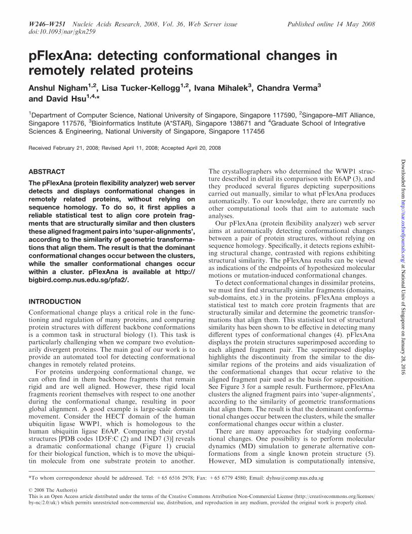
can often find in them backbone fragments that remainrigid and are well aligned. However, these rigid localfragments reorient themselves with respect to one anotherduring the conformational change, resulting in poorglobal alignment. A good example is large-scale domainmovement. Consider the HECT domain of the humanubiquitin ligase WWP1, which is homologous to thehuman ubiquitin ligase E6AP. Comparing their crystalstructures [PDB codes 1D5F:C (2) and 1ND7 (3)] revealsa dramatic conformational change (Figure 1) crucialfor their biological function, which is to move the ubiqui-tin molecule from one substrate protein to another.
The crystallographers who determined the WWP1 struc-ture described in detail its comparison with E6AP (3), andthey produced several figures depicting superpositionscarried out manually, similar to what pFlexAna producesautomatically. To our knowledge, there are currently noother computational tools that aim to automate suchanalyses.
Our pFlexAna (protein flexibility analyzer) web serveraims at automatically detecting conformational changesbetween a pair of protein structures, without relying onsequence homology. Specifically, it detects regions exhibit-ing structural change, contrasted with regions exhibitingstructural similarity. The pFlexAna results can be viewedas indications of the endpoints of hypothesized molecularmotions or mutation-induced conformational changes.
To detect conformational changes in dissimilar proteins,we must first find structurally similar fragments (domains,sub-domains, etc.) in the proteins. pFlexAna employs astatistical test to match core protein fragments that arestructurally similar and determine the geometric transfor-mations that align them. This statistical test of structuralsimilarity has been shown to be effective in detecting manydifferent types of conformational changes (4). pFlexAnadisplays the protein structures superimposed according toeach aligned fragment pair. The superimposed displayhighlights the discontinuity from the similar to the dis-similar regions of the proteins and aids visualization ofthe conformational changes that occur relative to thealigned fragment pair used as the basis for superposition.See Figure 3 for a sample result. Furthermore, pFlexAnaclusters the aligned fragment pairs into ‘super-alignments’,according to the similarity of geometric transformationsthat align them. The result is that the dominant conforma-tional changes occur between the clusters, while the smallerconformational changes occur within a cluster.
There are many approaches for studying conforma-tional changes. One possibility is to perform moleculardynamics (MD) simulation to generate alternative con-formations from a single known protein structure (5).However, MD simulation is computationally intensive,
*To whom correspondence should be addressed. Tel: +65 6516 2978; Fax: +65 6779 4580; Email: [email protected]
� 2008 The Author(s)
This is an Open Access article distributed under the terms of the Creative Commons Attribution Non-Commercial License (http://creativecommons.org/licenses/
by-nc/2.0/uk/) which permits unrestricted non-commercial use, distribution, and reproduction in any medium, provided the original work is properly cited.
at National U
niv of Singapore on January 28, 2016http://nar.oxfordjournals.org/
Dow
nloaded from
and usually it can only explore conformational changesthat are small in magnitude. An alternative approach is tocompare directly the structures of a protein in differentconformations. There are several good methods for this(4,6–8), but they can be applied only if the structures ofthe same protein in different conformations are available.
It is thus sometimes necessary to compare conforma-tions of related, but different proteins in order to inferconformational changes. One possibility is to performsequence alignment as a preprocessing step to match theproteins and then detect conformational changes betweenthem (9). This method, however, is restricted to proteinswith 90% sequence identity (9).
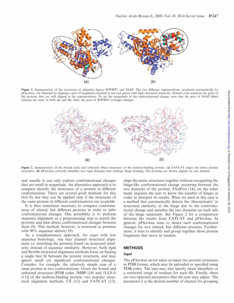
As a complementary approach, for cases with lowsequence homology, one may attempt structural align-ment i.e. matching the proteins based on structural simil-arity instead of sequence similarity. However, both rigidand flexible structural alignment methods focus on findinga single best fit between the protein structures, and mayignore small yet significant conformational changes.Consider, for example, the relatively simple case of asame protein in two conformations. Given the bound andunbound structures [PDB codes 3MBP (10) and 1LLS:A(11)] of the maltose-binding protein, two popular struc-tural alignment methods, CE (12) and FATCAT (13),
align the entire structures together without recognizing thehinge-like conformational change occurring between thetwo domains of the protein. FlexProt (14), on the otherhand, requires the user to know the number of hinges inorder to interpret its results. What we need in this case isa method that automatically detects the ‘discontinuity’ instructural similarity at the hinge due to the conforma-tional change and matches the two domains on each sideof the hinge separately. See Figure 2 for a comparisonbetween the results from FATCAT and pFlexAna. Ingeneral, pFlexAna aims to detect such conformationalchanges for two related, but different proteins. Further-more, it tries to identify and group together those proteinfragments that move in tandem.
METHODS
Input
The pFlexAna server takes as input two protein structuresin PDB format, which may be uploaded or specified usingPDB codes. The user may also specify chain identifiers ora restricted range of residues for each file. Finally, thereare two optional parameters that the user may adjust. Theparameter k is the desired number of clusters for grouping
Figure 1. Superposition of the structures of ubiquitin ligases WWWP1 and E6AP. The two different superpositions, produced automatically bypFlexAna, are obtained by aligning a pair of fragments (marked in red and green) with high structural similarity. Dotted ovals annotate the parts ofthe proteins that are well aligned in the superpositions. To see the magnitude of the conformational change, note that the pose of E6AP (blue)remains the same in both (a) and (b). Only the pose of WWWP1 (orange) changes.
Figure 2. Superposition of the bound (red) and unbound (blue) structures of the maltose-binding protein. (a) FATCAT aligns the entire proteinstructures. (b) pFlexAna correctly identifies two rigid domains that undergo hinge bending. The proteins are shown aligned on one domain.
Nucleic Acids Research, 2008, Vol. 36,Web Server issue W247
at National U
niv of Singapore on January 28, 2016http://nar.oxfordjournals.org/
Dow
nloaded from
the aligned fragments together, and � is the noise param-eter, which determines how strictly structural similarity isapplied. For low �-values, pFlexAna matches only frag-ments that are highly similar. For high �-values, it alsomatches fragments with weak similarity and ignores smalldifferences. Based on our experiences, �-values between0.2 and 0.4 work well for proteins with reasonably high-sequence homology (40% or higher). For proteins that areeven more distantly related, we find �-values up to 0.8 tobe useful as well, though this may lead to false positivematches in some cases.
Output
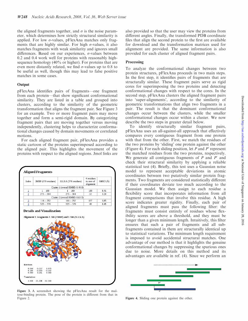
pFlexAna identifies pairs of fragments—one fragmentfrom each protein—that show significant conformationalsimilarity. They are listed in a table and grouped intoclusters, according to the similarity of the geometrictransformation that aligns the fragment pair. See Figure 3for an example. Two or more fragment pairs may movetogether and form a semi-rigid domain. By categorizingfragment pairs that are moving together versus movingindependently, clustering helps to characterize conforma-tional changes caused by domain movements or correlatedmotions.For each aligned fragment pair, pFlexAna provides a
static cartoon of the proteins superimposed according tothe aligned pair. This highlights the movement of theproteins with respect to the aligned regions. Jmol links are
also provided so that the user may view the proteins fromdifferent angles. Finally, the transformed PDB coordinatefiles that align the second protein to the first are availablefor download and the transformation matrices used foralignment are provided. The same information is alsoprovided for each cluster of aligned fragment pairs.
Processing
To analyze the conformational changes between twoprotein structures, pFlexAna proceeds in two main steps.In the first step, it identifies pairs of fragments that arestructurally similar. These fragment pairs serve as rigidcores for superimposing the two proteins and detectingconformational changes with respect to the cores. In thesecond step, pFlexAna clusters the aligned fragment pairsinto ‘super-alignments’, according to the similarity ofgeometric transformations that align two fragments in apair. The result is that the dominant conformationalchanges occur between the clusters, while the smallerconformational changes occur within a cluster. We nowdescribe the two steps in greater detail below.
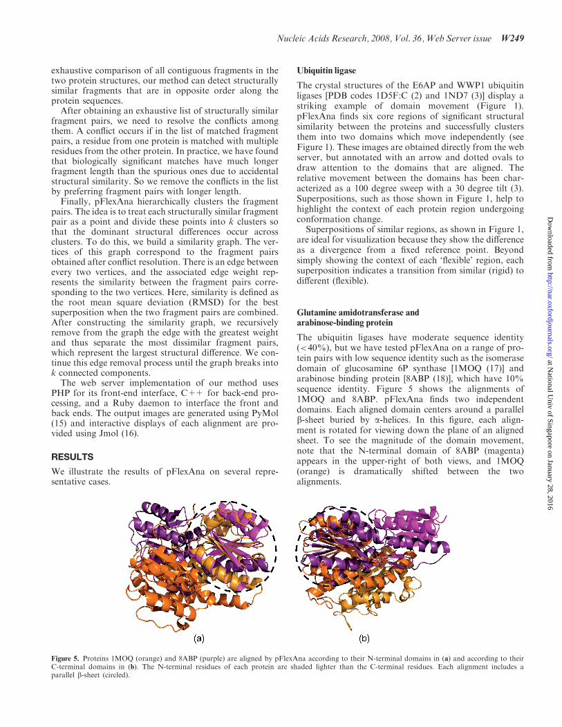
To identify structurally similar fragment pairs,pFlexAna uses an all-against-all approach that effectivelycompares every contiguous fragment from one proteinwith that from the other. First, we match the residues ofthe two proteins by ‘sliding’ one protein against the other(Figure 4). For each sliding position, let P and P0 representthe matched residues from the two proteins, respectively.We generate all contiguous fragments of P and P0 andcheck their structural similarity by applying a reliablestatistical test (4). Briefly, this test uses a Gaussian noisemodel to represent acceptable deviations in atomiccoordinates between two putatively similar protein frag-ments. Two fragments are considered statistically differentif their coordinates deviate too much according to theGaussian model. We then assign to each residue aflexibility score that incorporates information from allfragment comparisons that involve this residue. A highscore indicates greater rigidity. Finally, each pair ofaligned fragments must pass the following filter: thefragments must consist entirely of residues whose flex-ibility scores are above a threshold, and they must belonger than a given minimum length. Intuitively, this filterensures that such a pair of fragments and all sub-fragments contained in them are structurally identical upto statistical variations. The minimum length requirementis imposed to avoid accidental structural matches. Oneadvantage of our method is that it highlights the genuineconformational changes by suppressing the spurious onesdue to noise. More details on this method and itsadvantages are available in ref. (4). Since we perform an
Figure 3. A screenshot showing the pFlexAna result for the mal-tose-binding protein. The pose of the protein is different from that inFigure 2. Figure 4. Sliding one protein against the other.
W248 Nucleic Acids Research, 2008, Vol. 36,Web Server issue
at National U
niv of Singapore on January 28, 2016http://nar.oxfordjournals.org/
Dow
nloaded from
exhaustive comparison of all contiguous fragments in thetwo protein structures, our method can detect structurallysimilar fragments that are in opposite order along theprotein sequences.
After obtaining an exhaustive list of structurally similarfragment pairs, we need to resolve the conflicts amongthem. A conflict occurs if in the list of matched fragmentpairs, a residue from one protein is matched with multipleresidues from the other protein. In practice, we have foundthat biologically significant matches have much longerfragment length than the spurious ones due to accidentalstructural similarity. So we remove the conflicts in the listby preferring fragment pairs with longer length.
Finally, pFlexAna hierarchically clusters the fragmentpairs. The idea is to treat each structurally similar fragmentpair as a point and divide these points into k clusters sothat the dominant structural differences occur acrossclusters. To do this, we build a similarity graph. The ver-tices of this graph correspond to the fragment pairsobtained after conflict resolution. There is an edge betweenevery two vertices, and the associated edge weight rep-resents the similarity between the fragment pairs corre-sponding to the two vertices. Here, similarity is defined asthe root mean square deviation (RMSD) for the bestsuperposition when the two fragment pairs are combined.After constructing the similarity graph, we recursivelyremove from the graph the edge with the greatest weightand thus separate the most dissimilar fragment pairs,which represent the largest structural difference. We con-tinue this edge removal process until the graph breaks intok connected components.
The web server implementation of our method usesPHP for its front-end interface, C++ for back-end pro-cessing, and a Ruby daemon to interface the front andback ends. The output images are generated using PyMol(15) and interactive displays of each alignment are pro-vided using Jmol (16).
RESULTS
We illustrate the results of pFlexAna on several repre-sentative cases.
Ubiquitin ligase
The crystal structures of the E6AP and WWP1 ubiquitinligases [PDB codes 1D5F:C (2) and 1ND7 (3)] display astriking example of domain movement (Figure 1).pFlexAna finds six core regions of significant structuralsimilarity between the proteins and successfully clustersthem into two domains which move independently (seeFigure 1). These images are obtained directly from the webserver, but annotated with an arrow and dotted ovals todraw attention to the domains that are aligned. Therelative movement between the domains has been char-acterized as a 100 degree sweep with a 30 degree tilt (3).Superpositions, such as those shown in Figure 1, help tohighlight the context of each protein region undergoingconformation change.Superpositions of similar regions, as shown in Figure 1,
are ideal for visualization because they show the differenceas a divergence from a fixed reference point. Beyondsimply showing the context of each ‘flexible’ region, eachsuperposition indicates a transition from similar (rigid) todifferent (flexible).
Glutamine amidotransferase andarabinose-binding protein
The ubiquitin ligases have moderate sequence identity(<40%), but we have tested pFlexAna on a range of pro-tein pairs with low sequence identity such as the isomerasedomain of glucosamine 6P synthase [1MOQ (17)] andarabinose binding protein [8ABP (18)], which have 10%sequence identity. Figure 5 shows the alignments of1MOQ and 8ABP. pFlexAna finds two independentdomains. Each aligned domain centers around a parallelb-sheet buried by a-helices. In this figure, each align-ment is rotated for viewing down the plane of an alignedsheet. To see the magnitude of the domain movement,note that the N-terminal domain of 8ABP (magenta)appears in the upper-right of both views, and 1MOQ(orange) is dramatically shifted between the twoalignments.
Figure 5. Proteins 1MOQ (orange) and 8ABP (purple) are aligned by pFlexAna according to their N-terminal domains in (a) and according to theirC-terminal domains in (b). The N-terminal residues of each protein are shaded lighter than the C-terminal residues. Each alignment includes aparallel b-sheet (circled).
Nucleic Acids Research, 2008, Vol. 36,Web Server issue W249
at National U
niv of Singapore on January 28, 2016http://nar.oxfordjournals.org/
Dow
nloaded from
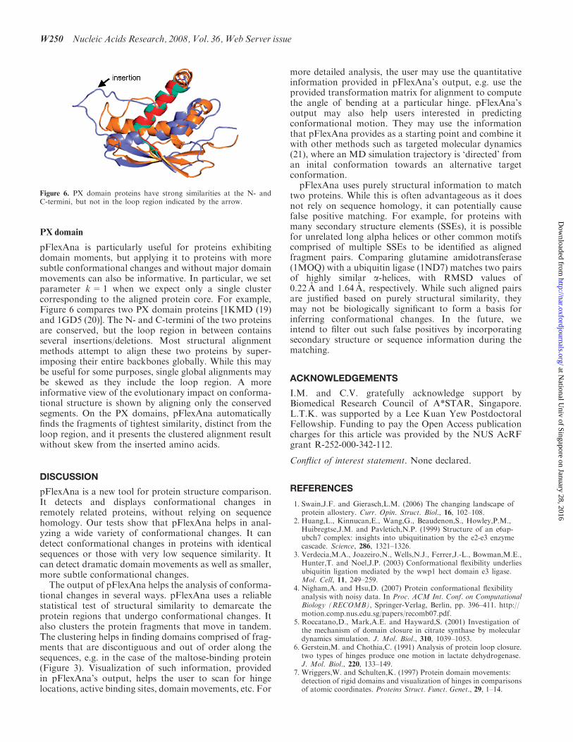
PX domain
pFlexAna is particularly useful for proteins exhibitingdomain moments, but applying it to proteins with moresubtle conformational changes and without major domainmovements can also be informative. In particular, we setparameter k=1 when we expect only a single clustercorresponding to the aligned protein core. For example,Figure 6 compares two PX domain proteins [1KMD (19)and 1GD5 (20)]. The N- and C-termini of the two proteinsare conserved, but the loop region in between containsseveral insertions/deletions. Most structural alignmentmethods attempt to align these two proteins by super-imposing their entire backbones globally. While this maybe useful for some purposes, single global alignments maybe skewed as they include the loop region. A moreinformative view of the evolutionary impact on conforma-tional structure is shown by aligning only the conservedsegments. On the PX domains, pFlexAna automaticallyfinds the fragments of tightest similarity, distinct from theloop region, and it presents the clustered alignment resultwithout skew from the inserted amino acids.
DISCUSSION
pFlexAna is a new tool for protein structure comparison.It detects and displays conformational changes inremotely related proteins, without relying on sequencehomology. Our tests show that pFlexAna helps in anal-yzing a wide variety of conformational changes. It candetect conformational changes in proteins with identicalsequences or those with very low sequence similarity. Itcan detect dramatic domain movements as well as smaller,more subtle conformational changes.The output of pFlexAna helps the analysis of conforma-
tional changes in several ways. pFlexAna uses a reliablestatistical test of structural similarity to demarcate theprotein regions that undergo conformational changes. Italso clusters the protein fragments that move in tandem.The clustering helps in finding domains comprised of frag-ments that are discontiguous and out of order along thesequences, e.g. in the case of the maltose-binding protein(Figure 3). Visualization of such information, providedin pFlexAna’s output, helps the user to scan for hingelocations, active binding sites, domain movements, etc. For
more detailed analysis, the user may use the quantitativeinformation provided in pFlexAna’s output, e.g. use theprovided transformation matrix for alignment to computethe angle of bending at a particular hinge. pFlexAna’soutput may also help users interested in predictingconformational motion. They may use the informationthat pFlexAna provides as a starting point and combine itwith other methods such as targeted molecular dynamics(21), where an MD simulation trajectory is ‘directed’ froman inital conformation towards an alternative targetconformation.
pFlexAna uses purely structural information to matchtwo proteins. While this is often advantageous as it doesnot rely on sequence homology, it can potentially causefalse positive matching. For example, for proteins withmany secondary structure elements (SSEs), it is possiblefor unrelated long alpha helices or other common motifscomprised of multiple SSEs to be identified as alignedfragment pairs. Comparing glutamine amidotransferase(1MOQ) with a ubiquitin ligase (1ND7) matches two pairsof highly similar a-helices, with RMSD values of0.22 A and 1.64 A, respectively. While such aligned pairsare justified based on purely structural similarity, theymay not be biologically significant to form a basis forinferring conformational changes. In the future, weintend to filter out such false positives by incorporatingsecondary structure or sequence information during thematching.
ACKNOWLEDGEMENTS
I.M. and C.V. gratefully acknowledge support byBiomedical Research Council of A*STAR, Singapore.L.T.K. was supported by a Lee Kuan Yew PostdoctoralFellowship. Funding to pay the Open Access publicationcharges for this article was provided by the NUS AcRFgrant R-252-000-342-112.
Conflict of interest statement. None declared.
REFERENCES
1. Swain,J.F. and Gierasch,L.M. (2006) The changing landscape ofprotein allostery. Curr. Opin. Struct. Biol., 16, 102–108.
2. Huang,L., Kinnucan,E., Wang,G., Beaudenon,S., Howley,P.M.,Huibregtse,J.M. and Pavletich,N.P. (1999) Structure of an e6ap-ubch7 complex: insights into ubiquitination by the e2-e3 enzymecascade. Science, 286, 1321–1326.
3. Verdecia,M.A., Joazeiro,N., Wells,N.J., Ferrer,J.-L., Bowman,M.E.,Hunter,T. and Noel,J.P. (2003) Conformational flexibility underliesubiquitin ligation mediated by the wwp1 hect domain e3 ligase.Mol. Cell, 11, 249–259.
4. Nigham,A. and Hsu,D. (2007) Protein conformational flexibilityanalysis with noisy data. In Proc. ACM Int. Conf. on ComputationalBiology (RECOMB), Springer-Verlag, Berlin, pp. 396–411. http://motion.comp.nus.edu.sg/papers/recomb07.pdf.
5. Roccatano,D., Mark,A.E. and Hayward,S. (2001) Investigation ofthe mechanism of domain closure in citrate synthase by moleculardynamics simulation. J. Mol. Biol., 310, 1039–1053.
6. Gerstein,M. and Chothia,C. (1991) Analysis of protein loop closure.two types of hinges produce one motion in lactate dehydrogenase.J. Mol. Biol., 220, 133–149.
7. Wriggers,W. and Schulten,K. (1997) Protein domain movements:detection of rigid domains and visualization of hinges in comparisonsof atomic coordinates. Proteins Struct. Funct. Genet., 29, 1–14.
Figure 6. PX domain proteins have strong similarities at the N- andC-termini, but not in the loop region indicated by the arrow.
W250 Nucleic Acids Research, 2008, Vol. 36,Web Server issue
at National U
niv of Singapore on January 28, 2016http://nar.oxfordjournals.org/
Dow
nloaded from
8. Hayward,S. and Berendsen,H.J.C. (1998) Systematic analysis ofdomain motions in proteins from conformational change: newresults on citrate synthase and t4 lysozyme. Proteins Struct. Funct.Genet., 30, 144–154.
9. Qi,G., Lee,R. and Hayward,S. (2005) A comprehensive and non-redundant database of protein domain movements. Bioinformatics,21, 2832–2838.
10. Quiocho,F.A., Spurlino,J.C. and Rodseth,L.E. (1997) Extensivefeatures of tight oligosaccharide binding revealed in high-resolutionstructures of the maltodextrin transport/chemosensory receptor.Structure, 5, 997–1015.
11. Rubin,S.M., Lee,S.Y., Ruiz,E.J., Pines,A. and Wemmer,D.E. (2002)Detection and characterization of xenon-binding sites in proteins by129xe nmr spectroscopy. J. Mol. Biol., 322, 425–440.
12. Shindyalov,I.N. and Bourne,P.E. (1998) Protein structure alignmentby incremental combinatorial extension (ce) of the optimal path.Protein Eng., 11, 739–747.
13. Ye,Y. and Godzik,A. (2003) Flexible structure alignment bychaining aligned fragment pairs allowing twists. Bioinformatics, 19(Suppl 2), ii246–ii255.
14. Shatsky,M., Wolfson,H.J. and Nussinov,R. (2002) Flexible proteinalignment and hinge detection. Proteins Struct. Funct. Genet., 48,242–256.
15. Delano,W.L. (2002) The PyMOL User’s Manual. DeLano Scientific,Palo Alto, CA.
16. Herraez,A. (2006) Biomolecules in the computer: Jmol to the rescue.Biochem. Mol. Biol. Educat., 34, 255–261.
17. Teplyakov,A., Obmolova,G., Badet-Denisot,M.A., Badet,B. andPolikarpov,I. (1998) Involvement of the c terminus in intramolec-ular nitrogen channeling in glucosamine 6-phosphate synthase:evidence from a 1.6 a crystal structure of the isomerase domain.Structure, 6, 1047–1055.
18. Vermersch,P.S., Lemon,D.D., Tesmer,J.J. and Quiocho,F.A. (1991)Sugar-binding and crystallographic studies of an arabinose-bindingprotein mutant (met108leu) that exhibits enhanced affinity andaltered specificity. Biochemistry, 30, 6861–6866.
19. Lu,J., Garcia,J., Dulubova,I., Sudhof,T.C. and Rizo,J. (2002)Solution structure of the vam7p px domain. Biochemistry, 41,5956–5962.
20. Hiroaki,H., Ago,T., Ito,T., Sumimoto,H. and Kohda,D. (2001)Solution structure of the px domain, a target of the sh3 domain.Nat. Struct. Biol., 8, 526–530.
21. van der Vaart,A. and Karplus,M. (2005) Simulation of conforma-tional transitions by the restricted perturbation-targeted moleculardynamics method. J. Chem. Phys., 122, 114903.
Nucleic Acids Research, 2008, Vol. 36,Web Server issue W251
at National U
niv of Singapore on January 28, 2016http://nar.oxfordjournals.org/
Dow
nloaded from