petechiae, purpura and vasculitis - cfpm ... - cfpm … · –diascopy refers to the use of a glass...
TRANSCRIPT

Petechiae, Purpura
and Vasculitis
Agnieszka Kupiec, MD
Georgetown Dermatology
Washington, DC
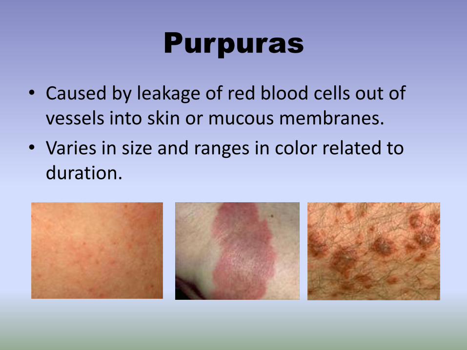
Purpuras
• Caused by leakage of red blood cells out of vessels into skin or mucous membranes.
• Varies in size and ranges in color related to duration.

Purpuras
• Either Non-Palpable or Palpable Purpura
– Non-palpable (flat): Petechiae (less then 3mm), Ecchymoses (more then 5mm)
– Palpable: Elevated Purpuras
Purpura
• The type of lesion present is usually indicative of the underlying pathogenesis:
– Macular purpura is typically non-inflammatory
– Palpable purpura is a sign of vascular inflammation (vasculitis)

Non-Palpable Purpura:
Ecchymoses
• A discoloration of the skin or mucous membranes resulting from extravasation of blood with color change over time with a characteristic transition of color ranging from blue-black, brown-yellow, or green.
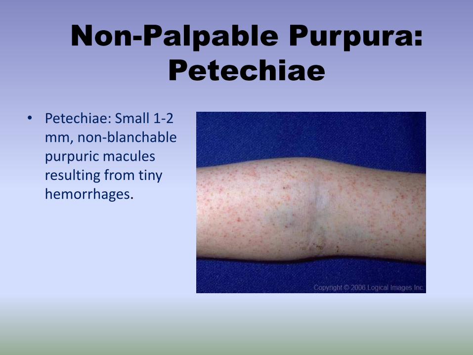
Non-Palpable Purpura:
Petechiae
• Petechiae: Small 1-2 mm, non-blanchable purpuric macules resulting from tiny hemorrhages.
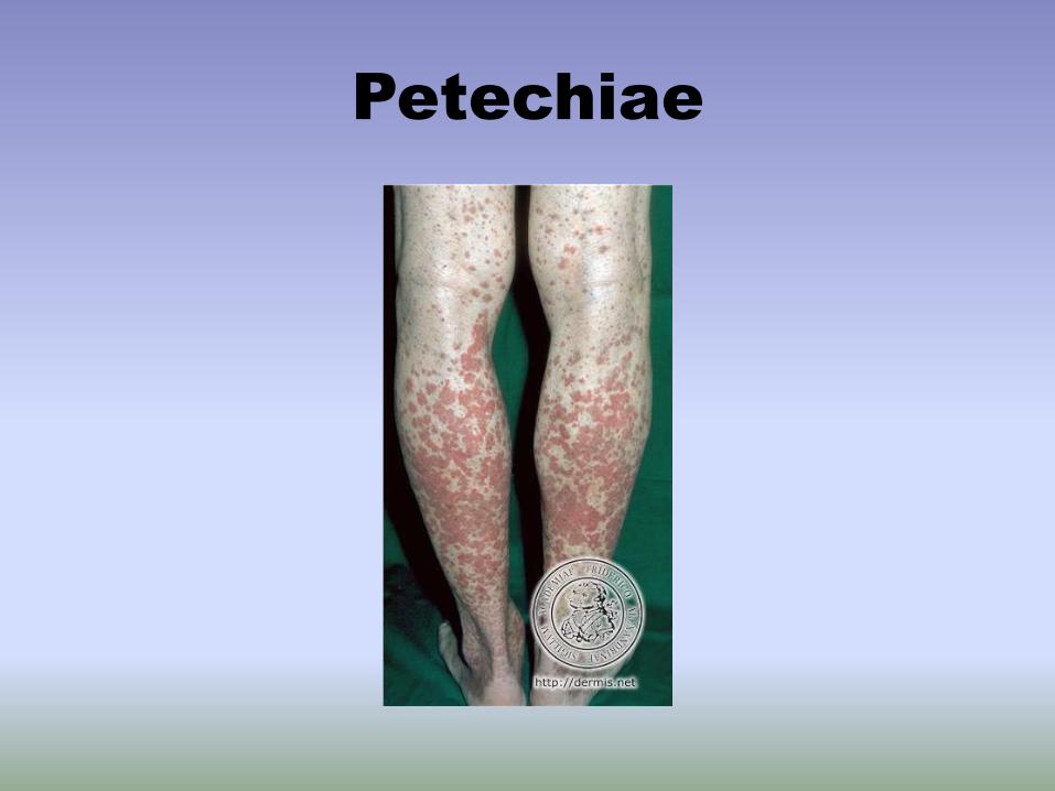
Petechiae

Palpable Purpura
• Palpable Purpura:
Raised and palpable
red or violaceous
discoloration of skin
or mucous
membranes due to
vascular inflammation
in the skin and
extravasation of red
blood cells.

Purpura: The Basics
• All forms do not blanch when pressed
– Diascopy refers to the use of a glass slide to apply pressure to the lesion in order to distinguish erythema secondary to vasodilation (blanchable with pressure), from erythrocyte extravasation (retains its red color)
• Purpura may result from hyper- and hypo-coagulable states, vascular dysfunction and extravascular causes
9
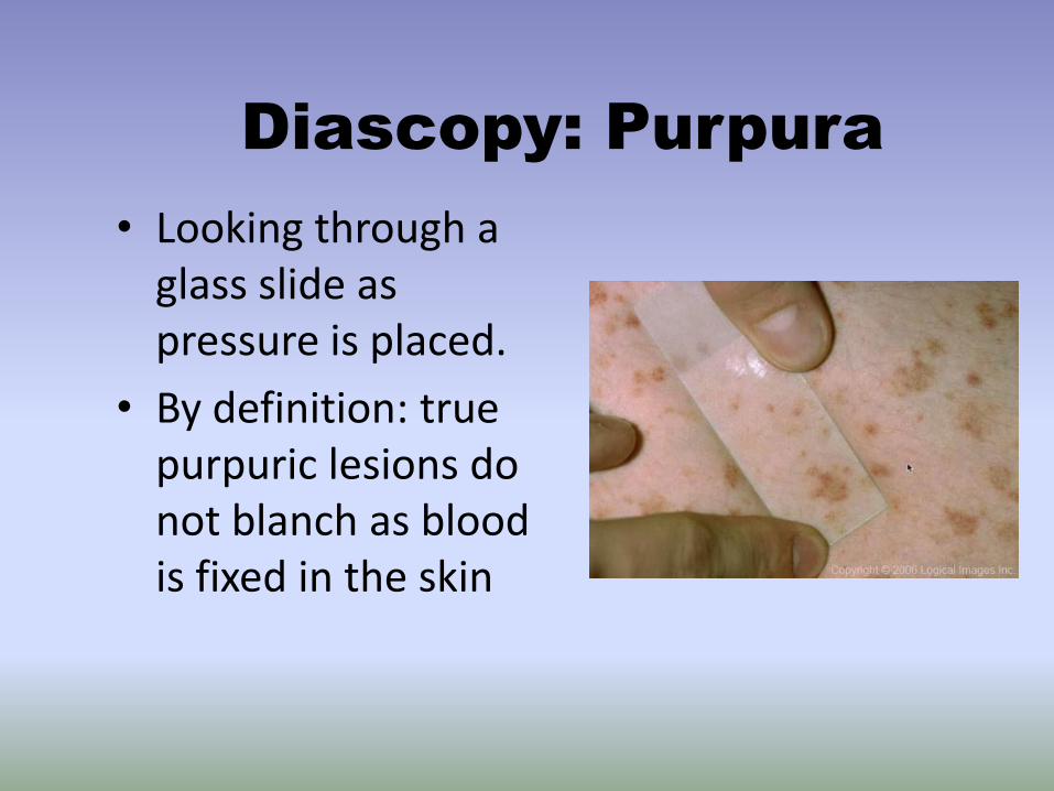
Diascopy: Purpura
• Looking through a glass slide as pressure is placed.
• By definition: true purpuric lesions do not blanch as blood is fixed in the skin

Examples of Purpura
Petechia
Ecchymosis
11
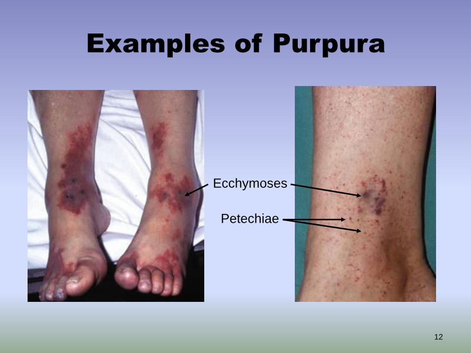
Examples of Purpura
Ecchymoses
Petechiae
12

Case One
13

Case One: History
HPI: 42-year-old man who presents to the ER with a 2-week
history of a rash on his abdomen and lower extremities.
PMH: hospitalization 1 year ago for community acquired
pneumonia
Medications: none
Allergies: none
Family history: unknown
Social history: without stable housing, no recent travel or
exposure to animals
Health-related behaviors: smokes 10 cigarettes/day, drinks 3-10
beers/day, limited access to food
ROS: easy bruising, bleeding from gums, overall fatigue
14
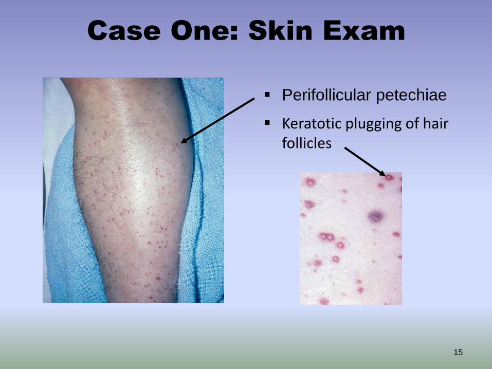
Case One: Skin Exam
Perifollicular petechiae
15
Keratotic plugging of hair follicles

Case One: Exam
hemorrhagic gingivitis
16
Case One, Question 1
Which of the following is the most likely
diagnosis?
a. Drug hypersensitivity reaction
b. Nutritional deficiency
c. Rocky mountain spotted fever
d. Urticaria
e. Vasculitis
17
Case One, Question 1
Answer: b
Which of the following is the most likely diagnosis?
a. Drug hypersensitivity reaction (typically without
purpuric lesions)
b. Nutritional deficiency
c. Urticaria (would expect raised edematous lesions, not
purpura)
d. Vasculitis (purpura would not be perifollicular and
would be palpable)
e. Rocky mountain spotted fever (no history of travel or
tick bite)
18

Vitamin C Deficiency -
Scurvy
• Scurvy results from insufficient vitamin C intake (e.g., fat diet, alcoholism), increased vitamin requirement (e.g., certain medications), and increased loss (e.g., dialysis)
• Vitamin C is required for normal collagen structure and its absence leads to skin and vessel fragility
19

Vitamin C Deficiency - Scurvy
Characteristic exam findings include:
• Perifollicular purpura
• Large ecchymoses on the lower legs
• Intramuscular and periosteal hemorrhage
• Keratotic plugging of hair follicles
• Hemorrhagic gingivitis (when patient has poor
oral hygiene)
Remember to take a dietary history in all
patients with purpura
20

Vitamin C Deficiency -
Scurvy

Case Two
22

Case Two: History
HPI: 19-year-old man who was admitted to the hospital with a
headache, stiff neck, high fever, and rash. His symptoms began 2-3
days prior to admission when he developed fevers with nausea and
vomiting.
PMH: splenectomy 3 years ago after a snowboarding accident
Medications: none
Allergies: none
Vaccination history: last vaccination as a child
Family history: non-contributory
Social history: attends a near-by state college, lives in a dormitory
Health-related behaviors: reports occasional alcohol use on the
weekends with 2-3 drinks per night, plays basketball with friends for
exercise
23
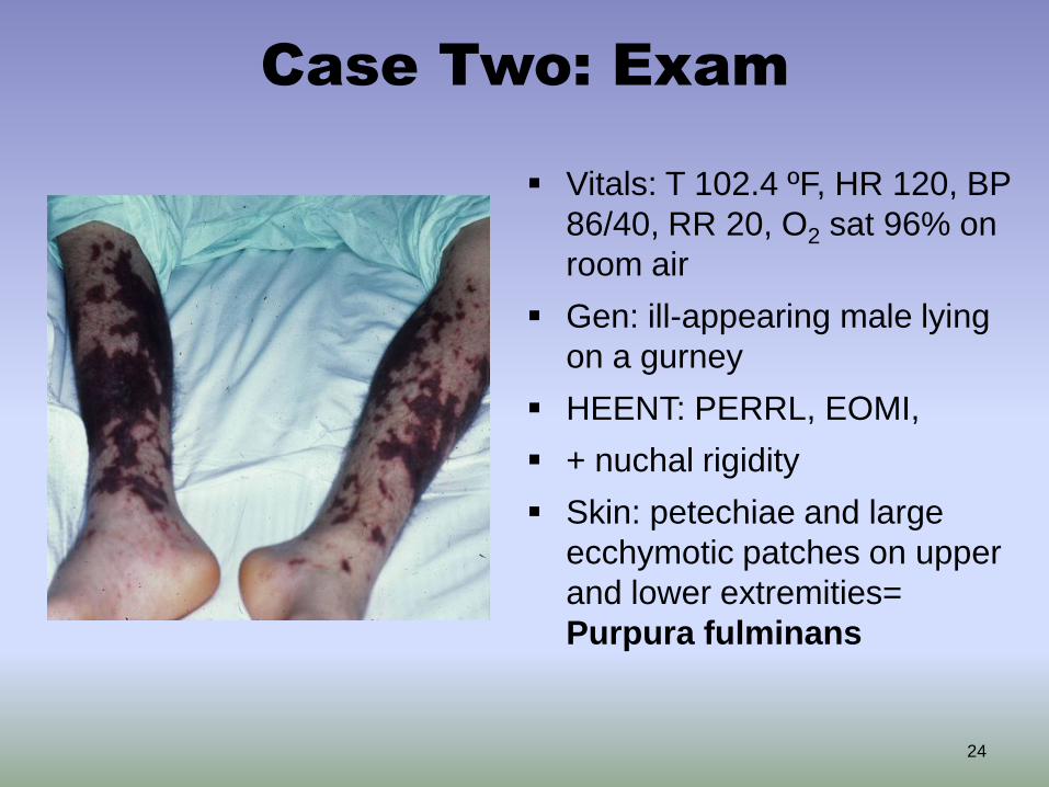
Case Two: Exam
Vitals: T 102.4 ºF, HR 120, BP
86/40, RR 20, O2 sat 96% on
room air
Gen: ill-appearing male lying
on a gurney
HEENT: PERRL, EOMI,
+ nuchal rigidity
Skin: petechiae and large
ecchymotic patches on upper
and lower extremities=
Purpura fulminans
24
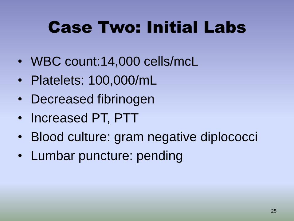
Case Two: Initial Labs
• WBC count:14,000 cells/mcL
• Platelets: 100,000/mL
• Decreased fibrinogen
• Increased PT, PTT
• Blood culture: gram negative diplococci
• Lumbar puncture: pending
25

Case Two, Question 1
Answer: a
In addition to fluid resuscitation, what is the most
needed treatment at this time?
a. IV antibiotics (may be started before lumbar
puncture)
b. IV corticosteroids (not unless suspicion for
pneumococcal meningitis is high)
c. Pain relief with oxycodone (not the patient’s primary
issue)
d. Plasmapheresis (not unless suspecting diagnosis of
thrombotic thrombocytopenic purpura – TTP)
26
Sepsis and DIC
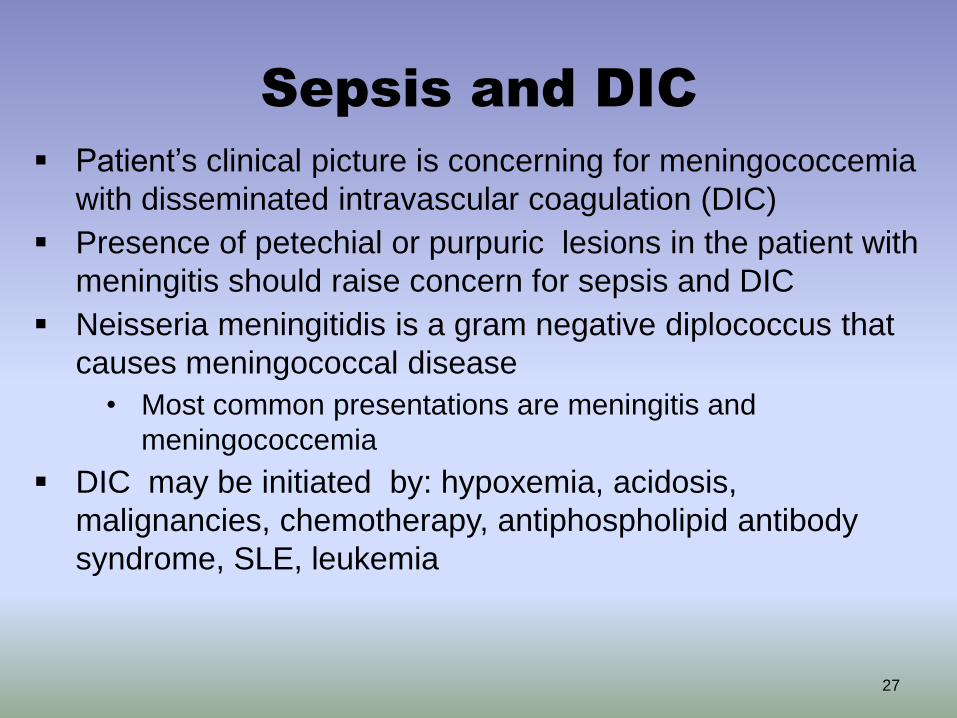
Patient’s clinical picture is concerning for meningococcemia
with disseminated intravascular coagulation (DIC)
Presence of petechial or purpuric lesions in the patient with
meningitis should raise concern for sepsis and DIC
Neisseria meningitidis is a gram negative diplococcus that
causes meningococcal disease
• Most common presentations are meningitis and
meningococcemia
DIC may be initiated by: hypoxemia, acidosis,
malignancies, chemotherapy, antiphospholipid antibody
syndrome, SLE, leukemia
27
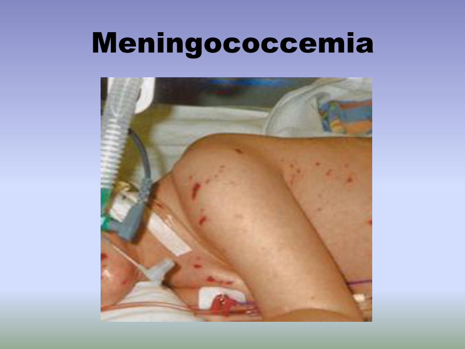
Meningococcemia
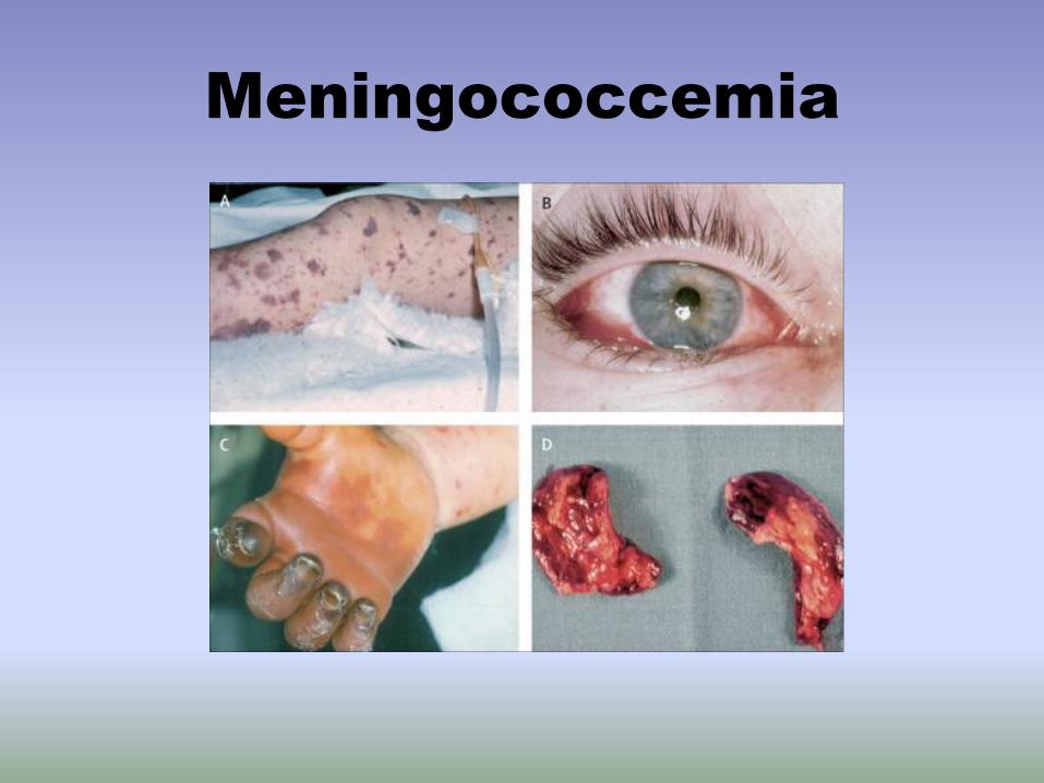
Meningococcemia
Disseminated Intravascular
Coagulation
• Skin lesions may be the initial manifestation
• Wide spread petechiae, ecchymoses, ischemic necrosis of the skin, and hemorrhagic bullae
• Purpura fulminans may supervene and progress to symetrical gangrene
• DIC results from unregulated intravascular
clotting resulting in depletion of clotting factors
and bleeding • The primary treatment is to treat the underlying condition

Purpura Fulminans - DIC

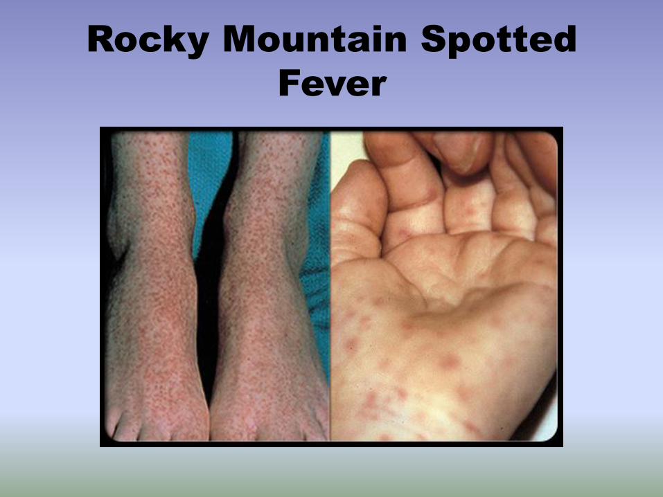
Rocky Mountain Spotted Fever
Another life-threatening diagnosis to consider in a patient with
a petechial rash is Rocky Mountain Spotted Fever (RMSF)
The most commonly fatal tickborne infection (caused by
Rickettsia rickettsii) in the US
A petechial rash is a frequent finding that usually occurs
several days after the onset of fever
Initially characterized by faint macules on the wrists or
ankles. As the disease progresses, the rash may become
petechial and involves the trunk, extremities, palms and soles
Majority of patients do not have the classic triad of fever, rash
and history of tick bite
32
Rocky Mountain Spotted
Fever

Rocky Mountain Spotted
Fever

Clinical Evaluation of Purpura
A history and physical exam is often all that is necessary
Important history items include:
• Family history of bleeding or thrombotic disorders (e.g., von
Willebrand disease)
• Use of drugs and medications (e.g., aspirin, warfarin) that
may affect platelet function and coagulation
• Medical conditions (e.g., liver disease) that may result in
altered coagulation
Complete blood count with differential and PT/PTT are
used to help assess platelet function and evaluate
coagulation states
35
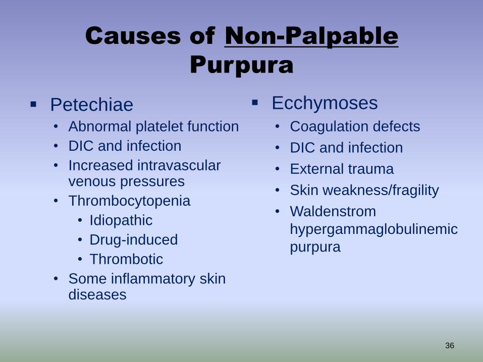
Causes of Non-Palpable
Purpura
Petechiae • Abnormal platelet function
• DIC and infection
• Increased intravascular venous pressures
• Thrombocytopenia
• Idiopathic
• Drug-induced
• Thrombotic
• Some inflammatory skin diseases
Ecchymoses
• Coagulation defects
• DIC and infection
• External trauma
• Skin weakness/fragility
• Waldenstrom
hypergammaglobulinemic
purpura
36

Palpable Purpura
37
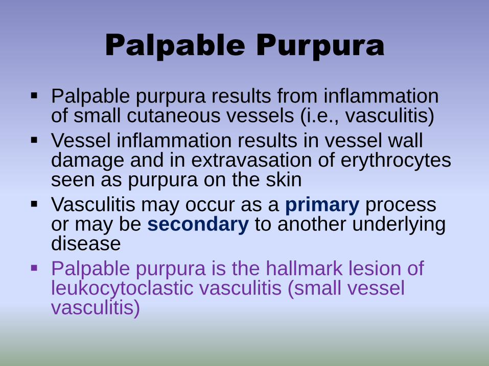
Palpable Purpura
Palpable purpura results from inflammation of small cutaneous vessels (i.e., vasculitis)
Vessel inflammation results in vessel wall damage and in extravasation of erythrocytes seen as purpura on the skin
Vasculitis may occur as a primary process or may be secondary to another underlying disease
Palpable purpura is the hallmark lesion of leukocytoclastic vasculitis (small vessel vasculitis)

Vasculitis Morphology
Vasculitis is classified by the vessel size affected (small,
medium, mixed or large)
Clinical morphology correlates with the size of the
affected blood vessels
• Small vessel: palpable purpura (urticarial lesions in rare cases,
e.g., urticarial vasculitis)
• Small to medium vessel: subcutaneous nodules, purpura and
FIXED livedo reticularis (also called livedo racemosa). Ulceration
and necrosis may be present in medium-vessel vasculitis.
• Large vessel: claudication, ulceration and necrosis
Diseases may involve more than one size of vessel
Systemic vasculitis may involve vessels in other organs
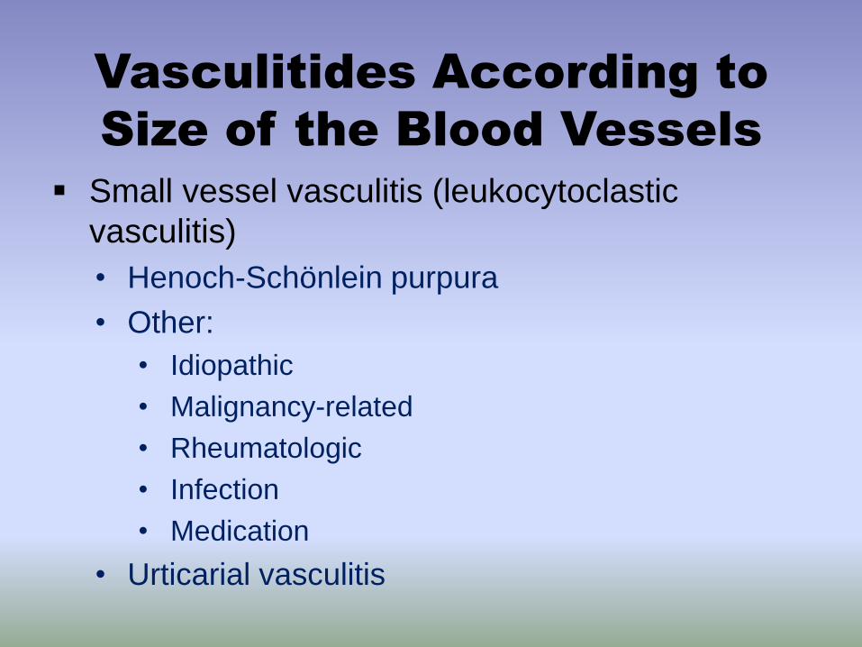
Vasculitides According to
Size of the Blood Vessels
Small vessel vasculitis (leukocytoclastic
vasculitis)
• Henoch-Schönlein purpura
• Other:
• Idiopathic
• Malignancy-related
• Rheumatologic
• Infection
• Medication
• Urticarial vasculitis
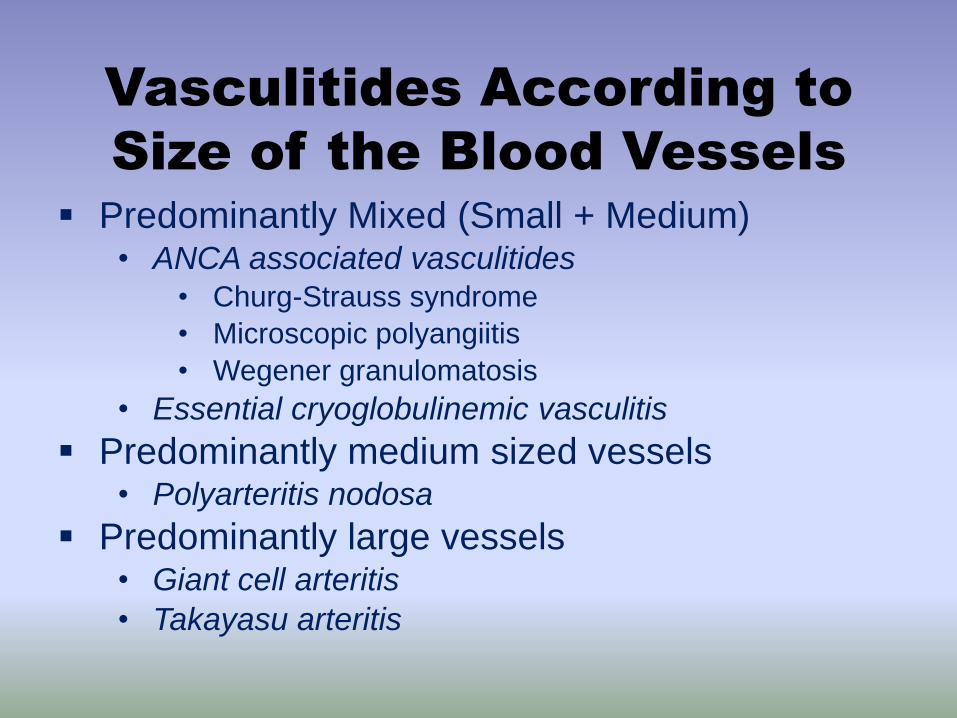
Vasculitides According to
Size of the Blood Vessels
Predominantly Mixed (Small + Medium) • ANCA associated vasculitides
• Churg-Strauss syndrome
• Microscopic polyangiitis
• Wegener granulomatosis
• Essential cryoglobulinemic vasculitis
Predominantly medium sized vessels • Polyarteritis nodosa
Predominantly large vessels • Giant cell arteritis
• Takayasu arteritis
Clinical Evaluation of
Vasculitis
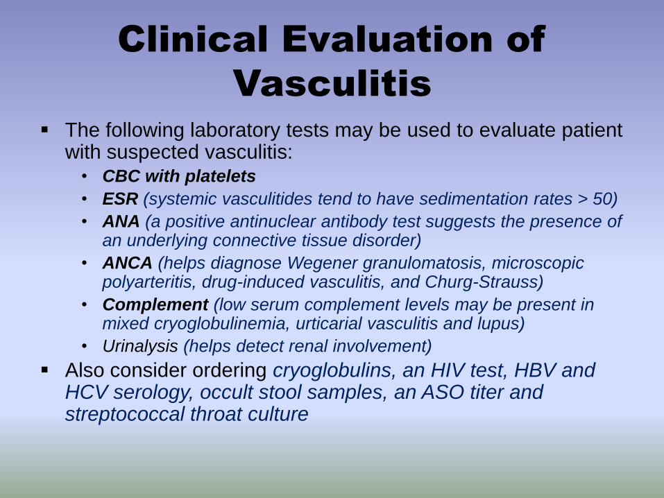
The following laboratory tests may be used to evaluate patient with suspected vasculitis:
• CBC with platelets
• ESR (systemic vasculitides tend to have sedimentation rates > 50)
• ANA (a positive antinuclear antibody test suggests the presence of an underlying connective tissue disorder)
• ANCA (helps diagnose Wegener granulomatosis, microscopic polyarteritis, drug-induced vasculitis, and Churg-Strauss)
• Complement (low serum complement levels may be present in mixed cryoglobulinemia, urticarial vasculitis and lupus)
• Urinalysis (helps detect renal involvement)
Also consider ordering cryoglobulins, an HIV test, HBV and HCV serology, occult stool samples, an ASO titer and streptococcal throat culture

Case Three
43

Case Three: History
HPI: 9-year-old girl with a 4-day history of abdominal pain and
rash on the lower extremities who was brought to the ER by her
mother. Her mother reported that the rash appeared suddenly
and was accompanied by joint pain of the knees and ankles and
aching abdominal pain. Over 3 days the rash changed from red
patches to more diffuse purple bumps.
PMH: no major illnesses or hospitalizations
Medications: none, up to date on vaccines
Allergies: none
Family history: no history of clotting or bleeding disorders
ROS: cough and runny nose a few weeks ago
44

Case Three: Skin Exam
Non-blanching erythematous
macules and papules on both legs
and feet (sparing the trunk, upper
extremities and face); diffuse
petechiae
45

Case Three, Question 1
In this clinical context, what test will
establish the diagnosis?
a. CBC
b. ESR
c. HIV test
d. Skin biopsy
e. Urinalysis
46

Case Three, Question 1
Answer: d
In this clinical context, what test will
establish the diagnosis?
a. CBC
b. ESR
c. HIV test
d. Skin biopsy (for routine microscopy and
direct immunofluorescence)
e. Urinalysis
47
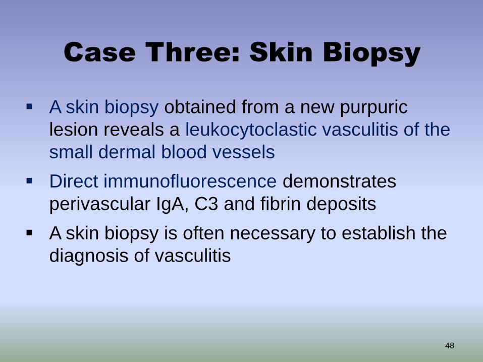
Case Three: Skin Biopsy
A skin biopsy obtained from a new purpuric
lesion reveals a leukocytoclastic vasculitis of the
small dermal blood vessels
Direct immunofluorescence demonstrates
perivascular IgA, C3 and fibrin deposits
A skin biopsy is often necessary to establish the
diagnosis of vasculitis
48
Case Three, Question 2
What is the most likely diagnosis?
a. Disseminated intravascular coagulation
b. Henoch-Schönlein Purpura
c. Idiopathic thrombocytopenic purpura
d. Sepsis
e. Urticaria
49
Case Three, Question 2
Answer: b
What is the most likely diagnosis?
a. Disseminated intravascular coagulation
b. Henoch-Schönlein Purpura
c. Idiopathic thrombocytopenic purpura
d. Sepsis
e. Urticaria
50

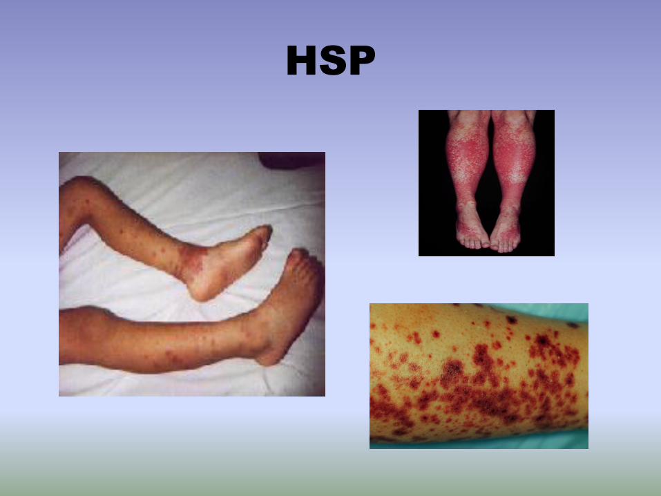
Henoch-Schonlein Purpura
• Henoch-Schönlein Purpura (HSP) is the most common form of systemic vasculitis in children – Characterized by palpable purpura (vasculitis), arthritis,
abdominal pain and kidney disease
• Primarily a childhood disease (between ages 3-15), but adults can also be affected
• HSP follows a seasonal pattern with a peak in incidence during the winter presumably due to association with a preceding viral or bacterial (streptococcal pharyngitis) infection
• Other bacterial infections, drugs, food and lymphoma may cause HSP 51

HSP: Diagnosis and
Evaluation
Diagnosis often made on clinical presentation
+/- skin biopsy
Skin biopsy shows leukocytoclastic vasculitis in
postcapillary venules (small vessel disease)
• Immune complexes in vessel walls contain IgA
deposition (the diagnostic feature of HSP)
Rule out streptococcal infection with an ASO or
throat culture

HSP: Evaluation and
Treatment
It is also important to look for systemic disease:
• Renal: Urinalysis, BUN/Cr
• Gastrointestinal: Stool guaiac
• HSP in adults may be a manifestation of underlying
malignancy
Natural History: most children completely
recover from HSP
• Some develop progressive renal disease
• More common in adults
Treatment is supportive +/- prednisone
HSP

Case Four
55

Case Four: History
45-year-old man who was admitted to the hospital
five weeks ago with acute bacterial endocarditis.
After an appropriate antibiotic regimen was
started and patient was stable, he was transferred
to a skilled nursing facility to finish a six-week
course of IV antibiotics.
On week #5, the patient developed a rash on his
lower extremities.
56
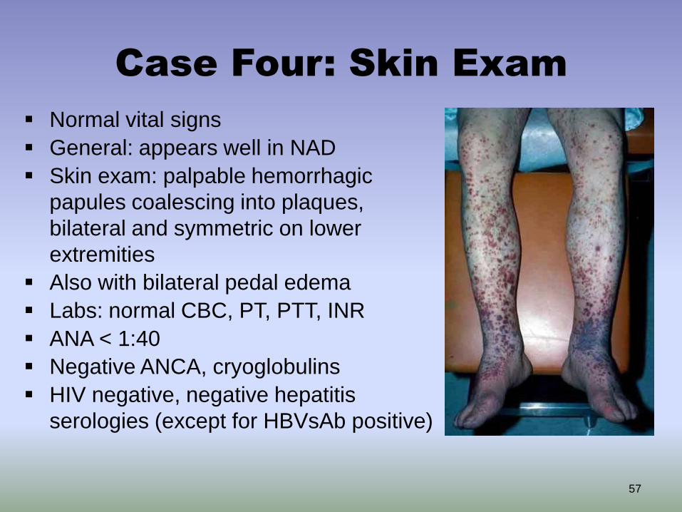
Case Four: Skin Exam
Normal vital signs
General: appears well in NAD
Skin exam: palpable hemorrhagic
papules coalescing into plaques,
bilateral and symmetric on lower
extremities
Also with bilateral pedal edema
Labs: normal CBC, PT, PTT, INR
ANA < 1:40
Negative ANCA, cryoglobulins
HIV negative, negative hepatitis
serologies (except for HBVsAb positive)
57

Case Four, Question 1
Which of the following is the most likely
cause of skin findings?
a. DIC secondary to sepsis
b. Leukocytoclastic vasculitis secondary to
antibiotics
c. Septic emboli with hemorrhage from
undiagnosed bacterial endocarditis
d. Urticarial vasculitis
58

Case Four, Question 1
Answer: b
Which of the following is the most likely cause of Mr.
Burton’s skin findings?
a. DIC secondary to sepsis (history and skin exam are less
concerning for sepsis. In DIC, coagulation studies are
abnormal)
b. Leukocytoclastic vasculitis secondary to antibiotics
c. Septic emboli with hemorrhage (these lesions tend to
occur on the distal extremities)
d. Urticarial vasculitis (presents with a different morphology,
which is urticarial)
59
Case Four, Question 2
A skin biopsy confirmed LCV. What else
should be done at this time?
a. Obtain a urinalysis
b. Start systemic steroid
c. Stop the IV antibiotics and replace with
another
d. All of the above
60

Case Four, Question 2
Answer: a & c
What else should be done at this time?
a. Obtain a urinalysis (detection of renal
involvement will impact treatment)
b. Start systemic steroid (typically used when
vasculitis is systemic or severe)
c. Stop the IV antibiotics and replace with
another (remove the offending agent)
d. All of the above
61

LCV: Etiology
• Acute infection – beta-hemolytic Streptococcus group A, and rarely Mycobacteruim tuberculosis
• Lymphoproliferative neoplasms
• Solid tumors: lungs, colon, prostate and breast
• Connective Tissue Disease

LCV: Evaluation
• CBC
• Urinanalysis
• ASO titer
• ANA
• Hepatitis serology
• ANCA
• Serum cryoglobulins
• Skin biopsy

LCV: Treatment
• Patients with normal UA and clinically well – nonaggressive treatment: rest , elevation of the leg, analgesics, avoidance of trauma and treat underlying cause
• NSAIDs,
• Colchicine, dapsone for chronic vasculitis
• Systemic corticosteroids for serious systemic manifestations or necrotic lesions

Leukocytoclastic vasculitis
Palpable hemorrhagic papules coalescing into plaques, bilateral on the lower extremities
65

Leukocytoclastic Vasculitis

Case Five
67

Case Five: History
HPI: 34-year-old previously healthy woman who was admitted to
the hospital with a 5-day history of fever, weight loss, joint
pain/swelling, paresthesias (both feet), and painful skin nodules
PMH: mild normocytic anemia
Medications: OCP, Malarone (malaria prophylaxis)
Allergies: sulfa
Family history: no autoimmune disease
Social history: worked in Haiti one month prior to admission; no
animal contacts
Health-related behaviors: no tobacco, alcohol, or drug use
ROS: reports no photosensitivity, malar rash, mucosal ulcers, dry
eyes/mouth, sore throat, abdominal pain, or dysuria
68

Case Five: Exam
Vitals: febrile
Gen: well-appearing, very thin
female
HEENT: clear, no ulcers noted
No lymphadenopathy
Normal cardiac, respiratory,
abdominal exams
Neurologic exam: decreased
sensation and reflexes in
bilateral legs
Joint exam: warmth/swelling at
hands, feet, ankles, knees
69
Skin: lower extremities with
multiple 6x8mm tender,
erythematous subcutaneous
nodules
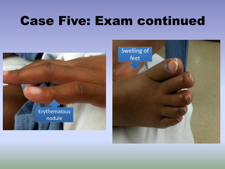
Case Five: Exam continued
Swelling of feet
Erythematous nodule

Case Five: Labs
• CBC: anemia
• ESR: 60 [0-15]; CRP: 46.8 [<6.3]
• Normal liver function, BUN/Cr
• UA: trace hemoglobin, trace protein
– 24 hour urine: increased protein
– Urine sediment: RBCs, WBCs, tubular casts; no dysmorphic RBCs
• CXR: negative
• ANCAs: negative
• ANA: negative
• Blood/urine cultures: negative
• Infectious work-up for parasites, bacteria, and viruses: negative
• Work-up for hematologic malignancy and hypercoagulability: negative
71
Case Five: Skin Biopsy
A skin biopsy obtained from a
subcutaneous nodule on leg reveals
inflammation of a medium-sized artery of
the skin
72

Case Five, Question 1
What is the most likely diagnosis?
a. Henoch-Schönlein Purpura
b. Erythema nodosum
c. Polyarteritis nodosa
d. Takayasu arteritis
e. Urticaria
73
Case Five, Question 1
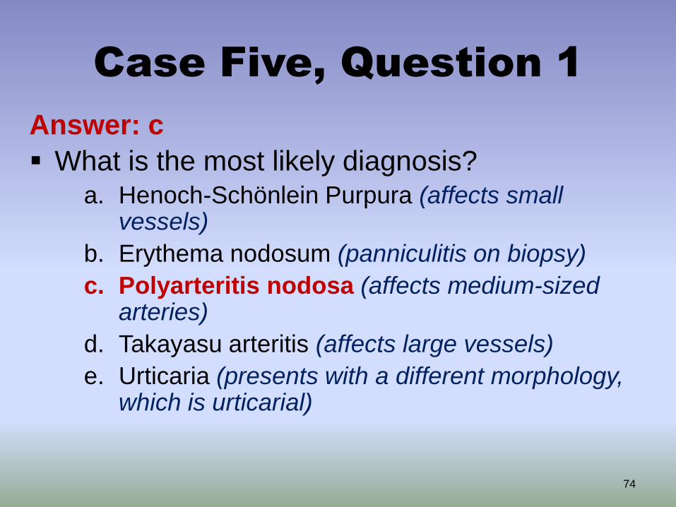
Answer: c
What is the most likely diagnosis? a. Henoch-Schönlein Purpura (affects small
vessels)
b. Erythema nodosum (panniculitis on biopsy)
c. Polyarteritis nodosa (affects medium-sized arteries)
d. Takayasu arteritis (affects large vessels)
e. Urticaria (presents with a different morphology, which is urticarial)
74
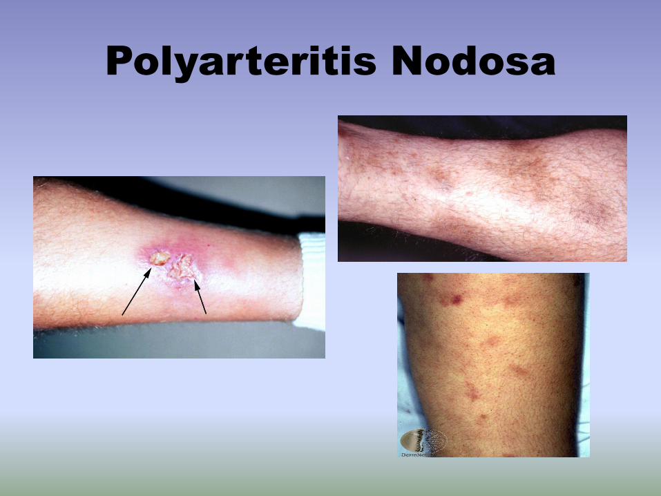
Polyarteritis Nodosa
Polyarteritis Nodosa (PAN) is a potentially systemic
disorder of necrotizing vasculitis of medium-sized
arteries
• Characterized by painful subcutaneous nodules, which
can ulcerate
• Patients may also present with livedo reticularis
Unknown etiology; may affect any organ (most often
skin, peripheral nerves, kidneys, joints, and GI tract)
PAN has been associated with HBV, HCV, HIV,
parvovirus B19, Crohn’s disease, strep and TB
infections and medications (minocycline)

Polyarteritis Nodosa-
Cutaneous
• ESR – the only laboratory abnormality
• P-ANCA may be present
• Most patients respond to aspirin, NSAIDs, prednisone
• In childhood – antibiotics may be used, since streptococcal infection is common
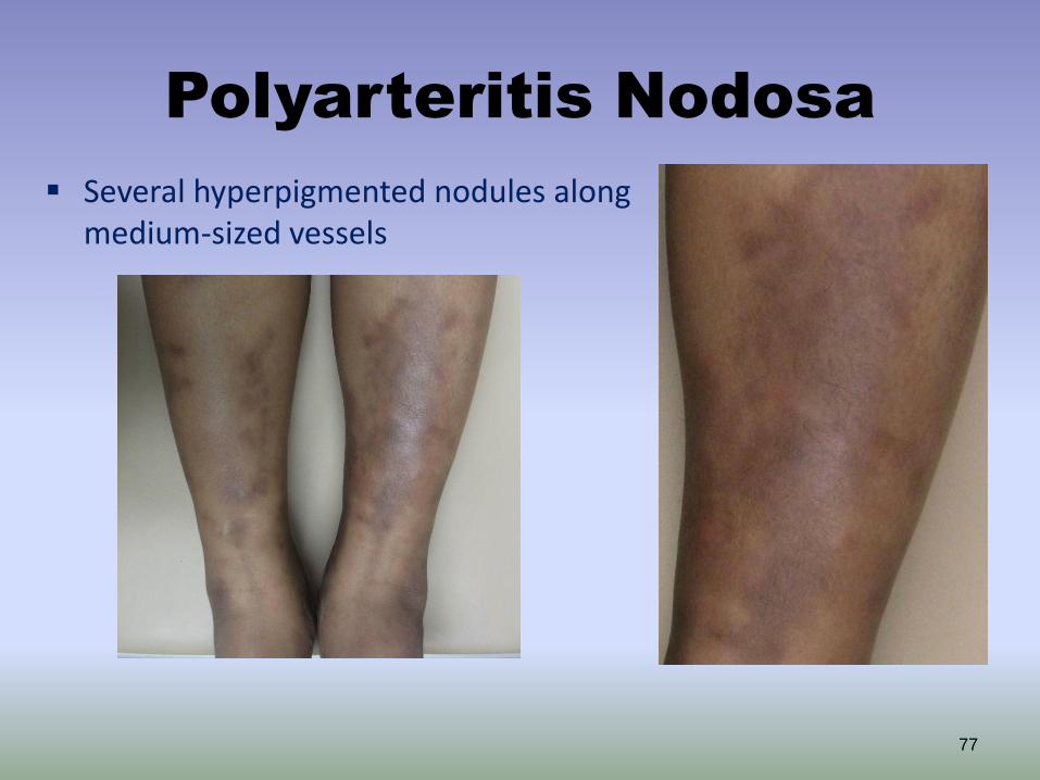
Polyarteritis Nodosa
77
Several hyperpigmented nodules along medium-sized vessels
Polyarteritis Nodosa

Systemic PAN
Key diagnostic features of this case
• Fever
• Leukocytosis with neutrophilia, trombocytosis
• Skin nodules with medium-sized artery inflammation on biopsy
• Renal involvement
• Paresthesias, decreased sensation and reflexes (i.e., mononeuritis multiplex)

Diagnosis: PAN
It is important to differentiate between cutaneous and
systemic PAN to help guide therapy
80
Cutaneous PAN
• Skin involvement +/- polyneuropathy, arthralgias, myalgias, fever
• More common in children (may follow a strep infection)
• Chronic benign course
Systemic PAN
• Neurological involvement common: mononeuritis multiplex, stroke
• Renal: hypertension
• Joint, Heart, GI, Liver also may be affected
• Orchitis in patients with HBV
Management: PAN
Chronic course (months-years);
exacerbations/remissions
Local wound care to any skin ulcerations
Patients without cutaneous PAN may be treated with
systemic corticosteroids alone
For patients with cardiac, gastrointestinal,
neurological, or renal involvement (i.e., systemic PAN):
• Specialty consultation for involved organs (especially
nephrology and neurology)
• Adjunctive therapy with cyclophosphamide
Treat any underlying infections (e.g., HBV)
Pigmentary Purpuric
Eruptions
• Presents on lower extremities with several clinical patterns
• The most common variant – Schamberg’s disease
• The other variants: – purpura anularis telangiectoides (Majocchi’s
disease)
– Gougerot-Blum syndrome (pigmented purpuric lichenoid dermatitis)

Pigmentary Purpuric
Eruptions Variants – cont.
• Ducas and Kapetanakis pigmented purpura
• Lichen aureus

Schamberg’s Disease
• Typical lesions: thumb-print sized and composedof aggregatesof pinhead-sized petechiae resemblings grains of cayenne pepper along with golden-brown hemosiderin staining
• The lesions begin on the lower legs with slow proximal extension
• The favored sites: lower shins and ankles

Schamberg’s Disease-
Treatment
• Topical steroids for 4-6 weeks
• Pentoxifyline 400mg three times a day for 2-3 weeks
• Oral rutoside 50 mg twice a day and ascorbic acid 500mg twice a day
• Support stocking in settings of venous stasis

Schamberg’s Disease
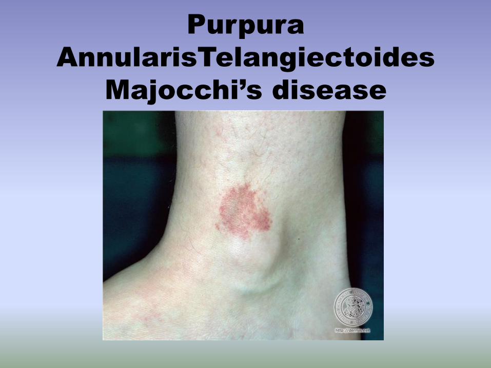
Purpura
AnnularisTelangiectoides
Majocchi’s disease

Gougerot-Blum Syndrome

Ducas and Kapetanakis

Lichen Aureus
Summary:
Petechiae and Purpura
• The term purpura is used to describe red-purple lesions that result from extravasation of the blood into the skin or mucous membranes
• Purpura may be palpable and non-palpable • Purpura does not blanch with pressure • Purpura may result from hyper- and hypocoagulable
states, vascular dysfunction and extravascular causes • Various life-threatening conditions present with
petechial rashes including meningococcemia and RMSF • The presence of petechial or purpuric lesions in a
septic patient should raise concern for DIC
Summary:
Palpable Purpura
• Palpable purpura results from underlying
blood vessel inflammation (vasculitis)
• Palpable purpura is the hallmark lesion of leukocytoclastic vasculitis
• The various etiologies of vasculitis may be categorized according to size of vessel affected
• A skin biopsy is usually necessary for the diagnosis of vasculitis