peripheral vascular disorders. objectives compare assessment findings typically present in patients...
TRANSCRIPT
Peripheral Vascular Disorders
ObjectivesCompare assessment findings typically present
in patients with peripheral arterial and peripheral venous disease.
Identify when venous thromboembolism (VTE) and complications of VTE occur.
List nursing interventions to help prevent VTE.Describe the nurse’s role in monitoring patients
who are receiving anticoagulants.Explain the treatment and care of patients with
aneurysms. Compare Raynaud’s and Buerger’s Disease.
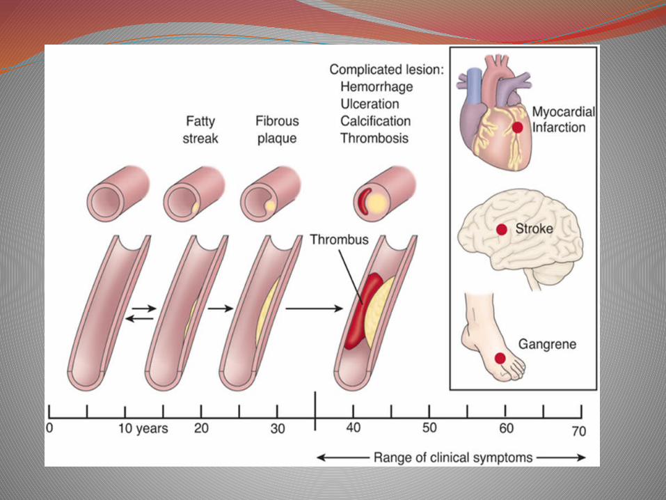
Peripheral Vascular DiseaseArteriosclerosis – thickening, loss of elasticity
and calcification of arterial wallsAtherosclerosis – a form of arteriosclerosis
deposits of fat and fibrin that obstruct and harden the arteries.
In the peripheral circulation these changes impair the blood supply to peripheral tissue peripheral vascular disease.
Peripheral Vascular Disease
Involves thickening of artery walls interferes with arterial blood flow to the lower extremities
Affects people in their 6th -8th decades of life.Risk factors – CHD, Diabetes Mellitus,
hypertension, cigarette smoking, elevated C-reactive protein
Regular daily exercise is a primary intervention for all types of PAD
Peripheral Vascular Disease
Assessment intermittent claudication rest pain inspection of the skin diminished/absence of peripheral pulses characteristics of arterial/venous insufficiency
Diagnostic Tests
AngiographyAnkle-Brachial IndexDoppler Ultrasound
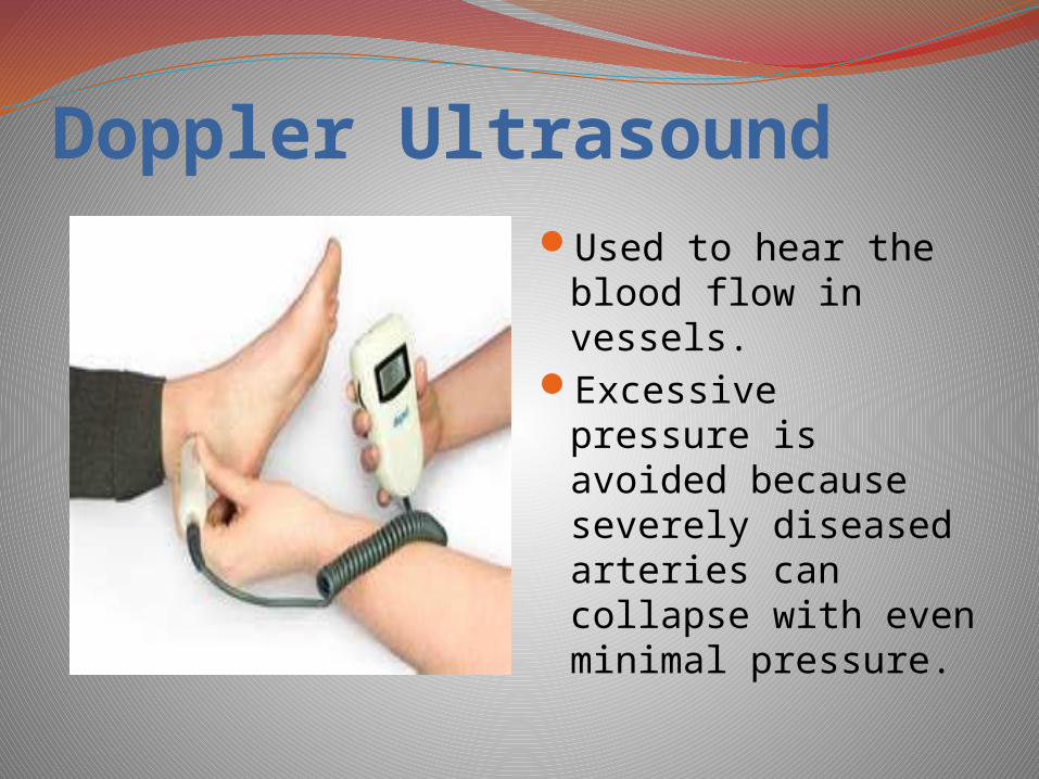
Doppler UltrasoundUsed to hear the
blood flow in vessels.Excessive pressure is
avoided because severely diseased arteries can collapse with even minimal pressure.
Ankle-Brachial Index
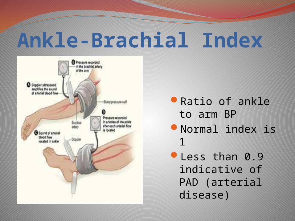
Ratio of ankle to arm BP
Normal index is 1Less than 0.9
indicative of PAD (arterial disease)
PAD TreatmentExercise /positioningTreatments –smoking
cessation/meticulous foot careRevascularizationDrug therapy
Trental (Pentoxifylline)Pletal (Cilostazol)Antiplatelet agents
ASA, Plavix
Impaired Tissue Perfusion
Assess peripheral pulsesPosition extremities dependentAvoid smokingEncourage exerciseUse foot cradles, lightweight blanketsAvoid electric heating pads/hot water bottles
Relieving PainAssess pain chronic, continuous, disablingLimits activitiesTeach pain relief/stress reduction techniquesAnalgesic: hydrocodone/acetaminophen (Vicodin) oxycodone/acetylsalicylic acid (Percodan) oxycodone/acetaminophen (Percocet)
Impaired Tissue Integrity
Assess and document skin conditionProvide meticulous daily skin care Regular inspection of extremities any
evidence of infection or inflammationApply egg crate/bed cradleGood nutrition, low-fat diet
Aneurysms
Abnormal dilatation of a blood vessel commonly at a site of weakness/tear in blood vessel wall.
Bulge or ballooning in wall of arteryHypertension is a major contributing factorCommonly affect the aorta/major peripheral
arteries
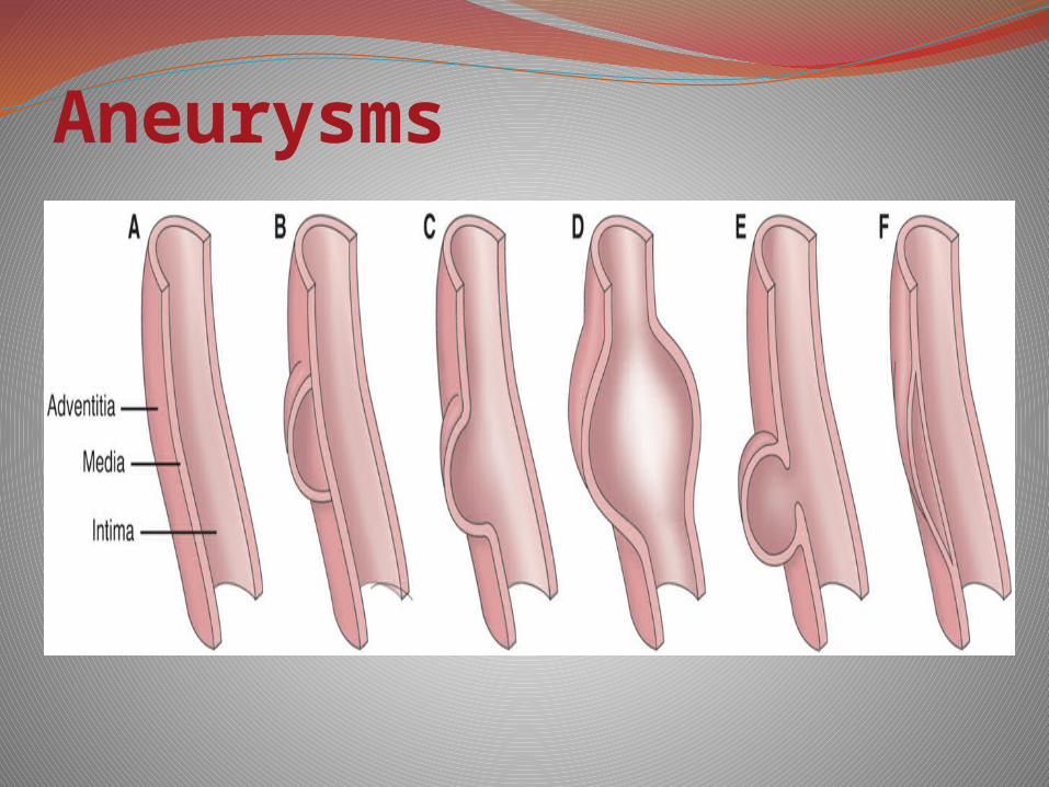
Aneurysms
Congenital or DiseaseTrue Aneurysm False AneurysmDissecting aneurysm
Aneurysms

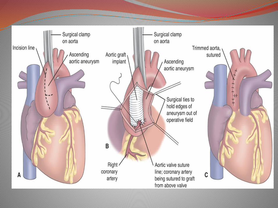
Thoracic Aortic AneurysmWeakening of the aortic wall by
arteriosclerosis & hypertensionCommon site for dissecting aneurysmFrequently asymptomaticSubsternal, neck or back painDyspnea, coughHoarseness, dysphagiaComplications Medical management
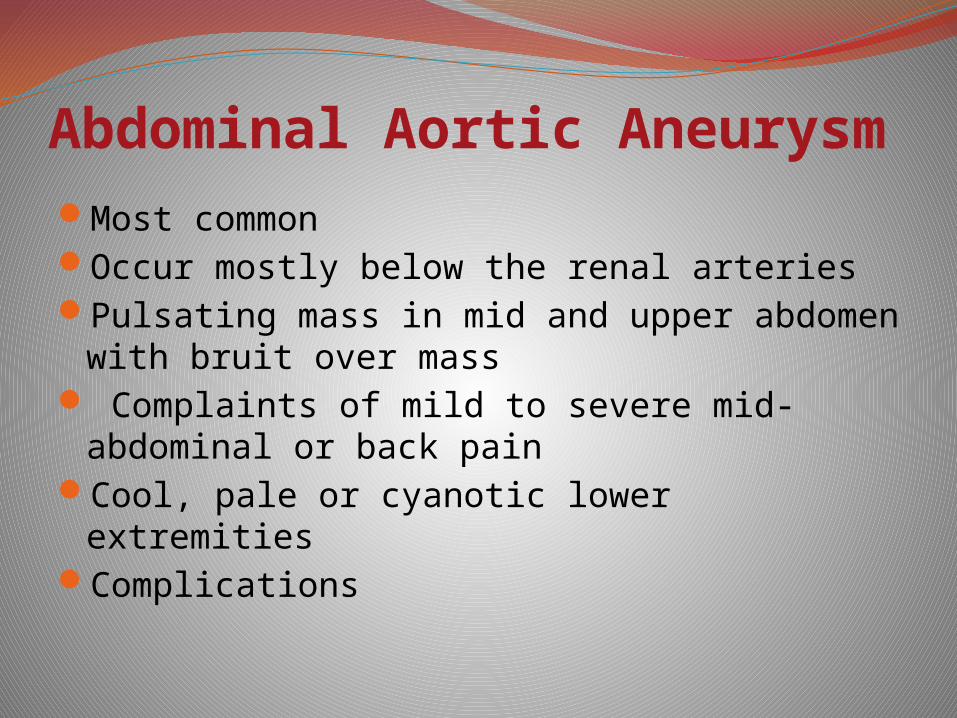
Abdominal Aortic Aneurysm
Most commonOccur mostly below the renal arteriesPulsating mass in mid and upper abdomen
with bruit over mass Complaints of mild to severe mid-abdominal
or back painCool, pale or cyanotic lower extremitiesComplications
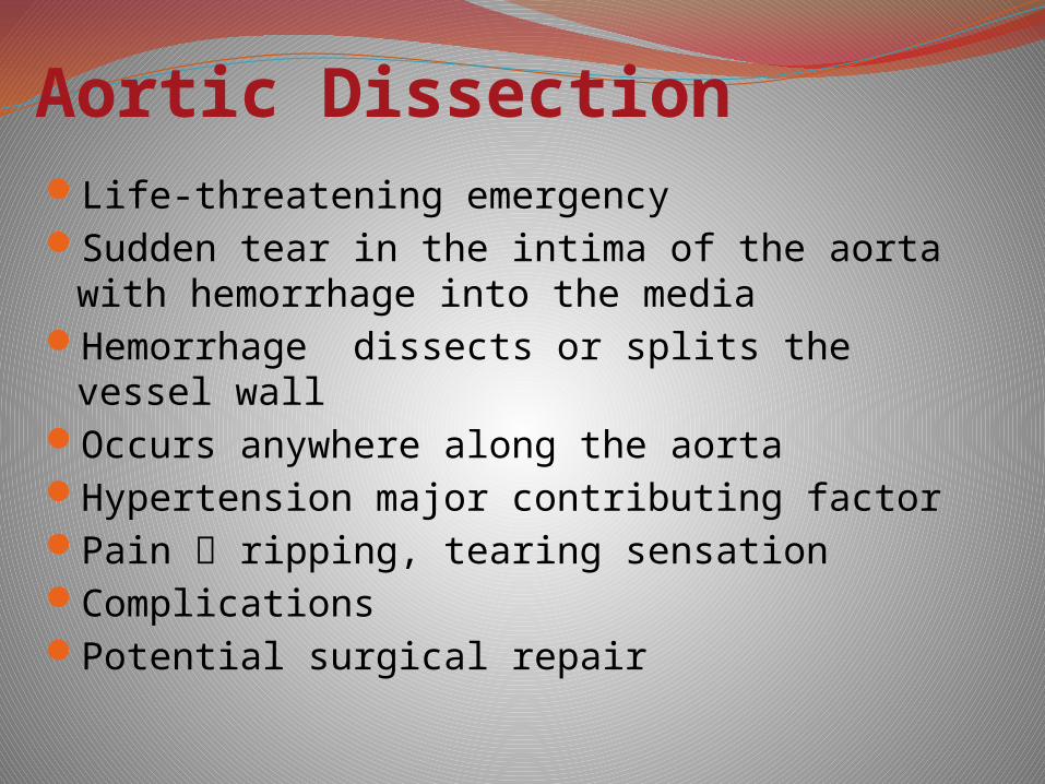
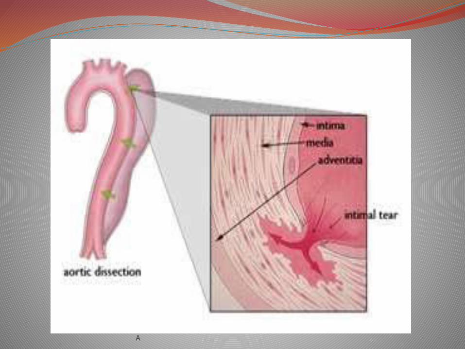
Aortic DissectionLife-threatening emergencySudden tear in the intima of the aorta with
hemorrhage into the mediaHemorrhage dissects or splits the vessel wallOccurs anywhere along the aortaHypertension major contributing factorPain ripping, tearing sensationComplicationsPotential surgical repair
A
Interdisciplinary CareDiagnosis Chest or abdominal X-rayCT scan Abdominal ultrasound
Treatment Antihypertensive medications Surgery – endovascular stent graft
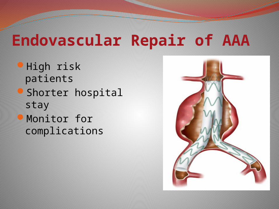
Endovascular Repair of AAA
High risk patientsShorter hospital stayMonitor for
complications
NURSING INTERVENTIONS
Risk for Ineffective Tissue PerfusionReduce the risk of aneurysm ruptureContinuously monitor cardiac rhythmReport manifestations of arterial embolismImmediate report changes in mental status or
symptoms of peripheral impairment

AnxietyExplain all procedures and treatmentsRespond to all questions honestlyProvide care in a calm, efficient mannerSpend time with the client
Venous ThrombosisBlood clot forms on the wall of the vein
inflammation, obstructed blood flowDVT – common complication of surgery and
immobilityVirchow’s triad – stasis of blood, vessel
damage, and increased blood coagulationDVT – usually asymptomatic dull aching pain possible tenderness, warmth along affected vein edema/cyanosis affected extremity
Venous DisordersInterdisciplinary CareDuplex venous ultrasonography
Plethysmography
MRI
DVT Prevention/Prophylaxis
Medications Low –molecular weight heparin
InterventionsRestMedicationsElevation of extremityCompression Therapy stockings external compression devices intermittent pneumatic compression devices
Heparin TherapyInterferes with the clotting cascade prevents
formation of stable fibrin clotMonitor/report any abnormal lab results and
aPTTAdminister deep subQUse infusion pump –when given IVProtamine Sulfate on handReport evidence of bleeding Patient teaching
Low-Molecular Weight Heparin
Lovenox (enoxaparin), Fragmin (dalteparin)Provide a more precise and predictable
anticoagulant effect than heparinSuitable for home-careReport excessive bruising/bleedingDo not take ASA, NSAIDsAdminister subQ
CoumadinInhibits synthesis of vitamin K dependent
clotting factors3 – 4 days until therapeuticMonitor INR (1.5 – 2.0)Take at the same time every day Bleeding precautionsAntidote: Vitamin KWear Medic-Alert tag

NURSING INTERVENTIONS
A
PainRegularly assess pain location
Measure calf and thigh diameter
Apply warm, moist heat to affected extremity
Ineffective Tissue Perfusion: Peripheral
Assess for peripheral pulses, skin integrity
Assess the skin of the affected lower leg and foot
Elevate extremities
Monitor lab results
Impaired Physical MobilityEncourage ROM exercise
Encourage frequent position changes
Encourage increased fluid and dietary fiber intake
Provide diversional activities
Leg UlcersExcavation of the skin surface that occurs
when inflamed necrotic tissue sloughs off.75% of leg ulcers chronic venous
insufficiency20% of leg ulcers arterial insufficiencyCellular metabolism cannot maintain energy
balance cell death (necrosis)
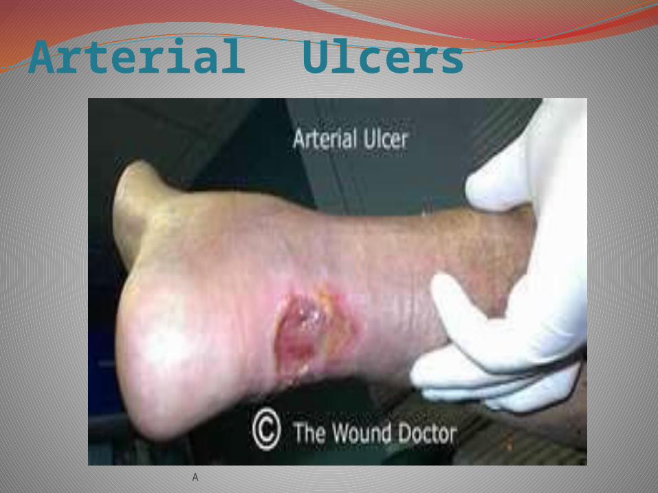
Arterial UlcersArterial thrombosis /arterial embolism =
tissue ischemia Ischemic tissue painful, pale, cool, cold Distal pulses may be absent Absence of hair on the toes or the legs Claudication present Ulcers are most likely perfectly round,
smooth edges, minimal drainage, no odor
A
Arterial Ulcers

Venous UlcersOver medial or anterior ankleLower leg edema/may be cyanoticAching, cramping painPulses present, may be difficult to palpateBrownish pigmentation to the skinSkin changes – stasis dermatitisShape – irregular border
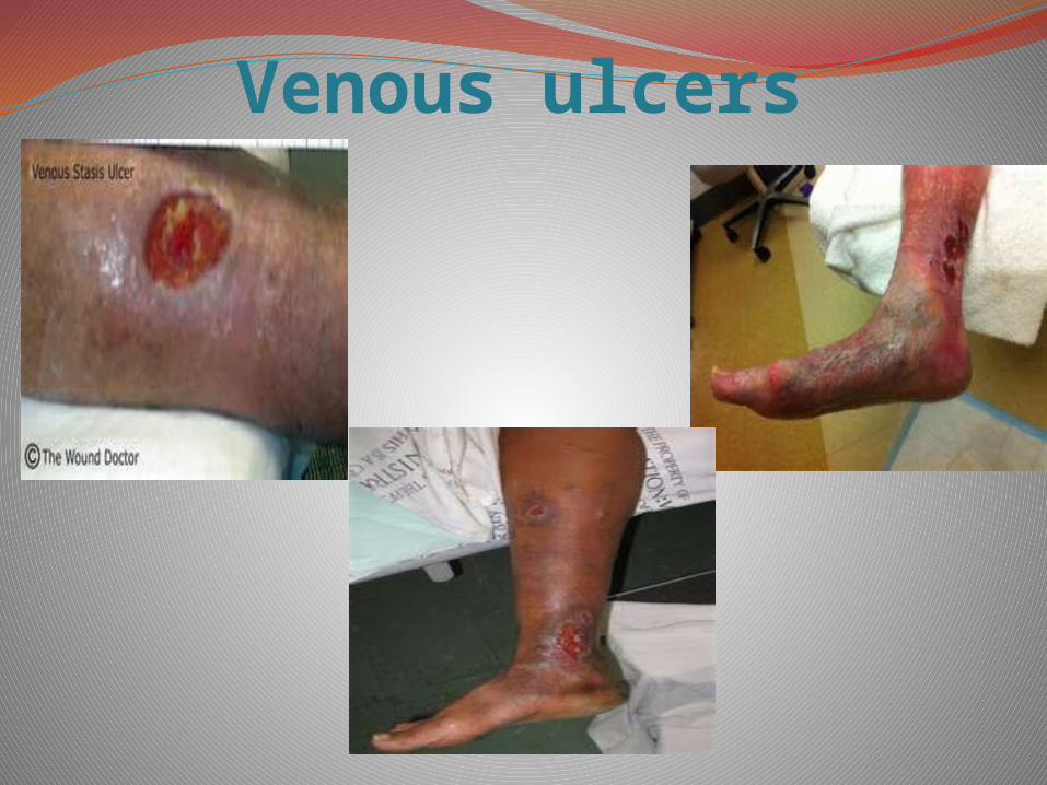
Venous ulcers
Interdisciplinary Care
Reduce edema
Treat ulcerations
Nursing AssessmentExtent and type of painPeripheral pulsesMobilityAssess for presence of infectionAssess nutrition
Mobility
With leg ulcers, activity is usually initially restricted to promote healing
Gradual progression of activityActivity to promote blood flow; encourage
patient to move about in bed and exercise upper extremities
Diversional activitiesPain medication prior to activities
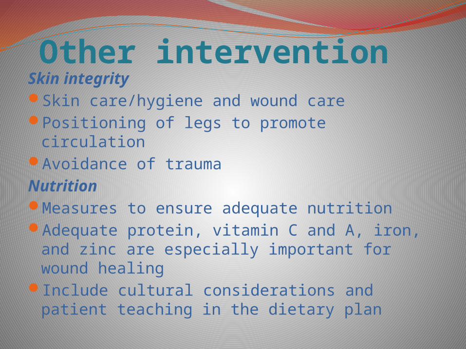
Other interventionSkin integritySkin care/hygiene and wound carePositioning of legs to promote circulationAvoidance of traumaNutritionMeasures to ensure adequate nutritionAdequate protein, vitamin C and A, iron, and
zinc are especially important for wound healing
Include cultural considerations and patient teaching in the dietary plan
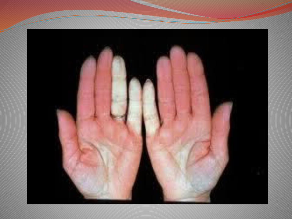
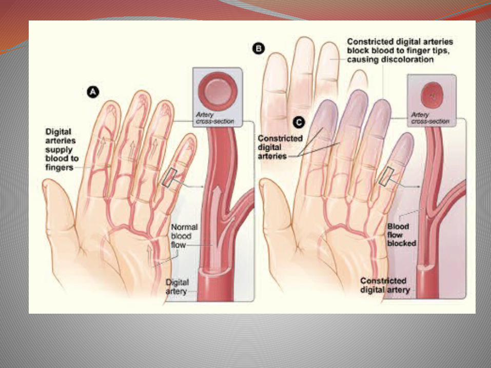
Raynaud’s DiseaseIntense vasospasm in the small arteries of the
fingersPallor, coldness, numbness, cyanosis and painOccurs in young womenAggravated by cold and stressBlue-white-red changesTreatmentVasodilators/Calcium Channel BlockersSympathectomyInterventions
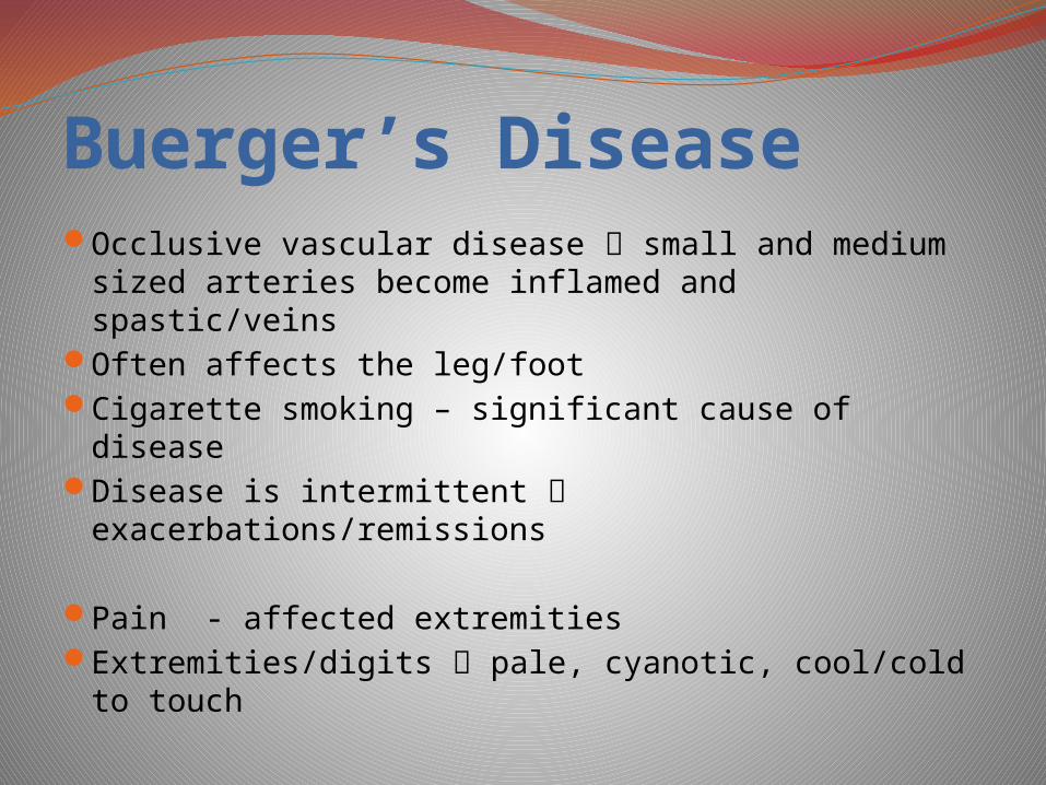
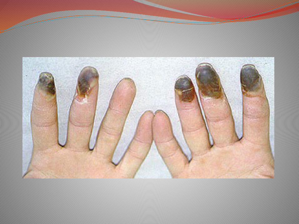
Buerger’s DiseaseOcclusive vascular disease small and medium
sized arteries become inflamed and spastic/veins Often affects the leg/footCigarette smoking – significant cause of diseaseDisease is intermittent exacerbations/remissions
Pain - affected extremitiesExtremities/digits pale, cyanotic, cool/cold to
touch
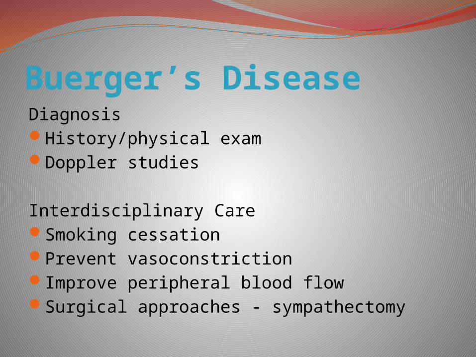
Buerger’s DiseaseDiagnosisHistory/physical examDoppler studies
Interdisciplinary CareSmoking cessationPrevent vasoconstrictionImprove peripheral blood flowSurgical approaches - sympathectomy

QUESTIONThe nurse notes that the client’s leg below
the knee is cool, pale, and dorsalis pedis & posterior tibia pulses are absent. The priority nursing intervention is to do which of the following?
a. Notify the healthcare provider.b. Prepare to initiate heparin therapy.c. Position the leg flat, supported in anatomic
position.d. Place a cradle over the leg to prevent
pressure from bedding.
QUESTIONAll of the following are appropriate home care
measures for the patient with PVD. Place them in order of priority.
1. Foot and leg care2. Smoking cessation3. Daily inspection of feet and legs4. Regular daily exercise5. Weight loss strategies
QUESTIONThe nurse evaluates her teaching plan for a patient
with deep vein thrombosis has been effective when the patient stated?
a. “I’ll use a hard-back chair, upright chair when sitting instead of my recliner.”
b. “I understand why I am not allowed to exercise for the next 6 weeks.”
c. “I’ll get my bloods drawn as scheduled and notify the doctor if I have unusual bleeding.”
d. “I’ll have my wife start to prepare low-cholesterol meals and will speak with the dietitian.”