peripheral inflammation acutely impairs human spatial ... · scanning was performed in 20 healthy...
TRANSCRIPT

ARCHIVAL REPORT
Peripheral Inflammation Acutely Impairs HumanSpatial Memory via Actions on Medial Temporal LobeGlucose Metabolism
Neil A. Harrison, Christian F. Doeller, Valerie Voon, Neil Burgess, and Hugo D. CritchleyBackground: Inflammation impairs cognitive performance and is implicated in the progression of neurodegenerative disorders. Rodentstudies demonstrated key roles for inflammatory mediators in many processes critical to memory, including long-term potentiation,synaptic plasticity, and neurogenesis. They also demonstrated functional impairment of medial temporal lobe (MTL) structures bysystemic inflammation. However, human data to support this position are limited.
Methods: Sequential fluorodeoxyglucose positron emission tomography together with experimentally induced inflammation was usedto investigate effects of a systemic inflammatory challenge on human MTL function. Fluorodeoxyglucose positron emission tomographyscanning was performed in 20 healthy participants before and after typhoid vaccination and saline control injection. After each scanningsession, participants performed a virtual reality spatial memory task analogous to the Morris water maze and a mirror-tracing proceduralmemory control task.
Results: Fluorodeoxyglucose positron emission tomography data demonstrated an acute reduction in human MTL glucose metabolismafter inflammation. The inflammatory challenge also selectively compromised human spatial, but not procedural, memory; this effectthat was independent of actions on motivation or psychomotor response. Effects of inflammation on parahippocampal and rhinalglucose metabolism directly mediated actions of inflammation on spatial memory.
Conclusions: These data demonstrate acute sensitivity of human MTL to mild peripheral inflammation, giving rise to associatedfunctional impairment in the form of reduced spatial memory performance. Our findings suggest a mechanism for the observedepidemiologic link between inflammation and risk of age-related cognitive decline and progression of neurodegenerative disordersincluding Alzheimer’s disease.
Key Words: Alzheimer’s disease, imaging, inflammation, memory,parahippocampus, PET
Although previously considered an immune-privileged site, itis now clear that the immune system plays an integral rolein many fundamental neuronal processes, including long-
term potentiation (LTP) (1,2), synaptic plasticity (3), and neuro-genesis (4), that are critical to learning and memory. In health,immune mechanisms regulate each of these processes and assistin the remodeling of neural circuits that promote learning andmemory (5). However, during systemic infection or injury (6), thispositive regulatory function is disrupted, resulting in acute memoryimpairments: When inflammation is severe, cognitive impairmentmay become persistent (7), and when chronic inflammation ispresent, age-related cognitive impairment is accelerated (8).Inflammation may drive the rapid progression of neurodegener-ative diseases such as Alzheimer’s disease (9).
From the Department of Psychiatry (NAH, HDC), Brighton and SussexMedical School, and Sackler Centre for Consciousness Science (NAH,HDC), University of Sussex, Falmer, United Kingdom; Donders Institutefor Brain, Cognition and Behaviour (CFD), Radboud University Nijme-gen, The Netherlands; Department of Psychiatry (VV), University ofCambridge, Cambridge, United Kingdom; and Institutes of CognitiveNeuroscience and Neurology (NB), University College London, London,United Kingdom.
Address correspondence to Neil A. Harrison, M.B.B.S., Ph.D., Clinical Ima-ging Sciences Centre, Brighton and Sussex Medical School, Universityof Sussex, Falmer, BN1 9RR, United Kingdom; E-mail: [email protected].
Received Oct 16, 2013; revised and accepted Jan 7, 2014.
0006-3223/$36.00http://dx.doi.org/10.1016/j.biopsych.2014.01.005
Structures in the medial temporal lobe (MTL) appear to beparticularly sensitive to effects of inflammation. This increasedsensitivity may be related to their relatively high receptor andmessenger RNA expression for proinflammatory cytokines (10,11)and their neural connectivity to regions such as the insula (12)that support cortical representations of peripheral inflammatorystates (13). Rodent studies emphasized the role of the hippo-campus; direct administration of inflammatory cytokines, partic-ularly interleukin (IL)-1, into the hippocampus selectively impairedspatial and contextual memory processes, including radial armand Morris water maze performance, and contextual, but notauditory-cued, fear conditioning (5,14,15). Similarly, over-expression of IL-1 messenger RNA within the hippocampus isassociated with delayed acquisition of spatial memory on theMorris water maze task (14). For synaptic plasticity underlying theencoding and recall of memories, LTP is arguably the keyneuronal mechanism. The cytokine IL-1 compromises both hippo-campal and dentate gyrus LTP (1,17,18) and may mediate bothage-dependent decreases in LTP (19) and the modulation of LTPby Aβ amyloid (20). Cytokine-induced inhibition of neurogenesiswithin the dentate gyrus is also alleviated by the microglialinhibitor minocycline (4). Together, these data highlight thecentral action of inflammatory mediators (cytokines such asIL-1) on MTL-dependent memory processes.
Inflammatory challenges administered outside the centralnervous system also induce IL-1 expression within brain regions,including the MTL (21). Peripherally induced inflammation alsoreplicates many of the direct actions of inflammatory cytokines onMTL-dependent memory (5,22,23). There are numerous mecha-nisms through which peripheral inflammation can engenderchanges in cytokine levels within sensitive brain regions. Circulatingcytokines may be actively transported across the blood-brain barrier
BIOL PSYCHIATRY 2014;76:585–593& 2014 Society of Biological Psychiatry

586 BIOL PSYCHIATRY 2014;76:585–593 N.A. Harrison et al.
(24) or activate microglia via the circumventricular organs (25) andvascular endothelium (26). However, local synthesis of IL-1 issuggested by the rapid upregulation of IL-1α and IL-1β geneexpression and the central predominance of the short half-life IL-1isoform in the context of mild systemic inflammatory challenge (21).Vagus nerve afferents show sensitivity to peripheral cytokines (27)and mild inflammatory challenge (28) indicating an additionalneurally mediated immune-brain pathway. Central vagus nervetargets show enhanced activity within 2–3 hours of peripheralinflammatory challenge in both rodents and humans (29,30).Electrical stimulation of vagus nerve afferents results in a rapidincrease in IL-1β expression within the hippocampus (31). Humoraland neurally mediated routes may communicate peripheral inflam-matory responses centrally to regions supporting memory processes.
These data from animal studies suggest mechanisms toaccount for human epidemiologic data linking increased periph-eral inflammation to accelerated cognitive aging and neuro-degeneration. However, it is unknown whether systemicinflammation modulates MTL function in humans. We used anexperimental inflammatory model, typhoid vaccination, togetherwith sequential fluorodeoxyglucose (FDG) positron emissiontomography (PET) scanning to quantify hypothesized effects ofperipheral inflammation on human MTL function and spatialmemory. In 20 healthy participants, three FDG-PET scans wereperformed immediately before and 4 hours and 8 hours aftertyphoid vaccination or control (saline) injection (Figure 1). Aftereach of the first two scanning sessions, participants performed aspatial memory task in which they learned and then recalled theidentity and location of two sets of 16 objects positioned within avirtual reality environment. This virtual reality task is analogous tothe Morris water maze (32), which is sensitive to inflammatoryeffects on object-location accuracy in rodents, and to the hiddentracer task, which is sensitive to lesions in discrete MTL structuresin humans (33). Recall of the spatial location and identity of bothsets of objects was tested again after the third scan to investigatedifferential effects of inflammation on early encoding and laterconsolidation processes. Participants also performed a mirror-tracing procedural memory task to test general effects ofinflammation on psychomotor responses and motor learning.
Methods and Materials
ParticipantsWe recruited 20 healthy male nonsmokers (mean age, 24.7 �
6.8 years old) and screened them for relevant physical or
www.sobp.org/journal
psychiatric illness; all were medication-free. Volunteers who hadreceived typhoid vaccine within 3 years or other vaccine within 6months were excluded. Participants were advised to avoidcaffeinated beverages, alcohol, high-fat meals, and excessiveexercise for 24 hours and steroidal or nonsteroidal drugs for 2weeks before testing. All participants fasted for 8 hours andconsumed only water until study completion. Written informedconsent was obtained from all participants, and procedures wereapproved by the Brighton East National Research EthicsCommittee.
Study DesignA randomized, double-blind, repeated measures crossover
design was used in which all participants underwent three FDG-PET imaging sessions each separated by 4 hours. After each of thefirst two scanning sessions, participants randomly received intra-muscular injections of either 0.025 mg Salmonella typhi vaccine(Typhim Vi; Aventis Pasteur MSD Ltd., Lyon, France) or 0.5 mLnormal saline. Of participants, 13 were randomly assigned to theearly inflammation group and received vaccination after the firstPET scan 1, and 7 were randomly assigned to the late inflamma-tion group and received vaccination after the second PET scan.This study design enabled us to control for nonspecific timeeffects as well as have sufficient participants (n ¼ 13) scanned 8hours after typhoid vaccination to test late effects of inflamma-tion. After each scan, participants performed a laptop-basedspatial memory task and a mirror-tracing procedural memorytask that took 35 min to complete. Vaccination or saline injectionwas given after the PET scan immediately before memory testing;this was done to minimize an already long testing day. We areaware of no data to suggest that peripherally induced inflamma-tion can impair memory at such a short latency, and if this werethe case, it would increase the risk of false-negative rather thanfalse-positive findings. A high-resolution inversion recovery echoplanar image was obtained to aid image registration.
Inflammatory ModelWe used a S. typhi vaccination model known to induce low-
grade inflammation without body temperature change (34).Blood (10 mL) was drawn into ethylenediamine tetraacetic acidBD Vacutainer tubes (Franklin Lakes, New Jersey) and centrifugedat 1250 � g for 10 min, and plasma was removed, aliquoted, andfrozen at �801C. Plasma IL-6, IL-1 receptor antagonist, and tumornecrosis factor alpha were assessed using high-sensitivityenzyme-linked immunosorbent assays (R&D Systems, Abingdon,United Kingdom). Limits of detection were .039 pg/mL, 6.26 pg/mL,
Figure 1. Study timeline. All participants completedthree fluorodeoxyglucose (FDG) positron emission tomo-graphy (PET) scans. Each scan was preceded by a blooddraw and followed by a memory testing session. The“early” inflammation group received the typhoid vacci-nation after the first PET scan (and saline injection afterthe second PET scan), and the “late” inflammation groupreceived the typhoid vaccination after the second PETscan (saline injection after the first scan). In the first twosessions (T1 and T2), participants encoded and thenrecalled the identity and spatial location of two sets ofobjects (object set 1 and set 2). In the third session (T3),participants recalled the identity and spatial location ofthe two sets of objects encoded at T1 and T2. Themirror-tracing task was performed at each of the threetesting sessions. MRI, magnetic resonance imaging.(Photo credit, copyright WGBH Educational Foundation.)
N.A. Harrison et al. BIOL PSYCHIATRY 2014;76:585–593 587
and .038 pg/mL with associated intra-assay and interassaycoefficients of variation of 7.4% and 7.8%, 5.3% and 8.6%, and5.3% and 8.4%.
Spatial Memory TaskUnrealEngine2 Runtime software (Epic Games, Cary, North
Carolina) was used to present a first-person perspective of a planesurrounded by a circular cliff (virtual diameter 180 m). Back-ground mountains, clouds, and the sun (created using Terragen;Planetside Software, Cheshire, United Kingdom) projected atinfinity were used to provide orientation cues. Two separatecounterbalanced arenas with grassy or rocky planes and differ-ently rendered mountains and clouds were used for the twoencoding sessions. Participants explored the arena for 2–3 minusing right-handed button presses to move forward, left, andright. Then 16 unique objects were sequentially presented withinthe arena, and participants were instructed to remember theiridentity and spatial location before picking them up. After allobjects were acquired, participants performed free recall of theobject identities. They then returned to the virtual reality environ-ment. A picture of one object was presented, following whichparticipants moved to where they thought the cued object hadbeen presented, indicated by a button press, and recorded theirconfidence (range, 1–5) for this location. For the first two sessions(T1 and T2), the object was shown again in its correct position,and participants collected it by running over it. The next objectwas cued after a variable intertrial interval. On the third session(T3), object recall and relocation phases were completed forobjects learned at T1 (set 1) and T2 (set 2). Performance wasindexed by accuracy of object spatial location: mean (1/distancefrom true object location in virtual meters) and number of objectsrecalled.
Procedural Memory TaskParticipants were asked to trace between two concentric five-
pointed stars viewed in a mirror as quickly and accurately aspossible. Both their hand and the concentric stars were obscuredfrom direct view. Time taken to complete two trials was used asan index of performance.
Image Acquisition and AnalysisThe PET scans (mean 155.3 � 11.8 MBq FDG) were acquired
for 35 min on a Siemens Biograph-64 PET-CT scanner (SiemensHealthcare, Erlangen, Germany) in three-dimensional dynamicacquisition mode. Participants lay supine with eyes open. Beforeeach PET acquisition, a low-dose computed tomography scan(120 kVp, 10 mA) was acquired for attenuation correction. Aftercorrection for scatter, random effects, and effects of attenuation,images were reconstructed in 1-min windows using Siemensproprietary iterative three-dimensional reconstruction schema (21iterations and 8 subsets). Individual 1-min scans were realignedand summed to produce a single 35-min activation scan persession, which was coregistered to subjects’ structural magneticresonance imaging scans and then spatially smoothed with an 8-mm full width at half maximum Gaussian kernel using standardSPM8 methods (Wellcome Trust Centre for Neuroimaging, Insti-tute of Neurology, United College London, London, United King-dom; http://www.fil.ion.ucl.ac.uk/spm).
Normalized images were included within a second-levelflexible-factorial analysis of variance (ANOVA) (repeated factortime, baseline, 4 hours, 8 hours; between-subject factor group,early, late inflammation). Main effects of time and group andgroup � time interaction were included in the model.
Normalization to a grand mean scaled value of 50 mL/100 g/min was applied, and global effects were included as nuisancecovariates in the general linear model (analysis of covariance).Correlations between changes in resting glucose metabolism andobject-location accuracy together with interactions with inflam-matory status (modeled using a dummy variable) were inves-tigated in a separate regression analysis.
Anatomic localization of MTL structures was based on Insaustiet al. (35), Monte Carlo simulation (1000 iterations) was used todetermine cluster extent thresholds for whole-brain correction atp � .01 (36), and a cluster threshold of �19 voxels was adopted.Regression analyses followed by Goodman test for mediationwere used to investigate relationships between inflammatorychallenge and changes in object-location accuracy and rightparahippocampal activity Montreal Neurological Institute (24,�32, �32) between encoding sessions T1 and T2. Mediationanalyses were performed using the interactive calculation tool formediation tests (http://quantpsy.org/sobel/sobel.htm).
Results
Inflammatory Responses to Typhoid VaccinationCytokine analyses confirmed significantly higher circulating
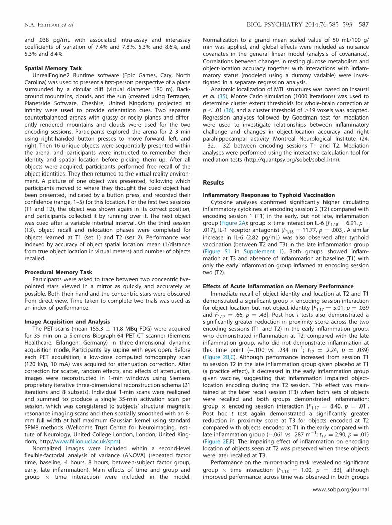
inflammatory cytokines at encoding session 2 (T2) compared withencoding session 1 (T1) in the early, but not late, inflammationgroup (Figure 2A): group � time interaction IL-6 [F1,18 ¼ 6.91, p ¼.017], IL-1 receptor antagonist [F1,18 ¼ 11.77, p ¼ .003]. A similarincrease in IL-6 (2.82 pg/mL) was also observed after typhoidvaccination (between T2 and T3) in the late inflammation group(Figure S1 in Supplement 1). Both groups showed inflam-mation at T3 and absence of inflammation at baseline (T1) withonly the early inflammation group inflamed at encoding sessiontwo (T2).
Effects of Acute Inflammation on Memory PerformanceImmediate recall of object identity and location at T2 and T1
demonstrated a significant group � encoding session interactionfor object location but not object identity [F1,17 ¼ 5.01, p ¼ .039and F1,17 ¼ .66, p ¼ .43]. Post hoc t tests also demonstrated asignificantly greater reduction in proximity score across the twoencoding sessions (T1 and T2) in the early inflammation group,who demonstrated inflammation at T2, compared with the lateinflammation group, who did not demonstrate inflammation atthis time point (�.100 vs. .234 m�1; t17 ¼ 2.24, p ¼ .039)(Figure 2B,C). Although performance increased from session T1to session T2 in the late inflammation group given placebo at T1(a practice effect), it decreased in the early inflammation groupgiven vaccine, suggesting that inflammation impaired object-location encoding during the T2 session. This effect was main-tained at the later recall session (T3) when both sets of objectswere recalled and both groups demonstrated inflammation:group � encoding session interaction [F1,17 ¼ 8.40, p ¼ .01].Post hoc t test again demonstrated a significantly greaterreduction in proximity score at T3 for objects encoded at T2compared with objects encoded at T1 in the early compared withlate inflammation group (�.061 vs. .287 m�1; t17 ¼ 2.90, p ¼ .01)(Figure 2E,F). The impairing effect of inflammation on encodinglocation of objects seen at T2 was preserved when these objectswere later recalled at T3.
Performance on the mirror-tracing task revealed no significantgroup � time interaction [F1,18 ¼ 1.00, p ¼ .33], althoughimproved performance across time was observed in both groups
www.sobp.org/journal
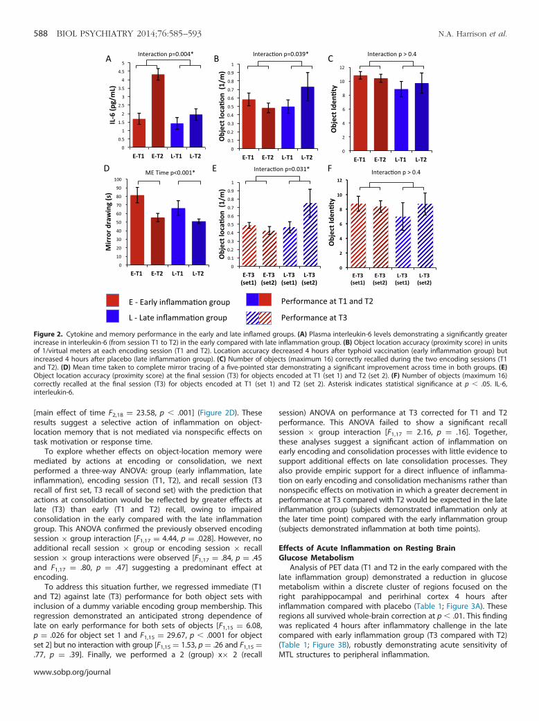
Figure 2. Cytokine and memory performance in the early and late inflamed groups. (A) Plasma interleukin-6 levels demonstrating a significantly greaterincrease in interleukin-6 (from session T1 to T2) in the early compared with late inflammation group. (B) Object location accuracy (proximity score) in unitsof 1/virtual meters at each encoding session (T1 and T2). Location accuracy decreased 4 hours after typhoid vaccination (early inflammation group) butincreased 4 hours after placebo (late inflammation group). (C) Number of objects (maximum 16) correctly recalled during the two encoding sessions (T1and T2). (D) Mean time taken to complete mirror tracing of a five-pointed star demonstrating a significant improvement across time in both groups. (E)Object location accuracy (proximity score) at the final session (T3) for objects encoded at T1 (set 1) and T2 (set 2). (F) Number of objects (maximum 16)correctly recalled at the final session (T3) for objects encoded at T1 (set 1) and T2 (set 2). Asterisk indicates statistical significance at p � .05. IL-6,interleukin-6.
588 BIOL PSYCHIATRY 2014;76:585–593 N.A. Harrison et al.
[main effect of time F2,18 ¼ 23.58, p � .001] (Figure 2D). Theseresults suggest a selective action of inflammation on object-location memory that is not mediated via nonspecific effects ontask motivation or response time.
To explore whether effects on object-location memory weremediated by actions at encoding or consolidation, we nextperformed a three-way ANOVA: group (early inflammation, lateinflammation), encoding session (T1, T2), and recall session (T3recall of first set, T3 recall of second set) with the prediction thatactions at consolidation would be reflected by greater effects atlate (T3) than early (T1 and T2) recall, owing to impairedconsolidation in the early compared with the late inflammationgroup. This ANOVA confirmed the previously observed encodingsession � group interaction [F1,17 ¼ 4.44, p ¼ .028]. However, noadditional recall session � group or encoding session � recallsession � group interactions were observed [F1,17 ¼ .84, p ¼ .45and F1,17 ¼ .80, p ¼ .47] suggesting a predominant effect atencoding.
To address this situation further, we regressed immediate (T1and T2) against late (T3) performance for both object sets withinclusion of a dummy variable encoding group membership. Thisregression demonstrated an anticipated strong dependence oflate on early performance for both sets of objects [F1,15 ¼ 6.08,p ¼ .026 for object set 1 and F1,15 ¼ 29.67, p � .0001 for objectset 2] but no interaction with group [F1,15 ¼ 1.53, p ¼ .26 and F1,15 ¼.77, p ¼ .39]. Finally, we performed a 2 (group) x� 2 (recall
www.sobp.org/journal
session) ANOVA on performance at T3 corrected for T1 and T2performance. This ANOVA failed to show a significant recallsession � group interaction [F1,17 ¼ 2.16, p ¼ .16]. Together,these analyses suggest a significant action of inflammation onearly encoding and consolidation processes with little evidence tosupport additional effects on late consolidation processes. Theyalso provide empiric support for a direct influence of inflamma-tion on early encoding and consolidation mechanisms rather thannonspecific effects on motivation in which a greater decrement inperformance at T3 compared with T2 would be expected in the lateinflammation group (subjects demonstrated inflammation only atthe later time point) compared with the early inflammation group(subjects demonstrated inflammation at both time points).
Effects of Acute Inflammation on Resting BrainGlucose Metabolism
Analysis of PET data (T1 and T2 in the early compared with thelate inflammation group) demonstrated a reduction in glucosemetabolism within a discrete cluster of regions focused on theright parahippocampal and perirhinal cortex 4 hours afterinflammation compared with placebo (Table 1; Figure 3A). Theseregions all survived whole-brain correction at p � .01. This findingwas replicated 4 hours after inflammatory challenge in the latecompared with early inflammation group (T3 compared with T2)(Table 1; Figure 3B), robustly demonstrating acute sensitivity ofMTL structures to peripheral inflammation.
Table 1. Brain Regions Showing an Acute Reduction in Resting Glucose Metabolism After Inflammatory Challenge
Side Region Coordinates Z Score Cluster puncorrected pcorrected
Inflammation-Induced Reductions in Glucose Metabolism (Early Inflammation Group)R Parahippocampus/perirhinal (36 �28 �24) 3.73 345 �.001 �.01R Fusiform gyrus (32 �54 �14) 3.89 190 �.001 �.01R Inferior temporal gyrus (57 �18 �33) 3.56 76 �.001 �.01R Temporal pole (57 11 �11) 3.42 28 �.001 �.01
Inflammation-Induced Reductions in Glucose Metabolism (Late Inflammation Group)R Entorhinal/perirhinal (21 �19 �27) 3.50 117 �.001 �.01L Entorhinal/perirhinal (�24 �13 �26) 3.67 71 �.001 �.01R Parahippocampus/perirhinal (33 �24 �24) 3.22 10 �.001
L, left; R, right.
N.A. Harrison et al. BIOL PSYCHIATRY 2014;76:585–593 589
To investigate whether this change in glucose metabolismbetween encoding sessions predicted changes in object-location accuracy. we next performed a regression analysis onthe PET data (i.e., T1 and T2 metabolism vs. T1 and T2 accuracy)(Table 2). This analysis revealed striking correlations betweenactivity change in bilateral parahippocampal and rhinal cortexand change in object-location accuracy across all participants(Figure 3C)—that is, there was a general relationship betweenchange in parahippocampal and rhinal glucose metabolism andchange in object-location accuracy. However, repetition of thisanalysis after inclusion of an interaction term coding groupmembership (early or late) also revealed a discrete contiguous
region within the right parahippocampal gyrus that mediatedthe detrimental effects of inflammation on object-locationencoding (Figures 3D and 4). In other words, inflammationdisrupted the relationship between parahippocampal metabo-lism and subsequent accuracy for object-location encoding.This interpretation was supported further by mediation analysis,which showed that inflammation induced changes in rightparahippocampal glucose metabolism (T1 and T2) MontrealNeurological Institute (24, �21, �32) that significantly medi-ated effects of inflammation on object-location memory (T1 andT2 accuracy) (Goodman test ¼ 3.58 [SE .74], p � .00035)(Figure 5).
Figure 3. Brain regions sensitive to acute inflammationand effects on object-location encoding. (A) Regionsshowing a greater reduction in glucose metabolism afterinflammation compared with placebo between sessions1 and 2. Contrast shown in T1 - T2 early group minus T1 -T2 late group. (B) Regions showing a greater reductionin glucose metabolism after inflammatory challenge inthe late compared with early inflamed group betweensessions 2 and 3. Contrast shown in T2 and T3 earlygroup compared with T2 and T3 late group. The y axis in(A) and (B) shows estimated glucose metabolism in mL/100 g/min. (C) Regions showing a positive correlationbetween change in object-location accuracy and changein glucose metabolism between the two encodingsessions (T1 and T2) across all participants. (D) Medialtemporal lobe region showing a significant group �location accuracy interaction between the two encodingsessions (T1 and T2). The y axis shows change in glucosemetabolism between T1 and T2 in mL/100 g/min. E, earlyinflammation group (received vaccine after first scan); L,late inflammation group (received vaccine after secondscan 2).
www.sobp.org/journal

Table 2. Brain Regions in Which Changes in Blood Glucose Metabolism Between the Two Encoding Sessions (T1 and T2) Predicted Associated Changes inMemory for Object Location
Side Region Coordinates Z Score Cluster puncorrected pcorrecteda
Regions Showing Positive Correlations Across All ParticipantsR Parahippocampus/perirhinal (27 �22 �27) 4.19 751 �.001 �.01L Parahippocampus/perirhinal (�29 �28 �26) 3.38 80 �.001 �.01R Precuneus (15 �60 45) 3.68 370 �.001 �.01L Inferior parietal lobule (�20 �51 54) 3.65 829 �.001 �.01R Inferior parietal lobule (33 �46 56) 3.31 99 �.001 �.01L Supplementary motor area (�15 �4 63) 3.55 95 �.001 �.01L Paracentral lobule (�3 �31 63) 3.36 221 �.001 �.01R Mid-frontal gyrus (35 3 46) 3.21 26 �.001 �.01R Mid-orbitofrontal gyrus (48 50 �9) 3.17 25 �.001 �.01
Medial Temporal Lobe Region Showing Interaction with GroupR Parahippocampus/perirhinal (24 �21 �32) 2.44 342 �.05 �.01b
L, left; R, right.apcorrected ¼ cluster survives whole-brain correction.bSurvives correction for a medial temporal lobe region of interest.
590 BIOL PSYCHIATRY 2014;76:585–593 N.A. Harrison et al.
Discussion
Systemic inflammation is associated with selective impairmentin human spatial memory but not MTL-independent proceduralmemory (37). Deficits in spatial memory were observed forobjects learned and recalled during systemic inflammation butnot objects learned in the absence of inflammation and recalledunder inflammatory conditions. This study suggests a predom-inant effect of inflammation on early encoding and consolidation
Figure 4. Right medial temporal lobe regions sensitive to inflammation,change in object-location accuracy, and interactions with inflammation.Cyan indicates regions showing a reduction in glucose metabolism afterinflammation (T1 - T2 early group minus T1 - T2 late group). Yellowindicates regions showing a positive correlation between change inobject-location accuracy (T1 and T2) and change in glucose metabolism(T1 and T2) across all participants. Red indicates area showing group �accuracy interaction—that is, the region mediating the effects of groupmembership (inflammation status) on change in accuracy for encodingobject location.
www.sobp.org/journal
processes rather than late consolidation and demonstrates arelative absence of state-dependent effects. In rodent contextualfear conditioning paradigms (5,23), inflammation impairs spatialmemory despite being induced after visuospatial information hasbeen attended to (and encoded) suggesting that our data arelikely mediated via an action on early consolidation processes.Although our analyses failed to demonstrate significant lateconsolidation effects, Figure 2B and E demonstrates a non-significant reduction in performance at T3 compared with T1 inthe early (early T3 [set 1] compared with early T1) but not lateinflammation group (late T3 [set 1] compared with late T1)consistent with a potential effect on late consolidation.
Resting glucose metabolism, particularly change in bilateralparahippocampus and perirhinal cortex metabolism immediatelybefore task performance, predicted change in accuracy acrossencoding sessions across all participants (Figures 2C and 3).However, this relationship was critically modulated by systemicinflammation. Within 4 hours of inflammatory challenge, glucosemetabolism decreased within perirhinal and entorhinal cortexand parahippocampus (Table 1). This effect was replicated inparticipants challenged after the first (Figure 3A) and second(Figure 3B) scanning sessions. A discrete subregion centered onthe right parahippocampus also predicted and mediated inflam-matory effects on subsequent object-location memory (Figures3D and 5). Together, these data demonstrate sensitivity of humanMTL structures, notably parahippocampus, to systemic
Resting Glucose Metabolism
Parahippocampus
Inflammation Object Location Memory
PATH A PATH B
PATH C
15.62 (4.31) P = 0.002
0.27 (0.009) P = 0.008
0.334 (0.149) P = 0.039
Figure 5. Mediation analysis showing that the changes in parahippo-campal glucose metabolism mediate the effects of inflammation onmemory for object location. Path coefficients (standard error of pathcoefficients) are shown for each path of the mediation model.

N.A. Harrison et al. BIOL PSYCHIATRY 2014;76:585–593 591
inflammation and provide mechanistic insight relevant to abroader literature linking severe or chronic inflammation to theattrition of human memory.
Studies investigating effects of inflammation on rodent spatialmemory to date have predominantly focused on actions on thehippocampus (1,5,15–17). We did not identify a major change inhippocampal glucose metabolism after inflammation or anyassociation between hippocampal glucose metabolism and sub-sequent memory performance. Although null results are hard tointerpret, learning object locations relative to the boundary in thistask correlates with functional magnetic resonance imaging signalfrom both right hippocampal and parahippocampal regions (32),suggesting greater sensitivity to detect metabolic changes in theparahippocampus. In addition, there is good evidence to suggeststrong parahippocampal involvement in this type of task. Neuro-psychological studies show that human performance on homo-logues of the Morris water maze and direct tests of object-location memory can be more strongly dependent on rightparahippocampal than hippocampal integrity (37–39). Studiesdemonstrate a central role for the right parahippocampus inhuman object-location memory and support our current findingof a critical role for the parahippocampus in mediatinginflammation-induced spatial memory impairments.
Right parahippocampal activity during object-location encod-ing has also been shown to predict subsequent retrieval successwith a spatial cue (40). In monkeys, one-trial memory for object-place associations (similar to our current task) appears to becritically dependent not on hippocampus but on posteriorparahippocampus (41). The contribution of the parahippocampusto within-scene object location and context memory is alsodissociable from the role of perirhinal cortex in object perceptionand memory (42–44). In rodents, perirhinal neurons respondselectively to objects and their previous occurrence (45,46) withselective lesions impairing performance on tasks requiring whole-object information (47). In contrast, rodents with postrhinal(parahippocampus) cortex lesions show impairment on taskssensitive to object location but not identity (48). Similar functionaldistinctions between perirhinal and parahippocampal activity arealso apparent in humans, with parahippocampal cortex activeduring object-location encoding and perirhinal cortex active toobjects alone (49). Our data suggest that systemic inflammationmay serve as a transient parahippocampal lesion resulting in adiscrete impairment in object-location memory.
The cellular mechanisms mediating this selective impairmentof human MTL function are unclear, although these mechanismsmay be usefully informed by rodent studies. For example, IL-1 hasbeen shown to reduce basal synaptic activity and synaptictransmission in a manner dependent on gamma-aminobutyricacid (50). It impairs LTP both dependent on and independent ofN-methyl-D-aspartate (1,2) and can decrease LTP-associatedglutamate release within the dentate gyrus (51). Although theseeffects are currently demonstrated only in rodent hippocampus,operation of either mechanism within human parahippocampusor perirhinal cortex could conceivably contribute to the observedreduction in glucose metabolism. Local or peripheral inflamma-tion can also impair hippocampal neurogenesis in proportion tothe associated increase in microglial activation (4). However,given the time course of this effect, it is unlikely to havecontributed to our results. Perhaps more pertinent is the role ofneurally mediated mechanisms. Peripheral inflammation has beenshown to increase rapidly activity within vagus nerve projectionareas in both rodents and humans (29,30), including insularcortex, a region that in primates has direct neural connectivity
to perirhinal and parahippocampal cortex (12), areas that providethe vast bulk of inputs into entorhinal cortex and the hippo-campal formation. Electrical stimulation of vagus nerve afferentsresults in a rapid increase in IL-1β expression within the hippo-campus (31). Activation of neurally mediated immune-braincommunicatory pathways may potentially modulate memoryprocesses even in the absence of significant signaling of inflam-mation across the blood-brain barrier at the endothelium (26) orcircumventricular organs (25).
A concern from rodent studies is that apparent effects ofinflammation on learning and memory may be confounded byactions on psychomotor speed (52). We also previously reportedpsychomotor slowing after typhoid vaccination (53). However, ourcurrent data strongly argue against a purely psychomotorexplanation for our effects. In particular, vaccination did notchange time taken to relocate objects; the late inflammationgroup showed no decrement in recall performance at time three,and mirror-tracing task performance was unimpaired by inflam-mation. As such, our data support and reinforce the interpretationof rodent studies.
One unresolved question is why we did not observe an effecton object identity memory, especially given reduced glucosemetabolism across an expansive MTL region encompassingperirhinal cortex. Although participants did not perform at ceiling,the relatively small number of exemplars may have reducedvariability associated with this measure, and consequently it mayhave been insensitive to subtle changes in object identitymemory. This interpretation is also suggested by data fromstudies that show more global reductions in memory after potentinflammatory challenges with lipopolysaccharide, which haveevoked decreased immediate verbal recall of story items, imme-diate and delayed spatial figural features, and word list learning(54). In another study, using low-dose lipopolysaccharide chal-lenge, declarative memory impairment was also inversely corre-lated with IL-6 levels (55).
Our study identifies a mechanism through which peripheralinflammation affects human spatial memory. This study hasimportant implications for understanding how chronic inflam-mation exacerbates age-related cognitive decline and plausiblythe increased risk of neurodegenerative disorders such asAlzheimer’s disease. Increased inflammatory markers areobserved in the MTL of patients with age-related cognitivedecline and Alzheimer’s disease (56). The profile of memoryimpairment observed in Alzheimer’s disease—selective impair-ment of MTL-dependent memory including impaired spatialmemory (57) with often striking preservation of proceduralmemory (58)—is similar to what we describe here. Nevertheless,it is uncertain whether they are the cause of cognitive symptomsor a consequence of a primary disease process. Increasedcirculating proinflammatory cytokines have been associatedwithin an increased risk of cognitive decline in both cross-sectional and prospective epidemiologic studies (8). Similarly,acute infections requiring admission to the intensive care unitconvey a significantly greater risk of subsequent cognitivedecline compared with other causes of intensive care unitadmission (7). In healthy middle-aged adults, levels of circulatinginflammatory cytokines are linked to the volume of MTLstructures, specifically hippocampus (59).
In conclusion, our data suggest that MTL structures are acutelysensitive to peripheral inflammation with consequent functionalimpairment. Peripheral inflammation results in an acute reductionin resting MTL glucose function associated with an acute declinein human spatial memory. This knowledge is motivation for
www.sobp.org/journal
592 BIOL PSYCHIATRY 2014;76:585–593 N.A. Harrison et al.
further investigation into the cognitive consequences of chronicor severe infections and inflammation.
This research was supported by Wellcome Trust IntermediateClinical Fellowships awarded to NAH and VV, European ResearchCouncil support to CFD and HDC, Netherlands Organisation forScientific Research support to CFD, and Medical Research Council(United Kingdom) and Wellcome Trust support to NB. We thank K.Miles for technical assistance with positron emission tomographyimaging and E. Cooper and P. Ghezzi for assistance with enzyme-linked immunosorbent assays.
The authors report no biomedical financial interests or potentialconflicts of interest.
Supplementary material cited in this article is available online athttp://dx.doi.org/10.1016/j.biopsych.2014.01.005.
1. Katsuki H, Nakai S, Hirai Y, Akaji K, Kiso Y, Satoh M (1990): Interleukin-1β inhibits long-term potentiation in the CA3 region of mousehippocampal slices. Eur J Pharmacol 181:323–332.
2. Schneider H, Pitossi F, Balschun D, Wagner A, del Rey A, BesedovskyHO (1998): A neuromodulatory role of interleukin-1beta in the hippo-campus. Proc Natl Acad Sci U S A 95:7778–7783.
3. Schafer DP, Lehrman EK, Kautzman AG, Koyama R, Mardinly AR,Yamasaki R, et al. (2012): Microglia sculpt postnatal neural circuits inan activity and complement-dependent manner. Neuron 74:691–705.
4. Ekdahl CT, Claasen JH, Bonde S, Kokaia Z, Lindvall O (2003):Inflammation is detrimental for neurogenesis in adult brain. Proc NatlAcad Sci U S A 100:13632–13637.
5. Yirmiya R, Goshen I (2011): Immune modulation of learning, memory,neural plasticity and neurogenesis. Brain Behav Immun 25:181–213.
6. Tynan RJ, Naicker S, Hinwood M, Nalivaiko E, Buller KM, Pow DV, et al.(2010): Chronic stress alters the density and morphology of microgliain a subset of stress-responsive brain regions. Brain Behav Immun 24:1058–1068.
7. Iwashyna TJ, Ely EW, Smith DM, Langa KM (2010): Long-term cognitiveimpairment and functional disability among survivors of severe sepsis.JAMA 304:1787–1794.
8. Weaver JD, Huang MH, Albert M, Harris T, Rowe JW, Seeman TE (2002):Interleukin-6 and risk of cognitive decline: MacArthur studies ofsuccessful aging. Neurology 59:371–378.
9. Perry VH, Cunningham C, Holmes C (2007): Systemic infections andinflammation affect chronic neurodegeneration. Nat Rev Immunol 7:161–167.
10. Ericsson A, Liu C, Hart RP, Sawchenko PE (1995): Type 1 interleukin-1receptor in the rat brain: Distribution, regulation, and relationship tosites of IL-1-induced cellular activation. J Comp Neurol 361:681–698.
11. Hawrylycz MJ, Lein ES, Guillozet-Bongaarts AL, Shen EH, Ng L, MillerJA, et al. (2012): An anatomically comprehensive atlas of the adulthuman brain transcriptome. Nature 489:391–399.
12. Sukuki WA, Amaral DG (1994): Perirhinal and parahippocampalcortices of the macaque monkey: cortical afferents. J Comp Neurol 350:497–533.
13. Critchley HD, Harrison NA (2013): Visceral influences on brain andbehaviour. Neuron 77:624–638.
14. Oitzl MS, van Oers H, Schobitz B, de Kloet ER (1993): Interleukin-1 beta,but not interleukin-6, impairs spatial navigation learning. Brain Res613:160–163.
15. Barrientos RM, Higgins EA, Sprunger DB, Watkins LR, Rudy JW, MaierSF (2002): Memory for context is impaired by a post context exposureinjection of interleukin-1 beta into dorsal hippocampus. Behav BrainRes 134:291–298.
16. Moore AH, Wu M, Shaftel SS, Graham KA, O’Banion MK (2009):Sustained expression of interleukin-1beta in mouse hippocampusimpairs spatial memory. Neuroscience 164:1484–1495.
17. Bellinger FP, Madamba S, Siggins GR (1993): Interleukin 1 beta inhibitssynaptic strength and long-term potentiation in the rat CA1 hippo-campus. Brain Res 628:227–234.
18. Cunningham AJ, Murray CA, O’Neill LA, Lynch MA, O’Connor JJ (1996):Interleukin-1 beta (IL-1 beta) and tumour necrosis factor (TNF) inhibit
www.sobp.org/journal
long-term potentiation in the rat dentate gyrus in vitro. Neurosci Lett203:17–20.
19. Murray CA, Lynch MA (1998): Evidence that increased hippocampalexpression of the cytokine interleukin-1 beta is a common trigger forage- and stress-induced impairments in long-term potentiation.J Neurosci 18:2974–2981.
20. Schmid AW, Lynch MA, Herron CE (2009): The effects of IL-1 receptorantagonist on beta amyloid mediated depression of LTP in the rat CA1in vivo. Hippocampus 19:670–676.
21. Ban E, Haour F, Lenstra R (1992): Brain interleukin 1 gene expressioninduced by peripheral lipopolysaccharide administration. Cytokine 4:48–54.
22. Gibertini M (1996): IL1 beta impairs relational but not proceduralrodent learning in a water maze task. Adv Exp Med Biol 402:207–217.
23. Pugh CR, Kumagawa K, Fleshner M, Watkins LR, Maier SF, Rudy JW,et al. (1998): Selective effects of peripheral lipopolysaccharide admin-istration on contextual and auditory-cue fear conditioning. BrainBehav Immun 12:212–229.
24. Banks WA, Erickson MA (2010): The blood-brain barrier and immunefunction and dysfunction. Neurobiol Dis 37:26–32.
25. Rivest S (2009): Regulation of innate immune responses in the brain.Nat Rev Immunol 9:429–439.
26. Saper CB, Romanovsky AA, Scammell TE (2012): Neural circuitryengaged by prostaglandins during the sickness syndrome. Nat Neuro-sci 15:1088–1095.
27. Goehler LE, Gaykema RP, Hansen MK, Anderson K, Maier SF, WatkinsLR (2000): Vagal immune-to-brain communication: A visceral chemo-sensory pathway. Auton Neurosci 85:49–59.
28. Hansen MK, O’Connor KA, Goehler LE, Watkins LR, Maier SF (2000): Thecontribution of the vagus nerve in interleukin-1β-induced fever isdependent on dose. Am J Physiol Regul Integr Comp Physiol 280:929–934.
29. Wan W, Wetmore L, Sorensen CM, Greenberg AH, Nance DM (1994):Neural and biochemical mediators of endotoxin and stress-inducedc-fos expression in the rat brain. Brain Res Bull 34:7–14.
30. Harrison NA, Brydon L, Walker C, Gray MA, Steptoe A, Dolan RJ, et al.(2009): Neural origins of human sickness in interoceptive responses toinflammation. Biol Psychiatry 66:415–422.
31. Hosoi T, Yasunobu O, Nomura Y (2000): Electrical stimulation ofafferent vagus nerve induces IL-1b expression in the brain andactivates HPA axis. Am J Physiol 279:141–147.
32. Doeller CF, King JA, Burgess N (2008): Parallel striatal and hippocampalsystems for landmarks and boundaries in spatial memory. Proc NatlAcad Sci U S A 105:5915–5920.
33. Bohbot VD, Kalina M, Stepankova K, Spackova N, Petrides M, Nadel L(1998): Spatial memory deficits in patients with lesions to the righthippocampus and to the right parahippocampal cortex. Neuropsycho-logia 36:1217–1238.
34. Hingorani AD, Cross J, Kharbanda RK, Mullen MJ, Bhagat K, Taylor M,et al. (2000): Acute systemic inflammation impairs endothelium-dependent dilatation in humans. Circulation 102:994–999.
35. Insausti R, Juottonen K, Soininen H, Insausti AM, Partanen K, Vainio P,et al. (1998): MR volumetric analysis of the human entorhinal,perirhinal, and temporopolar cortices. AJNR Am J Neuroradiol 19:659–671.
36. Slotnick SD, Moo LR, Segal JB, Hart J (2003): Distinct prefrontal cortexactivity associated with item memory and source memory for visualshapes. Brain Res Cogn Brain Res 17:75–82.
37. Gabrieli JDE, Corkin S, Mickel SF, Growdon JH (1993): Intact acquisitionand long-term retention of mirror-tracing skill in Alzheimer’s diseaseand in global amnesia. Behav Neurosci 107:899–910.
38. Bohbot VD, Corkin S (2007): Posterior parahippocampal place learningin H.M. Hippocampus 17:863–872.
39. Ploner CJ, Gaymard BM, Rivaud-Pechoux S, Baulac M, Clemenceau S,Samson S, et al. (2000): Lesions affecting the parahippocampal cortexyield spatial memory deficits in humans. Cerebral Cortex 10:1211–1216.
40. Sommer T, Rose M, Glascher J, Wolbers T, Buchel C (2005): Dissociablecontributions within the medial temporal lobe to encoding of object-location associations. Learn Mem 12:343–351.
41. Malkova L, Mishkin M (2003): One-trial memory for object-placeassociations after separate lesions of the hippocampus and para-hippocampal region in the monkey. J Neurosci 23:1956–1965.
N.A. Harrison et al. BIOL PSYCHIATRY 2014;76:585–593 593
42. Suzuki WL, Amaral DG (1994): Perirhinal and parahippocampal corticesof the macaque monkey: Cortical afferents. J Comp Neurol 350:497–533.
43. Eacott MJ, Gaffan EA (2005): The roles of perirhinal cortex, postrhinalcortex, and the fornix in memory for objects, contexts, and events inthe rat. Q J Exp Psychol B 58:202–217.
44. Buckley MJ, Gaffan D (1998): Perirhinal cortex ablation impairs visualobject identification. J Neurol 18:2268–2275.
45. Zhu XO, Brown MW, Aggleton JP (1995): Neuronal signalling ofinformation important to visual recognition memory in rat rhinaland neighbouring cortices. Eur J Neurosci 7:753–765.
46. Fahy EL, Riches IP, Brown MW (1993): Neuronal activity related tovisual recognition memory: Long-term memory and the encoding ofrecency and familiarity information in the primate anterior and medialinferior temporal and rhinal cortex. Exp Brain Res 96:457–472.
47. Norman G, Eacott MJ (2004): Impaired object recognition withincreasing levels of feature ambiguity in rats with perirhinal cortexlesions. Behav Brain Res 148:79–91.
48. Gaffan EA, Eacott MJ, Simpson EL (2000): Perirhinal ablation in ratsselectively impairs object identification in a simultaneous visualcomparison task. Behav Neurol 114:18–31.
49. Buffalo EA, Bellgowan PSF, Martin A (2006): Distinct roles for medialtemporal lobe structures in memory for objects and their locations.Learn Mem 13:638–643.
50. Ikegaya Y, Delcroix I, Iwakura Y, Matsuki N, Nishiyama N (2003):Interleukin-1β abrogates long-term depression of hippocampal CA1synaptic transmission. Synapse 47:54–57.
51. Canevari L, Richter-Levin G, Bliss TV (1994): LTP in the dentate gyrus isassociated with a persistent NMDA receptor-dependent enhancementof synaptosomal glutamate release. Brain Res 667:115–117.
52. Cunningham C, Sanderson DJ (2008): Malaise in the water maze:Untangling the effects of LPS and IL-1beta on learning and memory.Brain Behav Immun 22:1117–1127.
53. Brydon L, Harrison NA, Walker C, Steptoe A, Critchley HD (2008):Peripheral inflammation is associated with altered substantia nigra activityand psychomotor slowing in humans. Biol Psychiatry 63:1022–1029.
54. Reichenberg A, Yirmiya R, Schuld A, Kraus T, Haack M, Morag A, et al.(2001): Cytokine-associated emotional and cognitive disturbances inhumans. Arch Gen Psychiatry 58:445–452.
55. Krabbe KS, Reichenberg A, Yirmiya R, Smed A, Pedersen BK, Bruuns-gaard H (2005): Low-dose endotoxemia and human neuropsycholog-ical functions. Brain Behav Immun 19:453–460.
56. Akiyama H, Barger S, Barnum S, Bradt B, Bauer J, Cole GM, et al. (2000):Inflammation and Alzheimer’s disease. Neurobiol Aging 21:383–421.
57. Bird CM, Chan D, Hartley T, Pijnenburg YA, Rossor MN, Burgess N(2010): Topographical short-term memory differentiates Alzheimer’sdisease from frontotemporal lobar degeneration. Hippocampus 20:1154–1169.
58. van Halteren-van Tilborg IADA, Scherder EJA, Hulstijn W (2007): Motor-skill learning in Alzheimer’s disease: A review with an eye to theclinical practice. Neuropsychol Rev 17:203–212.
59. Marsland AL, Gianaros PJ, Abramowitch SM, Manuck SB, Hariri AR(2008): Interleukin-6 covaries inversely with hippocampal grey mattervolume in middle-aged adults. Biol Psychiatry 64:484–490.
www.sobp.org/journal