peripheral blood smear review - · pdf fileperipheral blood smear review ... spreader slide...
TRANSCRIPT
1
PERIPHERAL BLOOD SMEAR
REVIEW
Daniel Baiyee, MD
Complete Blood Count
• Automated cell counting
• Peripheral blood morphology
3
Lecture Outline
• COMMON RED BLOOD CELL CHANGES
• NORMAL SMEAR, ARTIFACTS, & REPORTING
• ANEMIA DUE TO ABNORMAL/IMPAIRED HB
SYNTHESIS
• ANEMIA DUE TO ABNORMAL/IMPAIRED DNA
SYNTHESIS
• HEMOLYTIC ANEMIA
• INFECTIOUS DISEASES
• PLATELET MORPHOLOGY
• WHITE BLOOD CELLS >> NEXT SESSION
4
5
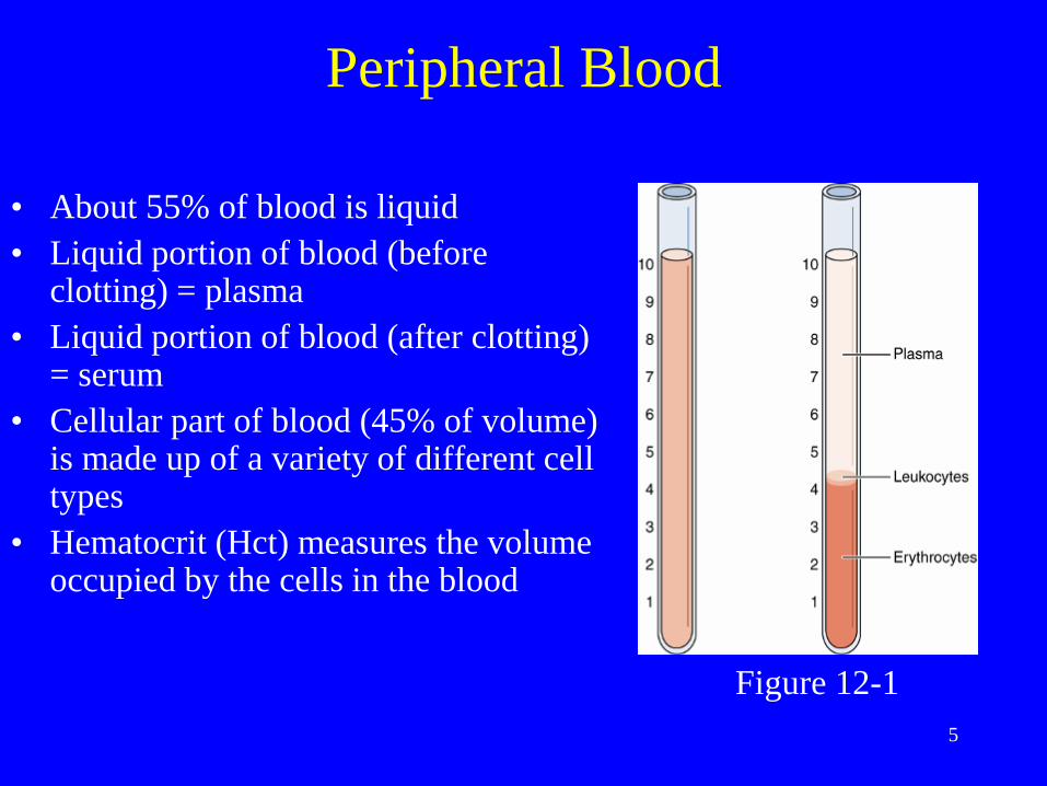
Peripheral Blood
• About 55% of blood is liquid
• Liquid portion of blood (before clotting) = plasma
• Liquid portion of blood (after clotting) = serum
• Cellular part of blood (45% of volume) is made up of a variety of different cell types
• Hematocrit (Hct) measures the volume occupied by the cells in the blood
Figure 12-1

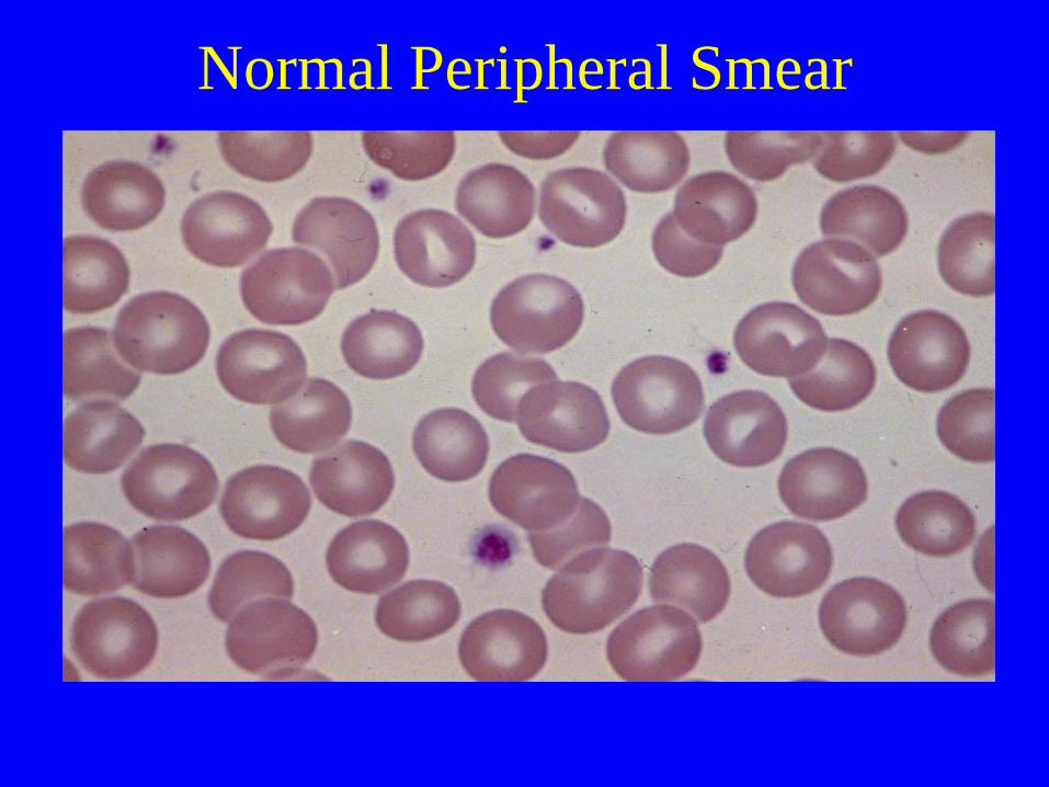
Normal Peripheral Smear
“More information can be gained from
examining the blood smear than
from any single hematologic procedure”
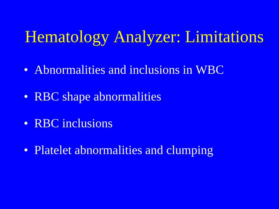
Hematology Analyzer: Limitations
• Abnormalities and inclusions in WBC
• RBC shape abnormalities
• RBC inclusions
• Platelet abnormalities and clumping

8
Cellular Components of Blood • Erythrocytes
– Red blood cells (RBCs)
• Leukocytes
– Also called white blood cells (WBCs)
– 5 majors types:
• Neutrophils (60%)
• Lymphocytes (30%)
• Monocytes (7%)
• Eosinophils (2%)
• Basophils (1%)
• Platelets
– Also called thrombocytes
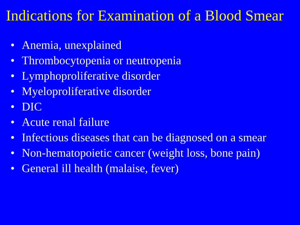
Indications for Examination of a Blood Smear
• Anemia, unexplained
• Thrombocytopenia or neutropenia
• Lymphoproliferative disorder
• Myeloproliferative disorder
• DIC
• Acute renal failure
• Infectious diseases that can be diagnosed on a smear
• Non-hematopoietic cancer (weight loss, bone pain)
• General ill health (malaise, fever)
10
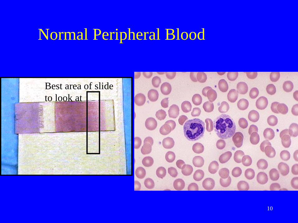
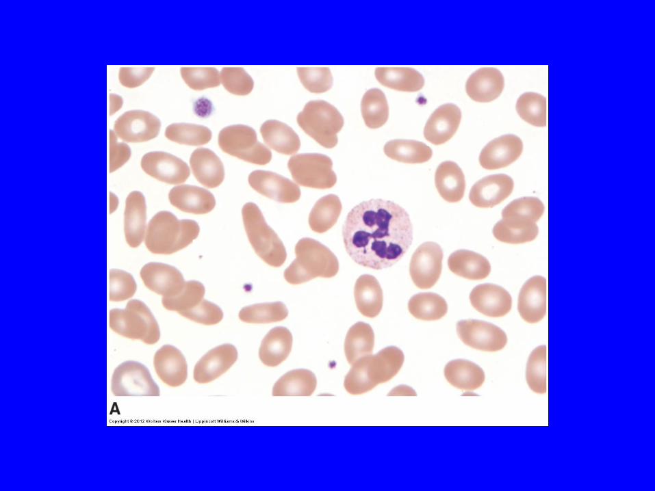
Normal Peripheral Blood
Best area of slide
to look at
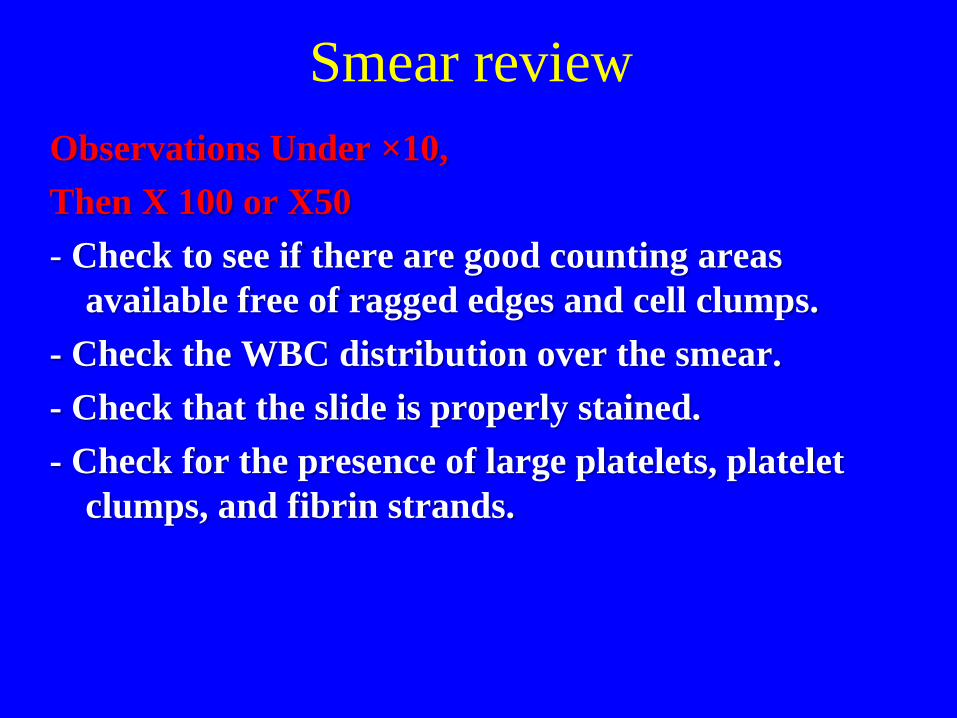
Smear review
Observations Under ×10,
Then X 100 or X50
- Check to see if there are good counting areas
available free of ragged edges and cell clumps.
- Check the WBC distribution over the smear.
- Check that the slide is properly stained.
- Check for the presence of large platelets, platelet
clumps, and fibrin strands.
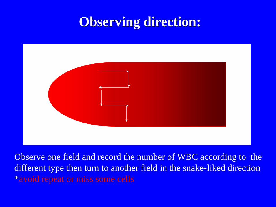
Observing direction:
Observe one field and record the number of WBC according to the
different type then turn to another field in the snake-liked direction
*avoid repeat or miss some cells
Normal blood smear
RBCs

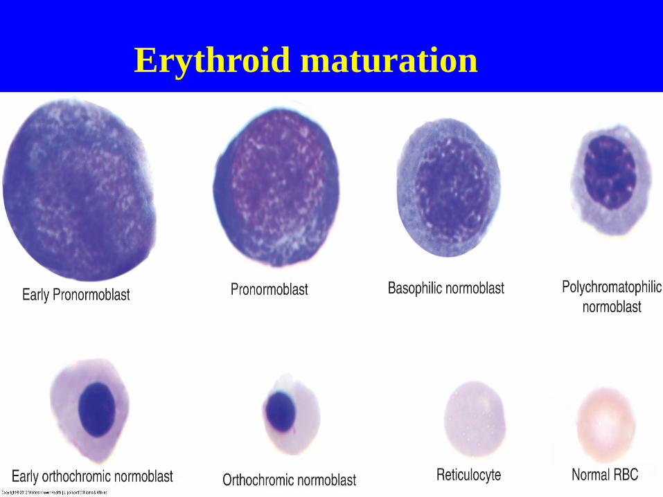
Erythroid maturation
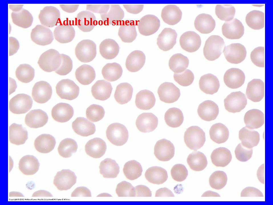
Adult blood smear
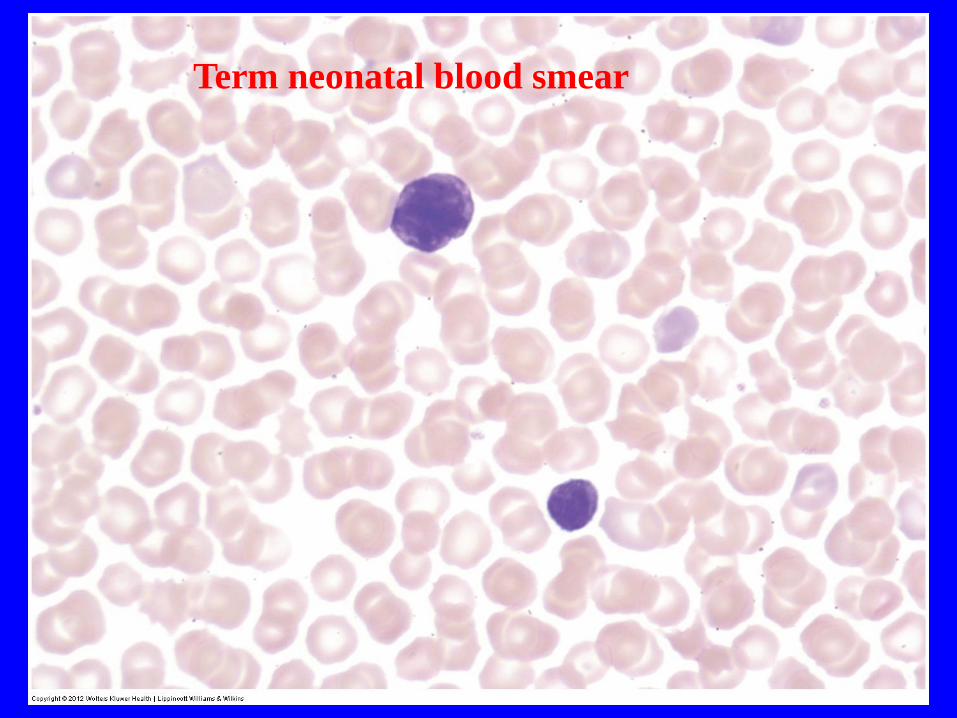
Term neonatal blood smear
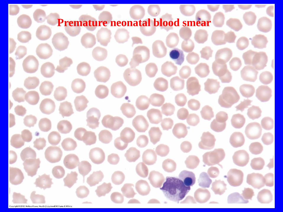
Premature neonatal blood smear

Neonatal blood smear
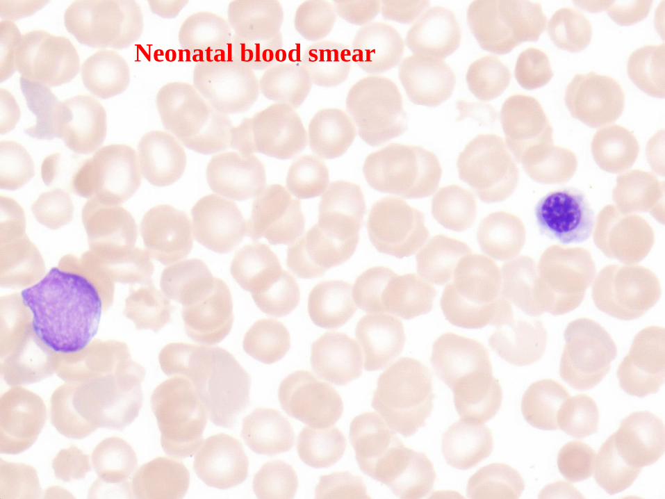
Neonatal blood smear
FEATURES OF NEONATAL
BLOOD SMEARS
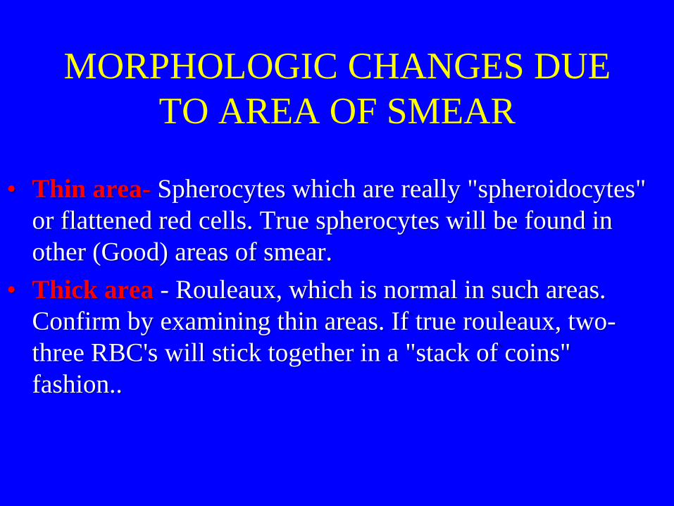
MORPHOLOGIC CHANGES DUE
TO AREA OF SMEAR
• Thin area- Spherocytes which are really "spheroidocytes"
or flattened red cells. True spherocytes will be found in
other (Good) areas of smear.
• Thick area - Rouleaux, which is normal in such areas.
Confirm by examining thin areas. If true rouleaux, two-
three RBC's will stick together in a "stack of coins"
fashion..
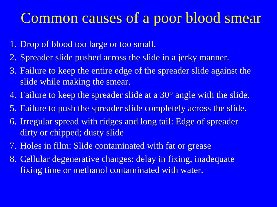
Common causes of a poor blood smear
1. Drop of blood too large or too small.
2. Spreader slide pushed across the slide in a jerky manner.
3. Failure to keep the entire edge of the spreader slide against the
slide while making the smear.
4. Failure to keep the spreader slide at a 30° angle with the slide.
5. Failure to push the spreader slide completely across the slide.
6. Irregular spread with ridges and long tail: Edge of spreader
dirty or chipped; dusty slide
7. Holes in film: Slide contaminated with fat or grease
8. Cellular degenerative changes: delay in fixing, inadequate
fixing time or methanol contaminated with water.
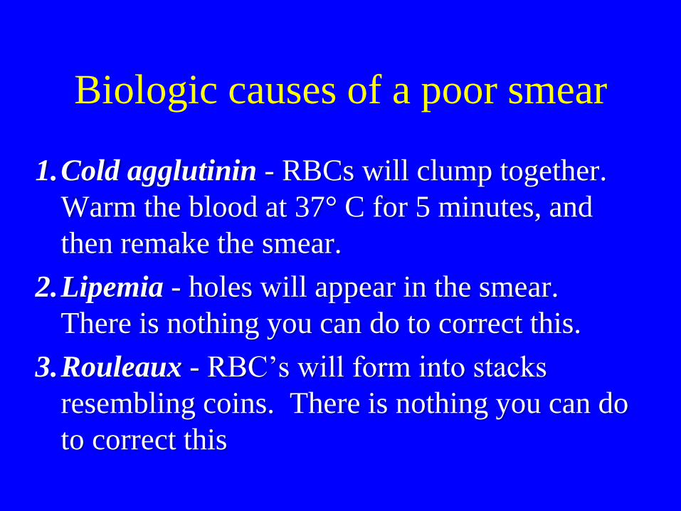
Biologic causes of a poor smear
1.Cold agglutinin - RBCs will clump together.
Warm the blood at 37° C for 5 minutes, and
then remake the smear.
2.Lipemia - holes will appear in the smear.
There is nothing you can do to correct this.
3.Rouleaux - RBC’s will form into stacks
resembling coins. There is nothing you can do
to correct this

Common Artifacts
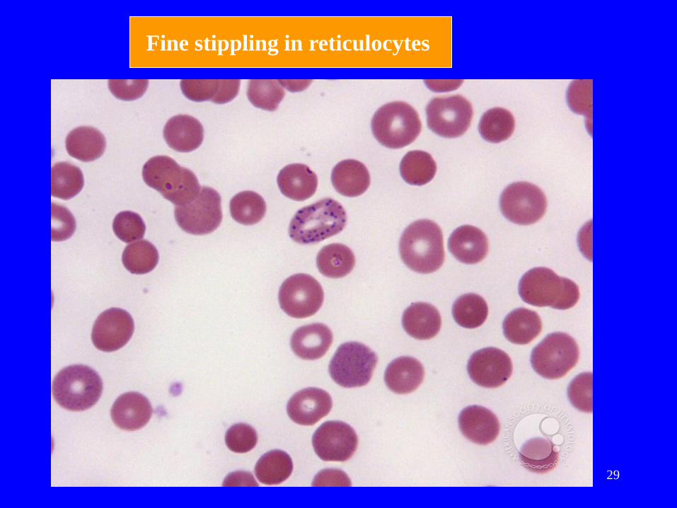
• Fine stippling in reticulocytes (slow air-drying)
• Crenated cells: Common artifact (Aged Blood,
Elevated PH, contact with glass and exposure to
moisture)
• EDTA effects on blood cells
• OLD Blood
• Artifactual Changes in RBC, WBC and Platelets

Age-related changes RBC:
-- Crenation (echinocyte formation), lysis,
hemoglobin crystallization.
WBC:
-- Swellling and smoothing of the nuclear chromatin
(mimicking band neutrophil formation), pyknosis
and karyhorrhexis of nuclei, cell smudging, and
prominence of Dohle bodies (mimicking toxic
change).
Platelets:
-- Clumping and, degranulation.
26
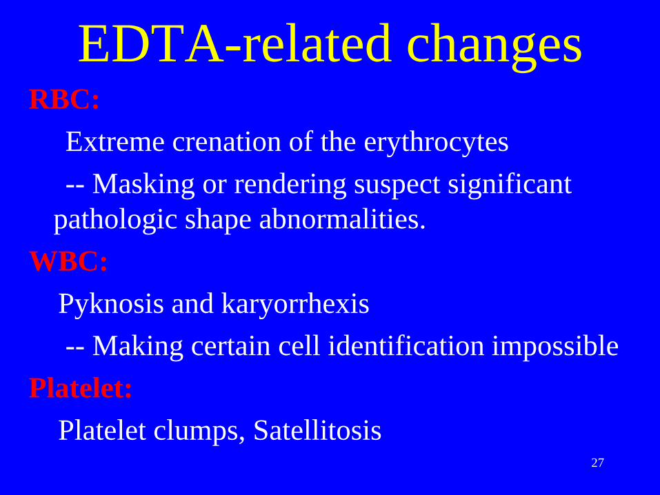
EDTA-related changes RBC:
Extreme crenation of the erythrocytes
-- Masking or rendering suspect significant
pathologic shape abnormalities.
WBC:
Pyknosis and karyorrhexis
-- Making certain cell identification impossible
Platelet:
Platelet clumps, Satellitosis
27
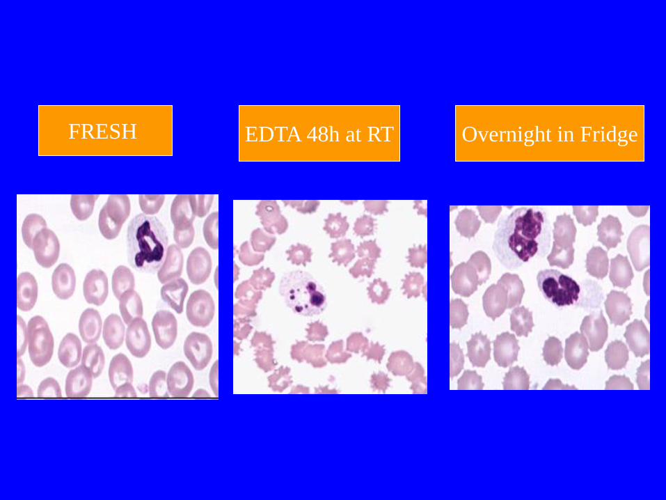
EDTA 48h at RT Overnight in Fridge FRESH
29
Fine stippling in reticulocytes
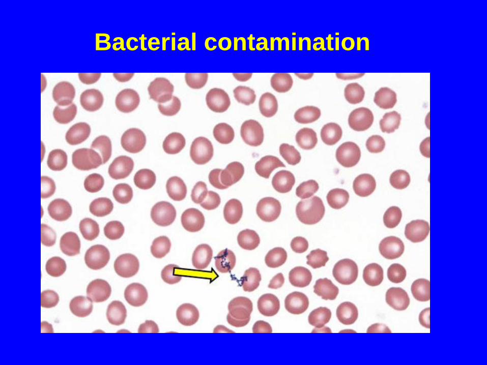
Bacterial contamination
ABNORMAL
DIFFERENTIALS 1. 200 Cell diff:
a. WBC > 15.0 (>20.0 for babies under 1 month and labor unit)
b. Three or more basophils seen.
2. If more than five immature WBC's are seen (or any blasts) let someone else diff slide and average results.
3. Correct WBC for NRBC's if you seen ten or more NRBCs/100 WBC.
4. Always indicate number of cells counted on diff.
5. If any cell type is extremely elevated (such as bands, monos, or eos > 20) indicate that you are aware of the abnormality by circling or checking on the card next to the results.
Reporting results
• Where possible use macrocytic and microcytic, rather than
simply anisocytosis alone, when describing red cell
morphology.
• Use specific cell morphology when possible, rather than
simply reporting poikilocytosis.
• When red cells are normocytic, normochromic, report out
as NORMAL. When abnormal morphology has been
noted, DO NOT indicate normal on the report form.
• EXAMPLE: 7-10 microcytic RBC's/OIF is reported out
as: 2+ microcytosis or Moderate microcytosis.

RBC Quantification
1
Peripheral Blood Morphology
Manual differential And
Morphology
RBC
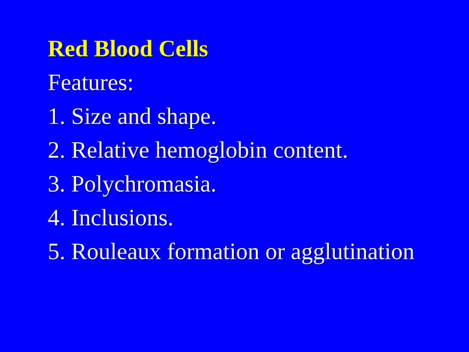
Red Blood Cells
Features:
1. Size and shape.
2. Relative hemoglobin content.
3. Polychromasia.
4. Inclusions.
5. Rouleaux formation or agglutination
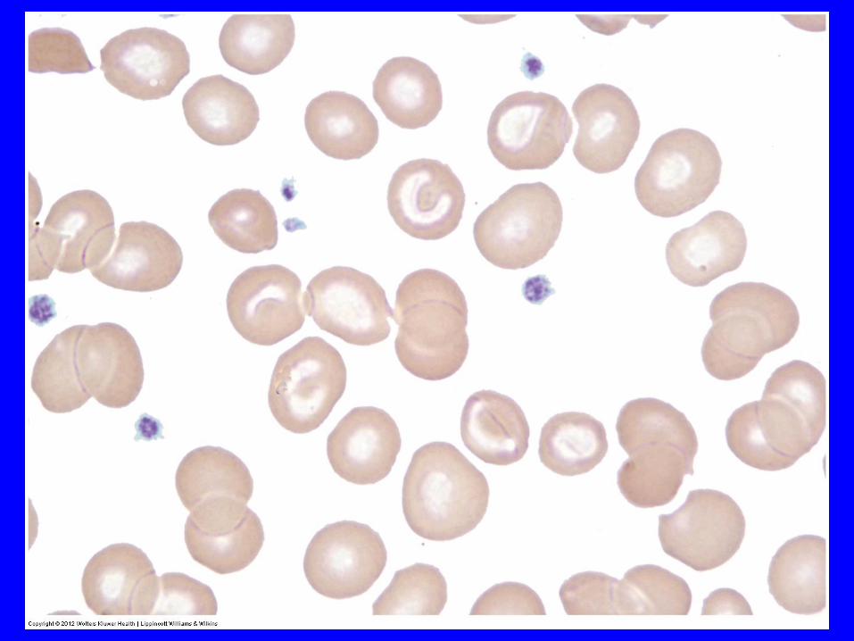
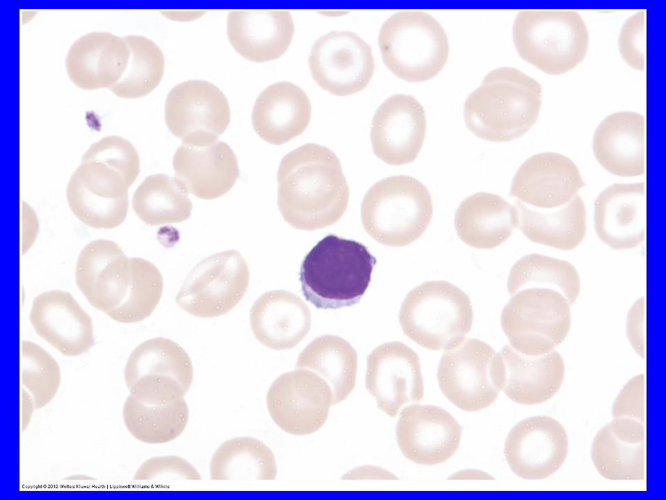
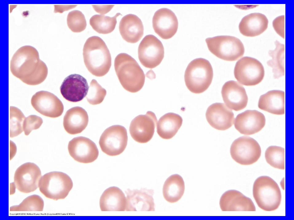
Normal Peripheral Smear
Common Red Blood Cell
Changes
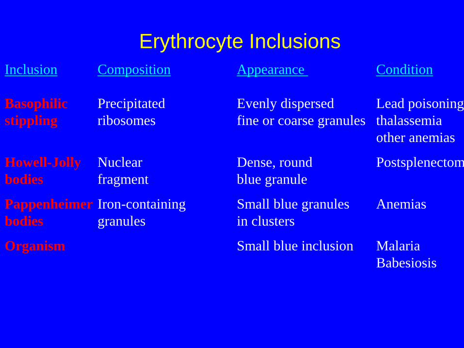
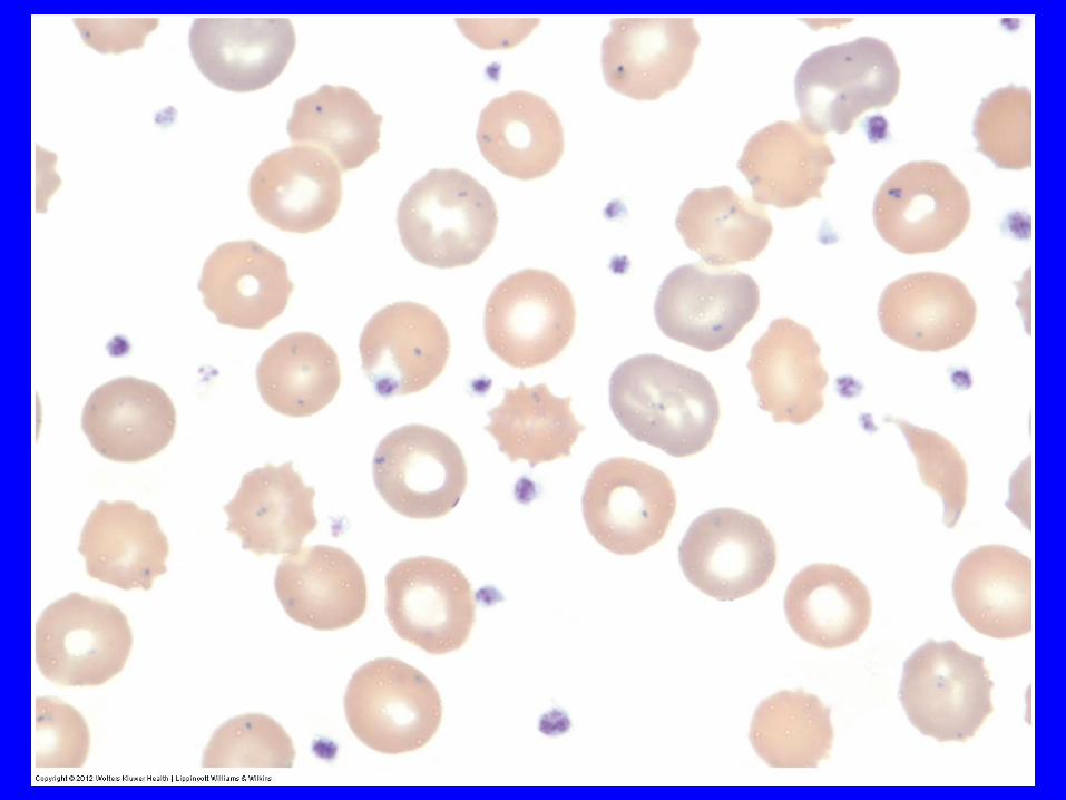
Erythrocyte Inclusions
Inclusion Composition Appearance Condition
Basophilic Precipitated Evenly dispersed Lead poisoning
stippling ribosomes fine or coarse granules thalassemia
other anemias
Howell-Jolly Nuclear Dense, round Postsplenectomy
bodies fragment blue granule
Pappenheimer Iron-containing Small blue granules Anemias
bodies granules in clusters
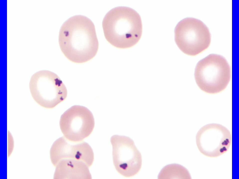
Organism Small blue inclusion Malaria
Babesiosis
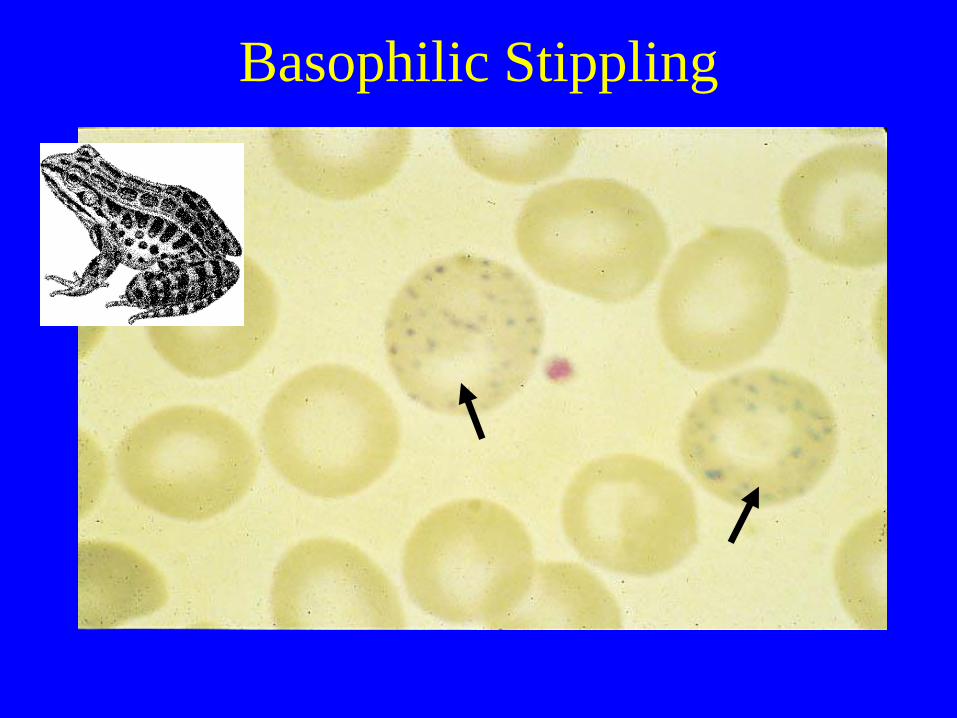
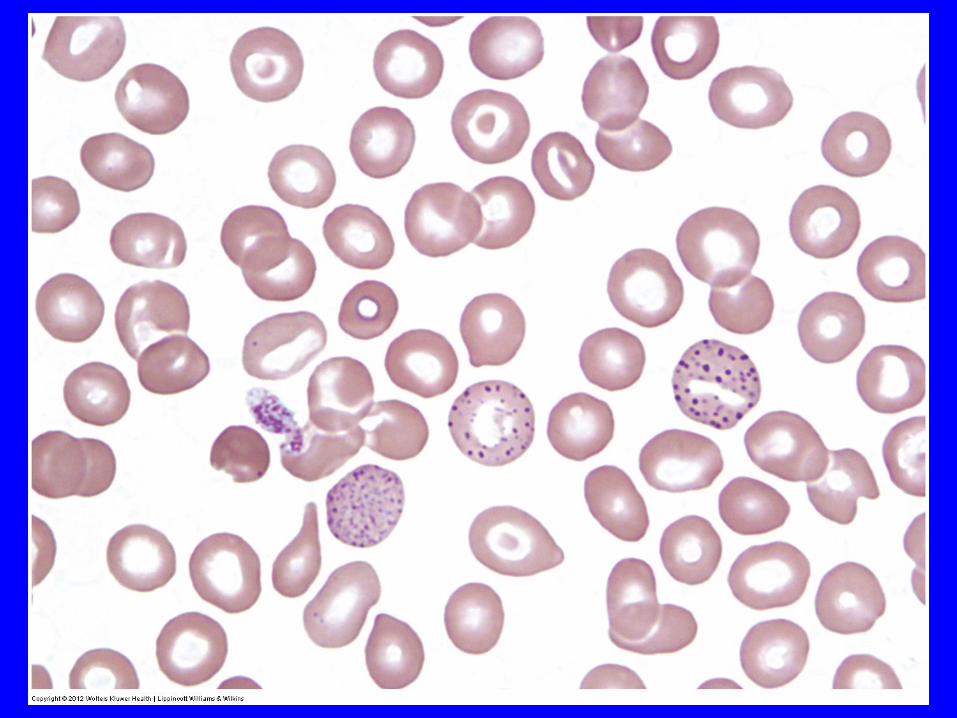
Basophilic Stippling
Malaria
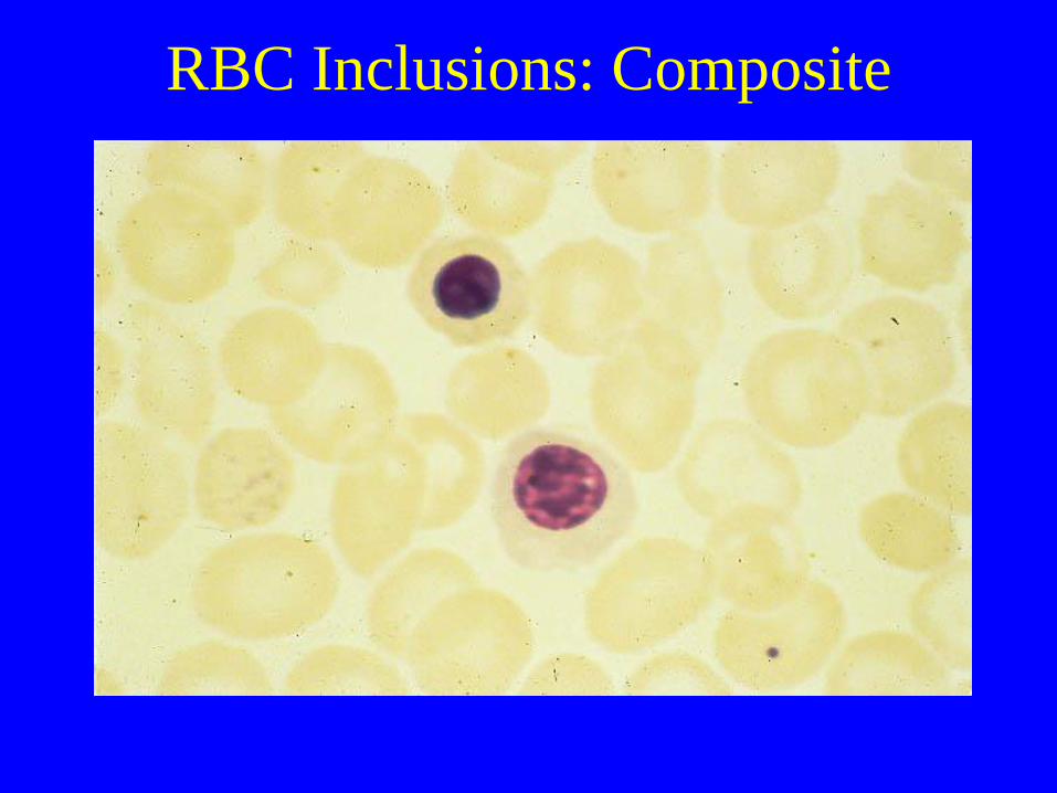
RBC Inclusions: Composite
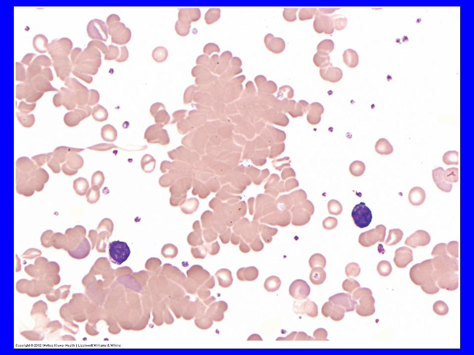
RBC Distribution Abnormalities
• Rouleaux formation Stacking of RBCs due to
increased plasma proteins
coating RBCs
• Agglutination Antibody-mediated
clumping;
temperature dependent
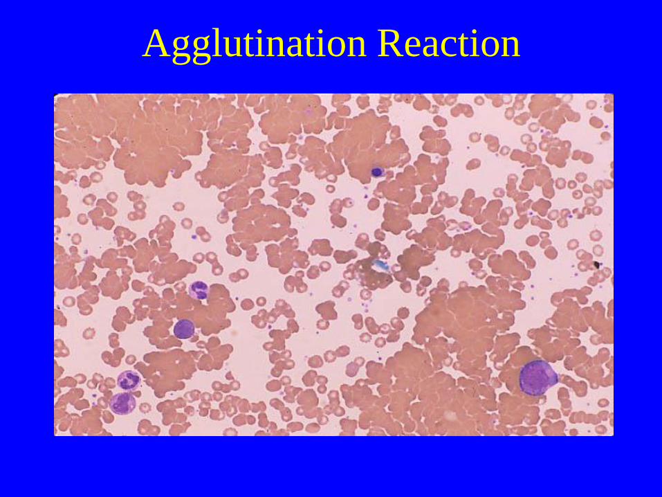
Agglutination Reaction

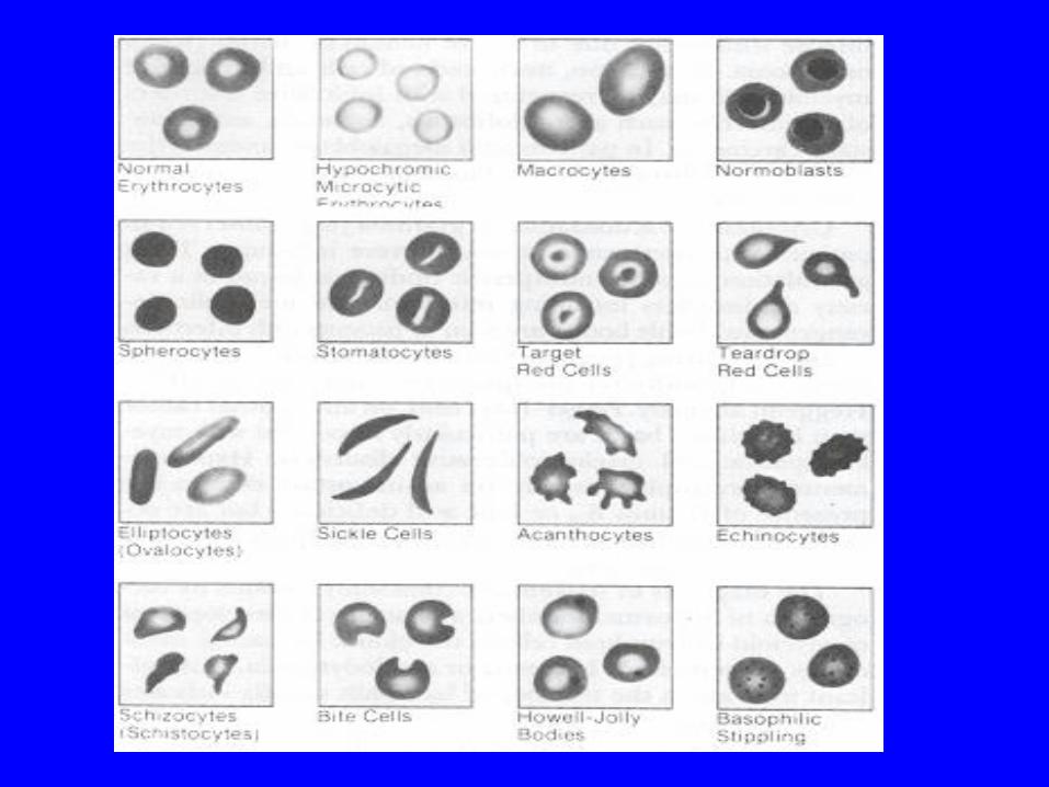
Variations in RBC Size and
Shape
• Anisocytosis Variations in size (e.g.microcytes)
• Poikilocytosis Variations in shape (e.g.target cell)
• Hypochromia Increased central pallor due to
• decrease in hemoglobin

Hypochromic Microcytic RBC
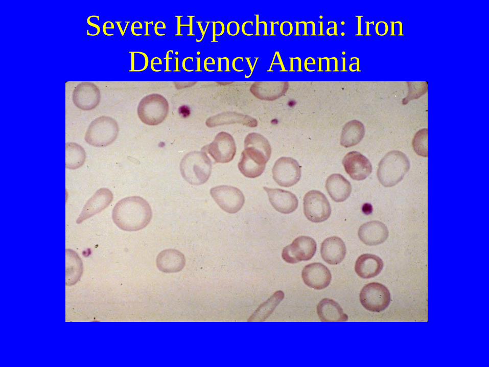
Severe Hypochromia: Iron
Deficiency Anemia
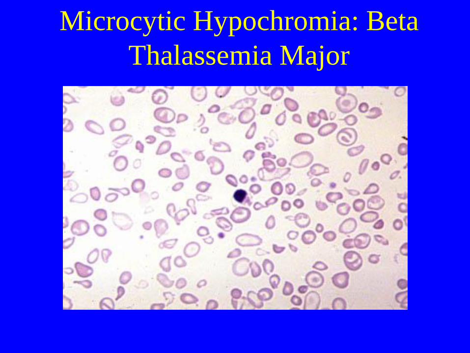
Microcytic Hypochromia: Beta
Thalassemia Major
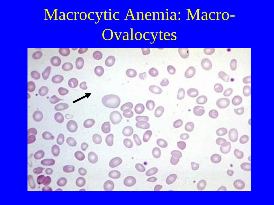
Macrocytic Anemia: Macro-
Ovalocytes

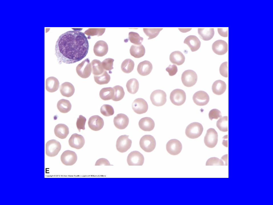
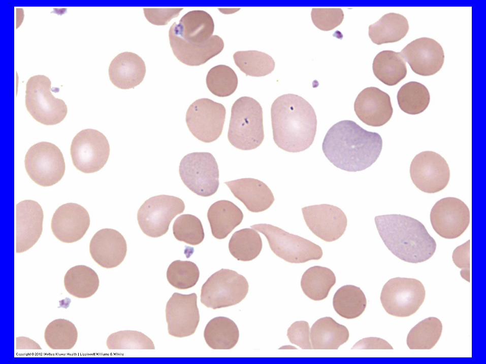
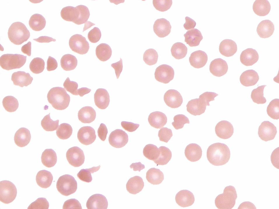
Shape Abnormalities of Erythrocytes
Terminology Description Condition
Target cells Central hemoglobin; target-shaped Liver disease; thalassemia:
Abnormal Hgb; iron deficiency
Echinocyte Short spicules, equally-spaced Uremia, hypokalemia, artifact
Acanthocyte Spiculated, irregular Liver disease (alcohol),
Post-splenectomy
Spherocyte Spherical, no central pallor HS, Immune hemolytic anemia
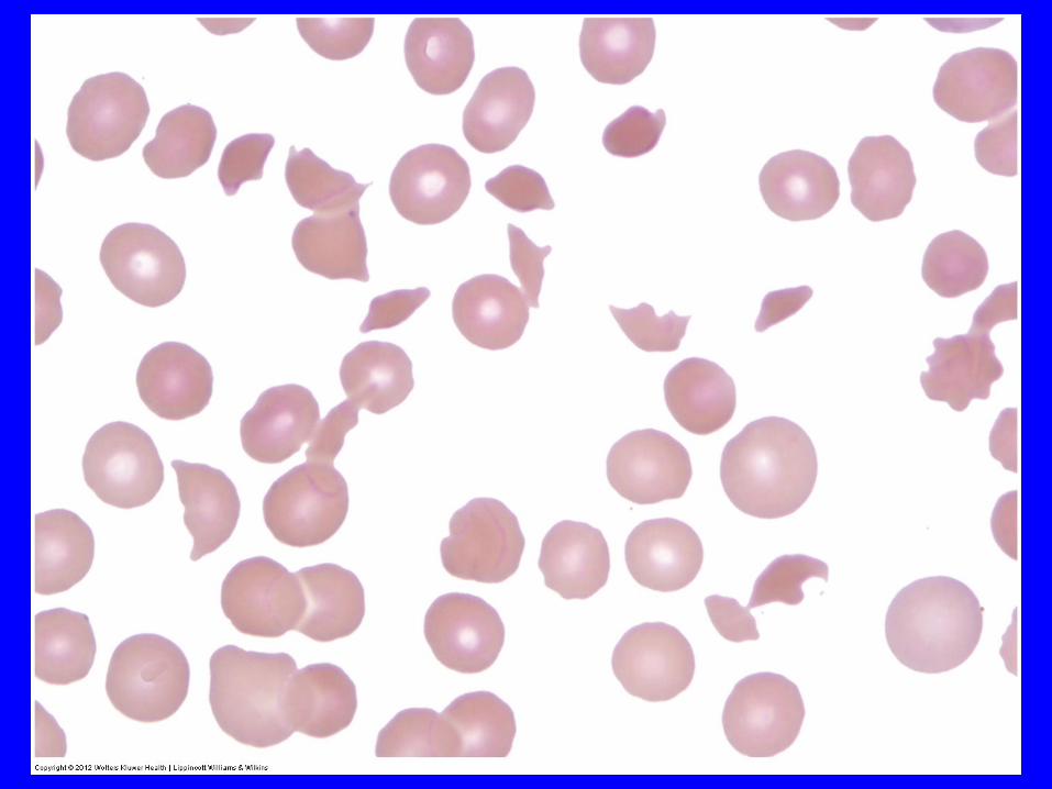
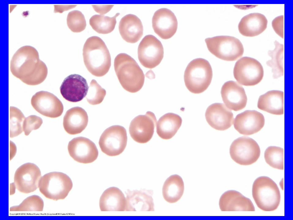
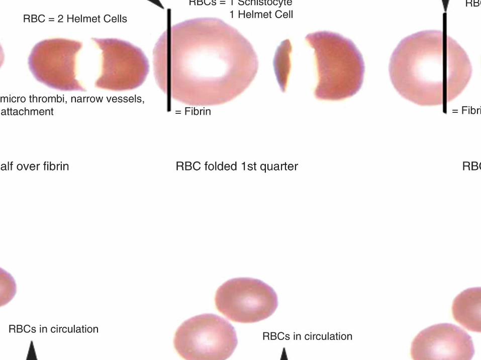
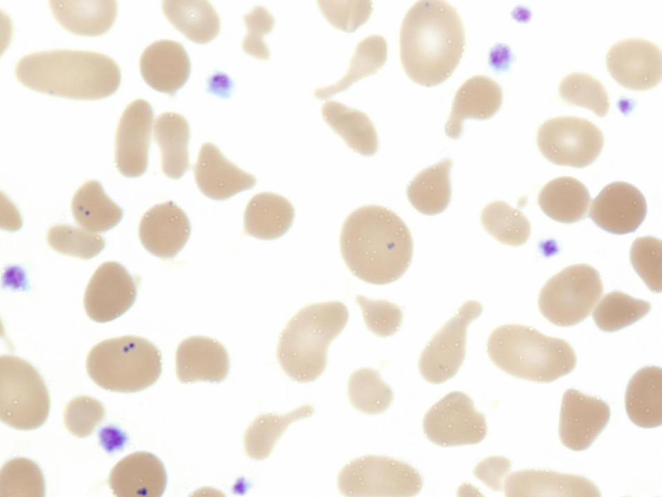
Schistocyte Fragmented RBC, helmet cells MAHA, burns
Ovalocyte Oval/elliptical shaped Hereditary elliptocytosis,
Megaloblastic anemia
Sickle cell bipolar spiculated shape Hgb S-containing
“banana” shaped hemoglobinopathy
Teardrop cell single elongated extremity Myelophthistic changes
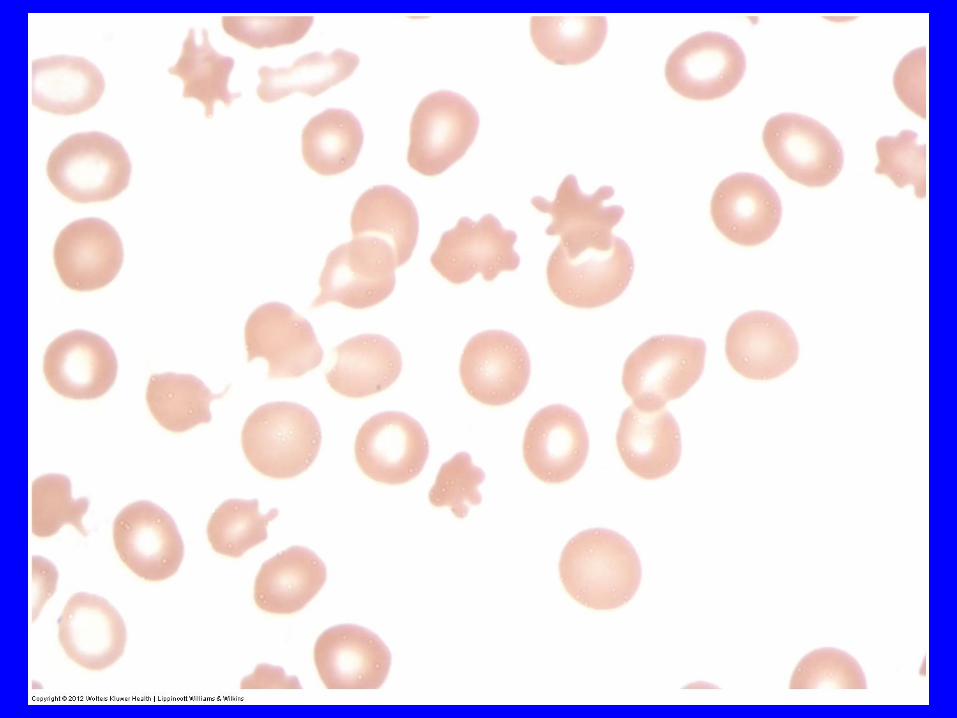
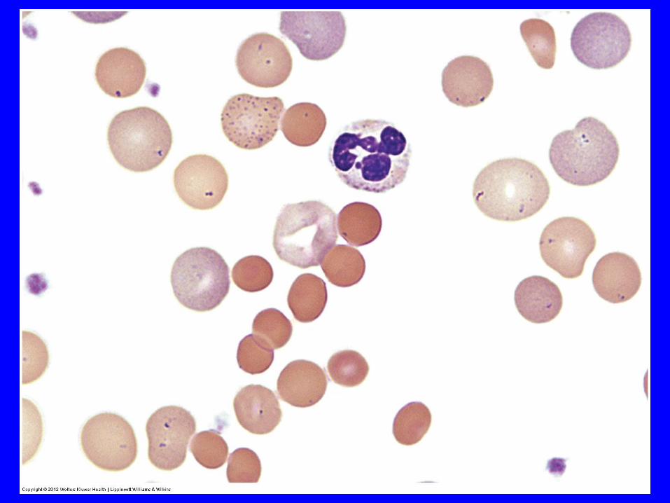
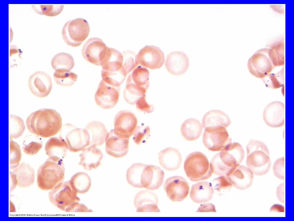
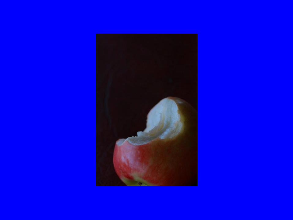
Bite cells Irregular gap in membrane G6PD deficiency
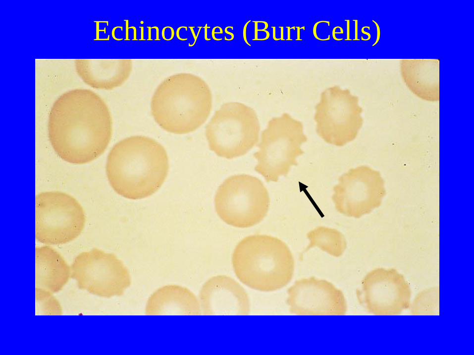
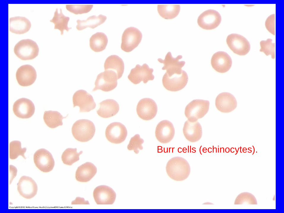
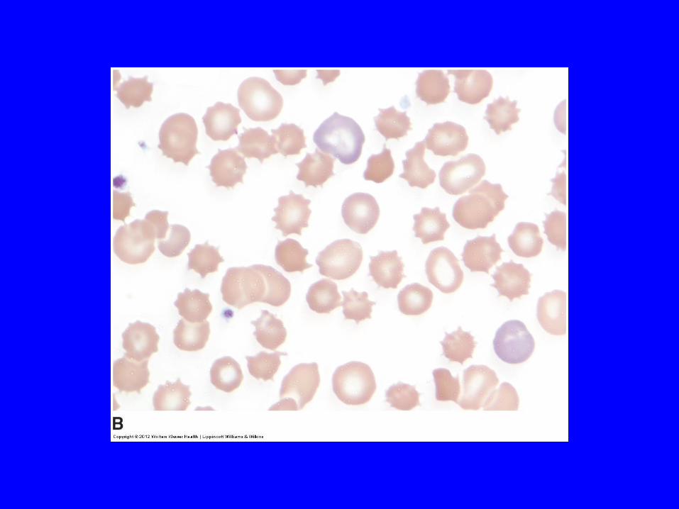
Echinocytes (Burr Cells)
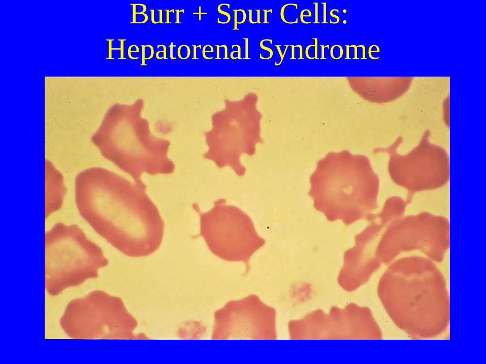
Burr + Spur Cells:
Hepatorenal Syndrome
Burr cells (echinocytes).


Echinocytes
(“Crenated cells, burr cells")
• Uremia
• HUS
• Crenated cells: Common artifact (Aged Blood, Elevated
PH, contact with glass and exposure to moisture)
• Post-splenectomy
• Hepatitis of the newborn
• Malabsorption states
• After administration of heparin
• Pyruvate kinase deficiency
• Phosphoglycerare kinase deficiency
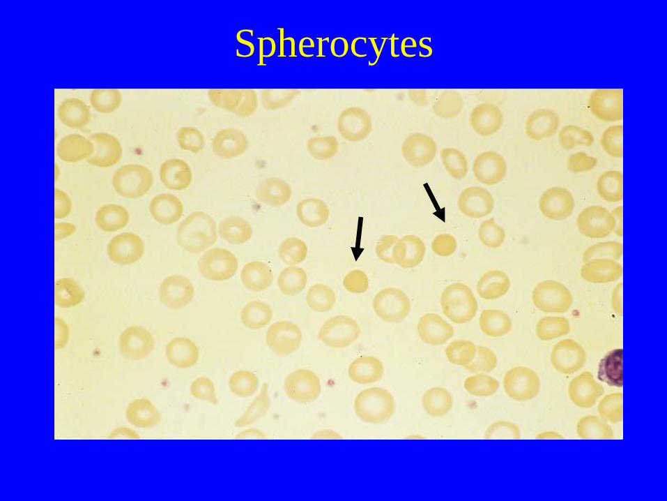
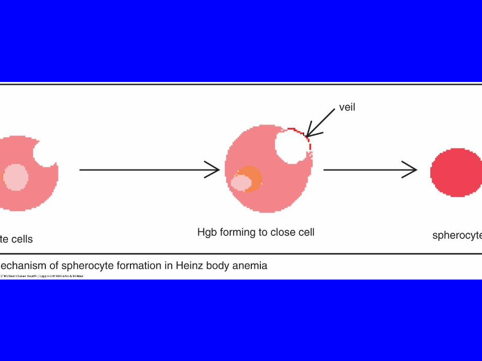
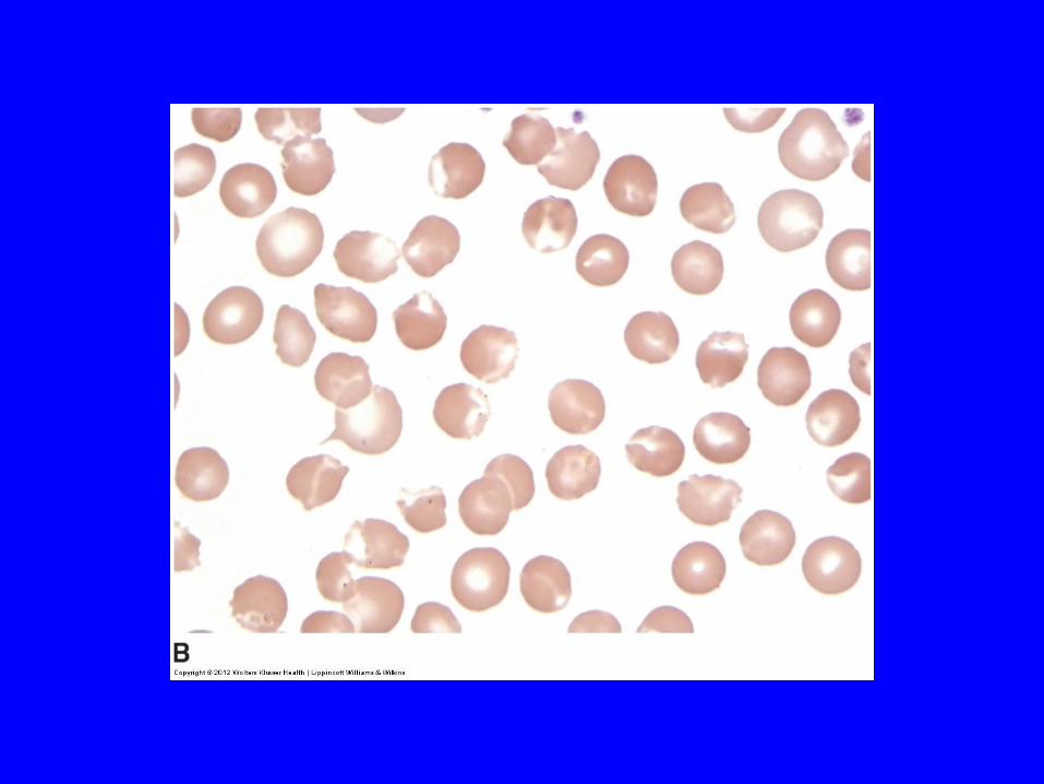
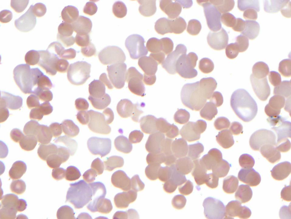
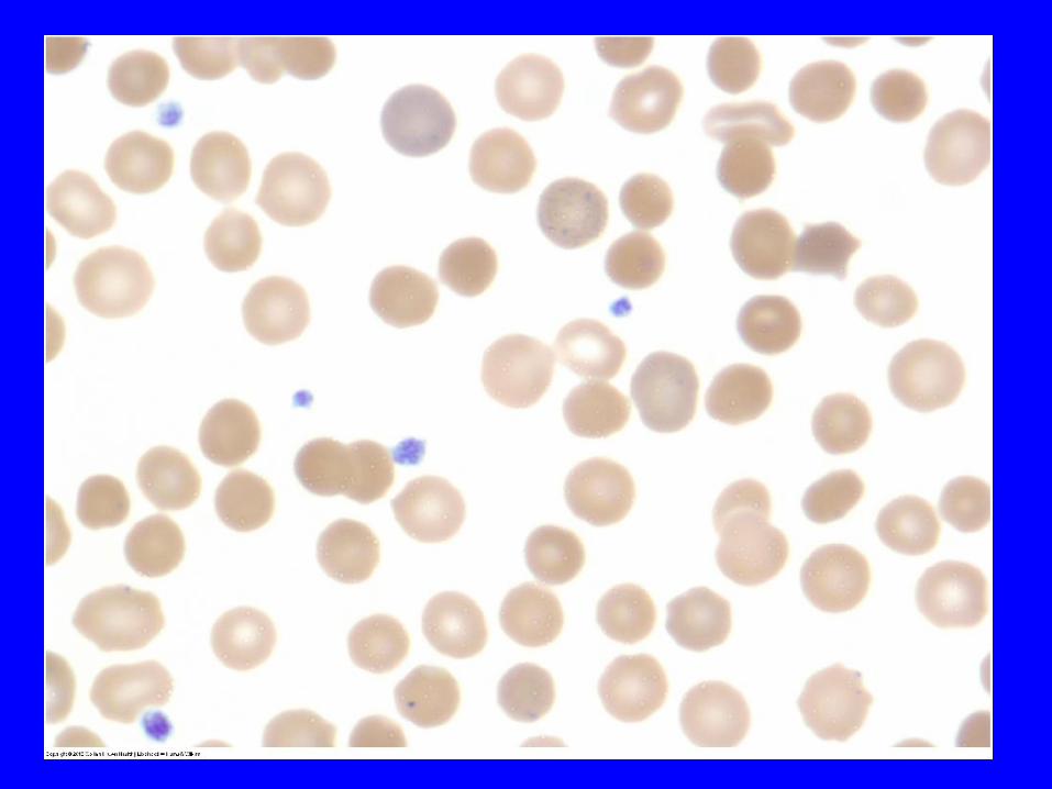
Spherocytes
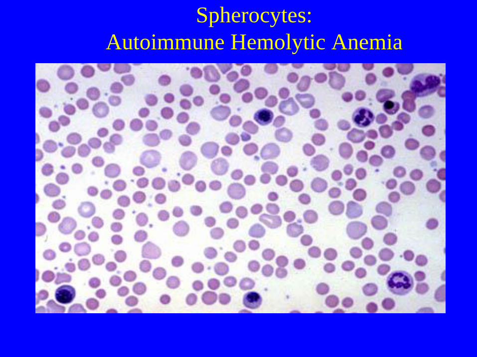
Spherocytes:
Autoimmune Hemolytic Anemia
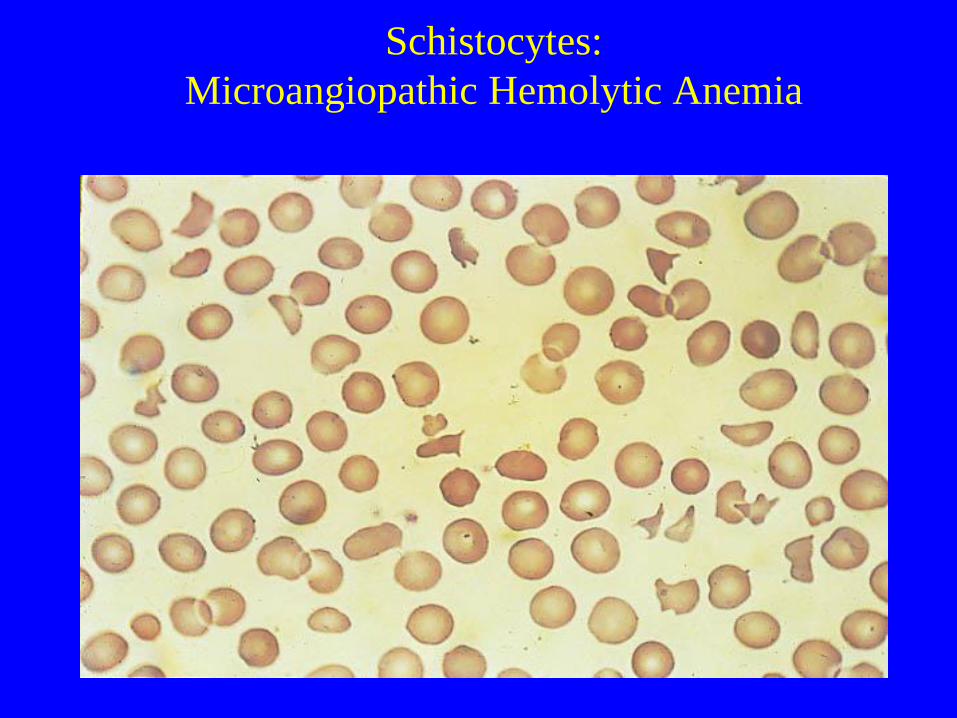
Schistocytes:
Microangiopathic Hemolytic Anemia


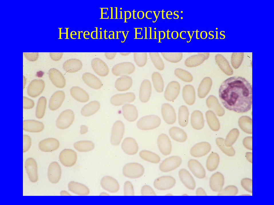
Elliptocytes:
Hereditary Elliptocytosis
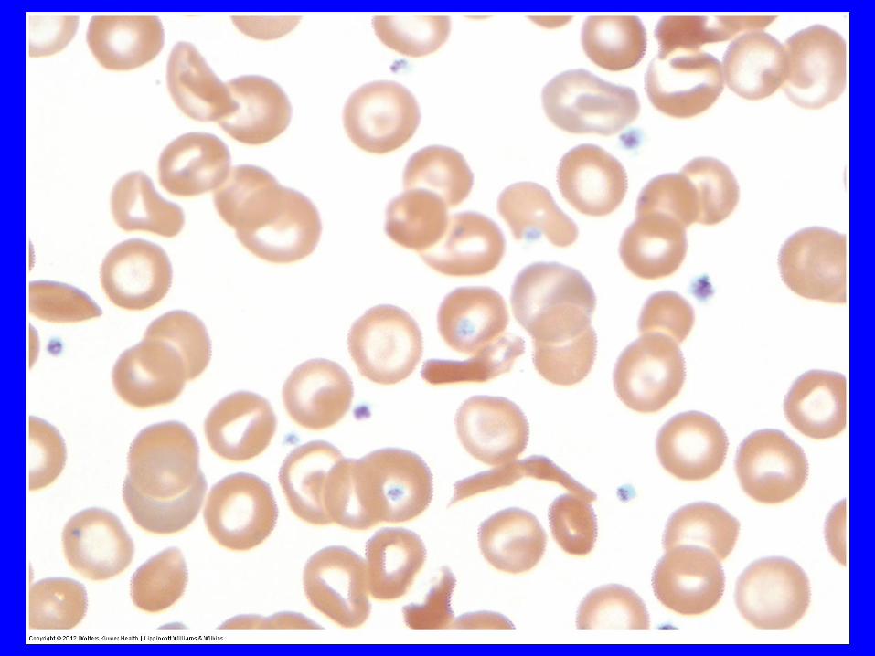
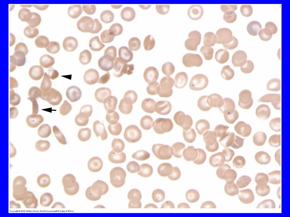
Sickle Cells

Target cells in Hemoglobin SC
Disease
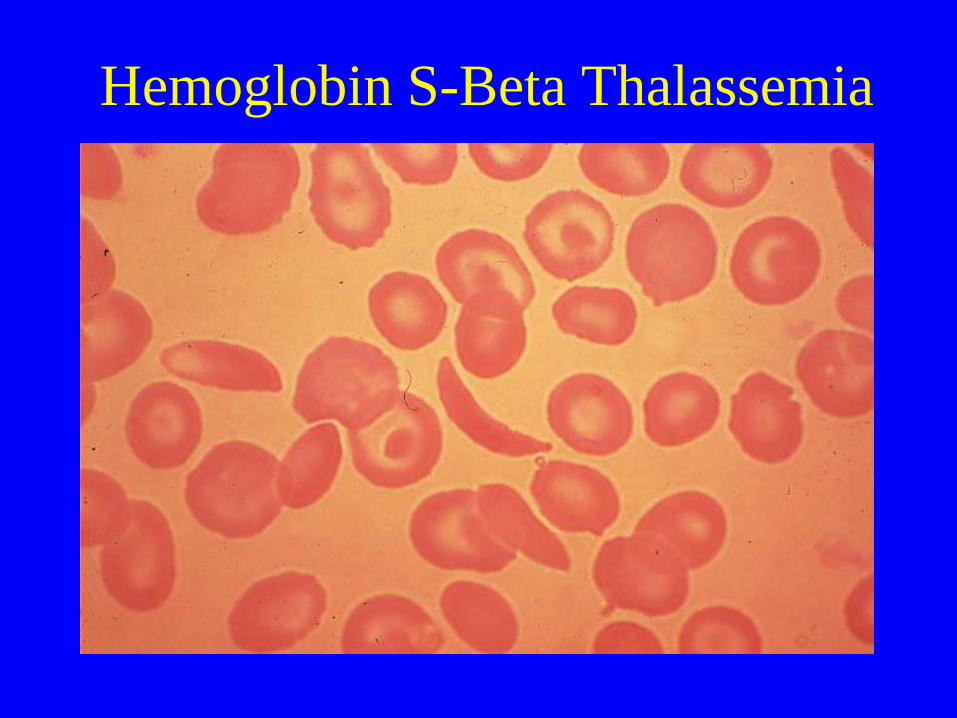
Hemoglobin S-Beta Thalassemia
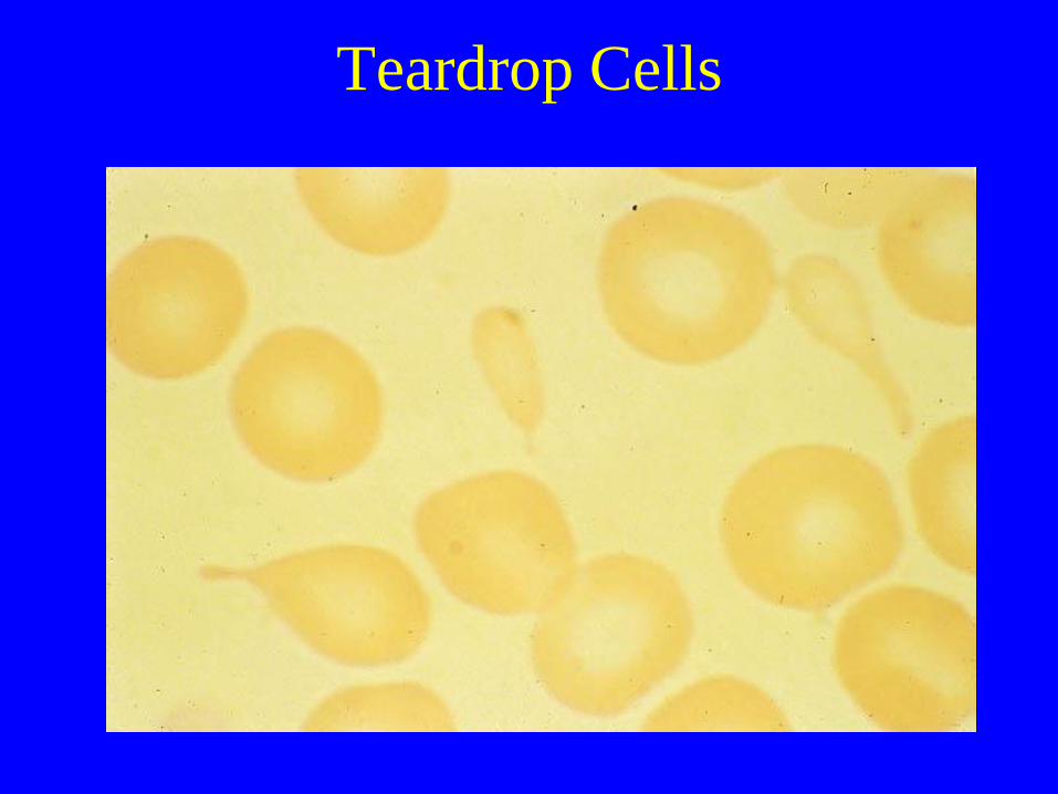
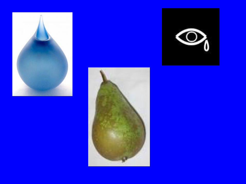
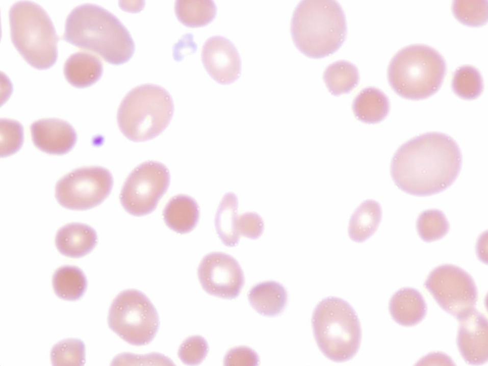
Teardrop Cells
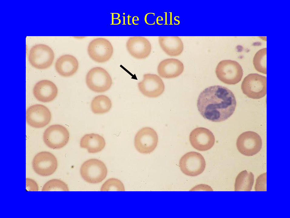
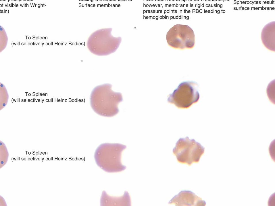
Bite Cells
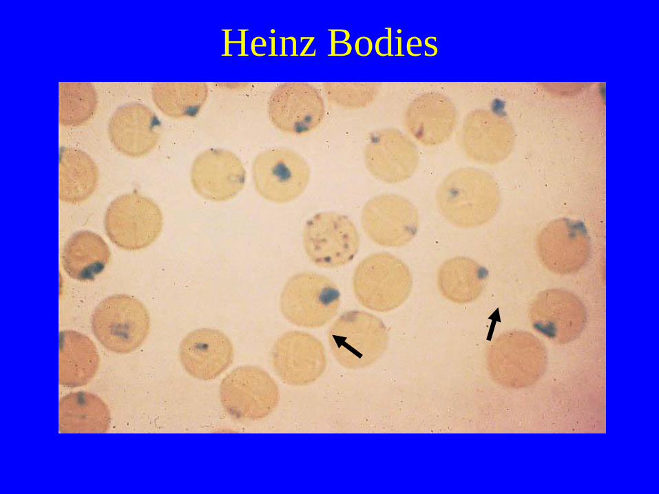
Heinz Bodies

Stomatocyte
• Hereditary or acquired hemolysis
• Hereditary stomatocytosis
• Alcoholic cirrhosis & acute alcoholism
• Obstructive liver disease
• Malignancy,
• Severe infection
• Treated acute leukemia
• Artifact.

Polychromatophilic red cells

Echinocytes
(“Crenated cells, burr cells")
• Uremia
• HUS
• Crenated cells: Common artifact (Aged Blood, Elevated
PH, contact with glass and exposure to moisture)
• Post-splenectomy
• Hepatitis of the newborn
• Malabsorption states
• After administration of heparin
• Pyruvate kinase deficiency
• Phosphoglycerare kinase deficiency
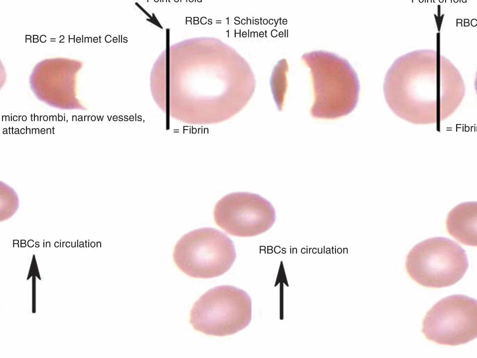

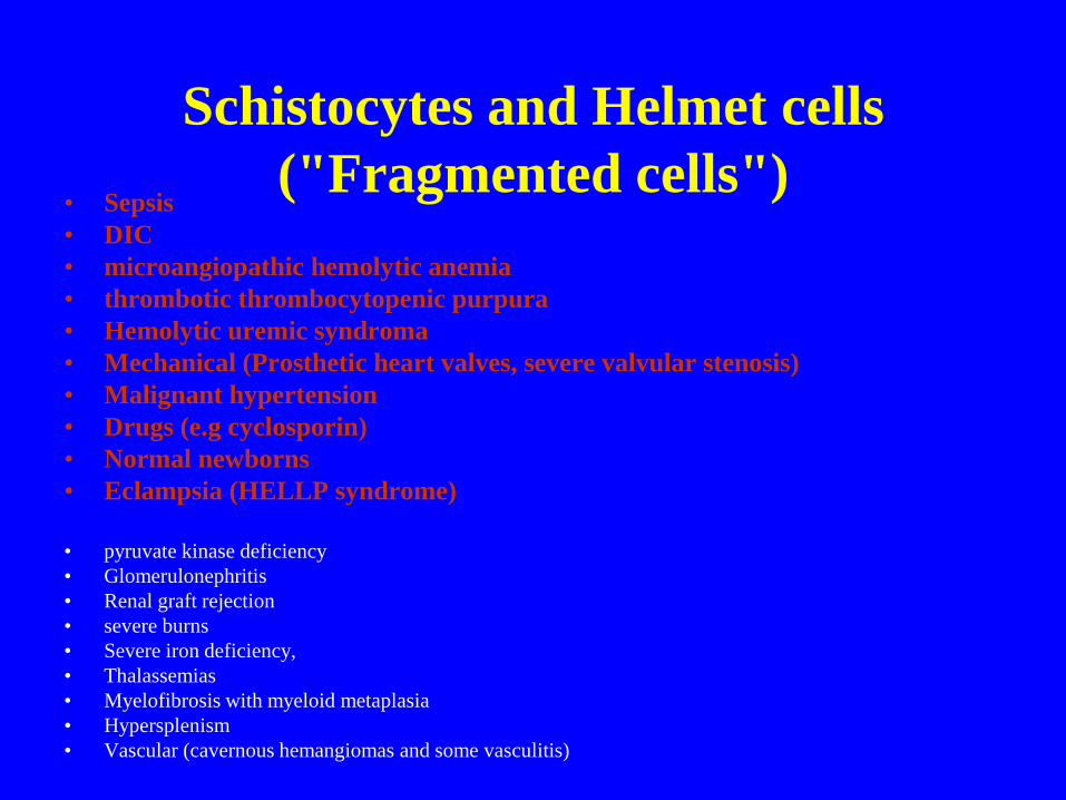
Schistocytes and Helmet cells
("Fragmented cells") • Sepsis
• DIC
• microangiopathic hemolytic anemia
• thrombotic thrombocytopenic purpura
• Hemolytic uremic syndroma
• Mechanical (Prosthetic heart valves, severe valvular stenosis)
• Malignant hypertension
• Drugs (e.g cyclosporin)
• Normal newborns
• Eclampsia (HELLP syndrome)
• pyruvate kinase deficiency
• Glomerulonephritis
• Renal graft rejection
• severe burns
• Severe iron deficiency,
• Thalassemias
• Myelofibrosis with myeloid metaplasia
• Hypersplenism
• Vascular (cavernous hemangiomas and some vasculitis)

Macrocyte Oval macrocyte
• Accelerated erythrocytosis.
• Myelodysplastic syndrome
• Megaloblastic anemia
• Fanconi anemia
• Congenital dyserythropoietic anemia (CDA types I and III)
Round macrocyte
• Liver disease
• Alcoholism
• Aplastic anemia
• Hypothyroidism

Rouleaux Formation
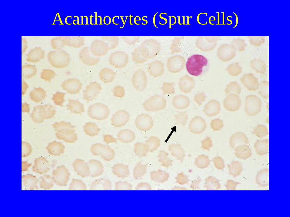
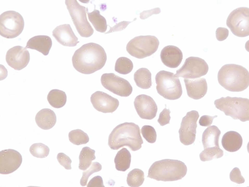
Acanthocytes (Spur Cells)
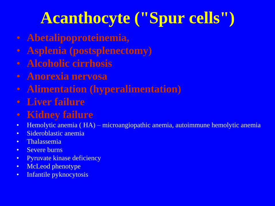
Acanthocyte ("Spur cells") • Abetalipoproteinemia,
• Asplenia (postsplenectomy)
• Alcoholic cirrhosis
• Anorexia nervosa
• Alimentation (hyperalimentation)
• Liver failure
• Kidney failure • Hemolytic anemia ( HA) – microangiopathic anemia, autoimmune hemolytic anemia
• Sideroblastic anemia
• Thalassemia
• Severe burns
• Pyruvate kinase deficiency
• McLeod phenotype
• Infantile pyknocytosis

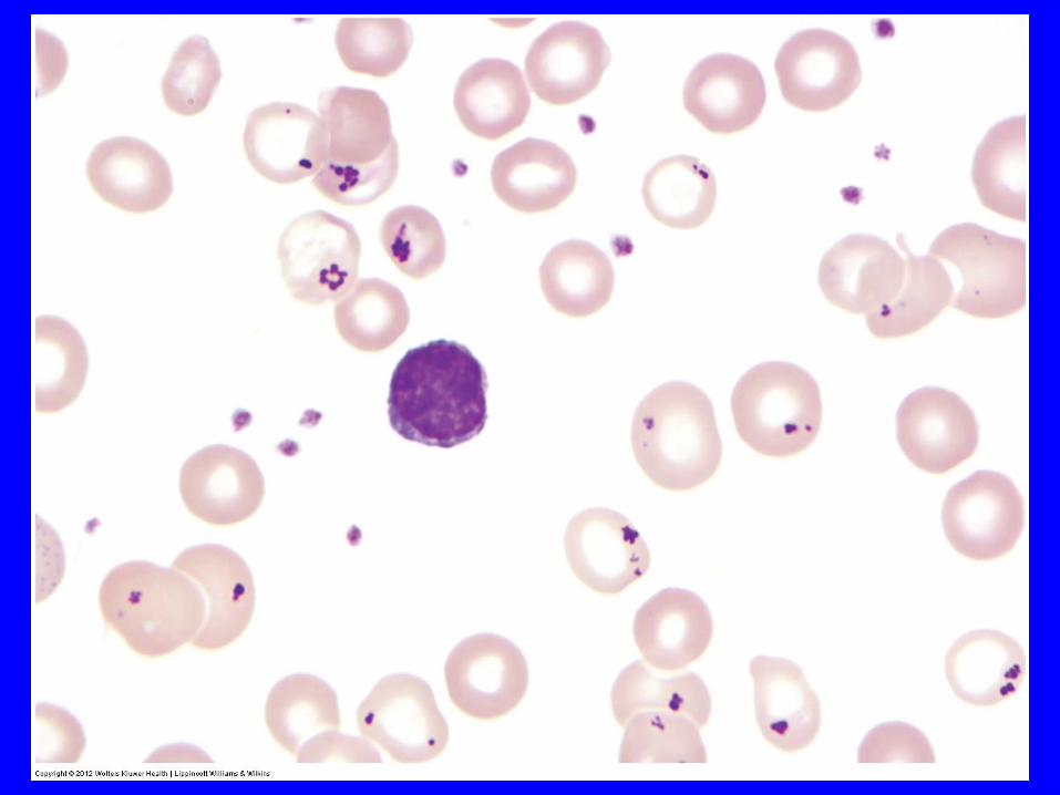
Target
Cells
Spur Cells
Target and Spur cells:
Changes in Liver Disease
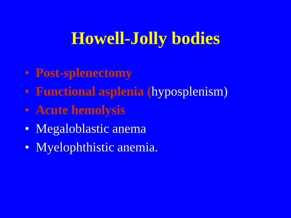
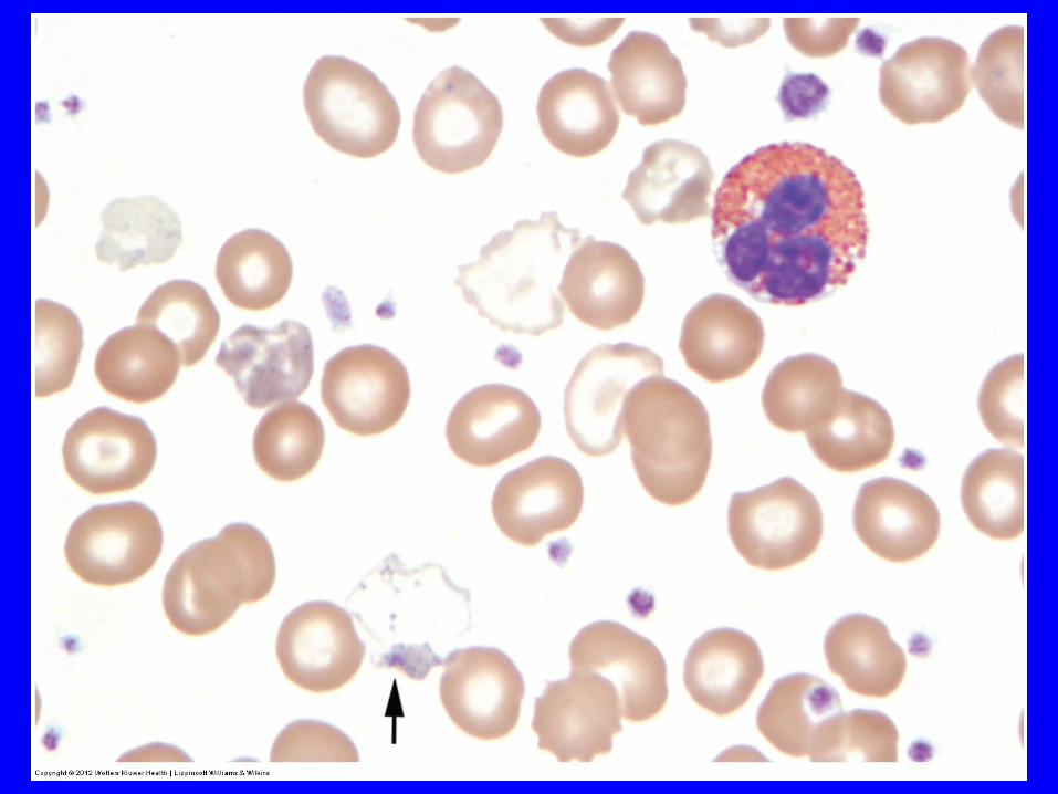
Howell-Jolly bodies
• Post-splenectomy
• Functional asplenia (hyposplenism)
• Acute hemolysis
• Megaloblastic anema
• Myelophthistic anemia.
Anemia Due to Abnormal/Impaired
Hb Synthesis
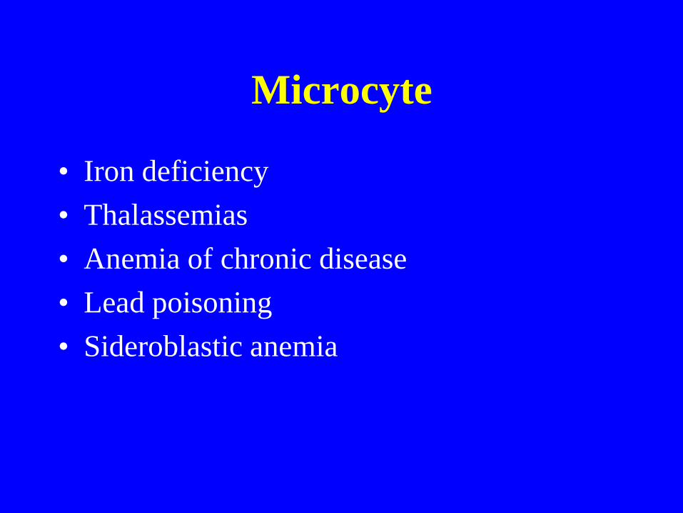
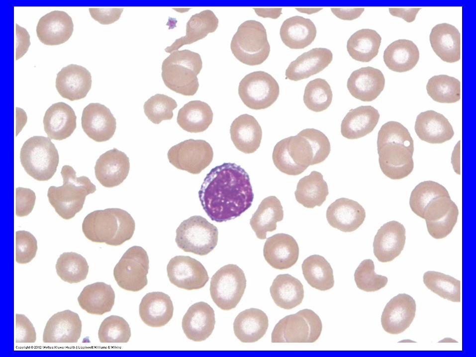
Microcyte
• Iron deficiency
• Thalassemias
• Anemia of chronic disease
• Lead poisoning
• Sideroblastic anemia
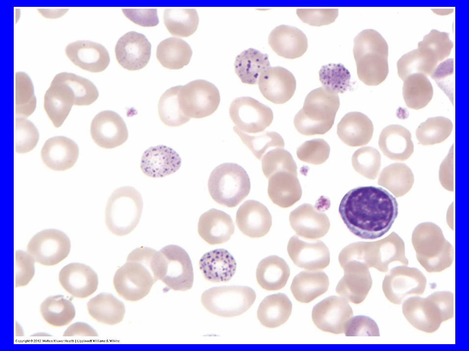
Basophilic stippling
• Heavy metal poisoning (e.g. lead and
arsenic)
• Hemoglobinopathies
• Thalassemias
• sideroblastic anemias
• pyrimidine-5’-nucleotidase deficiency
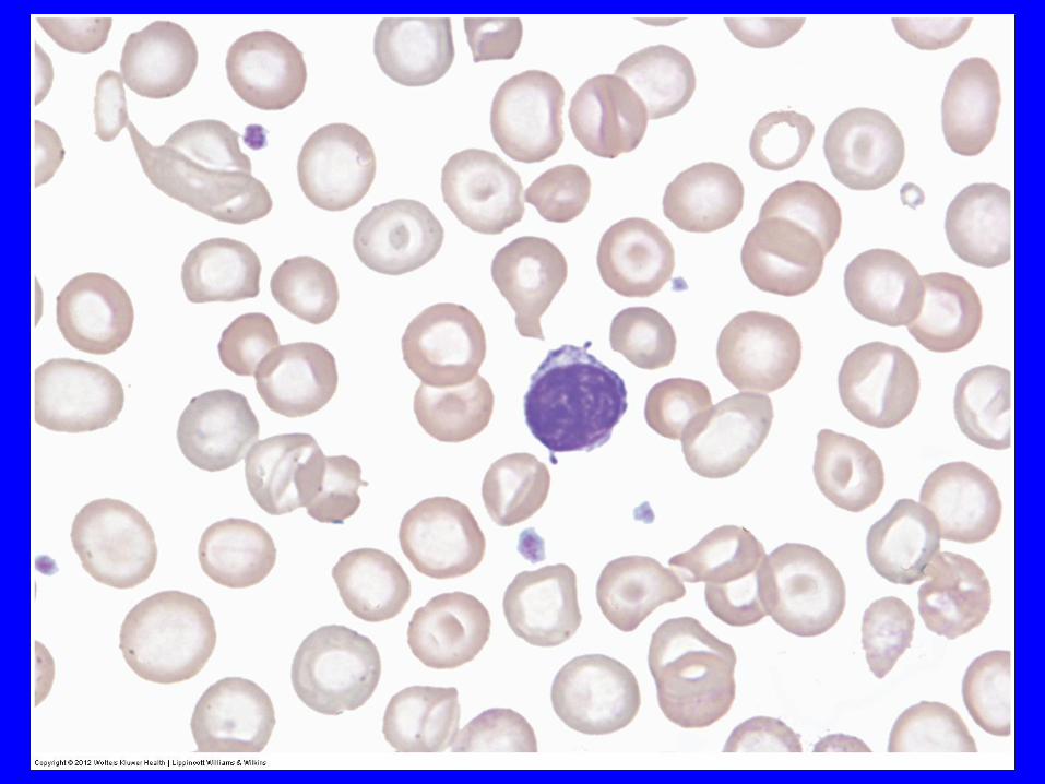
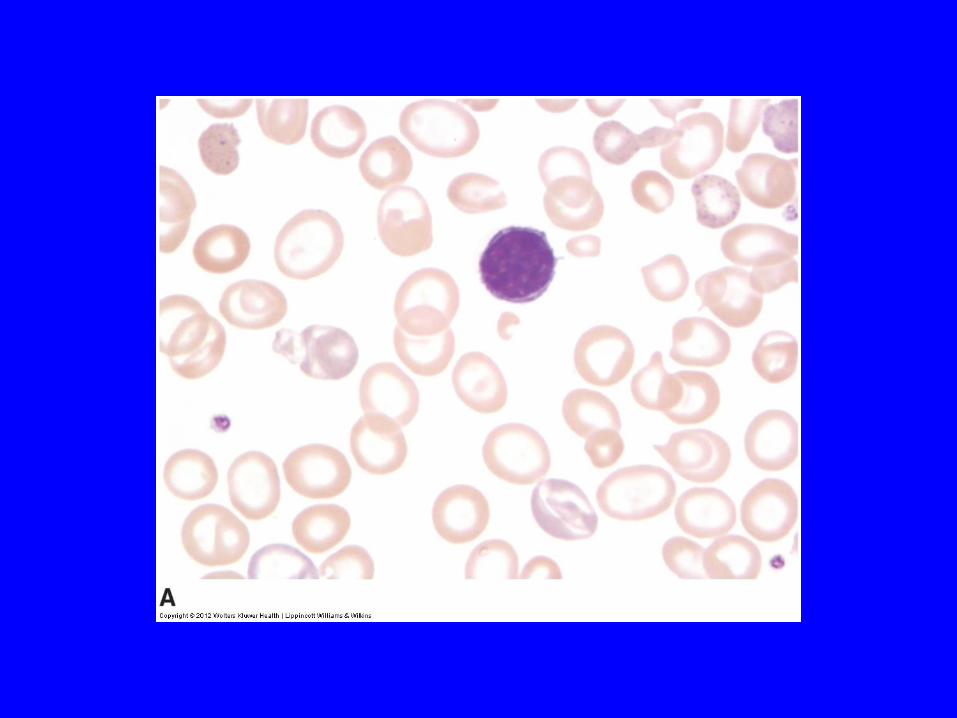
Target cells • Splenectomy
• Thalassemia
• Hemoglobinopathies (hemoglobin SS, SC, CC, EE, AE, sickle cell-thalassemia)
• iron deficiency anemia
• liver disease
• Postsplectomy
• familial lecithin-cholesterol acyltransferase (LCAT) deficiency.
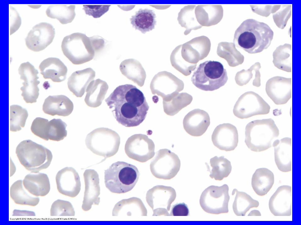
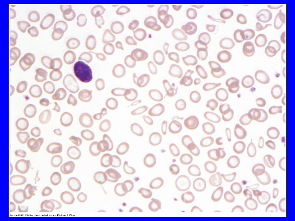
Nucleated red blood cells ("NRBCs")
• Acute bleeding
• Severe hemolysis
• Myelofibrosis
• Leukemia
• Myelophthisis
• Asplenia
• Newborn

Target cells • Splenectomy
• Thalassemia
• Hemoglobinopathies (hemoglobin SS, SC, CC, EE, AE, sickle cell-thalassemia)
• iron deficiency anemia
• liver disease
• Postsplectomy
• familial lecithin-cholesterol acyltransferase (LCAT) deficiency.

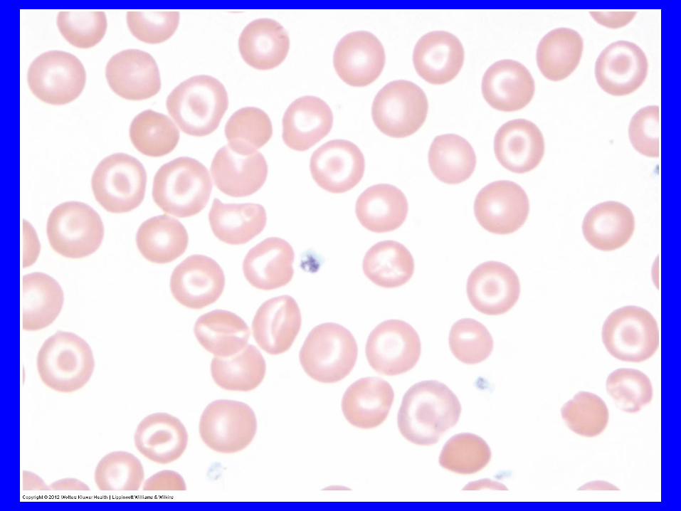
Sickle cells
• Hb S hemoglobinopathies:
- sickle cell anemia
- hemoglobin SC disease
- hemoglobin S-beta-thalassemia
- hemoglobin SD disease
- hemoglobin Memphis/S disease
Other hemoglobinopathies
- Hb I, Hb CHarlem, HbCCapetown
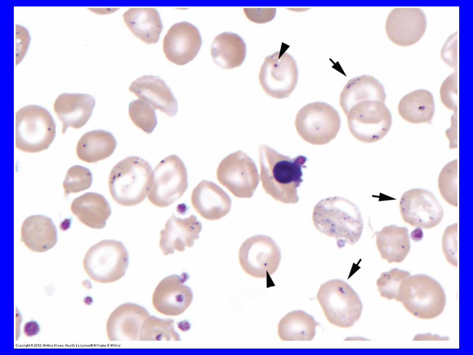
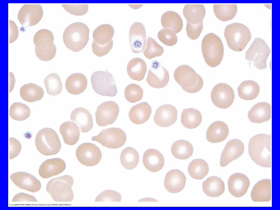
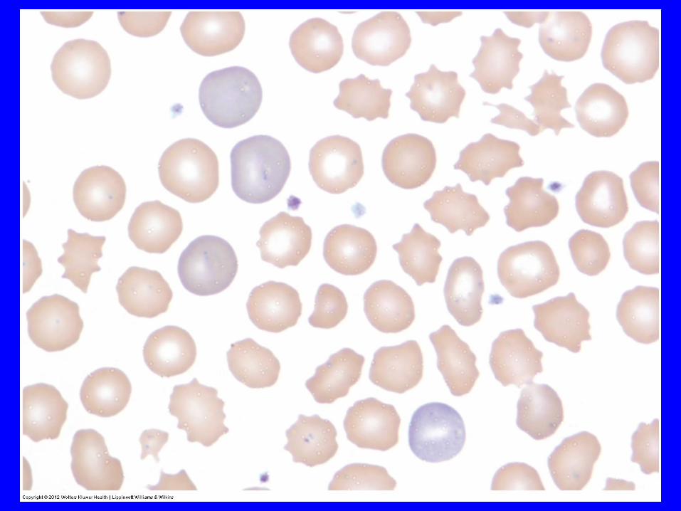
Anemia Due to Abnormal/Impaired
DNA Synthesis


Hemolytic Anemia

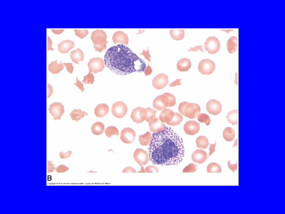
Autoagglutination
• Anti-RBC antibody
• Paraprotein
• Cold agglutinin disease
• Autoimmune hemolytic anemia
• Macroglobulinemia
• Hypergammaglobinemia
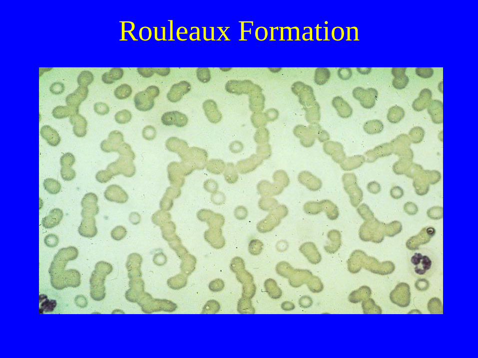
Rouleaux Formation

Rouleaux
• Acute and chronic inflammatory disorders
• Plasma cell dyscrasia
- Waldenstroms macroglobulinemia
- Multiple myeloma.

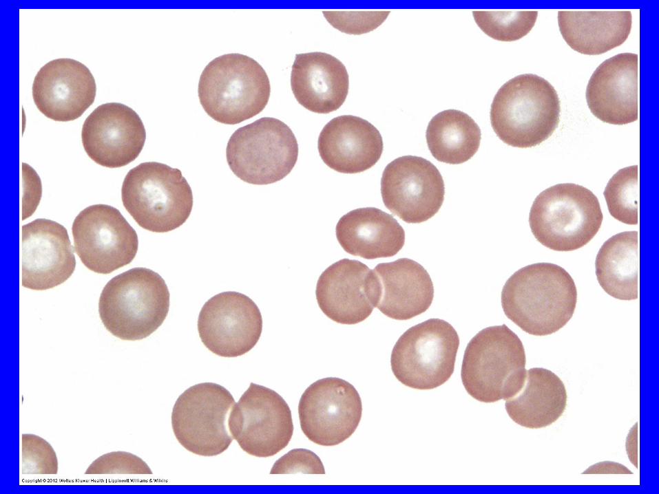
Spherocytes • Congenital (hereditary) spherocytosis
• Immune hemolytic anemias
• Microangiopathic hemolytic anemia (MAHA)
• Hypersplenism and post-splenectomy
• Myelofibrosis with myeloid metaplasia
• ABO/RH incompatibility
• Normal in newborn
• Oxidative changes – some hemoglobinopathies
• Malaria,
• Liver disease,
• Older population of transfused cells
• Artifact.
• Microspherocytes
- severe burns
- hereditary pyropoikilocytosis.

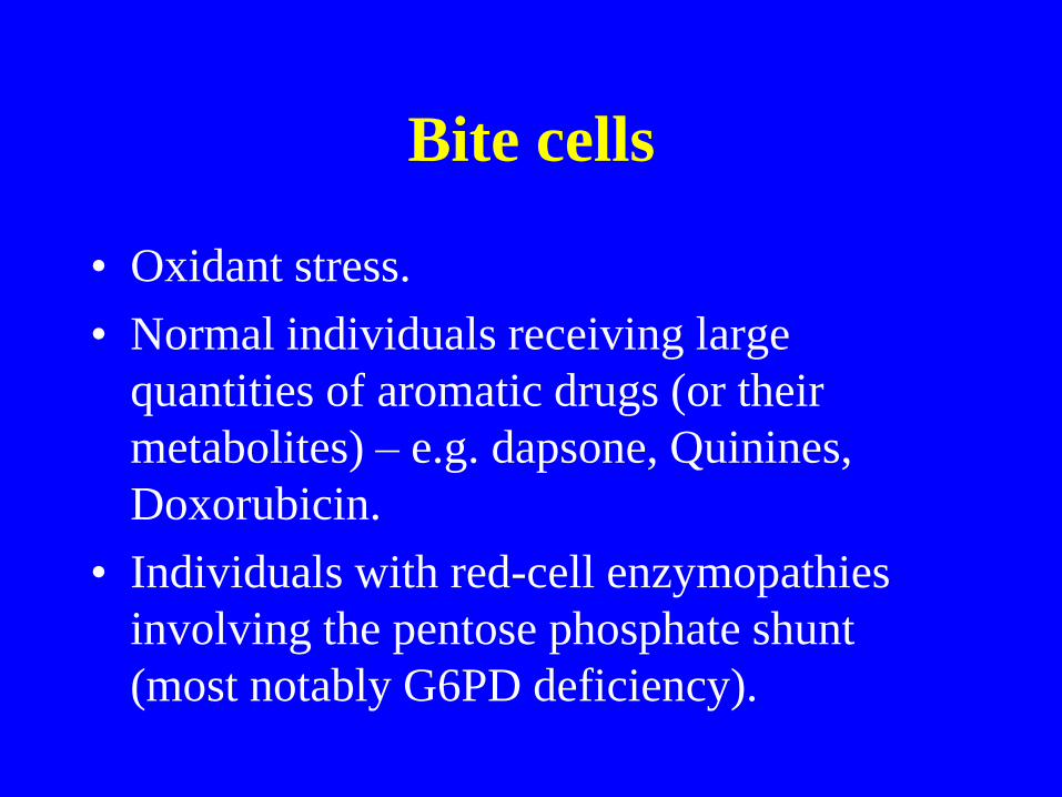
Bite cells
• Oxidant stress.
• Normal individuals receiving large
quantities of aromatic drugs (or their
metabolites) – e.g. dapsone, Quinines,
Doxorubicin.
• Individuals with red-cell enzymopathies
involving the pentose phosphate shunt
(most notably G6PD deficiency).
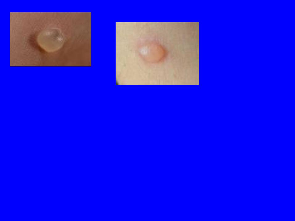
Blister cells
• Glucose-6-phosphate dehydrogenase (G-6-
PD) deficiency.
• Other oxidant stress


Spherocytes • Congenital (hereditary) spherocytosis
• Immune hemolytic anemias
• Microangiopathic hemolytic anemia (MAHA)
• Hypersplenism and post-splenectomy
• Myelofibrosis with myeloid metaplasia
• ABO/RH incompatibility
• Normal in newborn
• Oxidative changes – some hemoglobinopathies
• Malaria,
• Liver disease,
• Older population of transfused cells
• Artifact.
• Microspherocytes
- severe burns
- hereditary pyropoikilocytosis.
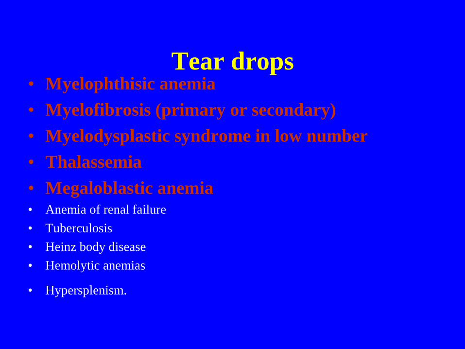
Tear drops • Myelophthisic anemia
• Myelofibrosis (primary or secondary)
• Myelodysplastic syndrome in low number
• Thalassemia
• Megaloblastic anemia • Anemia of renal failure
• Tuberculosis
• Heinz body disease
• Hemolytic anemias
• Hypersplenism.
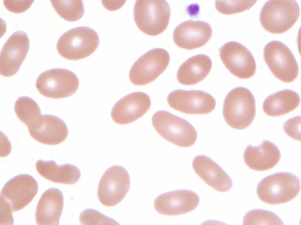
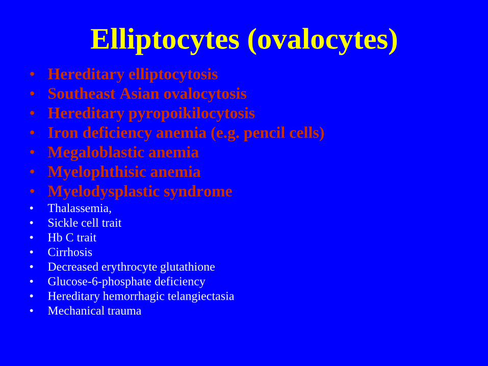
Elliptocytes (ovalocytes) • Hereditary elliptocytosis
• Southeast Asian ovalocytosis
• Hereditary pyropoikilocytosis
• Iron deficiency anemia (e.g. pencil cells)
• Megaloblastic anemia
• Myelophthisic anemia
• Myelodysplastic syndrome • Thalassemia,
• Sickle cell trait
• Hb C trait
• Cirrhosis
• Decreased erythrocyte glutathione
• Glucose-6-phosphate deficiency
• Hereditary hemorrhagic telangiectasia
• Mechanical trauma


RBC, RBC INCLUSIONS AND THEIR MIMICS
• ACANTHOCYTE VS ECHINOCYTE
• SPHEROCYTE VERSUS PSEUDOSPEROCYTE
• HOWELL-JOLLY BODIES VS PAPPENHEIMER
BODIES
• PLATELET OVERLYING A RED CELL VERSUS
HOWELL-JOLLY BODIES
• PLATELET OVERLYING A RED CELL VERSUS
INTRACELLULAR ORGANISMS (RING FOORM OF
BABESIA OR PLASMODIUM SPECIES)
161
RBC, RBC INCLUSIONS AND THEIR MIMICS
• BASOPHILIC STIPPLING VERSUS CLUSTERS
PAPPENHEIMER BODIES IN RBC
• ROULEAUX VERSUS AGGLTUINATION
• ELLIPTOCYTES VERSUS PARTIALLY SICKLED RED
CELLS
• TEAR DROP VERSUS TEAR DROP-LIKE CELLS
• PARTIALLY SICKLED CELLS VERSUS SICKLE-LIKE
CELLS
• BLISTER CELLS VERSUS BLISTER-LIKE CELLS
162