perioperative management of the pediatric patient with traumatic brain injury
TRANSCRIPT
REVIEW ARTICLE
Perioperative management of the pediatric patient withtraumatic brain injuryTarun Bhalla1, Elisabeth Dewhirst1, Amod Sawardekar1, Olamide Dairo1 & Joseph D. Tobias1,2
1 Departments of Anesthesiology, Nationwide Children’s Hospital and the Ohio State University, Columbus, OH, USA
2 Departments of Pediatrics, Nationwide Children’s Hospital and the Ohio State University, Columbus, OH, USA
Introduction
The National Center for Injury Prevention and Con-
trol estimates that over 510 000 traumatic brain inju-
ries (TBI) occur annually in children, 0–14 years of
age in the United States (1). Of these, there are 35 000
hospitalizations and 2000–3000 deaths. Men are 1.5
times more likely to sustain TBI than women (2).
Although falls are the leading cause of TBI when con-
sidering all children from 0 to 14 years of age, there
are etiological variations between age groups. Nonacci-
dental trauma (shaken baby syndrome or child abuse)
should always be in the differential diagnosis in infants
and young children (3). Motor vehicle accidents and
assault become more prevalent with increasing age.
Among motor vehicle-related injuries in this age
group, motor-pedestrian injuries are more common
than motor vehicle occupant injuries (4,5). School-aged
children exhibit a rise in bicycle-related injuries,
whereas adolescents experience a rise in motor vehicle
injuries, sports-related injuries, and assault (6). When
compared with data from the 1980s, although the mor-
tality of TBI has decreased, its total incidence has
more than tripled, possibly due to the increased popu-
lation, recognition of diagnosis, and presentation to
emergency departments (7).
With an improved understanding of the pathophysi-
ology of TBI, refinements of monitoring technology,
and ongoing research to determine optimal care, the
prognosis of TBI continues to improve. In 2003, the
Society of Critical Care Medicine published guidelines
for the acute management of severe TBI in infants,
children, and adolescents (8–10). To ensure their dis-
semination and visibility, the guidelines were published
simultaneously in three journals. As pediatric anesthe-
siologists are frequently involved in the perioperative
Keywords
neurosurgery; trauma; audit; general
anesthesia; traumatic brain injury;
adolescent; neuroanesthesia; child; age
Correspondence
Tarun Bhalla, Department of Anesthesiology
and Pain Medicine, Nationwide Children’s
Hospital, 700 Children’s Drive, Columbus,
OH 43205, USA
Email: [email protected]
Section Editor: Andrew Davidson
Accepted 3 March 2012
doi:10.1111/j.1460-9592.2012.03842.x
Summary
TBI and its sequelae remain a major healthcare issue throughout the world.
With an improved understanding of the pathophysiology of TBI, refine-
ments of monitoring technology, and ongoing research to determine opti-
mal care, the prognosis of TBI continues to improve. In 2003, the Society
of Critical Care Medicine published guidelines for the acute management
of severe TBI in infants, children, and adolescents. As pediatric anesthesiol-
ogists are frequently involved in the perioperative management of such
patients including their stabilization in the emergency department, familiar-
ity with these guidelines is necessary to limit preventable secondary damage
related to physiologic disturbances. This manuscript reviews the current
evidence-based medicine regarding the care of pediatric patients with TBI
as it relates to the perioperative care of such patients. The issues reviewed
include those related to initial stabilization, airway management, intra-
operative mechanical ventilation, hemodynamic support, administration of
blood and blood products, positioning, and choice of anesthetic technique.
The literature is reviewed regarding fluid management, glucose control,
hyperosmolar therapy, therapeutic hypothermia, and corticosteroids.
Whenever possible, management recommendations are provided.
Pediatric Anesthesia ISSN 1155-5645
ª 2012 Blackwell Publishing Ltd 1
management of such patients including their stabiliza-
tion in the emergency department, familiarity with
these guidelines is necessary to limit preventable sec-
ondary damage related to physiologic disturbances.
This manuscript aims to provide an up-to-date sum-
mary of recommendations for the management of
pediatric TBI as relevant to the field of anesthesia.
Pathophysiology of TBI
The pathophysiology and subsequent sequelae of TBI
involve both primary and secondary injuries to the
brain. Primary injury is caused by the initial trauma. It
involves the physical impact to the brain tissue from
acceleration to deceleration or rotational forces and
may result in skull fracture, brain contusion, intracra-
nial (intraparenchymal, epidural, and subdural) hema-
toma, or diffuse axonal injury (DAI) (11).
DAI refers to the widespread axonal damage caused
by the shearing forces that occur with rapid accelera-
tion and deceleration of the head. Motor vehicle acci-
dents are the most frequent cause; however, falls,
assault, and nonaccidental trauma are also implicated.
Occurring in approximately half of all patients with
TBI, it is a common injury with devastating effects.
Patients with DAI frequently do not regain conscious-
ness after their injury, leading to persistent vegetative
state. Patients who do recover have severe residual
impairment of cognitive function. Imaging may reveal
multiple white matter lesions, although as microscopic
injury may be more prevalent than macroscopic injury,
even MRI may miss DAI. Its presence can be inferred
when areas of punctate hemorrhage are visible in the
corpus callosum or the cerebral cortex. As well as the
primary injury, axonal damage in DAI may also result
from secondary biochemical cascades, leading to a
delayed onset or a clinical deterioration in a patient.
Recent attention has focused on the significance of
secondary injury and its effect on the eventual out-
come of patients with TBI. Inflammatory and excito-
toxic processes following injury result in further edema
formation, increases in intracranial pressure (ICP), and
reduced cerebral perfusion pressure (CPP) (12,13). Sec-
ondary injury may also result from physiologic
changes after the initial injury including hypoxemia
and hypotension. The adult literature have demon-
strated that the two major factors that result in sec-
ondary injury in patients with TBI are hypotension
defined as a systolic blood pressure £90 mmHg and
hypoxemia (PaO2 £ 60 mmHg) (13).
A challenge specific to the pediatric population is
the increased severity of cerebral edema as well as its
diffuse nature, when compared with adults (14). The
anatomic differences in the pediatric trauma patient
include an immature or unstable neck, a larger head-
to-torso ratio especially in infants, and a more compli-
ant skull. These characteristics may result in more pro-
found consequences from acceleration–deceleration
injuries. Additionally, as there is limited brain atrophy
in children in comparison with adults, there is little
room to compensate for edema in the rigid skull. The
immature brain also has a high water content and
lacks complete axonal myelination. Finally, inflamma-
tory mediators in the developing brain likely lead to
more significant edema when compared with adult
patients (15).
Initial stabilization and resuscitation
Given the potential for poor outcomes of TBI and the
significant impact of secondary insults, treatment strat-
egies are aimed not only at assessment and stabiliza-
tion, but also early therapeutic interventions to
decrease secondary injuries. Thus, the cornerstones of
modern TBI management are field resuscitation, expe-
ditious triage, emergent surgical evacuation of mass
lesions, control of ICP, support of CPP, multimodal
monitoring, and optimization of physiologic environ-
ment. The anesthesia provider is likely to be faced with
such patients either in the emergency department when
called upon for airway management or as these
patients are urgently transported to the operating
room for evacuation of space occupying lesions. As
secondary injury is preventable and treatable, the peri-
operative period remains a key time to initiate inter-
ventions that may improve the outcome of TBI.
Additionally, alterations in physiologic function
induced by anesthetic care or the surgical intervention
may result in secondary injury. The key components of
perioperative management include rapid evaluation,
ongoing resuscitation, early surgical intervention,
intensive monitoring, and anesthetic planning.
The initial assessment and stabilization of the
patient with TBI is usually achieved in the emergency
department. Following the initial assessment, resuscita-
tion, and stabilization, the patient is likely going to be
transported to the computed tomography (CT) scanner
and then to the operating room. As in many cases,
there are no family members available and the patient
is unable to provide any information, the anesthesia
provider will have limited historical data on which to
base their anesthetic care. Upon arrival, the anesthesia
team performs a rapid assessment of the patient. The
assessment should always begin with airway, breathing,
and circulation, followed by a rapid assessment of
neurologic status (GCS and papillary response) and
Perioperative management of the pediatric patient with neurotrauma T. Bhalla et al.
2 ª 2012 Blackwell Publishing Ltd
associated extracranial injuries. Information about the
time and mechanism of injury can be valuable. Associ-
ated injuries involving the thorax, abdomen, pelvis,
and long bones may be stable or evolve during the
perioperative period. Should intra-operative decompen-
sation occur, the potential impact of these associated
injuries must be considered when attempting to deter-
mine the etiology of new onset hemodynamic instabil-
ity, anemia, or ventilator difficulties.
Airway management
Patients with TBI requiring surgery will invariably
require endotracheal intubation. The decision to per-
form endotracheal intubation is not only determined
by the patient’s respiratory status or the need for an
operative procedure, but more importantly by their
Glasgow Coma Scale at the time of presentation. Cur-
rent trauma guidelines recommend immediate endotra-
cheal intubation for patients with a GCS <9 based on
the high probability that such an altered level of con-
sciousness is indicative of a brain injury of such sever-
ity that progressive cerebral edema is likely to occur
(16,17).
Airway management is complicated by a number of
factors including the urgency of situation with upper
airway obstruction or preexisting hypoxia, uncertainty
of cervical spine status, uncertainty regarding the air-
way because of the presence of blood, vomitus, debris
in the oral cavity or laryngo-pharyngeal injuries, a full
stomach, intracranial hypertension, and uncertain vol-
ume status. Although many of the tenets of airway
management are the same regardless of the clinical sce-
nario, all TBI patients should be considered to have a
full stomach and an underlying cervical spine injury
(CSI). Additionally, as patients may not be able to
effectively cooperative with a physical examination
that is key in ruling out a CSI by identifying neuro-
logic deficits or point tenderness in the neck, the air-
way is managed with the assumption that their may be
a CSI with a RSI and manual inline stabilization
(18,19). Such initiatives are imperative as the recent
data in the adult population suggest that an associated
CSI is not infrequent in patients with TBI. Although it
had been previously suggested that TBI patients had
an incidence of CSI similar to that of the general
trauma population, a higher incidence of CSI has been
noted in patients with TBI especially those with
increasing severity of injury as determined by low GCS
score and unconsciousness (20–22).
Of equal importance is the choice of agents used for
sedation/analgesia and neuoromuscular blockade dur-
ing RSI. Sodium thiopental, etomidate, and propofol
are commonly used in the operating room to induce
anesthesia before endotracheal intubation. All these
agents decrease the systemic hemodynamic response to
intubation, blunt increases in ICP, and decrease the
cerebral metabolic rate for oxygen (CMRO2). How-
ever, thiopental or propofol may not be appropriate
for critically ill patients given their vasodilatory and
negative inotropic effects that are exaggerated by co-
morbid conditions (23,24). In the trauma scenario,
alternative agents such as etomidate or ketamine are
frequently chosen (25). To date, the data regarding the
potential deleterious effects of etomidate on outcome
are limited to patients with presumed sepsis (26–28).
Outside of that scenario, there are no data to demon-
strate a deleterious effect on outcome despite the fact
that adrenal suppression does occur. Given that etomi-
date has a limited effect on MAP and effectively
decreases the CMRO2, thereby decreasing ICP, the net
effect is maintenance or an increase in CPP, thereby
making it a potentially valuable agent for patients with
TBI.
Ketamine’s popularity in the trauma setting relates
to its beneficial effects on cardiac and respiratory func-
tion. Ketamine generally increases heart rate and
blood pressure as well as provides bronchodilatation
because of the release of endogenous catecholamines
(29). In vitro and animal studies suggest ketamine has
direct negative inotropic properties (30,31), although
clinically the indirect sympathomimetic effects from
endogenous catecholamine release are generally over-
riding. The controversy with ketamine surrounds its
effects on ICP and whether it is contraindicated in
patients with TBI and altered intracranial compliance.
Although the literature from the 1970s and 1980s sug-
gested that ketamine increased ICP and was contrain-
dicated in patients with altered intracranial compliance
(32), more recent evidence has suggested no effect or
even that ketamine may decrease ICP when used to
prevent pain from invasive procedures (33–35).
Given the need to achieve rapid neuromuscular
blockade and optimal conditions for endotracheal intu-
bation, the choices for the neuromuscualar blocking
agent for RSI are succinylcholine and rocuronium
(36). Although succinylcholine may mildly increase
ICP (37–40), increases in ICP secondary to hypoxia
and hypercarbia are well documented and much more
likely to be clinically important. Therefore, rapid endo-
tracheal intubation and control of oxygenation and
ventilation are of paramount importance and far out-
weigh the risk of the mild increase in ICP from succi-
nylcholine. If sugammadex is locally available,
rocuronium may be a more feasible option. Although
frequently considered as an adjunct to endotracheal
T. Bhalla et al. Perioperative management of the pediatric patient with neurotrauma
ª 2012 Blackwell Publishing Ltd 3
intubation of patients with increase ICP, there is no
evidence-based medicine to show the efficacy of lido-
caine (41). In the pediatric population, the process of
endotracheal intubation with larygnoscopy in a patient
with increased ICP may result in bradycardia.
Although there are limited data to support its used,
pretreatment with atropine is generally suggested
(18,19). The incidence of bradycardia is further magni-
fied by associated hypoxemia, hypothermia, and the
administration of succinylcholine.
Intra-operative anesthetic care
The perioperative period may provide an opportunity
to either continue ongoing resuscitation or correct the
preexisting secondary insults that have not been ade-
quately addressed because of the urgency to get
patients to the operating room. Furthermore, surgery
and anesthesia may predispose the patient to the onset
of potentially new secondary injuries (such as intra-
operative hypotension because of surgical blood loss
or the effect of anesthetic agents, hyperglycemia
because of stress response), which may impact out-
come. The primary goals of anesthetic management
can be summarized as follows: (i) provide adequate
anesthesia and analgesia; (ii) optimize surgical condi-
tions; (iii) avoid secondary insults including hypoten-
sion, hypoxemia, hypocarbia, hypercarbia,
hypoglycemia, and hyperglycemia; (iv) maintain an
adequate CPP; and (v) avoid increases in ICP.
Respiratory support and mechanical ventilation
The basic tenets of ventilator support for the trauma
patient are the same as for all patients in the OR.
These include the provision of adequate oxygenation
and ventilation while limiting the potentially deleteri-
ous effects of mechanical ventilation on cardiovascular
function.
The goal of oxygenation is to maintain the PaO2 ‡60 mmHg (PaO2 ‡ 8 kPa) as CBF, CBV, and ICP
increases linearly with PaO2 values £ 60 mmHg
(PaO2 £ 8 kPa). Although PaO2 levels are routinely
‡60 mmHg (‡8 kPa) during anesthetic care, values
above this level have no impact on CBF, CBV, or
ICP. If adequate oxygenation cannot be maintained at
inspired oxygen concentrations (FiO2) of 0.5–0.6, the
usual practice is to increase positive end expiratory
pressure (PEEP). However, it must be remembered
that as PEEP increase, it may impact the overall CPP
because of its effects not only on cardiovascular func-
tion, but also its effects on ICP. Based on lung compli-
ance, PEEP is transmitted to the intracranial vault and
may impact ICP (42,43). Therefore, in patients with
TBI, it may be better to attempt to increase mean air-
way pressure and augment oxygenation by first
increasing the inspiratory time, FiO2, and peak inflat-
ing pressure rather than PEEP.
Although practiced routinely in the past for patients
with TBI, hyperventilation is generally avoided unless
there is a precipitous increase in ICP with impending
herniation (44,45). Prehospital ventilation has been
shown to have a significant impact on outcome with
increased in-hospital mortality in patients presenting
with either hypocarbia or hypercarbia (46). Others
have demonstrated a similar negative impact of in-hos-
pital hyperventilation on outcomes (47). Although
hyperventilation induces hypocapnia which causes
cerebral vasoconstriction and a reduction in CBF and
CBV and a corresponding decrease in ICP, there is a
disconnect between CBF and CMRO2 as hypocarbia
decreases CBF without effect CMRO2, thereby poten-
tially exposing the patient to ischemia especially during
the trauma period where there may be preexisting
alterations in CBF and autoregulation. Skippen et al.
(48) demonstrated a relationship between hypocarbia
and frequency of cerebral ischemia. Given this infor-
mation, hyperventilation should be used judiciously
intra-operatively for short-term control of ICP and to
facilitate surgical exposure during craniotomy. Prior to
dural closure, normocarbia should be restored to avoid
tension pneumocephalus. In situations where hyperven-
tilation is necessary, it may be beneficial to increase
the FiO2 as this may avoid the deleterious effects of
hypocarbia on CBF and cerebral tissue oxygenation
(49). Given the inaccuracy of ETCO2 monitoring and
the importance of PaCO2 control in these patients,
direct monitoring of arterial PaCO2 is recommended
(50,51).
Summary:
1. Maintain PaO2 ‡ 60 mmHg (PaO2 ‡ 8 kPa)
2. PEEP may increase ICP
3. Hyperventilation should only be used if impending
herniation
4. Avoid intra-operative hypoxemia
Hemodynamic support
As noted earlier, the role that hemodynamic support
may play in prevention of secondary injury cannot be
overemphasized. In both the adult and pediatric popu-
lation, data demonstrate that even a single periopera-
tive episode of hypotension can impact outcome
(52,53). A meta-analysis of 8721 patients (IMPACT
study) emphasizes the importance of secondary impact
and its effect on outcome (54). The study noted that
Perioperative management of the pediatric patient with neurotrauma T. Bhalla et al.
4 ª 2012 Blackwell Publishing Ltd
both hypotension and hypoxemia were significantly
associated with unfavorable 6-month outcome. Intra-
operative hypotension can be equally as devastating.
Pietropaoli et al. demonstrated that intra-operative
hypotension increased the incidence of mortality by a
factor of three (55). Additionally, the duration of
intra-operative hypotension was also inversely associ-
ated with functional outcome. Intra-operatively if
problems arise with the control of ICP, a slight eleva-
tion of the MAP may be effective in ameliorating such
issues. With intact autoregulation, as the MAP is
increased, there is a secondary cerebral vasoconstric-
tion to maintain CBF at baseline. The cerebral vaso-
constriction results in a decrease in CBV that may
decrease ICP in patients with altered intracranial com-
pliance. This relationship further demonstrates the
need to effectively control the intra-operative MAP.
Three small prospective, crossover trials have com-
pared norepinephrine with dopamine to treat hypoten-
sion and maintain MAP (56–58). There were no
differences in mean cerebral flow velocity, cerebral
oxygenation, or cerebral metabolism. However, the
authors reported that norepinephrine had a more pre-
dictable and consistent effect while dopamine resulted
in a higher ICP. A recent single-center retrospective
study of patients with severe TBI who received phenyl-
ephrine, norepinephrine, or dopamine reported that
phenylephrine resulted in the maximum increase in
MAP and CPP from baseline (59).
Summary:
1. Effective control of intra-operative MAP
2. A single intra-operative episode of hypotension can
effect outcome
3. Maintain optimal CPP
Anesthetic agents
There are significant differences when comparing the
effects of intravenous anesthetic agents and the volatile
agents in regard to their effects on CBF, CBV, and
CMRO2. However, to date, there are no evidence-
based data to demonstrate a significant difference in
outcome when comparing these two groups of agents.
The intravenous agents including thiopental, propofol,
and etomidate reduce CMRO2, which results in cere-
bral vasoconstriction and a decrease in CBF, CBV,
and ICP. However, agents such as thiopental and
propofol may result in significant hypotension and a
reduction in CPP. Additionally, with intact autoregula-
tion, the decrease in MAP is compensated for by cere-
bral vasodilatation to maintain CBF, which results in
an increase in CBV and may increase ICP. A similar
effect has been observed following the administration
of fentanyl and sufentanil although these agents have
no direct effect on the cerebral vasculature (60,61).
This effect can be treated by the administration of a
vasoactive agent such as phenylephrine to restore the
MAP back to baseline.
All of the volatile anesthetic agents (isoflurane, sevo-
flurane, and desflurane) decrease CMRO2 and may
cause cerebral vasodilatation, thereby increasing CBF,
CBV, and ICP. These effects are generally minimal at
exhaled concentrations of <1 minimum alveolar con-
centration. Therefore, they may be used in low concen-
trations in patients with TBI (62,63). However, nitrous
oxide should be avoided as it can increase CMRO2
causing cerebral vasodilatation and increased ICP (63).
As far as choice of neuromuscular blocking agent, the
issues related to succinylcholine and its potential
effects on ICP have been previously discussed. The
nondepolarizing agents have no direct effect on ICP
and can be used as needed to provide surgical relaxa-
tion and ensure immobility in critically ill patients.
Although neuromuscular blocking agents decrease oxy-
gen consumption and may transiently decrease ICP as
they eliminate thoracic skeletal tone and increase
venous drainage; postoperatively, their use is generally
not recommended (64). The adult literature clearly
demonstrates no outcome benefit and increased
adverse effects including infection and prolongation of
hospitalization (65).
Summary:
1. No significant difference in outcomes comparing
intravenous and inhalational anesthetic agents
2. Avoid nitrous oxide, increases CMRO2 and possible
increase in ICP
3. NMB are generally not recommended postopera-
tively, but are routinely used intra-operatively
ICP Monitoring
Preventing and treating intracranial hypertension that
would lead to deleterious secondary injury is vital to
the care of patients with TBI. While in common prac-
tice, monitoring of ICP for this purpose is presented as
an option rather than a guideline for children with
severe TBI (GCS < 8), because of insufficient evidence
in the pediatric population (8). Typically the choice of
monitor is between an intraparenchymal device and an
intraventricular device connected by catheter to an
external drain gauge. Correlation between the two has
been shown (66), and while the former may result in
less local tissue damage, the later allows CSF drainage
and is the suggested device in adult TBI guidelines
(67). An ICP greater or equal to 20 mmHg warrants
treatment in children. CPP should be maintained
T. Bhalla et al. Perioperative management of the pediatric patient with neurotrauma
ª 2012 Blackwell Publishing Ltd 5
above 40 mmHg; (68–70) however, there is no clear
age-related adjustment of this target value up to the
adult recommendation of 70 mmHg. Also in neonates
and infants, the minimum CPP is still unclear. Most
commonly, the device is placed at the end of surgery,
and thus, measured values are available for ICU care
and not intra-operatively, except in the case of a
patient returning to the OR. Complicating infection
from an ICP monitoring device is rare, and theoretical
complications of hemorrhage and seizure have not
been reported, indicating safety of ICP monitors (71).
Noninvasive transcranial doppler pulsatility index as
an indicator of ICP is controversial, however may be
useful on admission, having high sensitivity to predict
ICH and abnormal CPP (72). Another alternative to
direct ICP monitors is ocular ultrasound, which has
been suggested to detect ICH in adults.
Although not standard of care or in routine practice,
as the technology improves, perioperative cerebral
monitoring may become more commonplace (73–75).
Such technology may be particularly relevant in
patients receiving more aggressive systemic interven-
tions to manage refractory ICH, such as hyperventila-
tion. Jugular venous oxygen saturation (SjvO2),
measured by retrograde placement of a catheter from
the internal jugular into the jugular bulb (73), gives a
measure of cerebral oxygen delivery. In adults with
TBI, episodes of decreased SjvO2 have been associated
with poor neurologic outcome. Cerebral oximetry via
near infrared spectroscopy (NIRS) monitoring has
been shown to correlate with SjvO2, and in many cen-
ters, NIRS-based cerebral oximeters are used routinely
for children undergoing repair of congenital heart
defects. Other monitoring technologies to considered
in the perioperative period that may help in adjusting
therapies include transcranial Doppler ultrasonography
to evaluate middle cerebral blood flow and direct mea-
surement of brain tissue oxygenation using invasive
devices (73–75).
Summary:
1. ICP ‡ 20 mmHg warrants intervention
2. CPP should be maintained at >40 mmHg
3. Monitoring ICP helps to avoid further secondary
injury
Positioning
As the patient is moved and positioned on the operat-
ing room table, the head position is generally man-
dated by the surgical procedure to be performed.
However, several studies have demonstrated the poten-
tial impact of head positioning and head elevation on
ICP. Whenever feasible, the patient’s head should
remain in neutral and midline position to avoid jugular
venous obstruction. Flexion of the head, rotation of
the head to the right or the left, or lowering the
patient into the Trendelenburg position can signifi-
cantly increase ICP especially in patients with altered
intracranial compliance (76–78). A more controversial
issue that relates primarily to the ICU care of these
patients is whether head elevation should be routine.
While elevation of the head of the bed has been shown
to lower ICP by promoting venous drainage, some
studies have shown that a decrease in CPP may also
occur. Furthermore, rebound autoregulatory responses
to reduced CPP involve lowering of CBR to increase
CBV, which may actually predispose these patients to
episodes of intracranial hypertension and more prob-
lematic control of ICP (79). Overall, it has been sug-
gested that a moderate elevation of 15–30� is an
appropriate measure to lower ICP as long as adequate
CPP is vigilantly maintained and the degree of eleva-
tion titrated to the individual based on ICP and CPP
measurements (8). Head elevation has the added bene-
fit of minimizing ventilator-associated pneumonia.
Summary:
1. Head of bed should be maintained at 15–30�2. Reverse Trendelenburg may improve venous drain-
age, but may induce rebound increase in ICH
3. Patients head should remain neutral and midline
Fluid management including glucose control
Given the risks of hypovolemia and hypotension,
patients with TBI should receive initial volume resusci-
tation to achieve a normovolemic state. Although sig-
nificant fluid restriction was previously the acceptable
standard for patients with TBI, the current trend has
evolved to suggest that euvolemia is the optimal end-
point. In the consideration of fluid therapy, the issues
to be addressed include the type of fluid to be used
and the control of serum glucose. Issues related to
hyperosmolar therapy are discussed in the next section.
For the vast majority of patients, an isotonic fluid
should be chosen. Although lactated Ringers (LR) is
commonly used during the perioperative period, it
must be remembered that the sodium concentration of
130 mEqÆl)1 is less than that of the serum sodium con-
centration. When compared with the administration of
normal saline, patients receiving LR will have a
decrease in the serum sodium level and osmolarity
(80). Given this information, normal saline should be
used for the initial resuscitation and then the ongoing
provision of maintenance and replacement fluids for
the majority of patients with TBI. When comparing
the potential advantages and disadvantages of
Perioperative management of the pediatric patient with neurotrauma T. Bhalla et al.
6 ª 2012 Blackwell Publishing Ltd
crystalloid vs colloid, recent data from an adult trial
suggest that resuscitation with albumin containing flu-
ids may impact outcome (81). In the posthoc analysis
of a larger trial, the authors reported that the risk of
death was increased in patients who received albumin
compared with those who received normal saline.
Among patients with severe brain injury (GCS 3–8),
61 of 146 patients in the albumin group died (41.8%)
as compared with 32 of 144 in the saline group
(22.2%, relative risk of 1.88) (81).
Although hypertonic fluids have been shown to be
effective in the treatment of increased ICP (see below),
there are few data to demonstrate their superiority
over normal saline for the initial resuscitation of
patients with TBI. Prehospital hypertonic saline resus-
citation has been shown to be associated with a reduc-
tion in serum biomarker levels (S100B, neuron-specific
enolase and membrane basic protein) (82). However, a
prospective and randomized trial comparing prehospi-
tal resuscitation of TBI patients with hypotension
found no difference in neurologic outcome when com-
paring resuscitation with hypertonic saline or standard
fluid resuscitation protocols (83).
Another controversial issue regarding care of
patients with TBI is the association of hyperglycemia
with a poor outcome. As such, there remains some
divergence in the opinions regarding the need to
aggressively treat hyperglycemia in this population.
Causes of hyperglycemia after TBI include an increase
in gluconeogenesis and glycogenolysis from catechol-
amine response, cortisol release, and glucose intoler-
ance (84–86). Hyperglycemia is more prevalent after
severe TBI and in patients under 4 years old (84). Sec-
ondary brain injury from hyperglycemia can occur,
leading to an increase in glycolytic rates as shown by
increased lactate/pyruvate ratio, resulting in metabolic
acidosis within brain parenchyma, overproduction of
reactive oxygen species, and ultimately neuronal cell
death through alterations in immune, inflammatory,
and mitochondrial function (87). Several studies in
both children and adults support an association
between hyperglycemia and a poor neurologic outcome
following TBI; however, there are limited data to
prove whether treatment of the hyperglycemia will
improve outcome (88–90). In a retrospective review of
pediatric patients admitted to the regional trauma cen-
ter over a 1-year period, Cochrane et al. note that
patients who died had significantly higher admission
glucose values (mean of 267 vs 135 mgÆdl)1) and that
a serum glucose on admission of >300 mgÆdl)1
(16.7 mM) was uniformly fatal (88). Lam et al. (89)
noted that in severely injured patients with a GCS £8that a serum glucose ‡200 mgÆdl)1 (11.1 mM) was
associated with a worse outcome. Smith et al. (90)
reported that in children with severe traumatic brain
injury, hyperglycemia beyond the initial 48 h is associ-
ated with a greater likelihood of a poor neurologic
outcome.
Intensive insulin therapy for the control of hypergly-
cemia in critically ill adult patients has received a lot
of press in the literature following the report of Van
den Berghe et al. (91) which reported that intensive
insulin therapy (target blood glucose 80–110 mgÆdl)1)in critically ill patients was associated with lower mor-
tality. However, subsequent work has demonstrated
that this may not be that case and that there is an
increased risk of hypoglycemia (92,93). In a prospec-
tive trial, Billotta et al. (93) randomized 97 adults with
severe TBI to insulin therapy to maintain a blood glu-
cose at 80–120 mgÆdl)1 (4.4–6.7 mM) or conventional
insulin therapy to maintain a blood glucose concentra-
tion below 220 mgÆdl)1 (12.2 mM). They reported that
both the groups had similar mortality and neurologic
outcome at 6 months and a similar incidence of infec-
tious complications. Although the more strict control
of glucose resulted in a shorter duration of ICU stay,
there was a higher incidence of hypoglycemia. Given
the potential for an increased risk of hypoglycemia in
children and its potentially devastating neurologic con-
sequences if unrecognized and untreated, an aggressive
approach to the control of perioperative glucose can-
not universally be suggested in children with TBI.
Additionally, when reviewing the data, the studies
focus on ICU care and not the intraoperaive manage-
ment of such patients. Given these issues, it seems pru-
dent to suggest that glucose containing fluids not be
administered to the pediatric patient with TBI unless
the serum glucose is £70 mgÆdl)1. Given the potential
impact of both hyperglycemia and hypoglycemia, inter-
mittent monitoring of blood glucose concentrations
during intra-operative care is suggested. This can easily
be accomplished using bedside, point-of-care testing
devices. Moreover, the anesthesia provider may want
to take into account that there may be differential
effects of the individual anesthetic agents on blood
and brain glucose concentrations (94,95).
Summary:
1. Intra-operative euvolemia is optimal
2. Normal saline should be used as maintenance fluid
3. Glucose containing fluids should not be infused
unless the serum glucose is £70 mgÆdl)1 (3.9 mM)
Hyperosmolar therapy
Mannitol remains the standard agent used in hyper-
osmolar therapy. The principle of hyperosmolar
T. Bhalla et al. Perioperative management of the pediatric patient with neurotrauma
ª 2012 Blackwell Publishing Ltd 7
therapy is based on establishing an osmotic gradient
between the plasma and parenchymal brain tissue,
thereby reducing brain water content and thus ICP.
An intact blood–brain barrier is necessary for this
process. In addition to its osmotic effect, a rapid
decrease in ICP may be seen following its administra-
tion as mannitol improves blood rheology and
thereby results in an increase in cerebral vascular
resistance (cerebral vasoconstriction) with a decrease
in CBV and ICP. Available as a 20% solution (stan-
dard solution bag) or a 25% solution (bottle), dosing
regimens vary from 0.25 to 1 gmÆkg)1 and aim not to
exceed serum osmolarity of 320 mOsmÆl)1 (96,97).
The osmotic diuresis from mannitol can result in hyp-
ovolemia and hypotension, and concern has been
expressed regarding adverse effects following mannitol
administration if there is an intracranial collection of
blood (epidural or subdural hematoma). It is
postulated that a reduction in the brain volume may
result in relief of the tamponade that is preventing
ongoing bleeding. Therefore, it has been suggested
mannitol not be administered until the blood has
been evacuated.
More recently, there has been considerable interest
and work performed with the use of hypertonic saline
solutions. Generally, a 3% solution is used, although
concentrations up to 23.4% have been studied (98). A
serum sodium level of 145–160 mEqÆl)1 has been rec-
ommended, and while patients have tolerated a serum
osmolality up to 360 mOsmÆl)1, caution above
320 mOsmÆl)1 is recommended (99,100). In addition
to its effects on the brain, hypertonic saline has bene-
ficial hemodynamic effects including the restoration of
intravascular volume, increased inotropy, constriction
of capacitance vessels, and a decrease in resistance
vessels. These effects result in an increased MAP and
CPP in the majority of patients. Studies in both the
adult and pediatric population have demonstrated the
efficacy of these solutions in treating refractory
increases in ICP that have failed to respond to con-
ventional therapy including mannitol (99–102). Vats
et al. (103) retrospectively reviewed their experience
with hypertonic saline (5 mlÆkg)1 of a 3% solution) or
mannitol (0.5–1 gmÆkg)1) to treat increased ICP in a
cohort of pediatric patients ranging in age from
9 months to 16 years. Fifty-six doses of mannitol
were administered to 18 patients, while 82 doses of
hypertonic saline were administered to 25 patients.
Although both reduced ICP at 60 and 120 min, only
hypertonic saline reduced the ICP at 30 min and
increased the CPP at 60 and 120 min. Although the
majority of studies comparing these agents have been
performed in the ICU setting, the efficacy of hyper-
tonic saline has also been demonstrated intra-opera-
tively (104). In a prospective, blinded trial, adults
scheduled for elective resection of a supratentorial
brain tumor were randomized to receive 160 ml of
3% hypertonic saline or 150 ml of 20% mannitol.
The surgeon was asked to judge the brain relaxation
conditions as soft, adequate, or tight. Brain relaxation
was better in the patients who received hypertonic sal-
ine than in the mannitol group. Serum sodium was
higher in the hypertonic saline group, while urine out-
put was greater in patients receiving mannitol. No
difference was noted in fluid administered, length of
ICU stay, or length of hospital stay. Despite the data
suggesting improved efficacy with hypertonic saline,
there are no data to demonstrate an improvement in
outcome. As such, either agent should be considered
intra-operatively when the treatment of ICP is neces-
sary or to provide intra-operative brain relaxation.
However, hypertonic saline should be considered in
patients who fail to respond to conventional therapy
including mannitol.
Summary:
1. Mannitol remains the standard for hyperosmolar
therapy
2. Hypertonic saline, although efficacious, has not
demonstrated improved neurologic outcome
3. Hypertonic saline should be considered for patients
who may be refractory to mannitol therapy
Temperature control and therapeutic hypothermia
Moderate hypothermia decreases the cerebral meta-
bolic rate of oxygen consumption (CMRO2) resulting
in cerebral vasoconstriction, a decrease in CBF/CBV
and ICP. Additional potential benefits include an
attenuation of the alteration in the blood–brain barrier
related to TBI and a decrease in the release of excit-
atory neurotransmitters including lactate, which have
been implicated in secondary brain injury (105). A
recent meta-analysis in adults with TBI noted no sta-
tistically significant reduction in mortality although
there was an increase in the favorable neurologic out-
come in patients that received hypothermia, particu-
larly when cooling was maintained for more than 48 h
(106). However, the authors noted that the potential
benefits of hypothermia may be offset by a significant
increase in the risk of nosocomial infections including
pneumonia. By contrast, the National Acute Brain
Injury Study (NABIS: HII) failed to show benefit
when investigating 108 adults post-TBI randomized to
early induction of hypothermia (33�C within 4.4 h of
injury) or normothermia (107). Similarly, the pediatric
literature has shown no clear benefit from the use of
Perioperative management of the pediatric patient with neurotrauma T. Bhalla et al.
8 ª 2012 Blackwell Publishing Ltd
routine hypothermia following TBI (108). In fact, in
the prospective trial of 225 children, there was a rela-
tive risk of mortality of 1.4 in patients that received
hypothermia with a mortality rate of 21% in the hypo-
thermia group and 12% in the normothermia group
(108). There was an increased need for vasopressor use
and an increased incidence of hypotension in the hypo-
thermia group. Several adverse effects have been
reported with hypothermia including hypotension, bra-
dycardia, arrhythmias, sepsis, and coagulopathy
(109,110). It has been suggested that the latter may
negate any significant outcome impact noted by hypo-
thermia. Despite these issues, other investigators have
continued to demonstrate the safety of hypothermia
with the suggestion that additional trials are needed in
the pediatric population to determine whether there is
an outcome benefit (111). The overall consensus from
the most recent set of guidelines for the care of pediat-
ric patients with TBI suggests that there is currently
no published support for the use of therapeutic hypo-
thermia in pediatric patients with TBI (8–10).
Summary:
1. No class I evidence of improved outcomes for
induced hypothermia
2. There are potential adverse effects with induced
hypothermia, including hypotension, bradycardia,
arrhythmias, sepsis, and coagulopathy
Corticosteroids
Historically, corticosteroids were used as part of the
pharmacological management of patients with TBI,
based on literature reporting edema reduction and
improved outcome in patients with brain tumors.
However, trials that specifically evaluated the use of
corticosteroids in patients with TBI have failed to
show an improvement in outcome (112,113). More
recently, the large multicenter CRASH study found an
increased mortality or severe disability in adult
patients with TBI who received methylprednisolone
within 8 h of the event treated (114). Adverse effects
related to the administration of corticosteroids to criti-
cally ill patients include adrenal suppression, increased
risk of infection, and gastrointestinal bleeding.
Although there are no data specific to the pediatric
population, given the strength and size of the CRASH
trial, corticosteroids should not be administered to
pediatric patients with TBI.
Summary:
1. Steroids do not provide benefit in TBI patients
2. CRASH trial showed increased mortality in adults
receiving methylprednisolone suffering from TBI
Blood product administration and coagulation function
Given the multisystem involvement of patients with
TBI, anemia is a common component related to
extracranial injuries. In adults with TBI, anemia has
been shown to be associated with increased in-hospi-
tal mortality and poor outcome in TBI (115–117).
However, there are limited data to support guidelines
regarding the use of blood products in patients with
TBI. More recently, there has been significant atten-
tion in the literature regarding the potential deleteri-
ous effects of the use of blood products in critically
ill patients including an increased risk of nosocomial
infections, adult respiratory distress syndrome, and
longer hospital/ICU stays (118–120). Although not
specifically studied in the pediatric population with
TBI, data from other clinical scenarios involving crit-
ically ill infants and children have demonstrated no
benefit to a liberal transfusion practice (hemoglobin
10 gÆdl)1) vs a restrictive transfusion practice (hemo-
globin 7 gÆdl)1) (121,122).
In addition to the need for packed red blood cells,
transfusions of allogeneic blood products may be
required to treat coagulation disorders. Tissue factor,
which is released following brain injury, activates the
coagulation cascade resulting in thrombin formation
and depletion of coagulation factors. Coagulation
function may be further compromised by hypothermia,
acidosis, and hypocalcemia. Disturbances of coagula-
tion function are a frequent occurrence in patients with
TBI with a reported incidence of more than 30% in
adults with TBI (123). Risk factors for coagulation dis-
turbances associated with TBI include GCS £ 8, a
higher severity of illness score, and anatomic features
noted on CT imaging including cerebral edema, sub-
arachnoid hemorrhage, and a midline shift (124). As in
all critically ill pediatric patients, the use of blood
products to treat coagulation disturbances should be
based on laboratory parameters including the pro-
thrombin time (PT), partial thromboplastin time, and
platelet count. In addition to the use of blood prod-
ucts, both antifibrinolytic agents such as tranexamic
acid and pro-coagulant drugs such as recombinant fac-
tor VII (rFVIIa) have been used in trials and case ser-
ies in patients with TBI. Anecdotal evidence has
suggested the efficacy of rFVIIa even in pediatric
patients with TBI not only in the control of bleeding
that is unresponsive to conventional therapy, but also
as a means of allowing patients to be taken rapidly to
the operating room instead of waiting for the adminis-
tration of routine blood products to reverse the coagu-
lation disturbances (125,126). A review from the
T. Bhalla et al. Perioperative management of the pediatric patient with neurotrauma
ª 2012 Blackwell Publishing Ltd 9
Cochrane database reported that their analysis of two
randomized controlled revealed that the cohorts were
too small to draw a conclusion regarding the effective-
ness of rFVIIa for TBI patients (127). Although not
performed solely in patients with TBI, but rather adult
trauma patients in general; the CRASH-2 trial
reported decreased mortality in patients who received
tranexamic acid (128).
Summary:
1. There is no defined optimal transfusion endpoint
2. Treatment of clinical symptoms in conjunction with
coagulation laboratory results should drive therapy
Anticonvulsant prophylaxis
When compared with the adult population, infants and
small children are at an increased risk of posttraumatic
seizures (PTS) compared with adults, because of
increased excitability of the developing brain (129). The
incidence has been reported between 12% and 40% for
early PTS (within 7 days of injury) following moderate
to severe TBI with the incidence being higher based on
the severity of the injury and the age of the patient
(130,131). Children who are <2 years of age are 2.5
times more likely to suffer PTS. PTS has been identified
as a poor prognostic indicator of recovery in the pediat-
ric population with TBI. Seizure activity may lead to an
increase in CMRO2 and ICP, excessive neurotransmitter
release, and fluctuations in systemic blood pressure, fac-
tors that may contribute to secondary brain injury. The
administration of phenytoin reduces the incidence of
early PTS with no effect on late occurring PTS (7 days
postinjury) or overall outcome (132). It is therefore rec-
ommended that prophylactic anticonvulsant therapy be
considered only in the first week following severe TBI
in patients at high risk of seizure activity including
those with intraparenchymal blood.
Summary:
1. Children are at higher risk of suffering from post-
traumatic seizures
2. Seizure activity may worsen secondary brain injury
3. Prophylactic anticonvulsant therapy should be given
for the first 7 days posttrauma
Summary
TBI and its sequelae are a major healthcare issue
throughout the world. With an improved understand-
ing of the pathophysiology of TBI, refinements of
monitoring technology, and ongoing research to
determine optimal care, the prognosis of TBI contin-
ues to improve. Regardless of whether these children
are in the Pediatric ICU or the operating room,
knowledge of the guidelines from the Society of Crit-
ical Care Medicine regarding the acute management
of severe TBI in infants, children, and adolescents
may be useful in guiding therapeutic interventions.
Of particular note over the past decade has been the
increasing evidence of the importance of the preven-
tion of secondary injury and its role in a favorable
outcome. As such, meticulous attention to ventilatory
and hemodynamic management is vital for these
patients. Of prime importance during the periopera-
tive period is attention to the hemodynamic status as
even a single episode of hypotension has been shown
to worsen outcome. One area that merits further
investigation is the optimal CPP in the pediatric
population. Although there is general consensus in
the adult population that the CPP should be main-
tained at >70 mmHg, there are no specific data for
the various ranges along the pediatric population
(133). A lower limit of acceptability in the pediatric
population has been set at a CPP of 40 mmHg;
however, there are no age-specific guidelines (68).
Additionally, the intra-operative focus in the care of
these patients remains on correcting or preventing
physiologic disturbances that may result in secondary
injury to the brain including hypocarbia, hypercar-
bia, hypoxemia, hypoglycemia, and hyperglycemia.
Additional perioperative goals are outlined in
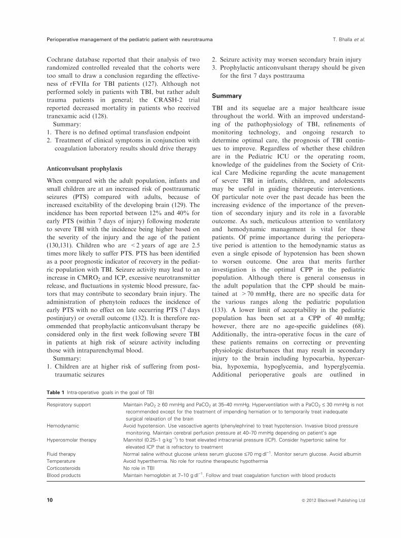
Table 1 Intra-operative goals in the goal of TBI
Respiratory support Maintain PaO2 ‡ 60 mmHg and PaCO2 at 35–40 mmHg. Hyperventilation with a PaCO2 £ 30 mmHg is not
recommended except for the treatment of impending herniation or to temporarily treat inadequate
surgical relaxation of the brain
Hemodynamic Avoid hypotension. Use vasoactive agents (phenylephrine) to treat hypotension. Invasive blood pressure
monitoring. Maintain cerebral perfusion pressure at 40–70 mmHg depending on patient’s age
Hyperosmolar therapy Mannitol (0.25–1 gÆkg)1) to treat elevated intracranial pressure (ICP). Consider hypertonic saline for
elevated ICP that is refractory to treatment
Fluid therapy Normal saline without glucose unless serum glucose £70 mgÆdl)1. Monitor serum glucose. Avoid albumin
Temperature Avoid hyperthermia. No role for routine therapeutic hypothermia
Corticosteroids No role in TBI
Blood products Maintain hemoglobin at 7–10 gÆdl)1. Follow and treat coagulation function with blood products
Perioperative management of the pediatric patient with neurotrauma T. Bhalla et al.
10 ª 2012 Blackwell Publishing Ltd
Table 1. As much of the clinical practice and care of
children with TBI continues to be extrapolated from
the adult literature, future studies focusing on the
care of children with TBI should be considered.
With appropriate attention to the current recommen-
dations perioperatively, the outcomes of TBI will
continue to improve.
Conflict of interest
No conflicts of interest declared.
References
1 Faul M, Xu L, Wald MM et al. Traumatic
Brain Injury in the United States: Emer-
gency Department Visits, Hospitalizations
and Deaths 2002–2006. Atlanta (GA): Cen-
ters for Disease Control and Prevention,
National Center for Injury Prevention and
Control 2010.
2 Schneier AJ, Shields BJ, Hostetler SG et al.
Incidence of pediatric traumatic brain injury
and associated hospital resource utilization
in the United States. Pediatrics 2006; 118:
483–492.
3 Duhaime AC, Christian CW, Rorke LB
et al. Non-accidental head injury in infants
– the ‘‘shaken-baby syndrome’’. N Engl J
Med 1998; 338: 1822–1829.
4 Agran PF, Anderson C, Winn D et al.
Rates of pediatric injuries by 3-month inter-
vals for children 0 to 3 years of age.
Pediatrics 2003; 111: e683–e692.
5 Durkin MS, Laraque D, Lubman I et al.
Epidemiology and prevention of traffic inju-
ries to urban children and adolescents.
Pediatrics 1999; 103: e74–e79.
6 Langlois JA, Rutland-Brown W, Thomas
KE. The incidence of traumatic brain injury
among children in the United States: differ-
ences by race. J Head Trauma Rehabil 2005;
20: 229–238.
7 Rivara FP. Childhood injuries. Dev Med
Child Neurol 1984; 26: 81–87.
8 Adelson PD, Bratton SL, Carney NA et al.
Guidelines for the acute medical manage-
ment of severe traumatic brain injury in
infants, children, and adolescents. Pediatr
Crit Care Med 2003; 4: S1–S75.
9 Adelson PD, Bratton SL, Carney NA et al.
Guidelines for the acute medical manage-
ment of severe traumatic brain injury in
infants, children, and adolescents. Crit Care
Med 2003; 31: S417–S491.
10 Adelson PD, Bratton SL, Carney NA et al.
Guidelines for the acute medical manage-
ment of severe traumatic brain injury in
infants, children, and adolescents. J Trauma
2003; 54: S235–S310.
11 Greve MW, Zink BJ. Pathophysiology of
traumatic brain injury. Mt Sinai J Med
2009; 76: 97–104.
12 Werner C, Engelhard K. Pathophysiology
of traumatic brain injury. Br J Anaesth
2007; 99: 4–9.
13 Chesnut RM, Marshall LF, Klauber
MR et al. The role of secondary brain
injury in determining outcome from
severe head injury. J Trauma 1993; 34:
216–222.
14 Lang DA, Teasdale GM, Macpherson P
et al. Diffuse brain swelling after head
injury: more often malignant in adults than
children? J Neurosurg 1994; 80: 675–680.
15 Kochanek PM. Pediatric traumatic brain
injury: quo vadis? Dev Neurosci 2006; 28:
244–255.
16 Davis DP, Koprowicz KM, Newgard CD
et al. The relationship between out-of-hospi-
tal airway management and outcome among
trauma patients with Glasgow Coma Scale
Scores of 8 or less. Prehosp Emerg Care
2011; 15: 184–192.
17 Davis DP, Peay J, Sise MJ et al. Prehospital
airway and ventilation management: a
trauma score and injury severity score-based
analysis. J Trauma 2010; 69: 294–301.
18 Tobias JD. Airway management for pediat-
ric emergencies. Pediatr Ann 1996; 25: 317–
320.
19 Tobias JD. Airway management in the pedi-
atric trauma patient. J Intensive Care Med
1998; 13: 1–14.
20 Crosby ET. Airway management in adults
after cervical spine trauma. Anesthesiology
2006; 104: 1293–1318.
21 Holly LT, Kelly DF, Counelis GJ et al. Cer-
vical spine trauma associated with moderate
and severe head injury: Incidence, risk fac-
tors, and injury characteristics. J Neurosurg
(Spine 3) 2002; 96: 285–291.
22 Demetriades D, Charalambides K, Chah-
wan S et al. Nonskeletal cervical spine inju-
ries: epidemiology and diagnostic pitfalls. J
Trauma 2000; 48: 724–727.
23 Tritapepe L, Voci P, Marino P et al. Cal-
cium chloride minimizes the hemodynamic
effects of propofol in patients undergoing
coronary artery bypass grafting. J Cardio-
thorac Vasc Anesth 1999; 13: 150–153.
24 Brussel T, Theissen JL, Vigfusson G et al.
Hemodynamic and cardiodynamic effects of
propofol and etomidate: negative inotropic
properties of propofol. Anesth Analg 1989;
69: 35–40.
25 Tobias JD. Etomidate: applications in pedi-
atric critical care and pediatric anesthesiol-
ogy. Pediatr Crit Care Med 2000; 1: 100–
106.
26 Annane D. ICU physicians should abandon
the use of etomidate. Intensive Care Med
2005; 31: 325–326.
27 den Brinker M, Hokken-Koelega AC,
Hazelzet JA et al. One single dose of etomi-
date negatively influences adrenocortical
performance for at least 24 hours in chil-
dren with meningococcal sepsis. Intensive
Care Med 2008; 34: 163–168.
28 Sprung CL, Annane D, Keh D et al. COR-
TICUS study group: hydrocortisone therapy
for patients with septic shock. JAMA 2002;
288: 862–871.
29 Chernow B, Laker R, Creuss D et al.
Plasma, urine, and cerebrospinal fluid cate-
cholamine concentrations during and after
ketamine sedation. Crit Care Med 1982; 10:
600–603.
30 Pagel PS, Kampine JP, Schmeling WT
et al. Ketamine depresses myocardial con-
tractility as evaluated by the preload recrui-
table stroke work relationship in chronically
instrumented dogs with autonomic nervous
system blockade. Anesthesiology 1992; 76:
564–572.
31 Gelissen HP, Epema AH, Henning RH
et al. Inotropic effects of propofol, thiopen-
tal, midazolam, etomidate, and ketamine on
isolated human atrial muscle. Anesthesiology
1996; 84: 397–403.
32 Schulte am Esch J, Pfeifer G, Thiemig I
et al. The influence of intravenous anaes-
thetic agents on primarily increased intra-
cranial pressure. Acta Neurochir (Wien)
1978; 45: 15–25.
33 Bar-Joseph G, Guilburd Y, Tamir A et al.
Effectiveness of ketamine in decreasing
intracranial pressure in children with intra-
cranial hypertension. J Neurosurg Pediatr
2009; 4: 40–46.
34 Albanese J, Arnaud S, Rey M et al. Keta-
mine decreases intracranial pressure and
electroencephalographic activity in trau-
matic brain injury patients during propofol
sedation. Anesthesiology 1997; 87: 1328–
1334.
35 Mayberg TS, Lam AM, Matta BF et al. Ke-
tamine does not increase cerebral blood flow
velocity or intracranial pressure during iso-
flurane/nitrous oxide anesthesia in patients
T. Bhalla et al. Perioperative management of the pediatric patient with neurotrauma
ª 2012 Blackwell Publishing Ltd 11
undergoing craniotomy. Anesth Analg 1995;
81: 84–89.
36 Perry JJ, Lee JS, Sillberg VA et al. Rocuro-
nium versus succinylcholine for rapid
sequence induction intubation. Cochrane
Database Syst Rev 2008; 2: CD002788.
37 Minton MD, Grosslight K, Stirt JA et al.
Increases in intracranial pressure from succi-
nylcholine: prevention by prior nondepolar-
izing blockade. Anesthesiology 1986; 65:
165–169.
38 Stirt JA, Grosslight KR, Bedford RF et al.
‘‘Defasciculation’’ with metocurine prevents
succinylcholine-induced increases in intra-
cranial pressure. Anesthesiology 1987; 67:
50–53.
39 Kovarik WD, Mayberg TS, Lam AM et al.
Succinylcholine does not change intracranial
pressure, cerebral blood flow velocity, or the
electroencephalogram in patients with neu-
rologic injury. Anesth Analg 1994; 78: 469–
473.
40 Clancy M, Halford S, Walls R et al. In
patients with head injuries who undergo
rapid sequence intubation using succinylcho-
line, does pretreatment with a competitive
neuromuscular blocking agent improve
outcome? A literature review. Emerg Med J
2001; 18: 373–375.
41 Robinson N, Clancy M. In patients with
head injury undergoing rapid sequence intu-
bation, does pretreatment with intravenous
lignocaine/lidocaine lead to an improved
neurological outcome? A review of the liter-
ature. Emerg Med J 2001; 18: 453–457.
42 Zhang XY, Yang ZJ, Wang QX et al.
Impact of positive end-expiratory pressure
on cerebral injury patients with hypoxemia.
Am J Emerg Med 2011; 29: 699–703.
43 Caricato A, Conti G, Della Corte F et al.
Effects of PEEP on the intracranial system
of patients with head injury and subarach-
noid hemorrhage: the role of respiratory
system compliance. J Trauma 2005; 58: 571–
576.
44 Stocchetti N, Maas AI, Chieregato A et al.
Hyperventilation in head injury: a review.
Chest 2005; 127: 1812–1827.
45 Adelson PD, Bratton SL, Carney NA et al.
Guidelines for the acute medical manage-
ment of severe traumatic brain injury in
infants, children, and adolescents. Chapter
12. Use of hyperventilation in the acute
management of severe pediatric traumatic
brain injury. Pediatr Crit Care Med 2003; 4:
S45–S48.
46 Dumont TM, Visioni AJ, Rughani AI et al.
Inappropriate prehospital ventilation in
severe traumatic brain injury increases in-
hospital mortality. J Neurotrauma 2010; 27:
1233–1241.
47 Muizelaar JP, Marmarou A, Ward JD et al.
Adverse effects of prolonged hyperventila-
tion in patients with severe head injury: a
randomized clinical trial. J Neurosurg 1991;
75: 731–739.
48 Skippen P, Seear M, Poskitt K et al. Effect
of hyperventilation on regional cerebral
blood flow in head-injured children. Crit
Care Med 1997; 25: 1402–1409.
49 Thiagarajan A, Goverdhan PD, Chari P
et al. The effect of hyperventilation and hy-
peroxia on cerebral venous oxygen satura-
tion in patients with traumatic brain injury.
Anesth Analg 1998; 87: 850–853.
50 Khan FA, Khan M, Abbasi S. Arterial to
end-tidal carbon dioxide difference in neuro-
surgical patients undergoing craniotomy: a
review of practice. J Pak Med Assoc 2007;
57: 446–448.
51 Ferber J, Juniewicz HM, Lechowicz-Gog-
owska EB et al. Arterial to end-tidal carbon
dioxide difference during craniotomy in
severely head-injured patients. Folia Med
Cracov 2001; 42: 141–152.
52 Marshall LF, Becker DP, Bowers SA et al.
The National Traumatic Coma Data Bank.
Part 1: design, purpose, goals, and results. J
Neurosurg 1983; 59: 276–284.
53 Tepas JJ III, DiScala C, Ramenofsky ML et
al. Mortality and head injury: the pediatric
perspective. Pediatr Surg 1990; 25: 92–95.
54 McHugh GS, Engel DC, Butcher I et al.
Prognostic value of secondary insults in
traumatic brain injury: results from the
IMPACT study. J Neurotrauma 2007; 24:
287–293.
55 Ract C, Vigue B. Comparison of the cere-
bral effects of dopamine and norepinephrine
in severely head-injured patients. Intensive
Care Med 2001; 27: 101–106.
56 Steiner LA, Johnston AJ, Czosnyka M et al.
Direct comparison of cerebrovascular effects
of norepinephrine and dopamine in head-
injured patients. Crit Care Med 2004; 32:
1049–1054.
57 Johnston AJ, Steiner LA, Chatfield DA et
al. Effect of cerebral perfusion pressure aug-
mentation with dopamine and norepineph-
rine on global and focal brain oxygenation
after traumatic brain injury. Intensive Care
Med 2004; 30: 791–797.
58 Sookplung P, Siriussawakul A, Malakouti
A et al. Vasopressor use and effect on
blood pressure after severe adult traumatic
brain injury. Neurocrit Care 2011; 15: 46–
54.
59 Pietropaoli JA, Rogers FB, Shackford SR et
al. The deleterious effects of intraoperative
hypotension on outcome in patients with
severe head injuries. J Trauma 1992; 33:
403–407.
60 Albanese J, Viviand X, Potie F et al. Sufen-
tanil, fentanyl, and alfentanil in head
trauma patients: a study on cerebral hemo-
dynamics. Crit Care Med 1999; 27: 407–411.
61 Schregel W, Weyerer W, Cunitz G. Opioids,
cerebral circulation and intracranial pres-
sure. Anaesthesist 1994; 43: 421–430.
62 Engelhard K, Werner C. Inhalational or
intravenous anesthetics for craniotomies?
Pro inhalational. Curr Opin Anaesthesiol
2006; 19: 504–508.
63 Schulte am Esch J, Thiemig I, Pfeifer G
et al. The influence of some inhalation ana-
esthetics on the intracranial pressure with
special reference to nitrous oxide. Anaesthe-
sist 1979; 28: 136–141.
64 Vernon DD, Witte MK. Effect of neuro-
muscular blockade on oxygen consumption
and energy expenditure in sedated, mechani-
cally ventilated children. Crit Care Med
2000; 28: 1569–1571.
65 Hsiang JK, Chesnut RM, Crisp CB et al.
Early, routine paralysis for intracranial pres-
sure control in severe head injury: is it nec-
essary? Crit Care Med 1994; 22: 1471–1476.
66 Exo J, Kochanek PM, Adelson PD et al.
Intracranial pressure-monitoring systems in
children with traumatic brain injury: com-
bining therapeutic and diagnostic tools. Pe-
diatr Crit Care Med 2011; 12: 560–565.
67 Bratton SL, Chestnut RM, Ghajar J et al.
Guidelines for the management of severe
traumatic brain injury. VI. Indications for
intracranial pressure monitoring. Neurotrau-
ma 2007; 24(Suppl 1): S1–S95.
68 Downard C, Hulka F, Mullins RJ et al.
Relationship of cerebral perfusion pressure
and survival in pediatric brain-injured
patients. J Trauma 2000; 49: 654–659.
69 Catala-Temprano A, Claret Teruel G, Cam-
bra Lasaosa FJ et al. Intracranial pressure
and cerebral perfusion pressure as risk fac-
tors in children with traumatic brain inju-
ries. J Neurosurg 2007; 106: 463–466.
70 Chambers IR, Kirkham FJ. What is the
optimal cerebral perfusion pressure in chil-
dren suffering from traumatic coma? Neuro-
surg Focus 2003; 15: E3.
71 Padayachy LC, Figaji AA, Bullock MR.
Intracranial pressure monitoring for trau-
matic brain injury in the modern era. Childs
Nerv Syst 2010; 26: 441–452.
72 Melo JR, Di Rocco F, Blanot S et al.
Transcranial Doppler can predict intracra-
nial hypertension in children with severe
traumatic brain injuries. Childs Nerv Syst
2011; 27: 979–984.
73 Schaffranietz L, Heinke W. The effect of
different ventilation regimes on jugular
venous oxygen saturation in elective neuro-
surgical patients. Neurol Res 1998; 20: S66–
S70.
74 Bratton SL, Chestnut RM, Ghajar J et al.
Guidelines for the management of severe
traumatic brain injury. X. Brain oxygen
monitoring and thresholds. J Neurotrauma
2007; 24: S65–S70.
Perioperative management of the pediatric patient with neurotrauma T. Bhalla et al.
12 ª 2012 Blackwell Publishing Ltd
75 Tobias JD. Cerebral oxygenation monitor-
ing: near infrared spectroscopy. Expert Rev
Med Devices 2006; 3: 235–243.
76 Ng I, Lim J, Wong HB. Effects of head pos-
ture on cerebral hemodynamics: its influ-
ences on intracranial pressure, cerebral
perfusion pressure, and cerebral oxygena-
tion. Neurosurgery 2004; 54: 593–597.
77 Mavrocordatos P, Bissonnette B, Ravussin
P. Effects of neck position and head eleva-
tion on intracranial pressure in anaesthe-
tized neurosurgical patients: preliminary
results. J Neurosurg Anesth 2000; 12: 10–14.
78 Hung OR, Hare GM, Brien S. Head eleva-
tion reduces head-rotation associated
increased ICP in patients with intracranial
tumours. Can J Anaesth 2000; 47: 415–420.
79 Rosner MJ, Coley IB. Cerebral perfusion
pressure, intracranial pressure, and head ele-
vation. J Neurosurg 1986; 65: 636–641.
80 Williams LE, Hildebrand KL, McCormick
SA et al. The effect of intravenous lactated
ringer’s solution versus 0.9% sodium chlo-
ride solution on serum osmolality in human
volunteers. Anesth Analg 1999; 88: 999–
1003.
81 Myburgh J, Cooper DJ, Finfer S et al. Sal-
ine or albumin for fluid resuscitation in
patients with traumatic brain injury. N Engl
J Med 2007; 357: 874–884.
82 Baker AJ, Rhind SG, Morrison LJ et al.
Resuscitation with hypertonic saline-dextran
reduces serum biomarker levels and corre-
lates with outcome in severe traumatic brain
injury patients. J Neurotrauma 2009; 26:
1227–1240.
83 Cooper DJ, Myles PS, McDermott FT et al.
Prehospital hypertonic saline resuscitation
of patients with hypotension and severe
traumatic brain injury: a randomized con-
trolled trial. JAMA 2004; 291: 1350–1357.
84 Sharma D, Jelacic J, Chennuri R et al. Inci-
dence and risk factors for perioperative
hyperglycemia in children with traumatic
brain injury. Anesth Analg 2009; 108: 81–89.
85 Young B, Ott L, Dempsey R et al. Rela-
tionship between admission hyperglycemia
and neurologic outcome of severely brain-
injured patients. Ann Surg 1989; 210: 466–
472.
86 Lipshutz AK, Gropper MA. Perioperative
glycemic control: an evidence-based review.
Anesthesiology 2009; 110: 408–421.
87 Rovlias A, Kotsou S. The influence of
hyperglycemia on neurological outcome in
patients with severe head injury. Neurosur-
gery 2000; 46: 335–342.
88 Cochran A, Scaife ER, Hansen KW et al.
Hyperglycemia and outcomes from Pediatric
traumatic brain injury. J Trauma 2003; 55:
1035–1038.
89 Lam AM, Winn HR, Cullen BF et al.
Hyperglycemia and neurological outcome in
patients with head injury. J Neurosurg 1991;
75: 545–551.
90 Smith RL, Lin JC, Adelson PD et al. Rela-
tionship between hyperglycemia and out-
come in children with severe traumatic brain
injury. Pediatr Crit Care Med 2012; 13: 85–
91.
91 Van den Berghe G, Wouters P, Weekers F
et al. Intensive insulin therapy in the criti-
cally ill patients. N Engl J Med 2001; 345:
1359–1367.
92 NICE-SUGAR Study Investigators, Finfer
S, Chittock DR et al. Intensive versus con-
ventional glucose control in critically ill
patients. N Engl J Med 2009; 306: 1283–
1297.
93 Billotta F, Caramia R, Cernak I et al.
Intensive insulin therapy after severe trau-
matic brain injury: a randomized clinical
trial. Neurocrit Care 2008; 9: 159–166.
94 Diltoer M, Camu F. Glucose homeostasis
and insulin secretion during isoflurane anes-
thesia in humans. Anesthesiology 1988; 68:
880–886.
95 Kitamura T, Ogawa M, Kawamura G et al.
The effects of sevoflurane and propofol on
glucose metabolism under aerobic condi-
tions in fed rats. Anesth Analg 2009; 109:
1479–1485.
96 Bennett TD, Statler KD, Korgenski EK et
al. Osmolar therapy in pediatric traumatic
brain injury. Crit Care Med 2012; 40: 208–
215.
97 Knapp JM. Hyperosmolar therapy in the
treatment of severe head injury in children:
mannitol and hypertonic saline. AACN Clin
Issues 2005; 16: 199–211.
98 Kerwin AJ, Schinco MA, Tepas JJ III. The
use of 23.4% hypertonic saline for the man-
agement of elevated intracranial pressure in
patients with severe traumatic brain injury:
a pilot study. J Trauma 2009; 67: 289–292.
99 Fisher B, Thomas D, Perterson B. Hyper-
tonic saline lowers raised intracranial pres-
sure in children after head trauma. J
Neurosurg Anesthesiol 1992; 4: 4–10.
100 Himmelseher S. Hypertonic saline solutions
for the treatment of intracranial hyperten-
sion. Curr Opin Anesthesiol 2007; 20: 414–
426.
101 Khanna S, Davis D, Peterson B et al. Use
of hypertonic saline in the treatment of
severe refractory posttraumatic intracranial
hypertension in pediatric traumatic brain
injury. Crit Care Med 2000; 28: 1144–1151.
102 Oddo M, Levine JM, Frangos S et al. Effect
of mannitol and hypertonic saline on cere-
bral oxygenation in patients with severe
traumatic brain injury and refractory intra-
cranial hypertension. J Neurol Neurosurg
Psychiatry 2009; 80: 916–920.
103 Vats A, Chambliss CR, Anand KJS et al. Is
hypertonic saline an effective alternative to
mannitol in the treatment of elevated intra-
cranial pressure in pediatric patients? Is
hypertonic saline an effective alternative to
mannitol in the treatment of elevated intra-
cranial pressure in pediatric patients? J
Intensive Care Med 1999; 14: 184–188.
104 Wu CT, Chen LC, Kuo CP et al. A com-
parison of 3% hypertonic saline and manni-
tol for brain relaxation during elective
supratentorial brain tumor surgery. Anesth
Analg 2010; 110: 903–907.
105 Metz C, Holzschuh M, Bein T et al. Moder-
ate Hypothermia in patients with severe
head injury: cerebral and extracerebral
effects. J Neurosurg 1996; 85: 533–541.
106 Peterson K, Carson S, Carney N. Hypother-
mia treatment for traumatic brain injury: a
systematic review and meta-analysis. J Neu-
rotrauma 2008; 25: 62–71.
107 Gl C (2002) The National Acute Brain
Injury Study: Hypothermia II (NABISH II).
http://www.clinicaltrials.gov.
108 Hutchison JS, Ward RE, Lacroix J et al.
Hypothermia therapy after traumatic brain
injury in children. N Engl J Med 2008; 358:
2447–2456.
109 Hutchison JS, Frndova H, Lo TY et al.
Impact of hypotension and low cerebral per-
fusion pressure on outcomes in children
treated with hypothermia therapy following
severe traumatic brain injury: a post hoc
analysis of the Hypothermia Pediatric Head
Injury Trial. Dev Neurosci 2010; 32: 406–
412.
110 Bourdages M, Bigras JL, Farrell CA et al.
Cardiac arrhythmias associated with severe
traumatic brain injury and hypothermia
therapy. Pediatr Crit Care Med 2010; 11:
408–414.
111 Adelson PD, Ragheb J, Kanev P et al.
Phase II clinical trial of moderate hypother-
mia after severe traumatic brain injury in
children. Neurosurgery 2005; 56: 740–775.
112 Saul TG, Ducker TB, Salcman M et al. Ste-
roids in severe head injury: a prospective
randomized clinical trial. J Neurosurgery
1981; 54: 596–600.
113 Cooper PR, Moody S, Clark WK et al.
Dexamethasone and severe head injury: a
Prospective double blind trial. Neurosurgery
1979; 51: 307–316.
114 Edwards P, Arango M, Balica L et al. Final
results of MRC CRASH, a randomised pla-
cebo-controlled trial of intravenous cortico-
steroid in adults with head injury-outcomes
at 6 months. Lancet 2005; 365: 1957–1959.
115 Alvarez M, Nava JM, Rue M et al. Mortal-
ity prediction in head trauma patients: per-
formance of Glasgow Coma Score and
general severity systems. Crit Care Med
1998; 26: 142–148.
116 Carlson AP, Schermer CR, Lu SW. Retro-
spective evaluation of anemia and transfu-
T. Bhalla et al. Perioperative management of the pediatric patient with neurotrauma
ª 2012 Blackwell Publishing Ltd 13
sion in traumatic brain injury. J Trauma
2006; 61: 567–571.
117 Salim A, Hadjizacharia P, DuBose J et al.
Role of anemia in traumatic brain injury. J
Am Coll Surg 2008; 207: 398–406.
118 Desmet L, Lacroix J. Transfusion in pediat-
rics. Crit Care Clin 2004; 20: 299–311.
119 Chaiwat O, Lang JD, Vavilala MS et al.
Early packed red blood cell transfusion and
acute respiratory distress syndrome after
trauma. Anesthesiology 2009; 110: 351–360.
120 Malone DL, Dunne J, Tracy JK et al.
Blood transfusion, independent of shock
severity, is associated with worse outcome
in trauma. J Trauma 2003; 54: 898–905.
121 Karam O, Tucci M, Ducruet T et al. Red
blood cell transfusion thresholds in pediatric
patients with sepsis. Pediatr Crit Care Med
2011; 12: 512–518.
122 Rouette J, Trottier H, Ducruet T et al. Red
blood cell transfusion in post-surgical inten-
sive care patients: a randomized clinical
trial. Ann Surg 2010; 251: 412–417.
123 Harhangi BS, Kompanje EJ, Leebeek FW
et al. Coagulation disorders after traumatic
brain injury. Acta Neurochir (Wien) 2008;
150: 165–175.
124 Talving P, Benfield R, Hadjizacharia P et al.
Coagulopathy in severe traumatic brain
injury: a prospective study. J Trauma 2009;
66: 55–61.
125 Morenski JD, Tobias JD, Jimenez DF.
Recombinant activated factor VII for cere-
bral injury-induced coagulopathy in pediatric
patients. Report of three cases and review of
the literature. J Neurosurg 2003; 98: 611–616.
126 Brown CV, Foulkrod KH, Lopez D et al.
Recombinant factor VIIa for the correction
of coagulopathy before emergent craniot-
omy in blunt trauma patients. J Trauma
2010; 68: 348–352.
127 Perel P, Roberts I, Shakur H et al. Haemo-
static drugs for traumatic brain injury.
Cochrane Database Syst Rev 2010; 1:
CD007877.
128 CRASH-2 trial collaborators, Shakur H,
Roberts I et al. Effects of tranexamic acid
on death, vascular occlusive events, and
blood transfusion in trauma patients with
significant haemorrhage (CRASH-2): a
randomised, placebo-controlled trial. Lancet
2010; 376: 23–32.
129 Liesemer K, Bratton SL, Zebrack CM et al.
Early post-traumatic seizures in moderate to
severe pediatric traumatic brain injury:
rates, risk factors, and clinical features. J
Neurotrauma 2011; 28: 755–762.
130 Tempkin NR, Dikmen SS, Winn HR. Post
traumatic seizures. Neurosurg Clin North
Am 1991; 2: 425–435.
131 Lewis RJ, Yee L, Inkelis SH et al. Clinical
predictors of post-traumatic seizures in chil-
dren with head trauma. Ann Emerg Med
1993; 22: 1114–1118.
132 Khan A, Banerjee A. The role of prophylac-
tic anticonvulsants in moderate to severe
head injury. Int J Emerg Med 2010; 3: 187–
191.
133 Marion DW, Spiegel TP. Changes in the
management of severe traumatic brain
injury: 1991–1997. Crit Care Med 2000; 28:
16–18.
Perioperative management of the pediatric patient with neurotrauma T. Bhalla et al.
14 ª 2012 Blackwell Publishing Ltd