percutaneous right brachial artery approach with 5f catheters for studying coronary artery disease
TRANSCRIPT
Catheterization and Cardiovascular Diagnosis 22:47-51 (1991)
Feature Topic: Small Size Catheter Trials
Percutaneous Right Brachial Artery Approach With 5F Catheters for Studying Coronary Artery Disease
J. Lupon-Roses, MD, FICA, E. Domingo, MD, FICA, FCCP, J. Angel, MD, FICA, FCCP, 1. Anivarro, MD, and J. Soler-Soler, MD
We prospectively studied 60 ischemic patients with 5F catheters (Pigtail and Amplatz) using the percutaneous right brachial artery approach (group I), in order to compare this technique with two groups of 100 patients each randomly studied by the femoral route with either 5F (group II) or 8F (group 111) catheters (Pigtail and Judkins). The following parameters were analyzed: need to change the initially elected catheter diameter or/and artery approach; technical difficulty for obtaining LV, LCA, and RCA angiograms; total time of X-ray exposure; quality image of LV, LCA, and RCA angiograms; incidence of arterial puncture related hematomas or total arterial occlusion; and duration of local compression after sheath removal. There were no differences between 5F brachial and femoral approaches except for the arterial compression time ( ~ ~ 0 . 0 1 ) and the X-ray exposure time @=0.03) which were longer with the brachial approach. Whatever the route used, 5F showed a mild increased difficulty (brachial p=O.OOl; femoral p=O.Ol) and a mild decreased quality image for LCA (branchial p=0.006; femoral ~ ~ 0 . 0 5 ) . Mild he- matomas were more frequent with 8F catheters ( ~ ~ 0 . 0 5 ) . The procedure could be com- pleted by the elected first artery and type of catheter (5F or 8F) in 57/60 patients in group I, in 95/100 in group II, and in 961100 in group 111 (nonsignificant differences). Thus, the percutaneous right brachial artery approach using 5F catheters is similar to the femoral artery approach with the same catheters. Although both of them showed a mild increased technical difficulty and a mild decreased quality image compared to 8F, mainly for LCA angiograms, they allowed complete and reliable angiograms reading and analysis.
Key words: coronary heart disease, angiography
INTRODUCTION
Technical improvements in angiographic catheters al- low smaller catheters to be used without compromising either flow rates during injection or torque control. The introduction of arteriography catheters with a smaller di- ameter was aimed at decreasing the morbidity directly related to arterial puncture and to possibly increasing the already proved safety of the progressively more frequent performance of outpatient cardiac catheterizations [ 1,2]. Five French catheters have already been successfully used in the percutaneous left brachial and femoral artery approach for studying patients with coronary artery dis- ease [ 1-61. Its advantages and limitations using the fem- oral route had been previously analyzed in comparison with 8F catheters [4-61. The purpose of this study is to prospectively assess the utility of 5F catheters for left heart cardiac catheterization including selective coronary angiographic procedures, using the percutaneous right brachial artery approach avoiding artery cut-down, and to compare the results of this technique with those pre- viously obtained using the femoral route and the Judkins technique [4,5].
0 1991 Wiley-Liss, Inc.
MATERIAL AND METHODS
We prospectively studied 60 ischemic patients (53 males and 7 females with a mean age of 56 years, range 40-76 years) with 5F catheters using the percutaneous right brachial artery approach (group I), and compared this technique with two groups of 100 patients each ran- domly studied by the femoral route with either 5F (group 11) and 8F catheters (group 111) catheters [4]. Collective antropometric data are specified in Table I. All but 2 patients of the group I were studied by the brachial route because of femoroiliac atherosclerosis.
The same multiple view cineangiographic study was accomplished in both series of patients for LV and cor- onary angiography. Pigtail catheters were used for LV
From the Servei de Cardiologia, Departament de Medicina, Hospital General Val1 d'Hebron, Barcelona, Spain.
Received March 13, 1990; revision accepted August 6 , 1990
Address reprint requests to Dr. J. Lupon-Roses, Servei de Cardiolo- gia, Departament de Medicina, Hospital General Vall d'Hebron, Pg. Vall d . Hebron sin, 08035 Barcelona, Spain.

48 Lupon-Roses et al.
TABLE 1. Collective AntroDometric Data*
Group I Group I1 Group I11 P
Age (Y) 56 t 8.(40-76) 56 2 9 (31-75) 54 t 9 (34-80) <0.05" Sex 53Mi7F 88Mi 12F 82Mi18F NS Weight (k) 71 2 14 (48-90) 70 i 10 (50-99) 70 i 10 (45-106) NS Height (cm) 165 2 8 (153-177) 164 2 7 (146-186) 164 t 8 (145-188) NS
*M, male; F, female; (y) , years; (k) , kilograms: (cm), centimeters. agetween groups I and 111.
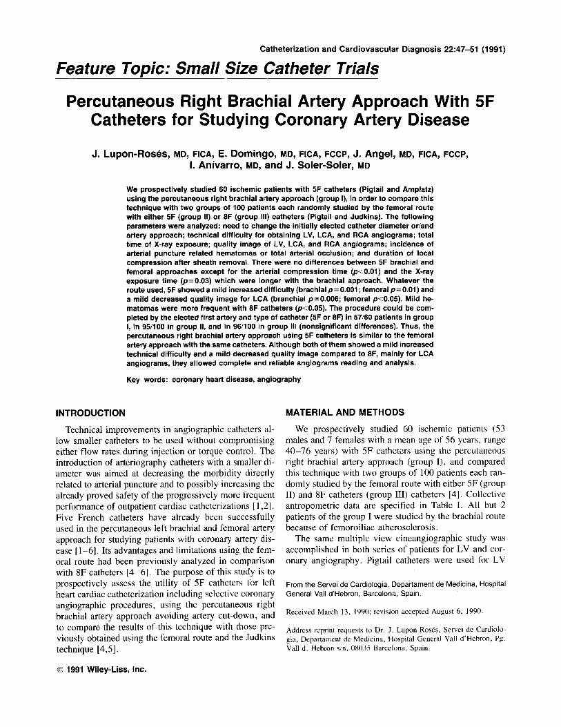
Fig. 1. LV angiogram performed with a 5 French pigtail cathe- ter by the right brachial approach, in the RAO projection.
angiograms whatever the route and the catheter diameter used. Preformed SF Amplatz catheters (USCI) were used for right and left coronary arteriographies in group I, and preformed Judkins catheters were used in groups 11 (SF USCI) and 111. Thirty-five to SO milliliters of iodinated hypoosmolar contrast material (Hexabrix) was adminis- tered with a pressure injector at a rate of 10-1.5 mm/sec for LV angiograms. Five to 8 milliliters of the same contrast was manually injected into the coronary arteries for each view. A C.G.R. X-ray system with a C arc allowing cranial and caudal angulations was used in all cases for X-ray imaging.
In group I the technique used was quite similar to that described by Fergusson and Kamada [7] although using a sheath with a valve and side arm, as others had done [8,9]: the brachial artery was palpated at the elbow crease and the overlying skin and subcutaneous tissue infiltrated with Mevipacain HCI . During careful palpa- tion of the artery an 18-gauge abbocath needle was in- serted at an angle of about 45" to the skin surface so as to transfix the vessel, and the needle was withdrawn. The cannula was then withdrawn slowly until arterial blood spurts, then it was angulated back and the 0.021-in. guidewire of the introducer set (USCI Hemaquet pediat- ric 006059 SF) was inserted into the artery, and advanced several centimeters. Then the cannula was withdrawn
and the teflon dilator and sheath were passed over the guidewire and into the brachial artery. A twisting motion was employed to minimize injury at the point of entry into the artery. Heparin (2,500 units) was injected through the side arm of the sheath. Pigtail and preformed Amplatz catheters were used for LV and coronary an- giograms, respectively, with the aid of the guidewire if necessary. After the exploration the sheath was removed before applying digital pressure to the puncture site. In most patients digital compression was combined with the inflation of a sphygmomanometer cuff around the upper arm to a level well above the patient's systolic blood pressure.
The following parameters were analyzed: need to change the initially elected catheter diameter or/and ar- tery approach; technical difficulty for obtaining LV, LCA, and RCA angiograms; total time of X-ray expo- sure; quality image of LV, LCA, and RCA angiograms; incidence of arterial puncture related hematomas or ar- terial total occlusion; and duration of local compression after sheath removal. Quality image and technical diffi- culty were evaluated semiquantitatively by the hemody- namist who performed the procedure.
The statistical analysis was done by means of chi square or Fisher's exact test for qualitative variables (de- pending on number of items) and Student's t test or U Mann-Whitney test for quantitative variables (depending on normal or nonnormal data distribution assessed by means of Kolmogorov-Smirnov test).
RESULTS
Results in groups I1 and 111 had been previously pub- lished [4] and are showed in Tables 11-IV. Significant differences were found in the greater easiness to cathe- terize LV (p<O.OS) and LCA (p = O . O l ) in group 111, in better quality image for LCA in group I11 (p<O.OS), longer X-ray exposure time in group I1 (P<O.OOl), longer arterial compression time in group I11 ( P <0.0001), and in a lower incidence of mild groin he- matomas in group I1 (p<O.OS).
Previous right brachial artery pulse was considered to be good in 47 patients, regular in 9, and fair in 4 patients. Brachial artery puncture was accomplished in the first
Catheters and Right Brachial Approach 49
Technical difficulty was classified as easy, difficult, or impossible. Table I displays these parameters for LV, RCA, and LCA angiograms for groups I to 111. The need of a guide-wire to enter the LV with the pigtail catheters was not considered a technical difficulty since most 5F pigtail catheters were introduced within the LV directly with a guide-wire. For statistical analysis technical dif- ficulty comparison was done between easy versus diffi- cult and impossible. There were no differences between groups 1 and I1 in technical difficulty (Table 11). LCA angiograms were significantly easier in group I11 than in group I ( p = O . O O l ; Table 11).
Total X-ray exposure time in group I was longer than in group I1 0, = 0.03) and was markedly longer than in group 111 (p<O.OOOl; Table 11).
Image quality was classified as good, regular, or bad. There were no bad examinations in any group. Regular examinations were considered those that were not excel- lent but good enough to allow complcte and reliable as- sessment of angiograms. Quality was graded by consen- sus between two experienced angiographers. The results of image quality for LV, RCA, and LCA angiograms in all groups are displayed in Table 111. Again there were no differences between groups I and 11. Image quality of LCA angiogram was mildly worse in group I than in group I11 0, = 0.006).
Neither moderate nor severe hematomas occurred in any group. There were only two mild hematomas in group I, but the differences with this group and the other two groups were not significants (Table IV). Postcathe- terization arterial compression time in group I was longer than in group I1 ( p < O . O I ) , but was similar to that in group I11 (Table IV).
There was one early arterial occlusion in group I that needed urgent surgery. At surgery no arterial dissection was found and arterial patency was restored after re- moval of a cylindrical thrombus whose length was that of the arterial sheath. The arterial occlusion was thus con- sidered to have been caused by a perisheath thrombal formation during the procedure (the procedure was nev- ertheless not specially long lasting).
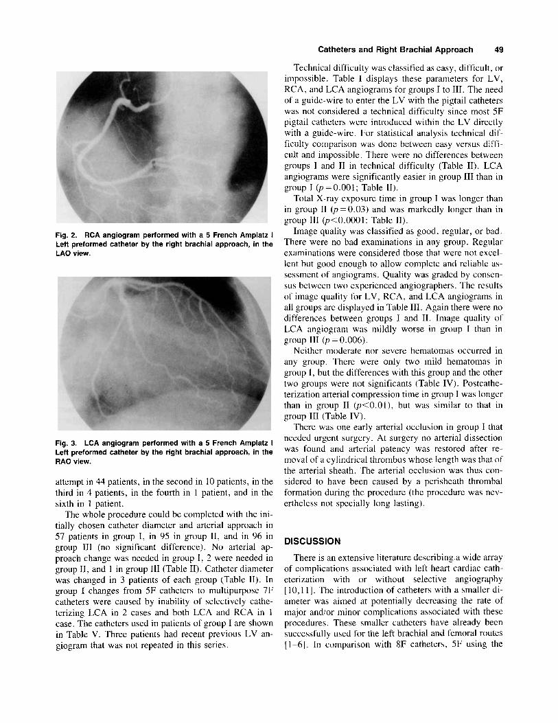
Fig. 2. RCA angiogram performed with a 5 French Amplatz I Left preformed catheter by the right brachial approach, in the LAO view.
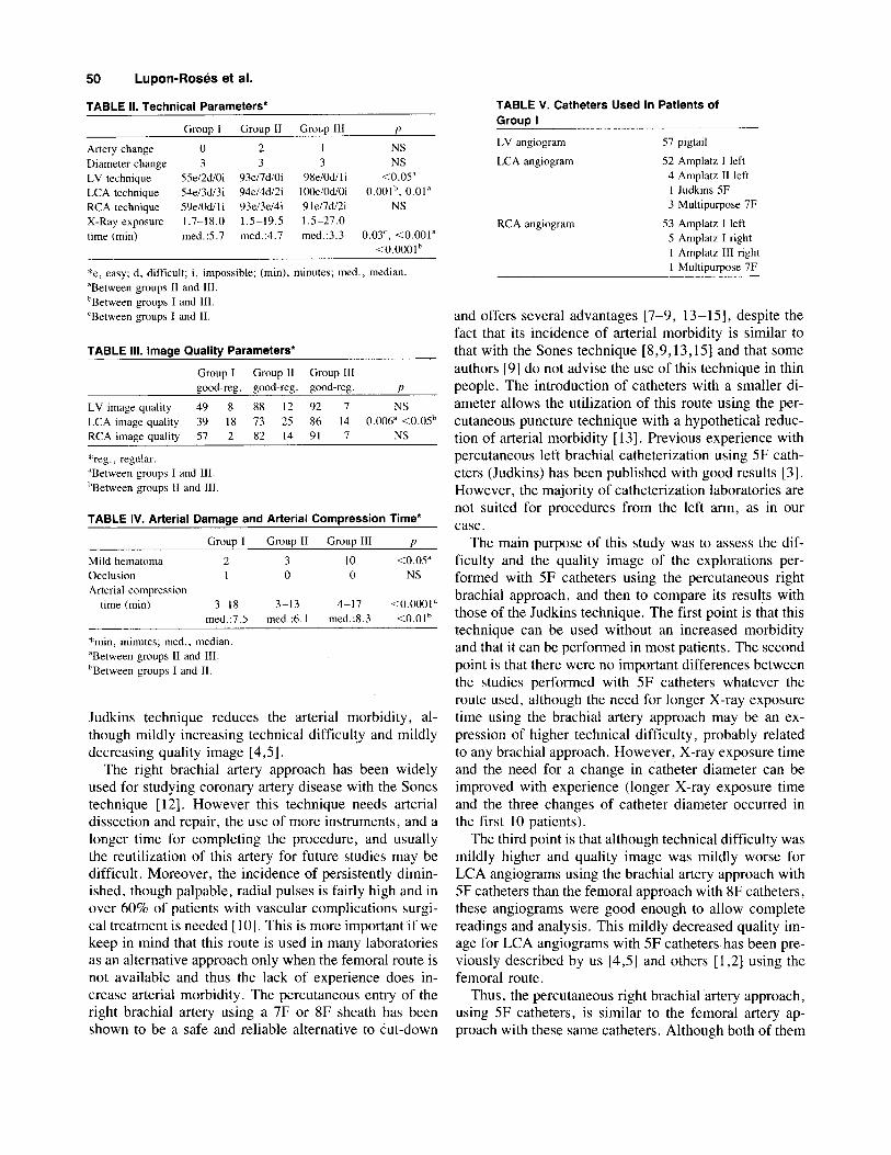
Fig. 3. LCA angiogram performed with a 5 French Amplatz I Leff preformed catheter by the right brachial approach, in the RAO view.
attempt in 44 patients, in the second in 10 patients, in the third in 4 patients, in the fourth in 1 patient, and in the sixth in 1 patient.
The whole procedure could be completed with the ini- tially chosen catheter diameter and arterial approach in 57 patients in group I, in 95 in group 11, and in 96 in group 111 (no significant difference). No arterial ap- proach change was needed in group I, 2 were needed in group 11, and 1 in group I11 (Table 11). Catheter diameter was changed in 3 patients of each group (Table 11). In group 1 changes from 5F catheters to multipurpose 7F catheters were caused by inability of selectively cathe- terizing LCA in 2 cases and both LCA and RCA in 1 case. The catheters used in patients of group I are shown in Table V. Three patients had recent previous LV an- giogram that was not repeated in this series.
DISCUSSION
There is an extensive literature describing a wide array of complications associated with left heart cardiac cath- eterization with or without selective angiography [ 10,111. The introduction of catheters with a smaller di- ameter was aimed at potentially decreasing the rate of major and/or minor complications associated with these procedures. These smaller catheters have already been successfully used for the left brachial and femoral routes [l-61. In comparison with 8F catheters, 5F using the
50 Lupon-Roses et al.
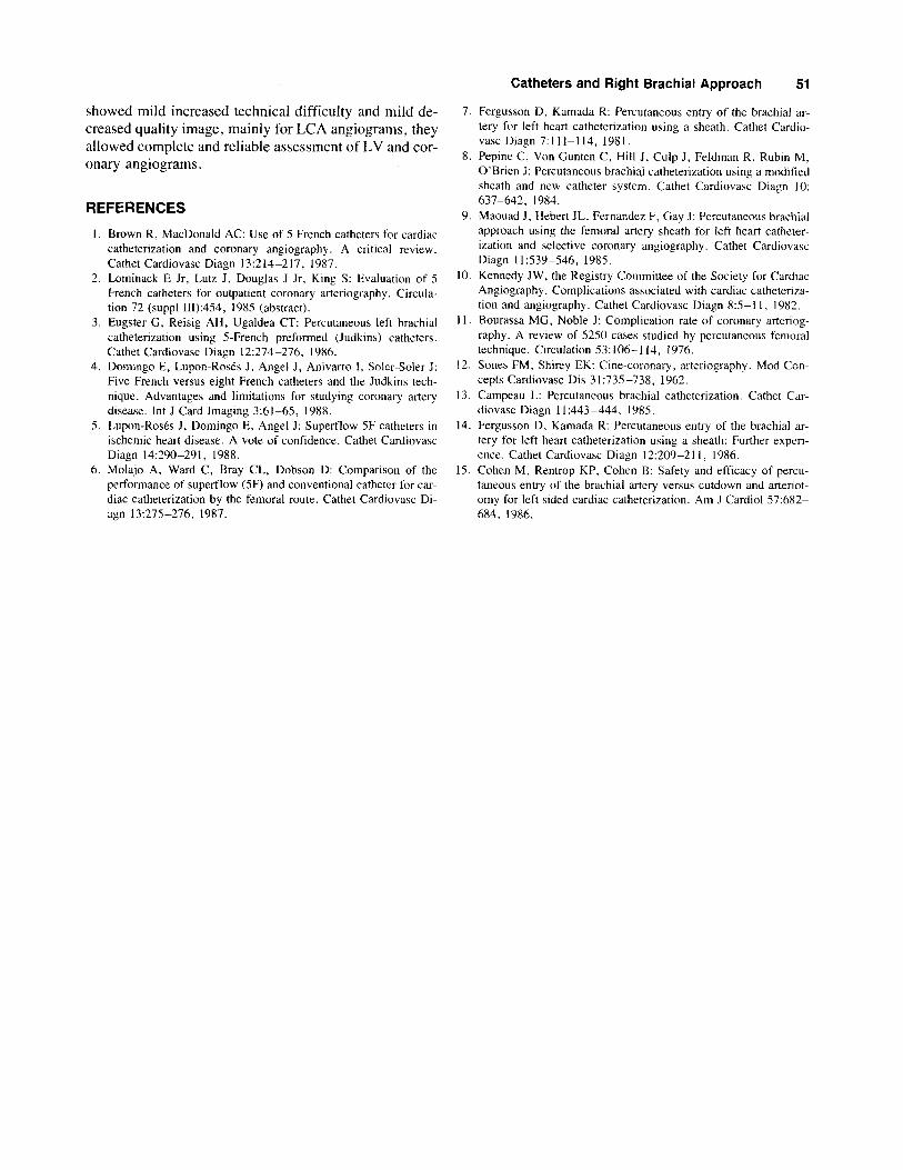
TABLE II. Technical Parameters*
Grouu I
Artery change 0 Diameter change 3 LV technique 55ei2diOi LCA technique 54ei3di3i RCA technique 59ei0dili X-Ray exposure 1.7-18.0 time (min) med . 5 . 7
Group I1 Group 111 P 2 1 NS 3 3 NS
93ei7diOi 98eiOdiI i C0.05" 94d4di2i IOOeiOdiOi 0.00Ib, 0.01" 93ei3ei4i 91el7di2i NS 1.5-19.5 1.5-27.0 med.:4.7 med.:3.3 0.03', <0.001"
<0.0001
*e, easy; d , difficult; i, impossible; (min), minutes; med., median. "Between groups 11 and 111. bBetween groups I and 111. 'Between groups I and 11.
TABLE 111. Image Quality Parameters*
Group I Group I1 Group I11 good-reg. good-reg. good-reg. P
LV image quality 49 8 88 12 92 7 NS LCA image quality 39 18 73 25 86 14 0.006" <0.05b RCA image quality 57 2 82 14 91 7 NS
*reg., regular. "Between groups I and 111. hBetween groups I1 and 111
TABLE IV. Arterial Damage and Arterial compression Time*
Grouo I Grouo I1 Grow 111 I)
Mild hematoma 2 3 10 <0.05" Occlusion 1 0 0 NS Arterial compression
time (min) 3-18 3-13 4-17 <0.0001" med.:7.5 med.:6.1 med.:8.3 <0.0Ih
~ ~~
*mm, minutes; rned., median "Between groups 11 and 111. bBetween groups I and 11.
Judkins technique reduces the arterial morbidity, al- though mildly increasing technical difficulty and mildly decreasing quality image [4,5].
The right brachial artery approach has been widely used for studying coronary artery disease with the Sones technique [ 121. However this technique needs arterial dissection and repair, the use of more instruments, and a longer time for completing the procedure, and usually the reutilization of this artery for future studies may be difficult. Moreover, the incidence of persistently dimin- ished, though palpable, radial pulses is fairly high and in over 60% of patients with vascular complications surgi- cal treatment is needed [lo]. This is more important if we keep in mind that this route is used in many laboratories as an alternative approach only when the femoral route is not available and thus the lack of experience does in- crease arterial morbidity. The percutaneous entry of the right brachial artery using a 7F or 8F sheath has been shown to be a safe and reliable alternative to cut-down
TABLE V. Catheters Used in Patients of GrouD I
LV angiogram 57 pigtail
LCA angiogram 52 Amplatz I left 4 Amplatz I1 left I Judkins 5F 3 Multipurpose 7F
5 Amplatz I right 1 Amplatz 111 right 1 Multipurpose 7F
RCA angiogram 53 Amplatz I left
and offers several advantages [7-9, 13 -1 51, despite the fact that its incidence of arterial morbidity is similar to that with the Sones technique [8,9,13,15] and that some authors [9] do not advise the use of this technique in thin people. The introduction of catheters with a smaller di- ameter allows the utilization of this route using the per- cutaneous puncture technique with a hypothetical reduc- tion of arterial morbidity [ 131. Previous experience with percutaneous left brachial catheterization using 5F cath- eters (Judkins) has been published with good results [3]. However, the majority of catheterization laboratories are not suited for procedures from the left arm, as in our case.
The main purpose of this study was to assess the dif- ficulty and the quality image of the explorations per- formed with 5F catheters using the percutaneous right brachial approach, and then to compare its results with those of the Judkins technique. The first point is that this technique can be used without an increased morbidity and that it can be performed in most patients. The second point is that there were no important differences between the studies performed with 5F catheters whatever the route used, although the need for longer X-ray exposure time using the brachial artery approach may be an ex- pression of higher technical difficulty, probably related to any brachial approach. However, X-ray exposure time and the need for a change in catheter diameter can be improved with experience (longer X-ray exposure time and the three changes of catheter diameter occurred in the first 10 patients).
The third point is that although technical difficulty was mildly higher and quality image was mildly worse for LCA angiograms using the brachial artery approach with 5F catheters than the femoral approach with 8F catheters, these angiograms were good enough to allow complete readings and analysis. This mildly decreased quality im- age for LCA angiograms with 5F catheters has been pre- viously described by us [4,5] and others [ 1,2] using the femoral route.
Thus, the percutaneous right brachial artery approach, using 5F catheters, is similar to the femoral artery ap- proach with these same catheters. Although both of them
Catheters and Right Brachial Approach 51
7. Fergusson D, Kamada R: Percutaneous entry of the brachial ar- tery for left heart catheterization using a sheath. Cathet Cardio- vasc Diagn 7:111-114, 1981.
8. Pepine C, Von Gunten C, Hill J, Culp J, Feldman R, Rubin M, O’Brien J: Percutaneous brachial catheterization using a modified sheath and new catheter system. Cathet Cardiovasc Diagn 10:
9. Maouad J , Hebert JL, Fernandez F, Gay J: Percutaneous brachial approach using the femoral artery sheath for left heart catheter- ization and selcctive coronary angiography . Cathet Cardiovasc Diagn 11539-546, 1985.
10. Kennedy JW, the Registry Committee of the Society for Cardiac Angiography. Complications associated with cardiac catheterizd- tion and angiography. Cathet Cardiovasc Diagn 8:5-1 I , 1982.
I 1 . Bourassa MG, Noble J: Complication rate of coronary arteriog- raphy. A review of 5250 cases studied by percutaneous femoral technique. Circulation 53: 106-1 14, 1976.
12. Sones FM, Shirey EK: Cine-coronary, arteriography. Mod Con- cepts Cardiovasc Dis 31:735-738, 1962.
13. Campeau L: Percutaneous brachial catheterization. Cathet Car- diovasc Diagn l1:443-444, 1985.
14. Fergusson D, Kamada R: Percutaneous entry of the brachial ar- tery for left heart catheterization using a sheath: Further experi- ence. Cathet Cardiovasc Diagn 12:209-211, 1986.
15. Cohen M, Rentrop KP, Cohen B: Safety and efficacy of percu- taneous entry of the brachial artery versus cutdown and arteriot- omy for left sided cardiac catheterization. Am J Cardiol 57:682- 684, 1986.
637-642, 1984.
showed mild increased technical difficulty and mild de- creased quality image, mainly for LCA angiograms, they allowed complete and reliable assessment of LV and cor- onary angiograms.
REFERENCES
1. Brown R , MacDonald AC: Use of 5 French catheters for cardiac catheterization and coronary angiography. A critical review. Cathet Cardiovasc Diagn 13:214-217, 1987.
2. Lominack E Jr, Lutz J, Douglas J Jr, King S: Evaluation of 5 French catheters for outpatient coronary arteriography. Circula- tion 72 (suppl III):454, 1985 (abstract).
3. Eugster G, Reisig AH, Ugaldea CT: Percutaneous left brachial catheterization using 5-French preformed (Judkins) catheters. Cathet Cardiovasc Diagn 12:274-276, 1986.
4. Domingo E, Lupon-Roses J, Angel J, Anivano I , Soler-Soler J: Five French versus eight French catheters and the Judkins tech- nique. Advantages and limitations for studying coronary artery disease. Int J Card Imaging 3:61-65, 1988.
5 . Lupon-RosCs J, Domingo E, Angel J: Superflow 5F catheters in ischemic heart disease. A vote of confidence. Cathet Cardiovasc Diagn 14:290-291, 1988.
6. Molajo A, Ward C, Bray CL, Dobson D: Comparison of the performance of superflow (SF) and conventional catheter for car- diac catheterization by the femoral route. Cathet Cardiovasc Di- agn 13:275-276, 1987.