peptide discotics : synthesis, self-assembly and application · peptide discotics synthesis,...
TRANSCRIPT
Peptide discotics : synthesis, self-assembly andapplicationCitation for published version (APA):Hout, van den, K. P. (2008). Peptide discotics : synthesis, self-assembly and application. Eindhoven:Technische Universiteit Eindhoven. https://doi.org/10.6100/IR635208
DOI:10.6100/IR635208
Document status and date:Published: 01/01/2008
Document Version:Publisher’s PDF, also known as Version of Record (includes final page, issue and volume numbers)
Please check the document version of this publication:
• A submitted manuscript is the version of the article upon submission and before peer-review. There can beimportant differences between the submitted version and the official published version of record. Peopleinterested in the research are advised to contact the author for the final version of the publication, or visit theDOI to the publisher's website.• The final author version and the galley proof are versions of the publication after peer review.• The final published version features the final layout of the paper including the volume, issue and pagenumbers.Link to publication
General rightsCopyright and moral rights for the publications made accessible in the public portal are retained by the authors and/or other copyright ownersand it is a condition of accessing publications that users recognise and abide by the legal requirements associated with these rights.
• Users may download and print one copy of any publication from the public portal for the purpose of private study or research. • You may not further distribute the material or use it for any profit-making activity or commercial gain • You may freely distribute the URL identifying the publication in the public portal.
If the publication is distributed under the terms of Article 25fa of the Dutch Copyright Act, indicated by the “Taverne” license above, pleasefollow below link for the End User Agreement:
www.tue.nl/taverne
Take down policyIf you believe that this document breaches copyright please contact us at:
providing details and we will investigate your claim.
Download date: 28. Dec. 2019
Peptide Discotics
Synthesis, self-assembly and application

Peptide Discotics
Synthesis, self-assembly and application
PROEFSCHRIFT
ter verkrijging van de graad van doctor aan de
Technische Universiteit Eindhoven, op gezag van de
Rector Magnificus, prof.dr.ir. C.J. van Duijn, voor een
commissie aangewezen door het College voor
Promoties in het openbaar te verdedigen
op donderdag 26 juni 2008 om 14.00 uur
door
Kelly Petronella Stelwagen-van den Hout
geboren te Drunen
Dit proefschrift is goedgekeurd door de promotor:
prof.dr. E.W. Meijer
Copromotoren:
dr. J.A.J.M. Vekemans
en
dr.ir. M.H.P. van Genderen
The research described in this thesis was financially supported by the Netherlands
Organisation for Scientific Research (NWO).
Omslagontwerp: Koen Pieterse
Druk: Gildeprint Drukkerijen B.V. te Enschede
A catalogue record is available from the Eindhoven University of Technology Library
ISBN: 978-90-386-1289-8

Voor mijn ouders
Voor Sjoerd

Table of contents
Chapter 1
Self-assembling contrast agents for MRI
1.1 Magnetic resonance imaging and the use of contrast agents 2 1.1.1 Applied strategies for contrast-enhancement 1.1.2 Synthetic approaches: towards more efficient contrast agents
1.2 Self-assembling paramagnetic amphiphiles for MR imaging 5
1.3 Self-assembly behaviour of C3-symmetrical discotics 9 1.3.1 Self-assembly of discotic molecules in apolar media 1.3.2 Self-assembly of discotic molecules in water
1.4 1H relaxation time measurements: study self-assembly in dilute solutions 16 1.4.1 Determining the critical micelle concentration 1.4.2 Probing the interaction of the biotin–avidin complex
1.5 Aim and outline of the thesis 17
1.6 References and notes 19
Chapter 2
Syntheses of peptide discotics with aliphatic solubilizing tails
2.1 Introduction 24
2.2 Syntheses of dipeptide discotics with an achiral glycine at the C-terminus 26
2.3 Syntheses of dipeptide discotics with a chiral phenylalanine at the C-terminus 28
2.4 Syntheses of monopeptide discotics 31
2.5 Conclusions 32
2.6 Experimental section 33
2.7 References and notes 46
Chapter 3
Self-assembly of peptide discotics with aliphatic solubilizing tails
3.1 Introduction 48
3.2 Self-assembly behaviour of dipeptide discotics in the neat state 50
3.3 Self-assembly behaviour of dipeptide discotics in solution 53
3.4 Stability of assemblies of dipeptide discotics 58
3.5 Self-assembly behaviour of monopeptide discotics in the neat state 60
3.6 Self-assembly behaviour of monopeptide discotics in solution 61
3.7 Stability of assemblies of monopeptide discotics 63
3.8 Conclusions 64
3.9 Experimental section 64
3.10 References and notes 65
Chapter 4
From apolar media to water: Self-assembly of a peptide-based discotic amphiphile
4.1 Introduction 68
4.2 Synthesis 69
4.3 Self-assembly in solution 70
4.4 Size of the self-assembled structures in water 72
4.5 Conclusions 76
4.6 Experimental section 76
4.7 References and notes 80
Chapter 5
Self-assembly of paramagnetic discotic amphiphiles
5.1 Introduction 84
5.2 Small GdIII-DTPA discotics 86 5.2.1 Syntheses 5.2.2 Self-assembly in solution 5.2.3 Conclusions
5.3 Medium GdIII-DTPA discotics 90 5.3.1 Syntheses
5.3.2 Self-assembly in solution 5.3.3 Conclusions
5.4 Large GdIII-DTPA discotics 95 5.4.1 Syntheses 5.4.2 Self-assembly in solution 5.4.3 Fitting of the T1 measurements
5.4.4 Evaluation of the aggregation concentration 5.4.5 Conclusions
5.5 Large GdIII-DOTA discotic 108 5.5.1 Synthesis 5.5.2 Longitudinal relaxation time measurements
5.5.3 Conclusions
5.6 Overall conclusions 112
5.7 Epilogue 112
5.8 Experimental section 115
5.9 Appendices 125
5.10 References and notes 130
Summary 133
Samenvatting 137
Curriculum Vitae 141
Dankwoord 143

11
Self-assembling contrast agents for MRI
ABSTRACT: Chemists have designed more efficient contrast agents for magnetic resonance
imaging for the past three decades. Several approaches and synthetic strategies are applied to
gain contrast-enhancement. Recently it has been shown that self-assembling contrast agents
based on paramagnetic amphiphiles allow combining the benefits of both high and low
molecular weight contrast agents, namely a high relaxivity and complete excretion from the
body, respectively. Thus far, research in this area has focused on the use of classical linear
amphiphiles mainly forming spherical objects, such as micelles. Discotic amphiphiles are until
now unexplored, but may be attractive candidates for self-assembling contrast agents in view of
their unique spatial features. In this chapter, currently reported systems based on classical
paramagnetic amphiphiles will be dealt with and the characteristic properties of discotic
amphiphiles and of their self-assembly will be highlighted.
Chapter 1
2
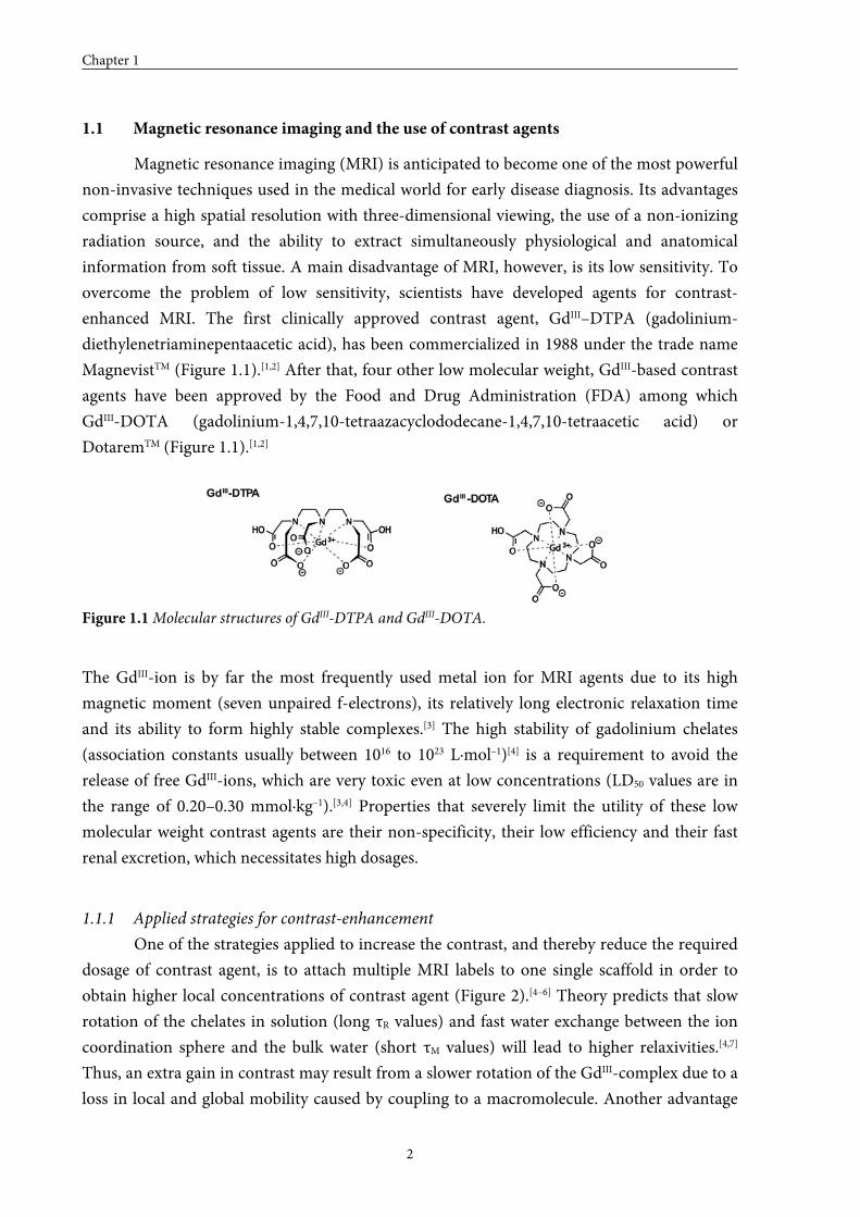
1.1 Magnetic resonance imaging and the use of contrast agents
Magnetic resonance imaging (MRI) is anticipated to become one of the most powerful
non-invasive techniques used in the medical world for early disease diagnosis. Its advantages
comprise a high spatial resolution with three-dimensional viewing, the use of a non-ionizing
radiation source, and the ability to extract simultaneously physiological and anatomical
information from soft tissue. A main disadvantage of MRI, however, is its low sensitivity. To
overcome the problem of low sensitivity, scientists have developed agents for contrast-
enhanced MRI. The first clinically approved contrast agent, GdIII–DTPA (gadolinium-
diethylenetriaminepentaacetic acid), has been commercialized in 1988 under the trade name
MagnevistTM (Figure 1.1).[1,2] After that, four other low molecular weight, GdIII-based contrast
agents have been approved by the Food and Drug Administration (FDA) among which
GdIII-DOTA (gadolinium-1,4,7,10-tetraazacyclododecane-1,4,7,10-tetraacetic acid) or
DotaremTM (Figure 1.1).[1,2]
Figure 1.1 Molecular structures of GdIII-DTPA and GdIII-DOTA.
The GdIII-ion is by far the most frequently used metal ion for MRI agents due to its high
magnetic moment (seven unpaired f-electrons), its relatively long electronic relaxation time
and its ability to form highly stable complexes.[3] The high stability of gadolinium chelates
(association constants usually between 1016 to 1023 L·mol–1)[4] is a requirement to avoid the
release of free GdIII-ions, which are very toxic even at low concentrations (LD50 values are in
the range of 0.20–0.30 mmol·kg–1).[3,4] Properties that severely limit the utility of these low
molecular weight contrast agents are their non-specificity, their low efficiency and their fast
renal excretion, which necessitates high dosages.
1.1.1 Applied strategies for contrast-enhancement
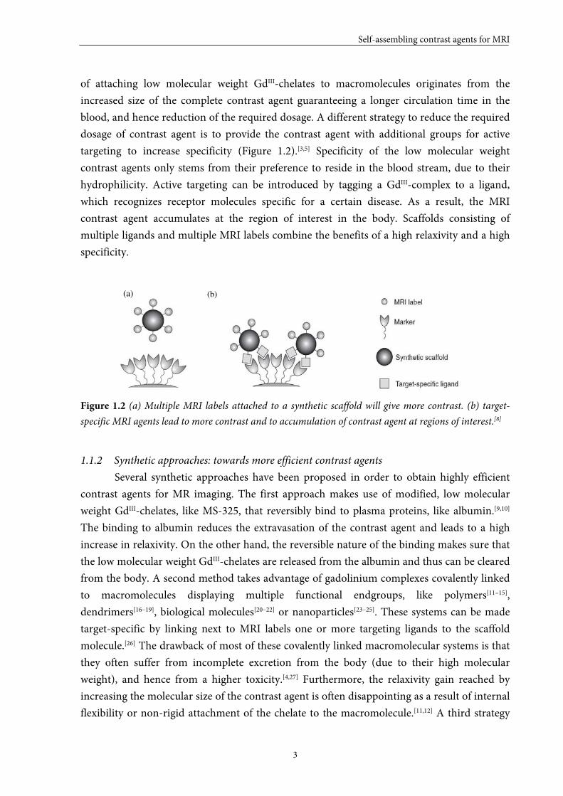
One of the strategies applied to increase the contrast, and thereby reduce the required
dosage of contrast agent, is to attach multiple MRI labels to one single scaffold in order to
obtain higher local concentrations of contrast agent (Figure 2).[4–6] Theory predicts that slow
rotation of the chelates in solution (long τR values) and fast water exchange between the ion
coordination sphere and the bulk water (short τM values) will lead to higher relaxivities.[4,7]
Thus, an extra gain in contrast may result from a slower rotation of the GdIII-complex due to a
loss in local and global mobility caused by coupling to a macromolecule. Another advantage
Self-assembling contrast agents for MRI
3
of attaching low molecular weight GdIII-chelates to macromolecules originates from the
increased size of the complete contrast agent guaranteeing a longer circulation time in the
blood, and hence reduction of the required dosage. A different strategy to reduce the required
dosage of contrast agent is to provide the contrast agent with additional groups for active
targeting to increase specificity (Figure 1.2).[3,5] Specificity of the low molecular weight
contrast agents only stems from their preference to reside in the blood stream, due to their
hydrophilicity. Active targeting can be introduced by tagging a GdIII-complex to a ligand,
which recognizes receptor molecules specific for a certain disease. As a result, the MRI
contrast agent accumulates at the region of interest in the body. Scaffolds consisting of
multiple ligands and multiple MRI labels combine the benefits of a high relaxivity and a high
specificity.
(a)
Figure 1.2 (a) Multiple MRI labels attached to a synthetic scaffold will give more contrast. (b) target-
specific MRI agents lead to more contrast and to accumulation of contrast agent at regions of interest.[8]
1.1.2 Synthetic approaches: towards more efficient contrast agents
Several synthetic approaches have been proposed in order to obtain highly efficient
contrast agents for MR imaging. The first approach makes use of modified, low molecular
weight GdIII-chelates, like MS-325, that reversibly bind to plasma proteins, like albumin.[9,10]
The binding to albumin reduces the extravasation of the contrast agent and leads to a high
increase in relaxivity. On the other hand, the reversible nature of the binding makes sure that
the low molecular weight GdIII-chelates are released from the albumin and thus can be cleared
from the body. A second method takes advantage of gadolinium complexes covalently linked
to macromolecules displaying multiple functional endgroups, like polymers[11–15],
dendrimers[16–19], biological molecules[20–22] or nanoparticles[23–25]. These systems can be made
target-specific by linking next to MRI labels one or more targeting ligands to the scaffold
molecule.[26] The drawback of most of these covalently linked macromolecular systems is that
they often suffer from incomplete excretion from the body (due to their high molecular
weight), and hence from a higher toxicity.[4,27] Furthermore, the relaxivity gain reached by
increasing the molecular size of the contrast agent is often disappointing as a result of internal
flexibility or non-rigid attachment of the chelate to the macromolecule.[11,12] A third strategy
(b)
Chapter 1
4
employs supramolecular systems like liposomes and micelles. Several studies describe the
encapsulation of MRI labels into liposome vesicles[28] or its immobilization into micelles[29–32]
or the liposome membrane[33–36]. These systems display easily controlled properties and good
pharmacological characteristics. Furthermore, it has been shown that supramolecular contrast
agents can be made target-specific by mixing in peptide amphiphiles displaying a certain
bioactive peptide sequence.[37–44] Another advantage of micellar systems is their reversible
nature, which allows combining the benefits of both high and low molecular weight contrast
agents, namely a high relaxivity and complete excretion from the body, respectively. This
unique set of properties has drawn the attention of many researchers. As a consequence, there
are many detailed investigations known concerning the use of paramagnetic amphiphiles.
So far, research in the area of self-assembling contrast agents has focused on the use of
linear paramagnetic amphiphiles. These amphiphiles consist of a hydrophilic gadolinium
containing head group and a hydrophobic alkyl tail and mainly form spherical assemblies,
such as micelles and liposomes, in solution. The linear amphiphiles that have been designed
by Hartgerink et al.[45–47] are an exception to this rule because these amphiphiles can self-
assemble into micellar rods and thus form columnar objects in solution. These amphiphiles,
called peptide amphiphiles, contain a hydrophilic peptide head group and a hydrophobic alkyl
tail. Gadolinium chelates have been linked to these peptide amphiphiles to create columnar
self-assembling contrast agents for MRI.[48,49]
The use of paramagnetic discotic amphiphiles has not been explored yet. Discotic
amphiphiles are a special class of amphiphiles that self-assemble into columnar structures.
Columnar self-assembling contrast agents based on discotic amphiphiles have some
advantages compared to spherical assemblies based on linear amphiphiles; firstly, they are able
to become very long, hence ensuring high local concentrations of gadolinium, and secondly,
the discotic amphiphiles making up the columns have a multivalent character, which leads to
higher local concentrations of contrast agent and opens possibilities for the use of asymmetric
target-specific discotics. Furthermore, the self-assembly of discotic molecules—C3-
symmetrical (C3)-discotics in particular—has been well-studied. C3-discotics have exerted
special attraction, since their symmetry often promotes the formation of helical aggregates, is
advantageous in synthesis design and rules out the importance of rotational order within the
assemblies. Most of the self-assembly studies currently reported have been performed in
apolar media, but the few studies describing the self-assembly of discotic amphiphiles in water
show similar characteristics. Synthesizing paramagnetic discotic amphiphiles might, therefore,
be an attractive strategy for creating a new class of self-assembling contrast agents. On the
other hand, if these self-assembling contrast agents are available, it would also be interesting
to investigate whether relaxation time measurements can be used as an extra tool to study the
self-assembly process of these discotic amphiphiles.
Self-assembling contrast agents for MRI
5
In the following sections, the relaxivities of currently reported self-assembling contrast
agents based on paramagnetic linear amphiphiles will be dealt with, and the fascinating self-
assembly behaviour of C3-symmetrical discotics in apolar media and in water will be
highlighted. Finally, the use of longitudinal relaxation time measurements as a tool to study
self-assembly processes will be briefly addressed.
1.2 Self-assembling paramagnetic amphiphiles for MR imaging
Paramagnetic amphiphiles are able to self-assemble into well-defined architectures,
which ideally exhibit four valuable properties: i) a high payload of GdIII-chelates, ii) an
enhanced ability to increase solvent proton relaxation rates due to a long molecular
reorientation time (τR), iii) an increased residence time in the blood by retarding the
extravasation that is typical for the small-sized GdIII-complexes commonly employed in MRI,
and/or iv) a dynamic character to ensure that the low molecular weight amphiphiles will
eventually be released and excreted from the body. A variety of GdIII-based micellar and
liposomal systems built up from paramagnetic linear amphiphiles has been designed and
characterized, and it has been shown that the longitudinal proton relaxivities of the aggregated
species are indeed considerably improved owing to the longer tumbling times of the GdIII-
complexes.[29–36]
The longitudinal 1H relaxation rate R1 is equal to the inverse of the longitudinal
relaxation time T1 (equation (E1)). For systems without a critical concentration (or a very low
critical concentration such as liposomes) equation (E2) holds, in which R1d is the diamagnetic
contribution (the relaxation rate of pure water), r1 represents the ionic relaxivity of the GdIII-
chelate (in mM–1s–1), and cGd is the GdIII-concentration (in mM).
11
1
TR = (E1)
Gddobs
crRR ×=− 111 (E2)
Gddobs
crRR ×=− a. n.111 (E3)
( )cmccrcmcrRR Gdadobs −+×=− 1
a. n.111 (E4)
For micelles, equations (E3) and (E4) can be derived. Equation (E3) describes the water 1H
relaxation rate in solutions below the cmc, in which r1n.a. represents the ionic relaxivity of non-
aggregated GdIII-chelate (in mM–1s–1). At concentrations greater than the cmc, the measured
relaxation rate is the sum of two contributions, one caused by the free monomer chelate
Chapter 1
6
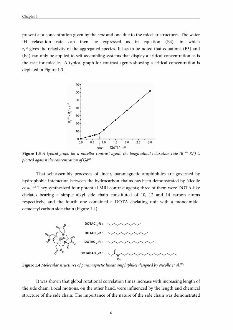
present at a concentration given by the cmc and one due to the micellar structures. The water 1H relaxation rate can then be expressed as in equation (E4), in which
r1 a gives the relaxivity of the aggregated species. It has to be noted that equations (E3) and
(E4) can only be applied to self-assembling systems that display a critical concentration as is
the case for micelles. A typical graph for contrast agents showing a critical concentration is
depicted in Figure 1.3.
Figure 1.3 A typical graph for a micellar contrast agent; the longitudinal relaxation rate (R1obs-R1d) is
plotted against the concentration of GdIII.
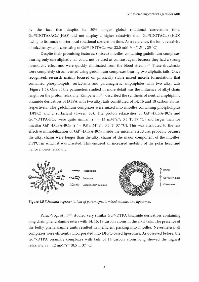
That self-assembly processes of linear, paramagnetic amphiphiles are governed by
hydrophobic interaction between the hydrocarbon chains has been demonstrated by Nicolle
et al.[50] They synthesized four potential MRI contrast agents; three of them were DOTA-like
chelates bearing a simple alkyl side chain constituted of 10, 12 and 14 carbon atoms
respectively, and the fourth one contained a DOTA chelating unit with a monoamide-
octadecyl carbon side chain (Figure 1.4).
Figure 1.4 Molecular structures of paramagnetic linear amphiphiles designed by Nicolle et al.[50]
It was shown that global rotational correlation times increase with increasing length of
the side chain. Local motions, on the other hand, were influenced by the length and chemical
structure of the side chain. The importance of the nature of the side chain was demonstrated
Self-assembling contrast agents for MRI
7
by the fact that despite its 30% longer global rotational correlation time,
GdIII(DOTASAC18)(H2O) did not display a higher relaxivity than GdIII(DOTAC14)-(H2O)
owing to its much shorter local rotational correlation time. As a reference, the ionic relaxivity
of micellar systems consisting of GdIII-DOTAC14 was 22.0 mM–1s–1 (1.5 T, 25 °C).
Despite their promising features, (mixed) micelles containing gadolinium complexes
bearing only one aliphatic tail could not be used as contrast agent because they had a strong
haemolytic effect and were quickly eliminated from the blood stream.[32] These drawbacks
were completely circumvented using gadolinium complexes bearing two aliphatic tails. Once
recognized, research mainly focused on physically stable mixed micelle formulations that
contained phospholipids, surfactants and paramagnetic amphiphiles with two alkyl tails
(Figure 1.5). One of the parameters studied in more detail was the influence of alkyl chain
length on the proton relaxivity. Kimpe et al.[51] described the synthesis of neutral amphiphilic
bisamide derivatives of DTPA with two alkyl tails constituted of 14, 16 and 18 carbon atoms,
respectively. The gadolinium complexes were mixed into micelles containing phospholipids
(DPPC) and a surfactant (Tween 80). The proton relaxivities of GdIII-DTPA-BC14 and
GdIII-DTPA-BC16 were quite similar (r1a = 13 mM–1s–1; 0.5 T, 37 °C) and larger than for
micellar GdIII-DTPA-BC18 (r1a = 9.8 mM–1s–1; 0.5 T, 37 °C). This was attributed to the less
effective immobilization of GdIII-DTPA-BC18 inside the micellar structure, probably because
the alkyl chains were longer than the alkyl chains of the major component of the micelles,
DPPC, in which it was inserted. This ensured an increased mobility of the polar head and
hence a lower relaxivity.
Figure 1.5 Schematic representations of paramagnetic mixed micelles and liposomes.
Parac-Vogt et al.[35] studied very similar GdIII-DTPA bisamide derivatives containing
long chain phenylalanine esters with 14, 16, 18 carbon atoms in the alkyl tails. The presence of
the bulky phenylalanine units resulted in inefficient packing into micelles. Nevertheless, all
complexes were efficiently incorporated into DPPC-based liposomes. As observed before, the
GdIII-DTPA bisamide complexes with tails of 14 carbon atoms long showed the highest
relaxivity, r1 = 12 mM–1s–1 (0.5 T, 37 °C).
Chapter 1
8
In the case of liposomes, the proton relaxivity could also be limited by a slow water
exchange rate between the liposome interior and exterior as was illustrated by Strijkers et al.[36]
They showed that unsaturated DOPC-based liposomes displayed a higher relaxivity compared
to saturated DSPC-based liposomes, and they attributed this effect to a less rigid DOPC
membrane that was more permeable to water. They also demonstrated that cholesterol was
needed in their system to obtain monodisperse unilamellar liposomes (Figure 1.5). The
highest relaxivity per GdIII-ion of 11.3 mM–1s–1 (0.625 T, 37 °C) was obtained for DOPC based
liposomes containing cholesterol. Gløgard et al.[33] also showed that DMPC-based liposomes,
which were in the liquid crystalline state, showed a higher relaxivity compared to DSPC-based
membranes that were in the solid gel state, which indicated that the relaxivity was again
exchange limited. They used fractional factorial design to find the liposome composition with
the highest relaxivity. The highest relaxivity, r1 = 52.0 mM–1s–1, was obtained in the region
with no cholesterol and low content of GdIII-chelate. However, it is questionable whether
liposomes without any cholesterol are stable monodisperse unilamellar liposomes. The same
authors also reported NMRD-profiles for DMPC/DMPG liposomes containing 5.5% GdIII-
HHD-DO3A and 20% cholesterol, which possessed an observed relaxivity of
43 mM–1s–1 (0.5 T, 39 °C).
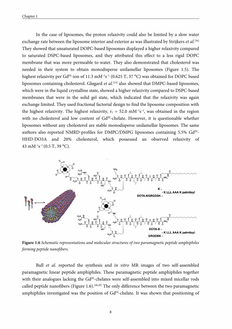
Figure 1.6 Schematic representations and molecular structures of two paramagnetic peptide amphiphiles
forming peptide nanofibers.
Bull et al. reported the synthesis and in vitro MR images of two self-assembled
paramagnetic linear peptide amphiphiles. These paramagnetic peptide amphiphiles together
with their analogues lacking the GdIII-chelates were self-assembled into mixed micellar rods
called peptide nanofibers (Figure 1.6).[48,49] The only difference between the two paramagnetic
amphiphiles investigated was the position of GdIII-chelate. It was shown that positioning of

Self-assembling contrast agents for MRI
9
the GdIII-chelate close to the hydrophobic tail of the molecule resulted in a higher relaxivity.
The ionic relaxivity of self-assembled amphiphiles displaying the chelator at the end of the
peptide fragment was 14.7 mM–1s–1 (1.5 T, 37 °C), while the ionic relaxivity of the self-
assembled amphiphiles having the chelator close to the alkyl tail equalled 21.5 mM–1s–1.
So far, the highest relaxivity per GdIII-ion was obtained by using liposomes (highest
relaxivity reported 43 mM–1s–1)[33] rather than mixed micelles (highest relaxivity reported
29 mM–1s–1)[31] or peptide nanofibers (highest relaxivity reported 21.5 mM–1s–1)[48]. However, it
should be noted that the range of reported relaxivities was rather broad (liposomes
r1 ≈ 5–43 mM–1s–1 and (mixed) micelles r1a ≈ 12–29 mM–1s–1)[25]. The field strengths and
temperatures at which these measurements have been performed are not identical so it is not
quite possible to compare these systems, but it gives a first indication. To judge whether the
observed proton relaxivities of self-assembling paramagnetic discotic amphiphiles are
promising, the proton relaxivities for self-assembled paramagnetic linear amphiphiles will be
used as a reference.
1.3 Self-assembly behaviour of C3-symmetrical discotics
The self-assembly of discotic molecules is studied in detail in the liquid crystalline
state, in the gel state and in solution. In general, C3-discotics are more thoroughly studied
compared to asymmetric discotics, since their symmetry offers some advantages; it is
compatible with chirality, it is advantageous in the synthetic design and, in many cases, it rules
out the importance of rotational order within the column. Most of the studies dealing with the
self-assembly of C3-discotics have been performed in apolar media, but the few studies
describing the self-assembly of discotic amphiphiles in water show similar characteristics,
which suggest that paramagnetic discotic amphiphiles may be applicable as self-assembling
contrast agents for MRI.
1.3.1 Self-assembly of discotic molecules in apolar media
In the 1990s, C3-symmetrical discotics based on cis-cis-1,3,5-cyclohexane-
tricarboxamide or 1,3,5-benzenetricarboxamide cores started to play an important role in the
area of nanoscience, because these molecules displayed a strong tendency to adopt appealing
conformations, like helices or propellers, as was demonstrated by their crystal structures.[52,53]
An important goal in this developing field was the identification of molecular subunits that
allow controlled formation of one-dimensional supramolecular constructs. New discotic
molecules were developed with the aim to investigate the effect of steric hindrance, pre-
orientation and the balance between secondary interactions on the stability of the aggregates.
Our laboratory started to investigate disc-like molecules that were built up by linking
three lipophilic, N-monoacetylated 2,2’-bipyridine-3,3’-diamine wedges to a central 1,3,5-
Chapter 1
10
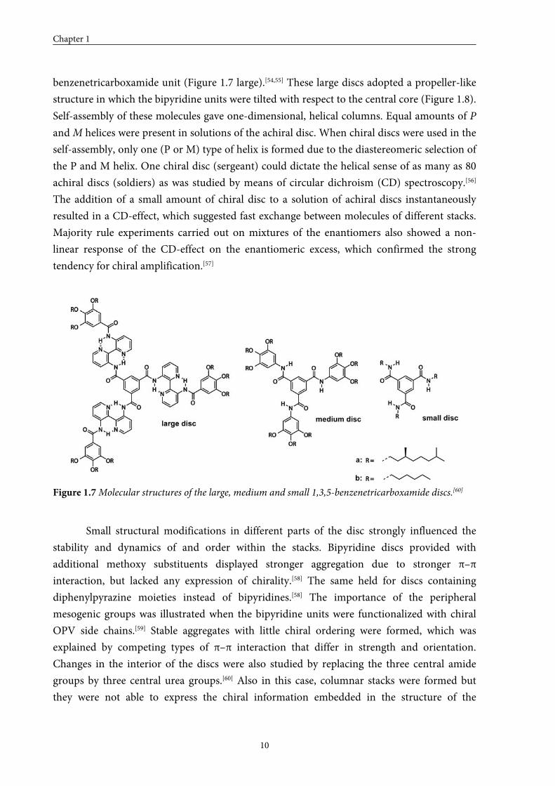
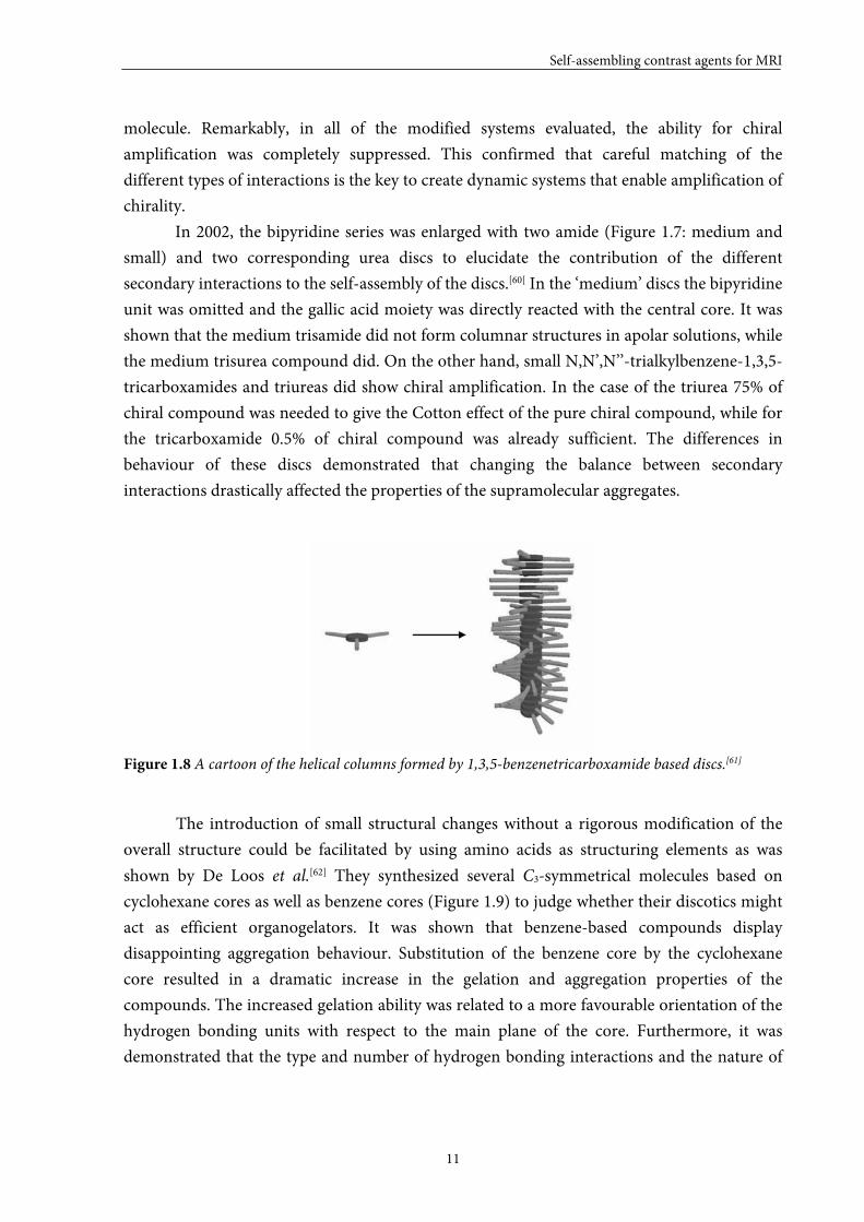
benzenetricarboxamide unit (Figure 1.7 large).[54,55] These large discs adopted a propeller-like
structure in which the bipyridine units were tilted with respect to the central core (Figure 1.8).
Self-assembly of these molecules gave one-dimensional, helical columns. Equal amounts of P
and M helices were present in solutions of the achiral disc. When chiral discs were used in the
self-assembly, only one (P or M) type of helix is formed due to the diastereomeric selection of
the P and M helix. One chiral disc (sergeant) could dictate the helical sense of as many as 80
achiral discs (soldiers) as was studied by means of circular dichroism (CD) spectroscopy.[56]
The addition of a small amount of chiral disc to a solution of achiral discs instantaneously
resulted in a CD-effect, which suggested fast exchange between molecules of different stacks.
Majority rule experiments carried out on mixtures of the enantiomers also showed a non-
linear response of the CD-effect on the enantiomeric excess, which confirmed the strong
tendency for chiral amplification.[57]
Figure 1.7 Molecular structures of the large, medium and small 1,3,5-benzenetricarboxamide discs.[60]
Small structural modifications in different parts of the disc strongly influenced the
stability and dynamics of and order within the stacks. Bipyridine discs provided with
additional methoxy substituents displayed stronger aggregation due to stronger π–π
interaction, but lacked any expression of chirality.[58] The same held for discs containing
diphenylpyrazine moieties instead of bipyridines.[58] The importance of the peripheral
mesogenic groups was illustrated when the bipyridine units were functionalized with chiral
OPV side chains.[59] Stable aggregates with little chiral ordering were formed, which was
explained by competing types of π–π interaction that differ in strength and orientation.
Changes in the interior of the discs were also studied by replacing the three central amide
groups by three central urea groups.[60[ Also in this case, columnar stacks were formed but
they were not able to express the chiral information embedded in the structure of the
Self-assembling contrast agents for MRI
11
molecule. Remarkably, in all of the modified systems evaluated, the ability for chiral
amplification was completely suppressed. This confirmed that careful matching of the
different types of interactions is the key to create dynamic systems that enable amplification of
chirality.
In 2002, the bipyridine series was enlarged with two amide (Figure 1.7: medium and
small) and two corresponding urea discs to elucidate the contribution of the different
secondary interactions to the self-assembly of the discs.[60[ In the ‘medium’ discs the bipyridine
unit was omitted and the gallic acid moiety was directly reacted with the central core. It was
shown that the medium trisamide did not form columnar structures in apolar solutions, while
the medium trisurea compound did. On the other hand, small N,N’,N’’-trialkylbenzene-1,3,5-
tricarboxamides and triureas did show chiral amplification. In the case of the triurea 75% of
chiral compound was needed to give the Cotton effect of the pure chiral compound, while for
the tricarboxamide 0.5% of chiral compound was already sufficient. The differences in
behaviour of these discs demonstrated that changing the balance between secondary
interactions drastically affected the properties of the supramolecular aggregates.
Figure 1.8 A cartoon of the helical columns formed by 1,3,5-benzenetricarboxamide based discs.[61]
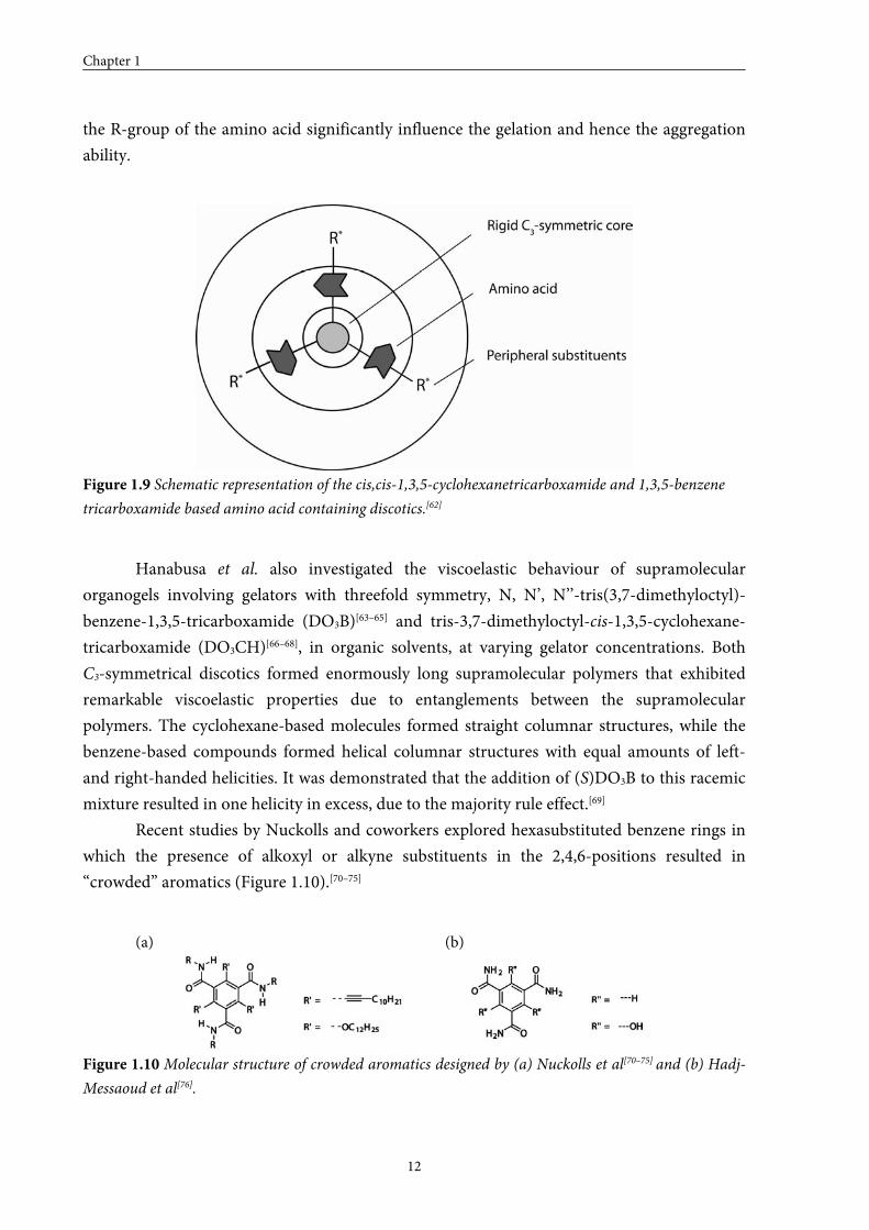
The introduction of small structural changes without a rigorous modification of the
overall structure could be facilitated by using amino acids as structuring elements as was
shown by De Loos et al.[62] They synthesized several C3-symmetrical molecules based on
cyclohexane cores as well as benzene cores (Figure 1.9) to judge whether their discotics might
act as efficient organogelators. It was shown that benzene-based compounds display
disappointing aggregation behaviour. Substitution of the benzene core by the cyclohexane
core resulted in a dramatic increase in the gelation and aggregation properties of the
compounds. The increased gelation ability was related to a more favourable orientation of the
hydrogen bonding units with respect to the main plane of the core. Furthermore, it was
demonstrated that the type and number of hydrogen bonding interactions and the nature of
Chapter 1
12
the R-group of the amino acid significantly influence the gelation and hence the aggregation
ability.
Figure 1.9 Schematic representation of the cis,cis-1,3,5-cyclohexanetricarboxamide and 1,3,5-benzene
tricarboxamide based amino acid containing discotics.[62]
Hanabusa et al. also investigated the viscoelastic behaviour of supramolecular
organogels involving gelators with threefold symmetry, N, N’, N’’-tris(3,7-dimethyloctyl)-
benzene-1,3,5-tricarboxamide (DO3B)[63–65] and tris-3,7-dimethyloctyl-cis-1,3,5-cyclohexane-
tricarboxamide (DO3CH)[66–68], in organic solvents, at varying gelator concentrations. Both
C3-symmetrical discotics formed enormously long supramolecular polymers that exhibited
remarkable viscoelastic properties due to entanglements between the supramolecular
polymers. The cyclohexane-based molecules formed straight columnar structures, while the
benzene-based compounds formed helical columnar structures with equal amounts of left-
and right-handed helicities. It was demonstrated that the addition of (S)DO3B to this racemic
mixture resulted in one helicity in excess, due to the majority rule effect.[69]
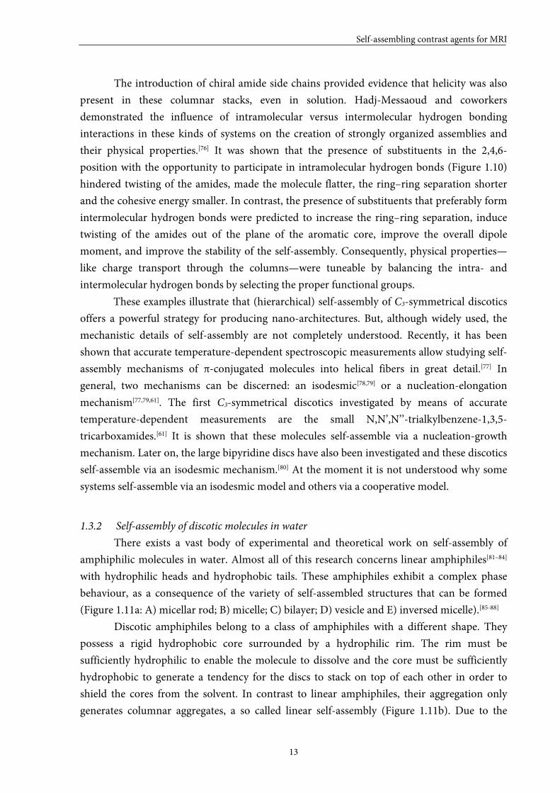
Recent studies by Nuckolls and coworkers explored hexasubstituted benzene rings in
which the presence of alkoxyl or alkyne substituents in the 2,4,6-positions resulted in
“crowded” aromatics (Figure 1.10).[70–75]
(a) (b)
Figure 1.10 Molecular structure of crowded aromatics designed by (a) Nuckolls et al[70–75] and (b) Hadj-
Messaoud et al[76].
Self-assembling contrast agents for MRI
13
The introduction of chiral amide side chains provided evidence that helicity was also
present in these columnar stacks, even in solution. Hadj-Messaoud and coworkers
demonstrated the influence of intramolecular versus intermolecular hydrogen bonding
interactions in these kinds of systems on the creation of strongly organized assemblies and
their physical properties.[76] It was shown that the presence of substituents in the 2,4,6-
position with the opportunity to participate in intramolecular hydrogen bonds (Figure 1.10)
hindered twisting of the amides, made the molecule flatter, the ring–ring separation shorter
and the cohesive energy smaller. In contrast, the presence of substituents that preferably form
intermolecular hydrogen bonds were predicted to increase the ring–ring separation, induce
twisting of the amides out of the plane of the aromatic core, improve the overall dipole
moment, and improve the stability of the self-assembly. Consequently, physical properties—
like charge transport through the columns—were tuneable by balancing the intra- and
intermolecular hydrogen bonds by selecting the proper functional groups.
These examples illustrate that (hierarchical) self-assembly of C3-symmetrical discotics
offers a powerful strategy for producing nano-architectures. But, although widely used, the
mechanistic details of self-assembly are not completely understood. Recently, it has been
shown that accurate temperature-dependent spectroscopic measurements allow studying self-
assembly mechanisms of π-conjugated molecules into helical fibers in great detail.[77] In
general, two mechanisms can be discerned: an isodesmic[78,79] or a nucleation-elongation
mechanism[77,79,61]. The first C3-symmetrical discotics investigated by means of accurate
temperature-dependent measurements are the small N,N’,N’’-trialkylbenzene-1,3,5-
tricarboxamides.[61] It is shown that these molecules self-assemble via a nucleation-growth
mechanism. Later on, the large bipyridine discs have also been investigated and these discotics
self-assemble via an isodesmic mechanism.[80] At the moment it is not understood why some
systems self-assemble via an isodesmic model and others via a cooperative model.
1.3.2 Self-assembly of discotic molecules in water
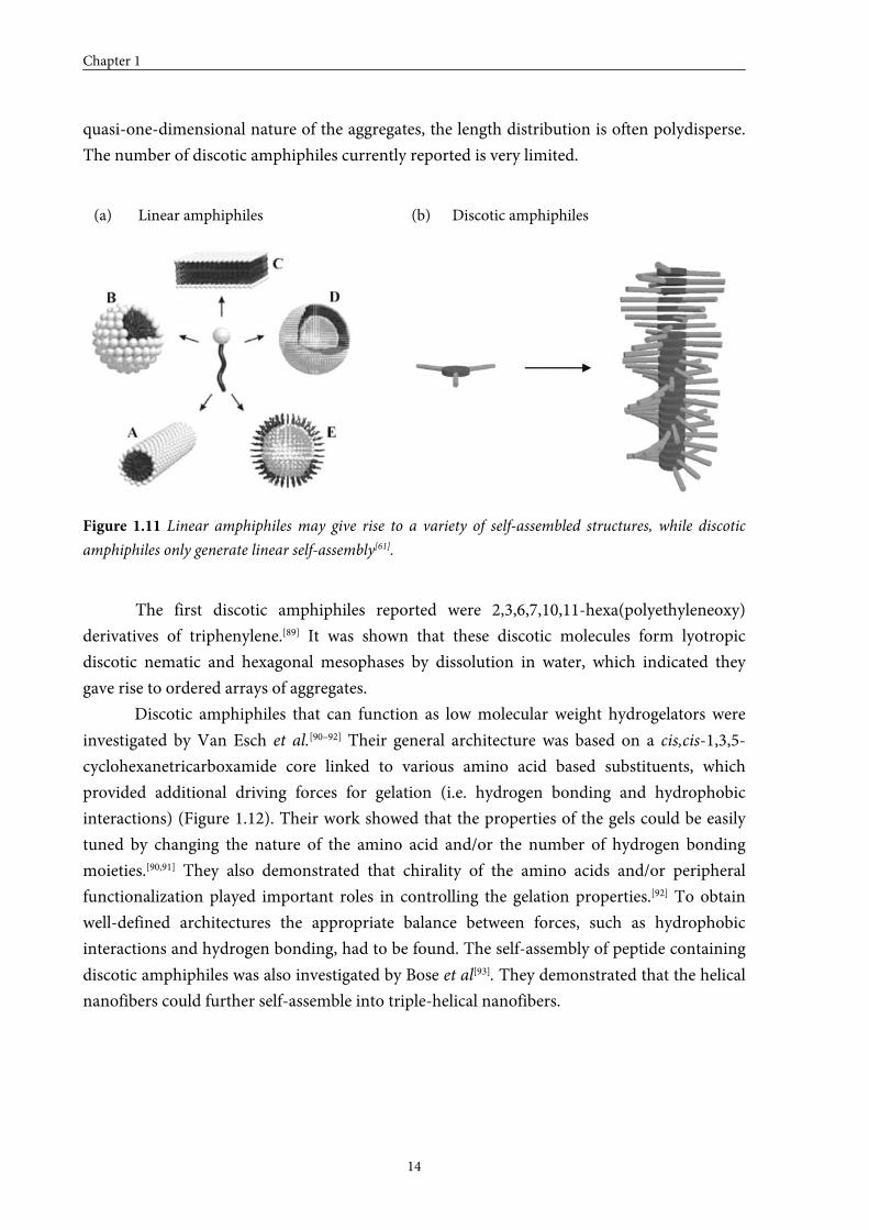
There exists a vast body of experimental and theoretical work on self-assembly of
amphiphilic molecules in water. Almost all of this research concerns linear amphiphiles[81–84]
with hydrophilic heads and hydrophobic tails. These amphiphiles exhibit a complex phase
behaviour, as a consequence of the variety of self-assembled structures that can be formed
(Figure 1.11a: A) micellar rod; B) micelle; C) bilayer; D) vesicle and E) inversed micelle).[85-88]
Discotic amphiphiles belong to a class of amphiphiles with a different shape. They
possess a rigid hydrophobic core surrounded by a hydrophilic rim. The rim must be
sufficiently hydrophilic to enable the molecule to dissolve and the core must be sufficiently
hydrophobic to generate a tendency for the discs to stack on top of each other in order to
shield the cores from the solvent. In contrast to linear amphiphiles, their aggregation only
generates columnar aggregates, a so called linear self-assembly (Figure 1.11b). Due to the
Chapter 1
14
quasi-one-dimensional nature of the aggregates, the length distribution is often polydisperse.
The number of discotic amphiphiles currently reported is very limited.
(a) Linear amphiphiles (b) Discotic amphiphiles
Figure 1.11 Linear amphiphiles may give rise to a variety of self-assembled structures, while discotic
amphiphiles only generate linear self-assembly[61].
The first discotic amphiphiles reported were 2,3,6,7,10,11-hexa(polyethyleneoxy)
derivatives of triphenylene.[89] It was shown that these discotic molecules form lyotropic
discotic nematic and hexagonal mesophases by dissolution in water, which indicated they
gave rise to ordered arrays of aggregates.
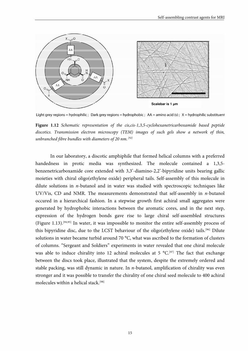
Discotic amphiphiles that can function as low molecular weight hydrogelators were
investigated by Van Esch et al.[90–92] Their general architecture was based on a cis,cis-1,3,5-
cyclohexanetricarboxamide core linked to various amino acid based substituents, which
provided additional driving forces for gelation (i.e. hydrogen bonding and hydrophobic
interactions) (Figure 1.12). Their work showed that the properties of the gels could be easily
tuned by changing the nature of the amino acid and/or the number of hydrogen bonding
moieties.[90,91] They also demonstrated that chirality of the amino acids and/or peripheral
functionalization played important roles in controlling the gelation properties.[92] To obtain
well-defined architectures the appropriate balance between forces, such as hydrophobic
interactions and hydrogen bonding, had to be found. The self-assembly of peptide containing
discotic amphiphiles was also investigated by Bose et al[93]. They demonstrated that the helical
nanofibers could further self-assemble into triple-helical nanofibers.
Self-assembling contrast agents for MRI
15
Scalebar is 1 µm
Figure 1.12 Schematic representation of the cis,cis-1,3,5-cyclohexanetricarboxamide based peptide
discotics. Transmission electron microscopy (TEM) images of such gels show a network of thin,
unbranched fibre bundles with diameters of 20 nm. [92]
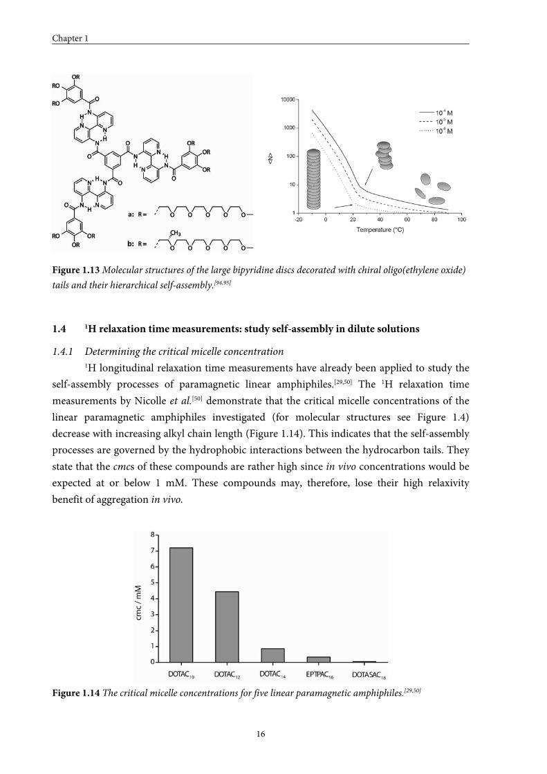
In our laboratory, a discotic amphiphile that formed helical columns with a preferred
handedness in protic media was synthesized. The molecule contained a 1,3,5-
benzenetricarboxamide core extended with 3,3’-diamino-2,2’-bipyridine units bearing gallic
moieties with chiral oligo(ethylene oxide) peripheral tails. Self-assembly of this molecule in
dilute solutions in n-butanol and in water was studied with spectroscopic techniques like
UV/Vis, CD and NMR. The measurements demonstrated that self-assembly in n-butanol
occured in a hierarchical fashion. In a stepwise growth first achiral small aggregates were
generated by hydrophobic interactions between the aromatic cores, and in the next step,
expression of the hydrogen bonds gave rise to large chiral self-assembled structures
(Figure 1.13).[94,95] In water, it was impossible to monitor the entire self-assembly process of
this bipyridine disc, due to the LCST behaviour of the oligo(ethylene oxide) tails.[96] Dilute
solutions in water became turbid around 70 °C, what was ascribed to the formation of clusters
of columns. “Sergeant and Soldiers” experiments in water revealed that one chiral molecule
was able to induce chirality into 12 achiral molecules at 5 °C.[97] The fact that exchange
between the discs took place, illustrated that the system, despite the extremely ordered and
stable packing, was still dynamic in nature. In n-butanol, amplification of chirality was even
stronger and it was possible to transfer the chirality of one chiral seed molecule to 400 achiral
molecules within a helical stack.[98]
Chapter 1
16
Figure 1.13 Molecular structures of the large bipyridine discs decorated with chiral oligo(ethylene oxide)
tails and their hierarchical self-assembly.[94,95]
1.4 1H relaxation time measurements: study self-assembly in dilute solutions
1.4.1 Determining the critical micelle concentration
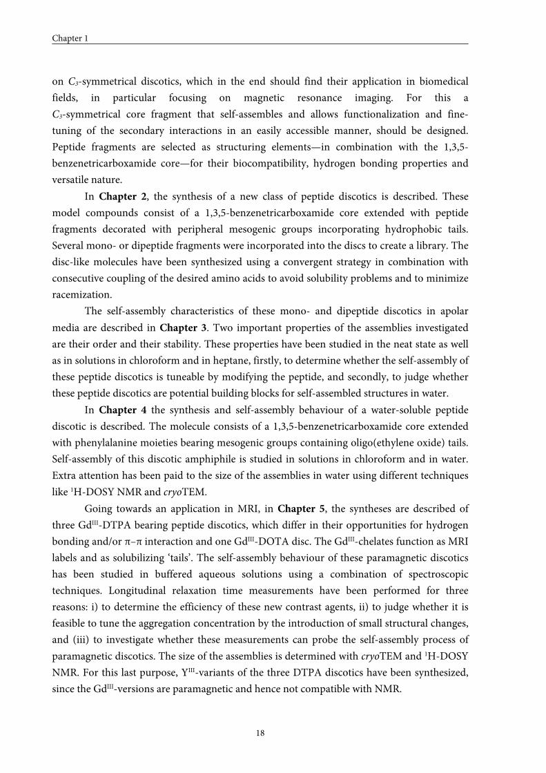
1H longitudinal relaxation time measurements have already been applied to study the
self-assembly processes of paramagnetic linear amphiphiles.[29,50] The 1H relaxation time
measurements by Nicolle et al.[50] demonstrate that the critical micelle concentrations of the
linear paramagnetic amphiphiles investigated (for molecular structures see Figure 1.4)
decrease with increasing alkyl chain length (Figure 1.14). This indicates that the self-assembly
processes are governed by the hydrophobic interactions between the hydrocarbon tails. They
state that the cmcs of these compounds are rather high since in vivo concentrations would be
expected at or below 1 mM. These compounds may, therefore, lose their high relaxivity
benefit of aggregation in vivo.
Figure 1.14 The critical micelle concentrations for five linear paramagnetic amphiphiles.[29,50]
Self-assembling contrast agents for MRI
17
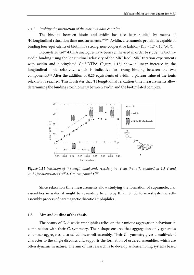
1.4.2 Probing the interaction of the biotin–avidin complex
The binding between biotin and avidin has also been studied by means of 1H longitudinal relaxation time measurements.[99,100] Avidin, a tetrameric protein, is capable of
binding four equivalents of biotin in a strong, non-cooperative fashion (Kass ≈ 1.7 × 10¹⁵ M–1).
Biotinylated GdIII-DTPA analogues have been synthesized in order to study the biotin–
avidin binding using the longitudinal relaxivity of the MRI label. MRI titration experiments
with avidin and biotinylated GdIII-DTPA (Figure 1.15) show a linear increase in the
longitudinal ionic relaxivity, which is indicative for strong binding between the two
components.[99] After the addition of 0.25 equivalents of avidin, a plateau value of the ionic
relaxivity is reached. This illustrates that 1H longitudinal relaxation time measurements allow
determining the binding stoichiometry between avidin and the biotinylated complex.
Figure 1.15 Variation of the longitudinal ionic relaxivity r1 versus the ratio avidin/1 at 1.5 T and
25 °C for biotinylated GdIII-DTPA compound 1.[99]
Since relaxation time measurements allow studying the formation of supramolecular
assemblies in water, it might be rewarding to employ this method to investigate the self-
assembly process of paramagnetic discotic amphiphiles.
1.5 Aim and outline of the thesis
The beauty of C3-discotic amphiphiles relies on their unique aggregation behaviour in
combination with their C3-symmetry. Their shape ensures that aggregation only generates
columnar aggregates, a so called linear self-assembly. Their C3-symmetry gives a multivalent
character to the single discotics and supports the formation of ordered assemblies, which are
often dynamic in nature. The aim of this research is to develop self-assembling systems based
Chapter 1
18
on C3-symmetrical discotics, which in the end should find their application in biomedical
fields, in particular focusing on magnetic resonance imaging. For this a
C3-symmetrical core fragment that self-assembles and allows functionalization and fine-
tuning of the secondary interactions in an easily accessible manner, should be designed.
Peptide fragments are selected as structuring elements—in combination with the 1,3,5-
benzenetricarboxamide core—for their biocompatibility, hydrogen bonding properties and
versatile nature.
In Chapter 2, the synthesis of a new class of peptide discotics is described. These
model compounds consist of a 1,3,5-benzenetricarboxamide core extended with peptide
fragments decorated with peripheral mesogenic groups incorporating hydrophobic tails.
Several mono- or dipeptide fragments were incorporated into the discs to create a library. The
disc-like molecules have been synthesized using a convergent strategy in combination with
consecutive coupling of the desired amino acids to avoid solubility problems and to minimize
racemization.
The self-assembly characteristics of these mono- and dipeptide discotics in apolar
media are described in Chapter 3. Two important properties of the assemblies investigated
are their order and their stability. These properties have been studied in the neat state as well
as in solutions in chloroform and in heptane, firstly, to determine whether the self-assembly of
these peptide discotics is tuneable by modifying the peptide, and secondly, to judge whether
these peptide discotics are potential building blocks for self-assembled structures in water.
In Chapter 4 the synthesis and self-assembly behaviour of a water-soluble peptide
discotic is described. The molecule consists of a 1,3,5-benzenetricarboxamide core extended
with phenylalanine moieties bearing mesogenic groups containing oligo(ethylene oxide) tails.
Self-assembly of this discotic amphiphile is studied in solutions in chloroform and in water.
Extra attention has been paid to the size of the assemblies in water using different techniques
like 1H-DOSY NMR and cryoTEM.
Going towards an application in MRI, in Chapter 5, the syntheses are described of
three GdIII-DTPA bearing peptide discotics, which differ in their opportunities for hydrogen
bonding and/or π–π interaction and one GdIII-DOTA disc. The GdIII-chelates function as MRI
labels and as solubilizing ‘tails’. The self-assembly behaviour of these paramagnetic discotics
has been studied in buffered aqueous solutions using a combination of spectroscopic
techniques. Longitudinal relaxation time measurements have been performed for three
reasons: i) to determine the efficiency of these new contrast agents, ii) to judge whether it is
feasible to tune the aggregation concentration by the introduction of small structural changes,
and (iii) to investigate whether these measurements can probe the self-assembly process of
paramagnetic discotics. The size of the assemblies is determined with cryoTEM and 1H-DOSY
NMR. For this last purpose, YIII-variants of the three DTPA discotics have been synthesized,
since the GdIII-versions are paramagnetic and hence not compatible with NMR.
Self-assembling contrast agents for MRI
19
1.6 References and notes
[1] S-P. Lin, J.J. Brown, J. Magn. Res. Imaging 2007, 25, 884–899.
[2] S. Laurent, L. vander Elst, R.N. Muller, Contrast Med. Mol. Imaging, 2006, 1 128–137.
[3] R.B. Lauffer, Chem. Rev. 1987, 87, 901–927.
[4] P. Caravan, J.J. Ellison, T.J. McMurry, R.B. Lauffer, Chem. Rev. 1999, 99, 2293–2352.
[5] V. Jacques, J.F. Desreux, Top. Curr. Chem. 2002, 221, 123–164.
[6] T. Barrett, H. Kobayashi, M. Brechbiel, P.L. Choyke, Eur. J. Radiol. 2006, 60, 353–366.
[7] É. Tóth, L. Helm, A.E. Merbach, Top. Curr. Chem. 2002, 221, 61–101.
[8] S. Langereis, Thesis: Dendritic MRI contrast agents: Synthetic strategies for targeting and multivalency;
Eindhoven University of Technology: Eindhoven (The Netherlands), 2005.
[9] R.B. Lauffer, D.J. Parmelee, S. Ouellet, R.P. Dolan, H. Sajiki, M. Scott, P.J. Bernard, E.M. Buchanan, K.
Y. Ong, Z. Tyeklár, K.S. Midelfort, T.J. McMurry, R.C. Walovitch, Acad. Radiol. 1996, 3, 356–358.
[10] P. Caravan, N.J. Cloutier, M.T. Greenfield, S.A. McDermid, S.U. Dunham, J.W.M. Bulte, J.D. Amedio
Jr., R.J. Looby, R.M. Supkowski, W. DeW. Horrocks Jr., T.J. McMurry, R.B. Lauffer, J. Am. Chem. Soc.
2002, 124, 3152–3162.
[11] V.S. Vexler, O. Clement, H. Schmitt-Willich, R.C. Brasch, J. Magn. Res. Imaging 1994, 4, 381–388.
[12] T.S. Desser, L.D. Rubin, H.H. Muller, F. Qing, S. Khodor, G. Zanazzi, S.W. Young, D.L. Ladd, J.A.
Wellons, K.E. Kellar, J. Magn. Res. Imaging 1994, 4, 467–472.
[13] F. Kiessling, M. Heilmann, T. Lammers, K. Ulbrich, V. Subr, P. Peschke, B. Waengler, W. Mier, H-H.
Schrenk, M. Bock, L. Schad, W. Semmler, Bioconjugate Chem. 2006, 17, 42–51.
[14] M. Spanoghe, D. Lanens, R. Dommisse, A. van der Linden, F. Alderweireldt, Magn. Res. Imaging 1992,
10, 913–917.
[15] P. Rongved, J. Klaveness, Carbohydr. Res. 1991, 214, 315–323.
[16] E.C. Wiener, M.W. Brechbiel, H. Brothers, R.L. Magin, O.A. Gansow, A.D. Tomalia, P.C. Lauterbur,
Magn. Reson. Med. 1994, 31, 1–8.
[17] S. Langereis, Q.G. de Lussanet, M.H.P. van Genderen, W.H. Backes, E.W. Meijer, Macromolecules 2004,
37, 3084–3091.
[18] B. Misselwitz, H. Schmitt-Willich, W. Ebert, T. Frenzel, H-J. Weinmann, Magn. Reson. Mater. Phys.,
Biol. Med. 2001, 12, 128–134.
[19] D.A. Fulton, E.M. Elemento, S. Aime, L. Chaabane, M. Botta, D. Parker, Chem. Commun. 2006, 1064–
1066.
[20] J.M. Hooker, A. Datta, M. Botta, K.N. Raymond, M.B. Francis, Nano Lett. 2007, 7, 2207–2210.
[21] D.J. Hnatowich, W.W. Layne, R.L. Childs, D. Lanteigne, M.A. Davis, T.W. Griffin, P.W. Doherty,
Science 1983, 220, 613–615.
[22] R.B. Lauffer, T.J. Bardy, Magn. Res. Imaging 1985, 3, 11–16.
[23] P-J. Debouttière, S. Roux, F. Vocanson, C. Billotey, O. Beuf, A. Favre-Réguillon, S. Pellet-Rostaing, R.
Lamartine, P. Perriat, O. Tillement, Adv. Funct. Mater. 2006, 16, 2330–2339.
[24] W.J. Rieter, J.S. Kim, M.L. Taylor, H. An, W. Lin, T. Tarrant, W. Lin, Angew. Chem. 2007, 119, 3754–
3756; Angew. Chem. Int. Ed. 2007, 46, 3680–3682.
[25] W.J.M. Mulder, G.J. Strijkers, G.A.F. van Tilborg, A.W. Griffioen, K. Nicolay, NMR Biomed. 2006, 19,
142–164.
[26] S. Langereis, A. Dirksen, T.M. Hackeng, M.H.P. van Genderen, E.W. Meijer, New J. Chem. 2007, 31,
1152–1160.
[27] R.B. Clarkson, Top. Curr. Chem. 2002, 221, 201–235.
Chapter 1
20
[28] S. Aime, L. Frullano, S. Geninatti Crich, Angew. Chem. 2002, 114, 1059–1061; Angew. Chem., Int. Ed.
2002, 41, 1017–1019.
[29] S. Torres, J.A. Martins, J.P. André, C.F.G.C. Geraldes, A.E. Merbach, E. Tóth, Chem. Eur. J. 2006, 12,
940–948.
[30] J.P. André, É. Tóth, H. Fisher, A. Seelig, H.R. Mäcke, A.E. Merbach, Chem. Eur. J. 1999, 5, 2977–2983.
[31] H. Tournier, R. Hyacinthe, M. Schneider, Acad. Radiol. 2002, 9, S20–S28.
[32] P.L. Anelli, L. Lattuada, V. Lorusso, M. Schneider, H. Tournier, F. Uggeri, Magma 2001, 12, 114–120.
[33] C. GlØgård, G. Stensrud, R. Hovland, S.L. Fossheim, J. Klaveness, Int. J. Pharm. 2002, 233, 131–140.
[34] M. Vaccaro, A. Accardo, D. Tesauro, G. Mangiapia, D. Löf, K. Schillén, O. Södermann, G. Morelli, L.
Paduano, Langmuir 2006, 22, 6635–6643.
[35] T.N. Parac-Vogt, K. Kimpe, S. Laurent, C. Piérart, L. vander Elst, R.N. Muller, K. Binnemans, Eur.
Biophys. J. 2006, 35, 136–144.
[36] G.J. Strijkers, W.J.M. Mulder, R.B. van Heeswijk, P.M. Frederik, P. Bomans, P.C.M.M. Magusin, K.
Nicolay, Magma 2005, 18, 186–192.
[37] G.A.F. van Tilborg, W.J.M. Mulder, N. Deckers, G. Storm, C.P.M. Reutelingsperger, G.J. Strijkers, K.
Nicolay, Bioconjugate Chem. 2006, 17, 741–749.
[38] W.J.M. Mulder, G.J. Strijkers, A.W. Griffioen, L. van Bloois, G. Molema, G. Storm, G.A. Koning, K.
Nicolay, Bioconjugate Chem. 2004, 15, 799–806.
[39] D.A. Sipkins, K. Gijbels, F.D. Tropper, M. Bednarski, K.C.P. Li, L. Steinman, J. Neuroimmunology 2000,
104, 1–9.
[40] A. Accardo, D. Tesauro, G. Morelli, E. Gianolio, S. Aime, M. Vaccaro, G. Mangiapia, L. Paduano, K.
Schillén, J. Biol. Inorg. Chem. 2007, 12, 267–276.
[41] F. Alhaique, I. Bertini, M. Fragai, M. Carafa, C. Luchinat, G. Parigi, Inorg. Chim. Acta 2002, 331, 151–
157.
[42] D. Tesauro, A. Accardo, E. Gianolio, L. Paduano, J. Teixeira, K. Schillén, S. Aime, G. Morelli,
ChemBioChem. 2007, 8, 950–955.
[43] F. Leclercq, M. Cohen-Ohana, N. Mignet, A. Sbarbati, J. Herscovici, D. Scherman, G. Byk, Bioconjugate
Chem. 2003, 14, 112–119.
[44] S.W.A. Reulen, W.W.T. Brusselaars, S. Langereis, W.J.M. Mulder, M. Breurken, M. Merkx, Bioconjugate
Chem. 2007, 18, 590–596.
[45] J.D. Hartgerink, E. Beniash, S.I. Stupp, Proc. Natl. Acad. Sci. U. S. A. 2002, 99, 5133–5138.
[46] J.D. Hartgerink, E. Beniash, S.I. Stupp, Science 2001, 294, 1684–1688.
[47] S.E. Paramonov, H.-W.Jun, J.D. Hartgerink, J. Am. Chem. Soc. 2006, 128, 7291–7298.
[48] S.R. Bull, M.O. Guler, R.E. Bras, T.J. Meade, S.I. Stupp, Nano Lett. 2005, 5, 1–4.
[49] S.R. Bull, M.O. Guler, R.E. Bras, P.N. Venkatasubramanian, S.I. Stupp, T.J. Meade, Bioconjugate Chem.
2005, 16, 1343–1348.
[50] G.M. Nicolle, E. Tóth, K-P. Eiseniener, H.R. Mäcke, A.E. Merbach, J. Biol. Inorg. Chem. 2002, 7, 757–
769.
[51] K. Kimpe, T.N. Parac-Vogt, S. Laurent, C. Piérart, L. vander Elst, R. N. Muller, K. Binnemans, Eur. J.
Inorg. Chem. 2003, 16, 3021–3027.
[52] E. Fan, J. Yang, S.J. Geib, T.C. Stoner, M.D. Hopkins, A.D. Hamilton, J. Chem. Soc., Chem. Commun.
1995, 1251–1252.
[53] M.P. Lightfoot, F.S. Mair, R.G. Pritchard, J.E. Warren, Chem. Commun. 1999, 1945–1946.
[54] A.R.A. Palmans, J.A.J.M. Vekemans, R.A. Hikmet, H. Fischer, E.W. Meijer, Chem. Eur. J. 1997, 3, 300–
307.
Self-assembling contrast agents for MRI
21
[55] A.R.A. Palmans, J.A.J.M. Vekemans, R.A. Hikmet, H. Fischer, E.W. Meijer, Adv. Mater. 1998, 10, 873–
876.
[56] A.R.A. Palmans, J.A.J.M. Vekemans, E.E. Havinga, E.W. Meijer, Angew. Chem. 1997, 109, 2763–2765;
Angew. Chem., Int. Ed. 1997, 36, 2648–2651.
[57] J. van Gestel, A.R.A. Palmans, B. Titulaer, J.A.J.M. Vekemans, E.W. Meijer, J. Am. Chem. Soc. 2005, 127,
5490–5494.
[58] E.W. Meijer, J.A.J.M. Vekemans, A.R.A. Palmans, P. Breure, J. de Kraker, L. Brunsveld, Polym. Prepr.
2000, 41, 902–903.
[59] J. van Herrikhuyzen, P. Jonkheijm, A.P.H.J. Schenning, E.W. Meijer, Org. Biomol. Chem. 2006, 4, 1539–
1545.
[60] J.J. van Gorp, J.A.J.M. Vekemans, E.W. Meijer, J. Am. Chem. Soc. 2002, 124, 14759–14769.
[61] M.M.J. Smulders, A.P.H.J. Schenning, E.W. Meijer, J. Am. Chem. Soc. 2007, 130, 606–611.
[62] M. de Loos, Thesis: Hydrogen-bonded low molecular weight gelators; University of Groningen:
Groningen (The Netherlands), 2005.
[63] K. Hanabusa, C. Kato, M. Kimura, H. Shirai, A. Kakehi, Chem. Lett. 1997, 429–430.
[64] A. Sakamoto, D. Ogata, T. Shikata, O. Urakawa, K. Hanabusa, Polymer 2006, 47, 956–960.
[65] T. Shikata, D. Ogata, K. Hanabusa, J. Phys. Chem. B 2004, 108, 508–514.
[66] K. Hanabusa, A. Kawakami, M. Kimura, H. Shirai, Chem. Lett. 1997, 191–192.
[67] A. Sakamoto, D. Ogata, T. Shikata, K. Hanabusa, Macromolecules 2005, 38, 8983–8986.
[68] T. Shikata, D. Ogata, K. Hanabusa, J. Soc. Reol. Jpn 2003, 31, 229–236.
[69] D. Ogata, T. Shikata, K. Hanabusa, J. Phys. Chem. B 2004, 108, 15503–15510.
[70] T.-Q. Nguyen, R. Martel, P. Avouris, M.L. Bushey, L. Brus, C. Nuckolls, J. Am. Chem. Soc. 2004, 126,
5234–5242.
[71] M.L. Bushey, T.-Q. Nguyen, W. Zhang, D. Horoszewski, C. Nuckolls, Angew. Chem. 2004, 116, 5562–
5570; Angew. Chem., Int. Ed. 2004, 43, 5446–5453.
[72] G.S. Tulevski, M.L. Bushey, J.L. Kosky, S.J.T. Ruter, C. Nuckolls, Angew. Chem. 2004, 116, 1872–1875;
Angew. Chem., Int. Ed. 2004, 43, 1836–1839.
[73] M.L. Bushey, A. Hwang, P.W. Stephens, C. Nuckolls, J. Am. Chem. Soc. 2001, 123, 8157–81558.
[74] M.L. Bushey, A. Hwang, P.W. Stephens, C. Nuckolls, Angew. Chem. 2002, 114, 2952–2955; Angew.
Chem., Int. Ed. 2002, 41, 2828–2831.
[75] M.L. Bushey, T.-Q. Nguyen, C. Nuckolls, J. Am. Chem. Soc. 2003, 125, 8264–8269.
[76] A. Rochefort, E. Bayard, S. Hadj-Messaoud, Adv. Mater. 2007, 19, 1992–1995.
[77] P. Jonkheijm, P. van der Schoot, A.P.H.J. Schenning, E.W. Meijer, Science 2006, 313, 80–83.
[78] R.B. Martin, Chem. Rev. 1996, 96, 3043–3064.
[79] P. van der Schoot, In Supramolecular polymers, 2nd ed.; A. Ciferri, Ed.; Taylor & Francis: London 2005.
[80] Manuscript in preparation.
[81] J.C.M. van Hest, D.A.P. Delnoye, M.W.P.L. Baars, M.H.P. van Genderen, E.W. Meijer, Science. 1995,
268, 1592–1595.
[82] J.T. Kunjappu, P. Somasundaran, Colloid Surf. A: Physicochem. Eng. Aspects 1996, 117, 1–5.
[83] Y. Chevalier, Curr. Opin. Colloid Interface Sci. 2002, 7, 3–11.
[84] K. Matsuoka, Y. Moroi, Curr. Opin. Colloid Interface Sci. 2003, 8, 227–235.
[85] J.N. Israelachvili, D.J. Mitchell, B.W. Ninham, J. Chem. Soc. Faraday Trans. II 1976, 72, 1525–1568.
[86] J.N. Israelachvili, D.J . Mitchell, B.W. Ninham, Biochim. Biophys. Acta 1977, 470, 185–201.
[87] F. Cavagnetto, A. Relini, Z. Mirghani, A. Gliozzi, D. Bertoia, A. Gambacorta, Biochim. Biophys. Acta
1992, 1106, 273–281.
Chapter 1
22
[88] S. Svenson, J. Disp. Sci. Technol. 2004, 25, 101–118.
[89] N. Boden, R.J. Bushby, C. Hardy, J. Physique Lett. 1985, 46, L-325–L-328.
[90] A. Heeres, C. van der Pol, M. Stuart, A. Friggeri, B.L. Feringa, J. van Esch, J. Am. Chem. Soc. 2003, 125,
14252–14253.
[91] K.J.C. van Bommel, C. van der Pol, I. Muizebelt, A. Friggeri, A. Heeres, A. Meetsma, B.L. Feringa, J. van
Esch, Angew. Chem. 2004, 116, 1695-1699; Angew. Chem., Int. Ed. 2004, 43, 1663–1667.
[92] A. Friggeri, C. van der Pol, K.J.C. van Bommel, A. Heeres, M. Stuart, B.L. Feringa, J. van Esch, Chem.
Eur. J. 2005, 11, 5353–5361.
[93] P.P. Bose, M.G.B. Drew, A.K. Das, A. Banerjee, Chem. Commun. 2006, 3196–3198.
[94] L. Brunsveld, H. Zhang, M. Glasbeek, J.A.J.M. Vekemans, E.W. Meijer, J. Am. Chem. Soc. 2000, 122,
6175–6182.
[95] P. van der Schoot, M.A.J. Michels, L. Brunsveld, R.P. Sijbesma, A. Ramzi, Langmuir, 2000, 16, 10076–
10083.
[96] L. Brunsveld, Thesis: Supramolecular chirality: From molecules to helical assemblies in polar media,
Eindhoven University of Technology, Eindhoven (The Netherlands), 2001.
[97] L. Brunsveld, B.G.G. Lohmeijer, J.A.J.M. Vekemans, E.W. Meijer, Chem. Commun. 2000, 2305-2306.
[98] L. Brunsveld, B.G.G. Lohmeijer, J.A.J.M. Vekemans, E.W. Meijer, J. Incl. Phenom. Macrocycl. Chem.
2001, 41, 61-64.
[99] S. Langereis, H-A.T. Kooistra, M.H.P. van Genderen, E.W. Meijer, Org. Biomol. Chem. 2004, 2, 1271–
1273.
[100] A. Dirksen, S. Langereis, B.F.M. de Waal, M.H.P. van Genderen, T.M. Hackeng, E.W. Meijer, Chem.
Commun. 2005, 2811–2813.
22
Syntheses of peptide discotics with aliphatic
solubilizing tails
ABSTRACT: The syntheses of C3-symmetrical molecules are described that consist of a
1,3,5-benzenetricarboxamide core extended with peptide fragments bearing peripheral
mesogenic groups incorporating aliphatic solubilizing tails. Seven dipeptide discs with
combinations of glycine and L- and/or D-phenylalanine are synthesized. It is shown that two
synthetic strategies, direct coupling of the dipeptide and consecutive coupling of the desired
amino acids, are needed in order to obtain all target discotics in high chemical and optical
purities. Secondly, two monopeptide discotics are manufactured. The amino acids incorporated
are L-alanine and L-phenylalanine. All compounds have been characterized with a combination
of analytical techniques including matrix assisted laser desorption/ionization time of flight mass
spectrometry (MALDI-TOF MS), 1H- and 13C-NMR spectroscopy, IR spectroscopy and elemental
analysis. The chemical purity of the final discotics has also been demonstrated with gel
permeation chromatography (GPC).
* Part of this work has been published: K. P. van den Hout, R. Martín-Rapún, J. A. J. M. Vekemans, E. W. Meijer,
Chem. Eur. J. 2007, 13, 8111–8123; X. Lou, K. P. van den Hout, M. H. C. J. van Houtem, J. L. J. van Dongen,
J. A. J. M. Vekemans, E. W. Meijer, J. Mass Spectrom. 2006, 41, 659–669.
Chapter 2
24
2.1 Introduction
In chemistry, C3-symmetrical molecules have attracted considerable attention in the
fields of asymmetric catalysis and molecular recognition.[1–3] Since the late 1980s,
C3-symmetrical (C3-)discotics, based on cis-cis-1,3,5-cyclohexanetricarboxamide or
1,3,5-benzenetricarboxamide cores, have also started to play an important role in the area of
nanoscience, because these molecules display a strong tendency to adopt appealing
conformations, like helices or propellers, as has been demonstrated by their crystal
structures.[4,5] An important goal in this developing field has been the identification of
molecular subunits that allow controlled formation of one-dimensional, supramolecular
constructs.[6–11] New C3-discotics are developed with the aim to investigate the effect of steric
hindrance, pre-orientation and the balance between secondary interactions on the stability of
the aggregates[11–14]. A better understanding of how to alter the aggregate stability—and hence
the dynamics—in a controlled way may lead to new applications in many fields, such as
electronics, biology, material science, etc.
One of the first C3-discotics synthesized in our group contained a 1,3,5-benzene-
tricarboxamide core extended with bipyridine units bearing apolar mesogenic groups.[15,16]
The importance of steric hindrance and careful matching of secondary interactions that
played a role in the self-assembly process was illustrated by studying the self-assembly
behaviour of analogous discs provided with additional methoxy groups on the bipyridine
units or with diphenylpyrazine moieties.[17] The importance of the periphery of the
disc-shaped molecules was highlighted when the bipyridine unit was functionalized with
peripheral, chiral OPVs.[18] A few years ago, a complete library of twelve C3-symmetrical
molecules with various π–π interacting groups and hydrogen bonding units was synthesized
in order to clarify the features governing self-assembly in apolar media.[19] The remaining
challenge is to develop discotic molecules that self-assemble and allow functionalization and
fine-tuning of the secondary interactions in an easily accessible manner. For this reason,
C3-symmetrical molecules that consist of a 1,3,5-benzenetricarboxamide centre extended with
peptide fragments bearing mesogenic groups with apolar tails, called peptide discotics, have
been designed (Figure 2.1). Peptide fragments are used as structuring elements because of
their biocompatibility, hydrogen bonding properties and versatile nature. Firstly, seven
dipeptide discotics with different combinations of glycine and L-and/or D-phenylalanine are
considered. Glycine has been selected because it is achiral and phenylalanine since it is chiral
and it carries a large hydrophobic substituent, which in principle can facilitate additional π–π
interaction to strengthen the self-assembly. Secondly, two monopeptide discotics are
evaluated. The amino acids incorporated are L-alanine and L-phenylalanine. L-Phenylalanine
is used since it is the general building block incorporated in the dipeptide discotics. L-Alanine
is incorporated to determine the effect of the phenyl group of phenylalanine on the stacking
properties.
Syntheses of peptide discotics with aliphatic solubilizing tails
25
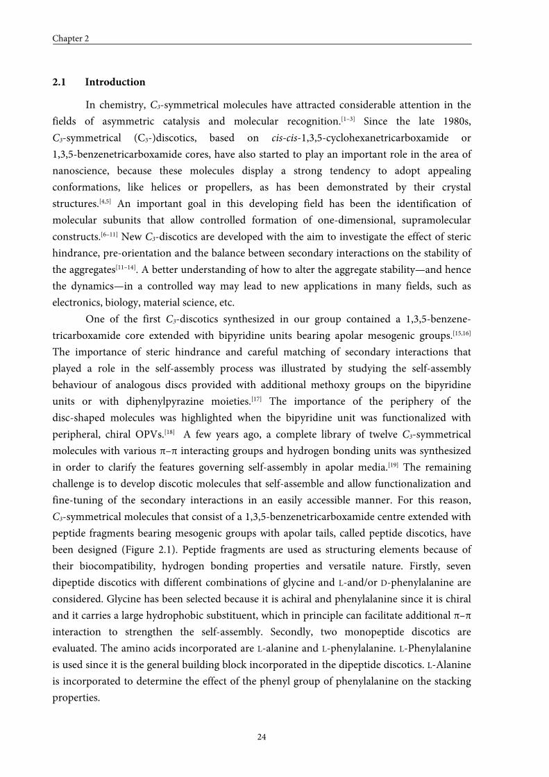
Figure 2.1 Library of peptide discotics; G = glycine, F = L-phenylalanine, f = D-phenylalanine,
A = alanine, cr = 1,3,5-benzenetricarbonyl core.
In this Chapter, the syntheses of nine C3-symmetrical peptide discotics are presented.
The self-assembly behaviour of these discs, both in the solid state and in solution, will be dealt
with in Chapter 3.
Chapter 2
26
2.2 Syntheses of dipeptide discotics with an achiral glycine at the C-terminus
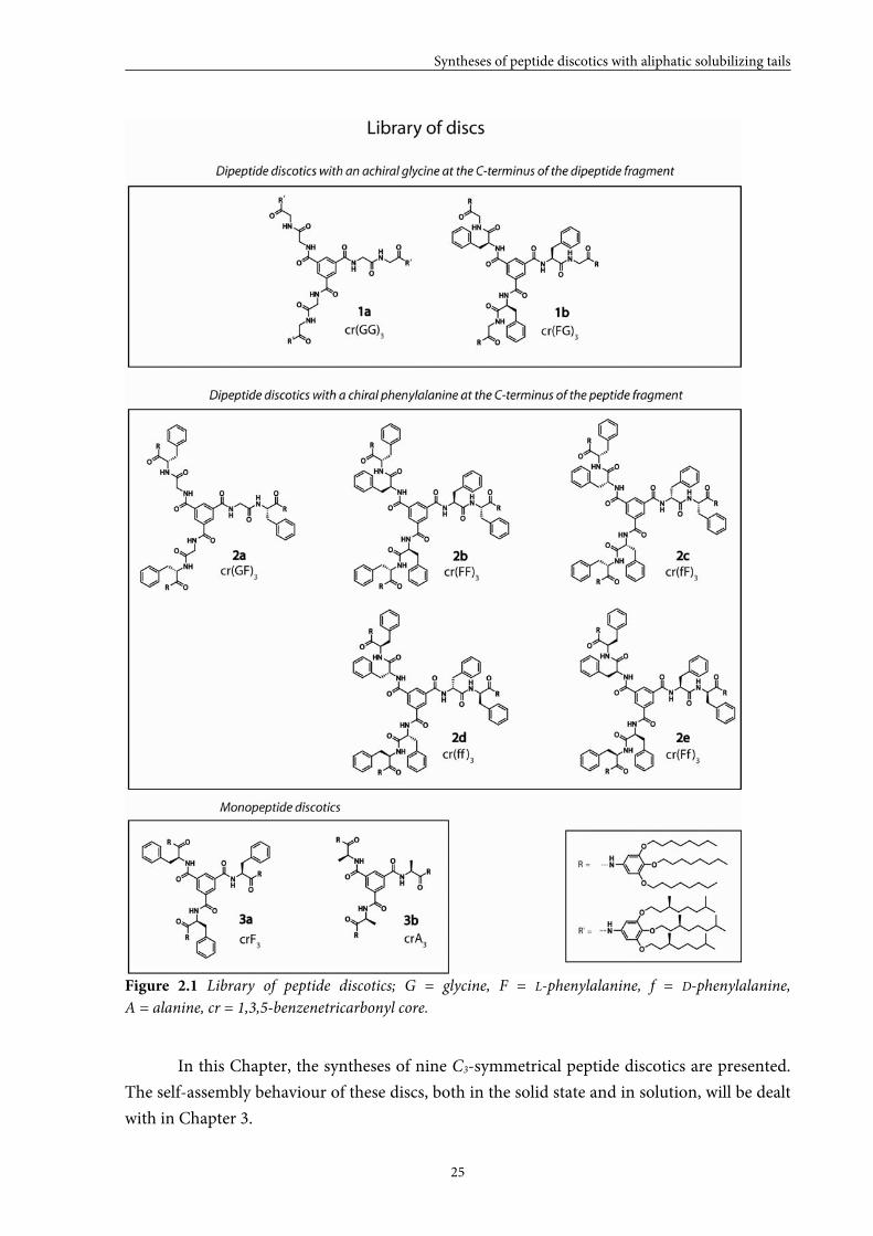
Dipeptide discotics cr(xG)3 (1a,b) containing a glycine at the C-terminus of the
dipeptide fragment were prepared as depicted in Scheme 2.1.
Scheme 2.1 Synthetic route towards discotics cr(xG)3 (1a,b): i) HBTU, DiPEA, DMF, RT, overnight,
~63%; ii) 1] TFA, RT, 1 h, 2] basic work-up, ~90% ; iii) Et3N, DCM, RT, overnight, ~65%.
3,4,5-Trioctyloxyaniline[20] and 3,4,5-tri((S)-3,7-dimethyloctyloxy)aniline[21] were synthesized
according to literature procedures. Direct coupling of the desired Boc-protected dipeptides
with the anilines was accomplished using HBTU as a coupling reagent and DiPEA as a base
and yielded pure compounds 4a,b. In these two cases, racemization could be excluded due to
the presence of an achiral glycine at the C-terminus of the peptide fragments. Deprotection of
4a,b with trifluoroacetic acid, followed by a basic work up, gave 5a,b. Finally, discotics 1a,b
were obtained after coupling of compounds 5a,b with 0.30 molar equivalents of trimesic
chloride in the presence of triethylamine as a base. The MALDI-TOF MS spectra of
compounds 4a, 5a and 1a are shown in Figure 2.2.

Syntheses of peptide discotics with aliphatic solubilizing tails
27
Figure 2.2 MALDI-TOF MS spectra for compounds 4a, 5a and 1a.
It is remarkable that besides protonated and cationized pseudomolecular ion signals strong
radical ion signals were observed for all compounds, including amino-terminated compounds
5a,b that were expected to be protonated prior to laser irradiation using acidic MALDI
matrices. The radical ion peaks [M]˙+ of compounds 4a, 5a and 1a could be observed at m/z =
775.5, m/z = 675.5 and m/z = 2183.7, respectively. Possible mechanisms for radical ion
formation were investigated with the employment of radical scavengers, with various matrices
and with direct laser desorption/ionization (LDI). Most likely the radicals were formed by
losing one electron from the aniline nitrogens, whereby the radicals were stabilized by
conjugation through the phenyl rings. It appeared that direct photo/thermal ionization of
analytes was an important route for the radical ion formation of compounds with trialkoxy
aniline/anilide groups.[22]
Chapter 2
28
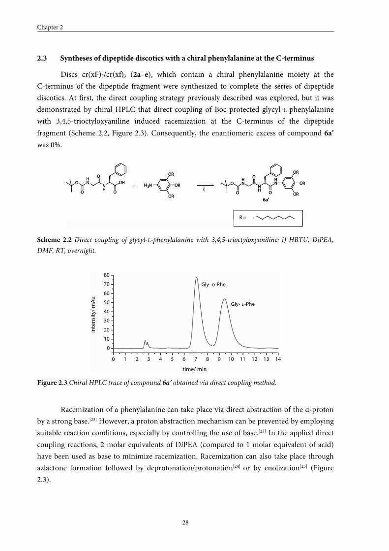
2.3 Syntheses of dipeptide discotics with a chiral phenylalanine at the C-terminus
Discs cr(xF)3/cr(xf)3 (2a–e), which contain a chiral phenylalanine moiety at the
C-terminus of the dipeptide fragment were synthesized to complete the series of dipeptide
discotics. At first, the direct coupling strategy previously described was explored, but it was
demonstrated by chiral HPLC that direct coupling of Boc-protected glycyl-L-phenylalanine
with 3,4,5-trioctyloxyaniline induced racemization at the C-terminus of the dipeptide
fragment (Scheme 2.2, Figure 2.3). Consequently, the enantiomeric excess of compound 6a’
was 0%.
Scheme 2.2 Direct coupling of glycyl-L-phenylalanine with 3,4,5-trioctyloxyaniline: i) HBTU, DiPEA,
DMF, RT, overnight.
Figure 2.3 Chiral HPLC trace of compound 6a’ obtained via direct coupling method.
Racemization of a phenylalanine can take place via direct abstraction of the α-proton
by a strong base.[23] However, a proton abstraction mechanism can be prevented by employing
suitable reaction conditions, especially by controlling the use of base.[23] In the applied direct
coupling reactions, 2 molar equivalents of DiPEA (compared to 1 molar equivalent of acid)
have been used as base to minimize racemization. Racemization can also take place through
azlactone formation followed by deprotonation/protonation[24] or by enolization[25] (Figure
2.3).
Syntheses of peptide discotics with aliphatic solubilizing tails
29
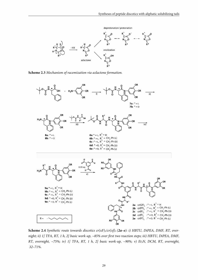
Scheme 2.3 Mechanism of racemization via azlactone formation.
Scheme 2.4 Synthetic route towards discotics cr(xF)3/cr(xf)3 (2a–e): i) HBTU, DiPEA, DMF, RT, over-
night; ii) 1] TFA, RT, 1 h, 2] basic work-up, ~85% over first two reaction steps; iii) HBTU, DiPEA, DMF,
RT, overnight, ~75%; iv) 1] TFA, RT, 1 h, 2] basic work-up, ~90%; v) Et3N, DCM, RT, overnight,
32–71%.
Chapter 2
30
It is known that the azlactone that is formed is less sensitive towards racemization
when R2 is an alkoxy (F-moc or Boc). Therefore, discotics cr(xF)3/cr(xf)3 (2a–e) have been
synthesized via consecutive coupling of the desired amino acids. The synthetic route towards
these discotics is depicted in Scheme 2.4. First, a single Boc-protected phenylalanine was
reacted with 3,4,5-trioctyloxyaniline using HBTU as a coupling reagent and DiPEA as a base.
Removal of the Boc-group with trifluoroactic acid, followed by a basic work-up gave free
amines 8a,b. Repetition of these consecutive steps with the desired amino acids and
compounds 8a,b as starting materials, gave dipeptide anilines 9a–e. Finally, discotics 2a–e
were obtained after coupling compounds 9a–e with 0.30 molar equivalents of trimesic
chloride in the presence of triethylamine as a base.
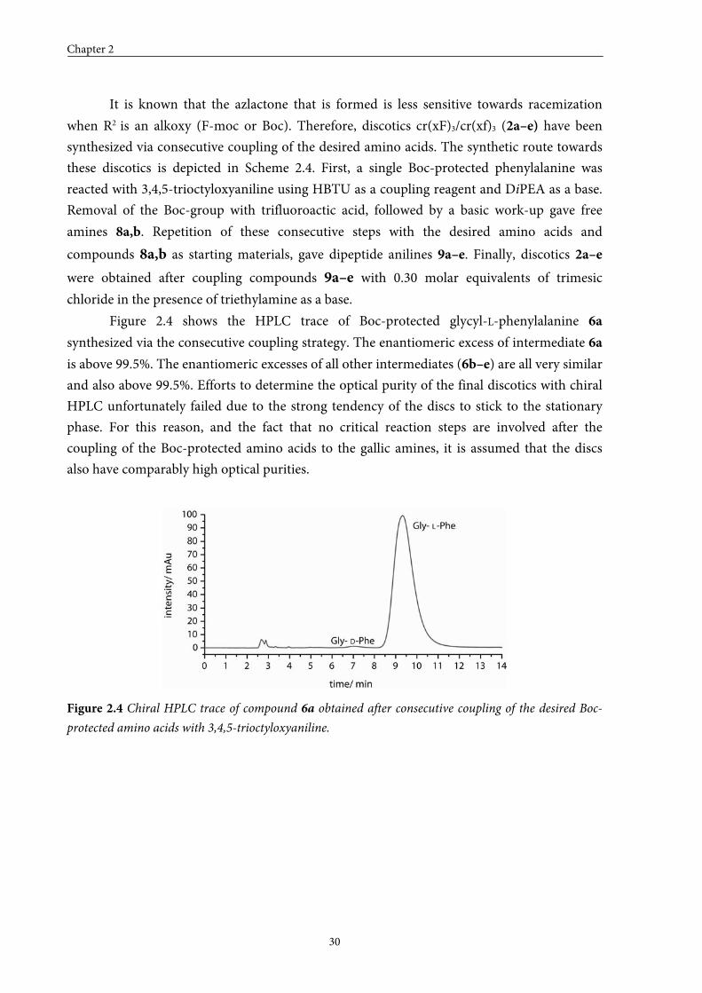
Figure 2.4 shows the HPLC trace of Boc-protected glycyl-L-phenylalanine 6a
synthesized via the consecutive coupling strategy. The enantiomeric excess of intermediate 6a
is above 99.5%. The enantiomeric excesses of all other intermediates (6b–e) are all very similar
and also above 99.5%. Efforts to determine the optical purity of the final discotics with chiral
HPLC unfortunately failed due to the strong tendency of the discs to stick to the stationary
phase. For this reason, and the fact that no critical reaction steps are involved after the
coupling of the Boc-protected amino acids to the gallic amines, it is assumed that the discs
also have comparably high optical purities.
Figure 2.4 Chiral HPLC trace of compound 6a obtained after consecutive coupling of the desired Boc-
protected amino acids with 3,4,5-trioctyloxyaniline.
Syntheses of peptide discotics with aliphatic solubilizing tails
31
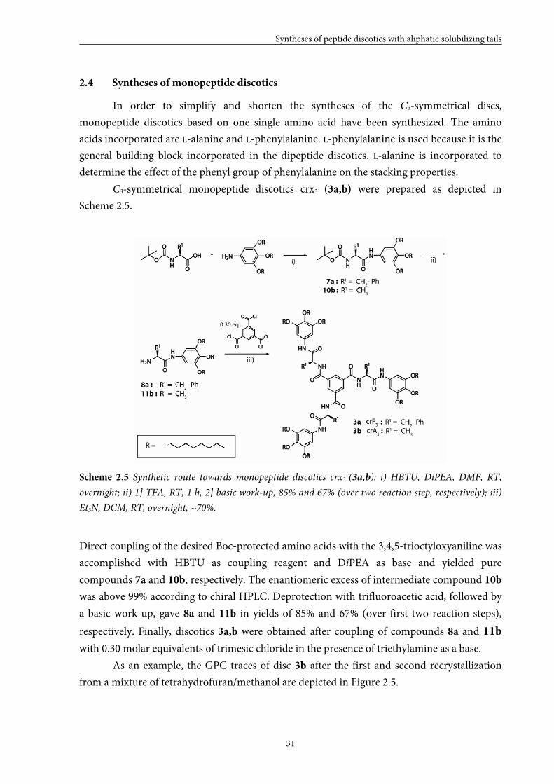
2.4 Syntheses of monopeptide discotics
In order to simplify and shorten the syntheses of the C3-symmetrical discs,
monopeptide discotics based on one single amino acid have been synthesized. The amino
acids incorporated are L-alanine and L-phenylalanine. L-phenylalanine is used because it is the
general building block incorporated in the dipeptide discotics. L-alanine is incorporated to
determine the effect of the phenyl group of phenylalanine on the stacking properties.
C3-symmetrical monopeptide discotics crx3 (3a,b) were prepared as depicted in
Scheme 2.5.
Scheme 2.5 Synthetic route towards monopeptide discotics crx3 (3a,b): i) HBTU, DiPEA, DMF, RT,
overnight; ii) 1] TFA, RT, 1 h, 2] basic work-up, 85% and 67% (over two reaction step, respectively); iii)
Et3N, DCM, RT, overnight, ~70%.
Direct coupling of the desired Boc-protected amino acids with the 3,4,5-trioctyloxyaniline was
accomplished with HBTU as coupling reagent and DiPEA as base and yielded pure
compounds 7a and 10b, respectively. The enantiomeric excess of intermediate compound 10b
was above 99% according to chiral HPLC. Deprotection with trifluoroacetic acid, followed by
a basic work up, gave 8a and 11b in yields of 85% and 67% (over first two reaction steps),
respectively. Finally, discotics 3a,b were obtained after coupling of compounds 8a and 11b
with 0.30 molar equivalents of trimesic chloride in the presence of triethylamine as a base.
As an example, the GPC traces of disc 3b after the first and second recrystallization
from a mixture of tetrahydrofuran/methanol are depicted in Figure 2.5.
Chapter 2
32
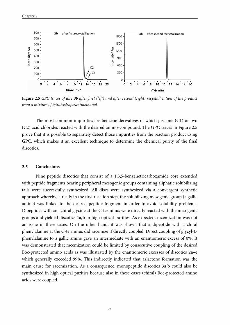
Figure 2.5 GPC traces of disc 3b after first (left) and after second (right) recystallization of the product
from a mixture of tetrahydrofuran/methanol.
The most common impurities are benzene derivatives of which just one (C1) or two
(C2) acid chlorides reacted with the desired amino-compound. The GPC traces in Figure 2.5
prove that it is possible to separately detect those impurities from the reaction product using
GPC, which makes it an excellent technique to determine the chemical purity of the final
discotics.
2.5 Conclusions
Nine peptide discotics that consist of a 1,3,5-benzenetricarboxamide core extended
with peptide fragments bearing peripheral mesogenic groups containing aliphatic solubilizing
tails were successfully synthesized. All discs were synthesized via a convergent synthetic
approach whereby, already in the first reaction step, the solubilizing mesogenic group (a gallic
amine) was linked to the desired peptide fragment in order to avoid solubility problems.
Dipeptides with an achiral glycine at the C-terminus were directly reacted with the mesogenic
groups and yielded discotics 1a,b in high optical purities. As expected, racemization was not
an issue in these cases. On the other hand, it was shown that a dipeptide with a chiral
phenylalanine at the C-terminus did racemize if directly coupled. Direct coupling of glycyl-L-
phenylalanine to a gallic amine gave an intermediate with an enantiomeric excess of 0%. It
was demonstrated that racemization could be limited by consecutive coupling of the desired
Boc-protected amino acids as was illustrated by the enantiomeric excesses of discotics 2a–e
which generally exceeded 99%. This indirectly indicated that azlactone formation was the
main cause for racemization. As a consequence, monopeptide discotics 3a,b could also be
synthesized in high optical purities because also in these cases (chiral) Boc-protected amino
acids were coupled.
Syntheses of peptide discotics with aliphatic solubilizing tails
33
2.6 Experimental section
General
Unless stated otherwise, all reagents and chemicals were obtained from commercial sources and used
without further purification. 3,4,5-Trioctyloxyaniline[20] and 3,4,5-tri((S)-3,7-dimethyloctyloxy)aniline[21] were
synthesized according to procedures described in the thesis of K. Pieterse (TU/e, 2001). Water was demineralised
prior to use. Dichloromethane and tetrahydrofuran were obtained by distillation over Merck molecular sieves
(4 Å). 1H-NMR, 1H-1H COSY and 19F-NMR spectra were recorded on a Varian Gemini 300 spectrometer, a
Varian Mercury Vx 400 spectrometer or a Varian Unity Inova 500 spectrometer at 298 K. Chemical shifts are
given in ppm (δ) values relative to tetramethylsilane (TMS). Splitting patterns are designated as s, singlet; d,
doublet; dd, double doublet; t, triplet; q, quartet; m, multiplet and br stands for broad. Neat state IR spectra were
measured at 298 K on a Perkin-Elmer 1605 FT-IR spectrophotometer. Matrix assisted laser
desorption/ionization mass spectra were obtained on a PerSeptive Biosystems Voyager DE-PRO spectrometer
using α-cyano-4-hydroxycinnamic acid (CHCA) and 2-[(2E)-3-(4-tert-butylphenyl)-2-methylprop-2-
enylidene]malononitrile (DCTB) as matrices. Elemental analyses were carried out using a Perkin Elmer 2400.
GPC measurements were performed on a Shimadzu system with a mixed column (2 × PL gel 3 µm, 100 Å), with
a flow of 1 mL·min–1, and tetrahydrofuran as eluting solvent. The injection volume was 50 µL and photodiode
array detection (260 nm) was applied. Chiral HPLC was performed on a Shimadzu system consisting of a
Shimadzu LC-10 ADvp pump, a Spark Midas autosampler and a Shimadzu SPD-10ADvp UV-Vis detector. Two
columns were used, a Daicel Chiralcel OD (0.46 × 25 cm) and a DNBPG (covalent 5 μm; 0.46 × 25 cm) both with
a 9/1 (v/v) hexane/isopropanol mixture as the eluent.
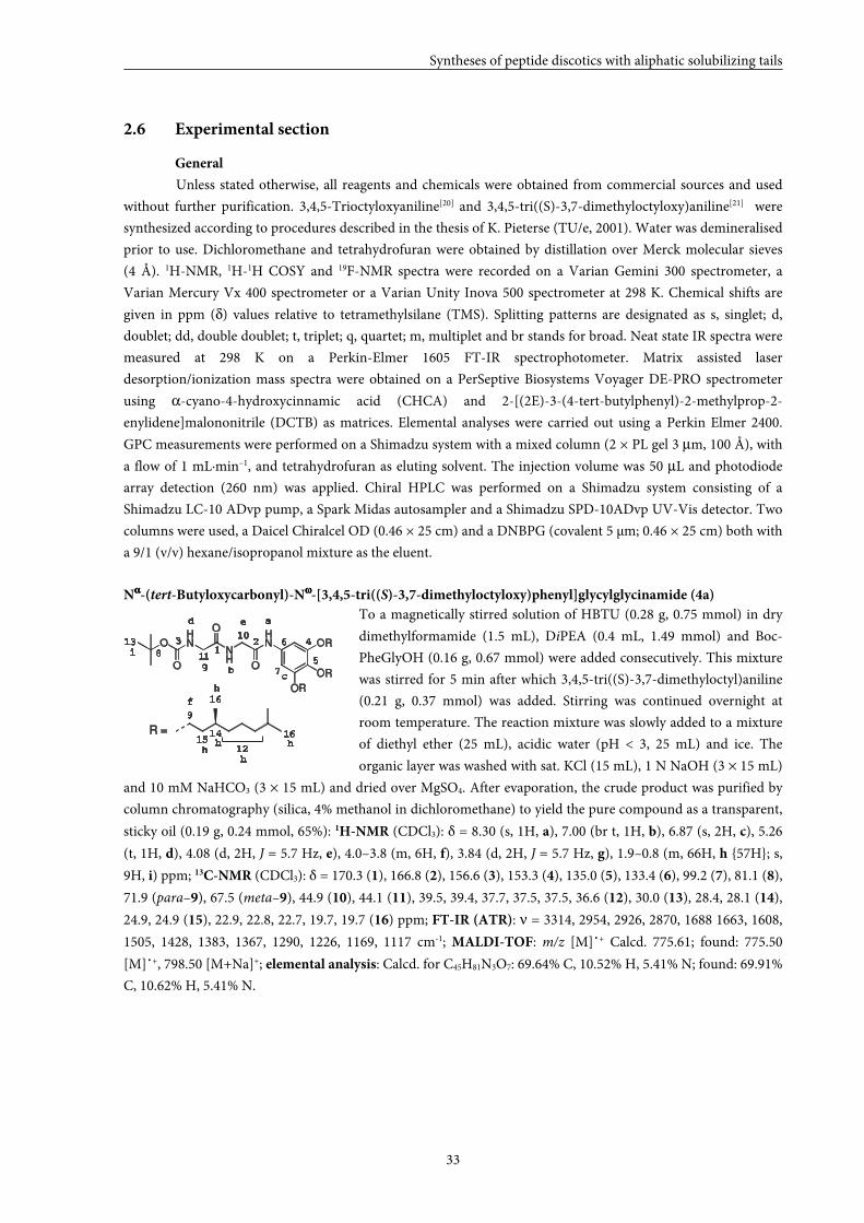
Nαααα-(tert-Butyloxycarbonyl)-Nωωωω-[3,4,5-tri((S)-3,7-dimethyloctyloxy)phenyl]glycylglycinamide (4a)
To a magnetically stirred solution of HBTU (0.28 g, 0.75 mmol) in dry
dimethylformamide (1.5 mL), DiPEA (0.4 mL, 1.49 mmol) and Boc-
PheGlyOH (0.16 g, 0.67 mmol) were added consecutively. This mixture
was stirred for 5 min after which 3,4,5-tri((S)-3,7-dimethyloctyl)aniline
(0.21 g, 0.37 mmol) was added. Stirring was continued overnight at
room temperature. The reaction mixture was slowly added to a mixture
of diethyl ether (25 mL), acidic water (pH < 3, 25 mL) and ice. The
organic layer was washed with sat. KCl (15 mL), 1 N NaOH (3 × 15 mL)
and 10 mM NaHCO3 (3 × 15 mL) and dried over MgSO4. After evaporation, the crude product was purified by
column chromatography (silica, 4% methanol in dichloromethane) to yield the pure compound as a transparent,
sticky oil (0.19 g, 0.24 mmol, 65%): 1H-NMR (CDCl3): δ = 8.30 (s, 1H, a), 7.00 (br t, 1H, b), 6.87 (s, 2H, c), 5.26
(t, 1H, d), 4.08 (d, 2H, J = 5.7 Hz, e), 4.0–3.8 (m, 6H, f), 3.84 (d, 2H, J = 5.7 Hz, g), 1.9–0.8 (m, 66H, h {57H}; s,
9H, i) ppm; 13C-NMR (CDCl3): δ = 170.3 (1), 166.8 (2), 156.6 (3), 153.3 (4), 135.0 (5), 133.4 (6), 99.2 (7), 81.1 (8),
71.9 (para–9), 67.5 (meta–9), 44.9 (10), 44.1 (11), 39.5, 39.4, 37.7, 37.5, 37.5, 36.6 (12), 30.0 (13), 28.4, 28.1 (14),
24.9, 24.9 (15), 22.9, 22.8, 22.7, 19.7, 19.7 (16) ppm; FT-IR (ATR): ν = 3314, 2954, 2926, 2870, 1688 1663, 1608,
1505, 1428, 1383, 1367, 1290, 1226, 1169, 1117 cm-1; MALDI-TOF: m/z [M]˙+ Calcd. 775.61; found: 775.50
[M]˙+, 798.50 [M+Na]+; elemental analysis: Calcd. for C45H81N3O7: 69.64% C, 10.52% H, 5.41% N; found: 69.91%
C, 10.62% H, 5.41% N.
OHN
NH
HN
O
O
O
OR
OR
OR1
23 4
5
6
7
8
10
11
13
9
12
1415
16
16R =
a
bc
d e
f
g
hh
h
hh
i
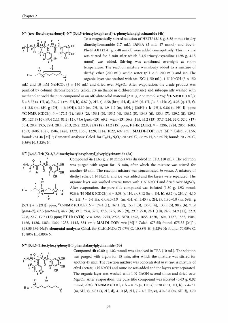
Chapter 2
34
Nαααα-(tert-Butyloxycarbonyl)-Nωωωω-(3,4,5-trioctyloxyphenyl)-L-phenylalanylglycinamide (4b)
To a magnetically stirred solution of HBTU (3.18 g, 8.38 mmol) in dry
dimethylformamide (17 mL), DiPEA (3 mL, 17 mmol) and Boc-L-
PheGlyOH (2.41 g, 7.48 mmol) were added consequtively. This mixture
was stirred for 5 min after which 3,4,5-trioctyloxyaniline (1.98 g, 4.15
mmol) was added. Stirring was continued overnight at room
temperature. The reaction mixture was slowly added to a mixture of
diethyl ether (200 mL), acidic water (pH < 3, 200 mL) and ice. The
organic layer was washed with sat. KCl (150 mL), 1 N NaOH (3 × 150
mL) and 10 mM NaHCO3 (3 × 150 mL) and dried over MgSO4. After evaporation, the crude product was
purified by column chromatography (silica, 2% methanol in dichloromethane) and subsequently washed with
methanol to yield the pure compound as an off-white solid material (2.00 g, 2.56 mmol, 62%): 1H-NMR (CDCl3):
δ = 8.27 (s, 1H, a), 7.4–7.1 (m, 5H, b), 6.87 (s, 2H, c), 6.58 (br t, 1H, d), 4.93 (d, 1H, J = 5.1 Hz, e), 4.28 (q, 1H, f),
4.1–3.8 (m, 8H, g {2H} + h {6H}), 3.10 (m, 2H, i), 1.9–1.2 (m, 45H, j {36H} + k {9H}), 0.86 (t, 9H, l) ppm; 13C-NMR (CDCl3): δ = 172.2 (1), 166.8 (2), 156.1 (3), 153.2 (4), 136.2 (5), 134.8 (6), 133.4 (7), 129.2 (8), 129.1
(9), 127.5 (10), 99.4 (11), 81.2 (12), 73.6 (para–13), 69.2 (meta–13), 56.8 (14), 44.2 (15), 37.7 (16), 32.0, 32.0, (17)
30.4, 29.7, 29.5, 29.4, 28.4 , 26.3, 26.2, 22.8, 22.8 (18), 14.2 (19) ppm; FT-IR (ATR): ν = 3296, 2924, 2855, 1683,
1653, 1606, 1525, 1504, 1428, 1379, 1365, 1228, 1114, 1022, 697 cm-1; MALDI-TOF: m/z [M]˙+ Calcd. 781.56;
found: 781.46 [M]˙+; elemental analysis: Calcd. for C46H75N3O7: 70.64% C, 9.67% H, 5.37% N; found: 70.72% C,
9.56% H, 5.32% N.
Nωωωω-[3,4,5-Tri((S)-3,7-dimethyloctyloxyphenyl]glycylglycinamide (5a)
Compound 4a (1.63 g, 2.10 mmol) was dissolved in TFA (10 mL). The solution
was purged with argon for 15 min, after which the mixture was stirred for
another 45 min. The reaction mixture was concentrated in vacuo. A mixture of
diethyl ether, 1 N NaOH and ice was added and the layers were separated. The
organic layer was washed several times with 1 N NaOH and dried over MgSO4.
After evaporation, the pure title compound was isolated (1.30 g, 1.92 mmol,
92%): 1H-NMR (CDCl3): δ = 8.58 (s, 1H, a), 8.12 (br t, 1H, b), 6.82 (s, 2H, c), 4.10
(d, 2H, J = 5.6 Hz, d), 4.0–3.9 (m, 6H, e), 3.45 (s, 2H, f), 1.90–0.8 (m, 59H, g
{57H} + h {2H}) ppm; 13C-NMR (CDCl3): δ = 174.4 (1), 167.1 (2), 153.3 (3), 135.0 (4), 133.5 (5), 98.9 (6), 71.9
(para–7), 67.5 (meta–7), 44.7 (8), 39.5, 39.4, 37.7, 37.5, 37.5, 36.5 (9), 29.9, 29.8, 28.1 (10), 24.9, 24.9 (11), 22.9,
22.8, 22.7, 19.7 (12) ppm; FT-IR (ATR): ν = 3286, 2954, 2926, 2870, 1698, 1655, 1620, 1604, 1527, 1555, 1504,
1466, 1426, 1383, 1366, 1233, 1115, 834 cm-1; MALDI-TOF: m/z [M]˙+ Calcd. 675.55; found: 675.55 [M]˙+,
698.55 [M+Na]+; elemental analysis: Calcd. for C40H73N3O5: 71.07% C, 10.88% H, 6.22% N; found: 70.95% C,
10.80% H, 6.09% N.
Nωωωω-[3,4,5-Trioctyloxy)phenyl]-L-phenylalanylglycinamide (5b)
Compound 4b (0.80 g, 1.02 mmol) was dissolved in TFA (10 mL). The solution
was purged with argon for 15 min, after which the mixture was stirred for
another 45 min. The reaction mixture was concentrated in vacuo. A mixture of
ethyl acetate, 1 N NaOH and some ice was added and the layers were separated.
The organic layer was washed with 1 N NaOH several times and dried over
MgSO4. After evaporation, the pure title compound was isolated (0.63 g, 0.92
mmol, 90%): 1H-NMR (CDCl3): δ = 8.75 (s, 1H, a), 8.20 (br t, 1H, b), 7.4–7.1
(m, 5H, c), 6.83 (s, 2H, d), 4.10 (d, 2H, J = 4.8 Hz, e), 4.0–3.8 (m, 6H, f), 3.70
1
23 4
5 6
7
8 10
11
12
13
15
16
17
18
a
b
c
d
ef
g
i
k
OHN
NH
HN
O
O
O
OR
OR
OR
R =
14
18
19
hj
l
9
1
2 3
45
6
8
8
a
bc
d
f
H2NNH
HN
O
O
OR
OR
OR
R =
7
9
10
12
1011
12
e
gg
g
g
g g
h
H2NNH
HN
O
O
OR
OR
OR
R =
a
1
2 3
4
56
78
9
10
11
1213
14
15
16
b
c
d
e
f
g
h
i
j
k
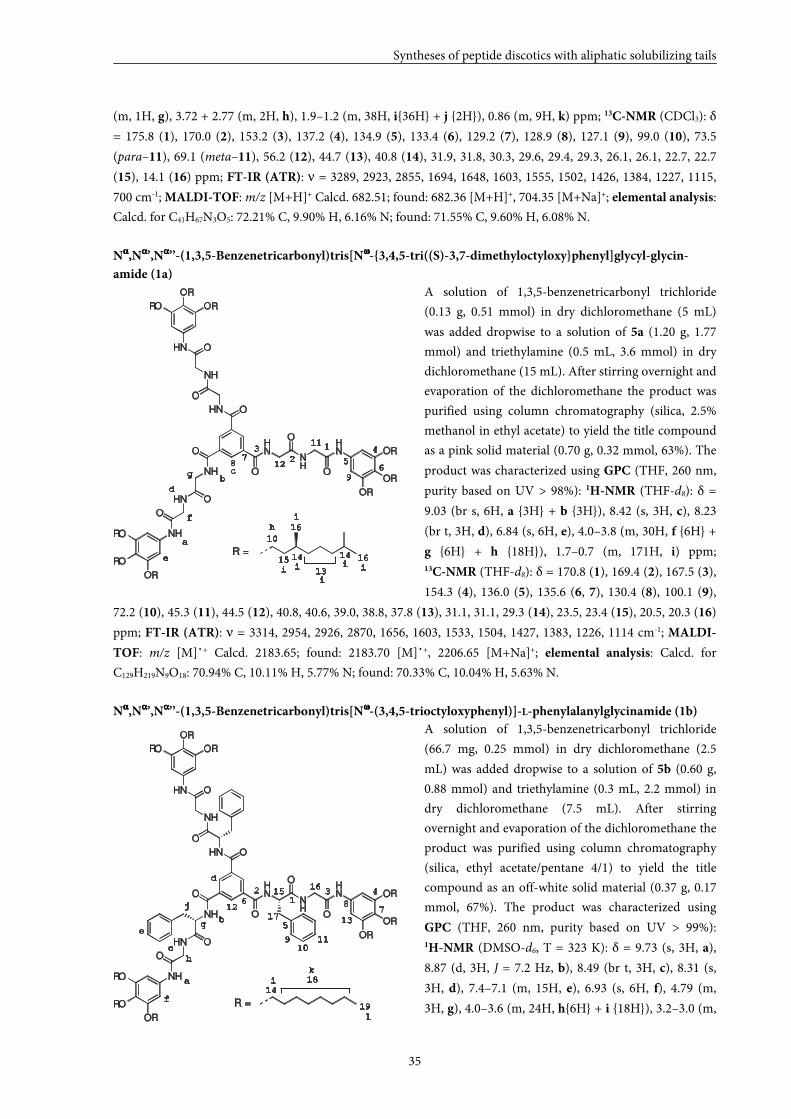
Syntheses of peptide discotics with aliphatic solubilizing tails
35
(m, 1H, g), 3.72 + 2.77 (m, 2H, h), 1.9–1.2 (m, 38H, i{36H} + j {2H}), 0.86 (m, 9H, k) ppm; 13C-NMR (CDCl3): δ
= 175.8 (1), 170.0 (2), 153.2 (3), 137.2 (4), 134.9 (5), 133.4 (6), 129.2 (7), 128.9 (8), 127.1 (9), 99.0 (10), 73.5
(para–11), 69.1 (meta–11), 56.2 (12), 44.7 (13), 40.8 (14), 31.9, 31.8, 30.3, 29.6, 29.4, 29.3, 26.1, 26.1, 22.7, 22.7
(15), 14.1 (16) ppm; FT-IR (ATR): ν = 3289, 2923, 2855, 1694, 1648, 1603, 1555, 1502, 1426, 1384, 1227, 1115,
700 cm-1; MALDI-TOF: m/z [M+H]+ Calcd. 682.51; found: 682.36 [M+H]+, 704.35 [M+Na]+; elemental analysis:
Calcd. for C41H67N3O5: 72.21% C, 9.90% H, 6.16% N; found: 71.55% C, 9.60% H, 6.08% N.
Nαααα,Nαααα’,Nαααα’’-(1,3,5-Benzenetricarbonyl)tris[Nωωωω-{3,4,5-tri((S)-3,7-dimethyloctyloxy}phenyl]glycyl-glycin-
amide (1a)
A solution of 1,3,5-benzenetricarbonyl trichloride
(0.13 g, 0.51 mmol) in dry dichloromethane (5 mL)
was added dropwise to a solution of 5a (1.20 g, 1.77
mmol) and triethylamine (0.5 mL, 3.6 mmol) in dry
dichloromethane (15 mL). After stirring overnight and
evaporation of the dichloromethane the product was
purified using column chromatography (silica, 2.5%
methanol in ethyl acetate) to yield the title compound
as a pink solid material (0.70 g, 0.32 mmol, 63%). The
product was characterized using GPC (THF, 260 nm,
purity based on UV > 98%): 1H-NMR (THF-d8): δ =
9.03 (br s, 6H, a {3H} + b {3H}), 8.42 (s, 3H, c), 8.23
(br t, 3H, d), 6.84 (s, 6H, e), 4.0–3.8 (m, 30H, f {6H} +
g {6H} + h {18H}), 1.7–0.7 (m, 171H, i) ppm; 13C-NMR (THF-d8): δ = 170.8 (1), 169.4 (2), 167.5 (3),
154.3 (4), 136.0 (5), 135.6 (6, 7), 130.4 (8), 100.1 (9),
72.2 (10), 45.3 (11), 44.5 (12), 40.8, 40.6, 39.0, 38.8, 37.8 (13), 31.1, 31.1, 29.3 (14), 23.5, 23.4 (15), 20.5, 20.3 (16)
ppm; FT-IR (ATR): ν = 3314, 2954, 2926, 2870, 1656, 1603, 1533, 1504, 1427, 1383, 1226, 1114 cm-1; MALDI-
TOF: m/z [M]˙+ Calcd. 2183.65; found: 2183.70 [M]˙+, 2206.65 [M+Na]+; elemental analysis: Calcd. for
C129H219N9O18: 70.94% C, 10.11% H, 5.77% N; found: 70.33% C, 10.04% H, 5.63% N.
Nαααα,Nαααα’,Nαααα’’-(1,3,5-Benzenetricarbonyl)tris[Nωωωω-(3,4,5-trioctyloxyphenyl)]-L-phenylalanylglycinamide (1b)
A solution of 1,3,5-benzenetricarbonyl trichloride
(66.7 mg, 0.25 mmol) in dry dichloromethane (2.5
mL) was added dropwise to a solution of 5b (0.60 g,
0.88 mmol) and triethylamine (0.3 mL, 2.2 mmol) in
dry dichloromethane (7.5 mL). After stirring
overnight and evaporation of the dichloromethane the
product was purified using column chromatography
(silica, ethyl acetate/pentane 4/1) to yield the title
compound as an off-white solid material (0.37 g, 0.17
mmol, 67%). The product was characterized using
GPC (THF, 260 nm, purity based on UV > 99%): 1H-NMR (DMSO-d6, T = 323 K): δ = 9.73 (s, 3H, a),
8.87 (d, 3H, J = 7.2 Hz, b), 8.49 (br t, 3H, c), 8.31 (s,
3H, d), 7.4–7.1 (m, 15H, e), 6.93 (s, 6H, f), 4.79 (m,
3H, g), 4.0–3.6 (m, 24H, h{6H} + i {18H}), 3.2–3.0 (m,
HN
NH
HN
O
O
OR
OR
OR
ONH
HN
NH
HN
NH
O
O
O
O
O
OHN
OR
RO
RO
RO
OR
OR
R =
1
2 3 4
5
67
8
910
11
12
16
14
15
1317
18
19
a
b
c
d
e
f
g
h
i
j
k
l
HN
NH
HN
O
O
OR
OR
OR
ONH
HN
NH
HN
NH
O
O
O
O
O
OHN
OR
RO
RO
RO
OR
OR
R =
1
2
3 4
56
78
9
10
11
12
13
141415
16
16
a
bc
d
e
f
h
i i
i
i
i i
g
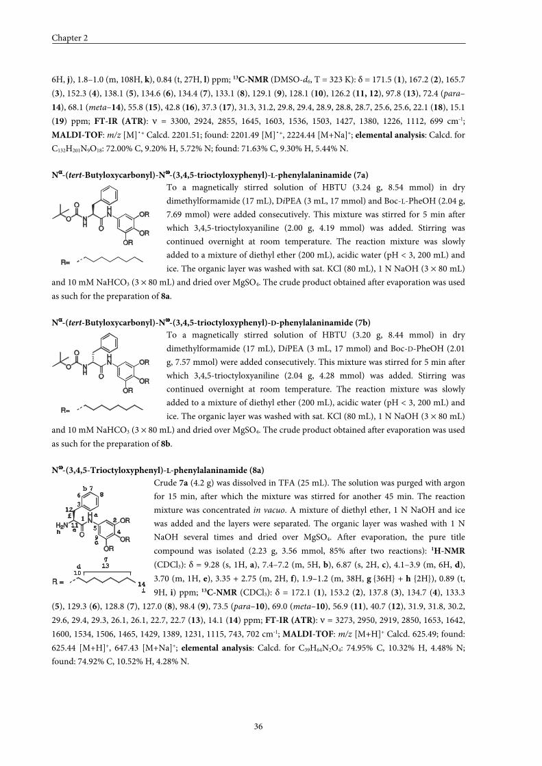
Chapter 2
36
6H, j), 1.8–1.0 (m, 108H, k), 0.84 (t, 27H, l) ppm; 13C-NMR (DMSO-d6, T = 323 K): δ = 171.5 (1), 167.2 (2), 165.7
(3), 152.3 (4), 138.1 (5), 134.6 (6), 134.4 (7), 133.1 (8), 129.1 (9), 128.1 (10), 126.2 (11, 12), 97.8 (13), 72.4 (para–
14), 68.1 (meta–14), 55.8 (15), 42.8 (16), 37.3 (17), 31.3, 31.2, 29.8, 29.4, 28.9, 28.8, 28.7, 25.6, 25.6, 22.1 (18), 15.1
(19) ppm; FT-IR (ATR): ν = 3300, 2924, 2855, 1645, 1603, 1536, 1503, 1427, 1380, 1226, 1112, 699 cm-1;
MALDI-TOF: m/z [M]˙+ Calcd. 2201.51; found: 2201.49 [M]˙+, 2224.44 [M+Na]+; elemental analysis: Calcd. for
C132H201N9O18: 72.00% C, 9.20% H, 5.72% N; found: 71.63% C, 9.30% H, 5.44% N.
Nαααα-(tert-Butyloxycarbonyl)-Nωωωω-(3,4,5-trioctyloxyphenyl)-L-phenylalaninamide (7a)
To a magnetically stirred solution of HBTU (3.24 g, 8.54 mmol) in dry
dimethylformamide (17 mL), DiPEA (3 mL, 17 mmol) and Boc-L-PheOH (2.04 g,
7.69 mmol) were added consecutively. This mixture was stirred for 5 min after
which 3,4,5-trioctyloxyaniline (2.00 g, 4.19 mmol) was added. Stirring was
continued overnight at room temperature. The reaction mixture was slowly
added to a mixture of diethyl ether (200 mL), acidic water (pH < 3, 200 mL) and
ice. The organic layer was washed with sat. KCl (80 mL), 1 N NaOH (3 × 80 mL)
and 10 mM NaHCO3 (3 × 80 mL) and dried over MgSO4. The crude product obtained after evaporation was used
as such for the preparation of 8a.
Nαααα-(tert-Butyloxycarbonyl)-Nωωωω-(3,4,5-trioctyloxyphenyl)-D-phenylalaninamide (7b)
To a magnetically stirred solution of HBTU (3.20 g, 8.44 mmol) in dry
dimethylformamide (17 mL), DiPEA (3 mL, 17 mmol) and Boc-D-PheOH (2.01
g, 7.57 mmol) were added consecutively. This mixture was stirred for 5 min after
which 3,4,5-trioctyloxyaniline (2.04 g, 4.28 mmol) was added. Stirring was
continued overnight at room temperature. The reaction mixture was slowly
added to a mixture of diethyl ether (200 mL), acidic water (pH < 3, 200 mL) and
ice. The organic layer was washed with sat. KCl (80 mL), 1 N NaOH (3 × 80 mL)
and 10 mM NaHCO3 (3 × 80 mL) and dried over MgSO4. The crude product obtained after evaporation was used
as such for the preparation of 8b.
Nωωωω-(3,4,5-Trioctyloxyphenyl)-L-phenylalaninamide (8a)
Crude 7a (4.2 g) was dissolved in TFA (25 mL). The solution was purged with argon
for 15 min, after which the mixture was stirred for another 45 min. The reaction
mixture was concentrated in vacuo. A mixture of diethyl ether, 1 N NaOH and ice
was added and the layers were separated. The organic layer was washed with 1 N
NaOH several times and dried over MgSO4. After evaporation, the pure title
compound was isolated (2.23 g, 3.56 mmol, 85% after two reactions): 1H-NMR
(CDCl3): δ = 9.28 (s, 1H, a), 7.4–7.2 (m, 5H, b), 6.87 (s, 2H, c), 4.1–3.9 (m, 6H, d),
3.70 (m, 1H, e), 3.35 + 2.75 (m, 2H, f), 1.9–1.2 (m, 38H, g {36H} + h {2H}), 0.89 (t,
9H, i) ppm; 13C-NMR (CDCl3): δ = 172.1 (1), 153.2 (2), 137.8 (3), 134.7 (4), 133.3
(5), 129.3 (6), 128.8 (7), 127.0 (8), 98.4 (9), 73.5 (para–10), 69.0 (meta–10), 56.9 (11), 40.7 (12), 31.9, 31.8, 30.2,
29.6, 29.4, 29.3, 26.1, 26.1, 22.7, 22.7 (13), 14.1 (14) ppm; FT-IR (ATR): ν = 3273, 2950, 2919, 2850, 1653, 1642,
1600, 1534, 1506, 1465, 1429, 1389, 1231, 1115, 743, 702 cm-1; MALDI-TOF: m/z [M+H]+ Calcd. 625.49; found:
625.44 [M+H]+, 647.43 [M+Na]+; elemental analysis: Calcd. for C39H64N2O4: 74.95% C, 10.32% H, 4.48% N;
found: 74.92% C, 10.52% H, 4.28% N.
NH
HN
O
OR
OR
OR
R =
O
O
NH
HN
O
OR
OR
OR
R =
O
O
H2N
HN
O
OR
OR
OR
R =
10
13
14
d
h
i
1 2
3
45
6
78
9
11
12a
b
c
e
f
g
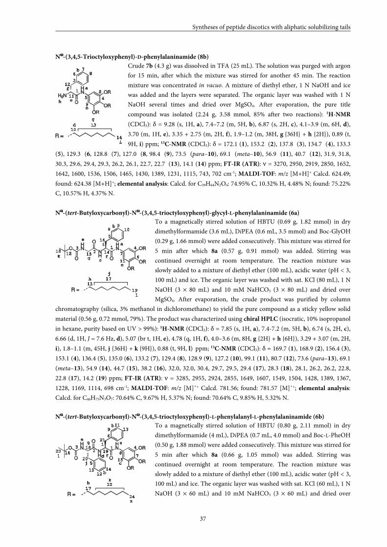
Syntheses of peptide discotics with aliphatic solubilizing tails
37
Nωωωω-(3,4,5-Trioctyloxyphenyl)-D-phenylalaninamide (8b)
Crude 7b (4.3 g) was dissolved in TFA (25 mL). The solution was purged with argon
for 15 min, after which the mixture was stirred for another 45 min. The reaction
mixture was concentrated in vacuo. A mixture of diethyl ether, 1 N NaOH and ice
was added and the layers were separated. The organic layer was washed with 1 N
NaOH several times and dried over MgSO4. After evaporation, the pure title
compound was isolated (2.24 g, 3.58 mmol, 85% after two reactions): 1H-NMR
(CDCl3): δ = 9.28 (s, 1H, a), 7.4–7.2 (m, 5H, b), 6.87 (s, 2H, c), 4.1–3.9 (m, 6H, d),
3.70 (m, 1H, e), 3.35 + 2.75 (m, 2H, f), 1.9–1.2 (m, 38H, g {36H} + h {2H}), 0.89 (t,
9H, i) ppm; 13C-NMR (CDCl3): δ = 172.1 (1), 153.2 (2), 137.8 (3), 134.7 (4), 133.3
(5), 129.3 (6, 128.8 (7), 127.0 (8, 98.4 (9), 73.5 (para–10), 69.1 (meta–10), 56.9 (11), 40.7 (12), 31.9, 31.8,
30.3, 29.6, 29.4, 29.3, 26.2, 26.1, 22.7, 22.7 (13), 14.1 (14) ppm; FT-IR (ATR): ν = 3270, 2950, 2919, 2850, 1652,
1642, 1600, 1536, 1506, 1465, 1430, 1389, 1231, 1115, 743, 702 cm-1; MALDI-TOF: m/z [M+H]+ Calcd. 624.49;
found: 624.38 [M+H]+; elemental analysis: Calcd. for C39H64N2O4: 74.95% C, 10.32% H, 4.48% N; found: 75.22%
C, 10.57% H, 4.37% N.
Nαααα-(tert-Butyloxycarbonyl)-Nωωωω-(3,4,5-trioctyloxyphenyl)-glycyl-L-phenylalaninamide (6a)
To a magnetically stirred solution of HBTU (0.69 g, 1.82 mmol) in dry
dimethylformamide (3.6 mL), DiPEA (0.6 mL, 3.5 mmol) and Boc-GlyOH
(0.29 g, 1.66 mmol) were added consecutively. This mixture was stirred for
5 min after which 8a (0.57 g, 0.91 mmol) was added. Stirring was
continued overnight at room temperature. The reaction mixture was
slowly added to a mixture of diethyl ether (100 mL), acidic water (pH < 3,
100 mL) and ice. The organic layer was washed with sat. KCl (80 mL), 1 N
NaOH (3 × 80 mL) and 10 mM NaHCO3 (3 × 80 mL) and dried over
MgSO4. After evaporation, the crude product was purified by column
chromatography (silica, 3% methanol in dichloromethane) to yield the pure compound as a sticky yellow solid
material (0.56 g, 0.72 mmol, 79%). The product was characterized using chiral HPLC (isocratic, 10% isopropanol
in hexane, purity based on UV > 99%): 1H-NMR (CDCl3): δ = 7.85 (s, 1H, a), 7.4-7.2 (m, 5H, b), 6.74 (s, 2H, c),
6.66 (d, 1H, J = 7.6 Hz, d), 5.07 (br t, 1H, e), 4.78 (q, 1H, f), 4.0–3.6 (m, 8H, g {2H} + h {6H}), 3.29 + 3.07 (m, 2H,
i), 1.8–1.1 (m, 45H, j {36H} + k {9H}), 0.88 (t, 9H, l) ppm; 13C-NMR (CDCl3): δ = 169.7 (1), 168.9 (2), 156.4 (3),
153.1 (4), 136.4 (5), 135.0 (6), 133.2 (7), 129.4 (8), 128.9 (9), 127.2 (10), 99.1 (11), 80.7 (12), 73.6 (para–13), 69.1
(meta–13), 54.9 (14), 44.7 (15), 38.2 (16), 32.0, 32.0, 30.4, 29.7, 29.5, 29.4 (17), 28.3 (18), 28.1, 26.2, 26.2, 22.8,
22.8 (17), 14.2 (19) ppm; FT-IR (ATR): ν = 3285, 2955, 2924, 2855, 1649, 1607, 1549, 1504, 1428, 1389, 1367,
1228, 1169, 1114, 698 cm-1; MALDI-TOF: m/z [M]˙+ Calcd. 781.56; found: 781.57 [M]˙+; elemental analysis:
Calcd. for C46H75N3O7: 70.64% C, 9.67% H, 5.37% N; found: 70.64% C, 9.85% H, 5.32% N.
Nαααα-(tert-Butyloxycarbonyl)-Nωωωω-(3,4,5-trioctyloxyphenyl)-L-phenylalanyl-L-phenylalaninamide (6b)
To a magnetically stirred solution of HBTU (0.80 g, 2.11 mmol) in dry
dimethylformamide (4 mL), DiPEA (0.7 mL, 4.0 mmol) and Boc-L-PheOH
(0.50 g, 1.88 mmol) were added consecutively. This mixture was stirred for
5 min after which 8a (0.66 g, 1.05 mmol) was added. Stirring was
continued overnight at room temperature. The reaction mixture was
slowly added to a mixture of diethyl ether (100 mL), acidic water (pH < 3,
100 mL) and ice. The organic layer was washed with sat. KCl (60 mL), 1 N
NaOH (3 × 60 mL) and 10 mM NaHCO3 (3 × 60 mL) and dried over
1013
14
d
h
i
1 2
3
45
6
78
9
11
12a
b
c
e
f
g
R =
H2N
HN
O
OR
OR
OR
10
13
14O
HN
NH
HN
O
O
O
OR
OR
OR
R =
d
j
l
12
3 4
5
67
89
11
12
a
b
c
e
fg15
16
17
18
19
h
i
k
12
3 4
5
6
78
10
11
12
13
15
16
17
18
a
b
c
d
e
f
g i
k
OHN
NH
HN
O
O
O
OR
OR
OR
R =
14
19
h
j
9
20
21
22
23
24
b
l
m
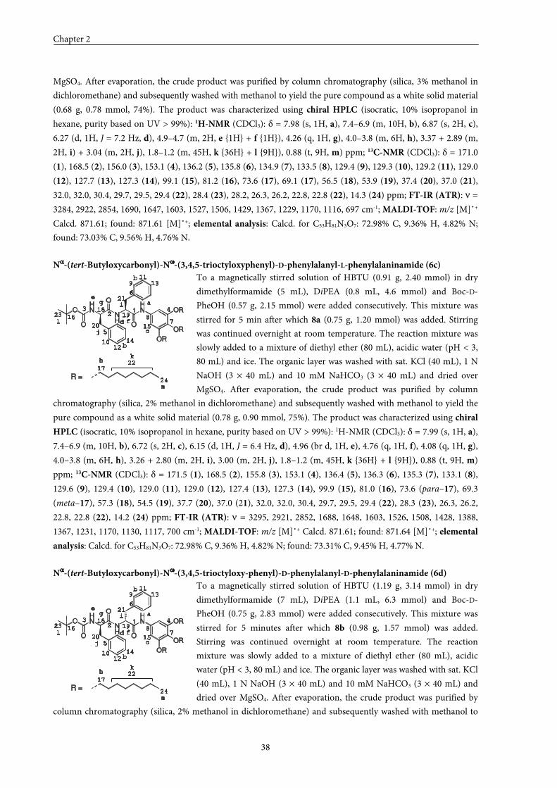
Chapter 2
38
MgSO4. After evaporation, the crude product was purified by column chromatography (silica, 3% methanol in
dichloromethane) and subsequently washed with methanol to yield the pure compound as a white solid material
(0.68 g, 0.78 mmol, 74%). The product was characterized using chiral HPLC (isocratic, 10% isopropanol in
hexane, purity based on UV > 99%): 1H-NMR (CDCl3): δ = 7.98 (s, 1H, a), 7.4–6.9 (m, 10H, b), 6.87 (s, 2H, c),
6.27 (d, 1H, J = 7.2 Hz, d), 4.9–4.7 (m, 2H, e {1H} + f {1H}), 4.26 (q, 1H, g), 4.0–3.8 (m, 6H, h), 3.37 + 2.89 (m,
2H, i) + 3.04 (m, 2H, j), 1.8–1.2 (m, 45H, k {36H} + l {9H}), 0.88 (t, 9H, m) ppm; 13C-NMR (CDCl3): δ = 171.0
(1), 168.5 (2), 156.0 (3), 153.1 (4), 136.2 (5), 135.8 (6), 134.9 (7), 133.5 (8), 129.4 (9), 129.3 (10), 129.2 (11), 129.0
(12), 127.7 (13), 127.3 (14), 99.1 (15), 81.2 (16), 73.6 (17), 69.1 (17), 56.5 (18), 53.9 (19), 37.4 (20), 37.0 (21),
32.0, 32.0, 30.4, 29.7, 29.5, 29.4 (22), 28.4 (23), 28.2, 26.3, 26.2, 22.8, 22.8 (22), 14.3 (24) ppm; FT-IR (ATR): ν =
3284, 2922, 2854, 1690, 1647, 1603, 1527, 1506, 1429, 1367, 1229, 1170, 1116, 697 cm-1; MALDI-TOF: m/z [M]˙+
Calcd. 871.61; found: 871.61 [M]˙+; elemental analysis: Calcd. for C53H81N3O7: 72.98% C, 9.36% H, 4.82% N;
found: 73.03% C, 9.56% H, 4.76% N.
Nαααα-(tert-Butyloxycarbonyl)-Nωωωω-(3,4,5-trioctyloxyphenyl)-D-phenylalanyl-L-phenylalaninamide (6c)
To a magnetically stirred solution of HBTU (0.91 g, 2.40 mmol) in dry
dimethylformamide (5 mL), DiPEA (0.8 mL, 4.6 mmol) and Boc-D-
PheOH (0.57 g, 2.15 mmol) were added consecutively. This mixture was
stirred for 5 min after which 8a (0.75 g, 1.20 mmol) was added. Stirring
was continued overnight at room temperature. The reaction mixture was
slowly added to a mixture of diethyl ether (80 mL), acidic water (pH < 3,
80 mL) and ice. The organic layer was washed with sat. KCl (40 mL), 1 N
NaOH (3 × 40 mL) and 10 mM NaHCO3 (3 × 40 mL) and dried over
MgSO4. After evaporation, the crude product was purified by column
chromatography (silica, 2% methanol in dichloromethane) and subsequently washed with methanol to yield the
pure compound as a white solid material (0.78 g, 0.90 mmol, 75%). The product was characterized using chiral
HPLC (isocratic, 10% isopropanol in hexane, purity based on UV > 99%): 1H-NMR (CDCl3): δ = 7.99 (s, 1H, a),
7.4–6.9 (m, 10H, b), 6.72 (s, 2H, c), 6.15 (d, 1H, J = 6.4 Hz, d), 4.96 (br d, 1H, e), 4.76 (q, 1H, f), 4.08 (q, 1H, g),
4.0–3.8 (m, 6H, h), 3.26 + 2.80 (m, 2H, i), 3.00 (m, 2H, j), 1.8–1.2 (m, 45H, k {36H} + l {9H}), 0.88 (t, 9H, m)
ppm; 13C-NMR (CDCl3): δ = 171.5 (1), 168.5 (2), 155.8 (3), 153.1 (4), 136.4 (5), 136.3 (6), 135.3 (7), 133.1 (8),
129.6 (9), 129.4 (10), 129.0 (11), 129.0 (12), 127.4 (13), 127.3 (14), 99.9 (15), 81.0 (16), 73.6 (para–17), 69.3
(meta–17), 57.3 (18), 54.5 (19), 37.7 (20), 37.0 (21), 32.0, 32.0, 30.4, 29.7, 29.5, 29.4 (22), 28.3 (23), 26.3, 26.2,
22.8, 22.8 (22), 14.2 (24) ppm; FT-IR (ATR): ν = 3295, 2921, 2852, 1688, 1648, 1603, 1526, 1508, 1428, 1388,
1367, 1231, 1170, 1130, 1117, 700 cm-1; MALDI-TOF: m/z [M]˙+ Calcd. 871.61; found: 871.64 [M]˙+; elemental
analysis: Calcd. for C53H81N3O7: 72.98% C, 9.36% H, 4.82% N; found: 73.31% C, 9.45% H, 4.77% N.
Nαααα-(tert-Butyloxycarbonyl)-Nωωωω-(3,4,5-trioctyloxy-phenyl)-D-phenylalanyl-D-phenylalaninamide (6d)
To a magnetically stirred solution of HBTU (1.19 g, 3.14 mmol) in dry
dimethylformamide (7 mL), DiPEA (1.1 mL, 6.3 mmol) and Boc-D-
PheOH (0.75 g, 2.83 mmol) were added consecutively. This mixture was
stirred for 5 minutes after which 8b (0.98 g, 1.57 mmol) was added.
Stirring was continued overnight at room temperature. The reaction
mixture was slowly added to a mixture of diethyl ether (80 mL), acidic
water (pH < 3, 80 mL) and ice. The organic layer was washed with sat. KCl
(40 mL), 1 N NaOH (3 × 40 mL) and 10 mM NaHCO3 (3 × 40 mL) and
dried over MgSO4. After evaporation, the crude product was purified by
column chromatography (silica, 2% methanol in dichloromethane) and subsequently washed with methanol to
23 4
5
6
78
10
11
12
13
15
16
17
18
a
b
c
d
e
f
g i
k
14
19
h
j
9
20
21
22
23
24
b
l
m
1
R =
OHN
NH
HN
O
O
O
OR
OR
OR
23 4
5
6
78
10
11
12
13
15
16
17
18
a
b
c
d
e
f
g i
k
14
19
h
j
9
20
21
22
23
24
b
l
m
1
R =
OHN
NH
HN
O
O
O
OR
OR
OR
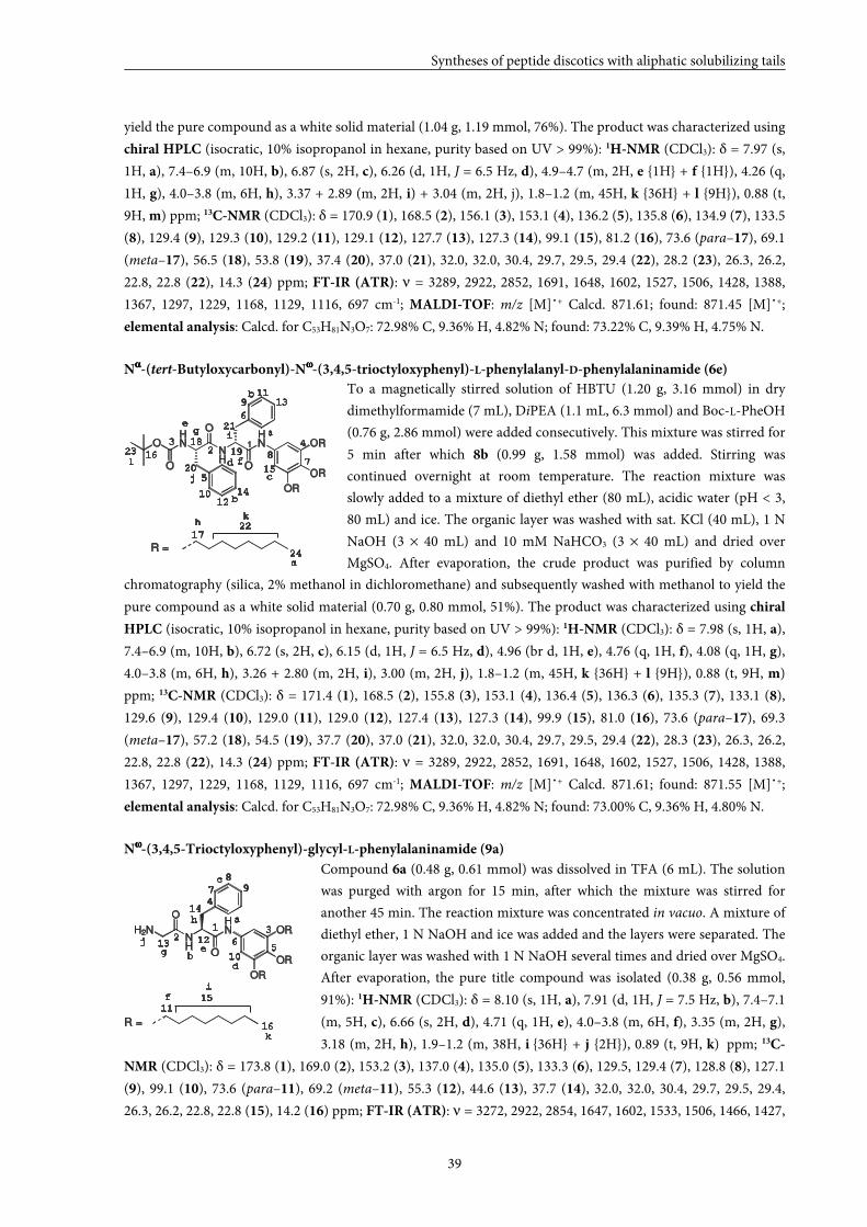
Syntheses of peptide discotics with aliphatic solubilizing tails
39
yield the pure compound as a white solid material (1.04 g, 1.19 mmol, 76%). The product was characterized using
chiral HPLC (isocratic, 10% isopropanol in hexane, purity based on UV > 99%): 1H-NMR (CDCl3): δ = 7.97 (s,
1H, a), 7.4–6.9 (m, 10H, b), 6.87 (s, 2H, c), 6.26 (d, 1H, J = 6.5 Hz, d), 4.9–4.7 (m, 2H, e {1H} + f {1H}), 4.26 (q,
1H, g), 4.0–3.8 (m, 6H, h), 3.37 + 2.89 (m, 2H, i) + 3.04 (m, 2H, j), 1.8–1.2 (m, 45H, k {36H} + l {9H}), 0.88 (t,
9H, m) ppm; 13C-NMR (CDCl3): δ = 170.9 (1), 168.5 (2), 156.1 (3), 153.1 (4), 136.2 (5), 135.8 (6), 134.9 (7), 133.5
(8), 129.4 (9), 129.3 (10), 129.2 (11), 129.1 (12), 127.7 (13), 127.3 (14), 99.1 (15), 81.2 (16), 73.6 (para–17), 69.1
(meta–17), 56.5 (18), 53.8 (19), 37.4 (20), 37.0 (21), 32.0, 32.0, 30.4, 29.7, 29.5, 29.4 (22), 28.2 (23), 26.3, 26.2,
22.8, 22.8 (22), 14.3 (24) ppm; FT-IR (ATR): ν = 3289, 2922, 2852, 1691, 1648, 1602, 1527, 1506, 1428, 1388,
1367, 1297, 1229, 1168, 1129, 1116, 697 cm-1; MALDI-TOF: m/z [M]˙+ Calcd. 871.61; found: 871.45 [M]˙+;
elemental analysis: Calcd. for C53H81N3O7: 72.98% C, 9.36% H, 4.82% N; found: 73.22% C, 9.39% H, 4.75% N.
Nαααα-(tert-Butyloxycarbonyl)-Nωωωω-(3,4,5-trioctyloxyphenyl)-L-phenylalanyl-D-phenylalaninamide (6e)
To a magnetically stirred solution of HBTU (1.20 g, 3.16 mmol) in dry
dimethylformamide (7 mL), DiPEA (1.1 mL, 6.3 mmol) and Boc-L-PheOH
(0.76 g, 2.86 mmol) were added consecutively. This mixture was stirred for
5 min after which 8b (0.99 g, 1.58 mmol) was added. Stirring was
continued overnight at room temperature. The reaction mixture was
slowly added to a mixture of diethyl ether (80 mL), acidic water (pH < 3,
80 mL) and ice. The organic layer was washed with sat. KCl (40 mL), 1 N
NaOH (3 × 40 mL) and 10 mM NaHCO3 (3 × 40 mL) and dried over
MgSO4. After evaporation, the crude product was purified by column
chromatography (silica, 2% methanol in dichloromethane) and subsequently washed with methanol to yield the
pure compound as a white solid material (0.70 g, 0.80 mmol, 51%). The product was characterized using chiral
HPLC (isocratic, 10% isopropanol in hexane, purity based on UV > 99%): 1H-NMR (CDCl3): δ = 7.98 (s, 1H, a),
7.4–6.9 (m, 10H, b), 6.72 (s, 2H, c), 6.15 (d, 1H, J = 6.5 Hz, d), 4.96 (br d, 1H, e), 4.76 (q, 1H, f), 4.08 (q, 1H, g),
4.0–3.8 (m, 6H, h), 3.26 + 2.80 (m, 2H, i), 3.00 (m, 2H, j), 1.8–1.2 (m, 45H, k {36H} + l {9H}), 0.88 (t, 9H, m)
ppm; 13C-NMR (CDCl3): δ = 171.4 (1), 168.5 (2), 155.8 (3), 153.1 (4), 136.4 (5), 136.3 (6), 135.3 (7), 133.1 (8),
129.6 (9), 129.4 (10), 129.0 (11), 129.0 (12), 127.4 (13), 127.3 (14), 99.9 (15), 81.0 (16), 73.6 (para–17), 69.3
(meta–17), 57.2 (18), 54.5 (19), 37.7 (20), 37.0 (21), 32.0, 32.0, 30.4, 29.7, 29.5, 29.4 (22), 28.3 (23), 26.3, 26.2,
22.8, 22.8 (22), 14.3 (24) ppm; FT-IR (ATR): ν = 3289, 2922, 2852, 1691, 1648, 1602, 1527, 1506, 1428, 1388,
1367, 1297, 1229, 1168, 1129, 1116, 697 cm-1; MALDI-TOF: m/z [M]˙+ Calcd. 871.61; found: 871.55 [M]˙+;
elemental analysis: Calcd. for C53H81N3O7: 72.98% C, 9.36% H, 4.82% N; found: 73.00% C, 9.36% H, 4.80% N.
Nωωωω-(3,4,5-Trioctyloxyphenyl)-glycyl-L-phenylalaninamide (9a)
Compound 6a (0.48 g, 0.61 mmol) was dissolved in TFA (6 mL). The solution
was purged with argon for 15 min, after which the mixture was stirred for
another 45 min. The reaction mixture was concentrated in vacuo. A mixture of
diethyl ether, 1 N NaOH and ice was added and the layers were separated. The
organic layer was washed with 1 N NaOH several times and dried over MgSO4.
After evaporation, the pure title compound was isolated (0.38 g, 0.56 mmol,
91%): 1H-NMR (CDCl3): δ = 8.10 (s, 1H, a), 7.91 (d, 1H, J = 7.5 Hz, b), 7.4–7.1
(m, 5H, c), 6.66 (s, 2H, d), 4.71 (q, 1H, e), 4.0–3.8 (m, 6H, f), 3.35 (m, 2H, g),
3.18 (m, 2H, h), 1.9–1.2 (m, 38H, i {36H} + j {2H}), 0.89 (t, 9H, k) ppm; 13C-
NMR (CDCl3): δ = 173.8 (1), 169.0 (2), 153.2 (3), 137.0 (4), 135.0 (5), 133.3 (6), 129.5, 129.4 (7), 128.8 (8), 127.1
(9), 99.1 (10), 73.6 (para–11), 69.2 (meta–11), 55.3 (12), 44.6 (13), 37.7 (14), 32.0, 32.0, 30.4, 29.7, 29.5, 29.4,
26.3, 26.2, 22.8, 22.8 (15), 14.2 (16) ppm; FT-IR (ATR): ν = 3272, 2922, 2854, 1647, 1602, 1533, 1506, 1466, 1427,
23 4
5
6
78
10
11
12
13
15
16
17
18a
b
c
d
e
f
gi
k
14
19
h
j
9
20
21
22
23
24
b
l
m
1
R =
OHN
NH
HN
O
O
O
OR
OR
OR
10
13
14
d
i
k
12
3
4
56
7
8
9
11
12
a
b
c
e
f
g
15
16
h
j
R =
H2NNH
HN
O
O
OR
OR
OR
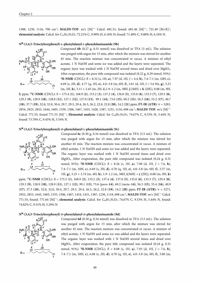
Chapter 2
40
1388, 1230, 1116, 700 cm-1; MALDI-TOF: m/z [M]˙+ Calcd. 681.51; found: 681.46 [M]˙+, 721.49 [M+K]+;
elemental analysis: Calcd. for C41H67N3O5: 72.21% C, 9.90% H, 6.16% N; found: 71.48% C, 9.80% H, 6.16% N.
Nωωωω-(3,4,5-Trioctyloxyphenyl)-L-phenylalanyl-L-phenylalaninamide (9b)
Compound 6b (0.27 g, 0.31 mmol) was dissolved in TFA (3 mL). The solution
was purged with argon for 15 min, after which the mixture was stirred for another
45 min. The reaction mixture was concentrated in vacuo. A mixture of ethyl
acetate, 1 N NaOH and some ice was added and the layers were separated. The
organic layer was washed with 1 N NaOH several times and dried over MgSO4.
After evaporation, the pure title compound was isolated (0.22 g, 0.29 mmol, 93%): 1H-NMR (CDCl3): δ = 8.32 (s, 1H, a), 7.97 (d, 1H, J = 4.4, b), 7.4–7.1 (m, 10H, c),
6.69 (s, 2H, d), 4.77 (q, 1H, e), 4.0–3.8 (m, 6H, f), 3.61 (d, 1H, J = 5.6 Hz, g), 3.23
(m, 2H, h), 3.11 + 2.45 (m, 2H, i),1.9–1.2 (m, 38H, j {36H} + k {2H}), 0.88 (m, 9H,
l) ppm; 13C-NMR (CDCl3): δ = 175.4 (1), 168.9 (2), 153.2 (3), 137.2 (4), 136.8 (5), 135.0 (6), 133.3 (7), 129.5 (8),
129.3 (9), 128.9 (10), 128.8 (11), 127.1 (12), 127.0 (13), 99.1 (14), 73.6 (15), 69.2 (15), 56.3 (16), 55.2 (17), 40.7
(18), 37.7 (19), 32.0, 32.0, 30.4, 29.7, 29.5, 29.4, 26.3, 26.2, 22.8, 22.8 (20), 14.2 (21) ppm; FT-IR (ATR): ν = 3283,
2954, 2923, 2855, 1644, 1605, 1539, 1506, 1467, 1455, 1428, 1387, 1231, 1116, 698 cm-1; MALDI-TOF: m/z [M]˙+
Calcd. 771.55; found 771.53 [M]˙+; Elemental analysis: Calcd. for C48H73N3O5: 74.67% C, 9.53% H, 5.44% N,
found: 73.78% C, 9.45% H, 5.54% N.
Nωωωω-(3,4,5-Trioctyloxyphenyl)-D-phenylalanyl-L-phenylalaninamide (9c)
Compound 6c (0.30 g, 0.34 mmol) was dissolved in TFA (3.5 mL). The solution
was purged with argon for 15 min, after which the mixture was stirred for
another 45 min. The reaction mixture was concentrated in vacuo. A mixture of
ethyl acetate, 1 N NaOH and some ice was added and the layers were separated.
The organic layer was washed with 1 N NaOH several times and dried over
MgSO4. After evaporation, the pure title compound was isolated (0.24 g, 0.31
mmol, 91%): 1H-NMR (CDCl3): δ = 8.26 (s, 1H, a), 7.90 (d, 1H, J = 7.6, b),
7.4–7.1 (m, 10H, c), 6.69 (s, 2H, d), 4.70 (q, 1H, e), 4.0–3.8 (m, 6H, f), 3.57 (m,
1H, g), 3.23 + 2.74 (m, 4H, h), 1.9–1.2 (m, 38H, i{36H} + j {2H}), 0.88 (m, 9H, k)
ppm; 13C-NMR (CDCl3): δ = 175.5 (1), 168.9 (2), 153.2 (3), 137.4 (4), 137.0 (5), 135.0 (6), 133.3 (7), 129.4 (8),
129.3 (9), 128.9 (10), 128.9 (11), 127.1 (12), 99.1 (13), 73.6 (para–14), 69.2 (meta–14), 56.3 (15), 55.4 (16), 40.9
(17), 37.3 (18), 32.0, 32.0, 30.4, 29.7, 29.5, 29.4, 26.3, 26.2, 22.8 (19), 14.2 (20) ppm; FT-IR (ATR): ν = 3271,
2922, 2853, 1643, 1603, 1535, 1506, 1467, 1454, 1431, 1387, 1230, 1118, 698 cm-1; MALDI-TOF: m/z [M]˙+ Calcd.
771.55; found: 771.64 [M]˙+; elemental analysis: Calcd. for C48H73N3O5: 74.67% C, 9.53% H, 5.44% N, found:
74.82% C, 9.51% H, 5.29% N.
Nωωωω-(3,4,5-Trioctyloxyphenyl)-D-phenylalanyl-D-phenylalaninamide (9d)
Compound 6d (0.30 g, 0.34 mmol) was dissolved in TFA (3.5 mL). The solution
was purged with argon for 15 min, after which the mixture was stirred for
another 45 min. The reaction mixture was concentrated in vacuo. A mixture of
ethyl acetate, 1 N NaOH and some ice was added and the layers were separated.
The organic layer was washed with 1 N NaOH several times and dried over
MgSO4. After evaporation, the pure title compound was isolated (0.24 g, 0.31
mmol, 91%): 1H-NMR (CDCl3): δ = 8.08 (s, 1H, a), 7.93 (d, 1H, J = 7.6, b),
7.4–7.1 (m, 10H, c), 6.68 (s, 2H, d), 4.70 (q, 1H, e), 4.0–3.8 (m, 6H, f), 3.60 (m,
12 3
4
5
67
810
11
12
12
14
16
17a
b
c
d
e
f
g
i
13
15
h
9
18
19
20
c
k
h
j
R =
H2NNH
HN
O
O
OR
OR
OR
R =
H2NNH
HN
O
O
OR
OR
OR1
2 3
4
5
67
810
11
12
13
15
17
18a
b
c
d
e
f
g
j
14
16
i
9
19
20
21
c
l
h
k
12 3
4
5
67
810
11
12
13
15
17
18a
b
c
d
e
f
g
j
14
16
i
9
19
20
21
c
l
R =
H2NNH
HN
O
O
OR
OR
ORh
k
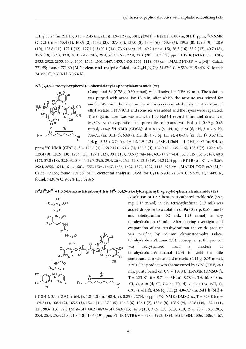
Syntheses of peptide discotics with aliphatic solubilizing tails
41
1H, g), 3.23 (m, 2H, h), 3.11 + 2.45 (m, 2H, i), 1.9–1.2 (m, 38H, j {36H} + k {2H}), 0.88 (m, 9H, l) ppm; 13C-NMR
(CDCl3): δ = 175.4 (1), 168.9 (2), 153.2 (3), 137.4 (4), 137.0 (5), 135.0 (6), 133.3 (7), 129.5 (8), 129.3 (9), 128.9
(10), 128.8 (11), 127.1 (12), 127.1 (13),99.1 (14), 73.6 (para–15), 69.2 (meta–15), 56.3 (16), 55.2 (17), 40.7 (18),
37.5 (19), 32.0, 32.0, 30.4, 29.7, 29.5, 29.4, 26.3, 26.2, 22.8, 22.8 (20), 14.2 (21) ppm; FT-IR (ATR): ν = 3283,
2955, 2922, 2855, 1646, 1606, 1540, 1506, 1467, 1455, 1430, 1231, 1119, 698 cm-1; MALDI-TOF: m/z [M]˙+ Calcd.
771.55; found: 771.60 [M]˙+; elemental analysis: Calcd. for C48H73N3O5: 74.67% C, 9.53% H, 5.44% N, found:
74.35% C, 9.53% H, 5.36% N.
Nωωωω-(3,4,5-Trioctyloxyphenyl)-L-phenylalanyl-D-phenylalaninamide (9e)
Compound 6e (0.78 g, 0.90 mmol) was dissolved in TFA (9 mL). The solution
was purged with argon for 15 min, after which the mixture was stirred for
another 45 min. The reaction mixture was concentrated in vacuo. A mixture of
ethyl acetate, 1 N NaOH and some ice was added and the layers were separated.
The organic layer was washed with 1 N NaOH several times and dried over
MgSO4. After evaporation, the pure title compound was isolated (0.49 g, 0.63
mmol, 71%): 1H-NMR (CDCl3): δ = 8.13 (s, 1H, a), 7.90 (d, 1H, J = 7.6, b),
7.4–7.1 (m, 10H, c), 6.68 (s, 2H, d), 4.70 (q, 1H, e), 4.0–3.8 (m, 6H, f), 3.57 (m,
1H, g), 3.23 + 2.74 (m, 4H, h), 1.9–1.2 (m, 38H, i {36H} + j {2H}), 0.87 (m, 9H, k)
ppm; 13C-NMR (CDCl3): δ = 175.6 (1), 168.9 (2), 153.3 (3), 137.3 (4), 137.0 (5), 135.1 (6), 133.3 (7), 129.4 (8),
129.4 (9), 128.9 (10), 128.9 (11), 127.1 (12), 99.1 (13), 73.6 (para–14), 69.3 (meta–14), 56.3 (15), 55.5 (16), 40.8
(17), 37.0 (18), 32.0, 32.0, 30.4, 29.7, 29.5, 29.4, 26.3, 26.2, 22.8, 22.8 (19), 14.2 (20) ppm; FT-IR (ATR): ν = 3265,
2924, 2855, 1644, 1614, 1603, 1555, 1504, 1467, 1454, 1427, 1379, 1229, 1115, 698 cm-1; MALDI-TOF: m/z [M]˙+
Calcd. 771.55; found: 771.58 [M]˙+; elemental analysis: Calcd. for C48H73N3O5: 74.67% C, 9.53% H, 5.44% N,
found: 74.81% C, 9.62% H, 5.32% N.
Nαααα,Nαααα’,Nαααα’’-(1,3,5-Benzenetricarbonyl)tris[Nωωωω-(3,4,5-trioctyloxyphenyl)]-glycyl-L-phenylalaninamide (2a)
A solution of 1,3,5-benzenetricarbonyl trichloride (45.4
mg, 0.17 mmol) in dry tetrahydrofuran (1.7 mL) was
added dropwise to a solution of 9a (0.39 g, 0.57 mmol)
and triethylamine (0.2 mL, 1.43 mmol) in dry
tetrahydrofuran (5 mL). After stirring overnight and
evaporation of the tetrahydrofuran the crude product
was purified by column chromatography (silica,
tetrahydrofuran/hexane 2/1). Subsequently, the product
was recrystallized from a mixture of
tetrahydrofuran/methanol (2/3) to yield the title
compound as a white solid material (0.12 g, 0.05 mmol,
32%). The product was characterized by GPC (THF, 260
nm, purity based on UV ~ 100%): 1H-NMR (DMSO-d6,
T = 323 K): δ = 9.71 (s, 3H, a), 8.78 (t, 3H, b), 8.48 (s,
3H, c), 8.18 (d, 3H, J = 7.5 Hz, d), 7.3–7.1 (m, 15H, e),
6.91 (s, 6H, f), 4.66 (q, 3H, g), 4.0–3.7 (m, 24H, h {6H} +
i {18H}), 3.1 + 2.9 (m, 6H, j), 1.8–1.0 (m, 108H, k), 0.85 (t, 27H, l) ppm; 13C-NMR (DMSO-d6, T = 323 K): δ =
169.2 (1), 168.4 (2), 165.5 (3), 152.1 (4), 137.3 (5), 134.3 (6), 134.1 (7), 133.6 (8), 128.9 (9), 127.8 (10), 126.1 (11,
12), 98.6 (13), 72.3 (para–14), 68.2 (meta–14), 54.6 (15), 42.6 (16), 37.5 (17), 31.0, 31.0, 29.6, 28.7, 28.6, 28.5,
28.4, 25.4, 25.3, 21.8, 21.8 (18), 13.6 (19) ppm; FT-IR (ATR): ν = 3280, 2923, 2854, 1651, 1604, 1536, 1506, 1467,
12 3
4
5
67
810
11
12
12
14
16
17a
b
c
d
e
f
g
i
13
15
h
9
18
19
20
c
k
h
j
R =
H2NNH
HN
O
O
OR
OR
OR
HN
NH
HN
O
O
OR
OR
OR
ONH
HN
NH
HN
NH
O
O
O
O
O
OHN
OR
RO
RO
RO
OR
OR
R =
k
l
11
f
1516
10
13
14
d
123
4
5
6
78
9 12
a
bc
e
g
h
i
17
18
19
j
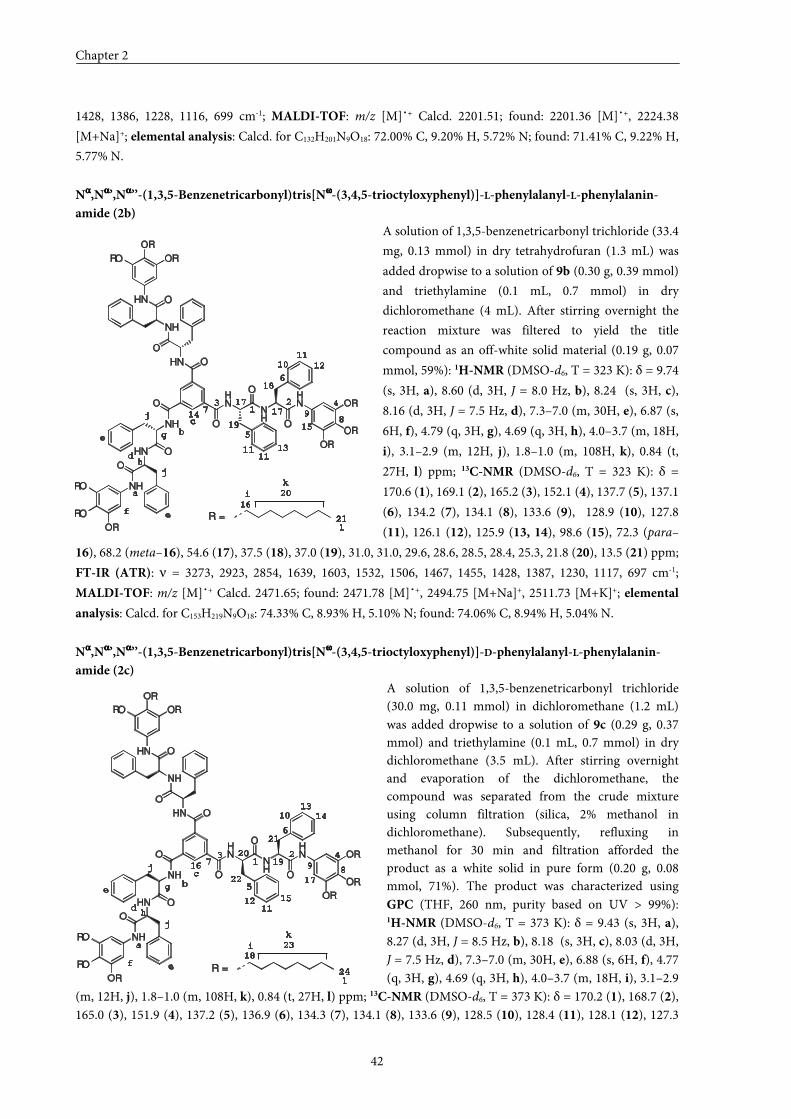
Chapter 2
42
1428, 1386, 1228, 1116, 699 cm-1; MALDI-TOF: m/z [M]˙+ Calcd. 2201.51; found: 2201.36 [M]˙+, 2224.38
[M+Na]+; elemental analysis: Calcd. for C132H201N9O18: 72.00% C, 9.20% H, 5.72% N; found: 71.41% C, 9.22% H,
5.77% N.
Nαααα,Nαααα’,Nαααα’’-(1,3,5-Benzenetricarbonyl)tris[Nωωωω-(3,4,5-trioctyloxyphenyl)]-L-phenylalanyl-L-phenylalanin-
amide (2b)
A solution of 1,3,5-benzenetricarbonyl trichloride (33.4
mg, 0.13 mmol) in dry tetrahydrofuran (1.3 mL) was
added dropwise to a solution of 9b (0.30 g, 0.39 mmol)
and triethylamine (0.1 mL, 0.7 mmol) in dry
dichloromethane (4 mL). After stirring overnight the
reaction mixture was filtered to yield the title
compound as an off-white solid material (0.19 g, 0.07
mmol, 59%): 1H-NMR (DMSO-d6, T = 323 K): δ = 9.74
(s, 3H, a), 8.60 (d, 3H, J = 8.0 Hz, b), 8.24 (s, 3H, c),
8.16 (d, 3H, J = 7.5 Hz, d), 7.3–7.0 (m, 30H, e), 6.87 (s,
6H, f), 4.79 (q, 3H, g), 4.69 (q, 3H, h), 4.0–3.7 (m, 18H,
i), 3.1–2.9 (m, 12H, j), 1.8–1.0 (m, 108H, k), 0.84 (t,
27H, l) ppm; 13C-NMR (DMSO-d6, T = 323 K): δ =
170.6 (1), 169.1 (2), 165.2 (3), 152.1 (4), 137.7 (5), 137.1
(6), 134.2 (7), 134.1 (8), 133.6 (9), 128.9 (10), 127.8
(11), 126.1 (12), 125.9 (13, 14), 98.6 (15), 72.3 (para–
16), 68.2 (meta–16), 54.6 (17), 37.5 (18), 37.0 (19), 31.0, 31.0, 29.6, 28.6, 28.5, 28.4, 25.3, 21.8 (20), 13.5 (21) ppm;
FT-IR (ATR): ν = 3273, 2923, 2854, 1639, 1603, 1532, 1506, 1467, 1455, 1428, 1387, 1230, 1117, 697 cm-1;
MALDI-TOF: m/z [M]˙+ Calcd. 2471.65; found: 2471.78 [M]˙+, 2494.75 [M+Na]+, 2511.73 [M+K]+; elemental
analysis: Calcd. for C153H219N9O18: 74.33% C, 8.93% H, 5.10% N; found: 74.06% C, 8.94% H, 5.04% N.
Nαααα,Nαααα’,Nαααα’’-(1,3,5-Benzenetricarbonyl)tris[Nωωωω-(3,4,5-trioctyloxyphenyl)]-D-phenylalanyl-L-phenylalanin-
amide (2c)
A solution of 1,3,5-benzenetricarbonyl trichloride (30.0 mg, 0.11 mmol) in dichloromethane (1.2 mL) was added dropwise to a solution of 9c (0.29 g, 0.37 mmol) and triethylamine (0.1 mL, 0.7 mmol) in dry dichloromethane (3.5 mL). After stirring overnight and evaporation of the dichloromethane, the compound was separated from the crude mixture using column filtration (silica, 2% methanol in dichloromethane). Subsequently, refluxing in methanol for 30 min and filtration afforded the product as a white solid in pure form (0.20 g, 0.08 mmol, 71%). The product was characterized using GPC (THF, 260 nm, purity based on UV > 99%): 1H-NMR (DMSO-d6, T = 373 K): δ = 9.43 (s, 3H, a), 8.27 (d, 3H, J = 8.5 Hz, b), 8.18 (s, 3H, c), 8.03 (d, 3H, J = 7.5 Hz, d), 7.3–7.0 (m, 30H, e), 6.88 (s, 6H, f), 4.77 (q, 3H, g), 4.69 (q, 3H, h), 4.0–3.7 (m, 18H, i), 3.1–2.9
(m, 12H, j), 1.8–1.0 (m, 108H, k), 0.84 (t, 27H, l) ppm; 13C-NMR (DMSO-d6, T = 373 K): δ = 170.2 (1), 168.7 (2), 165.0 (3), 151.9 (4), 137.2 (5), 136.9 (6), 134.3 (7), 134.1 (8), 133.6 (9), 128.5 (10), 128.4 (11), 128.1 (12), 127.3
j
k
18
19
12
f
15
16
10 14
d
123 4
5
6
78
9
13
a
b
c
e
i
g17
e
h
j
l
11
21
20
22
23
R =
HN
NH
HN
O
O
OR
OR
OR
ONH
HN
NH
HN
NH
O
O
O
O
O
OHN
OR
RO
RO
RO
OR
OR
24
HN
NH
HN
O
O
OR
OR
OR
ONH
HN
NH
HN
NH
O
O
O
O
O
OHN
OR
RO
RO
RO
OR
OR
R =
j
k
14
18
19
11
f
15
16
10
13d
123 4
5
6
7
89
12
a
b
c
e
i
g
17
e
h
j
l
11
21
20
17
11
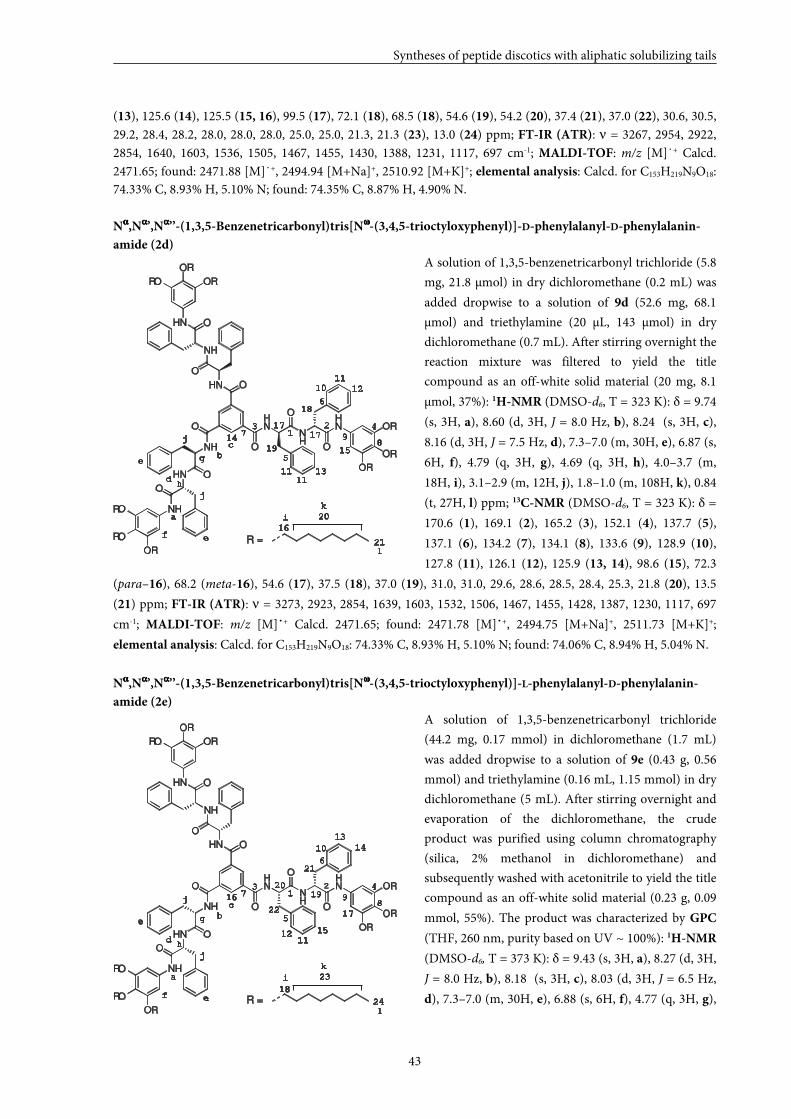
Syntheses of peptide discotics with aliphatic solubilizing tails
43
(13), 125.6 (14), 125.5 (15, 16), 99.5 (17), 72.1 (18), 68.5 (18), 54.6 (19), 54.2 (20), 37.4 (21), 37.0 (22), 30.6, 30.5, 29.2, 28.4, 28.2, 28.0, 28.0, 28.0, 25.0, 25.0, 21.3, 21.3 (23), 13.0 (24) ppm; FT-IR (ATR): ν = 3267, 2954, 2922, 2854, 1640, 1603, 1536, 1505, 1467, 1455, 1430, 1388, 1231, 1117, 697 cm-1; MALDI-TOF: m/z [M]˙+ Calcd. 2471.65; found: 2471.88 [M]˙+, 2494.94 [M+Na]+, 2510.92 [M+K]+; elemental analysis: Calcd. for C153H219N9O18: 74.33% C, 8.93% H, 5.10% N; found: 74.35% C, 8.87% H, 4.90% N.
Nαααα,Nαααα’,Nαααα’’-(1,3,5-Benzenetricarbonyl)tris[Nωωωω-(3,4,5-trioctyloxyphenyl)]-D-phenylalanyl-D-phenylalanin-
amide (2d)
A solution of 1,3,5-benzenetricarbonyl trichloride (5.8
mg, 21.8 µmol) in dry dichloromethane (0.2 mL) was
added dropwise to a solution of 9d (52.6 mg, 68.1
µmol) and triethylamine (20 µL, 143 µmol) in dry
dichloromethane (0.7 mL). After stirring overnight the
reaction mixture was filtered to yield the title
compound as an off-white solid material (20 mg, 8.1
µmol, 37%): 1H-NMR (DMSO-d6, T = 323 K): δ = 9.74
(s, 3H, a), 8.60 (d, 3H, J = 8.0 Hz, b), 8.24 (s, 3H, c),
8.16 (d, 3H, J = 7.5 Hz, d), 7.3–7.0 (m, 30H, e), 6.87 (s,
6H, f), 4.79 (q, 3H, g), 4.69 (q, 3H, h), 4.0–3.7 (m,
18H, i), 3.1–2.9 (m, 12H, j), 1.8–1.0 (m, 108H, k), 0.84
(t, 27H, l) ppm; 13C-NMR (DMSO-d6, T = 323 K): δ =
170.6 (1), 169.1 (2), 165.2 (3), 152.1 (4), 137.7 (5),
137.1 (6), 134.2 (7), 134.1 (8), 133.6 (9), 128.9 (10),
127.8 (11), 126.1 (12), 125.9 (13, 14), 98.6 (15), 72.3
(para–16), 68.2 (meta-16), 54.6 (17), 37.5 (18), 37.0 (19), 31.0, 31.0, 29.6, 28.6, 28.5, 28.4, 25.3, 21.8 (20), 13.5
(21) ppm; FT-IR (ATR): ν = 3273, 2923, 2854, 1639, 1603, 1532, 1506, 1467, 1455, 1428, 1387, 1230, 1117, 697
cm-1; MALDI-TOF: m/z [M]˙+ Calcd. 2471.65; found: 2471.78 [M]˙+, 2494.75 [M+Na]+, 2511.73 [M+K]+;
elemental analysis: Calcd. for C153H219N9O18: 74.33% C, 8.93% H, 5.10% N; found: 74.06% C, 8.94% H, 5.04% N.
Nαααα,Nαααα’,Nαααα’’-(1,3,5-Benzenetricarbonyl)tris[Nωωωω-(3,4,5-trioctyloxyphenyl)]-L-phenylalanyl-D-phenylalanin-
amide (2e)
A solution of 1,3,5-benzenetricarbonyl trichloride
(44.2 mg, 0.17 mmol) in dichloromethane (1.7 mL)
was added dropwise to a solution of 9e (0.43 g, 0.56
mmol) and triethylamine (0.16 mL, 1.15 mmol) in dry
dichloromethane (5 mL). After stirring overnight and
evaporation of the dichloromethane, the crude
product was purified using column chromatography
(silica, 2% methanol in dichloromethane) and
subsequently washed with acetonitrile to yield the title
compound as an off-white solid material (0.23 g, 0.09
mmol, 55%). The product was characterized by GPC
(THF, 260 nm, purity based on UV ~ 100%): 1H-NMR
(DMSO-d6, T = 373 K): δ = 9.43 (s, 3H, a), 8.27 (d, 3H,
J = 8.0 Hz, b), 8.18 (s, 3H, c), 8.03 (d, 3H, J = 6.5 Hz,
d), 7.3–7.0 (m, 30H, e), 6.88 (s, 6H, f), 4.77 (q, 3H, g),
j
k
14
18
19
11
f
15
16
10
13d
123 4
5
6
7
89
12
a
b
c
e
i
g
17
e
h
j
l
11
21
20
17
R =
HN
NH
HN
O
O
OR
OR
OR
ONH
HN
NH
HN
NH
O
O
O
O
O
OHN
OR
RO
RO
RO
OR
OR
11
j
k
18
19
12
f
15
16
10 14
d
13 4
5
6
7
89
13
a
b
c
e
i
g17
e
h
j
l
11
21
20
R =
2HN
NH
HN
O
O
OR
OR
OR
ONH
HN
NH
HN
NH
O
O
O
O
O
OHN
OR
RO
RO
RO
OR
OR
22
23
24
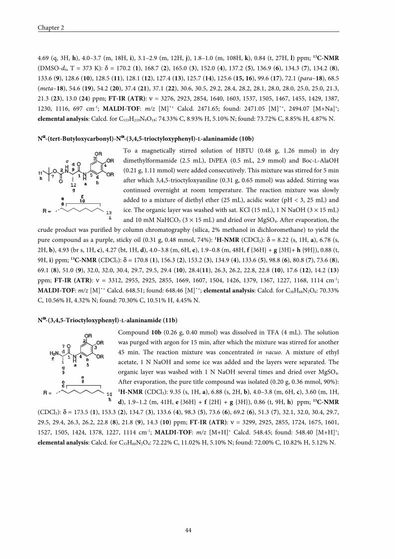
Chapter 2
44
4.69 (q, 3H, h), 4.0–3.7 (m, 18H, i), 3.1–2.9 (m, 12H, j), 1.8–1.0 (m, 108H, k), 0.84 (t, 27H, l) ppm; 13C-NMR
(DMSO-d6, T = 373 K): δ = 170.2 (1), 168.7 (2), 165.0 (3), 152.0 (4), 137.2 (5), 136.9 (6), 134.3 (7), 134.2 (8),
133.6 (9), 128.6 (10), 128.5 (11), 128.1 (12), 127.4 (13), 125.7 (14), 125.6 (15, 16), 99.6 (17), 72.1 (para–18), 68.5
(meta–18), 54.6 (19), 54.2 (20), 37.4 (21), 37.1 (22), 30.6, 30.5, 29.2, 28.4, 28.2, 28.1, 28.0, 28.0, 25.0, 25.0, 21.3,
21.3 (23), 13.0 (24) ppm; FT-IR (ATR): ν = 3276, 2923, 2854, 1640, 1603, 1537, 1505, 1467, 1455, 1429, 1387,
1230, 1116, 697 cm-1; MALDI-TOF: m/z [M]˙+ Calcd. 2471.65; found: 2471.05 [M]˙+, 2494.07 [M+Na]+;
elemental analysis: Calcd. for C153H219N9O18: 74.33% C, 8.93% H, 5.10% N; found: 73.72% C, 8.85% H, 4.87% N.
Nαααα-(tert-Butyloxycarbonyl)-Nωωωω-(3,4,5-trioctyloxyphenyl)-L-alaninamide (10b)
To a magnetically stirred solution of HBTU (0.48 g, 1.26 mmol) in dry
dimethylformamide (2.5 mL), DiPEA (0.5 mL, 2.9 mmol) and Boc-L-AlaOH
(0.21 g, 1.11 mmol) were added consecutively. This mixture was stirred for 5 min
after which 3,4,5-trioctyloxyaniline (0.31 g, 0.65 mmol) was added. Stirring was
continued overnight at room temperature. The reaction mixture was slowly
added to a mixture of diethyl ether (25 mL), acidic water (pH < 3, 25 mL) and
ice. The organic layer was washed with sat. KCl (15 mL), 1 N NaOH (3 × 15 mL)
and 10 mM NaHCO3 (3 × 15 mL) and dried over MgSO4. After evaporation, the
crude product was purified by column chromatography (silica, 2% methanol in dichloromethane) to yield the
pure compound as a purple, sticky oil (0.31 g, 0.48 mmol, 74%): 1H-NMR (CDCl3): δ = 8.22 (s, 1H, a), 6.78 (s,
2H, b), 4.93 (br s, 1H, c), 4.27 (bt, 1H, d), 4.0–3.8 (m, 6H, e), 1.9–0.8 (m, 48H, f {36H} + g {3H}+ h {9H}), 0.88 (t,
9H, i) ppm; 13C-NMR (CDCl3): δ = 170.8 (1), 156.3 (2), 153.2 (3), 134.9 (4), 133.6 (5), 98.8 (6), 80.8 (7), 73.6 (8),
69.1 (8), 51.0 (9), 32.0, 32.0, 30.4, 29.7, 29.5, 29.4 (10), 28.4(11), 26.3, 26.2, 22.8, 22.8 (10), 17.6 (12), 14.2 (13)
ppm; FT-IR (ATR): ν = 3312, 2955, 2925, 2855, 1669, 1607, 1504, 1426, 1379, 1367, 1227, 1168, 1114 cm-1;
MALDI-TOF: m/z [M]˙+ Calcd. 648.51; found: 648.46 [M]˙+; elemental analysis: Calcd. for C38H68N2O6: 70.33%
C, 10.56% H, 4.32% N; found: 70.30% C, 10.51% H, 4.45% N.
Nωωωω-(3,4,5-Trioctyloxyphenyl)-L-alaninamide (11b)
Compound 10b (0.26 g, 0.40 mmol) was dissolved in TFA (4 mL). The solution
was purged with argon for 15 min, after which the mixture was stirred for another
45 min. The reaction mixture was concentrated in vacuo. A mixture of ethyl
acetate, 1 N NaOH and some ice was added and the layers were separated. The
organic layer was washed with 1 N NaOH several times and dried over MgSO4.
After evaporation, the pure title compound was isolated (0.20 g, 0.36 mmol, 90%): 1H-NMR (CDCl3): 9.35 (s, 1H, a), 6.88 (s, 2H, b), 4.0–3.8 (m, 6H, c), 3.60 (m, 1H,
d), 1.9–1.2 (m, 41H, e {36H} + f {2H} + g {3H}), 0.86 (t, 9H, h) ppm; 13C-NMR
(CDCl3): δ = 173.5 (1), 153.3 (2), 134.7 (3), 133.6 (4), 98.3 (5), 73.6 (6), 69.2 (6), 51.3 (7), 32.1, 32.0, 30.4, 29.7,
29.5, 29.4, 26.3, 26.2, 22.8 (8), 21.8 (9), 14.3 (10) ppm; FT-IR (ATR): ν = 3299, 2925, 2855, 1724, 1675, 1601,
1527, 1505, 1424, 1378, 1227, 1114 cm-1; MALDI-TOF: m/z [M+H]+ Calcd. 548.45; found: 548.40 [M+H]+;
elemental analysis: Calcd. for C33H60N2O4: 72.22% C, 11.02% H, 5.10% N; found: 72.00% C, 10.82% H, 5.12% N.
1
a
b
c
d
e
f
g
h
2
3
4
5
6
7
9
8
10R =
H2N
O
NH
OR
OR
OR
1
a b
cd
e f
i
g
12
13
h
2
3
4
567
9
810
R =
11
HN
O
O
O
NH
OR
OR
OR
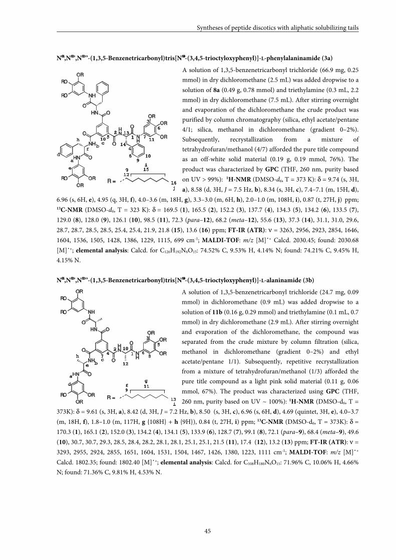
Syntheses of peptide discotics with aliphatic solubilizing tails
45
Nαααα,Nαααα’,Nαααα’’-(1,3,5-Benzenetricarbonyl)tris[Nωωωω-(3,4,5-trioctyloxyphenyl)]-L-phenylalaninamide (3a)
A solution of 1,3,5-benzenetricarbonyl trichloride (66.9 mg, 0.25
mmol) in dry dichloromethane (2.5 mL) was added dropwise to a
solution of 8a (0.49 g, 0.78 mmol) and triethylamine (0.3 mL, 2.2
mmol) in dry dichloromethane (7.5 mL). After stirring overnight
and evaporation of the dichloromethane the crude product was
purified by column chromatography (silica, ethyl acetate/pentane
4/1; silica, methanol in dichloromethane (gradient 0–2%).
Subsequently, recrystallization from a mixture of
tetrahydrofuran/methanol (4/7) afforded the pure title compound
as an off-white solid material (0.19 g, 0.19 mmol, 76%). The
product was characterized by GPC (THF, 260 nm, purity based
on UV > 99%): 1H-NMR (DMSO-d6, T = 373 K): δ = 9.74 (s, 3H,
a), 8.58 (d, 3H, J = 7.5 Hz, b), 8.34 (s, 3H, c), 7.4–7.1 (m, 15H, d),
6.96 (s, 6H, e), 4.95 (q, 3H, f), 4.0–3.6 (m, 18H, g), 3.3–3.0 (m, 6H, h), 2.0–1.0 (m, 108H, i), 0.87 (t, 27H, j) ppm; 13C-NMR (DMSO-d6, T = 323 K): δ = 169.5 (1), 165.5 (2), 152.2 (3), 137.7 (4), 134.3 (5), 134.2 (6), 133.5 (7),
129.0 (8), 128.0 (9), 126.1 (10), 98.5 (11), 72.3 (para–12), 68.2 (meta–12), 55.6 (13), 37.3 (14), 31.1, 31.0, 29.6,
28.7, 28.7, 28.5, 28.5, 25.4, 25.4, 21.9, 21.8 (15), 13.6 (16) ppm; FT-IR (ATR): ν = 3263, 2956, 2923, 2854, 1646,
1604, 1536, 1505, 1428, 1386, 1229, 1115, 699 cm-1; MALDI-TOF: m/z [M]˙+ Calcd. 2030.45; found: 2030.68
[M]˙+; elemental analysis: Calcd. for C126H192N6O15: 74.52% C, 9.53% H, 4.14% N; found: 74.21% C, 9.45% H,
4.15% N.
Nαααα,Nαααα’,Nαααα’’-(1,3,5-Benzenetricarbonyl)tris[Nωωωω-(3,4,5-trioctyloxyphenyl)]-L-alaninamide (3b)
A solution of 1,3,5-benzenetricarbonyl trichloride (24.7 mg, 0.09
mmol) in dichloromethane (0.9 mL) was added dropwise to a
solution of 11b (0.16 g, 0.29 mmol) and triethylamine (0.1 mL, 0.7
mmol) in dry dichloromethane (2.9 mL). After stirring overnight
and evaporation of the dichloromethane, the compound was
separated from the crude mixture by column filtration (silica,
methanol in dichloromethane (gradient 0–2%) and ethyl
acetate/pentane 1/1). Subsequently, repetitive recrystallization
from a mixture of tetrahydrofuran/methanol (1/3) afforded the
pure title compound as a light pink solid material (0.11 g, 0.06
mmol, 67%). The product was characterized using GPC (THF,
260 nm, purity based on UV ~ 100%): 1H-NMR (DMSO-d6, T =
373K): δ = 9.61 (s, 3H, a), 8.42 (d, 3H, J = 7.2 Hz, b), 8.50 (s, 3H, c), 6.96 (s, 6H, d), 4.69 (quintet, 3H, e), 4.0–3.7
(m, 18H, f), 1.8–1.0 (m, 117H, g {108H} + h {9H}), 0.84 (t, 27H, i) ppm; 13C-NMR (DMSO-d6, T = 373K): δ =
170.3 (1), 165.1 (2), 152.0 (3), 134.2 (4), 134.1 (5), 133.9 (6), 128.7 (7), 99.1 (8), 72.1 (para–9), 68.4 (meta–9), 49.6
(10), 30.7, 30.7, 29.3, 28.5, 28.4, 28.2, 28.1, 28.1, 25.1, 25.1, 21.5 (11), 17.4 (12), 13.2 (13) ppm; FT-IR (ATR): ν =
3293, 2955, 2924, 2855, 1651, 1604, 1531, 1504, 1467, 1426, 1380, 1223, 1111 cm-1; MALDI-TOF: m/z [M]˙+
Calcd. 1802.35; found: 1802.40 [M]˙+; elemental analysis: Calcd. for C108H180N6O15: 71.96% C, 10.06% H, 4.66%
N; found: 71.36% C, 9.81% H, 4.53% N.
1
a
bc
d
e
f
i
g
12
13
h
2
3
4
5
67
9
8
10
11
R =
HN
ONH
HN
HN
NH
O
O
O
O
O
NH
RO
RO
OR
OR
OR
OR
RO
OR
OR
1
a
bc
d
e
f
HN
ONH
HN
HN
NH
O
O
O
O
O
NH
RO
RO
OR
OR
OR
OR
RO
OR
ORR =
i
12
g
j
15
16
h
2
3
4
5
6
7
98 10
10 11
13
14
Chapter 2
46
2.7 References and notes
[1] C. Moberg, Angew. Chem. 1998, 110, 260–281; Angew. Chem., Int. Ed. 1998, 37, 248–268.
[2] S.E. Gibson, M.P. Castaldi, Chem. Commun. 2006, 3045–3062.
[3] S.E. Gibson, M.P. Castaldi, Angew. Chem. 2006, 118, 4834–4837; Angew. Chem., Int. Ed. 2006, 45, 4718–
4720.
[4] E. Fan, J. Yang, S.J. Geib, T.C. Stoner, M.D. Hopkins, A.D. Hamilton, J. Chem. Soc., Chem. Commun.
1995, 1251–1252.
[5] M.P. Lightfoot, F.S. Mair, R.G. Pritchard, J.E. Warren, Chem. Commun. 1999, 1945–1946.
[6] Y. Matsunaga, N. Miyajima, Y. Nakayasu, S. Sakai, M. Yonenaga, Bull. Chem. Soc. Jpn. 1988, 61, 207–
210.
[7] Y. Yasuda, E. Ishi, H. Inada, Y. Shirota, Chem. Lett. 1996, 575–576.
[8] K. Hanabusa, C. Kato, M. Kimura, H. Shirai, A. Kakehi, Chem. Lett. 1997, 429–430.
[9] S.Y. Ryu, S. Kim, J. Seo, Y-W. Kim, O-H. Kwon, D-J. Jang, S.Y. Park, Chem. Commun. 2004, 70–71.
[10] S.J. Lee, C.R. Park, J.Y. Chang, Langmuir 2004, 20, 9513–9519.
[11] K.J.C. van Bommel, C. van der Pol, I. Muizebelt, A. Friggeri, A. Heeres, A. Meetsma, B.L. Jeringa, J. van
Esch, Angew. Chem. 2004, 116, 1695–1699; Angew. Chem., Int. Ed. 2004, 43, 1663–1667.
[12] T-Q. Nguyen, R. Martel, P. Avouris, M.L. Bushey, L. Brus, C. Nuckolls, J. Am. Chem. Soc. 2004, 126,
5234–5242.
[13] P.P. Bose, M.G.B. Drew, A.K. Das, A. Banerjee, Chem. Commun. 2006, 3196–3198.
[14] M. de Loos, Thesis: Hydrogen-bonded low molecular weight gelators; University of Groningen:
Groningen (The Netherlands), 2005.
[15] A.R.A. Palmans, J.A.J.M. Vekemans, E.E. Havinga, E.W. Meijer, Angew. Chem. 1997, 109, 2763–2765;
Angew. Chem., Int. Ed. 1997, 36, 2648–2651.
[16] A.R.A. Palmans, J.A.J.M. Vekemans, R.A. Hikmet, H. Fischer, E.W. Meijer, Adv. Mater. 1998, 10, 873–
876.
[17] E.W. Meijer, J.A.J.M. Vekemans, A.R.A. Palmans, P. Breure, J. de Kraker, L. Brunsveld, Polym. Prepr.
2000, 41, 902–903.
[18] J. van Herrikhuyzen, P. Jonkheijm, A.P.H.J. Schenning, E.W. Meijer, Org. Biomol. Chem. 2006, 4, 1539–
1545.
[19] J.J. van Gorp, J.A.J.M. Vekemans, E.W. Meijer, J. Am. Chem. Soc. 2002, 124, 14759–14769.
[20] 3,4,5-Trioctyloxyaniline was synthesized as described for 3,4,5-tridodecyloxyaniline by K. Pieterse,
Thesis: Electron deficient materials based on azaheterocycles, Eindhoven University of Technology,
Eindhoven (The Netherlands), 2001. The obtained data correspond to those described for the
compound by A. Zinsou, M. Veber, H. Strzelecka, C. Jallabert, P. Fourré, New J. Chem. 1993, 17, 309.
[21] K. Pieterse, Thesis: Electron deficient materials based on azaheterocycles, Eindhoven University of
Technology, Eindhoven (The Netherlands), 2001.
[22] X. Lou, K.P. van den Hout, M.H.C.J. van Houtem, J.L.J. van Dongen, J.A.J.M. Vekemans, E.W. Meijer, J.
Mass Spectrom. 2006, 41, 659–669.
[23] Y. Okada, Curr. Org. Chem. 2001, 5, 1–43.
[24] A. Berkessel, F. Cleemann, S. Mukherjee, T.N. Müller, J. Lex, Angew. Chem. 2005, 117, 817–821; Angew.
Chem., Int. Ed. 2005, 44, 807–811.
[25] K. Gottwald, D. Seebach, Tetrahedron 1999, 55, 723–738.
33
Self-assembly of peptide discotics with aliphatic
solubilizing tails
Characterization in the neat state and in solution
ABSTRACT: The self-assembly of C3-symmetrical molecules that consist of a 1,3,5-
benzenetricarboxamide core extended with three peptide fragments decorated with peripheral
mesogenic groups containing aliphatic solubilizing tails is described. Small structural
modifications in the peptide fragments have been applied to demonstrate their influence on the
stability of the self-assembled structures. Seven dipeptide discotics have been investigated, all
with different combinations of glycine and L- and/or D-phenylalanine. Characterization of these
discotics in the neat state using DSC and polarized optical microscopy (POM) and in solution
with CD, UV/Vis, 1H-NMR and IR reveals that there is a clear upward trend in the stack
stability, going from the glycine-phenylalanine combinations to the phenylalanine-
phenylalanine motifs. A larger hydrophobic core and additional π–π interaction contribute to
enhanced stability of the stacks. In addition, stacking properties of two monopeptide discotics
containing an L-phenylalanine or an L-alanine fragment have been investigated and compared,
both in the neat state and in solution. It is unambiguously shown that the L-phenylalanine based
discs form more stable stacks compared to the L-alanine based discs.
* Part of this work has been published: K.P. van den Hout, R. Martín-Rapún, J. A. J. M. Vekemans, E. W. Meijer,
Chem. Eur. J. 2007, 13, 8111–8123.
Chapter 3
48
3.1 Introduction
Self-assembled architectures are envisioned to serve as important building blocks in
the fields of biology as well as material science.[1–11] The interplay of secondary interactions
fulfils a key role in the formation of stable conformers that still can express their reversible
nature. It is known that minor changes in the molecule may dramatically influence the subtle
balance of non-covalent interactions between molecules, in the most extreme cases resulting
in the loss of reversibility or in preventing the formation of stable self-assembled
structures.[12–16] The challenge is to develop new molecules that self-assemble and allow
functionalization and fine-tuning of the structure in an easily accessible manner.
Peptide fragments are widely used as structuring elements in self-assembled
architectures for their biocompatibility, hydrogen-bonding properties and versatile
nature.[17–25] Amphiphilic dendritic dipeptides that self-assemble into helical pores have been
described by Percec and coworkers.[26] Stupp and collaborators have reported peptide
amphiphiles that self-assemble into nanofibers and they demonstrate the ability of such
systems to tolerate chemical modifications by changing the peptide sequence.[27] In the group
of Van Esch excellent low molecular weight hydrogelators have been developed that possess a
cyclohexane core extended with peptides.[28] They show that the properties of the gels can
easily be tuned by changing the hydrophobic substituents or the number of hydrogen bonding
moieties. Recently, Kimizuka and coworkers have also reported C3-symmetrical conjugates
that bear three β-sheet forming peptides (FKFEFKFE) that form anti-parallel β-sheets in
water.[29] These C3-symmetrical peptide conjugates spontaneously self-assemble into virus-
sized peptide nanospheres.
In our laboratory, the self-assembly of various 1,3,5-benzenetricarboxamide
derivatives, which have a strong tendency to form columnar aggregates in solution, has been
extensively studied.[30–33] In our present design the 1,3,5-benzenetricarboxamide centre is
extended with peptide fragments providing additional driving forces for self-assembly (like
hydrogen bonding and π–π interaction).
In this Chapter, the self-assembly behaviour of all nine peptide discotics introduced in
the previous Chapter (Figure 1) will be evaluated. The self-assembly processes of all these
molecules are studied in the neat state using DSC, POM and XRD as well as in solution with
CD, UV/Vis, low concentration 1H-NMR and IR spectroscopy.
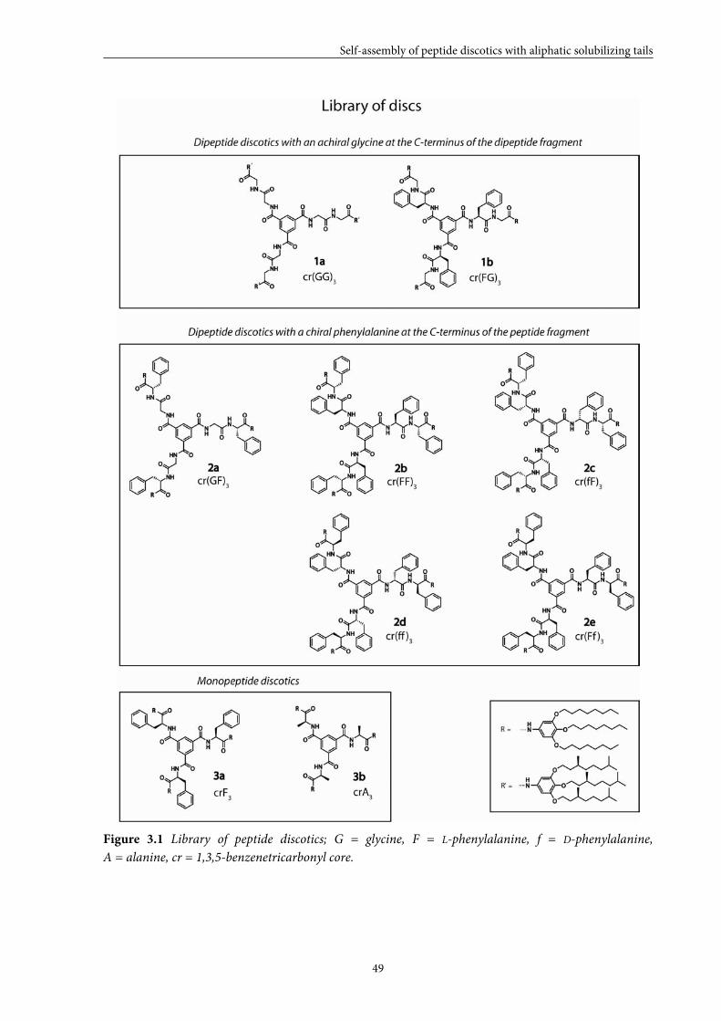
Self-assembly of peptide discotics with aliphatic solubilizing tails
49
Figure 3.1 Library of peptide discotics; G = glycine, F = L-phenylalanine, f = D-phenylalanine,
A = alanine, cr = 1,3,5-benzenetricarbonyl core.
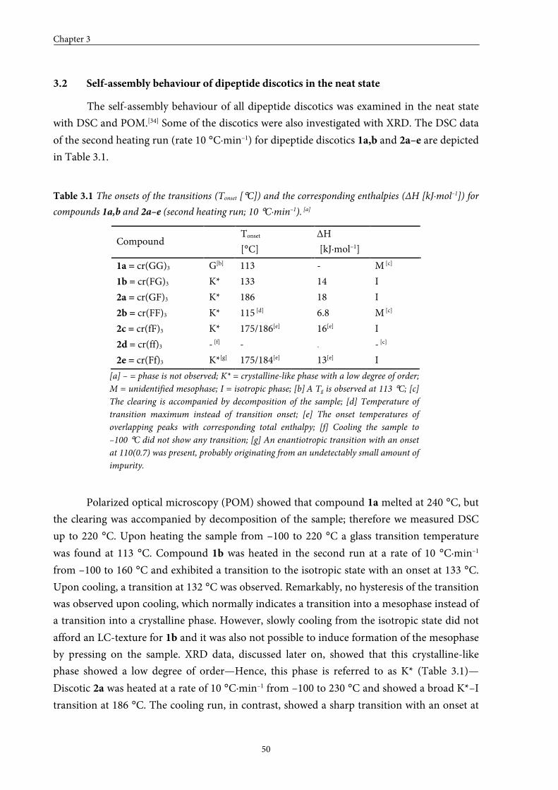
Chapter 3
50
3.2 Self-assembly behaviour of dipeptide discotics in the neat state
The self-assembly behaviour of all dipeptide discotics was examined in the neat state
with DSC and POM.[34] Some of the discotics were also investigated with XRD. The DSC data
of the second heating run (rate 10 °C·min–1) for dipeptide discotics 1a,b and 2a–e are depicted
in Table 3.1.
Table 3.1 The onsets of the transitions (Tonset [°C]) and the corresponding enthalpies (ΔH [kJ·mol–1]) for
compounds 1a,b and 2a–e (second heating run; 10 °C·min–1). [a]
Compound Tonset
[°C]
H ΔH
[kJ·mol–1]
1a = cr(GG)3 G[b] 113 - M [c]
1b = cr(FG)3 K* 133 14 I
2a = cr(GF)3 K* 186 18 I
2b = cr(FF)3 K* 115 [d] 6.8 M [c]
2c = cr(fF)3 K* 175/186[e] 16[e] I
2d = cr(ff)3 - [f] - - - [c]
2e = cr(Ff)3 K*[g] 175/184[e] 13[e] I
[a] – = phase is not observed; K* = crystalline-like phase with a low degree of order;
M = unidentified mesophase; I = isotropic phase; [b] A Tg is observed at 113 °C; [c]
The clearing is accompanied by decomposition of the sample; [d] Temperature of
transition maximum instead of transition onset; [e] The onset temperatures of
overlapping peaks with corresponding total enthalpy; [f] Cooling the sample to
–100 °C did not show any transition; [g] An enantiotropic transition with an onset
at 110(0.7) was present, probably originating from an undetectably small amount of
impurity.
Polarized optical microscopy (POM) showed that compound 1a melted at 240 °C, but
the clearing was accompanied by decomposition of the sample; therefore we measured DSC
up to 220 °C. Upon heating the sample from –100 to 220 °C a glass transition temperature
was found at 113 °C. Compound 1b was heated in the second run at a rate of 10 °C·min–1
from –100 to 160 °C and exhibited a transition to the isotropic state with an onset at 133 °C.
Upon cooling, a transition at 132 °C was observed. Remarkably, no hysteresis of the transition
was observed upon cooling, which normally indicates a transition into a mesophase instead of
a transition into a crystalline phase. However, slowly cooling from the isotropic state did not
afford an LC-texture for 1b and it was also not possible to induce formation of the mesophase
by pressing on the sample. XRD data, discussed later on, showed that this crystalline-like
phase showed a low degree of order—Hence, this phase is referred to as K* (Table 3.1)—
Discotic 2a was heated at a rate of 10 °C·min–1 from –100 to 230 °C and showed a broad K*–I
transition at 186 °C. The cooling run, in contrast, showed a sharp transition with an onset at
Self-assembly of peptide discotics with aliphatic solubilizing tails
51
176 °C. Polarized optical microscopy pointed out that a monotropic mesophase was present at
around 202 °C. The texture of the mesophase could be grown as is shown in Figure 3.2.
Probably, the enthalpy associated to this I–M transition was too small to be detected with
DSC.
Figure 3.2 Polarized optical microscopy texture of compound 2a at 187 °C.
Homochiral enantiomers cr(FF)3 (2b) and cr(ff)3 (2d) started to show some
decomposition around 240 °C, before reaching the clearing point (around 305 °C); therefore
these discotics were heated from –100 to 220 °C in DSC. Discotic 2b showed a reversible
transition with a maximum at 115 °C in the heating run and a maximum at 107 °C in the
cooling run. Surprisingly, enantiomer 2d did not exhibit any transition upon cooling to
–100 °C. Heterochiral enantiomers cr(fF) (2c) and cr(Ff)3 (2e) melted around 200 °C. Upon
heating discotic 2e from –100 to 220 °C three transitions could be discerned, a small
transition with a maximum at 110 °C and a transition that was overlapping the transition to
the isotropic state with a onset at 175 °C. Upon cooling, just one I–K* transition was observed
with a maximum at 153 °C. The DSC curve of discotic 2c was similar to the one of 2e, except
for the small transition at 110 °C in the heating run, which was missing. Most likely, this small
peak originated from a small amount of impurity, which could not be detected by MALDI-
TOF MS and GPC.
Dipeptide discotics 1a,b and 2a,b,e have also been investigated with XRD. The data are
gathered in table 3.2. The XRD patterns of compounds 1a,b consist of an intense peak and a
diffuse halo in the small and the wide angle region, respectively. The position of the former is
temperature-independent up to the clearing point and corresponds to a distance of 32 Å for
1a and 27 Å for 1b, which are in the range of the diameters of the molecules. The diffuse halo
is attributed to the aliphatic chains in the periphery although the associated spacings, 4.8 Å for
1a and 4.6 Å for 1b at room temperature, are somehow larger than the expected 4.4–4.6 Å.
Increasing the temperature leads to an increase in the distance corresponding to the halo.
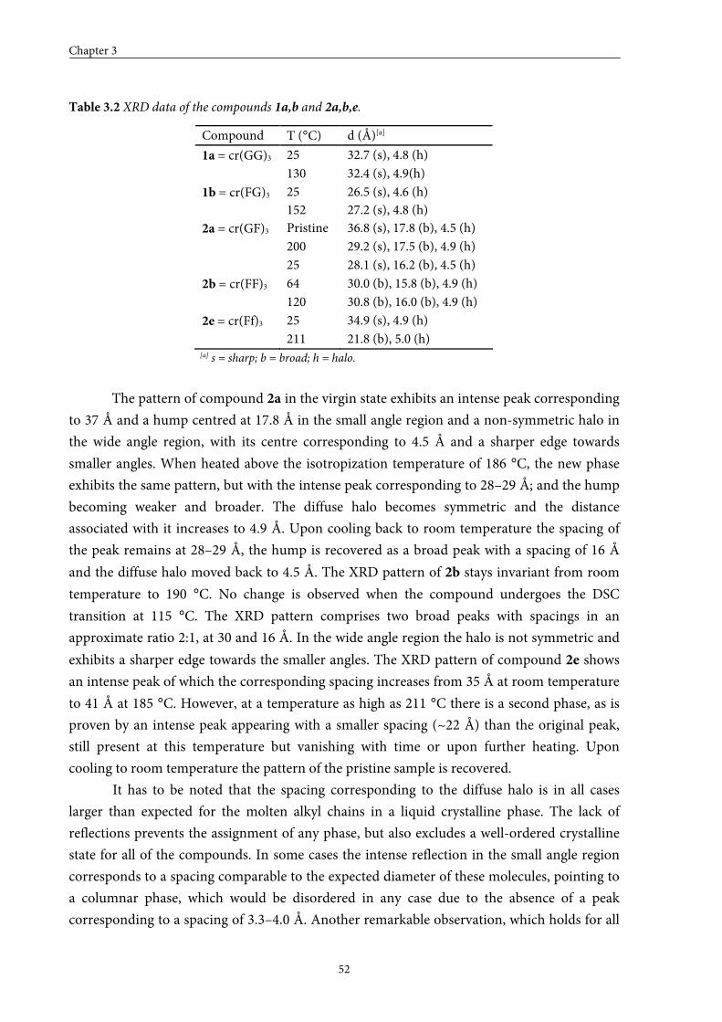
Chapter 3
52
Table 3.2 XRD data of the compounds 1a,b and 2a,b,e.
Compound T (°C) d (Å)[a]
1a = cr(GG)3 25 32.7 (s), 4.8 (h)
130 32.4 (s), 4.9(h)
1b = cr(FG)3 25 152
26.5 (s), 4.6 (h) 27.2 (s), 4.8 (h)
2a = cr(GF)3 Pristine 36.8 (s), 17.8 (b), 4.5 (h)
200 29.2 (s), 17.5 (b), 4.9 (h)
25 28.1 (s), 16.2 (b), 4.5 (h)
2b = cr(FF)3 64 30.0 (b), 15.8 (b), 4.9 (h)
120 30.8 (b), 16.0 (b), 4.9 (h)
2e = cr(Ff)3 25 34.9 (s), 4.9 (h)
211 21.8 (b), 5.0 (h) [a] s = sharp; b = broad; h = halo.
The pattern of compound 2a in the virgin state exhibits an intense peak corresponding
to 37 Å and a hump centred at 17.8 Å in the small angle region and a non-symmetric halo in
the wide angle region, with its centre corresponding to 4.5 Å and a sharper edge towards
smaller angles. When heated above the isotropization temperature of 186 °C, the new phase
exhibits the same pattern, but with the intense peak corresponding to 28–29 Å; and the hump
becoming weaker and broader. The diffuse halo becomes symmetric and the distance
associated with it increases to 4.9 Å. Upon cooling back to room temperature the spacing of
the peak remains at 28–29 Å, the hump is recovered as a broad peak with a spacing of 16 Å
and the diffuse halo moved back to 4.5 Å. The XRD pattern of 2b stays invariant from room
temperature to 190 °C. No change is observed when the compound undergoes the DSC
transition at 115 °C. The XRD pattern comprises two broad peaks with spacings in an
approximate ratio 2:1, at 30 and 16 Å. In the wide angle region the halo is not symmetric and
exhibits a sharper edge towards the smaller angles. The XRD pattern of compound 2e shows
an intense peak of which the corresponding spacing increases from 35 Å at room temperature
to 41 Å at 185 °C. However, at a temperature as high as 211 °C there is a second phase, as is
proven by an intense peak appearing with a smaller spacing (~22 Å) than the original peak,
still present at this temperature but vanishing with time or upon further heating. Upon
cooling to room temperature the pattern of the pristine sample is recovered.
It has to be noted that the spacing corresponding to the diffuse halo is in all cases
larger than expected for the molten alkyl chains in a liquid crystalline phase. The lack of
reflections prevents the assignment of any phase, but also excludes a well-ordered crystalline
state for all of the compounds. In some cases the intense reflection in the small angle region
corresponds to a spacing comparable to the expected diameter of these molecules, pointing to
a columnar phase, which would be disordered in any case due to the absence of a peak
corresponding to a spacing of 3.3–4.0 Å. Another remarkable observation, which holds for all
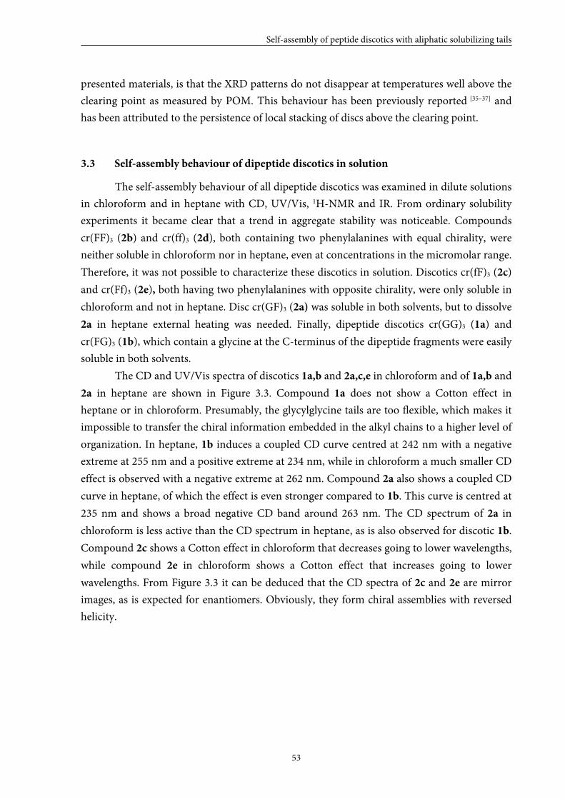
Self-assembly of peptide discotics with aliphatic solubilizing tails
53
presented materials, is that the XRD patterns do not disappear at temperatures well above the
clearing point as measured by POM. This behaviour has been previously reported [35–37] and
has been attributed to the persistence of local stacking of discs above the clearing point.
3.3 Self-assembly behaviour of dipeptide discotics in solution
The self-assembly behaviour of all dipeptide discotics was examined in dilute solutions
in chloroform and in heptane with CD, UV/Vis, 1H-NMR and IR. From ordinary solubility
experiments it became clear that a trend in aggregate stability was noticeable. Compounds
cr(FF)3 (2b) and cr(ff)3 (2d), both containing two phenylalanines with equal chirality, were
neither soluble in chloroform nor in heptane, even at concentrations in the micromolar range.
Therefore, it was not possible to characterize these discotics in solution. Discotics cr(fF)3 (2c)
and cr(Ff)3 (2e), both having two phenylalanines with opposite chirality, were only soluble in
chloroform and not in heptane. Disc cr(GF)3 (2a) was soluble in both solvents, but to dissolve
2a in heptane external heating was needed. Finally, dipeptide discotics cr(GG)3 (1a) and
cr(FG)3 (1b), which contain a glycine at the C-terminus of the dipeptide fragments were easily
soluble in both solvents.
The CD and UV/Vis spectra of discotics 1a,b and 2a,c,e in chloroform and of 1a,b and
2a in heptane are shown in Figure 3.3. Compound 1a does not show a Cotton effect in
heptane or in chloroform. Presumably, the glycylglycine tails are too flexible, which makes it
impossible to transfer the chiral information embedded in the alkyl chains to a higher level of
organization. In heptane, 1b induces a coupled CD curve centred at 242 nm with a negative
extreme at 255 nm and a positive extreme at 234 nm, while in chloroform a much smaller CD
effect is observed with a negative extreme at 262 nm. Compound 2a also shows a coupled CD
curve in heptane, of which the effect is even stronger compared to 1b. This curve is centred at
235 nm and shows a broad negative CD band around 263 nm. The CD spectrum of 2a in
chloroform is less active than the CD spectrum in heptane, as is also observed for discotic 1b.
Compound 2c shows a Cotton effect in chloroform that decreases going to lower wavelengths,
while compound 2e in chloroform shows a Cotton effect that increases going to lower
wavelengths. From Figure 3.3 it can be deduced that the CD spectra of 2c and 2e are mirror
images, as is expected for enantiomers. Obviously, they form chiral assemblies with reversed
helicity.
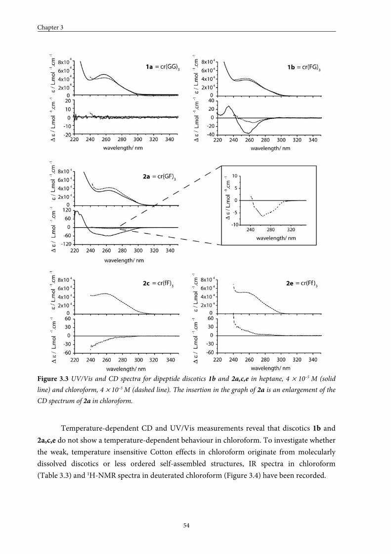
Chapter 3
54
Figure 3.3 UV/Vis and CD spectra for dipeptide discotics 1b and 2a,c,e in heptane, 4 × 10⁻⁵ M (solid
line) and chloroform, 4 × 10⁻⁵ M (dashed line). The insertion in the graph of 2a is an enlargement of the
CD spectrum of 2a in chloroform.
Temperature-dependent CD and UV/Vis measurements reveal that discotics 1b and
2a,c,e do not show a temperature-dependent behaviour in chloroform. To investigate whether
the weak, temperature insensitive Cotton effects in chloroform originate from molecularly
dissolved discotics or less ordered self-assembled structures, IR spectra in chloroform
(Table 3.3) and 1H-NMR spectra in deuterated chloroform (Figure 3.4) have been recorded.
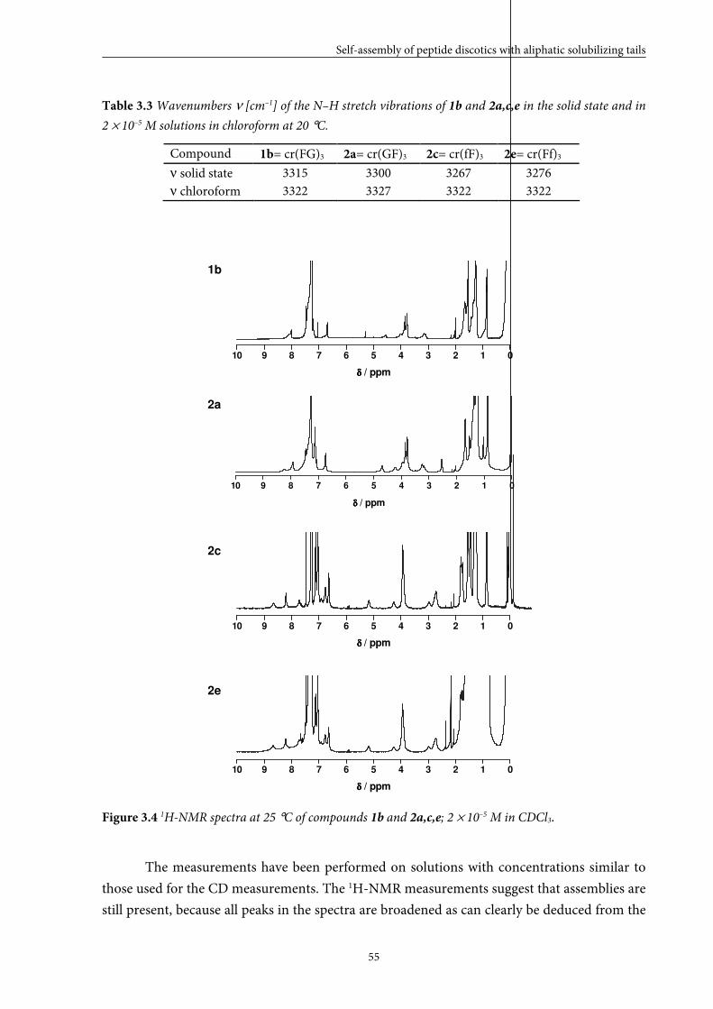
Self-assembly of peptide discotics with aliphatic solubilizing tails
55
Table 3.3 Wavenumbers ν [cm–1] of the N–H stretch vibrations of 1b and 2a,c,e in the solid state and in
2 × 10⁻⁵ M solutions in chloroform at 20 °C.
Compound 1b= cr(FG)3 2a= cr(GF)3 2c= cr(fF)3 2e= cr(Ff)3
ν solid state 3315 3300 3267 3276 ν chloroform 3322 3327 3322 3322
10 9 8 7 6 5 4 3 2 1 0
δδδδ / ppm
10 9 8 7 6 5 4 3 2 1 0
δδδδ / ppm
10 9 8 7 6 5 4 3 2 1 0
δ δ δ δ / ppm
10 9 8 7 6 5 4 3 2 1 0
δδδδ / ppm
Figure 3.4 1H-NMR spectra at 25 °C of compounds 1b and 2a,c,e; 2 × 10⁻⁵ M in CDCl3.
The measurements have been performed on solutions with concentrations similar to
those used for the CD measurements. The 1H-NMR measurements suggest that assemblies are
still present, because all peaks in the spectra are broadened as can clearly be deduced from the
1b
2c
2a
2e
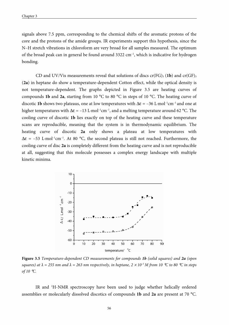
Chapter 3
56
signals above 7.5 ppm, corresponding to the chemical shifts of the aromatic protons of the
core and the protons of the amide groups. IR experiments support this hypothesis, since the
N–H stretch vibrations in chloroform are very broad for all samples measured. The optimum
of the broad peak can in general be found around 3322 cm–1, which is indicative for hydrogen
bonding.
CD and UV/Vis measurements reveal that solutions of discs cr(FG)3 (1b) and cr(GF)3
(2a) in heptane do show a temperature-dependent Cotton effect, while the optical density is
not temperature-dependent. The graphs depicted in Figure 3.5 are heating curves of
compounds 1b and 2a, starting from 10 °C to 80 °C in steps of 10 °C. The heating curve of
discotic 1b shows two plateaus, one at low temperatures with ∆ε = –36 L·mol–1cm–1 and one at
higher temperatures with ∆ε = –13 L·mol–1cm–1, and a melting temperature around 62 °C. The
cooling curve of discotic 1b lies exactly on top of the heating curve and these temperature
scans are reproducible, meaning that the system is in thermodynamic equilibrium. The
heating curve of discotic 2a only shows a plateau at low temperatures with
∆ε = –53 L·mol–1cm–1. At 80 °C, the second plateau is still not reached. Furthermore, the
cooling curve of disc 2a is completely different from the heating curve and is not reproducible
at all, suggesting that this molecule possesses a complex energy landscape with multiple
kinetic minima.
Figure 3.5 Temperature-dependent CD measurements for compounds 1b (solid squares) and 2a (open
squares) at λ = 255 nm and λ = 263 nm respectively, in heptane, 2 × 10⁻⁵ M from 10 °C to 80 °C in steps
of 10 °C.
IR and 1H-NMR spectroscopy have been used to judge whether helically ordered
assemblies or molecularly dissolved discotics of compounds 1b and 2a are present at 70 °C.
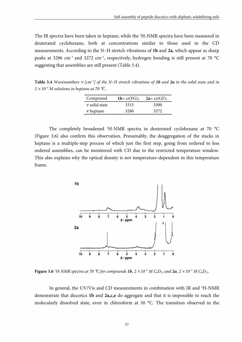
Self-assembly of peptide discotics with aliphatic solubilizing tails
57
The IR spectra have been taken in heptane, while the 1H-NMR spectra have been measured in
deuterated cyclohexane, both at concentrations similar to those used in the CD
measurements. According to the N–H stretch vibrations of 1b and 2a, which appear as sharp
peaks at 3286 cm–1 and 3272 cm–1, respectively, hydrogen bonding is still present at 70 °C
suggesting that assemblies are still present (Table 3.4).
Table 3.4 Wavenumbers ν [cm–1] of the N–H stretch vibrations of 1b and 2a in the solid state and in
2 × 10⁻⁵ M solutions in heptane at 70 °C.
Compound 1b= cr(FG)3 2a= cr(GF)3
ν solid state 3315 3300 ν heptane 3286 3272
The completely broadened 1H-NMR spectra in deuterated cyclohexane at 70 °C
(Figure 3.6) also confirm this observation. Presumably, the deaggregation of the stacks in
heptane is a multiple-step process of which just the first step, going from ordered to less
ordered assemblies, can be monitored with CD due to the restricted temperature window.
This also explains why the optical density is not temperature-dependent in this temperature
frame.
10 9 8 7 6 5 4 3 2 1 0
10 9 8 7 6 5 4 3 2 1 0δδδδ / ppm
δδδδ / ppm
Figure 3.6 1H-NMR spectra at 70 °C for compounds 1b, 2 × 10⁻⁵ M C6D12 and 2a, 2 × 10⁻⁵ M C6D12.
In general, the UV/Vis and CD measurements in combination with IR and 1H-NMR
demonstrate that discotics 1b and 2a,c,e do aggregate and that it is impossible to reach the
molecularly dissolved state, even in chloroform at 50 °C. The transition observed in the
1b
2a
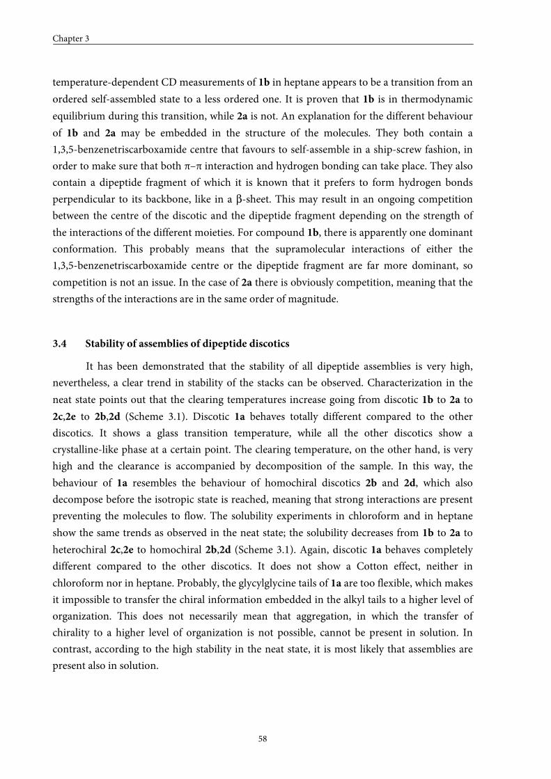
Chapter 3
58
temperature-dependent CD measurements of 1b in heptane appears to be a transition from an
ordered self-assembled state to a less ordered one. It is proven that 1b is in thermodynamic
equilibrium during this transition, while 2a is not. An explanation for the different behaviour
of 1b and 2a may be embedded in the structure of the molecules. They both contain a
1,3,5-benzenetriscarboxamide centre that favours to self-assemble in a ship-screw fashion, in
order to make sure that both π–π interaction and hydrogen bonding can take place. They also
contain a dipeptide fragment of which it is known that it prefers to form hydrogen bonds
perpendicular to its backbone, like in a β-sheet. This may result in an ongoing competition
between the centre of the discotic and the dipeptide fragment depending on the strength of
the interactions of the different moieties. For compound 1b, there is apparently one dominant
conformation. This probably means that the supramolecular interactions of either the
1,3,5-benzenetriscarboxamide centre or the dipeptide fragment are far more dominant, so
competition is not an issue. In the case of 2a there is obviously competition, meaning that the
strengths of the interactions are in the same order of magnitude.
3.4 Stability of assemblies of dipeptide discotics
It has been demonstrated that the stability of all dipeptide assemblies is very high,
nevertheless, a clear trend in stability of the stacks can be observed. Characterization in the
neat state points out that the clearing temperatures increase going from discotic 1b to 2a to
2c,2e to 2b,2d (Scheme 3.1). Discotic 1a behaves totally different compared to the other
discotics. It shows a glass transition temperature, while all the other discotics show a
crystalline-like phase at a certain point. The clearing temperature, on the other hand, is very
high and the clearance is accompanied by decomposition of the sample. In this way, the
behaviour of 1a resembles the behaviour of homochiral discotics 2b and 2d, which also
decompose before the isotropic state is reached, meaning that strong interactions are present
preventing the molecules to flow. The solubility experiments in chloroform and in heptane
show the same trends as observed in the neat state; the solubility decreases from 1b to 2a to
heterochiral 2c,2e to homochiral 2b,2d (Scheme 3.1). Again, discotic 1a behaves completely
different compared to the other discotics. It does not show a Cotton effect, neither in
chloroform nor in heptane. Probably, the glycylglycine tails of 1a are too flexible, which makes
it impossible to transfer the chiral information embedded in the alkyl tails to a higher level of
organization. This does not necessarily mean that aggregation, in which the transfer of
chirality to a higher level of organization is not possible, cannot be present in solution. In
contrast, according to the high stability in the neat state, it is most likely that assemblies are
present also in solution.
Self-assembly of peptide discotics with aliphatic solubilizing tails
59
Scheme 3.1 Cores of compounds 1b and 2a–e shown in order of increasing stability.
Scheme 3.1 demonstrates that the stack stability increases when two phenylalanines are
present in the core (2b–e). An explanation for the increased stability may be that two
phenylalanines provide a larger hydrophobic core and additional π–π interaction. The
measurements also reveal that the stack stability is higher when two phenylalanines with equal
chirality (2b,2d) are present compared to two phenylalanines with opposite chirality (2c,2e).
This may be due to more efficient packing when two phenylalanines with equal chirality are
introduced. Unexpectedly, the data also point out that when a combination of
L-phenylalanine and glycine is used, the stack stability increases when the
L-phenylalanine is situated remote from the core. To explain this observation, a more detailed
understanding of the self-assembly mechanism is required. When the centre starts to self-
assemble first, followed by the mesogenic groups at the periphery, it would be more beneficial
to have the phenylalanine close to the centre to enlarge the core and provide a more confined
packing. Nevertheless, the opposite is observed, which suggests that stacking is not initiated
from the centre of the discotics, but from the periphery. Preliminary investigation of the
stacking mechanism of 1b in solution with CD also supports this hypothesis.
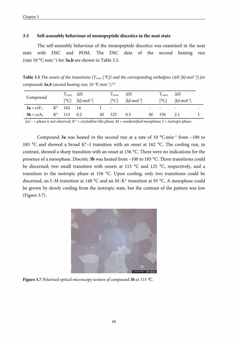
Chapter 3
60
3.5 Self-assembly behaviour of monopeptide discotics in the neat state
The self-assembly behaviour of the monopeptide discotics was examined in the neat
state with DSC and POM. The DSC data of the second heating run
(rate 10 °C·min–1) for 3a,b are shown in Table 3.5.
Table 3.5 The onsets of the transitions (Tonset [°C]) and the corresponding enthalpies (ΔH [kJ·mol–1]) for
compounds 3a,b (second heating run; 10 °C·min–1).[a]
Compound Tonset
[°C]
ΔH
[kJ·mol-1]
Tonset
[°C]
ΔH
[kJ·mol-1]
Tonset
[°C]
ΔH
[kJ·mol-1]
3a = crF3 K* 162 14 I - - - - - -
3b = crA3 K* 113 0.2 M 125 0.3 M 156 2.1 I
[a] – = phase is not observed; K* = crystalline-like phase; M = unidentified mesophase; I = isotropic phase.
Compound 3a was heated in the second run at a rate of 10 °C·min–1 from –100 to
185 °C and showed a broad K*–I transition with an onset at 162 °C. The cooling run, in
contrast, showed a sharp transition with an onset at 156 °C. There were no indications for the
presence of a mesophase. Discotic 3b was heated from –100 to 185 °C. Three transitions could
be discerned; two small transition with onsets at 113 °C and 125 °C, respectively, and a
transition to the isotropic phase at 156 °C. Upon cooling, only two transitions could be
discerned, an I–M transition at 148 °C and an M–K* transition at 95 °C. A mesophase could
be grown by slowly cooling from the isotropic state, but the contrast of the pattern was low
(Figure 3.7).
Figure 3.7 Polarized optical microscopy texture of compound 3b at 115 °C.
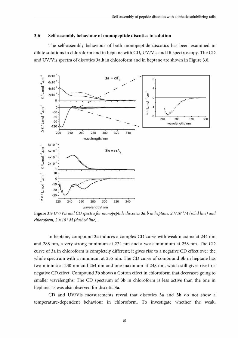
Self-assembly of peptide discotics with aliphatic solubilizing tails
61
3.6 Self-assembly behaviour of monopeptide discotics in solution
The self-assembly behaviour of both monopeptide discotics has been examined in
dilute solutions in chloroform and in heptane with CD, UV/Vis and IR spectroscopy. The CD
and UV/Vis spectra of discotics 3a,b in chloroform and in heptane are shown in Figure 3.8.
Figure 3.8 UV/Vis and CD spectra for monopeptide discotics 3a,b in heptane, 2 × 10⁻⁵ M (solid line) and
chloroform, 2 × 10⁻⁵ M (dashed line).
In heptane, compound 3a induces a complex CD curve with weak maxima at 244 nm
and 288 nm, a very strong minimum at 224 nm and a weak minimum at 258 nm. The CD
curve of 3a in chloroform is completely different; it gives rise to a negative CD effect over the
whole spectrum with a minimum at 255 nm. The CD curve of compound 3b in heptane has
two minima at 230 nm and 264 nm and one maximum at 248 nm, which still gives rise to a
negative CD effect. Compound 3b shows a Cotton effect in chloroform that decreases going to
smaller wavelengths. The CD spectrum of 3b in chloroform is less active than the one in
heptane, as was also observed for discotic 3a.
CD and UV/Vis measurements reveal that discotics 3a and 3b do not show a
temperature-dependent behaviour in chloroform. To investigate whether the weak,
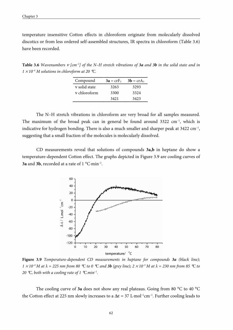
Chapter 3
62
temperature insensitive Cotton effects in chloroform originate from molecularly dissolved
discotics or from less ordered self-assembled structures, IR spectra in chloroform (Table 3.6)
have been recorded.
Table 3.6 Wavenumbers ν [cm–1] of the N–H stretch vibrations of 3a and 3b in the solid state and in
1 × 10⁻⁴ M solutions in chloroform at 20 °C.
Compound 3a = crF3 3b = crA3
ν solid state 3263 3293 ν chloroform 3300
3421 3324 3423
The N–H stretch vibrations in chloroform are very broad for all samples measured.
The maximum of the broad peak can in general be found around 3322 cm–1, which is
indicative for hydrogen bonding. There is also a much smaller and sharper peak at 3422 cm–1,
suggesting that a small fraction of the molecules is molecularly dissolved.
CD measurements reveal that solutions of compounds 3a,b in heptane do show a
temperature-dependent Cotton effect. The graphs depicted in Figure 3.9 are cooling curves of
3a and 3b, recorded at a rate of 1 °C·min–1.
Figure 3.9 Temperature-dependent CD measurements in heptane for compounds 3a (black line);
1 × 10⁻⁴ M at λ = 225 nm from 80 °C to 0 °C and 3b (grey line); 2 × 10⁻⁵ M at λ = 230 nm from 85 °C to
20 °C, both with a cooling rate of 1 °C.min–1.
The cooling curve of 3a does not show any real plateaus. Going from 80 °C to 40 °C
the Cotton effect at 225 nm slowly increases to a ∆ε = 37 L·mol–1cm–1. Further cooling leads to
Self-assembly of peptide discotics with aliphatic solubilizing tails
63
a more rapid decrease of the CD signal. At around 25 °C, the Cotton effect changes sign and,
finally, at 0 °C it seems that a plateau will be reached with ∆ε = –106 L·mol–1cm–1.
Unfortunately, the cooling curves are not reproducible. This behaviour has already been
observed before for certain dipeptide discotics and illustrates that these molecules display a
very complex energy landscape. The cooling curve of 3b shows two plateaus, one at low
temperatures at around ∆ε = –30 L·mol–1cm–1 and one at higher temperatures with
∆ε = 8 L·mol–1cm–1, and a melting temperature around 45 °C. The cooling curve of 3b is
reproducible, but does not lie exactly on top of the heating curve, meaning that the system is
not in thermodynamic equilibrium. IR spectroscopy has been performed to judge whether
helically ordered assemblies or molecularly dissolved discotics of 3a and 3b are present in
heptane at 70 °C. The IR spectra are taken in heptane at concentrations similar to those used
in the CD measurements. According to the N–H stretch vibrations of 3a and 3b, which appear
as sharp peaks at 3289 cm–1 and 3351 cm–1 respectively, hydrogen bonding is still present
(Table 3.7). The hydrogen bonds present in assemblies of 3a are stronger than the ones for 3b,
suggesting that, when a phenylalanine is used as structuring element, more stable assemblies
are formed.
Table 3.7 Wavenumbers ν [cm–1] of the N–H stretch vibrations of 3a and 3b in the solid state and in
1 × 10⁻⁴ M solutions in heptane at 80 °C.
Compound 3a = crF3 3b = crA3
ν solid state 3263 3293 ν heptane 3289 3351
3.7 Stability of assemblies of monopeptide discotics
It has been demonstrated that the stabilities of the monopeptide assemblies are still
very high despite the lack of one amide functionality compared to the dipeptide discotics.
Characterization in the neat state points out that the clearing temperature of the
phenylalanine-based disc 3a (162 °C) is somewhat higher than the one of the alanine-based
disc 3b (156 °C), indicating that the interactions between the molecules are somewhat
stronger in the case of 3a. In solution, the same trend has been observed with IR experiments
performed in heptane, which show that hydrogen bonding is stronger for 3a than for 3b as
can be deduced from a smaller wavenumber for the N–H stretch vibration of the former.
Explanation for the increased stability of 3a may be that the extra phenyl group provides more
confinement of space and/or additional π–π interaction. The additional phenyl moiety also
plays a role in determining the degree of order present in the assemblies, since significantly
larger Cotton effects are obtained for 3a compared to 3b, especially in heptane. The
monopeptide discotics can, in terms of stability and order within the stacks, be situated in
Chapter 3
64
between dipeptide discotics 1b [cr(FG)3] and 2a [cr(GF)3]. For this reason, phenylalanine-
based discotic 3a looks the most attractive candidate for future functionalization; the
assemblies of 3a possess appropriate stack stability in relation with the dipeptide discotics,
while from a synthetic point a view they are much easier to prepare than the dipeptide
discotics.
3.8 Conclusions
The self-assembly behaviour of seven dipeptide discotics and two monopeptide
discotics is described both in the neat state and in solution. Although all discotics form very
stable assemblies, a clear trend in the stability of the different stacks is observed depending on
the peptide fragment incorporated.
The assemblies are not very well-ordered in the neat state. Furthermore, some of the
self-assembled structures show very complex energy landscapes in solution. This indicates
that small changes in the balance between the secondary interactions originating from the
benzenetricarboxamide core and the peptide fragments have a strong influence on the order
within the stack. The balance between these supramolecular interactions is very subtle and a
small modification can already make the difference between thermodynamically stable or
completely frustrated stacks.
The results presented over here show that it is possible to alter the stacking properties
of these peptide discotics by modifying the peptide fragments, but the results also stress the
importance of understanding the self-assembly mechanisms in order to clarify the self-
assembly behaviour of the different C3-symmetrical molecules. Finally, it is shown that
phenylalanine-based discotic 3a is the most attractive candidate for future functionalization,
since assemblies of 3a possess appropriate stabilities and are easily accessible.
3.9 Experimental section
General
UV/VIS spectra were obtained on a Perkin Elmer Lambda 40 spectrometer. CD measurements were
performed on a Jasco J-600 spectropolarimeter connected to a Jasco PTC-348WI temperature controller.
Infrared spectra of the liquid samples were recorded using a Biorad FTS 6000 spectrometer. The heatable cell
used to perform the measurements was controlled by a home made temperature controller and fitted with KBr
windows and a Teflon spacer that makes the path length 1 mm. Each spectrum was recorded with a resolution of
4 cm–1, co-adding 200 scans. Low concentration 1H-NMR spectra were recorded on a Varian Unity Inova 500
spectrometer. The optical properties of the neat state were determined using a Jenaval polarization microscope
equipped with a Linkam THMS 600 heating device and a Polaroid digital camera model PDMC-2. DSC data
were collected on a Perkin Elmer Pyris 1 under nitrogen atmosphere. The X-ray measurements were carried out
on a home-built system consisting of a sealed X-ray tube (Cu Kα radiation), a primary graphite monochromator,
a pinhole collimator, a sample stage, and a Siemens Hi-Star area detector. All components were mounted on an
Self-assembly of peptide discotics with aliphatic solubilizing tails
65
optical bench to provide maximal flexibility and easy alignment. Samples were prepared in a 0.9 mm diameter
Lindemann capillary tube and mounted in a home-built furnace, based on a TMS94 Linkam hot stage. Wide
angle X-ray (WAXS) patterns were recorded with a sample-to-detector distance of 8.1 cm.
Synthesis
The syntheses of compounds 1a,b, 2a–e and 3a,b are described in Chapter 2.
3.10 References and notes
[1] L.J. Prins, D.N. Reinhoudt, P. Timmerman, Angew. Chem. 2001, 113, 2446-2492; Angew. Chem., Int. Ed.
2001, 40, 2382–2426.
[2] D.J. Hill, M.J. Mio, R.J. Prince, T.S. Hughes, J.S. Moore, Chem. Rev. 2001, 101, 3893–4011.
[3] L. Brunsveld, B.J.B. Folmer, E.W. Meijer, R.P. Sijbesma, Chem. Rev. 2001, 101, 4071–4097.
[4] D.C. Sherrington, K.A. Taskinen, Chem. Soc. Rev. 2001, 30, 83–93.
[5] L.M. Greig, D. Philip, Chem. Soc. Rev. 2001, 30, 287–302.
[6] D.T. Bong, T.D. Clark, J.R. Granja, M.R. Ghadiri, Angew. Chem. 2001, 113, 1016-1041; Angew. Chem.,
Int. Ed. 2001, 40, 989–1011.
[7] T. Kato, Science, 2002, 295, 2414–2418.
[8] A. Ciferri, Macromol. Rapid Commun. 2002, 23, 511–529.
[9] H.M. Keizer, R.P. Sijbesma, Chem. Soc. Rev. 2005, 34, 226–234.
[10] A.L. Sisson, M.R. Shah, S. Bhosale, S. Matile, Chem. Soc. Rev. 2006, 35, 1269–1286.
[11] K. Sada, M. Takeuchi, N. Fujita, M. Numata, S. Shinkai, Chem. Soc. Rev. 2007, 36, 415–435.
[12] E.W. Meijer, J.A.J.M. Vekemans, A.R.A. Palmans, P. Breure, J. de Kraker, L. Brunsveld, Polym. Prep.
2000, 41, 902–903.
[13] J.J. van Gorp, Thesis: Helices by hydrogen bonding, folding and stacking of chiral supramolecular
scaffolds; Eindhoven University of Technology: Eindhoven (The Netherlands), 2004.
[14] M. de Loos, Thesis: Hydrogen-bonded low molecular weight gelators; University of Groningen:
Groningen (The Netherlands), 2005.
[15] V. Percec, A.E. Dulcey, M. Peterca, M. IIies, M.J. Sienkowska, P.A. Heiney, J. Am. Chem. Soc. 2005, 127,
17902–17909.
[16] R.E. Gillard, F.M. Raymo, J.F. Stoddart, Chem. Eur. J. 1997, 3, 1933–1940.
[17] S. Zhang, X. Zhao, J. Mater. Chem. 2004, 14, 2082–2086.
[18] K. Rajagopal, J.P. Schneider, Curr. Opin. Struct. Biol. 2004, 14, 480–486.
[19] N. Ashkenasy, W.S. Horne, M.R. Ghadiri, Biomaterials 2006, 2, 99–102.
[20] J.R. Dunetz, C. Sandstrom, E.R. Young, P. Baker, S.A. van Name, T. Cathopolous, R. Fairman, J.C. de
Paula, K.S. Åkerfeldt, Org. Lett. 2005, 7, 2559–2561.
[21] J. Couet, J.D. Jeyaprakash, S. Samuel, A. Kopyshev, S. Santer, M. Biesalski, Angew. Chem. 2005, 117,
3361-3365; Angew. Chem., Int. Ed. 2005, 44, 3297–3301.
[22] A. Rösler, H-A. Klok, I.W. Hamley, V. Castelletto, O.O. Mykhaylyk, Biomacromolecules 2003, 4, 859–
863.
[23] H-A. Klok, G.W.M. Vandermeulen, H. Nuhn, A. Rösler, I.W. Hamley, V. Castelletto, H. Xu, S.S. Sheiko,
Faraday Discuss. 2004, 128, 29–41.
[24] D. Aili, K. Enander, J. Rydberg, I. Lundström, L. Baltzer, B. Liedberg, J. Am. Chem. Soc. 2006, 128,
2194–2195.
[25] P.P. Bose, M.G.B. Drew, A.K. Das, A. Banerjee, Chem. Commun. 2006, 3196–3198.
Chapter 3
66
[26] V. Percec, A.E. Dulcey, V.S.K. Balagurusamy, Y. Miura, J. Smidrkal, M. Peterca, S. Nummelin, U.
Edlund, S.D. Hudson, P.A. Heiney, D.A. Hu, S.N. Magonov, S.A. Vinogradov, Nature, 2004, 430, 764–
768.
[27] J.D. Hartgerink, E. Beniash, S.I. Stupp, Proc. Natl. Acad. Sci. U. S. A. 2002, 99, 5133–5138.
[28] K.J.C. van Bommel, C. van der Pol, I. Muizebelt, A. Friggeri, A. Heeres, A. Meetsma, B.L. Feringa, J. van
Esch, Angew. Chem. 2004, 116, 1695-1699; Angew. Chem., Int. Ed. 2004, 43, 1663–1667.
[29] K. Matsuura, K. Murasato, N. Kimizuka, J. Am. Chem. Soc. 2005, 127, 10148–10149.
[30] A.R.A. Palmans, J.A.J.M. Vekemans, H. Fischer, R.A. Hikmet, E.W. Meijer, Chem. Eur. J. 1997, 3, 300–
307.
[31] L. Brunsveld, H. Zhang, M. Glasbeek, J.A.J.M. Vekemans, E.W. Meijer, J. Am. Chem. Soc. 2000, 122,
6175–6182.
[32] L. Brunsveld, B.G.G. Lohmeijer, J.A.J.M. Vekemans, E.W. Meijer, J. Incl. Phenom. Macrocycl. Chem.
2001, 41, 61–64.
[33] J.J. van Gorp, J.A.J.M. Vekemans, E.W. Meijer, J. Am. Chem. Soc. 2002, 124, 14759–14769.
[34] A preliminary AFM study did not give information about the dimensions of the assemblies.
[35] R. Festag, R. Kleppinger, M. Soliman, J.H. Wendorff, G. Lattermann, G. Staufer, Liq. Cryst. 1992, 11,
699–710.
[36] R. Kleppinger, C.P. Lillya, C. Yang, J. Am. Chem. Soc. 1997, 119, 4097–4102.
[37] A. Hayer, V. de Halleux, A. Köhler, A. El-Garoughy, E.W. Meijer, J. Barberá, J. Tant, J. Levin, M.
Lehmann, J.C. Gierschner, Y.H. Geerts, J. Phys. Chem. B 2006, 110, 7653–7659.
44
From apolar media to water:
Self-assembly of a peptide-based discotic amphiphile
Characterization in solution
ABSTRACT: A C3-symmetrical molecule with a 1,3,5-benzenetricarboxamide core decorated with
L-phenylalanine residues bearing peripheral mesogenic groups containing oligo(ethylene oxide)
tails is described. A convergent synthetic strategy, similar to the one reported for the
monopeptide discotics described in Chapter 2 has been applied to obtain the target product. To
investigate the stacking properties of phenylalanine-based discotic 1 a combination of techniques
including UV/Vis spectroscopy, circular dichroism, NMR spectroscopy and cryoTEM has been
applied. The measurements demonstrate that this molecule forms ordered aggregates in
solutions in chloroform and in water. The stacks are well-defined, nanometer-sized objects
(containing 11 monomers within an aggregate), of which the size is independent of
concentration within the investigated concentration regime (10–5 to 10–3 M). Their water-
solubility in combination with their well-defined nanometer-size and their ability to display
multiple functional groups make these assemblies potential supramolecular scaffolds for
biomedical applications.
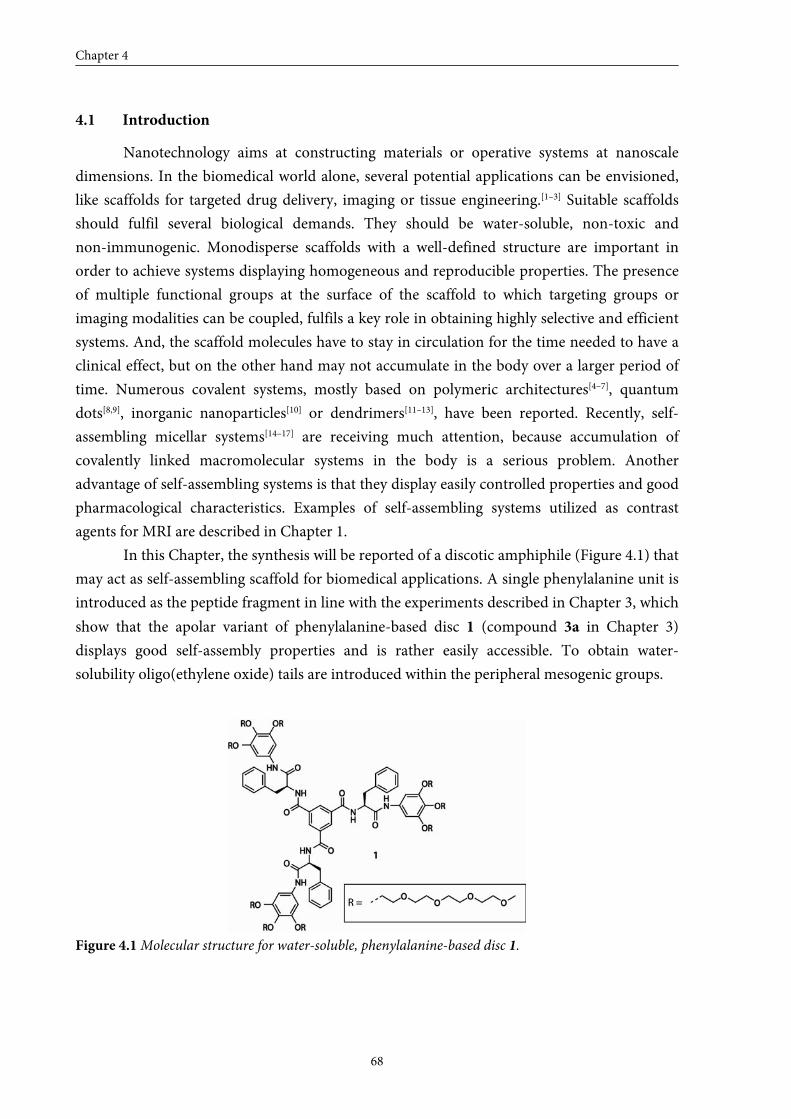
Chapter 4
68
4.1 Introduction
Nanotechnology aims at constructing materials or operative systems at nanoscale
dimensions. In the biomedical world alone, several potential applications can be envisioned,
like scaffolds for targeted drug delivery, imaging or tissue engineering.[1–3] Suitable scaffolds
should fulfil several biological demands. They should be water-soluble, non-toxic and
non-immunogenic. Monodisperse scaffolds with a well-defined structure are important in
order to achieve systems displaying homogeneous and reproducible properties. The presence
of multiple functional groups at the surface of the scaffold to which targeting groups or
imaging modalities can be coupled, fulfils a key role in obtaining highly selective and efficient
systems. And, the scaffold molecules have to stay in circulation for the time needed to have a
clinical effect, but on the other hand may not accumulate in the body over a larger period of
time. Numerous covalent systems, mostly based on polymeric architectures[4–7], quantum
dots[8,9], inorganic nanoparticles[10] or dendrimers[11–13], have been reported. Recently, self-
assembling micellar systems[14–17] are receiving much attention, because accumulation of
covalently linked macromolecular systems in the body is a serious problem. Another
advantage of self-assembling systems is that they display easily controlled properties and good
pharmacological characteristics. Examples of self-assembling systems utilized as contrast
agents for MRI are described in Chapter 1.
In this Chapter, the synthesis will be reported of a discotic amphiphile (Figure 4.1) that
may act as self-assembling scaffold for biomedical applications. A single phenylalanine unit is
introduced as the peptide fragment in line with the experiments described in Chapter 3, which
show that the apolar variant of phenylalanine-based disc 1 (compound 3a in Chapter 3)
displays good self-assembly properties and is rather easily accessible. To obtain water-
solubility oligo(ethylene oxide) tails are introduced within the peripheral mesogenic groups.
Figure 4.1 Molecular structure for water-soluble, phenylalanine-based disc 1.
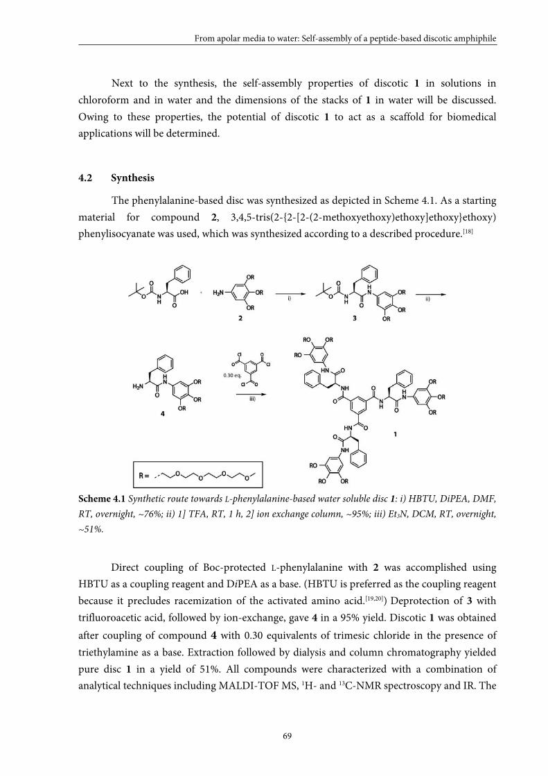
From apolar media to water: Self-assembly of a peptide-based discotic amphiphile
69
Next to the synthesis, the self-assembly properties of discotic 1 in solutions in
chloroform and in water and the dimensions of the stacks of 1 in water will be discussed.
Owing to these properties, the potential of discotic 1 to act as a scaffold for biomedical
applications will be determined.
4.2 Synthesis
The phenylalanine-based disc was synthesized as depicted in Scheme 4.1. As a starting
material for compound 2, 3,4,5-tris(2-{2-[2-(2-methoxyethoxy)ethoxy]ethoxy}ethoxy)
phenylisocyanate was used, which was synthesized according to a described procedure.[18]
Scheme 4.1 Synthetic route towards L-phenylalanine-based water soluble disc 1: i) HBTU, DiPEA, DMF,
RT, overnight, ~76%; ii) 1] TFA, RT, 1 h, 2] ion exchange column, ~95%; iii) Et3N, DCM, RT, overnight,
~51%.
Direct coupling of Boc-protected L-phenylalanine with 2 was accomplished using
HBTU as a coupling reagent and DiPEA as a base. (HBTU is preferred as the coupling reagent
because it precludes racemization of the activated amino acid.[19,20]) Deprotection of 3 with
trifluoroacetic acid, followed by ion-exchange, gave 4 in a 95% yield. Discotic 1 was obtained
after coupling of compound 4 with 0.30 equivalents of trimesic chloride in the presence of
triethylamine as a base. Extraction followed by dialysis and column chromatography yielded
pure disc 1 in a yield of 51%. All compounds were characterized with a combination of
analytical techniques including MALDI-TOF MS, 1H- and 13C-NMR spectroscopy and IR. The
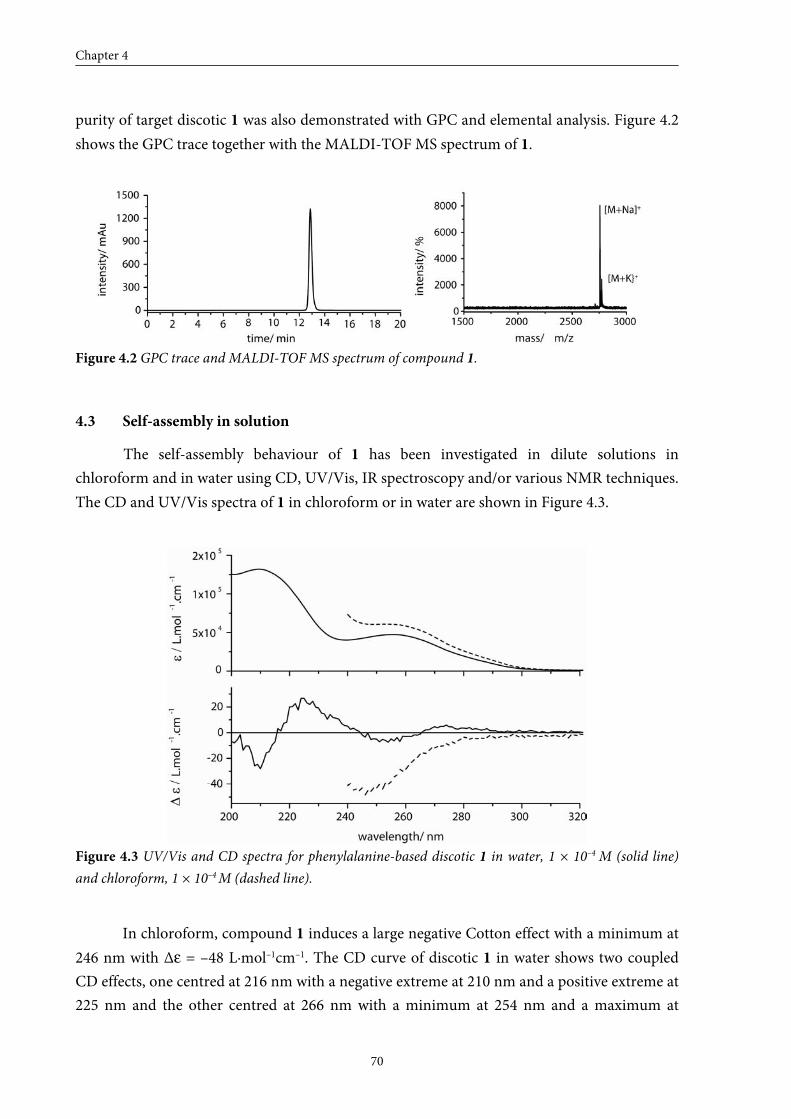
Chapter 4
70
purity of target discotic 1 was also demonstrated with GPC and elemental analysis. Figure 4.2
shows the GPC trace together with the MALDI-TOF MS spectrum of 1.
Figure 4.2 GPC trace and MALDI-TOF MS spectrum of compound 1.
4.3 Self-assembly in solution
The self-assembly behaviour of 1 has been investigated in dilute solutions in
chloroform and in water using CD, UV/Vis, IR spectroscopy and/or various NMR techniques.
The CD and UV/Vis spectra of 1 in chloroform or in water are shown in Figure 4.3.
Figure 4.3 UV/Vis and CD spectra for phenylalanine-based discotic 1 in water, 1 × 10⁻⁴ M (solid line)
and chloroform, 1 × 10⁻⁴ M (dashed line).
In chloroform, compound 1 induces a large negative Cotton effect with a minimum at
246 nm with Δε = –48 L·mol–1cm–1. The CD curve of discotic 1 in water shows two coupled
CD effects, one centred at 216 nm with a negative extreme at 210 nm and a positive extreme at
225 nm and the other centred at 266 nm with a minimum at 254 nm and a maximum at
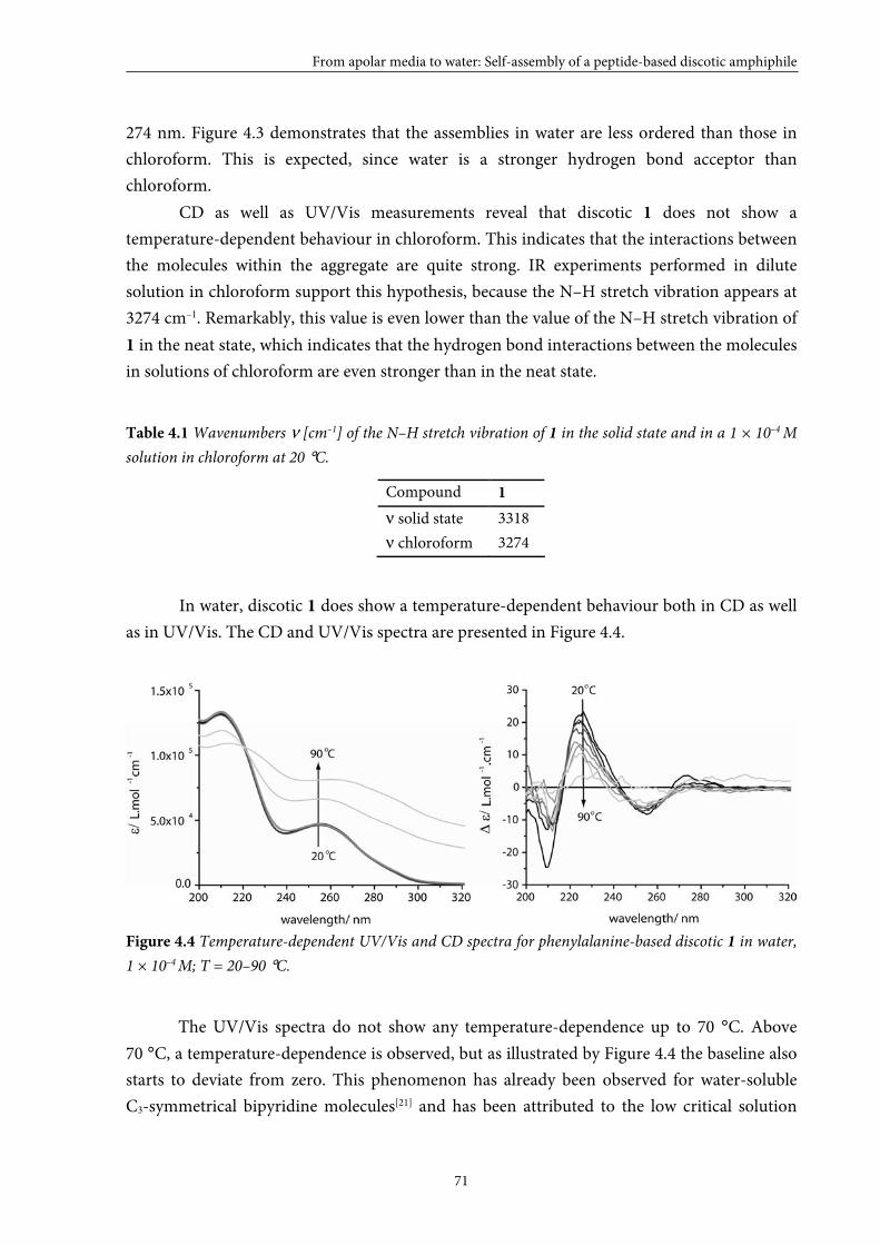
From apolar media to water: Self-assembly of a peptide-based discotic amphiphile
71
274 nm. Figure 4.3 demonstrates that the assemblies in water are less ordered than those in
chloroform. This is expected, since water is a stronger hydrogen bond acceptor than
chloroform.
CD as well as UV/Vis measurements reveal that discotic 1 does not show a
temperature-dependent behaviour in chloroform. This indicates that the interactions between
the molecules within the aggregate are quite strong. IR experiments performed in dilute
solution in chloroform support this hypothesis, because the N–H stretch vibration appears at
3274 cm–1. Remarkably, this value is even lower than the value of the N–H stretch vibration of
1 in the neat state, which indicates that the hydrogen bond interactions between the molecules
in solutions of chloroform are even stronger than in the neat state.
Table 4.1 Wavenumbers ν [cm–1] of the N–H stretch vibration of 1 in the solid state and in a 1 × 10⁻⁴ M
solution in chloroform at 20 °C.
Compound 1
ν solid state 3318
ν chloroform 3274
In water, discotic 1 does show a temperature-dependent behaviour both in CD as well
as in UV/Vis. The CD and UV/Vis spectra are presented in Figure 4.4.
Figure 4.4 Temperature-dependent UV/Vis and CD spectra for phenylalanine-based discotic 1 in water,
1 × 10⁻⁴ M; T = 20–90 °C.
The UV/Vis spectra do not show any temperature-dependence up to 70 °C. Above
70 °C, a temperature-dependence is observed, but as illustrated by Figure 4.4 the baseline also
starts to deviate from zero. This phenomenon has already been observed for water-soluble
C3-symmetrical bipyridine molecules[21] and has been attributed to the low critical solution
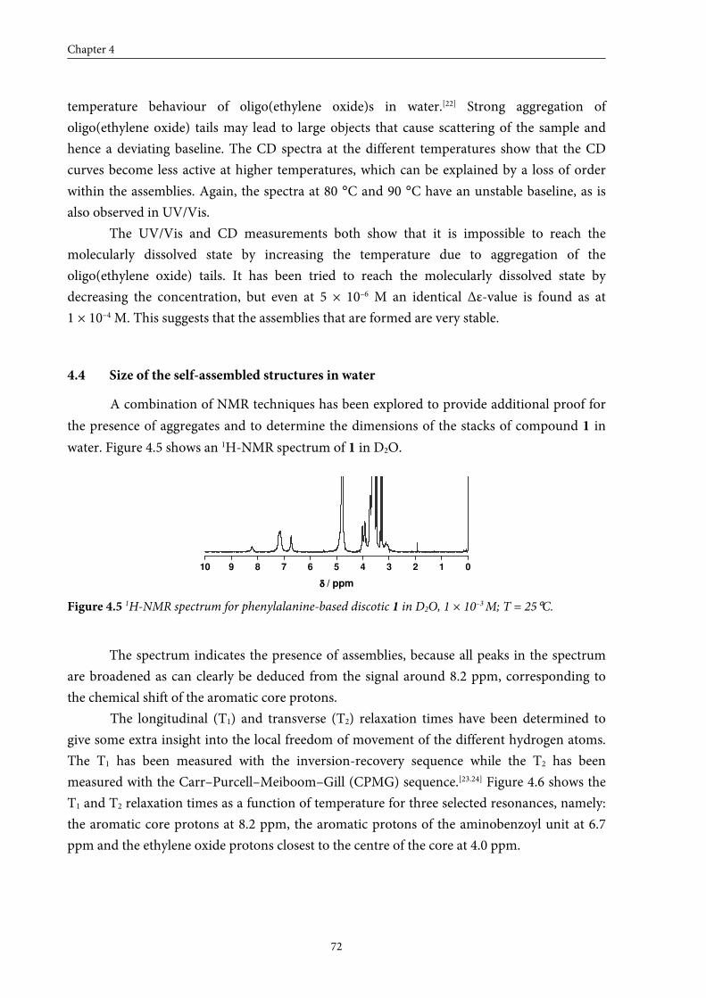
Chapter 4
72
temperature behaviour of oligo(ethylene oxide)s in water.[22] Strong aggregation of
oligo(ethylene oxide) tails may lead to large objects that cause scattering of the sample and
hence a deviating baseline. The CD spectra at the different temperatures show that the CD
curves become less active at higher temperatures, which can be explained by a loss of order
within the assemblies. Again, the spectra at 80 °C and 90 °C have an unstable baseline, as is
also observed in UV/Vis.
The UV/Vis and CD measurements both show that it is impossible to reach the
molecularly dissolved state by increasing the temperature due to aggregation of the
oligo(ethylene oxide) tails. It has been tried to reach the molecularly dissolved state by
decreasing the concentration, but even at 5 × 10⁻⁶ M an identical Δε-value is found as at
1 × 10⁻⁴ M. This suggests that the assemblies that are formed are very stable.
4.4 Size of the self-assembled structures in water
A combination of NMR techniques has been explored to provide additional proof for
the presence of aggregates and to determine the dimensions of the stacks of compound 1 in
water. Figure 4.5 shows an 1H-NMR spectrum of 1 in D2O.
10 9 8 7 6 5 4 3 2 1 0
δδδδ / ppm
Figure 4.5 1H-NMR spectrum for phenylalanine-based discotic 1 in D2O, 1 × 10⁻³ M; T = 25°C.
The spectrum indicates the presence of assemblies, because all peaks in the spectrum
are broadened as can clearly be deduced from the signal around 8.2 ppm, corresponding to
the chemical shift of the aromatic core protons.
The longitudinal (T1) and transverse (T2) relaxation times have been determined to
give some extra insight into the local freedom of movement of the different hydrogen atoms.
The T1 has been measured with the inversion-recovery sequence while the T2 has been
measured with the Carr–Purcell–Meiboom–Gill (CPMG) sequence.[23.24] Figure 4.6 shows the
T1 and T2 relaxation times as a function of temperature for three selected resonances, namely:
the aromatic core protons at 8.2 ppm, the aromatic protons of the aminobenzoyl unit at 6.7
ppm and the ethylene oxide protons closest to the centre of the core at 4.0 ppm.
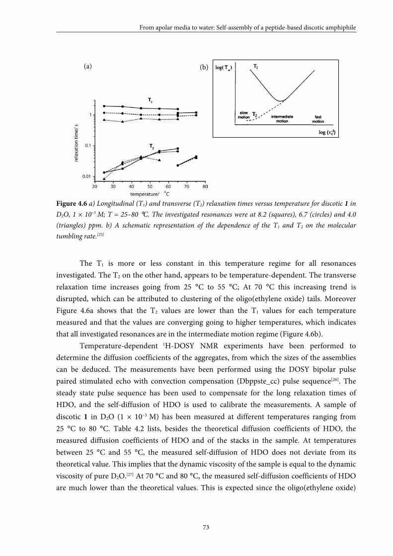
From apolar media to water: Self-assembly of a peptide-based discotic amphiphile
73
Figure 4.6 a) Longitudinal (T1) and transverse (T2) relaxation times versus temperature for discotic 1 in
D2O, 1 × 10⁻³ M; T = 25–80 °C. The investigated resonances were at 8.2 (squares), 6.7 (circles) and 4.0
(triangles) ppm. b) A schematic representation of the dependence of the T1 and T2 on the molecular
tumbling rate.[25]
The T1 is more or less constant in this temperature regime for all resonances
investigated. The T2 on the other hand, appears to be temperature-dependent. The transverse
relaxation time increases going from 25 °C to 55 °C; At 70 °C this increasing trend is
disrupted, which can be attributed to clustering of the oligo(ethylene oxide) tails. Moreover
Figure 4.6a shows that the T2 values are lower than the T1 values for each temperature
measured and that the values are converging going to higher temperatures, which indicates
that all investigated resonances are in the intermediate motion regime (Figure 4.6b).
Temperature-dependent 1H-DOSY NMR experiments have been performed to
determine the diffusion coefficients of the aggregates, from which the sizes of the assemblies
can be deduced. The measurements have been performed using the DOSY bipolar pulse
paired stimulated echo with convection compensation (Dbppste_cc) pulse sequence[26]. The
steady state pulse sequence has been used to compensate for the long relaxation times of
HDO, and the self-diffusion of HDO is used to calibrate the measurements. A sample of
discotic 1 in D2O (1 × 10⁻³ M) has been measured at different temperatures ranging from
25 °C to 80 °C. Table 4.2 lists, besides the theoretical diffusion coefficients of HDO, the
measured diffusion coefficients of HDO and of the stacks in the sample. At temperatures
between 25 °C and 55 °C, the measured self-diffusion of HDO does not deviate from its
theoretical value. This implies that the dynamic viscosity of the sample is equal to the dynamic
viscosity of pure D2O.[27] At 70 °C and 80 °C, the measured self-diffusion coefficients of HDO
are much lower than the theoretical values. This is expected since the oligo(ethylene oxide)
(a) (b)
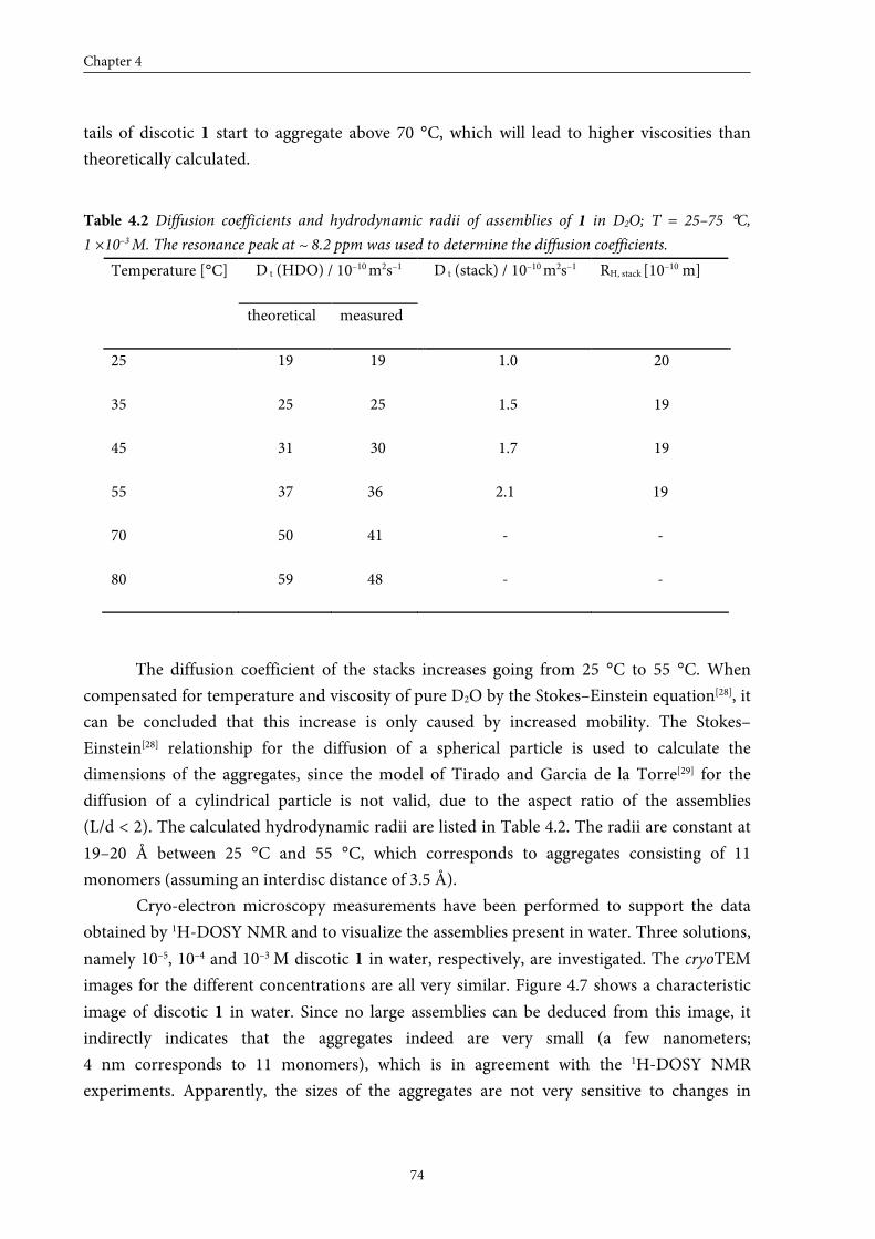
Chapter 4
74
tails of discotic 1 start to aggregate above 70 °C, which will lead to higher viscosities than
theoretically calculated.
Table 4.2 Diffusion coefficients and hydrodynamic radii of assemblies of 1 in D2O; T = 25–75 °C,
1 ×10⁻³ M. The resonance peak at ~ 8.2 ppm was used to determine the diffusion coefficients.
D t (HDO) / 10–10 m2s–1 Temperature [°C]
theoretical measured
D t (stack) / 10–10 m2s–1 RH, stack [10–10 m]
25 19 19 1.0 20
35 25 25 1.5 19
45 31 30 1.7 19
55 37 36 2.1 19
70 50 41 - -
80 59 48 - -
The diffusion coefficient of the stacks increases going from 25 °C to 55 °C. When
compensated for temperature and viscosity of pure D2O by the Stokes–Einstein equation[28], it
can be concluded that this increase is only caused by increased mobility. The Stokes–
Einstein[28] relationship for the diffusion of a spherical particle is used to calculate the
dimensions of the aggregates, since the model of Tirado and Garcia de la Torre[29] for the
diffusion of a cylindrical particle is not valid, due to the aspect ratio of the assemblies
(L/d < 2). The calculated hydrodynamic radii are listed in Table 4.2. The radii are constant at
19–20 Å between 25 °C and 55 °C, which corresponds to aggregates consisting of 11
monomers (assuming an interdisc distance of 3.5 Å).
Cryo-electron microscopy measurements have been performed to support the data
obtained by 1H-DOSY NMR and to visualize the assemblies present in water. Three solutions,
namely 10–5, 10–4 and 10–3 M discotic 1 in water, respectively, are investigated. The cryoTEM
images for the different concentrations are all very similar. Figure 4.7 shows a characteristic
image of discotic 1 in water. Since no large assemblies can be deduced from this image, it
indirectly indicates that the aggregates indeed are very small (a few nanometers;
4 nm corresponds to 11 monomers), which is in agreement with the 1H-DOSY NMR
experiments. Apparently, the sizes of the aggregates are not very sensitive to changes in
From apolar media to water: Self-assembly of a peptide-based discotic amphiphile
75
temperature or concentration, which indicates that the stacks have a relatively well-defined
size within the investigated concentration regime (10–5 to 10–3 M).
Figure 4.7 CryoTEM image for discotic 1 in water; 1 × 10⁻⁴ M. Images were obtained with an underfocus
value of –1.5 μm.
The fact that only 11 monomers are able to stack on top of each other is presumably
caused by steric crowding, but so far no experimental proof for this hypothesis has been
found. However, there are other discotic amphiphiles reported that resemble disc 1 and show
similar characteristics in apolar media. These discotics have been investigated by De Loos et
al.[30] and possess a 1,3,5-benzenetricarboxamide core decorated with phenylalanine moieties
each containing one alkyl tail. They demonstrate that these discotics are rather poor
organogelators compared to their cis-cis-1,3,5-cyclohexanetricarboxamide analogues. This
suggests that aggregates of benzene-based discotics are much smaller in size (restricted
growth) compared to the aggregates of cyclohexane-based discs. When combining these
results with our findings, a possible relationship between the restricted growth and the
combination of the 1,3,5-benzenetricarboxamide core with the phenylalanines is deduced.
Our hypothesis is supported by the work of Hanabusa et al.[31] They show that small
1,3,5-benzenetricarboxamide discotics lacking the phenylalanine moiety do form long fibres
and, therefore, do show good gelating properties. Regarding disc 1, the additional
aminobenzene units may also play a critical role in the self-assembly process, since it is
possible to stack the phenyl groups of phenylalanines on top of the aminobenzene units by
means of π–π interaction.
Chapter 4
76
4.5 Conclusions
In this chapter the synthesis of phenylalanine-based disc 1, using a convergent
synthetic approach, is described. Disc 1 self-assembles in solutions in chloroform and in water
as has been demonstrated by means of CD. The molecularly dissolved state can not be
reached, neither via raising the temperature due to clustering of the oligo(ethylene oxide) tails
nor via lowering the concentration; even at a concentration as low as 5 × 10⁻⁶ M aggregates are
still present, which suggests that the aggregates are quite stable. NMR techniques in
combination with cryoTEM have been performed and show that the stacks are well-defined
nanometer-sized objects (at an estimate 11 monomers within an aggregate). The sizes of the
aggregates are almost independent of concentration (investigated concentration 10–5 M to
10–3 M) and temperature (10–3 M, 25–55 °C). The combination of properties like, water
solubility and a well-defined nanometer-size makes these discotic molecules attractive
supramolecular scaffolds for drug delivery or imaging.
4.6 Experimental section
General
Compounds 1–4 were all kindly provided by B. F. M. de Waal. Unless stated otherwise, all reagents and
chemicals were obtained from commercial sources and used without further purification. Water was
demineralised prior to use. Dichloromethane and tetrahydrofuran were obtained by distillation over Merck
molecular sieves (4 Å). 1H-NMR, 1H-1H COSY and 19F-NMR spectra were recorded on a Varian Gemini 300
spectrometer, a Varian Mercury Vx 400 spectrometer or a Varian Unity Inova 500 spectrometer at 298 K.
Chemical shifts are given in ppm (δ) values relative to tetramethylsilane (TMS). Splitting patterns are designated
as s, singlet; d, doublet; dd, double doublet; t, triplet; q, quartet; m, multiplet and br stands for broad. Neat state
IR spectra were measured at 298 K on a Perkin-Elmer 1605 FT-IR spectrophotometer. Matrix assisted laser
desorption/ionization mass spectra were obtained on a PerSeptive Biosystems Voyager DE-PRO spectrometer
using α-cyano-4-hydroxycinnamic acid (CHCA) and 2-[(2E)-3-(4-tert-butylphenyl)-2-methylprop-2-
enylidene]malononitrile (DCTB) as matrices. Elemental analyses were carried out using a Perkin Elmer 2400.
UV/VIS spectra were obtained on a Perkin Elmer Lambda 40 spectrometer. CD measurements were performed
on a Jasco J-600 spectropolarimeter connected to a Jasco PTC-348WI temperature controller. Infrared spectra of
the liquid samples were recorded using a Biorad FTS 6000 spectrometer. The heatable cell used to perform the
measurements was fitted with KBr windows and a Teflon spacer that makes the path length 1 mm. Each
spectrum was recorded with a resolution of 4 cm–1, co-adding 200 scans. The longitudinal and transverse
relaxation time and 1H-DOSY NMR experiments were recorded on a Varian Unity Inova 500 spectrometer
operating at 500.618 MHz and equipped with a 5 mm ID-PFG probe from Varian. Additional, experimental 1H-DOSY NMR data can be found at the end of this experimental section. Cryogenic transmission microscopy
measurements were performed on a FEI Titan Krios TEM equipped with a field emission gun (FEG) operating at
300 kV. Images were recorded using a 2k x 2k Gatan CCD camera equipped with a post column Gatan energy
filter (GIF). TEM grids, (R2/2 Quantifoil Jena grids) were purchased from Aurion. The Quantifoil grids
underwent a surface plasma treatment using a Cressington 208 carbon coater operating at 5 mA for 40 s prior to
the vitrification procedure. The cryoTEM images of the discotics were obtained by applying small aliquots (3 µL)
of the aqueous discotic solutions to Quantifoil grids (R2/2 Quantifoil Jena) within the environmental chamber
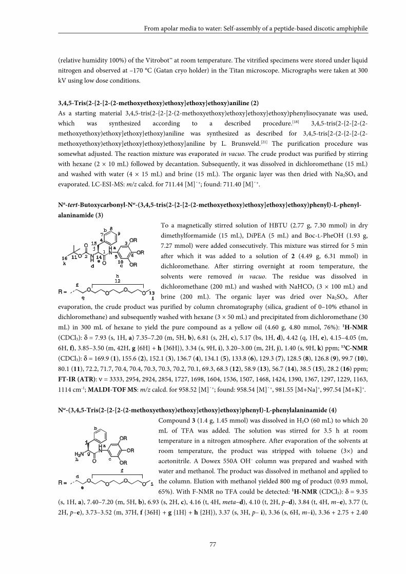
From apolar media to water: Self-assembly of a peptide-based discotic amphiphile
77
(relative humidity 100%) of the Vitrobot™ at room temperature. The vitrified specimens were stored under liquid
nitrogen and observed at –170 °C (Gatan cryo holder) in the Titan microscope. Micrographs were taken at 300
kV using low dose conditions.
3,4,5-Tris(2-{2-[2-(2-methoxyethoxy)ethoxy]ethoxy}ethoxy)aniline (2)
As a starting material 3,4,5-tris(2-{2-[2-(2-methoxyethoxy)ethoxy]ethoxy}ethoxy)phenylisocyanate was used,
which was synthesized according to a described procedure.[18] 3,4,5-tris(2-{2-[2-(2-
methoxyethoxy)ethoxy]ethoxy}ethoxy)aniline was synthesized as described for 3,4,5-tris[2-(2-{2-[2-(2-
methoxyethoxy)ethoxy]ethoxy}ethoxy)ethoxy]aniline by L. Brunsveld.[21] The purification procedure was
somewhat adjusted. The reaction mixture was evaporated in vacuo. The crude product was purified by stirring
with hexane (2 × 10 mL) followed by decantation. Subsequently, it was dissolved in dichloromethane (15 mL)
and washed with water (4 × 15 mL) and brine (15 mL). The organic layer was then dried with Na2SO4 and
evaporated. LC-ESI-MS: m/z calcd. for 711.44 [M]˙+; found: 711.40 [M]˙+.
Nα-tert-Butoxycarbonyl-Nω-(3,4,5-tris(2-{2-[2-(2-methoxyethoxy)ethoxy]ethoxy}ethoxy)phenyl)-L-phenyl-
alaninamide (3)
To a magnetically stirred solution of HBTU (2.77 g, 7.30 mmol) in dry
dimethylformamide (15 mL), DiPEA (5 mL) and Boc-L-PheOH (1.93 g,
7.27 mmol) were added consecutively. This mixture was stirred for 5 min
after which it was added to a solution of 2 (4.49 g, 6.31 mmol) in
dichloromethane. After stirring overnight at room temperature, the
solvents were removed in vacuo. The residue was dissolved in
dichloromethane (200 mL) and washed with NaHCO3 (3 × 100 mL) and
brine (200 mL). The organic layer was dried over Na2SO4. After
evaporation, the crude product was purified by column chromatography (silica, gradient of 0–10% ethanol in
dichloromethane) and subsequently washed with hexane (3 × 50 mL) and precipitated from dichloromethane (30
mL) in 300 mL of hexane to yield the pure compound as a yellow oil (4.60 g, 4.80 mmol, 76%): 1H-NMR
(CDCl3): δ = 7.93 (s, 1H, a) 7.35–7.20 (m, 5H, b), 6.81 (s, 2H, c), 5.17 (bs, 1H, d), 4.42 (q, 1H, e), 4.15–4.05 (m,
6H, f), 3.85–3.50 (m, 42H, g {6H} + h {36H}), 3.34 (s, 9H, i), 3.20–3.00 (m, 2H, j), 1.40 (s, 9H, k) ppm; 13C-NMR
(CDCl3): δ = 169.9 (1), 155.6 (2), 152.1 (3), 136.7 (4), 134.1 (5), 133.8 (6), 129.3 (7), 128.5 (8), 126.8 (9), 99.7 (10),
80.1 (11), 72.2, 71.7, 70.4, 70.4, 70.3, 70.3, 70.2, 70.1, 69.3, 68.3 (12), 58.9 (13), 56.7 (14), 38.5 (15), 28.2 (16) ppm;
FT-IR (ATR): ν = 3333, 2954, 2924, 2854, 1727, 1698, 1604, 1536, 1507, 1468, 1424, 1390, 1367, 1297, 1229, 1163,
1114 cm-1; MALDI-TOF MS: m/z calcd. for 958.52 [M]˙+; found: 958.54 [M]˙+, 981.55 [M+Na]+, 997.54 [M+K]+.
Nω-(3,4,5-Tris(2-{2-[2-(2-methoxyethoxy)ethoxy]ethoxy}ethoxy)phenyl)-L-phenylalaninamide (4)
Compound 3 (1.4 g, 1.45 mmol) was dissolved in H2O (60 mL) to which 20
mL of TFA was added. The solution was stirred for 3.5 h at room
temperature in a nitrogen atmosphere. After evaporation of the solvents at
room temperature, the product was stripped with toluene (3×) and
acetonitrile. A Dowex 550A OH– column was prepared and washed with
water and methanol. The product was dissolved in methanol and applied to
the column. Elution with methanol yielded 800 mg of product (0.93 mmol,
65%). With F-NMR no TFA could be detected: 1H-NMR (CDCl3): δ = 9.35
(s, 1H, a), 7.40–7.20 (m, 5H, b), 6.93 (s, 2H, c), 4.16 (t, 4H, meta–d), 4.10 (t, 2H, p–d), 3.84 (t, 4H, m–e), 3.77 (t,
2H, p–e), 3.73–3.52 (m, 37H, f {36H} + g {1H} + h {2H}), 3.37 (s, 3H, p– i), 3.36 (s, 6H, m–i), 3.36 + 2.75 + 2.40
12
34
5 6
7 9
1011
8
a
b
cd
f
g
e
12
13
h
i
j
k
OO
OOR =
NH
HN
OR
O
OR
OR
O
O
14
15
16
a
b
c
d
f
g
e
OO
OOR =
h
i
H2N
HN
OR
O
OR
OR
j
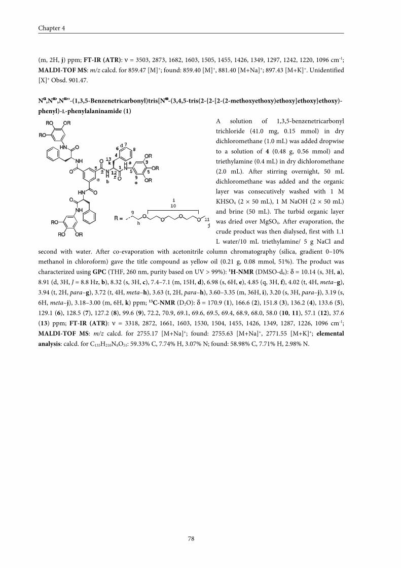
Chapter 4
78
(m, 2H, j) ppm; FT-IR (ATR): ν = 3503, 2873, 1682, 1603, 1505, 1455, 1426, 1349, 1297, 1242, 1220, 1096 cm-1;
MALDI-TOF MS: m/z calcd. for 859.47 [M]+; found: 859.40 [M]+, 881.40 [M+Na]+; 897.43 [M+K]+. Unidentified
[X]+ Obsd. 901.47.
Nαααα,Nαααα’,Nαααα’’-(1,3,5-Benzenetricarbonyl)tris[Nωωωω-(3,4,5-tris(2-{2-[2-(2-methoxyethoxy)ethoxy]ethoxy}ethoxy)-
phenyl)-L-phenylalaninamide (1)
A solution of 1,3,5-benzenetricarbonyl
trichloride (41.0 mg, 0.15 mmol) in dry
dichloromethane (1.0 mL) was added dropwise
to a solution of 4 (0.48 g, 0.56 mmol) and
triethylamine (0.4 mL) in dry dichloromethane
(2.0 mL). After stirring overnight, 50 mL
dichloromethane was added and the organic
layer was consecutively washed with 1 M
KHSO4 (2 × 50 mL), 1 M NaOH (2 × 50 mL)
and brine (50 mL). The turbid organic layer
was dried over MgSO4. After evaporation, the
crude product was then dialysed, first with 1.1
L water/10 mL triethylamine/ 5 g NaCl and
second with water. After co-evaporation with acetonitrile column chromatography (silica, gradient 0–10%
methanol in chloroform) gave the title compound as yellow oil (0.21 g, 0.08 mmol, 51%). The product was
characterized using GPC (THF, 260 nm, purity based on UV > 99%): 1H-NMR (DMSO-d6): δ = 10.14 (s, 3H, a),
8.91 (d, 3H, J = 8.8 Hz, b), 8.32 (s, 3H, c), 7.4–7.1 (m, 15H, d), 6.98 (s, 6H, e), 4.85 (q, 3H, f), 4.02 (t, 4H, meta–g),
3.94 (t, 2H, para–g), 3.72 (t, 4H, meta–h), 3.63 (t, 2H, para–h), 3.60–3.35 (m, 36H, i), 3.20 (s, 3H, para–j), 3.19 (s,
6H, meta–j), 3.18–3.00 (m, 6H, k) ppm; 13C-NMR (D2O): δ = 170.9 (1), 166.6 (2), 151.8 (3), 136.2 (4), 133.6 (5),
129.1 (6), 128.5 (7), 127.2 (8), 99.6 (9), 72.2, 70.9, 69.1, 69.6, 69.5, 69.4, 68.9, 68.0, 58.0 (10, 11), 57.1 (12), 37.6
(13) ppm; FT-IR (ATR): ν = 3318, 2872, 1661, 1603, 1530, 1504, 1455, 1426, 1349, 1287, 1226, 1096 cm-1;
MALDI-TOF MS: m/z calcd. for 2755.17 [M+Na]+; found: 2755.63 [M+Na]+, 2771.55 [M+K]+; elemental
analysis: calcd. for C135H210N6O51: 59.33% C, 7.74% H, 3.07% N; found: 58.98% C, 7.71% H, 2.98% N.
NH
OHN
OR
OHN
NH
O
O
NH
O
OHN
RO
RO OR
RO
RO OR
OR
OR
OO
OOR =
a
bc
d
f
g
e
h
i
j
k1
2
3
4
55
67
8
9
5
10
11
12
13
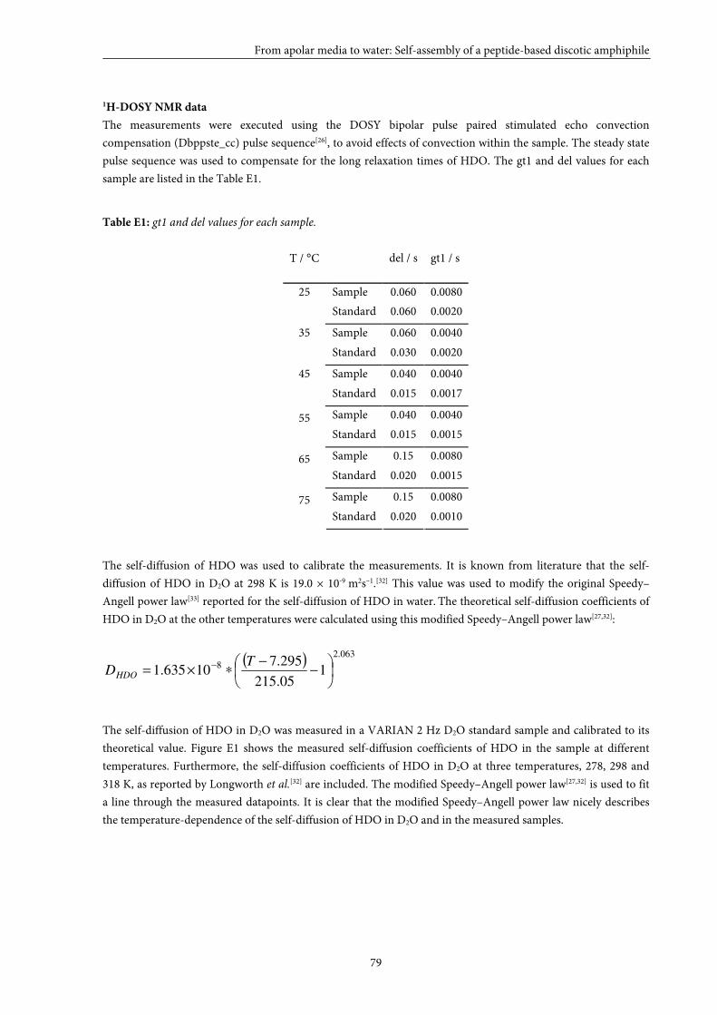
From apolar media to water: Self-assembly of a peptide-based discotic amphiphile
79
1H-DOSY NMR data
The measurements were executed using the DOSY bipolar pulse paired stimulated echo convection
compensation (Dbppste_cc) pulse sequence[26], to avoid effects of convection within the sample. The steady state
pulse sequence was used to compensate for the long relaxation times of HDO. The gt1 and del values for each
sample are listed in the Table E1.
Table E1: gt1 and del values for each sample.
T / °C del / s gt1 / s
Sample 0.060 0.0080 25
Standard 0.060 0.0020
Sample 0.060 0.0040 35
Standard 0.030 0.0020
Sample 0.040 0.0040 45
Standard 0.015 0.0017
Sample 0.040 0.0040 55 Standard 0.015 0.0015
Sample 0.15 0.0080 65 Standard 0.020 0.0015
Sample 0.15 0.0080 75 Standard 0.020 0.0010
The self-diffusion of HDO was used to calibrate the measurements. It is known from literature that the self-
diffusion of HDO in D2O at 298 K is 19.0 × 10⁻⁹ m2s–1.[32] This value was used to modify the original Speedy–
Angell power law[33] reported for the self-diffusion of HDO in water. The theoretical self-diffusion coefficients of
HDO in D2O at the other temperatures were calculated using this modified Speedy–Angell power law[27,32]:
( ) 063.2
8 105.215
295.710635.1
−
−∗×= − T
DHDO
The self-diffusion of HDO in D2O was measured in a VARIAN 2 Hz D2O standard sample and calibrated to its
theoretical value. Figure E1 shows the measured self-diffusion coefficients of HDO in the sample at different
temperatures. Furthermore, the self-diffusion coefficients of HDO in D2O at three temperatures, 278, 298 and
318 K, as reported by Longworth et al.[32] are included. The modified Speedy–Angell power law[27,32] is used to fit
a line through the measured datapoints. It is clear that the modified Speedy–Angell power law nicely describes
the temperature-dependence of the self-diffusion of HDO in D2O and in the measured samples.
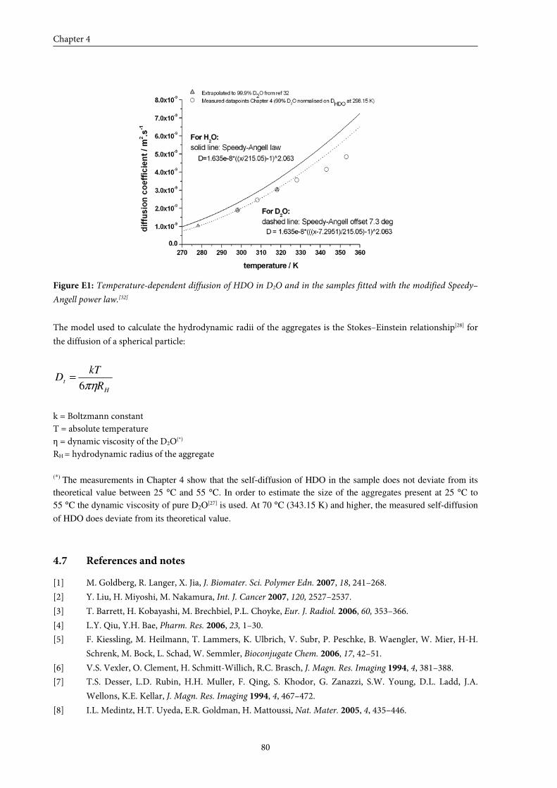
Chapter 4
80
Figure E1: Temperature-dependent diffusion of HDO in D2O and in the samples fitted with the modified Speedy–
Angell power law.[32]
The model used to calculate the hydrodynamic radii of the aggregates is the Stokes–Einstein relationship[28] for
the diffusion of a spherical particle:
H
tR
kTD
πη6=
k = Boltzmann constant T = absolute temperature η = dynamic viscosity of the D2O(*)
RH = hydrodynamic radius of the aggregate (*) The measurements in Chapter 4 show that the self-diffusion of HDO in the sample does not deviate from its
theoretical value between 25 °C and 55 °C. In order to estimate the size of the aggregates present at 25 °C to
55 °C the dynamic viscosity of pure D2O[27] is used. At 70 °C (343.15 K) and higher, the measured self-diffusion
of HDO does deviate from its theoretical value.
4.7 References and notes
[1] M. Goldberg, R. Langer, X. Jia, J. Biomater. Sci. Polymer Edn. 2007, 18, 241–268.
[2] Y. Liu, H. Miyoshi, M. Nakamura, Int. J. Cancer 2007, 120, 2527–2537.
[3] T. Barrett, H. Kobayashi, M. Brechbiel, P.L. Choyke, Eur. J. Radiol. 2006, 60, 353–366.
[4] L.Y. Qiu, Y.H. Bae, Pharm. Res. 2006, 23, 1–30.
[5] F. Kiessling, M. Heilmann, T. Lammers, K. Ulbrich, V. Subr, P. Peschke, B. Waengler, W. Mier, H-H.
Schrenk, M. Bock, L. Schad, W. Semmler, Bioconjugate Chem. 2006, 17, 42–51.
[6] V.S. Vexler, O. Clement, H. Schmitt-Willich, R.C. Brasch, J. Magn. Res. Imaging 1994, 4, 381–388.
[7] T.S. Desser, L.D. Rubin, H.H. Muller, F. Qing, S. Khodor, G. Zanazzi, S.W. Young, D.L. Ladd, J.A.
Wellons, K.E. Kellar, J. Magn. Res. Imaging 1994, 4, 467–472.
[8] I.L. Medintz, H.T. Uyeda, E.R. Goldman, H. Mattoussi, Nat. Mater. 2005, 4, 435–446.
From apolar media to water: Self-assembly of a peptide-based discotic amphiphile
81
[9] W.J.M. Mulder, A.W. Griffioen, G.J. Strijkers, D.P. Cormode, K. Nicolay, Z.A. Fayad, Nanomedicine
2007, 2, 307–324.
[10] Z.P. Xu, Q.H. Zeng, G.Q. Lu, A.B. Yu, Chem. Eng. Sci. 2006, 61, 1027–1040.
[11] U. Boas, P.M.H. Heegard, Chem. Soc. Rev. 2004, 33, 43–63.
[12] E.R. Gillies, J.M.J. Fréchet, Drug Discovery Today 2005, 10, 35–43.
[13] S. Langereis, A. Dirksen, T.M. Hackeng, M.H.P. van Genderen, E.W. Meijer, New J. Chem. 2007, 31,
1152–1160.
[14] A. Barge, G. Cravotto, B. Robaldo, E. Gianolio, S. Aime, J. Incl. Phenom. Macrocycl. Chem. 2007, 57,
489–495.
[15] S.R. Bull, M.O. Guler, R.E. Bras, P.N. Venkatasubramanian, S.I. Stupp, T.J. Meade, Bioconjugate Chem.
2005, 16, 1343–1348.
[16] G.M. Nicolle, E. Tóth, K-P. Eisenwiener, H.R. Mäcke, A.E. Merbach, J. Biol. Inorg. Chem. 2002, 7, 757–
769.
[17] E.R. Gillies, T.B. Jonsson, J.M.J. Fréchet, J. Am. Chem. Soc. 2004, 126, 11936–11943.
[18] M.A.C. Broeren, B.F.M. de Waal, M.H.P. van Genderen, H.M.H.F. Sanders, G. Fytas, E.W. Meijer, J.
Am. Chem. Soc. 2005, 127, 10334–10343.
[19] R. Knorr, A. Trzeciak, W. Bannwarth, D. Gillessen, Tetrahedron Lett. 1989, 30, 1927–1930.
[20] The enantiomeric excess of intermediate 3 was not determined with chiral HPLC.
[21] L. Brunsveld, Thesis: Supramolecular chirality from molecules to helical assemblies in polar media ,
Eindhoven University of Technology, Eindhoven (The Netherlands), 2001.
[22] F. Jr. Bailey, J. Koleske, Poly (Ethylene Oxide) Academic Press Inc., New York, 1976.
[23] H.Y. Carr, E.M. Purcell, Phys. Rev. 1954, 94, 630–638.
[24] S. Meiboom, D. Gill, Rev. Scient. Instr. 1958, 29, 688–691.
[25] Claridge T.D.W.; Tetrahedron Organic Chemistry Series Volume 19: High Resolution NMR Techniques
in Organic Chemistry, Elsevier, 1999, 13–44.
[26] A. Jerschow, N. Müller, J. Magn. Reson. 1997, 125, 372–375.
[27] C. H. Cho, J. Urquidi, S. Singh, G. Wilse Robinson, J. Phys. Chem. B 1999, 103, 1991–1994.
[28] Stokes, G.G.; Trans. Cambridge Philos. Soc., 1845, 8, 287.
[29] Tirado, M.M.; Garcia de la Torre, J.; J. Chem Phys., 1979, 71, 2581–2587.
[30] M. de Loos, Thesis: Hydrogen-bonded low molecular weight gelators; University of Groningen:
Groningen (The Netherlands), 2005.
[31] A. Sakamoto, D. Ogata, T. Shikata, O. Urakawa, K. Hanabusa, Polymer 2006, 47, 956–960.
[32] L.G. Longsworth, J. Phys. Chem. 1960, 64, 1914–1917.
[33] R.J. Speedy, C.A. Angell, J. Chem Phys. 1976, 65, 851–858.

55
Self-assembly of paramagnetic discotic amphiphiles
ABSTRACT: The syntheses are described of four paramagnetic discotic molecules, which all
possess 1,3,5-benzenetricarboxamide cores, but differ in their linker units and/or peripheral
chelating ligands. Three of the discs contain peripheral GdIII-DTPAs and are referred to as small
(1a), medium (2a) and large (3a) GdIII-DTPA discotics, respectively. Going from disc 1a to disc
3a, the capability to undergo secondary interactions and the hydrophobic character both
increase. Discotic 4 is very similar to disc 3a; the only difference is that the peripheral
GdIII-DTPA moieties are replaced by GdIII-DOTA units. The self-assembly behaviour of these
molecules has been studied in aqueous solution using longitudinal relaxation time (T1)
measurements. Concentration-dependent T1 measurements demonstrate that the self-assembly
process of paramagnetic discotics can indeed be probed. Furthermore, by modifying the number
of secondary interactions and the size of the hydrophobic core, the self-assembly processes of
these molecules can be tuned, which might be important to meet the requirements for use in
vivo. From the four paramagnetic discotics synthesized, GdIII-complex 3a is the most promising
candidate for a columnar self-assembling contrast agent, in view of its low aggregation
concentration and its high ionic relaxivity in the aggregated form (~30 mM–1s–1 at 20 °C and
1.5 T). Furthermore, the well-defined nanometer-size of assemblies of 3a may be advantageous
in relation to its pharmacokinetic properties.
Chapter 5
84
5.1 Introduction
Since the approval of the first magnetic resonance (MR) contrast agent in 1988, there
has been a remarkable growth in the utilization of these agents in the clinics. To date, it is
estimated that almost 50% of all the MR examinations are contrast-enhanced. Most of the
clinically approved contrast agents[1,2] are complexes incorporating gadolinium (GdIII) as this
ion is highly paramagnetic and has a long electronic relaxation time. Drawbacks of the
approved contrast agents are their non-specificity and their low efficiency, which only stems
from their preference to be in the blood stream, in view of their hydrophilicity. Therefore,
there is a strong need for new contrast agents with improved performance. In recent years,
various strategies have been explored in order to improve the proton relaxivity of MRI
contrast agents.[3–5] Low molecular weight contrast agents have been coupled to various
macromolecules, either covalently[6–13] or non-covalently[14–17], to reduce the molecular
tumbling rate of the GdIII-complex and, as a result, to increase the proton relaxivity. However,
a high molecular weight alone does not guarantee a low tumbling rate and hence a high
proton relaxivity. In fact, many flexible linear polymeric systems do not show an appreciable
increase in proton relaxivity as molecular weights increase beyond 10 kDa, indicating that
segmental flexibility dominates the tumbling rate of the GdIII-complex.[18,19] Another problem
of covalently linked macromolecular systems is non-specific accumulation in the body due to
their high molecular weight. As a result, self-assembling contrast agents have recently gained
attention owing to their easily controlled properties, good pharmacological characteristics
and/or their reversible nature, which allows combining the benefits of both low and high
molecular weight contrast agents. Modified micelles[20,21], liposomes[22–26], peptide nanofibers[27]
and a system based on cyclodextrins[28] with ionic relaxivities ranging from 4.1 to 43 mM–1s–1
(at 0.5 T, 25 °C) were reported as potential self-assembling contrast agents for MRI. In
addition, it has been shown that T1 measurements can be used to probe the self-assembly
process of these systems, as was reported for biotin–avidin complexes[29,30] and micelles[31,32]
(see Chapter 1).
So far, research has focused on the use of classical linear amphiphiles mainly forming
spherical self-assembled structures, leaving the use of discotic amphiphiles unexplored.
Discotic molecules are a special class of amphiphiles that form columnar objects in solution.
Compared to spherical assemblies, columnar structures have the advantage of being able to
become very long, hence ensuring higher local concentrations of gadolinium. Another benefit
of the use of discotic amphiphiles compared to linear amphiphiles is their multivalent
character, which leads to higher molecular relaxivities and opens possibilities for the use of
asymmetric target-specific discotics. Furthermore, the self-assembly of discotics, especially
C3-symmetrical (C3)-discotics, has been well-studied. In our laboratory, the self-assembly
behaviour of various 1,3,5-benzenetricarboxamide-based discotics has been extensively
investigated.[33–37] In Chapter 4, it has been shown that L-phenylalanine-based discotic
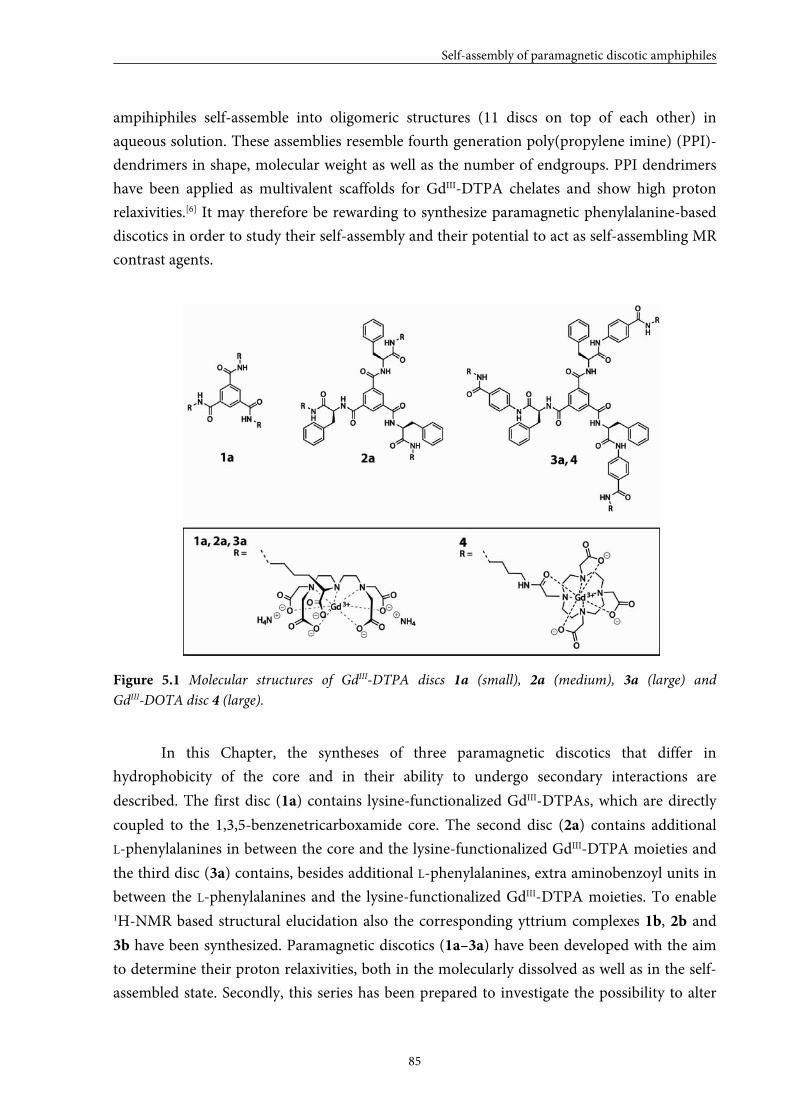
Self-assembly of paramagnetic discotic amphiphiles
85
ampihiphiles self-assemble into oligomeric structures (11 discs on top of each other) in
aqueous solution. These assemblies resemble fourth generation poly(propylene imine) (PPI)-
dendrimers in shape, molecular weight as well as the number of endgroups. PPI dendrimers
have been applied as multivalent scaffolds for GdIII-DTPA chelates and show high proton
relaxivities.[6] It may therefore be rewarding to synthesize paramagnetic phenylalanine-based
discotics in order to study their self-assembly and their potential to act as self-assembling MR
contrast agents.
Figure 5.1 Molecular structures of GdIII-DTPA discs 1a (small), 2a (medium), 3a (large) and
GdIII-DOTA disc 4 (large).
In this Chapter, the syntheses of three paramagnetic discotics that differ in
hydrophobicity of the core and in their ability to undergo secondary interactions are
described. The first disc (1a) contains lysine-functionalized GdIII-DTPAs, which are directly
coupled to the 1,3,5-benzenetricarboxamide core. The second disc (2a) contains additional
L-phenylalanines in between the core and the lysine-functionalized GdIII-DTPA moieties and
the third disc (3a) contains, besides additional L-phenylalanines, extra aminobenzoyl units in
between the L-phenylalanines and the lysine-functionalized GdIII-DTPA moieties. To enable 1H-NMR based structural elucidation also the corresponding yttrium complexes 1b, 2b and
3b have been synthesized. Paramagnetic discotics (1a–3a) have been developed with the aim
to determine their proton relaxivities, both in the molecularly dissolved as well as in the self-
assembled state. Secondly, this series has been prepared to investigate the possibility to alter
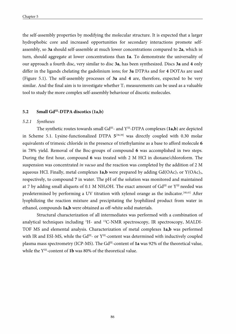
Chapter 5
86
the self-assembly properties by modifying the molecular structure. It is expected that a larger
hydrophobic core and increased opportunities for secondary interactions promote self-
assembly, so 3a should self-assemble at much lower concentrations compared to 2a, which in
turn, should aggregate at lower concentrations than 1a. To demonstrate the universality of
our approach a fourth disc, very similar to disc 3a, has been synthesized. Discs 3a and 4 only
differ in the ligands chelating the gadolinium ions; for 3a DTPAs and for 4 DOTAs are used
(Figure 5.1). The self-assembly processes of 3a and 4 are, therefore, expected to be very
similar. And the final aim is to investigate whether T1 measurements can be used as a valuable
tool to study the more complex self-assembly behaviour of discotic molecules.
5.2 Small GdIII-DTPA discotics (1a,b)
5.2.1 Syntheses
The synthetic routes towards small GdIII- and YIII-DTPA complexes (1a,b) are depicted
in Scheme 5.1. Lysine-functionalized DTPA 5[38,39] was directly coupled with 0.30 molar
equivalents of trimesic chloride in the presence of triethylamine as a base to afford molecule 6
in 78% yield. Removal of the Boc-groups of compound 6 was accomplished in two steps.
During the first hour, compound 6 was treated with 2 M HCl in dioxane/chloroform. The
suspension was concentrated in vacuo and the reaction was completed by the addition of 2 M
aqueous HCl. Finally, metal complexes 1a,b were prepared by adding Gd(OAc)3 or Y(OAc)3,
respectively, to compound 7 in water. The pH of the solution was monitored and maintained
at 7 by adding small aliquots of 0.1 M NH4OH. The exact amount of GdIII or YIII needed was
predetermined by performing a UV titration with xylenol orange as the indicator.[40,41] After
lyophilizing the reaction mixture and precipitating the lyophilized product from water in
ethanol, compounds 1a,b were obtained as off-white solid materials.
Structural characterization of all intermediates was performed with a combination of
analytical techniques including 1H- and 13C-NMR spectroscopy, IR spectroscopy, MALDI-
TOF MS and elemental analysis. Characterization of metal complexes 1a,b was performed
with IR and ESI-MS, while the GdIII- or YIII-content was determined with inductively coupled
plasma mass spectrometry (ICP-MS). The GdIII-content of 1a was 92% of the theoretical value,
while the YIII-content of 1b was 80% of the theoretical value.
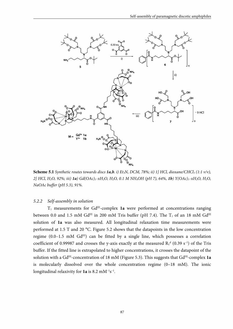
Self-assembly of paramagnetic discotic amphiphiles
87
Scheme 5.1 Synthetic routes towards discs 1a,b. i) Et3N, DCM, 78%; ii) 1] HCl, dioxane/CHCl3 (1:1 v/v),
2] HCl, H2O, 92%; iii) 1a) Gd(OAc)3 ·xH2O, H2O, 0.1 M NH4OH (pH 7), 64%, 1b) Y(OAc)3 ·xH2O, H2O,
NaOAc buffer (pH 5.3), 91%.
5.2.2 Self-assembly in solution
T1 measurements for GdIII-complex 1a were performed at concentrations ranging
between 0.0 and 1.5 mM GdIII in 200 mM Tris buffer (pH 7.4). The T1 of an 18 mM GdIII
solution of 1a was also measured. All longitudinal relaxation time measurements were
performed at 1.5 T and 20 °C. Figure 5.2 shows that the datapoints in the low concentration
regime (0.0–1.5 mM GdIII) can be fitted by a single line, which possesses a correlation
coefficient of 0.99987 and crosses the y-axis exactly at the measured R1d (0.39 s–1) of the Tris
buffer. If the fitted line is extrapolated to higher concentrations, it crosses the datapoint of the
solution with a GdIII-concentration of 18 mM (Figure 5.3). This suggests that GdIII-complex 1a
is molecularly dissolved over the whole concentration regime (0–18 mM). The ionic
longitudinal relaxivity for 1a is 8.2 mM–1s–1.
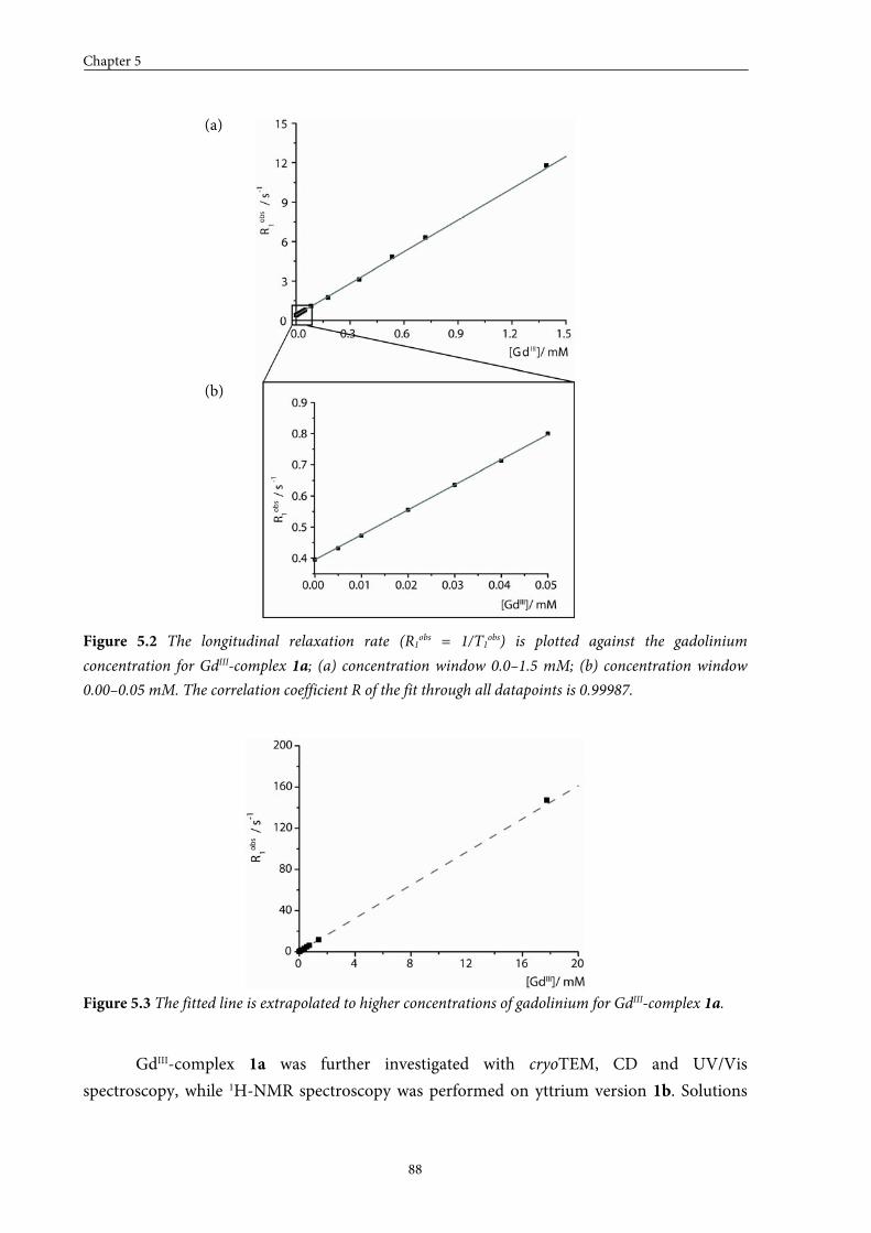
Chapter 5
88
Figure 5.2 The longitudinal relaxation rate (R1obs = 1/T1obs) is plotted against the gadolinium
concentration for GdIII-complex 1a; (a) concentration window 0.0–1.5 mM; (b) concentration window
0.00–0.05 mM. The correlation coefficient R of the fit through all datapoints is 0.99987.
Figure 5.3 The fitted line is extrapolated to higher concentrations of gadolinium for GdIII-complex 1a.
GdIII-complex 1a was further investigated with cryoTEM, CD and UV/Vis
spectroscopy, while 1H-NMR spectroscopy was performed on yttrium version 1b. Solutions
(a)
(b)
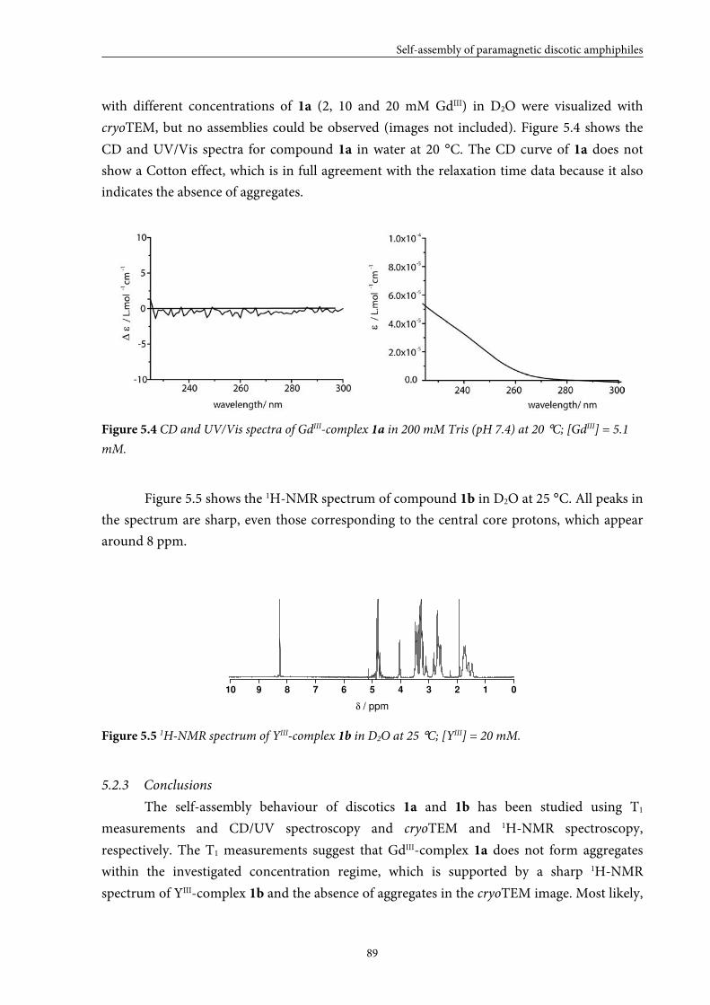
Self-assembly of paramagnetic discotic amphiphiles
89
with different concentrations of 1a (2, 10 and 20 mM GdIII) in D2O were visualized with
cryoTEM, but no assemblies could be observed (images not included). Figure 5.4 shows the
CD and UV/Vis spectra for compound 1a in water at 20 °C. The CD curve of 1a does not
show a Cotton effect, which is in full agreement with the relaxation time data because it also
indicates the absence of aggregates.
Figure 5.4 CD and UV/Vis spectra of GdIII-complex 1a in 200 mM Tris (pH 7.4) at 20 °C; [GdIII] = 5.1
mM.
Figure 5.5 shows the 1H-NMR spectrum of compound 1b in D2O at 25 °C. All peaks in
the spectrum are sharp, even those corresponding to the central core protons, which appear
around 8 ppm.
10 9 8 7 6 5 4 3 2 1 0
δ / ppm
Figure 5.5 1H-NMR spectrum of YIII-complex 1b in D2O at 25 °C; [YIII] = 20 mM.
5.2.3 Conclusions
The self-assembly behaviour of discotics 1a and 1b has been studied using T1
measurements and CD/UV spectroscopy and cryoTEM and 1H-NMR spectroscopy,
respectively. The T1 measurements suggest that GdIII-complex 1a does not form aggregates
within the investigated concentration regime, which is supported by a sharp 1H-NMR
spectrum of YIII-complex 1b and the absence of aggregates in the cryoTEM image. Most likely,
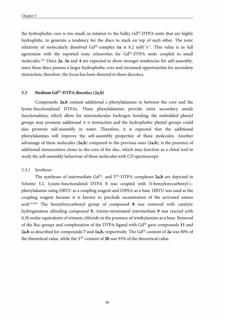
Chapter 5
90
the hydrophobic core is too small, in relation to the bulky GdIII-DTPA units that are highly
hydrophilic, to generate a tendency for the discs to stack on top of each other. The ionic
relaxivity of molecularly dissolved GdIII-complex 1a is 8.2 mM–1s–1. This value is in full
agreement with the reported ionic relaxivities for GdIII-DTPA units coupled to small
molecules.[42] Discs 2a, 3a and 4 are expected to show stronger tendencies for self-assembly,
since these discs possess a larger hydrophobic core and increased opportunities for secondary
interaction, therefore, the focus has been directed to these discotics.
5.3 Medium GdIII-DTPA discotics (2a,b)
Compounds 2a,b contain additional L-phenylalanines in between the core and the
lysine-functionalized DTPAs. These phenylalanines provide extra secondary amide
functionalities, which allow for intermolecular hydrogen bonding; the embedded phenyl
groups may promote additional π–π interaction and the hydrophobic phenyl groups could
also promote self-assembly in water. Therefore, it is expected that the additional
phenylalanines will improve the self-assembly properties of these molecules. Another
advantage of these molecules (2a,b) compared to the previous ones (1a,b), is the presence of
additional stereocentres closer to the core of the disc, which may function as a chiral tool to
study the self-assembly behaviour of these molecules with CD spectroscopy.
5.3.1 Syntheses
The syntheses of intermediate GdIII- and YIII-DTPA complexes 2a,b are depicted in
Scheme 5.2. Lysine-functionalized DTPA 5 was coupled with N-benzyloxycarbonyl-L-
phenylalanine using HBTU as a coupling reagent and DiPEA as a base. HBTU was used as the
coupling reagent because it is known to preclude racemization of the activated amino
acid.[43,44] The benzyloxycarbonyl group of compound 8 was removed with catalytic
hydrogenation affording compound 9. Amino-terminated intermediate 9 was reacted with
0.30 molar equivalents of trimesic chloride in the presence of triethylamine as a base. Removal
of the Boc-groups and complexation of the DTPA-ligand with GdIII gave compounds 11 and
2a,b as described for compounds 7 and 1a,b, respectively. The GdIII-content of 2a was 80% of
the theoretical value, while the YIII-content of 2b was 93% of the theoretical value.
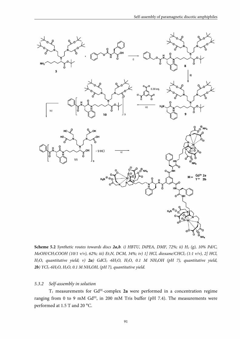
Self-assembly of paramagnetic discotic amphiphiles
91
Scheme 5.2 Synthetic routes towards discs 2a,b. i) HBTU, DiPEA, DMF, 72%; ii) H2 (g), 10% Pd/C,
MeOH/CH3COOH (10/1 v/v), 62%; iii) Et3N, DCM, 34%; iv) 1] HCl, dioxane/CHCl3 (1:1 v/v), 2] HCl,
H2O, quantitative yield; v) 2a) GdCl3 ·6H2O, H2O, 0.1 M NH4OH (pH 7), quantitative yield,
2b) YCl3 ·6H2O, H2O, 0.1 M NH4OH, (pH 7), quantitative yield.
5.3.2 Self-assembly in solution
T1 measurements for GdIII-complex 2a were performed in a concentration regime
ranging from 0 to 9 mM GdIII, in 200 mM Tris buffer (pH 7.4). The measurements were
performed at 1.5 T and 20 °C.
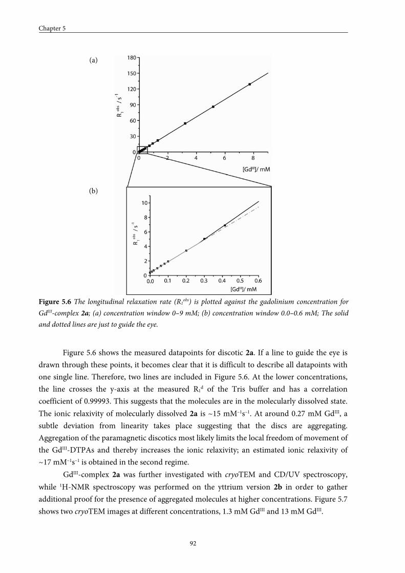
Chapter 5
92
Figure 5.6 The longitudinal relaxation rate (R1obs) is plotted against the gadolinium concentration for
GdIII-complex 2a; (a) concentration window 0–9 mM; (b) concentration window 0.0–0.6 mM; The solid
and dotted lines are just to guide the eye.
Figure 5.6 shows the measured datapoints for discotic 2a. If a line to guide the eye is
drawn through these points, it becomes clear that it is difficult to describe all datapoints with
one single line. Therefore, two lines are included in Figure 5.6. At the lower concentrations,
the line crosses the y-axis at the measured R1d of the Tris buffer and has a correlation
coefficient of 0.99993. This suggests that the molecules are in the molecularly dissolved state.
The ionic relaxivity of molecularly dissolved 2a is ~15 mM–1s–1. At around 0.27 mM GdIII, a
subtle deviation from linearity takes place suggesting that the discs are aggregating.
Aggregation of the paramagnetic discotics most likely limits the local freedom of movement of
the GdIII-DTPAs and thereby increases the ionic relaxivity; an estimated ionic relaxivity of
~17 mM–1s–1 is obtained in the second regime.
GdIII-complex 2a was further investigated with cryoTEM and CD/UV spectroscopy,
while 1H-NMR spectroscopy was performed on the yttrium version 2b in order to gather
additional proof for the presence of aggregated molecules at higher concentrations. Figure 5.7
shows two cryoTEM images at different concentrations, 1.3 mM GdIII and 13 mM GdIII.
(a)
(b)
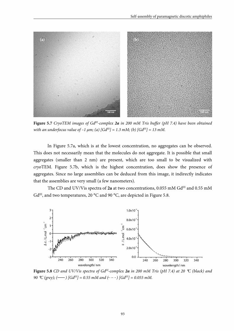
Self-assembly of paramagnetic discotic amphiphiles
93
Figure 5.7 CryoTEM images of GdIII-complex 2a in 200 mM Tris buffer (pH 7.4) have been obtained
with an underfocus value of –1 μm; (a) [GdIII] = 1.3 mM; (b) [GdIII] = 13 mM.
In Figure 5.7a, which is at the lowest concentration, no aggregates can be observed.
This does not necessarily mean that the molecules do not aggregate. It is possible that small
aggregates (smaller than 2 nm) are present, which are too small to be visualized with
cryoTEM. Figure 5.7b, which is the highest concentration, does show the presence of
aggregates. Since no large assemblies can be deduced from this image, it indirectly indicates
that the assemblies are very small (a few nanometers).
The CD and UV/Vis spectra of 2a at two concentrations, 0.055 mM GdIII and 0.55 mM
GdIII, and two temperatures, 20 °C and 90 °C, are depicted in Figure 5.8.
Figure 5.8 CD and UV/Vis spectra of GdIII-complex 2a in 200 mM Tris (pH 7.4) at 20 °C (black) and
90 °C (grey); ( ) [GdIII] = 0.55 mM and ( ) [GdIII] = 0.055 mM.
(a) (b)
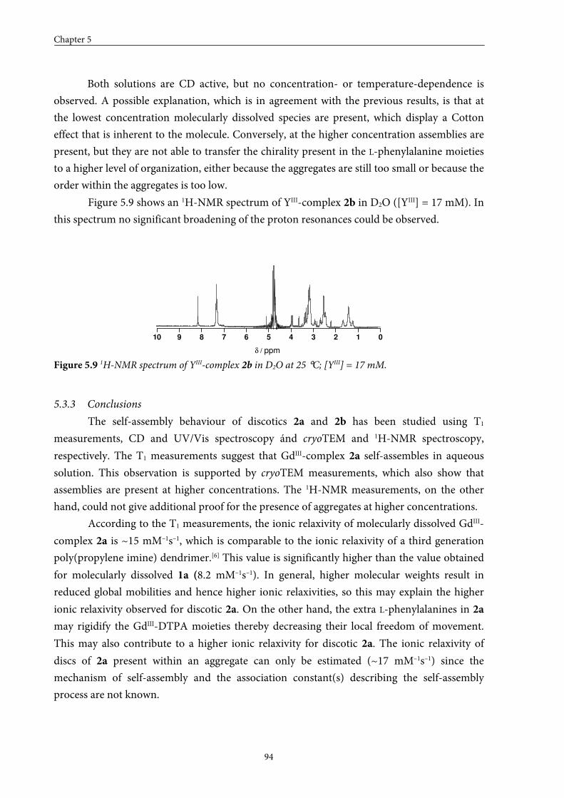
Chapter 5
94
Both solutions are CD active, but no concentration- or temperature-dependence is
observed. A possible explanation, which is in agreement with the previous results, is that at
the lowest concentration molecularly dissolved species are present, which display a Cotton
effect that is inherent to the molecule. Conversely, at the higher concentration assemblies are
present, but they are not able to transfer the chirality present in the L-phenylalanine moieties
to a higher level of organization, either because the aggregates are still too small or because the
order within the aggregates is too low.
Figure 5.9 shows an 1H-NMR spectrum of YIII-complex 2b in D2O ([YIII] = 17 mM). In
this spectrum no significant broadening of the proton resonances could be observed.
10 9 8 7 6 5 4 3 2 1 0
δ / ppm
Figure 5.9 1H-NMR spectrum of YIII-complex 2b in D2O at 25 °C; [YIII] = 17 mM.
5.3.3 Conclusions
The self-assembly behaviour of discotics 2a and 2b has been studied using T1
measurements, CD and UV/Vis spectroscopy ánd cryoTEM and 1H-NMR spectroscopy,
respectively. The T1 measurements suggest that GdIII-complex 2a self-assembles in aqueous
solution. This observation is supported by cryoTEM measurements, which also show that
assemblies are present at higher concentrations. The 1H-NMR measurements, on the other
hand, could not give additional proof for the presence of aggregates at higher concentrations.
According to the T1 measurements, the ionic relaxivity of molecularly dissolved GdIII-
complex 2a is ~15 mM–1s–1, which is comparable to the ionic relaxivity of a third generation
poly(propylene imine) dendrimer.[6] This value is significantly higher than the value obtained
for molecularly dissolved 1a (8.2 mM–1s–1). In general, higher molecular weights result in
reduced global mobilities and hence higher ionic relaxivities, so this may explain the higher
ionic relaxivity observed for discotic 2a. On the other hand, the extra L-phenylalanines in 2a
may rigidify the GdIII-DTPA moieties thereby decreasing their local freedom of movement.
This may also contribute to a higher ionic relaxivity for discotic 2a. The ionic relaxivity of
discs of 2a present within an aggregate can only be estimated (~17 mM–1s–1) since the
mechanism of self-assembly and the association constant(s) describing the self-assembly
process are not known.
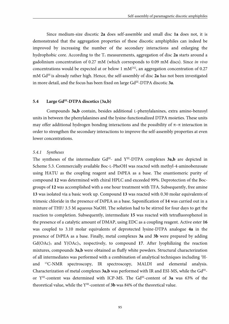
Self-assembly of paramagnetic discotic amphiphiles
95
Since medium-size discotic 2a does self-assemble and small disc 1a does not, it is
demonstrated that the aggregation properties of these discotic amphiphiles can indeed be
improved by increasing the number of the secondary interactions and enlarging the
hydrophobic core. According to the T1 measurements, aggregation of disc 2a starts around a
gadolinium concentration of 0.27 mM (which corresponds to 0.09 mM discs). Since in vivo
concentrations would be expected at or below 1 mM[32], an aggregation concentration of 0.27
mM GdIII is already rather high. Hence, the self-assembly of disc 2a has not been investigated
in more detail, and the focus has been fixed on large GdIII-DTPA discotic 3a.
5.4 Large GdIII-DTPA discotics (3a,b)
Compounds 3a,b contain, besides additional L-phenylalanines, extra amino-benzoyl
units in between the phenylalanines and the lysine-functionalized DTPA moieties. These units
may offer additional hydrogen bonding interactions and the possibility of π–π interaction in
order to strengthen the secondary interactions to improve the self-assembly properties at even
lower concentrations.
5.4.1 Syntheses
The syntheses of the intermediate GdIII- and YIII-DTPA complexes 3a,b are depicted in
Scheme 5.3. Commercially available Boc-L-PheOH was reacted with methyl-4-aminobenzoate
using HATU as the coupling reagent and DiPEA as a base. The enantiomeric purity of
compound 12 was determined with chiral HPLC and exceeded 99%. Deprotection of the Boc-
groups of 12 was accomplished with a one hour treatment with TFA. Subsequently, free amine
13 was isolated via a basic work up. Compound 13 was reacted with 0.30 molar equivalents of
trimesic chloride in the presence of DiPEA as a base. Saponification of 14 was carried out in a
mixture of THF/ 3.5 M aqueous NaOH. The solution had to be stirred for four days to get the
reaction to completion. Subsequently, intermediate 15 was reacted with tetrafluorophenol in
the presence of a catalytic amount of DMAP, using EDC as a coupling reagent. Active ester 16
was coupled to 3.10 molar equivalents of deprotected lysine-DTPA analogue 4a in the
presence of DiPEA as a base. Finally, metal complexes 3a and 3b were prepared by adding
Gd(OAc)3 and Y(OAc)3, respectively, to compound 17. After lyophilizing the reaction
mixtures, compounds 3a,b were obtained as fluffy white powders. Structural characterization
of all intermediates was performed with a combination of analytical techniques including 1H-
and 13C-NMR spectroscopy, IR spectroscopy, MALDI and elemental analysis.
Characterization of metal complexes 3a,b was performed with IR and ESI-MS, while the GdIII-
or YIII-content was determined with ICP-MS. The GdIII-content of 3a was 63% of the
theoretical value, while the YIII-content of 3b was 84% of the theoretical value.
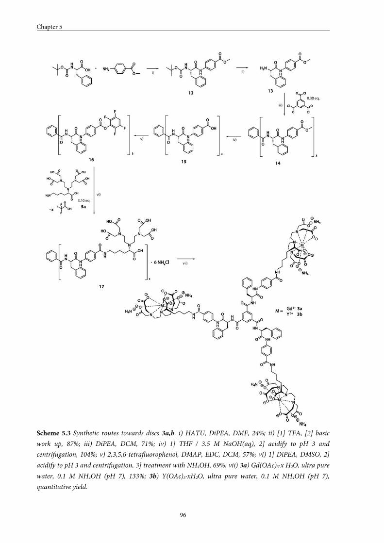
Chapter 5
96
Scheme 5.3 Synthetic routes towards discs 3a,b. i) HATU, DiPEA, DMF, 24%; ii) [1] TFA, [2] basic
work up, 87%; iii) DiPEA, DCM, 71%; iv) 1] THF / 3.5 M NaOH(aq), 2] acidify to pH 3 and
centrifugation, 104%; v) 2,3,5,6-tetrafluorophenol, DMAP, EDC, DCM, 57%; vi) 1] DiPEA, DMSO, 2]
acidify to pH 3 and centrifugation, 3] treatment with NH4OH, 69%; vii) 3a) Gd(OAc)3∙x H2O, ultra pure
water, 0.1 M NH4OH (pH 7), 133%; 3b) Y(OAc)3∙xH2O, ultra pure water, 0.1 M NH4OH (pH 7),
quantitative yield.

Self-assembly of paramagnetic discotic amphiphiles
97
5.4.2 Self-assembly in solution
T1 measurements on GdIII-complex 3a were performed in a concentration regime
ranging from 0 to 4 mM GdIII, in 200 mM Tris buffer (pH 7.4). The measurements were
performed at 1.5 T and 20 °C. Figure 5.10 shows the measured datapoints for discotic 3a.
Figure 5.10 The longitudinal relaxation rate (R1obs) is plotted against the gadolinium concentration for
GdIII-complex 3a; (a) concentration window 0–4 mM; (b) concentration window 0.0–0.4 mM; (c)
concentration window 0.00–0.05 mM. The lines are just to guide the eye.
(a)
(b)
(c)
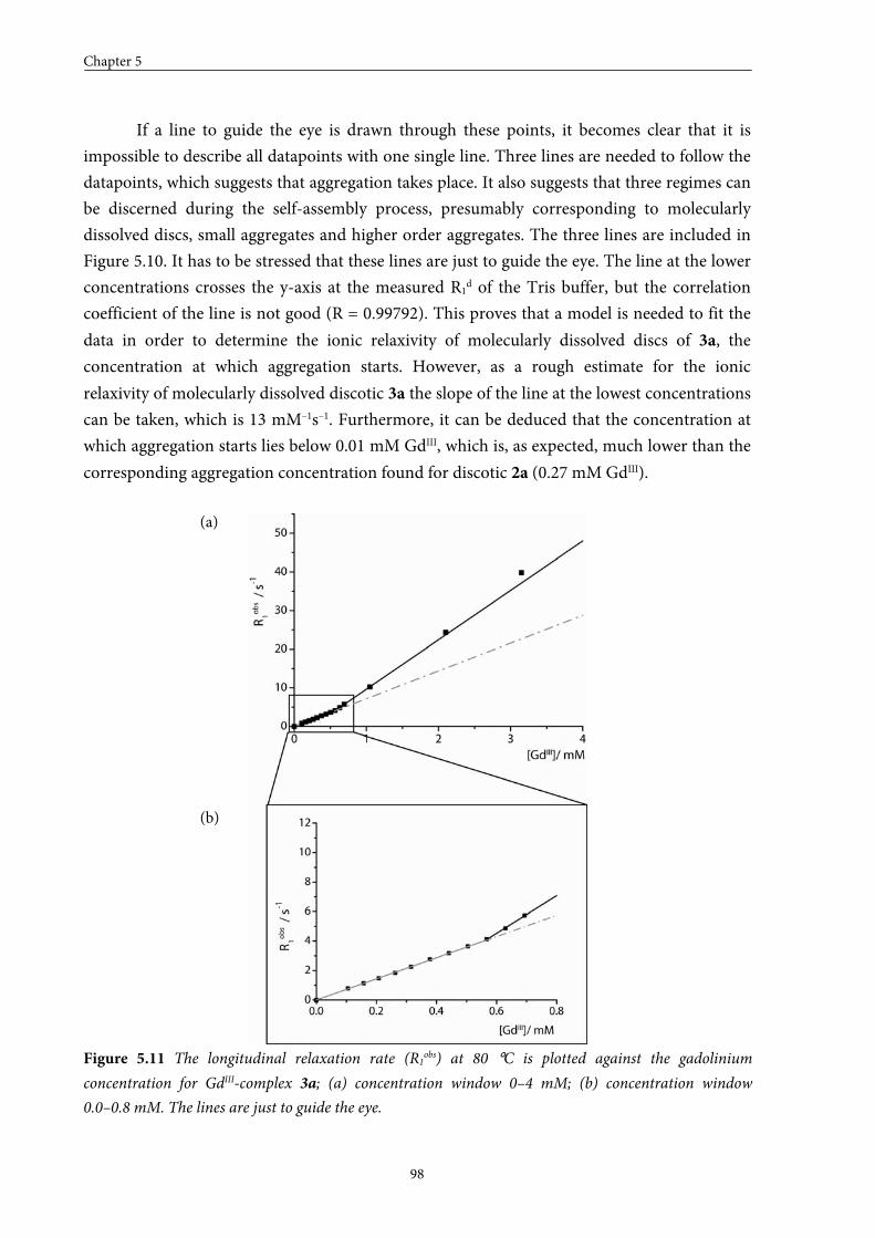
Chapter 5
98
If a line to guide the eye is drawn through these points, it becomes clear that it is
impossible to describe all datapoints with one single line. Three lines are needed to follow the
datapoints, which suggests that aggregation takes place. It also suggests that three regimes can
be discerned during the self-assembly process, presumably corresponding to molecularly
dissolved discs, small aggregates and higher order aggregates. The three lines are included in
Figure 5.10. It has to be stressed that these lines are just to guide the eye. The line at the lower
concentrations crosses the y-axis at the measured R1d of the Tris buffer, but the correlation
coefficient of the line is not good (R = 0.99792). This proves that a model is needed to fit the
data in order to determine the ionic relaxivity of molecularly dissolved discs of 3a, the
concentration at which aggregation starts. However, as a rough estimate for the ionic
relaxivity of molecularly dissolved discotic 3a the slope of the line at the lowest concentrations
can be taken, which is 13 mM–1s–1. Furthermore, it can be deduced that the concentration at
which aggregation starts lies below 0.01 mM GdIII, which is, as expected, much lower than the
corresponding aggregation concentration found for discotic 2a (0.27 mM GdIII).
Figure 5.11 The longitudinal relaxation rate (R1obs) at 80 °C is plotted against the gadolinium
concentration for GdIII-complex 3a; (a) concentration window 0–4 mM; (b) concentration window
0.0–0.8 mM. The lines are just to guide the eye.
(a)
(b)
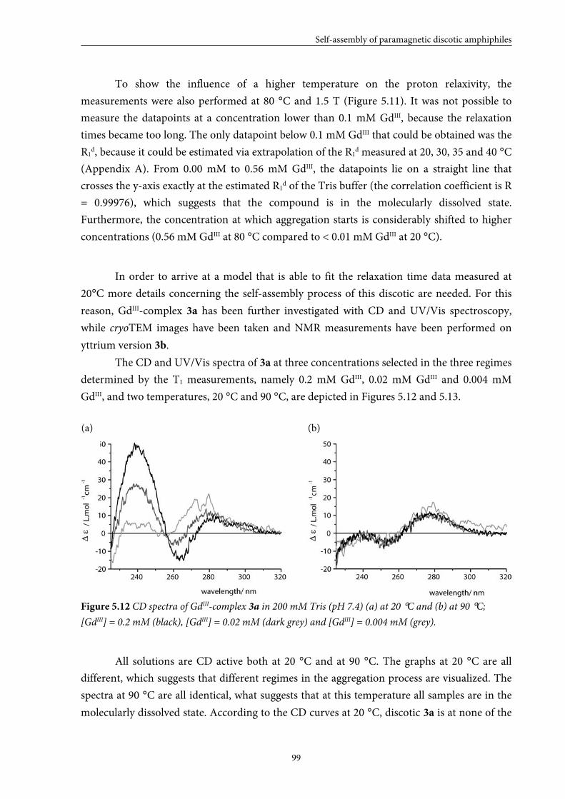
Self-assembly of paramagnetic discotic amphiphiles
99
To show the influence of a higher temperature on the proton relaxivity, the
measurements were also performed at 80 °C and 1.5 T (Figure 5.11). It was not possible to
measure the datapoints at a concentration lower than 0.1 mM GdIII, because the relaxation
times became too long. The only datapoint below 0.1 mM GdIII that could be obtained was the
R1d, because it could be estimated via extrapolation of the R1
d measured at 20, 30, 35 and 40 °C
(Appendix A). From 0.00 mM to 0.56 mM GdIII, the datapoints lie on a straight line that
crosses the y-axis exactly at the estimated R1d of the Tris buffer (the correlation coefficient is R
= 0.99976), which suggests that the compound is in the molecularly dissolved state.
Furthermore, the concentration at which aggregation starts is considerably shifted to higher
concentrations (0.56 mM GdIII at 80 °C compared to < 0.01 mM GdIII at 20 °C).
In order to arrive at a model that is able to fit the relaxation time data measured at
20°C more details concerning the self-assembly process of this discotic are needed. For this
reason, GdIII-complex 3a has been further investigated with CD and UV/Vis spectroscopy,
while cryoTEM images have been taken and NMR measurements have been performed on
yttrium version 3b.
The CD and UV/Vis spectra of 3a at three concentrations selected in the three regimes
determined by the T1 measurements, namely 0.2 mM GdIII, 0.02 mM GdIII and 0.004 mM
GdIII, and two temperatures, 20 °C and 90 °C, are depicted in Figures 5.12 and 5.13.
Figure 5.12 CD spectra of GdIII-complex 3a in 200 mM Tris (pH 7.4) (a) at 20 °C and (b) at 90 °C;
[GdIII] = 0.2 mM (black), [GdIII] = 0.02 mM (dark grey) and [GdIII] = 0.004 mM (grey).
All solutions are CD active both at 20 °C and at 90 °C. The graphs at 20 °C are all
different, which suggests that different regimes in the aggregation process are visualized. The
spectra at 90 °C are all identical, what suggests that at this temperature all samples are in the
molecularly dissolved state. According to the CD curves at 20 °C, discotic 3a is at none of the
(a) (b)
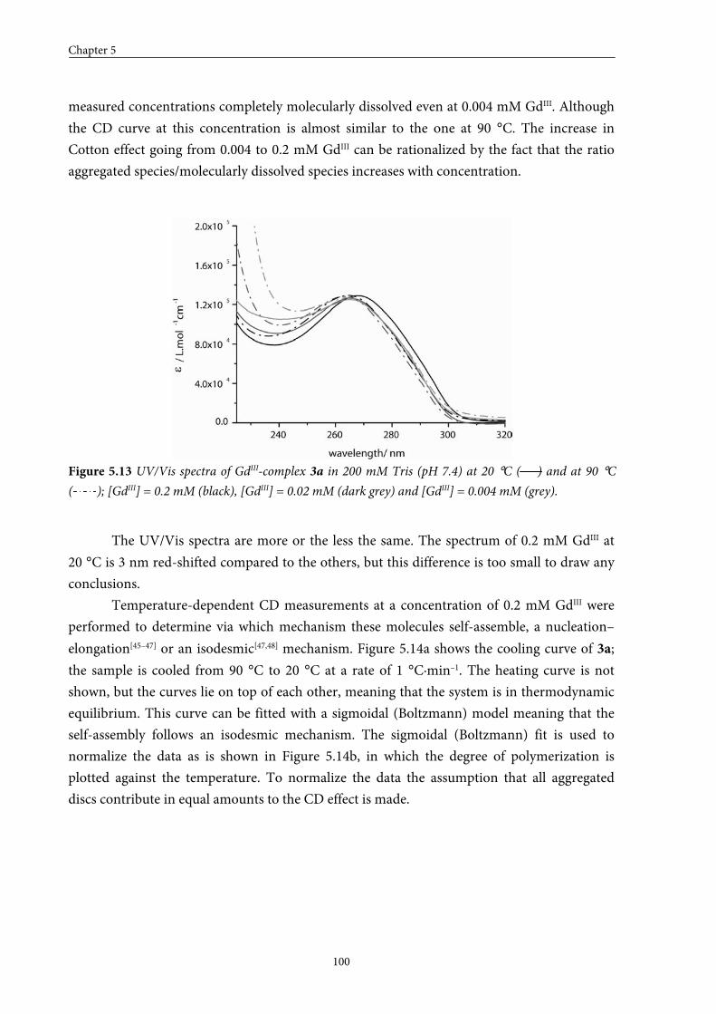
Chapter 5
100
measured concentrations completely molecularly dissolved even at 0.004 mM GdIII. Although
the CD curve at this concentration is almost similar to the one at 90 °C. The increase in
Cotton effect going from 0.004 to 0.2 mM GdIII can be rationalized by the fact that the ratio
aggregated species/molecularly dissolved species increases with concentration.
Figure 5.13 UV/Vis spectra of GdIII-complex 3a in 200 mM Tris (pH 7.4) at 20 °C ( ) and at 90 °C
( ); [GdIII] = 0.2 mM (black), [GdIII] = 0.02 mM (dark grey) and [GdIII] = 0.004 mM (grey).
The UV/Vis spectra are more or the less the same. The spectrum of 0.2 mM GdIII at
20 °C is 3 nm red-shifted compared to the others, but this difference is too small to draw any
conclusions.
Temperature-dependent CD measurements at a concentration of 0.2 mM GdIII were
performed to determine via which mechanism these molecules self-assemble, a nucleation–
elongation[45–47] or an isodesmic[47,48] mechanism. Figure 5.14a shows the cooling curve of 3a;
the sample is cooled from 90 °C to 20 °C at a rate of 1 °C·min–1. The heating curve is not
shown, but the curves lie on top of each other, meaning that the system is in thermodynamic
equilibrium. This curve can be fitted with a sigmoidal (Boltzmann) model meaning that the
self-assembly follows an isodesmic mechanism. The sigmoidal (Boltzmann) fit is used to
normalize the data as is shown in Figure 5.14b, in which the degree of polymerization is
plotted against the temperature. To normalize the data the assumption that all aggregated
discs contribute in equal amounts to the CD effect is made.
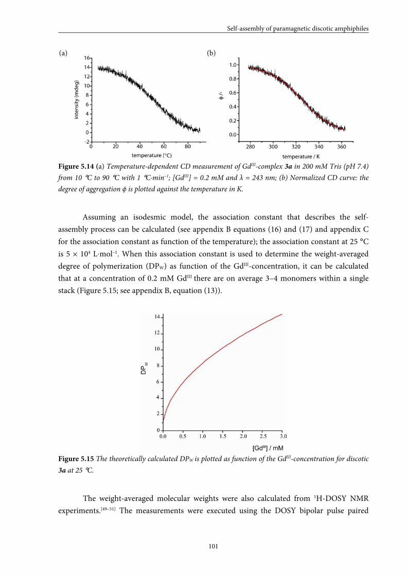
Self-assembly of paramagnetic discotic amphiphiles
101
Figure 5.14 (a) Temperature-dependent CD measurement of GdIII-complex 3a in 200 mM Tris (pH 7.4)
from 10 °C to 90 °C with 1 °C·min–1; [GdIII] = 0.2 mM and λ = 243 nm; (b) Normalized CD curve: the
degree of aggregation ϕ is plotted against the temperature in K.
Assuming an isodesmic model, the association constant that describes the self-
assembly process can be calculated (see appendix B equations (16) and (17) and appendix C
for the association constant as function of the temperature); the association constant at 25 °C
is 5 × 10⁴ L⋅mol–1. When this association constant is used to determine the weight-averaged
degree of polymerization (DPW) as function of the GdIII-concentration, it can be calculated
that at a concentration of 0.2 mM GdIII there are on average 3–4 monomers within a single
stack (Figure 5.15; see appendix B, equation (13)).
Figure 5.15 The theoretically calculated DPW is plotted as function of the GdIII-concentration for discotic
3a at 25 °C.
The weight-averaged molecular weights were also calculated from 1H-DOSY NMR
experiments.[49–51] The measurements were executed using the DOSY bipolar pulse paired
(a) (b)
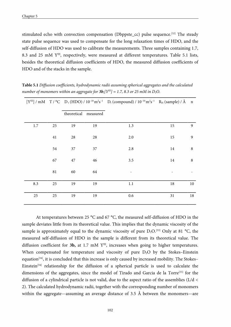
Chapter 5
102
stimulated echo with convection compensation (Dbppste_cc) pulse sequence.[52] The steady
state pulse sequence was used to compensate for the long relaxation times of HDO, and the
self-diffusion of HDO was used to calibrate the measurements. Three samples containing 1.7,
8.3 and 25 mM YIII, respectively, were measured at different temperatures. Table 5.1 lists,
besides the theoretical diffusion coefficients of HDO, the measured diffusion coefficients of
HDO and of the stacks in the sample.
Table 5.1 Diffusion coefficients, hydrodynamic radii assuming spherical aggregates and the calculated
number of monomers within an aggregate for 3b;[YIII] = 1.7, 8.3 or 25 mM in D2O.
D t (HDO) / 10–10 m2s–1 [YIII] / mM T / °C
theoretical measured
Dt (compound) / 10–10 m2s–1 RH (sample) / Å n
25 19 19 1.3 15 9
41 28 28 2.0 15 9
54 37 37 2.8 14 8
67 47 46 3.5 14 8
1.7
81 60 64 - - -
8.3 25 19 19 1.1 18 10
25 25 19 19 0.6 31 18
At temperatures between 25 °C and 67 °C, the measured self-diffusion of HDO in the
sample deviates little from its theoretical value. This implies that the dynamic viscosity of the
sample is approximately equal to the dynamic viscosity of pure D2O.[53] Only at 81 °C, the
measured self-diffusion of HDO in the sample is different from its theoretical value. The
diffusion coefficient for 3b, at 1.7 mM YIII, increases when going to higher temperatures.
When compensated for temperature and viscosity of pure D2O by the Stokes–Einstein
equation[54], it is concluded that this increase is only caused by increased mobility. The Stokes–
Einstein[54] relationship for the diffusion of a spherical particle is used to calculate the
dimensions of the aggregates, since the model of Tirado and Garcia de la Torre[55] for the
diffusion of a cylindrical particle is not valid, due to the aspect ratio of the assemblies (L/d <
2). The calculated hydrodynamic radii, together with the corresponding number of monomers
within the aggregate—assuming an average distance of 3.5 Å between the monomers—are
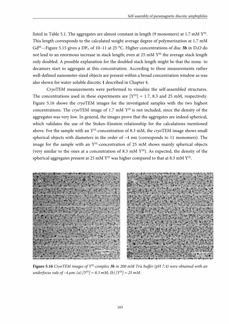
Self-assembly of paramagnetic discotic amphiphiles
103
listed in Table 5.1. The aggregates are almost constant in length (9 monomers) at 1.7 mM YIII.
This length corresponds to the calculated weight average degree of polymerization at 1.7 mM
GdIII—Figure 5.15 gives a DPw of 10–11 at 25 °C. Higher concentrations of disc 3b in D2O do
not lead to an enormous increase in stack length; even at 25 mM YIII the average stack length
only doubled. A possible explanation for the doubled stack length might be that the nona- to
decamers start to aggregate at this concentration. According to these measurements rather
well-defined nanometer-sized objects are present within a broad concentration window as was
also shown for water-soluble discotic 1 described in Chapter 4.
CryoTEM measurements were performed to visualize the self-assembled structures.
The concentrations used in these experiments are [YIII] = 1.7, 8.3 and 25 mM, respectively.
Figure 5.16 shows the cryoTEM images for the investigated samples with the two highest
concentrations. The cryoTEM image of 1.7 mM YIII is not included, since the density of the
aggregates was very low. In general, the images prove that the aggregates are indeed spherical,
which validates the use of the Stokes–Einstein relationship for the calculations mentioned
above. For the sample with an YIII-concentration of 8.3 mM, the cryoTEM image shows small
spherical objects with diameters in the order of ~4 nm (corresponds to 11 monomers). The
image for the sample with an YIII-concentration of 25 mM shows mainly spherical objects
(very similar to the ones at a concentration of 8.3 mM YIII). As expected, the density of the
spherical aggregates present at 25 mM YIII was higher compared to that at 8.3 mM YIII.
Figure 5.16 CryoTEM images of YIII-complex 3b in 200 mM Tris buffer (pH 7.4) were obtained with an
underfocus vale of –4 μm; (a) [YIII] = 8.3 mM; (b) [YIII] = 25 mM.
(a) (b)
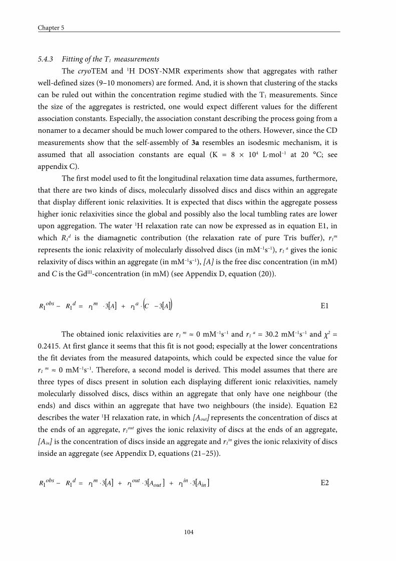
Chapter 5
104
5.4.3 Fitting of the T1 measurements
The cryoTEM and 1H DOSY-NMR experiments show that aggregates with rather
well-defined sizes (9–10 monomers) are formed. And, it is shown that clustering of the stacks
can be ruled out within the concentration regime studied with the T1 measurements. Since
the size of the aggregates is restricted, one would expect different values for the different
association constants. Especially, the association constant describing the process going from a
nonamer to a decamer should be much lower compared to the others. However, since the CD
measurements show that the self-assembly of 3a resembles an isodesmic mechanism, it is
assumed that all association constants are equal (K = 8 × 10⁴ L⋅mol–1 at 20 °C; see
appendix C).
The first model used to fit the longitudinal relaxation time data assumes, furthermore,
that there are two kinds of discs, molecularly dissolved discs and discs within an aggregate
that display different ionic relaxivities. It is expected that discs within the aggregate possess
higher ionic relaxivities since the global and possibly also the local tumbling rates are lower
upon aggregation. The water 1H relaxation rate can now be expressed as in equation E1, in
which R1d is the diamagnetic contribution (the relaxation rate of pure Tris buffer), r1m
represents the ionic relaxivity of molecularly dissolved discs (in mM–1s–1), r1 a gives the ionic
relaxivity of discs within an aggregate (in mM–1s–1), [A] is the free disc concentration (in mM)
and C is the GdIII-concentration (in mM) (see Appendix D, equation (20)).
[ ] [ ]( )ACrArRRamdobs 33 1111 −⋅+⋅=− E1
The obtained ionic relaxivities are r1 m ≈ 0 mM–1s–1 and r1 a = 30.2 mM–1s–1 and χ2 =
0.2415. At first glance it seems that this fit is not good; especially at the lower concentrations
the fit deviates from the measured datapoints, which could be expected since the value for
r1 m ≈ 0 mM–1s–1. Therefore, a second model is derived. This model assumes that there are
three types of discs present in solution each displaying different ionic relaxivities, namely
molecularly dissolved discs, discs within an aggregate that only have one neighbour (the
ends) and discs within an aggregate that have two neighbours (the inside). Equation E2
describes the water 1H relaxation rate, in which [Aout] represents the concentration of discs at
the ends of an aggregate, r1out gives the ionic relaxivity of discs at the ends of an aggregate,
[Ain] is the concentration of discs inside an aggregate and r1in gives the ionic relaxivity of discs
inside an aggregate (see Appendix D, equations (21–25)).
[ ] [ ] [ ]inin
outoutmdobs
ArArArRR 333 11111 ⋅+⋅+⋅=− E2
Self-assembly of paramagnetic discotic amphiphiles
105
Figure 5.17 shows the water 1H relaxation rate data obtained for disc 3a together with the fit
based on model 2. By using this model three ionic relaxivities and the sum-of-squares are
determined, r1 m = 9.2 mM–1s–1, r1 out = 25.5 mM–1s–1, r1 in = 31.3 mM–1s–1 and χ2 = 0.02554. This
model seems to describe the measured datapoints very well, even at the lower GdIII-
concentrations. A comparison of the two models using the corrected Akaike’s information
criteria, which takes the number of parameters and the sum-of-squares (χ2) into account,
supports our findings since it is shown that the probability that model 1 is correct is 0%,
while the probability that model 2 is correct is 100%.
Figure 5.17 Model 2 is used to fit the measured relaxation rate data for discotic 3a obtained in 200 mM
Tris (pH 7.4) at 20 °C and 1.5 T. (a) concentration regime from 0.0–3.2 mM GdIII; (b) concentration
regime from 0.0–0.4 mM GdIII; (c) concentration regime from 0.00–0.05 mM GdIII.
The gain in relaxivity going from a molecularly dissolved disc to a disc within an
aggregate (either at the end or inside) is high, while the gain in relaxivity going from a disc at
the end of an aggregate to a disc inside an aggregate is much smaller. This indicates that the
ionic relaxivity of a dimer is not very different from the ionic relaxivity of a decamer.
5.4.4 Evaluation of the aggregation concentration
Assuming an isodesmic model and an association constant of 8 × 10⁴ L·mol–1 at 20 °C,
the concentration at which aggregation of disc 3a starts can be estimated and lies around
3 × 10⁻⁴ mM GdIII. This value cannot directly be compared to the critical micelle
concentration of micellar contrast agents, since the mechanisms of self-assembly are
completely different. At the point at which the discotic molecules start to aggregate almost all
discs are in the molecularly dissolved state (isodesmic self-assembly), while at the critical
micelle concentration almost all linear amphiphiles are incorporated into aggregated
structures (extreme case of nucleation–elongation).
(b) (c) (a)
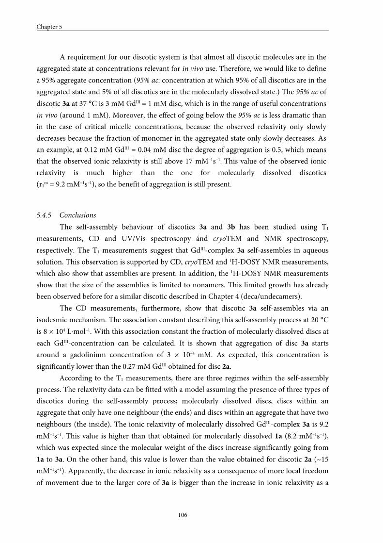
Chapter 5
106
A requirement for our discotic system is that almost all discotic molecules are in the
aggregated state at concentrations relevant for in vivo use. Therefore, we would like to define
a 95% aggregate concentration (95% ac: concentration at which 95% of all discotics are in the
aggregated state and 5% of all discotics are in the molecularly dissolved state.) The 95% ac of
discotic 3a at 37 °C is 3 mM GdIII = 1 mM disc, which is in the range of useful concentrations
in vivo (around 1 mM). Moreover, the effect of going below the 95% ac is less dramatic than
in the case of critical micelle concentrations, because the observed relaxivity only slowly
decreases because the fraction of monomer in the aggregated state only slowly decreases. As
an example, at 0.12 mM GdIII = 0.04 mM disc the degree of aggregation is 0.5, which means
that the observed ionic relaxivity is still above 17 mM–1s–1. This value of the observed ionic
relaxivity is much higher than the one for molecularly dissolved discotics
(r1m = 9.2 mM–1s–1), so the benefit of aggregation is still present.
5.4.5 Conclusions
The self-assembly behaviour of discotics 3a and 3b has been studied using T1
measurements, CD and UV/Vis spectroscopy ánd cryoTEM and NMR spectroscopy,
respectively. The T1 measurements suggest that GdIII-complex 3a self-assembles in aqueous
solution. This observation is supported by CD, cryoTEM and 1H-DOSY NMR measurements,
which also show that assemblies are present. In addition, the 1H-DOSY NMR measurements
show that the size of the assemblies is limited to nonamers. This limited growth has already
been observed before for a similar discotic described in Chapter 4 (deca/undecamers).
The CD measurements, furthermore, show that discotic 3a self-assembles via an
isodesmic mechanism. The association constant describing this self-assembly process at 20 °C
is 8 × 10⁴ L⋅mol–1. With this association constant the fraction of molecularly dissolved discs at
each GdIII-concentration can be calculated. It is shown that aggregation of disc 3a starts
around a gadolinium concentration of 3 × 10⁻⁴ mM. As expected, this concentration is
significantly lower than the 0.27 mM GdIII obtained for disc 2a.
According to the T1 measurements, there are three regimes within the self-assembly
process. The relaxivity data can be fitted with a model assuming the presence of three types of
discotics during the self-assembly process; molecularly dissolved discs, discs within an
aggregate that only have one neighbour (the ends) and discs within an aggregate that have two
neighbours (the inside). The ionic relaxivity of molecularly dissolved GdIII-complex 3a is 9.2
mM–1s–1. This value is higher than that obtained for molecularly dissolved 1a (8.2 mM–1s–1),
which was expected since the molecular weight of the discs increase significantly going from
1a to 3a. On the other hand, this value is lower than the value obtained for discotic 2a (~15
mM–1s–1). Apparently, the decrease in ionic relaxivity as a consequence of more local freedom
of movement due to the larger core of 3a is bigger than the increase in ionic relaxivity as a
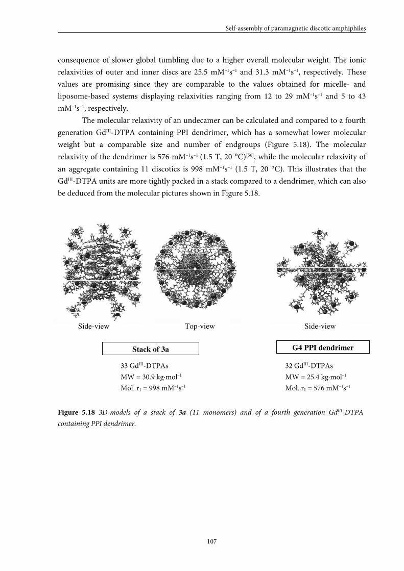
Self-assembly of paramagnetic discotic amphiphiles
107
consequence of slower global tumbling due to a higher overall molecular weight. The ionic
relaxivities of outer and inner discs are 25.5 mM–1s–1 and 31.3 mM–1s–1, respectively. These
values are promising since they are comparable to the values obtained for micelle- and
liposome-based systems displaying relaxivities ranging from 12 to 29 mM–1s–1 and 5 to 43
mM–1s–1, respectively.
The molecular relaxivity of an undecamer can be calculated and compared to a fourth
generation GdIII-DTPA containing PPI dendrimer, which has a somewhat lower molecular
weight but a comparable size and number of endgroups (Figure 5.18). The molecular
relaxivity of the dendrimer is 576 mM–1s–1 (1.5 T, 20 °C)[56], while the molecular relaxivity of
an aggregate containing 11 discotics is 998 mM–1s–1 (1.5 T, 20 °C). This illustrates that the
GdIII-DTPA units are more tightly packed in a stack compared to a dendrimer, which can also
be deduced from the molecular pictures shown in Figure 5.18.
Figure 5.18 3D-models of a stack of 3a (11 monomers) and of a fourth generation GdIII-DTPA
containing PPI dendrimer.
33 GdIII-DTPAs 32 GdIII-DTPAs
MW = 30.9 kg⋅mol–1 MW = 25.4 kg⋅mol–1
Mol. r1 = 998 mM–1s–1 Mol. r1 = 576 mM–1s–1
G4 PPI dendrimer Stack of 3a
Top-view Side-view Side-view
Chapter 5
108
5.5 Large GdIII-DOTA discotic (4)
Because large DTPA discotics 3a,b displayed high relaxivities and low aggregation
concentrations, the ligand in 3 was replaced. Large DOTA discotic 4 was synthesized to show
the universality of our approach. Switching the ligand from DTPA to DOTA may result in a
higher association constant, since electrostatic repulsion does not play a role in the case of
GdIII-DOTA. This complex is neutral, which makes it easier for the GdIII-DOTA discs to self-
assemble. GdIII-DTPA complexes, on the other hand, have a charge of 2–, so the overall charge
of one discotic is 6–. Electrostatic repulsion may, therefore, hinder aggregation. The ionic
relaxivities for the molecularly dissolved discs are expected to be comparable since DOTA and
DTPA ligands display similar ionic relaxivities.[3]
5.5.1 Synthesis
GdIII-DOTA discotic 4 was prepared as depicted in Scheme 5.4. DOTA derivative 18
was kindly provided by SyMO-Chem BV. Activated disc 16 was reacted with 3.20 equivalents
of compound 18 in deuterated DMSO in the presence of an excess of base. Dilution of the
reaction mixture with ultra pure water and consecutive acidification to pH 3 with
concentrated HCl did not result in precipitation of target compound 19, hence the aqueous
solution was lyophilized to yield crude 19 as a off-white solid material. Purification by eluting
a solution of crude 19 in water over a size-exclusion PD10 column followed by preparative RP
HPLC gave pure disc 19. Finally, metal complex 4 was prepared by adding an excess of
Gd(OAc)3 to compound 19 in water. The pH of the solution was monitored and maintained at
7 by adding pyridine. After stirring overnight, a CHELEX 100 sodium form resin was added
to capture the excess of gadolinium ions. After three days, the removal of the GdIII-ions was
completed as was indicated by an UV test experiment with xylenol orange.[40,41] The solution
was carefully isolated from the resin beads and was subsequently lyophilized to yield the
product as a white fluffy powder.
Structural characterization of discotic 4 was performed with LC-MS. The GdIII-content
of disc 4 was determined with ICP-MS. The values for the observed GdIII- and YIII-contents for
all complexes (1–3) were very close to their theoretical values, typically between 80–100%,
except for discotics 4 for which a GdIII-content of 28% was found. This may be attributed to
the presence of residual salts originating from the pyridine, which was not used during the
complexation of the other discotics. Despite this low number, the self-assembly of discotic 4
has been evaluated in a preliminary study.
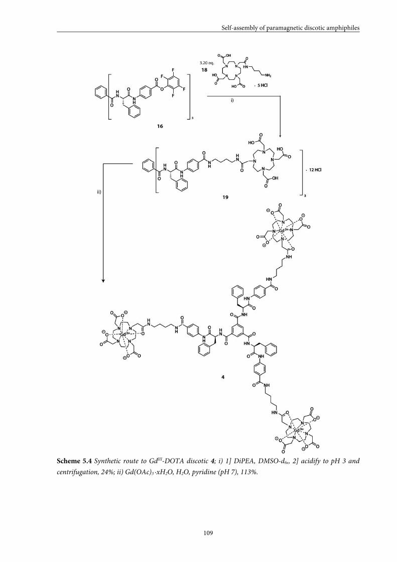
Self-assembly of paramagnetic discotic amphiphiles
109
Scheme 5.4 Synthetic route to GdIII-DOTA discotic 4; i) 1] DiPEA, DMSO-d6,, 2] acidify to pH 3 and
centrifugation, 24%; ii) Gd(OAc)3 ·xH2O, H2O, pyridine (pH 7), 113%.
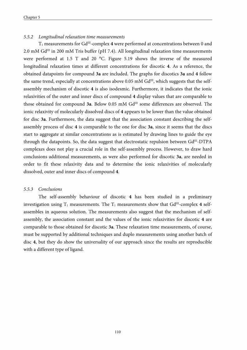
Chapter 5
110
5.5.2 Longitudinal relaxation time measurements
T1 measurements for GdIII-complex 4 were performed at concentrations between 0 and
2.0 mM GdIII in 200 mM Tris buffer (pH 7.4). All longitudinal relaxation time measurements
were performed at 1.5 T and 20 °C. Figure 5.19 shows the inverse of the measured
longitudinal relaxation times at different concentrations for discotic 4. As a reference, the
obtained datapoints for compound 3a are included. The graphs for discotics 3a and 4 follow
the same trend, especially at concentrations above 0.05 mM GdIII, which suggests that the self-
assembly mechanism of discotic 4 is also isodesmic. Furthermore, it indicates that the ionic
relaxivities of the outer and inner discs of compound 4 display values that are comparable to
those obtained for compound 3a. Below 0.05 mM GdIII some differences are observed. The
ionic relaxivity of molecularly dissolved discs of 4 appears to be lower than the value obtained
for disc 3a. Furthermore, the data suggest that the association constant describing the self-
assembly process of disc 4 is comparable to the one for disc 3a, since it seems that the discs
start to aggregate at similar concentrations as is estimated by drawing lines to guide the eye
through the datapoints. So, the data suggest that electrostatic repulsion between GdIII-DTPA
complexes does not play a crucial role in the self-assembly process. However, to draw hard
conclusions additional measurements, as were also performed for discotic 3a, are needed in
order to fit these relaxivity data and to determine the ionic relaxivities of molecularly
dissolved, outer and inner discs of compound 4.
5.5.3 Conclusions
The self-assembly behaviour of discotic 4 has been studied in a preliminary
investigation using T1 measurements. The T1 measurements show that GdIII-complex 4 self-
assembles in aqueous solution. The measurements also suggest that the mechanism of self-
assembly, the association constant and the values of the ionic relaxivities for discotic 4 are
comparable to those obtained for discotic 3a. These relaxation time measurements, of course,
must be supported by additional techniques and duplo measurements using another batch of
disc 4, but they do show the universality of our approach since the results are reproducible
with a different type of ligand.
Self-assembly of paramagnetic discotic amphiphiles
111
Figure 5.19 The longitudinal relaxation rate (R1obs) is plotted against the gadolinium concentration for
GdIII-complex 4; As a reference, the relaxation rates of GdIII-complex 3a are included; (a) concentration
window 0.0–2.5 mM; (b) concentration window 0.00–0.35 mM; (c) concentration window 0.00–0.04
mM.
(a)
(b)
(c)
Chapter 5
112
5.6 Overall conclusions
Four paramagnetic discotics that differ in their complexity and capability to undergo
secondary interactions have been successfully synthesized. As expected, it has been shown that
increased opportunities for secondary interactions and larger hydrophobic cores lead to lower
concentrations at which aggregation starts; the aggregation concentration decreased going
from GdIII-DTPA discotic 1a (does not aggregate) to 2a (aggregation starts at 0.27 mM GdIII)
to 3a (aggregation starts at 3 × 10⁻⁴ mM GdIII). Furthermore, it has been shown that changing
the ligand from DTPA to DOTA only slightly affects the self-assembly process, which
illustrates the universality of our approach. To be able to alter self-assembly processes and
thus aggregation concentrations is very important because it allows designing systems that
may benefit from their aggregation in vivo. The self-assembly studies also point out that, next
to CD spectroscopy, T1 measurements can be used to probe the self-assembly process of the
paramagnetic discotics described in this chapter. However, it has to be noted that self-
assembly mechanisms can be most easily studied using CD spectroscopy, since it takes less
time and the data are easier to interpret.
From the three GdIII-DTPA discotics synthesized, GdIII-complex 3a is the most
promising building block for a supramolecular contrast agent for MRI, in view of its low
aggregation concentration, its high ionic relaxivity in the aggregated form (~30 mM–1s–1 at
20 °C and 1.5 T) and its rather well-defined assemblies with dimensions in the nanometer
range. In vitro these paramagnetic assemblies show higher ionic relaxivities than fourth
generation GdIII-DTPA containing PPI dendrimers, which are comparable in size, molecular
weight and the number of endgroups, and they also show higher relaxivities compared to
many of the micellar and liposomal contrast agents reported thus far. Future research should
point out whether these new self-assembling contrast agents based on discotic amphiphiles
also show high proton relaxivities in vivo.
5.7 Epilogue
The results described in this chapter show that paramagnetic discotic molecules can be
designed in such a way that they display good self-assembling properties and high molecular
relaxivities in 200 mM Tris (pH 7.4) in vitro. Future research is, however, necessary in order
to determine whether this new type of self-assembling contrast agents offers opportunities for
use in vivo.
First of all, the influence of the presence of serum proteins, like BSA, on the
performance of discotic 3a should be investigated. These proteins may affect the behaviour of
discs of 3a in the molecularly dissolved and/or the self-assembled state, because they are
known to bind to hydrophobic molecules. Especially the core of discotic 3a is quite
hydrophobic, so aspecific binding may play an important role in vivo. For molecularly
Self-assembly of paramagnetic discotic amphiphiles
113
dissolved species this may be beneficial because it is known that non-covalent binding of a
small molecule to a protein leads to an increased ionic relaxivity and an increased residence
time in the body, while the paramagnetic complex can still be excreted from the body due to
the reversible nature of the binding. For self-assembled structures, the effect of aspecific
binding on their performance is not so straightforward. It is proposed that there are three
possibilities; firstly, there is no aspecific binding because the hydrophilic GdIII-complexes
shield the core of the stack from the protein, secondly, the entire stack of 3a binds to the
serum protein, and thirdly, aspecific binding of the discs to the protein is stronger than the
binding between the discs, hence the paramagnetic assemblies break up. In the first two
scenarios, the molecular relaxivity of the assemblies stays the same or increases somewhat, in
the third scenario, the molecular relaxivity decreases since the benefit from aggregation has
dissappeared. Preliminary in vitro relaxation time measurements of discotic 3a in serum and
in blood suggest that the assemblies do break up, since the observed proton relaxivities are
much lower than expected. It seems that stronger interactions between the discs are needed to
keep the assemblies intact. The results described in Chapters 2 and 3 illustrate that by using
phenylalanyl-phenylalanines as structuring elements in the core of the discotic, the stack
stability can be increased. The introduction of an additional phenylalanine in the structure of
3a may, therefore, avoid deaggregation. Another strategy for creating stronger interactions
between discs may be based on the incorporation of fluorophobic interactions. This type of
interaction is unknown to proteins, so proteins are not able to disturb fluorophobic
interactions between discs. One way to obtain fluorinated discs is by replacing the
phenylalanines by fluorinated phenylalanines. Future research should focus on these aspects
because self-assembly of paramagnetic discotic molecules is a key requirement for obtaining
systems displaying high relaxivities.
Secondly, it is important to investigate the dynamics of the system for reasons
concerning excretion from the body, stability of the assemblies and possibilities for
introducing active targeting. Excretion of paramagnetic complexes is important, due to the
toxicity of gadolinium ions that may be released from the complexes. Assemblies of discotic
3a are relatively small (diameters in the order of 4 nm) hence accumulation in the body
should not be an issue. Therefore, in this particular case reversible binding between discs
within an aggregate is not a necessity for excretion. However, when replacing the 1,3,5-
benzenetricarboxamide centre of disc 3a by a cis-cis-1,3,5-cyclohexanetricarboxamide centre
self-assembling contrast agents forming long columnar structures in solution presumably
become accessible. These agents may act as potential intravascular agents because of their high
molecularly weight. For these types of systems, reversible binding between the discs within an
aggregate is required to ensure complete excretion of the paramagnetic complexes from the
body. On the other hand, the assembly must be stable enough, so the interactions between the
discs within the aggregate should not be too weak (this is a general requirement and thus
Chapter 5
114
holds also for assemblies of discotic 3a); otherwise the benefit of a high relaxivity due to
aggregation vanishes. As a consequence, the dynamics must be balanced in an appropriate
way for an optimal performance in vivo. Dynamic systems may also open possibilities for the
introduction of active targeting in an easily accessible manner demanding less synthetic effort,
since symmetrical discs with similar core-fragments but displaying peptide sequences at their
periphery may simply be mixed with paramagnetic discs. In this way, even more efficient
contrast agents can be prepared.
And finally, the potential of the use of asymmetric discotic molecules containing
peripheral GdIII-complexes and an active peptide sequence should be investigated because this
may be an alternative strategy for introducing active targeting to the system. The main
advantage of this method compared to the method using at least two types of symmetrical
discs is that the molecularly dissolved paramagnetic discs themselve display peptide sequences
and thus are target-specific. Drawback, on the other hand, is that it takes much more synthetic
effort to synthesize asymmetric discs incorporating various peptide sequences.
All these aspects together may give new insights in the performance of paramagnetic
discotic amphiphiles in vivo. This is of general importance in order to demonstrate whether
paramagnetic discotic molecules are efficient self-assembling contrast agents for MR imaging.
Self-assembly of paramagnetic discotic amphiphiles
115
5.8 Experimental section
General
Unless stated otherwise, all reagents and chemicals were obtained from commercial sources and used
without further purification. Lysine-DTPA 5 and its benzyloxycarbonyl protected analogue 5b were synthesized
according to literature.[38,39] Water was demineralised prior to use. Dichloromethane and tetrahydrofuran were
obtained by distillation over Merck molecular sieves (4Å). Semi-preparative HPLC was performed on a Varian
Prostar HPLC system with a Vydac C18 column (2.5 × 20 cm, flow 10 mL⋅min–1), eluted with a linear gradient of
30–50% CH3CN in 0.1% aqueous TFA in 90 min. 1H-NMR, 1H-1H COSY, 13C-NMR and 19F-NMR spectra were
recorded on a Varian Gemini 300 spectrometer, a Varian Mercury Vx 400 spectrometer or a Varian Unity Inova
500 spectrometer at 298 K. Chemical shifts are given in ppm (δ) values relative to tetramethylsilane (TMS).
Splitting patterns are designated as s, singlet; br s, broad singlet, d, doublet; dd, double doublet; t, triplet; q,
quartet; m, multiplet. Neat state IR spectra were measured at 298 K on a Perkin-Elmer 1605 FT-IR
spectrophotometer. Matrix assisted laser desorption/ionization mass spectra were obtained on a PerSeptive
Biosystems Voyager DE-PRO spectrometer using α-cyano-4-hydroxycinnamic acid (CHCA) and 2-[(2E)-3-(4-
tert-butylphenyl)-2-methylprop-2-enylidene]malononitrile (DCTB) as matrices. (LC)-ESI-MS measurements
were performed on a Finnigan LCQ Deca XP Max ion trap mass spectrometer (Thermo Electron Corporation,
San Jose, USA). Elemental analyses were carried out using a Perkin Elmer 2400. GdIII- and YIII-contents were
determined by means of inductively coupled plasma mass spectrometry (ICP-MS) conducted by Philips
Research-Materials Analysis in Eindhoven. Chiral HPLC was performed on a Shimadzu system consisting of a
Shimadzu LC-10 ADvp pump, a Spark Midas autosampler and a Shimadzu SPD-10ADvp UV-Vis detector. A
DNBPG column (covalent, 5 μm; 0.46 x 25 cm) was used, with a 9/1 (v/v) hexane/isopropanol mixture as the
eluent. The T1 measurements were performed at 1.5 T on a Bruker Minispec mq60 NMR analyzer. The sample
volume was 300 µL and the samples were measured in tubes with a diameter of 7.5 mm. Longitudinal relaxation
times were determined with an inversion recovery pulse sequence. GdIII-concentrations of the aqueous solutions
used for the T1-relaxivity measurements were determined by means of inductively coupled plasma mass
spectrometry (ICP-MS) conducted by Philips Research-Materials Analysis in Eindhoven. UV/VIS spectra were
obtained on a Perkin Elmer Lambda 40 spectrometer. CD measurements were performed on a Jasco J815 CD
spectrometer connected to a Jasco PTC-348WI temperature controller. Cryogenic transmission microscopy
measurements were performed on a FEI Titan Krios TEM equipped with a field emission gun (FEG) operating at
300 kV. Images were recorded using a 2k x 2k Gatan CCD camera equipped with a post column Gatan energy
filter (GIF). TEM grids, (R2/2 Quantifoil Jena grids) were purchased from Aurion. The Quantifoil grids
underwent a surface plasma treatment using a Cressington 208 carbon coater operating at 5 mA for 40 s prior to
the vitrification procedure. The cryoTEM images of the discotics were obtained by applying small aliquots (3 µL)
of the aqueous discotic solutions to Quantifoil grids (R2/2 Quantifoil Jena) within the environmental chamber
(relative humidity 100%) of the Vitrobot™ at room temperature. The vitrified specimens were stored under liquid
nitrogen and observed at –170°C (Gatan cryo holder) in the Titan microscope. Micrographs were taken at 300
kV using low dose conditions. 1H-DOSY NMR experiments were recorded on a Varian Unity Inova 500
spectrometer operating at 500.618 MHz and equipped with a 5 mm ID-PFG probe from Varian. Additional,
experimental 1H-DOSY NMR data can be found at the end of this experimental section.
tert-Butyl 6-amino-2-[bis-{2-[bis-(tert-butoxycarbonylmethyl)amino]ethyl}amino]hexanoate (5)
A detailed description of the synthesis of this lysine-DTPA building block was reported.[38,39]
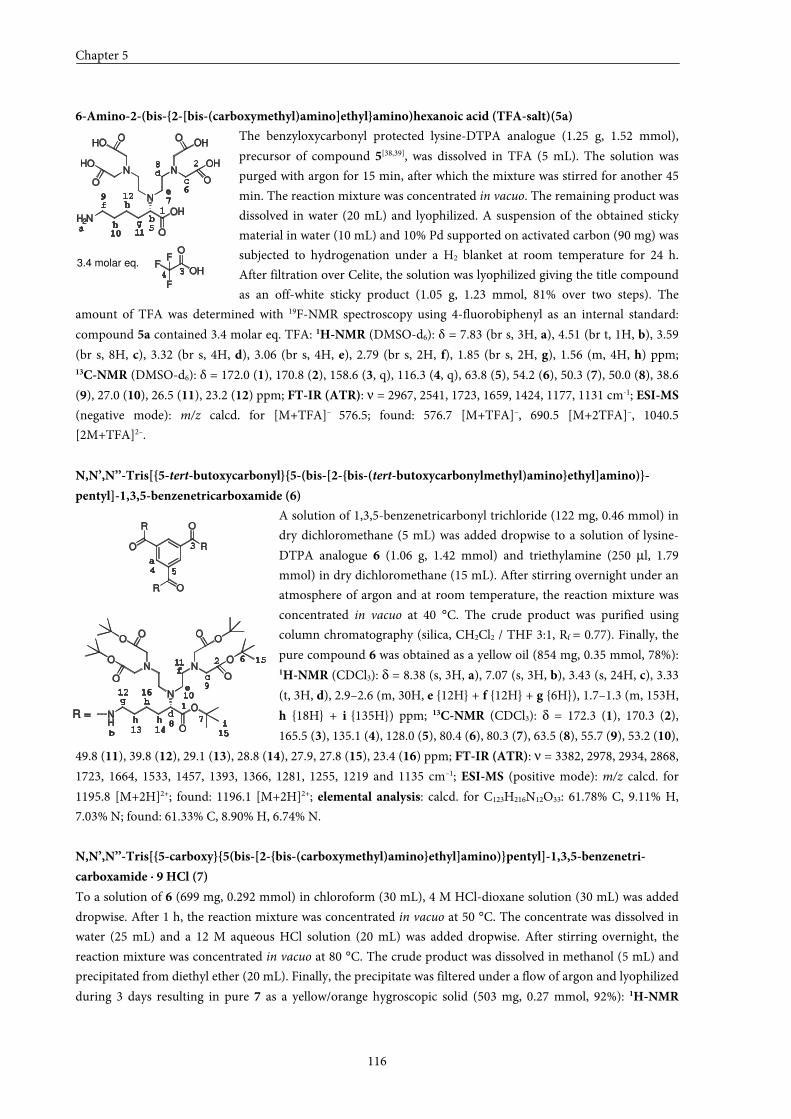
Chapter 5
116
6-Amino-2-(bis-{2-[bis-(carboxymethyl)amino]ethyl}amino)hexanoic acid (TFA-salt)(5a)
The benzyloxycarbonyl protected lysine-DTPA analogue (1.25 g, 1.52 mmol),
precursor of compound 5[38,39], was dissolved in TFA (5 mL). The solution was
purged with argon for 15 min, after which the mixture was stirred for another 45
min. The reaction mixture was concentrated in vacuo. The remaining product was
dissolved in water (20 mL) and lyophilized. A suspension of the obtained sticky
material in water (10 mL) and 10% Pd supported on activated carbon (90 mg) was
subjected to hydrogenation under a H2 blanket at room temperature for 24 h.
After filtration over Celite, the solution was lyophilized giving the title compound
as an off-white sticky product (1.05 g, 1.23 mmol, 81% over two steps). The
amount of TFA was determined with 19F-NMR spectroscopy using 4-fluorobiphenyl as an internal standard:
compound 5a contained 3.4 molar eq. TFA: 1H-NMR (DMSO-d6): δ = 7.83 (br s, 3H, a), 4.51 (br t, 1H, b), 3.59
(br s, 8H, c), 3.32 (br s, 4H, d), 3.06 (br s, 4H, e), 2.79 (br s, 2H, f), 1.85 (br s, 2H, g), 1.56 (m, 4H, h) ppm; 13C-NMR (DMSO-d6): δ = 172.0 (1), 170.8 (2), 158.6 (3, q), 116.3 (4, q), 63.8 (5), 54.2 (6), 50.3 (7), 50.0 (8), 38.6
(9), 27.0 (10), 26.5 (11), 23.2 (12) ppm; FT-IR (ATR): ν = 2967, 2541, 1723, 1659, 1424, 1177, 1131 cm-1; ESI-MS
(negative mode): m/z calcd. for [M+TFA]– 576.5; found: 576.7 [M+TFA]–, 690.5 [M+2TFA]–, 1040.5
[2M+TFA]2–.
N,N’,N’’-Tris[{5-tert-butoxycarbonyl}{5-(bis-[2-{bis-(tert-butoxycarbonylmethyl)amino}ethyl]amino)}-
pentyl]-1,3,5-benzenetricarboxamide (6)
A solution of 1,3,5-benzenetricarbonyl trichloride (122 mg, 0.46 mmol) in
dry dichloromethane (5 mL) was added dropwise to a solution of lysine-
DTPA analogue 6 (1.06 g, 1.42 mmol) and triethylamine (250 µl, 1.79
mmol) in dry dichloromethane (15 mL). After stirring overnight under an
atmosphere of argon and at room temperature, the reaction mixture was
concentrated in vacuo at 40 °C. The crude product was purified using
column chromatography (silica, CH2Cl2 / THF 3:1, Rf = 0.77). Finally, the
pure compound 6 was obtained as a yellow oil (854 mg, 0.35 mmol, 78%): 1H-NMR (CDCl3): δ = 8.38 (s, 3H, a), 7.07 (s, 3H, b), 3.43 (s, 24H, c), 3.33
(t, 3H, d), 2.9–2.6 (m, 30H, e {12H} + f {12H} + g {6H}), 1.7–1.3 (m, 153H,
h {18H} + i {135H}) ppm; 13C-NMR (CDCl3): δ = 172.3 (1), 170.3 (2),
165.5 (3), 135.1 (4), 128.0 (5), 80.4 (6), 80.3 (7), 63.5 (8), 55.7 (9), 53.2 (10),
49.8 (11), 39.8 (12), 29.1 (13), 28.8 (14), 27.9, 27.8 (15), 23.4 (16) ppm; FT-IR (ATR): ν = 3382, 2978, 2934, 2868,
1723, 1664, 1533, 1457, 1393, 1366, 1281, 1255, 1219 and 1135 cm–1; ESI-MS (positive mode): m/z calcd. for
1195.8 [M+2H]2+; found: 1196.1 [M+2H]2+; elemental analysis: calcd. for C123H216N12O33: 61.78% C, 9.11% H,
7.03% N; found: 61.33% C, 8.90% H, 6.74% N.
N,N’,N’’-Tris[{5-carboxy}{5(bis-[2-{bis-(carboxymethyl)amino}ethyl]amino)}pentyl]-1,3,5-benzenetri-
carboxamide ⋅⋅⋅⋅ 9 HCl (7)
To a solution of 6 (699 mg, 0.292 mmol) in chloroform (30 mL), 4 M HCl-dioxane solution (30 mL) was added
dropwise. After 1 h, the reaction mixture was concentrated in vacuo at 50 °C. The concentrate was dissolved in
water (25 mL) and a 12 M aqueous HCl solution (20 mL) was added dropwise. After stirring overnight, the
reaction mixture was concentrated in vacuo at 80 °C. The crude product was dissolved in methanol (5 mL) and
precipitated from diethyl ether (20 mL). Finally, the precipitate was filtered under a flow of argon and lyophilized
during 3 days resulting in pure 7 as a yellow/orange hygroscopic solid (503 mg, 0.27 mmol, 92%): 1H-NMR
H2N
N
N N
OH
O
O
O O
O
HO
HO OH
OH
OHF
F
FO
a
b
cd
e
f
gh
h1
2
34
5
6
7
8
9
10 11
12
3.4 molar eq.
b
c
d
e
f
g1
2
3
4 5
6
78
9
1012
a
h
h
hi
NH
N
N N
O
O
O
O O
O
O
O O
O
R
O
OR
O
R
R =
11
13 14 15
16
15
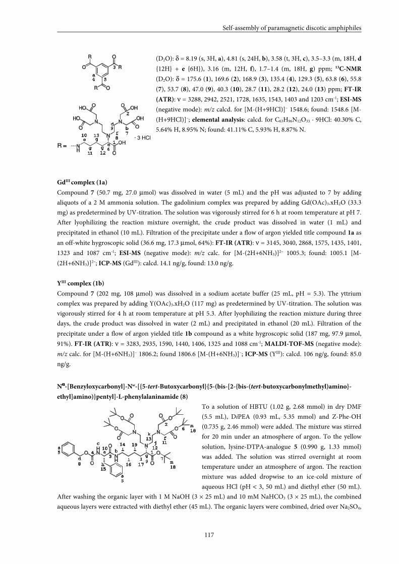
Self-assembly of paramagnetic discotic amphiphiles
117
(D2O): δ = 8.19 (s, 3H, a), 4.81 (s, 24H, b), 3.58 (t, 3H, c), 3.5–3.3 (m, 18H, d
{12H} + e {6H}), 3.16 (m, 12H, f), 1.7–1.4 (m, 18H, g) ppm; 13C-NMR
(D2O): δ = 175.6 (1), 169.6 (2), 168.9 (3), 135.4 (4), 129.3 (5), 63.8 (6), 55.8
(7), 53.7 (8), 47.0 (9), 40.3 (10), 28.7 (11), 28.2 (12), 24.0 (13) ppm; FT-IR
(ATR): ν = 3288, 2942, 2521, 1728, 1635, 1543, 1403 and 1203 cm-1; ESI-MS
(negative mode): m/z calcd. for [M-(H+9HCl)]– 1548.6; found: 1548.6 [M-
(H+9HCl)]–; elemental analysis: calcd. for C63H96N12O33 · 9HCl: 40.30% C,
5.64% H, 8.95% N; found: 41.11% C, 5.93% H, 8.87% N.
GdIII complex (1a)
Compound 7 (50.7 mg, 27.0 µmol) was dissolved in water (5 mL) and the pH was adjusted to 7 by adding
aliquots of a 2 M ammonia solution. The gadolinium complex was prepared by adding Gd(OAc)3.xH2O (33.3
mg) as predetermined by UV-titration. The solution was vigorously stirred for 6 h at room temperature at pH 7.
After lyophilizing the reaction mixture overnight, the crude product was dissolved in water (1 mL) and
precipitated in ethanol (10 mL). Filtration of the precipitate under a flow of argon yielded title compound 1a as
an off-white hygroscopic solid (36.6 mg, 17.3 µmol, 64%): FT-IR (ATR): ν = 3145, 3040, 2868, 1575, 1435, 1401,
1323 and 1087 cm-1; ESI-MS (negative mode): m/z calc. for [M-(2H+6NH3)]2– 1005.3; found: 1005.1 [M-
(2H+6NH3)]2–; ICP-MS (GdIII): calcd. 14.1 ng/g, found: 13.0 ng/g.
YIII complex (1b)
Compound 7 (202 mg, 108 µmol) was dissolved in a sodium acetate buffer (25 mL, pH = 5.3). The yttrium
complex was prepared by adding Y(OAc)3.xH2O (117 mg) as predetermined by UV-titration. The solution was
vigorously stirred for 4 h at room temperature at pH 5.3. After lyophilizing the reaction mixture during three
days, the crude product was dissolved in water (2 mL) and precipitated in ethanol (20 mL). Filtration of the
precipitate under a flow of argon yielded title 1b compound as a white hygroscopic solid (187 mg, 97.9 µmol,
91%). FT-IR (ATR): ν = 3283, 2935, 1590, 1440, 1406, 1325 and 1088 cm-1; MALDI-TOF-MS (negative mode):
m/z calc. for [M-(H+6NH3)]– 1806.2; found 1806.6 [M-(H+6NH3)]–; ICP-MS (YIII): calcd. 106 ng/g, found: 85.0
ng/g.
Nαααα-[Benzyloxycarbonyl]-Nω-[{5-tert-Butoxycarbonyl}{5-(bis-[2-{bis-(tert-butoxycarbonylmethyl)amino}-
ethyl]amino)}pentyl]-L-phenylalaninamide (8)
To a solution of HBTU (1.02 g, 2.68 mmol) in dry DMF
(5.5 mL), DiPEA (0.93 mL, 5.35 mmol) and Z-Phe-OH
(0.735 g, 2.46 mmol) were added. The mixture was stirred
for 20 min under an atmosphere of argon. To the yellow
solution, lysine-DTPA-analogue 5 (0.990 g, 1.33 mmol)
was added. The solution was stirred overnight at room
temperature under an atmosphere of argon. The reaction
mixture was added dropwise to an ice-cold mixture of
aqueous HCl (pH < 3, 50 mL) and diethyl ether (50 mL).
After washing the organic layer with 1 M NaOH (3 × 25 mL) and 10 mM NaHCO3 (3 × 25 mL), the combined
aqueous layers were extracted with diethyl ether (45 mL). The organic layers were combined, dried over Na2SO4,
OHN
NH
O
O N
N N
O
O O
O
O
O
O
O
O
O
b
c
d
e
f
g1
2
34
5
6
7
89
10 12
a
h
i
1113
14
1516
j
k
l
l
lm
m
a
5
1718
18
19
b
c
d
e
f
g
1
2
3
4 5
6
78
9
10
12
a
NH
N
N N
OH
O
O
O O
O
HO
HO OH
OH
R
O
OR
O
R
R =
11
13g
g
· 3 HCl
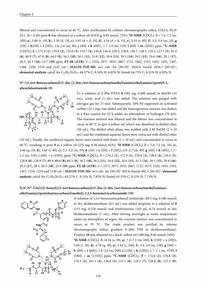
Chapter 5
118
filtered and concentrated in vacuo at 40 °C. After purification by column chromatography (silica, CH2Cl2 /Et3N
25:1, Rf = 0.26) pure 8 was obtained as a yellow oil (0.976 g, 0.95 mmol, 72%): 1H-NMR (CDCl3): δ = 7.4–7.2 (m,
10H, a), 5.88 (t, 1H, b), 5.50 (d, 1H, c), 5.05 (d + d, 2H, d), 4.34 (d + d, 1H, e), 3.43 (s, 8H, f), 3.2–3.0 (m, 5H, g
{1H} + h{2H} + i {2H}), 2.8–2.6 (m, 8H, j {4H} + k {4H}), 1.7–1.0 (m, 51H, l {6H} + m {45H}) ppm; 13C-NMR
(CDCl3): δ = 172.9 (1), 170.8 (2), 170.6 (3), 155.7 (4), 136.8, 136.4, 129.5, 128.8, 128.7, 128.3, 128.2, 127.1 (5), 81.0
(6), 80.9 (7), 67.0 (8), 64.2 (9), 56.5 (10), 56.1 (11), 53.9 (12), 50.4 (13), 39.5 (14), 39.2 (15), 29.6 (16), 29.1 (17),
28.5, 28.3 (18), 23.7 (19) ppm; FT-IR (ATR): ν = 3324, 2977, 2933, 2867, 1722, 1662, 1533, 1455, 1392, 1367,
1284, 1250, 1218 and 1147 cm-1; MALDI-TOF-MS: m/z calc. for [M+H]+ 1026.6; found: 1026.7 [M+H]+;
elemental analysis: calcd. for C55H87N5O13: 64.37% C, 8.54% H, 6.82% N; found: 64.73% C, 8.51% H, 6.92% N.
Nω-[{5-tert-Butoxycarbonyl}{5-(bis-[2-{bis-(tert-butoxycarbonylmethyl)amino}ethyl]amino)}pentyl]-L-
phenylalaninamide (9)
To a solution of Z-Phe-DTPA 8 (502 mg, 0.489 mmol) in MeOH (10
mL), acetic acid (1 mL) was added. The solution was purged with
nitrogen gas for 15 min. Subsequently, 10% Pd supported on activated
carbon (32.2 mg) was added and the heterogeneous mixture was shaken
in a Parr-reactor for 22 h under an atmosphere of hydrogen (70 psi).
The reaction mixture was filtered and the filtrate was concentrated in
vacuo at 60 °C to give a yellow oil, which was dissolved in diethyl ether
(20 mL). The diethyl ether phase was washed with 2 M NaOH (2 × 10
mL) and the combined aqueous layers were extracted with diethyl ether
(10 mL). Finally, the combined organic layers were washed with brine (2 × 10 mL) and concentrated in vacuo at
60 °C, resulting in pure 9 as a yellow oil (270 mg, 0.30 mmol, 62%): 1H-NMR (CDCl3): δ = 7.4–7.2 (m, 5H, a),
3.60 (q, 1H, b), 3.43 (s, 8H, c), 3.3–3.2 (m, 5H, d {1H} + e {2H} + f {2H}), 2.9–2.7 (m, 8H, g {4H} + h {4H}), 1.7–
1.2 (m, 51H, i {6H} + j {45H}) ppm; 13C-NMR (CDCl3): δ = 174.2 (1), 172.9 (2), 170.8 (3), 138.4 (4), 129.4 (5),
128.8 (6), 126.9 (7), 80.9, 80.8 (8), 64.5 (9), 56.7 (10), 56.2 (11), 54.0 (12), 50.6 (13), 41.3 (14), 39.1 (15), 29.9 (16),
29.5 (17), 28.5, 28.3 (18), 23.9 (19) ppm; FT-IR (ATR): ν = 3373, 2977, 2933, 2867, 1725, 1673, 1520, 1455, 1392,
1367, 1254, 1219 and 1149 cm-1; MALDI-TOF-MS: m/z calc. for [M+H]+ 892.6; found: 892.4 [M+H]+; elemental
analysis: calcd. for C47H81N5O11: 63.27% C, 9.15% H, 7.85% N; found: 63.55% C, 9.11% H, 7.73% N.
N,N’,N’’-Tris[{(S)-benzyl}{(5-tert-butoxycarbonyl)(5-[bis-{2-(bis-[tert-butoxycarbonylmethyl]amino)-
ethyl}amino])pentylaminocarbonyl}methyl]-1,3,5-benzenetricarboxamide (10)
A solution of 1,3,5-benzenetricarbonyl trichloride (49.5 mg, 0.186 mmol)
in dry dichloromethane (0.5 mL) was added dropwise to a solution of 9
(515 mg, 0.578 mmol) and triethylamine (103 µL, 0.74 mmol) in dry
dichloromethane (5 mL). After stirring overnight at room temperature
under an atmosphere of argon, the reaction mixture was concentrated in
vacuo at 35 °C. The crude product was purified by column
chromatography (silica, gradient 0–20% THF in dichloromethane).
Product 10 was obtained as a thick, yellow oil (180 mg, 0.06 mmol, 34%): 1H-NMR (CDCl3): δ =8.34 (s, 3H, a), 7.4–7.2 (m, 18H, b {15H} + c {3H}),
5.94 (t, 3H, d), 4.78 (q, 3H, e), 3.43 (s, 24H, f), 3.4–3.0 (m, 15H, g {3H} +
h {6H} + i {6H}), 2.8–2.6 (m, 24H, j {12H} + k {12H}), 1.7–1.1 (m, 153H, l
{18H} + m {135H}) ppm; 13C-NMR (CDCl3): δ = 172.8 (1), 170.8 (2),
170.3 (3), 165.1 (4), 136.8 (5), 135.1 (6), 129.5 (7), 128.8 (8), 127.3 (9),
H2NNH
O N
N N
O
O O
O
O
O
O
O
O
O
b
c
d
e
f
g
12
3
4
56
7
8
9
10
a
h
i
i
i
j
j
17
8
11
12
13
14
15
16
18
18
19
b
c
d
e
f
g1
2
34
5
6
7
89
10
12
a
h
i
NH
N
N N
O
O
O
O O
O
O
O O
O
NH
O
OHN
O
NH
R
R
O
O
OR
R =11
13
14
15
16 j
k
l
l
l m
m
17 18
8
10
16
19
19
20
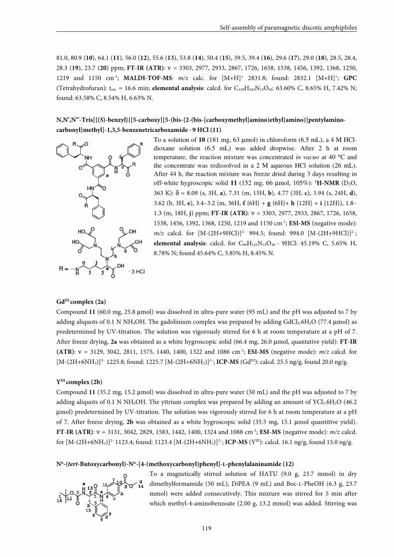
Self-assembly of paramagnetic discotic amphiphiles
119
81.0, 80.9 (10), 64.1 (11), 56.0 (12), 55.6 (13), 53.8 (14), 50.4 (15), 39.5, 39.4 (16), 29.6 (17), 29.0 (18), 28.5, 28.4,
28.3 (19), 23.7 (20) ppm; FT-IR (ATR): ν = 3303, 2977, 2933, 2867, 1726, 1658, 1538, 1456, 1392, 1368, 1250,
1219 and 1150 cm-1; MALDI-TOF-MS: m/z calc. for [M+H]+ 2831.8; found: 2832.1 [M+H]+; GPC
(Tetrahydrofuran): tret. = 16.6 min; elemental analysis: calcd. for C150H243N15O36: 63.60% C, 8.65% H, 7.42% N;
found: 63.58% C, 8.54% H, 6.63% N.
N,N’,N’’-Tris[{(S)-benzyl}{[5-carboxy][5-(bis-{2-(bis-[carboxymethyl]amino)ethyl}amino)]pentylamino-
carbonyl}methyl]-1,3,5-benzenetricarboxamide ⋅⋅⋅⋅ 9 HCl (11)
To a solution of 10 (181 mg, 63 µmol) in chloroform (6.5 mL), a 4 M HCl-
dioxane solution (6.5 mL) was added dropwise. After 2 h at room
temperature, the reaction mixture was concentrated in vacuo at 40 °C and
the concentrate was redissolved in a 2 M aqueous HCl solution (26 mL).
After 44 h, the reaction mixture was freeze dried during 3 days resulting in
off-white hygroscopic solid 11 (152 mg, 66 µmol, 105%): 1H-NMR (D2O,
363 K): δ = 8.09 (s, 3H, a), 7.31 (m, 15H, b), 4.77 (3H, c), 3.94 (s, 24H, d),
3.62 (b, 3H, e), 3.4–3.2 (m, 36H, f {6H} + g {6H}+ h {12H} + i {12H}), 1.8–
1.3 (m, 18H, j) ppm; FT-IR (ATR): ν = 3303, 2977, 2933, 2867, 1726, 1658,
1538, 1456, 1392, 1368, 1250, 1219 and 1150 cm-1; ESI-MS (negative mode):
m/z calcd. for [M-(2H+9HCl)]2– 994.5; found: 994.0 [M-(2H+9HCl)]2–;
elemental analysis: calcd. for C90H123N15O36 · 9HCl: 45.19% C, 5.65% H,
8.78% N; found 45.64% C, 5.85% H, 8.45% N.
GdIII complex (2a)
Compound 11 (60.0 mg, 25.8 µmol) was dissolved in ultra-pure water (95 mL) and the pH was adjusted to 7 by
adding aliquots of 0.1 N NH4OH. The gadolinium complex was prepared by adding GdCl3.6H2O (77.4 µmol) as
predetermined by UV-titration. The solution was vigorously stirred for 6 h at room temperature at a pH of 7.
After freeze drying, 2a was obtained as a white hygroscopic solid (66.4 mg, 26.0 µmol, quantative yield): FT-IR
(ATR): ν = 3129, 3042, 2811, 1575, 1440, 1400, 1322 and 1086 cm-1; ESI-MS (negative mode): m/z calcd. for
[M-(2H+6NH3)]2– 1225.8; found: 1225.7 [M-(2H+6NH3)]2–; ICP-MS (GdIII): calcd. 25.5 ng/g, found 20.0 ng/g.
YIII complex (2b)
Compound 11 (35.2 mg, 15.2 µmol) was dissolved in ultra-pure water (50 mL) and the pH was adjusted to 7 by
adding aliquots of 0.1 N NH4OH. The yttrium complex was prepared by adding an amount of YCl3.6H2O (46.2
µmol) predetermined by UV-titration. The solution was vigorously stirred for 6 h at room temperature at a pH
of 7. After freeze drying, 2b was obtained as a white hygroscopic solid (35.5 mg, 15.1 µmol quantitive yield).
FT-IR (ATR): ν = 3131, 3042, 2829, 1583, 1442, 1400, 1324 and 1088 cm-1; ESI-MS (negative mode): m/z calcd.
for [M-(2H+6NH3)]2– 1123.4; found: 1123.4 [M-(2H+6NH3)]2–; ICP-MS (YIII): calcd. 16.1 ng/g, found 15.0 ng/g.
Nα-(tert-Butoxycarbonyl)-Nω-[4-(methoxycarbonyl)phenyl]-L-phenylalaninamide (12)
To a magnetically stirred solution of HATU (9.0 g, 23.7 mmol) in dry
dimethylformamide (50 mL), DiPEA (9 mL) and Boc-L-PheOH (6.3 g, 23.7
mmol) were added consecutively. This mixture was stirred for 5 min after
which methyl-4-aminobenzoate (2.00 g, 13.2 mmol) was added. Stirring was
b
c
d
e
f
g
a
h
i
NH
N
N N
OH
O
O
O O
O
HO
HO OH
OH
NH
O
OHN
O
NH
R
R
O
O
OR
R =j
j
j · 3 HCl
OHN
NH
O
O
O
O
b
c
d
e
f
g
1
2
3
5
8
9
a
h
i
6
7
4
1011
12
13 14
15
16
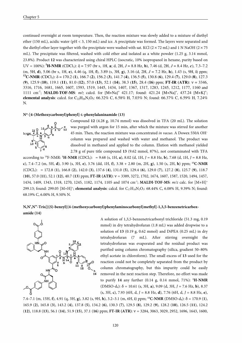
Chapter 5
120
continued overnight at room temperature. Then, the reaction mixture was slowly added to a mixture of diethyl
ether (150 mL), acidic water (pH < 3, 150 mL) and ice. A precipitate was formed. The layers were separated and
the diethyl ether layer together with the precipitate were washed with sat. KCl (2 × 72 mL) and 1 N NaOH (2 × 75
mL). The precipitate was filtered, washed with cold ether and isolated as a white powder (1.25 g, 3.14 mmol,
23.8%). Product 12 was characterized using chiral HPLC (isocratic, 10% isopropanol in hexane, purity based on
UV = 100%): 1H-NMR (CDCl3): δ = 7.97 (br s, 1H, a; d, 2H, J = 8.8 Hz, b), 7.46 (d, 2H, J = 8.4 Hz, c), 7.3–7.2
(m, 5H, d), 5.06 (br s, 1H, e), 4.46 (q, 1H, f), 3.89 (s, 3H, g), 3.16 (d, 2H, J = 7.2 Hz, h), 1.43 (s, 9H, i) ppm; 13
C-NMR (CDCl3): δ = 170.2 (1), 166.7 (2), 156.2 (3), 141.7 (4), 136.5 (5), 130.8 (6), 129.4 (7), 129.0 (8), 127.3
(9), 125.9 (10), 119.1 (11), 81.0 (12), 57.0 (13), 52.1 (14), 38.3 (15), 28.4 (16) ppm; FT-IR (ATR): ν = 3346,
3316, 1716, 1681, 1665, 1607, 1593, 1519, 1445, 1434, 1407, 1367, 1317, 1283, 1245, 1212, 1177, 1160 and
1111 cm-1; MALDI-TOF-MS: m/z calcd. for [M+Na]+ 421.17; found: 421.24 [M+Na]+, 437.24 [M+K]+;
elemental analysis: calcd. for C22H26N2O5: 66.32% C, 6.58% H, 7.03% N; found: 66.37% C, 6.59% H, 7.24%
N.
Nω-[4-(Methoxycarbonyl)phenyl]-L-phenylalaninamide (13)
Compound 12 (4.28 g, 10.74 mmol) was dissolved in TFA (20 mL). The solution
was purged with argon for 15 min, after which the mixture was stirred for another
45 min. Then, the reaction mixture was concentrated in vacuo. A Dowex 550A OH–
column was prepared and washed with water and methanol. The product was
dissolved in methanol and applied to the column. Elution with methanol yielded
2.78 g of pure title compound 13 (9.62 mmol, 87%), not contaminated with TFA
according to 19F-NMR: 1H-NMR (CDCl3): = 9.68 (s, 1H, a), 8.02 (d, 1H, J = 8.8 Hz, b), 7.68 (d, 1H, J = 8.8 Hz,
c), 7.4–7.2 (m, 5H, d), 3.90 (s, 3H, e), 3.76 (dd, 1H, f), 3.38 + 2.80 (m, 2H, g), 1.50 (s, 2H, h) ppm; 13C-NMR
(CDCl3): = 172.8 (1), 166.8 (2), 142.0 (3), 137.6 (4), 131.0 (5), 129.4 (6), 129.0 (7), 127.2 (8), 125.7 (9), 118.7
(10), 57.0 (11), 52.1 (12), 40.7 (13) ppm; FT-IR (ATR): ν = 3389, 3272, 1702, 1674, 1607, 1587, 1520, 1494, 1457,
1434, 1409, 1343, 1318, 1270, 1245, 1182, 1174, 1105 and 1074 cm-1; MALDI-TOF-MS: m/z calc. for [M+H]+
299.13; found: 299.05 [M+H]+; elemental analysis: calcd. for C17H18N2O3: 68.44% C, 6.08% H, 9.39% N; found:
68.19% C, 6.00% H, 9.50% N.
N,N’,N’’-Tris[{(S)-benzyl}{4-(methoxycarbonyl)phenylaminocarbonyl}methyl]-1,3,5-benzenetricarbox-
amide (14)
A solution of 1,3,5-benzenetricarbonyl trichloride (51.3 mg, 0.19
mmol) in dry tetrahydrofuran (1.8 mL) was added dropwise to a
solution of 13 (0.19 g, 0.62 mmol) and DiPEA (0.25 mL) in dry
tetrahydrofuran (7 mL). After stirring overnight the
tetrahydrofuran was evaporated and the residual product was
purified using column chromatography (silica, gradient 50–80%
ethyl acetate in chloroform). The small excess of 13 used for the
reaction could not be completely separated from the product by
column chromatography, but this impurity could be easily
removed in the next reaction step. Therefore, no effort was made
to purify 14 any further (0.14 g, 0.14 mmol, 71%): 1H-NMR
(DMSO-d6): δ = 10.61 (s, 3H, a), 9.09 (d, 3H, J = 7.6 Hz, b), 8.37
(s, 3H, c), 7.93 (6H, d, J = 8.8 Hz, d), 7.76 (6H, d, J = 8.8 Hz, e),
7.4–7.1 (m, 15H, f), 4.91 (q, 3H, g), 3.82 (s, 9H, h), 3.2–3.1 (m, 6H, i) ppm; 13C-NMR (DMSO-d6): δ = 170.9 (1),
165.9 (2), 165.8 (3), 143.2 (4), 137.8 (5), 134.2 (6), 130.3 (7), 129.5 (8), 129.2 (9), 128.2 (10), 126.5 (11), 124.2
(12), 118.8 (13), 56.1 (14), 51.9 (15), 37.1 (16) ppm; FT-IR (ATR): ν = 3284, 3063, 3029, 2952, 1696, 1643, 1600,
H2NNH
O
O
O
bc
d
e
f
g
1
2
5
ah
4
3
6
7
8
910
11 12
13
NH
HN
HN
NH
NH
HN
O
O
O
O
O
O
O
O
O
O
OO
bc
d
e
f
g1
2
3
5
6
7a
h
i
4
8
9
10
11
1213
14
15
16
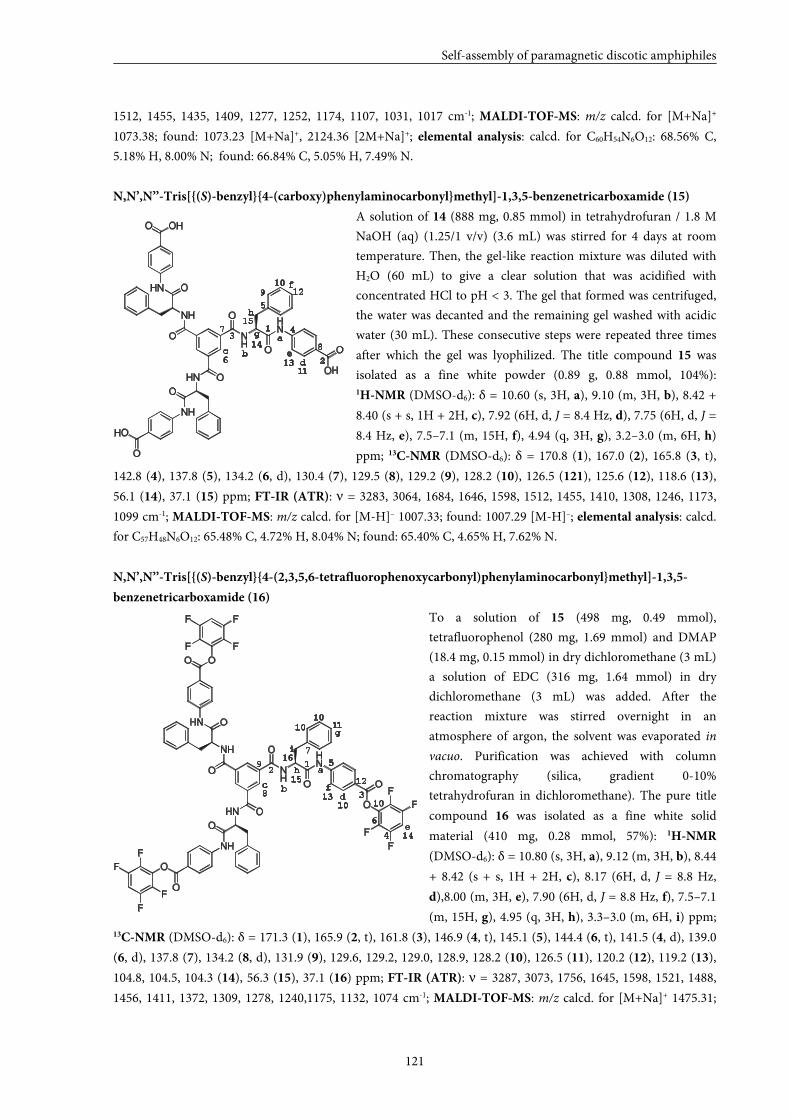
Self-assembly of paramagnetic discotic amphiphiles
121
1512, 1455, 1435, 1409, 1277, 1252, 1174, 1107, 1031, 1017 cm-1; MALDI-TOF-MS: m/z calcd. for [M+Na]+
1073.38; found: 1073.23 [M+Na]+, 2124.36 [2M+Na]+; elemental analysis: calcd. for C60H54N6O12: 68.56% C,
5.18% H, 8.00% N; found: 66.84% C, 5.05% H, 7.49% N.
N,N’,N’’-Tris[{(S)-benzyl}{4-(carboxy)phenylaminocarbonyl}methyl]-1,3,5-benzenetricarboxamide (15)
A solution of 14 (888 mg, 0.85 mmol) in tetrahydrofuran / 1.8 M
NaOH (aq) (1.25/1 v/v) (3.6 mL) was stirred for 4 days at room
temperature. Then, the gel-like reaction mixture was diluted with
H2O (60 mL) to give a clear solution that was acidified with
concentrated HCl to pH < 3. The gel that formed was centrifuged,
the water was decanted and the remaining gel washed with acidic
water (30 mL). These consecutive steps were repeated three times
after which the gel was lyophilized. The title compound 15 was
isolated as a fine white powder (0.89 g, 0.88 mmol, 104%): 1H-NMR (DMSO-d6): δ = 10.60 (s, 3H, a), 9.10 (m, 3H, b), 8.42 +
8.40 (s + s, 1H + 2H, c), 7.92 (6H, d, J = 8.4 Hz, d), 7.75 (6H, d, J =
8.4 Hz, e), 7.5–7.1 (m, 15H, f), 4.94 (q, 3H, g), 3.2–3.0 (m, 6H, h)
ppm; 13C-NMR (DMSO-d6): δ = 170.8 (1), 167.0 (2), 165.8 (3, t),
142.8 (4), 137.8 (5), 134.2 (6, d), 130.4 (7), 129.5 (8), 129.2 (9), 128.2 (10), 126.5 (121), 125.6 (12), 118.6 (13),
56.1 (14), 37.1 (15) ppm; FT-IR (ATR): ν = 3283, 3064, 1684, 1646, 1598, 1512, 1455, 1410, 1308, 1246, 1173,
1099 cm-1; MALDI-TOF-MS: m/z calcd. for [M-H]– 1007.33; found: 1007.29 [M-H]–; elemental analysis: calcd.
for C57H48N6O12: 65.48% C, 4.72% H, 8.04% N; found: 65.40% C, 4.65% H, 7.62% N.
N,N’,N’’-Tris[{(S)-benzyl}{4-(2,3,5,6-tetrafluorophenoxycarbonyl)phenylaminocarbonyl}methyl]-1,3,5-
benzenetricarboxamide (16)
To a solution of 15 (498 mg, 0.49 mmol),
tetrafluorophenol (280 mg, 1.69 mmol) and DMAP
(18.4 mg, 0.15 mmol) in dry dichloromethane (3 mL)
a solution of EDC (316 mg, 1.64 mmol) in dry
dichloromethane (3 mL) was added. After the
reaction mixture was stirred overnight in an
atmosphere of argon, the solvent was evaporated in
vacuo. Purification was achieved with column
chromatography (silica, gradient 0-10%
tetrahydrofuran in dichloromethane). The pure title
compound 16 was isolated as a fine white solid
material (410 mg, 0.28 mmol, 57%): 1H-NMR
(DMSO-d6): δ = 10.80 (s, 3H, a), 9.12 (m, 3H, b), 8.44
+ 8.42 (s + s, 1H + 2H, c), 8.17 (6H, d, J = 8.8 Hz,
d),8.00 (m, 3H, e), 7.90 (6H, d, J = 8.8 Hz, f), 7.5–7.1
(m, 15H, g), 4.95 (q, 3H, h), 3.3–3.0 (m, 6H, i) ppm; 13C-NMR (DMSO-d6): δ = 171.3 (1), 165.9 (2, t), 161.8 (3), 146.9 (4, t), 145.1 (5), 144.4 (6, t), 141.5 (4, d), 139.0
(6, d), 137.8 (7), 134.2 (8, d), 131.9 (9), 129.6, 129.2, 129.0, 128.9, 128.2 (10), 126.5 (11), 120.2 (12), 119.2 (13),
104.8, 104.5, 104.3 (14), 56.3 (15), 37.1 (16) ppm; FT-IR (ATR): ν = 3287, 3073, 1756, 1645, 1598, 1521, 1488,
1456, 1411, 1372, 1309, 1278, 1240,1175, 1132, 1074 cm-1; MALDI-TOF-MS: m/z calcd. for [M+Na]+ 1475.31;
NH
HN
HN
NH
NH
HN
O
O
O
O
O
O
OH
O
O
HO
OHO
bc
de
f
g1
2
3
5
6
a
h
4715
9
10
8
11
12
13
14
NH
HN
HN
NH
NH
HN
O
O
O
O
O
O
O
O
O
O
OO
F
F
F
F
F
F
F
F
F
F
F
F
1a
bc
d
e
f
g
h
i
2
3
4
5
6
7
8
9
10
10
1010
11
12
13
14
15
16
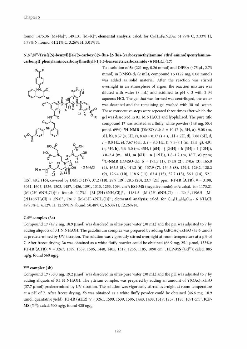
Chapter 5
122
found: 1475.36 [M+Na]+, 1491.31 [M+K]+; elemental analysis: calcd. for C75H48F12N6O12: 61.99% C, 3.33% H,
5.78% N; found: 61.21% C, 3.26% H, 5.01% N.
N,N’,N’’-Tris[{(S)-benzyl}{[4-{(5-carboxy)(5-[bis-{2-[bis-(carboxymethyl)amino]ethyl}amino])pentylamino-
carbonyl}]phenylaminocarbonyl}methyl]-1,3,5-benzenetricarboxamide ⋅⋅⋅⋅ 6 NH4Cl (17)
To a solution of 5a (221 mg, 0.26 mmol) and DiPEA (475 µL, 2.73
mmol) in DMSO-d6 (2 mL), compound 15 (122 mg, 0.08 mmol)
was added as solid material. After the reaction was stirred
overnight in an atmosphere of argon, the reaction mixture was
diluted with water (8 mL) and acidified to pH < 3 with 2 M
aqueous HCl. The gel that was formed was centrifuged, the water
was decanted and the remaining gel washed with 30 mL water.
These consecutive steps were repeated three times after which the
gel was dissolved in 0.1 M NH4OH and lyophilized. The pure title
compound 17 was isolated as a fluffy, white powder (148 mg, 55.4
µmol, 69%): 1H-NMR (DMSO-d6): δ = 10.47 (s, 3H, a), 9.08 (m,
3H, b), 8.57 (s, 3H, c), 8.40 + 8.37 (s + s, 1H + 2H, d), 7.88 (6H, d,
J = 8.0 Hz, e), 7.67 (6H, d, J = 8.0 Hz, f), 7.5–7.1 (m, 15H, g), 4.91
(q, 3H, h), 3.6–3.0 (m, 45H, i {6H} +j {24H} + k {3H} + l {12H}),
3.0–2.4 (m, 18H, m {6H}+ n {12H}), 1.8–1.2 (m, 18H, o) ppm; 13C-NMR (DMSO-d6): δ = 173.5 (1), 171.8 (2), 170.6 (3), 165.8
(4), 165.5 (5), 141.2 (6), 137.9 (7), 134.3 (8), 129.4, 129.2, 128.2
(9), 126.4 (10), 118.6 (11), 63.4 (12), 57.7 (13), 56.1 (14), 52.1
(15), 48.2 (16), covered by DMSO (17), 37.2 (18), 28.9 (19), 28.5 (20), 23.7 (21) ppm; FT-IR (ATR): ν = 3198,
3031, 1603, 1536, 1503, 1457, 1436, 1391, 1313, 1253, 1094 cm-1; ESI-MS (negative mode): m/z calcd. for 1173.21
[M-(2H+6NH4Cl)]2–; found: 1173.1 [M-(2H+6NH4Cl)]2–, 1184.5 [M-(2H+6NH4Cl) + Na]2–,1196.5 [M-
(2H+6NH4Cl) + 2Na]2–, 781.7 [M-(3H+6NH4Cl)]3–; elemental analysis: calcd. for C111H138N18O39 ∙ 6 NH4Cl:
49.95% C, 6.12% H, 12.59% N; found: 50.48% C, 6.63% H, 12.26% N.
GdIII complex (3a)
Compound 17 (49.2 mg, 18.9 µmol) was dissolved in ultra-pure water (30 mL) and the pH was adjusted to 7 by
adding aliquots of 0.1 N NH4OH. The gadolinium complex was prepared by adding Gd(OAc)3.xH2O (43.6 µmol)
as predetermined by UV-titration. The solution was vigorously stirred overnight at room temperature at a pH of
7. After freeze drying, 3a was obtained as a white fluffy powder could be obtained (66.9 mg, 25.1 µmol, 133%):
FT-IR (ATR): ν = 3267, 1589, 1539, 1506, 1440, 1405, 1319, 1256, 1185, 1090 cm-1; ICP-MS (GdIII): calcd. 885
ng/g, found 560 ng/g.
YIII complex (3b)
Compound 17 (50.0 mg, 19.2 µmol) was dissolved in ultra-pure water (30 mL) and the pH was adjusted to 7 by
adding aliquots of 0.1 N NH4OH. The yttrium complex was prepared by adding an amount of Y(OAc)3.xH2O
(37.7 µmol) predetermined by UV-titration. The solution was vigorously stirred overnight at room temperature
at a pH of 7. After freeze drying, 3b was obtained as a white fluffy powder could be obtained (46.6 mg, 18.9
µmol, quantative yield). FT-IR (ATR): ν = 3261, 1599, 1539, 1506, 1440, 1408, 1319, 1257, 1185, 1091 cm-1; ICP-
MS (YIII): calcd. 500 ng/g, found 420 ng/g.
a
b
c
de
f
g
h
i
j
k
l
m
n
o
o
o
1
2
3
4
56
7
8
9
11
12
13
14
15
16
NH
HN
HN
NH
NH
HN
O
O
O
O
O
O
O
O
O
O
RO
R
R
NH
N
N N
OH
O
O
O O
O
HO
HO OH
OH
R =
99
9
9
10
17
18
19 20
21
· 2 NH4Cl
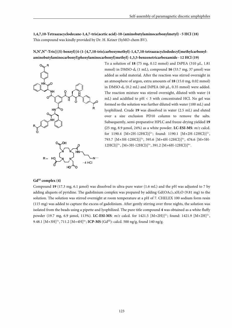
Self-assembly of paramagnetic discotic amphiphiles
123
1,4,7,10-Tetraazacyclodecane-1,4,7-tris(acetic acid)-10-(aminobutylaminocarbonylmetyl) ⋅⋅⋅⋅ 5 HCl (18)
This compound was kindly provided by Dr. H. Keizer (SyMO-chem BV).
N,N’,N’’-Tris[{(S)-benzyl}{4-(1-[4,7,10-tris{carboxymethyl}-1,4,7,10-tetraazacyclododecyl]methylcarbonyl-
aminobutylaminocarbonyl)phenylaminocarbonyl}methyl]-1,3,5-benzenetricarboxamide ⋅⋅⋅⋅ 12 HCl (19)
To a solution of 18 (75 mg, 0.12 mmol) and DiPEA (310 µL, 1.81
mmol) in DMSO-d6 (1 mL), compound 16 (53.7 mg, 37 µmol) was
added as solid material. After the reaction was stirred overnight in
an atmosphere of argon, extra amounts of 18 (15.0 mg, 0.02 mmol)
in DMSO-d6 (0.2 mL) and DiPEA (60 µL, 0.35 mmol) were added.
The reaction mixture was stirred overnight, diluted with water (4
mL) and acidified to pH < 3 with concentrated HCl. No gel was
formed so the solution was further diluted with water (100 mL) and
lyophilized. Crude 19 was dissolved in water (2.5 mL) and eluted
over a size exclusion PD10 column to remove the salts.
Subsequently, semi-preparative HPLC and freeze-drying yielded 19
(25 mg, 8.9 µmol, 24%) as a white powder. LC-ESI-MS: m/z calcd.
for 1190.4 [M+2H-12HCl)]2+; found: 1190.1 [M+2H-12HCl)]2+,
793.7 [M+3H-12HCl)]3+, 595.6 [M+4H-12HCl)]4+, 476.6 [M+5H-
12HCl)]5+, [M+3H-12HCl)]3+, 391.2 [M+6H-12HCl)]6+.
GdIII complex (4)
Compound 19 (17.3 mg, 6.1 µmol) was dissolved in ultra-pure water (1.6 mL) and the pH was adjusted to 7 by
adding aliquots of pyridine. The gadolinium complex was prepared by adding Gd(OAc)3.xH2O (9.81 mg) to the
solution. The solution was stirred overnight at room temperature at a pH of 7. CHELEX 100 sodium form resin
(115 mg) was added to capture the excess of gadolinium. After gently stirring over three nights, the solution was
isolated from the beads using a pipette and lyophilized. The pure title compound 4 was obtained as a white fluffy
powder (19.7 mg, 6.9 µmol, 113%). LC-ESI-MS: m/z calcd. for 1421.5 [M+2H)]2+; found: 1421.9 [M+2H]2+,
9.48.1 [M+3H]3+, 711.2 [M+4H]4+; ICP-MS (GdIII): calcd. 500 ng/g, found 140 ng/g.
NH
HN
HN
NH
NH
HN
O
O
O
O
O
O
O
O
RO
R
R
R =
N N
N N
O
HN
OHOO
HO
O OH
NH
· 4 HCl
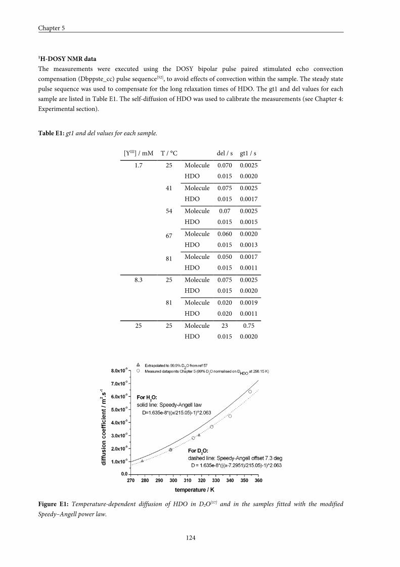
Chapter 5
124
1H-DOSY NMR data
The measurements were executed using the DOSY bipolar pulse paired stimulated echo convection
compensation (Dbppste_cc) pulse sequence[52], to avoid effects of convection within the sample. The steady state
pulse sequence was used to compensate for the long relaxation times of HDO. The gt1 and del values for each
sample are listed in Table E1. The self-diffusion of HDO was used to calibrate the measurements (see Chapter 4:
Experimental section).
Table E1: gt1 and del values for each sample.
[YIII] / mM T / °C del / s gt1 / s
Molecule 0.070 0.0025 25
HDO 0.015 0.0020
Molecule 0.075 0.0025 41
HDO 0.015 0.0017
Molecule 0.07 0.0025 54
HDO 0.015 0.0015
Molecule 0.060 0.0020 67 HDO 0.015 0.0013
Molecule 0.050 0.0017
1.7
81 HDO 0.015 0.0011
Molecule 0.075 0.0025 25
HDO 0.015 0.0020
Molecule 0.020 0.0019
8.3
81
HDO 0.020 0.0011
Molecule 23 0.75 25 25
HDO 0.015 0.0020
Figure E1: Temperature-dependent diffusion of HDO in D2O[57] and in the samples fitted with the modified
Speedy–Angell power law.
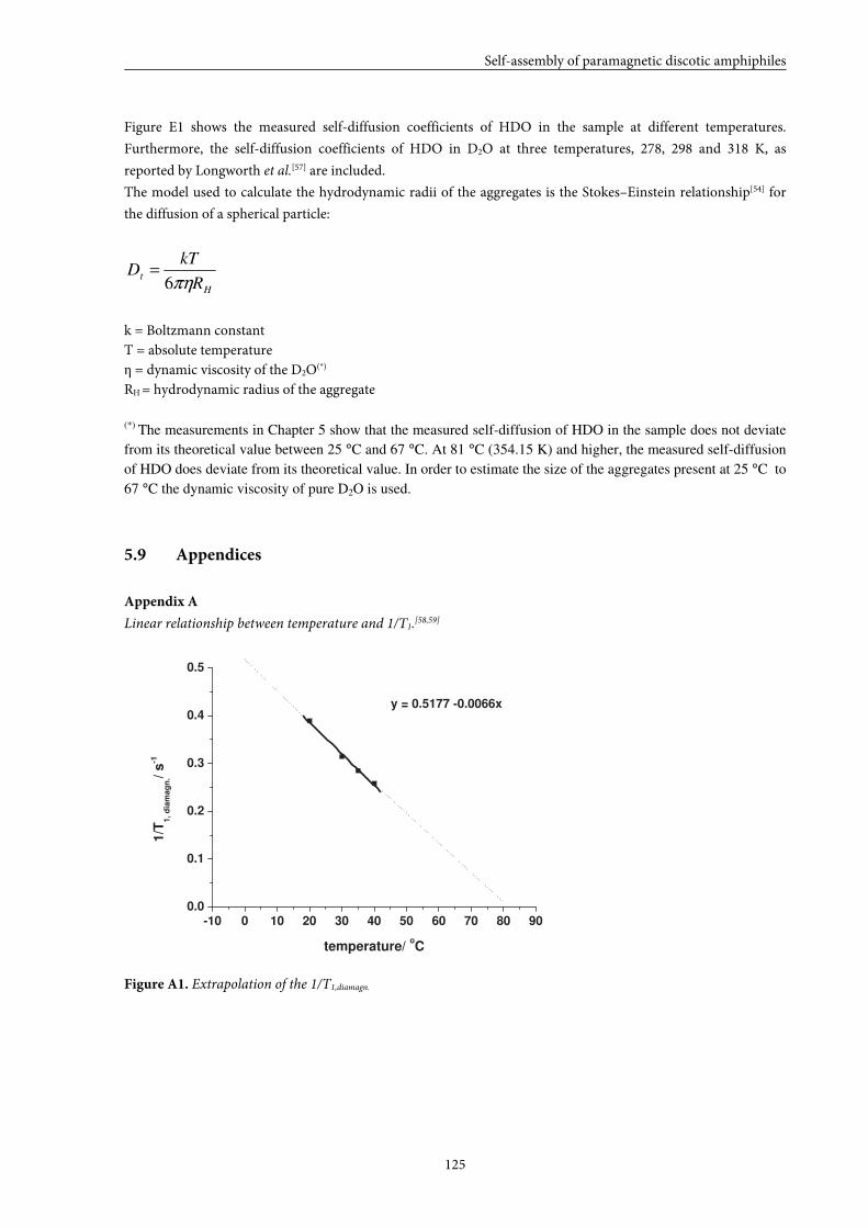
Self-assembly of paramagnetic discotic amphiphiles
125
Figure E1 shows the measured self-diffusion coefficients of HDO in the sample at different temperatures.
Furthermore, the self-diffusion coefficients of HDO in D2O at three temperatures, 278, 298 and 318 K, as
reported by Longworth et al.[57] are included.
The model used to calculate the hydrodynamic radii of the aggregates is the Stokes–Einstein relationship[54] for
the diffusion of a spherical particle:
H
tR
kTD
πη6=
k = Boltzmann constant T = absolute temperature η = dynamic viscosity of the D2O(*)
RH = hydrodynamic radius of the aggregate (*) The measurements in Chapter 5 show that the measured self-diffusion of HDO in the sample does not deviate
from its theoretical value between 25 °C and 67 °C. At 81 °C (354.15 K) and higher, the measured self-diffusion
of HDO does deviate from its theoretical value. In order to estimate the size of the aggregates present at 25 °C to
67 °C the dynamic viscosity of pure D2O is used.
5.9 Appendices
Appendix A
Linear relationship between temperature and 1/T1.[58,59]
-10 0 10 20 30 40 50 60 70 80 90 0.0
0.1
0.2
0.3
0.4
0.5
y = 0.5177 -0.0066x
1/T
1 , d
iam
ag
n. /
s -1
temperature/ o C
Figure A1. Extrapolation of the 1/T1,diamagn.
Chapter 5
126
Appendix B
Theory isodesmic polymerization
Consider the following polymerization of free discotics [A] in solution
[ ]
[ ]
[ ]
[ ] [ ]
[ ]
[ ] [ ]
[ ]
[ ] [ ](1)
AA
A
k
kK AAA
AA
A
k
kK A4AA
AA
A
k
kK AAA
A
A
k
kK AAA
1i
i
i
iii
ik_
ik1i
3
4
4
44
4k_
4k3
2
3
3
333
3k_
3k2
2
2
2
222
2k_
2k
−−
←→−
−
← →
−
←→
−
← →
==+
⋅
⋅
⋅
==+
==+
==+
According to equation (1), the molar concentration of all aggregates with different degree of polymerization i at
equilibrium can be written as:
[ ] [ ]
[ ] [ ] [ ] [ ]
[ ] [ ] [ ] [ ]
[ ] [ ] [ ] [ ] [ ]
[ ]
[ ] (4) Ac
(3) Aic
(2) KA AK...KK AAK A
AKKKAAKA
AKKAAKA
AKA
1i
ip
1i
it
i
2j
jii
23i1-iii
4234344
323233
222
∑
∑
∏
∞
=
∞
=
=
=
=
⋅
⋅
⋅
===
⋅
⋅
⋅
==
==
=
Where ct is the initial total concentration of free discotics, and cp is the total concentration of all i-mers including
the free disotics. In the isodesmic model, the equilibrium constants for all free discotic additions are identical (K2
= K3 = … = Ki = … = K). Therefore, equation (2) reduces to:
Self-assembly of paramagnetic discotic amphiphiles
127
[ ] [ ]( ) [ ] (5) AKAKKA i1ii1i
−− ==
And the mole fraction of an i-mer is defined as:
[ ](6)
c
Ax
t
ii =
The degree of aggregation ϕ, which can be deduced from spectroscopic measurements and can be written as:
(7) x1 1−=φ
Due to equation (5), the initial total free disc concentration can be written as:
[ ]( ) (8) AKiKc
1i
i1t ∑
∞
=
−=
If K[A] < 1 this sum can be conveniently written as :
[ ][ ]( )
(9) AK1
Ac
2t−
=
And cp is written as:
[ ][ ]( )
(10) AK1
Acp
−=
The free disc concentration [A] can be isolated from equation (9):
[ ] (11) Kc
1K4c1K2c
2
1A
2t
tt +−+=
For each given value of K and initial concentration ct, the corresponding concentration of free disc [A] can be
calculated and hence, the concentration of each i-mer too. The mole fraction of each i-mer is then defined via
equation (6).
The weight average degree of polymerization PDw is defined by equation (12):
(12)
iN
Ni
DP
1i
i
1i
i2
w
∑
∑∞
=
∞
==
The numerator of equation (12) is proportional to the summation of the molar concentrations of all i-mers
multiplied by i2, whereas the denominator is proportional to the initial free disc concentration, ct. In other words,
equation (12) can be written as:
Chapter 5
128
[ ] [ ][ ]
(13) K[A]1
AK1
c
AKi
c
Ai
DPt
1i
i1i2
t
1i
i2
w−
+===
∑∑∞
=
−∞
=
The number average degree of polymerization DPN is defined by equation (14):
(14)
N
iN
DP
1i
i
1i
i
N
∑
∑∞
=
∞
==
Where Ni is the number of aggregates of a given i-mer. Now, the numerator of equation (14) is proportional to
the initial free disc concentration, ct, whereas the denominator is proportional to the summation of the molar
concentrations of all the i-mers. In other words, equation (14) can be written as:
[ ][ ]
(15) K[A]1
1c
A
cDP
1i
iA1-iK
t
1i
i
tN
−===
∑∑∞
=
∞
=
By combining equations (6), (7) and (15), the degree of aggregation ϕ can be expressed in terms of DPN. So DPN
can be calculated via equation (16):
[ ][ ]( )
(16) DPAK1
1
c
Ax1 2
N2t
1 =−
===−φ
Now DPN is known as function of the temperature, the association constant at each temperature can be
determined via equation (17). This equation is a combination of equations (11) and (15) and gives:
(17) 14Kc2
1
2
1DP tN ++=
Since it is more convenient to express equation (15) in GdIII concentrations instead of disc concentrations. ct, the
total initial concentration of free discs will be replaced by C, the total initial concentration of GdIII:
(18) 3
Cct =
Thus [A] used in equation (15) becomes:
[ ] (19)
K3
C
1K3
C41K
3
C2
2
1A
2
+−+
=
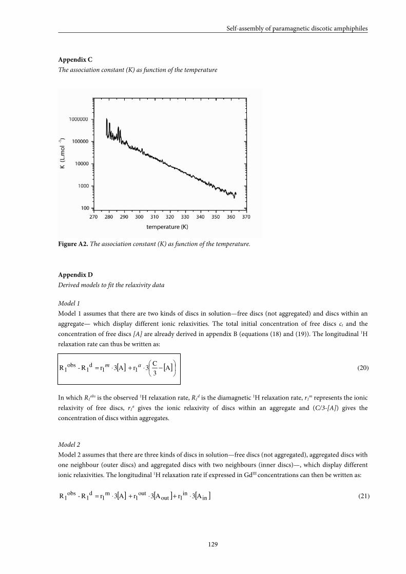
Self-assembly of paramagnetic discotic amphiphiles
129
Appendix C
The association constant (K) as function of the temperature
Figure A2. The association constant (K) as function of the temperature.
Appendix D
Derived models to fit the relaxivity data
Model 1
Model 1 assumes that there are two kinds of discs in solution—free discs (not aggregated) and discs within an
aggregate— which display different ionic relaxivities. The total initial concentration of free discs ct and the
concentration of free discs [A] are already derived in appendix B (equations (18) and (19)). The longitudinal 1H
relaxation rate can thus be written as:
[ ] [ ] (20) A3
C3r A3r R-R 11
d1
obs1
−⋅+⋅= am
In which R1obs is the observed 1H relaxation rate, R1d is the diamagnetic 1H relaxation rate, r1m represents the ionic
relaxivity of free discs, r1a gives the ionic relaxivity of discs within an aggregate and (C/3-[A]) gives the
concentration of discs within aggregates.
Model 2
Model 2 assumes that there are three kinds of discs in solution—free discs (not aggregated), aggregated discs with
one neighbour (outer discs) and aggregated discs with two neighbours (inner discs)—, which display different
ionic relaxivities. The longitudinal 1H relaxation rate if expressed in GdIII concentrations can then be written as:
[ ] [ ] [ ] (21) A3r A3r A3r R-R inin
1outout
1m
1d
1obs
1 ⋅+⋅+⋅=
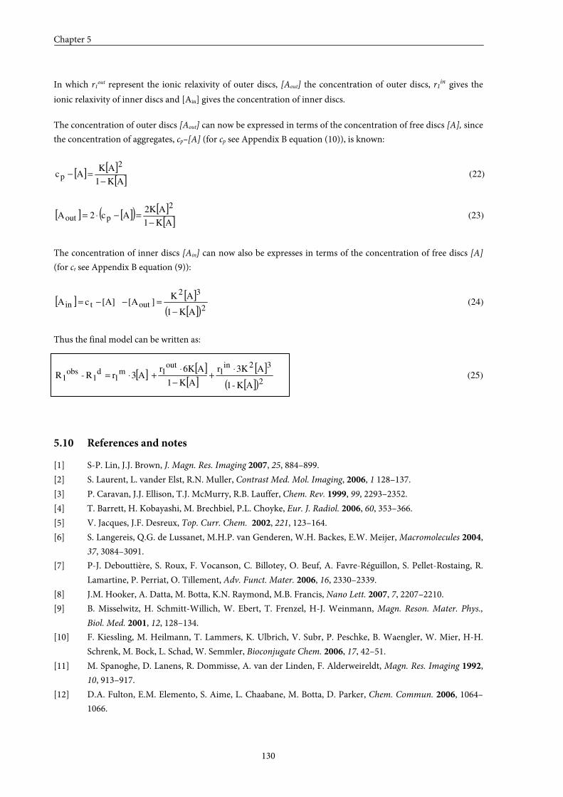
Chapter 5
130
In which r1out represent the ionic relaxivity of outer discs, [Aout] the concentration of outer discs, r1in gives the
ionic relaxivity of inner discs and [Ain] gives the concentration of inner discs.
The concentration of outer discs [Aout] can now be expressed in terms of the concentration of free discs [A], since
the concentration of aggregates, cp–[A] (for cp see Appendix B equation (10)), is known:
[ ] [ ][ ]
(22) AK1
AK Ac
2
p−
=−
[ ] [ ]( ) [ ][ ]
(23) AK1
A2K Ac2A
2
pout−
=−⋅=
The concentration of inner discs [Ain] can now also be expresses in terms of the concentration of free discs [A]
(for ct see Appendix B equation (9)):
[ ] [ ][ ]( )
(24) AK1
AK][A[A]c A
2
32
outtin−
=−−=
Thus the final model can be written as:
[ ][ ]
[ ][ ]
[ ]( )(25)
AK-1
A3Kr
AK1
A6Kr A3r R-R
2
32in1
out1m
1d
1obs
1⋅
+−
⋅+⋅=
5.10 References and notes
[1] S-P. Lin, J.J. Brown, J. Magn. Res. Imaging 2007, 25, 884–899.
[2] S. Laurent, L. vander Elst, R.N. Muller, Contrast Med. Mol. Imaging, 2006, 1 128–137.
[3] P. Caravan, J.J. Ellison, T.J. McMurry, R.B. Lauffer, Chem. Rev. 1999, 99, 2293–2352.
[4] T. Barrett, H. Kobayashi, M. Brechbiel, P.L. Choyke, Eur. J. Radiol. 2006, 60, 353–366.
[5] V. Jacques, J.F. Desreux, Top. Curr. Chem. 2002, 221, 123–164.
[6] S. Langereis, Q.G. de Lussanet, M.H.P. van Genderen, W.H. Backes, E.W. Meijer, Macromolecules 2004,
37, 3084–3091.
[7] P-J. Debouttière, S. Roux, F. Vocanson, C. Billotey, O. Beuf, A. Favre-Réguillon, S. Pellet-Rostaing, R.
Lamartine, P. Perriat, O. Tillement, Adv. Funct. Mater. 2006, 16, 2330–2339.
[8] J.M. Hooker, A. Datta, M. Botta, K.N. Raymond, M.B. Francis, Nano Lett. 2007, 7, 2207–2210.
[9] B. Misselwitz, H. Schmitt-Willich, W. Ebert, T. Frenzel, H-J. Weinmann, Magn. Reson. Mater. Phys.,
Biol. Med. 2001, 12, 128–134.
[10] F. Kiessling, M. Heilmann, T. Lammers, K. Ulbrich, V. Subr, P. Peschke, B. Waengler, W. Mier, H-H.
Schrenk, M. Bock, L. Schad, W. Semmler, Bioconjugate Chem. 2006, 17, 42–51.
[11] M. Spanoghe, D. Lanens, R. Dommisse, A. van der Linden, F. Alderweireldt, Magn. Res. Imaging 1992,
10, 913–917.
[12] D.A. Fulton, E.M. Elemento, S. Aime, L. Chaabane, M. Botta, D. Parker, Chem. Commun. 2006, 1064–
1066.
Self-assembly of paramagnetic discotic amphiphiles
131
[13] R.B. Lauffer, D.J. Parmelee, S. Ouellet, R.P. Dolan, H. Sajiki, M. Scott, P.J. Bernard, E.M. Buchanan, K.
Y. Ong, Z. Tyeklár, K.S. Midelfort, T.J. McMurry, R.C. Walovitch, Acad. Radiol. 1996, 3, 356–358.
[14] S. Aime, L. Frullano, S. Geninatti Crich, Angew. Chem. 2002, 114, 1059–1061; Angew. Chem., Int. Ed.
2002, 41, 1017–1019.
[15] P. Caravan, N.J. Cloutier, M.T. Greenfield, S.A. McDermid, S.U. Dunham, J.W.M. Bulte, J.D. Amedio
Jr., R.J. Looby, R.M. Supkowski, W. DeW. Horrocks Jr., T.J. McMurry, R.B. Lauffer, J. Am. Chem. Soc.
2002, 124, 3152–3162.
[16] J.B. Livramento, E. Tóth, A. Sour, A. Borel, A.E. Merbach, R. Ruloff, Angew. Chem. 2005, 117, 1504–
1508; Angew. Chem., Int. Ed. 2005, 44, 1480–1484.
[17] L. Prinzen, R-J. J.H.M. Miserus, A. Dirksen, T.M. Hackeng, N. Deckers, N.J. Bitsch, R.T.A. Megens, K.
Douma, J.W. Heemskerk, M.E. Kooi, P.M. Frederik, D.W. Slaaf, M.A.M.J. van Zandvoort, C.P.M.
Reutelingsperger, Nano Lett. 2007, 7, 93–100.
[18] V.S. Vexler, O. Clement, H. Schmitt-Willich, R.C. Brasch, J. Magn. Res. Imaging 1994, 4, 381–388.
[19] T.S. Desser, L.D. Rubin, H.H. Muller, F. Qing, S. Khodor, G. Zanazzi, S.W. Young, D.L. Ladd, J.A.
Wellons, K.E. Kellar, J. Magn. Res. Imaging 1994, 4, 467–472.
[20] H-J. Raatschen, R. Swain, D.M. Shames, Y. Fu, Z. Boyd, M.L. Zierhut, M.F. Wendland, B. Misselwitz, H-
J. Weinmann, K-J. Wolf, R.C. Brasch, Contrast Med. Mol. Imaging 2006, 1, 113–120.
[21] H.Y. Lee, H.W. Jee, S.M. Seo, B.K. Kwak, G. Khang, S.H. Cho, Bioconjugate Chem. 2006, 17, 700–706.
[22] G.A.F. van Tilborg, W.J.M. Mulder, N. Deckers, G. Storm, C.P.M. Reutelingsperger, G.J. Strijkers, K.
Nicolay, Bioconjugate Chem. 2006, 17, 741–749.
[23] T.N. Parac-Vogt, K. Kimpe, S. Laurent, C. Piérart, L. vander Elst, R.N. Muller, K. Binnemans, Eur.
Biophys. J. 2006, 35, 136–144.
[24] M. Vaccaro, A. Accardo, D. Tesauro, G. Mangiapia, D. Löf, K. Schillén, O. Södermann, G. Morelli, L.
Paduano, Langmuir 2006, 22, 6635–6643.
[25] C. GlØgård, G. Stensrud, R. Hovland, S.L. Fossheim, J. Klaveness, Int. J. Pharm. 2002, 233, 131–140.
[26] A. Accardo, D. Tesauro, G. Morelli, E. Gianolio, S. Aime, M. Vaccaro, G. Mangiapia, L. Paduano, K.
Schillén, J. Biol. Inorg. Chem. 2007, 12, 267–276.
[27] S.R. Bull, M.O. Guler, R.E. Bras, P.N. Venkatasubramanian, S.I. Stupp, T.J. Meade, Bioconjugate Chem.
2005, 16, 1343–1348.
[28] A. Barge, G. Cravotto, B. Robaldo, E. Gianolio, S. Aime, J. Incl. Phenom. Macrocycl. Chem. 2007, 57,
489–495.
[29] S. Langereis, H-A.T. Kooistra, M.H.P. van Genderen, E.W. Meijer, Org. Biomol. Chem. 2004, 2, 1271–
1273.
[30] A. Dirksen, S. Langereis, B.F.M. de Waal, M.H.P. van Genderen, T.M. Hackeng, E.W. Meijer, Chem.
Commun. 2005, 2811–2813.
[31] S. Torres, J.A. Martins, J.P. André, C.F.G.C. Geraldes, A.E. Merbach, E. Tóth, Chem. Eur. J. 2006, 12,
940–948.
[32] G.M. Nicolle, E. Tóth, K-P. Eiseniener, H.R. Mäcke, A.E. Merbach, J. Biol. Inorg. Chem. 2002, 7, 757–
769.
[33] A.R.A. Palmans, J.A.J.M. Vekemans, H. Fischer, R.A. Hikmet, E.W. Meijer, Chem. Eur. J. 1997, 3, 300–
307.
[34] L. Brunsveld, H. Zhang, M. Glasbeek, J.A.J.M. Vekemans, E.W. Meijer, J. Am. Chem. Soc. 2000, 122,
6175–6182.
[35] L. Brunsveld, B.G.G. Lohmeijer, J.A.J.M. Vekemans, E.W. Meijer, J. Incl. Phenom. Macrocycl. Chem.
2001, 41, 61-64.
Chapter 5
132
[36] J.J. van Gorp, J.A.J.M. Vekemans, E.W. Meijer, J. Am. Chem. Soc. 2002, 124, 14759–14769.
[37] K.P. van den Hout, R. Martín-Rapún, J.A.J.M. Vekemans, E.W. Meijer, Chem. Eur. J. 2007, 13, 8111–
8123.
[38] P.L. Anelli, F. Fedeli, O. Gazzotti, L. Lattuada, G. Lux, F. Rebasti, Bioconjugate Chem. 1999, 10, 137–140.
[39] M.A. Williams, H. Rapoport, J. Org. Chem. 1993, 58, 1151–1158.
[40] A. Barge, G. Cravotto, E. Gianolio, F. Fedeli, Contrast Med. Mol. Imaging 2006, 1, 184–188.
[41] B.L. Gupta, Talanta 1974, 21, 683–684.
[42] S. Langereis, Thesis: Dendritic MRI contrast agents, synthetic strategies for targeting and multivalency,
Eindhoven University of Technology, Eindhoven (The Netherlands), 2005.
[43] R. Knorr, A. Trzeciak, W. Bannwarth, D. Gillessen, Tetrahedron Lett. 1989, 30, 1927–1930.
[44] Enantiomeric excess of intermediate 5 was not determined with chiral HPLC.
[45] M.M.J. Smulders, A.P.H.J. Schenning, E.W. Meijer, J. Am. Chem. Soc. 2007, 130, 606–611.
[46] P. Jonkheijm, P. van der Schoot, A.P.H.J. Schenning, E.W. Meijer, Science 2006, 313, 80–83.
[47] P. van der Schoot, In Supramolecular polymers; 2nd ed.; A. Ciferri, Ed. Taylor & Francis: London 2005.
[48] R.B. Martin, Chem. Rev. 1996, 96, 3043–3064.
[49] G. Fleischer, Polymer 1985, 26, 1677–1682.
[50] R. Raghavan, T.L. Maver, F.D. Blum, Macromolecules 1987, 20, 814–820.
[51] A. Chen, D. Wu, C.S. Johnson Jr, J. Am. Chem. Soc. 1995, 117, 7965–7970.
[52] A. Jerschow, N. Müller, J. Magn. Reson. 1997, 125, 372–375.
[53] C.H. Cho, J. Urquidi, S. Singh, G. Wilse Robinson, J. Phys. Chem. B 1999, 103, 1991–1994.
[54] G.G. Stokes, Trans Cambridge Philos. Soc. 1845, 8, 287.
[55] M.M. Tirado, J. Garcia de la Torre, J. Chem. Phys.1979, 71, 2581–2587.
[56] This value is estimated from a plot in which the ionic relaxivity is plotted against the molecular weight
for different generations of GdIII-DTPA containing PPI dendrimers as described by S. Langereis[6].
[57] L.G. Longsworth, J. Phys. Chem. 1960, 64, 1914–1917.
[58] T.R. Nelson, S.M. Tung, Magn. Res. Imaging 1987, 5, 189–199.
[59] P.A. Bottemly, T.H. Forster, R.E. Argersinger, L.M. Pfeifer, Med. Phys. 1984, 11, 425–448.
Summary
133
PEPTIDE DISCOTICS
Synthesis, self-assembly and application
Summary
Magnetic resonance imaging (MRI) is one of the most valuable non-invasive imaging
techniques used in the medical world for early diagnosis of disease. This can in part be
attributed to the recent development of MRI contrast agents. Most of the currently applied
contrast agents are gadolinium(III) complexes of diethylenetriaminepentaacetic acid
(GdIII-DTPA). Properties that severely limit the utility of these low molecular weight contrast
agents are their non-specificity, their low contrast efficiency and their fast renal excretion,
which necessitates high dosages. In recent years, various strategies have been explored in
order to improve the proton relaxivities of MRI contrast agents. Self-assembling contrast
agents, such as paramagnetic micelles or liposomes, have recently gained attention due to
their easily controlled properties and good pharmacological characteristics. An additional
benefit of micelles is their dynamic nature, which allows combining the benefits of both low
and high molecular weight contrast agents. Designing paramagnetic discotic amphiphiles
might be a promising strategy for creating self-assembling contrast agents of a different shape
and hence displaying different properties. Columnar self-assembling contrast agents based on
discotic molecules may in the first place become very long, hence ensuring high local
concentrations of gadolinium, and secondly, the discotic amphiphiles building up the
columns are multivalent, which ensures higher local concentrations of contrast agent and
opens possibilities for the use of asymmetric target-specific discotics. Moreover, the self-
assembly of C3-symmetrical discotic molecules has been well-studied. In this thesis, a strategy
towards new self-assembling contrast agents based on paramagnetic discotic amphiphiles has
been developed and described. Peptide fragments are used as structuring elements because of
their biocompatibility, hydrogen bonding properties and versatile nature.
In Chapter 1 a general introduction into the intruiging properties of self-assembling
linear paramagnetic amphiphiles forming liposomal or micellar contrast agents is described.
Furthermore, the self-assembling properties of C3-symmetrical discotics in apolar media as
well as in water are addressed in this Chapter.
In Chapter 2, the synthesis of a new class of discotic molecules is presented. These
molecules, which function as model compounds, consist of a 1,3,5-benzenetricarboxamide
core extended with peptide fragments decorated with mesogenic groups incorporating
hydrophobic tails. Several dipeptide fragments consisting of glycine, D-phenylalanine and/or
L-phenylalanine and monopeptide fragments, either L-alanine or L-phenylalanine, have been
incorporated to be able to study the effects of increased hydrophobicity and extra
opportunities for secondary interactions on the self-assembly properties. All discotic
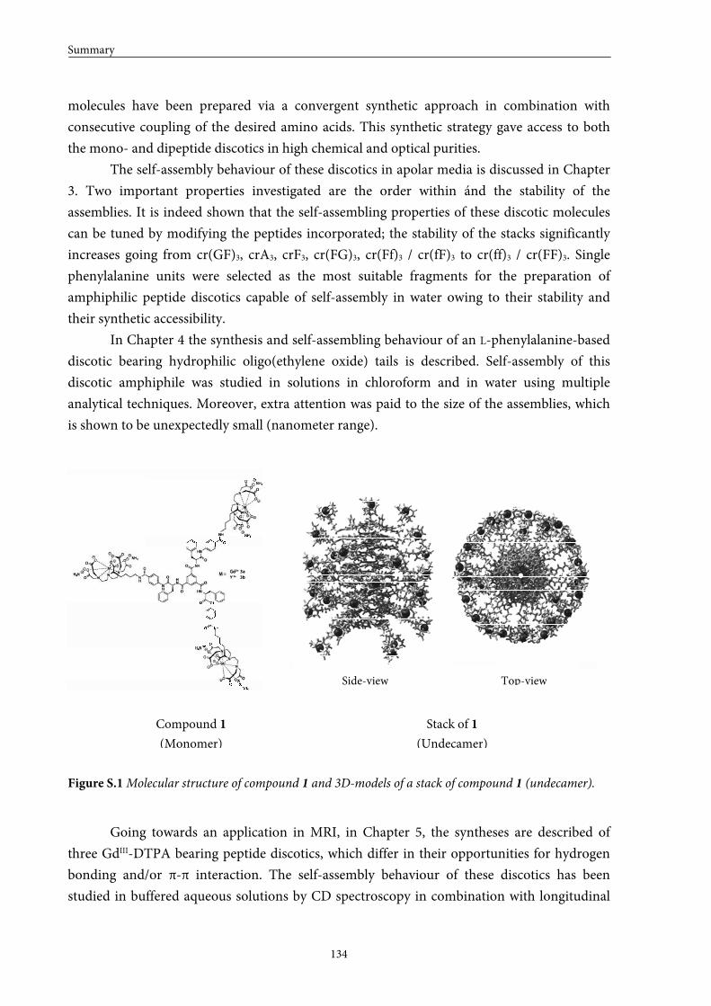
Summary
134
molecules have been prepared via a convergent synthetic approach in combination with
consecutive coupling of the desired amino acids. This synthetic strategy gave access to both
the mono- and dipeptide discotics in high chemical and optical purities.
The self-assembly behaviour of these discotics in apolar media is discussed in Chapter
3. Two important properties investigated are the order within ánd the stability of the
assemblies. It is indeed shown that the self-assembling properties of these discotic molecules
can be tuned by modifying the peptides incorporated; the stability of the stacks significantly
increases going from cr(GF)3, crA3, crF3, cr(FG)3, cr(Ff)3 / cr(fF)3 to cr(ff)3 / cr(FF)3. Single
phenylalanine units were selected as the most suitable fragments for the preparation of
amphiphilic peptide discotics capable of self-assembly in water owing to their stability and
their synthetic accessibility.
In Chapter 4 the synthesis and self-assembling behaviour of an L-phenylalanine-based
discotic bearing hydrophilic oligo(ethylene oxide) tails is described. Self-assembly of this
discotic amphiphile was studied in solutions in chloroform and in water using multiple
analytical techniques. Moreover, extra attention was paid to the size of the assemblies, which
is shown to be unexpectedly small (nanometer range).
Figure S.1 Molecular structure of compound 1 and 3D-models of a stack of compound 1 (undecamer).
Going towards an application in MRI, in Chapter 5, the syntheses are described of
three GdIII-DTPA bearing peptide discotics, which differ in their opportunities for hydrogen
bonding and/or π-π interaction. The self-assembly behaviour of these discotics has been
studied in buffered aqueous solutions by CD spectroscopy in combination with longitudinal
Top-view Side-view
Compound 1
(Monomer)
Stack of 1
(Undecamer)
Side-view Top-view
Summary
135
relaxation time measurements. It has been established that GdIII-DTPA discs with larger
hydrophobic cores give rise to stronger stacking interactions and hence increased aggregate
stabilities, higher ionic relaxivities and lower aggregation concentrations. Therefore, the
largest GdIII-DTPA disc 1 (Figure S.1) has been studied in more detail. A model, which
distinguishes three types of discotic molecules has been derived to describe the relaxation time
data. This model is shown to fit the relaxation time data pretty accurately and hence has been
applied to calculate the ionic relaxivities of the molecularly dissolved discs and of the
assemblies present in solution. Furthermore, 1H-DOSY NMR measurements in combination
with cryoTEM have been performed and show that the size of the assemblies is comparable to
the size of the assemblies obtained for the phenylalanine-based discotics described in Chapter
4 (Figure S.1). Finally, the universality of our appraoch has been illustrated with the synthesis
and characterization of a large disc in which the GdIII-DTPA complexes are replaced by
GdIII-DOTAs. The relaxation time measurements of this GdIII-DOTA disc indeed show that
the obtained results are reproducible with a different type of ligand.
In this thesis a first step was made towards the use of paramagnetic discotic
amphiphiles as self-assembling contrast agents for MRI. In the epilogue the author’s personal
opinion on the perspectives of the research is described. Hopefully, this thesis may contribute
to the development of new target-specific, self-assembling contrast agents for MRI in the near
future.

Samenvatting
137
PEPTIDE DISCOTEN
Synthese, zelf-assemblage en applicatie
Samenvatting
Magnetic resonance imaging (MRI) is één van de meest belangrijke niet-invasieve
beeldende technieken die toegepast wordt in de medische wereld om in een vroeg stadium
diagnoses te stellen. Dit is mede te danken aan de recente ontwikkelingen op het gebied van
MRI contrastmiddelen. In ziekenhuizen worden momenteel vooral gadoliniumcomplexen van
diethyleentriaminepenta-azijnzuur (GdIII-DTPA) als contrastmiddel gebruikt. Een nadeel van
deze laagmoleculaire contrastmiddelen is de hoge dosis die vereist is vanwege hun aspecifieke
verdeling in het lichaam, lage efficiëntie en snelle uitscheiding via de nieren. Het onderzoek
heeft zich daarom de afgelopen jaren voornamelijk gericht op het ontwikkelen van
efficiëntere contrastmiddelen. Tegenwoordig staan zelf-assemblerende contrastmiddelen,
zoals paramagnetische lineaire amfifielen, die bolvormige aggregaten vormen (micellen en
liposomen), veelvuldig in de belangstelling vanwege hun makkelijk te controleren en goede
farmacologische eigenschappen. Verder hebben micellen een extra voordeel, namelijk, hun
dynamische karakter, wat het mogelijk maakt om de voordelen van laag- en hoogmoleculaire
contrastmiddelen te combineren. Het prepareren van paramagnetische schijfvormige
amfifielen zou een veelbelovende strategie kunnen zijn om zelf-assemblerende
contrastmiddelen te prepareren met andere dimensies en dus met andere eigenschappen.
Cilindervormige contrastmiddelen gebaseerd op schijf-vormige moleculen (discoten) bezitten
de mogelijkheid om heel lang te worden, wat kan leiden tot een hoge lokale concentratie aan
gadolinium. Bovendien zijn de discoten, waaruit de cylinders opgebouwd zijn, multivalent.
Ook dit resulteert in een hogere lokale concentratie aan gadolinium en biedt de mogelijkheid
tot het gebruik van asymmetrische target-specifieke discoten. Daar komt bij dat het zelf-
assemblage gedrag van C3-symmetrische discoten grondig onderzocht is. In dit proefschrift is
een nieuwe strategie voor het verkrijgen van zelf-assemblerende contrastmiddelen gebaseerd
op schijfvormige peptiden ontwikkeld en beschreven. Peptiden zijn gebruikt als structurerend
element vanwege hun biocompatibiliteit, diversiteit en waterbrugvormende eigenschappen.
In Hoofdstuk 1 wordt een algemene inleiding omtrent de intrigerende eigenschappen
van zelf-assemblerende lineaire paramagnetische amfifielen beschreven. Tevens worden de
zelf-assemblerende eigenschappen van C3-symmetrische discoten in apolaire oplosmiddelen
en in water besproken.
In Hoofdstuk 2 wordt de synthese van een nieuwe klasse van schijfvormige moleculen
gepresenteerd. Deze modelstructuren bestaan uit een 1,3,5-benzeentricarboxamide centrum
verlengd met peptidefragmenten die gekoppeld zijn aan mesogene groepen met hydrofobe
staarten. Dipeptide fragmenten bestaande uit glycine, D-fenylalanine en/of L-fenylalanine en
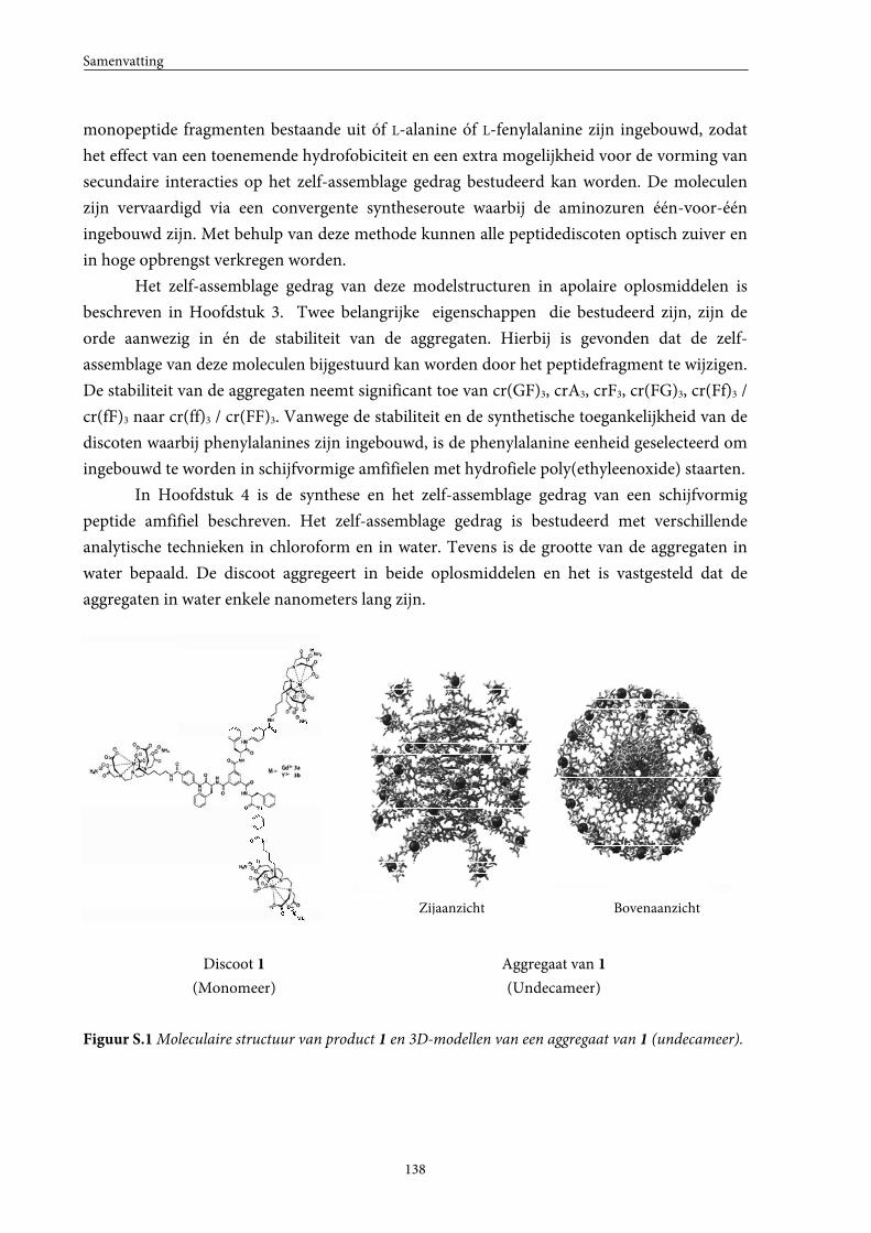
Samenvatting
138
monopeptide fragmenten bestaande uit óf L-alanine óf L-fenylalanine zijn ingebouwd, zodat
het effect van een toenemende hydrofobiciteit en een extra mogelijkheid voor de vorming van
secundaire interacties op het zelf-assemblage gedrag bestudeerd kan worden. De moleculen
zijn vervaardigd via een convergente syntheseroute waarbij de aminozuren één-voor-één
ingebouwd zijn. Met behulp van deze methode kunnen alle peptidediscoten optisch zuiver en
in hoge opbrengst verkregen worden.
Het zelf-assemblage gedrag van deze modelstructuren in apolaire oplosmiddelen is
beschreven in Hoofdstuk 3. Twee belangrijke eigenschappen die bestudeerd zijn, zijn de
orde aanwezig in én de stabiliteit van de aggregaten. Hierbij is gevonden dat de zelf-
assemblage van deze moleculen bijgestuurd kan worden door het peptidefragment te wijzigen.
De stabiliteit van de aggregaten neemt significant toe van cr(GF)3, crA3, crF3, cr(FG)3, cr(Ff)3 /
cr(fF)3 naar cr(ff)3 / cr(FF)3. Vanwege de stabiliteit en de synthetische toegankelijkheid van de
discoten waarbij phenylalanines zijn ingebouwd, is de phenylalanine eenheid geselecteerd om
ingebouwd te worden in schijfvormige amfifielen met hydrofiele poly(ethyleenoxide) staarten.
In Hoofdstuk 4 is de synthese en het zelf-assemblage gedrag van een schijfvormig
peptide amfifiel beschreven. Het zelf-assemblage gedrag is bestudeerd met verschillende
analytische technieken in chloroform en in water. Tevens is de grootte van de aggregaten in
water bepaald. De discoot aggregeert in beide oplosmiddelen en het is vastgesteld dat de
aggregaten in water enkele nanometers lang zijn.
Figuur S.1 Moleculaire structuur van product 1 en 3D-modellen van een aggregaat van 1 (undecameer).
Top-view Side-view
Discoot 1
(Monomeer)
Aggregaat van 1
(Undecameer)
Zijaanzicht Bovenaanzicht
Samenvatting
139
Voor een toepassing in MRI is, in Hoofdstuk 5, de synthese van drie GdIII-DTPA
gelabelde discoten, die verschillen in hun mogelijkheid tot waterstofbrugvorming en π-π
interactie, behandeld. Het zelf-assemblage gedrag van deze moleculen in een gebufferde
oplossing is onderzocht met CD spectroscopie en longitudinale relaxiviteitsmetingen. Hierbij
is gevonden dat schijven met een grotere hydrofobe kern sterker aggregeren en hogere
ionische relaxiviteiten vertonen. Om deze reden is de grote GdIII-DTPA discoot 1 (Figuur S.1)
in meer detail bestudeerd. Er is een model afgeleid dat onderscheid maakt tussen drie type
schijven om de relaxiviteitsdata te beschrijven. Dit model beschrijft de experimentele data vrij
nauwkeurig en is daarom toegepast om de ionische relaxiviteiten van moleculair opgeloste
schijven en aggregaten te berekenen. Daarnaast zijn 1H-DOSY NMR en cryoTEM metingen
uitgevoerd om de grootte van de paramagnetische aggregaten te bepalen. De grootte bleek
vergelijkbaar met de grootte van de aggregaten beschreven in Hoofdstuk 4 (Figuur S.1).
Tenslotte, is een discoot gesynthetiseerd en gekarakteriseerd waarbij de GdIII-DTPAs zijn
vervangen door GdIII-DOTAs om zo de universeelheid van deze benadering te benadrukken.
De relaxiviteitsmetingen laten inderdaad zien dat de data gereproduceerd kunnen worden met
een discoot met een ander type ligand.
In dit proefschrift is een eerste stap gemaakt in de richting van het gebruik van
paramagnetische discoten als zelf-assemblerende contrastmiddelen voor MRI. In de epiloog
geeft de autheur haar persoonlijke visie over de toekomst van dit onderzoek. Hopelijk kan dit
proefschrift bijdragen aan de ontwikkeling van efficiëntere target-specifieke zelf-
assemblerende contrastmiddelen voor MRI.

Curriculum Vitae
141
Curriculum Vitae
Kelly van den Hout werd geboren op 19 oktober 1979 te Drunen. Na haar
VWO-opleiding aan het d’Oultremontcollege te Drunen begon zij aan de
studie Scheikundige Technologie aan de Technische Universiteit Eindhoven.
Tijdens haar studie liep zij 4 maanden stage bij DSM Pharmaceuticals te
Venlo. De studie werd in 2003 afgerond met een onderzoek naar multi-
functionele contrastmiddelen voor MRI. Aansluitend trad zij in dienst van de Technische
Universiteit Eindhoven als promovenda binnen de capaciteitsgroep Macromoleculaire en
Organische Chemie van de faculteit Scheikundige Technologie. Onder leiding van prof.dr.
E.W. Meijer, dr. J.A.J.M. Vekemans en dr.ir. M.H.P. van Genderen werkte zij tot en met juni
2008 aan haar promotieonderzoek. De belangrijkste resultaten hiervan staan beschreven in dit
proefschrift. Vanaf 1 januari 2008 is de auteur gestart met de éénjarige eerstegraads
lerarenopleiding scheikunde aan de Eindhoven School of Education.
Kelly van den Hout was born in Drunen, the Netherlands, on October 19, 1979. After
secondary education at the d’ Oultremontcollege in Drunen, she started studying Chemical
Engineering at the Eindhoven University of Technology. During her master, a traineeship of 4
months was performed at DSM Pharmaceuticals in Venlo. The master degree was obtained in
2003 with an investigation into multi-functional contrast agents for MRI. From December
2003 to June 2008 she worked as a PhD student at the Eindhoven University of Technology in
the laboratory of Macromolecular and Organic Chemistry at the department of Chemistry
and Chemical Engineering. Under the supervision of prof. dr. E.W. Meijer, dr. J.A.J.M.
Vekemans and dr.ir. M.H.P. van Genderen she worked on self-assembling discotic molecules
with the aim to design new supramolecular contrast agents for MRI. The most important
results of the research are described in this thesis. As of Januari 2008 the author started a one-
year master programme in Chemistry Education, which will lead to a first degree teacher
qualification in chemistry, at the Eindhoven School of Education.

Dankwoord
143
Dankwoord
Na vier jaar hard werken is het einde van een plezierige en leerzame promotieperiode
bij SMO in zicht. Graag maak ik dan ook van de gelegenheid gebruik om iedereen te
bedanken die heeft bijgedragen aan de totstandkoming van dit proefschrift. Er zijn een aantal
mensen die ik in het bijzonder wil bedanken.
Allereerst wil ik mijn promotor Bert Meijer bedanken. Bert, jouw enthousiasme en
jouw geloof in mij als onderzoeker hebben ervoor gezorgd dat ik vier jaar geleden besloot om
te gaan promoveren. Een beslissing waar ik zeker geen spijt van heb! Met evenveel vertrouwen
heb je me nu de mogelijkheid gegeven om tijdens mijn promotie aan de lerarenopleiding te
beginnen, waar ik je ook zeer dankbaar voor ben. Daarnaast zou ik Jef Vekemans en Marcel
van Genderen graag willen bedanken voor de waardevolle discusies, opbouwende kritiek en
razendsnelle correcties van de vele manuscripten; ik had me geen betere copromotoren
kunnen wensen. Jef, jouw passie voor de organische synthese is zeker op mij overgeslagen en
ik zal er alles aan doen om dit weer verder door te geven aan mijn eigen leerlingen. Marcel,
jouw gestructureerdheid en vakdidactische kwaliteiten zijn een voorbeeld voor mij. Net zo’n
goede docent worden als jij is dan ook mijn streven.
Dank aan prof. K. Nicolay en prof. J. van Esch voor het plaatsnemen in mijn
kerncommissie en het corrigeren van mijn manuscript. I would like to thank Dr. J. Hartgerink
for reading the manuscript and participating in my committee.
Ook wil ik dr. Sander Langereis bedanken voor zijn deelname aan de promotie-
commissie. Sander, jouw promotieonderzoek naar dendritische MRI contrastmiddelen is een
ware inspiratiebron voor mij geweest. Bedankt voor alles wat ik van je geleerd heb, het
corrigeren van hoofdstuk 5 en de ICP metingen die je bij Philips voor mij uit hebt laten
voeren.
Als promovenda heb ik de eer gehad om een aantal studenten te begeleiden. Leandra,
bedankt voor jouw bijdrage aan het initiële onderzoek en heel veel succes met het afronden
van je promotie. Rob Kleijnen, je wist je de synthese heel erg snel eigen te maken, wat ik heel
erg knap vind. De opleiding scheikundige technologie zul je ongetwijfeld met een mooi
resultaat afronden. Harald Albers, dankzij jouw labmotto “niet brommen, maar kolommen”,
heb je heel wat complexe structuren gesynthetiseerd, waarvan de meeste zijn opgenomen in
hoofdstuk 5. Ik ben je hier erg dankbaar voor en wens je heel veel succes met je eigen
promotieonderzoek bij het NKI in Amsterdam. Luc Starmans, de relaxiviteitsmetingen die jij
op het einde van mijn promotie hebt uitgevoerd, hebben zeker tot nieuwe inzichten geleid,
bedankt hiervoor.
Maarten Smulders bedankt voor alle hulp met de CD metingen, de nuttige discussies
over de CD data en natuurlijk de hulp met de modellen. Tom de Greef en Thomas Hermans
Dankwoord
144
ben ik zeer erkentelijk voor de hulp met de 1H-DOSY NMR metingen. Tom, jouw bijdrage
aan de modellen voor de relaxiviteitsmetingen heb ik ook erg op prijs gesteld. Rafael Martín-
Rapún, thanks for all the XRD measurements you performed for me and the usefull
discussions we had about the results. I wish you all the best in Spain. Nicole Papen-Botterhuis
en Philippe Leclere, bedankt voor de moeite die jullie hebben genomen om AFM voor mij te
meten. Nico Sommerdijk, Paul Bomans en Matthijn Vos wil ik bedanken voor de cryoTEM
metingen en Otto van Asselen voor de hulp bij de IR metingen in oplossing. Bas de Waal,
hartstikke bedankt voor de syntheses die je voor mij hebt uitgevoerd. Dankzij jou heb ik me
niet hoeven verdiepen in de opzuivering van ethyleenoxide derivaten (zie hoofdtsuk 4).
SyMO-Chem BV. in de persoon van Henk Keizer, wil ik bedanken voor het verstrekken van
het DOTA ligand. Joost van Dongen, Ralf Bovee en Xianwen Lou, bedankt voor de
ondersteuning bij de vele analyses. Lou, I am very proud on the article we published in the
journal of mass spectrometry. Koen Pieterse, je bent niet alleen een uitstekend grafisch
ontwerper van covers en proefschriften (ik ben apetrots op onze Chemistry cover), ook het
simulatiewerk van jou en Michel Debertrand heeft ons erg geholpen. Gustav Strijkers en Erik
Sanders, bedankt voor alle hulp en waardevolle discussies op het gebied van de
relaxiviteitsmetingen. Het secretariaat, bestaande uit Hanneke Veldhoen, Joke Rediker, Ingrid
Dirkx, Carine van der Vaart en Angela Jones bedankt voor al jullie hulp. Hans Damen, ben ik
dankbaar voor het regelen van alle chemicaliën waarvan ik veelvuldig gebruik heb gemaakt.
Henk Eding, bedankt voor het uitvoeren van de vele element analyses, je gezelligheid en de
thee die altijd voor me klaar stond.
Aangezien ik heel wat tijd op het lab heb doorgebracht wil ik ook al mijn labgenoten
van lab 2 bedanken voor de gezellige tijd! Ook wil ik mijn kamergenoten van STO4.43 Nicole,
Edith en Tristan en ‘oud’-kamergenoten Patricia, Hinke en Tobias bedanken voor de steun en
gezelligheid van de afgelopen jaren. Verder wil ik Eva, Linda, Hinke, Patricia, Nicole, Jolanda,
Ingrid, Sagitta, Daniela, Maarten, Michel, Martin, Arjan, Frank en Dirk bedanken voor de
gezamenlijke lunches, fruitjes, volleybalweekenden en andere uitstapjes.
Mieke, Elleke, Carla, Esther, Lian, Astrid, Femke, Sofie en Ellen, ik heb altijd veel
plezier aan onze volleybalwedstrijden en weekendjes weg beleefd en ik hoop dan ook dat we
ons plan om weer samen te gaan volleyballen daadwerkelijk zullen oppakken.
Ive, Micheal, Susanne, Martijn, Katriene, Tamara, Jon-Sjo, Betty, Rob, Kim, Mark,
Ramona en Kjeld, bedankt voor al jullie steun de afgelopen tijd. Ik hoop dat onze vriendschap
nog vele jaren mag duren.
Voor mijn paranimfen, Jolanda Spiering en Nicole Papen-Botterhuis, schieten
woorden tekort. We hebben in de afgelopen jaren zoveel mooie, maar ook moeilijke
Dankwoord
145
momenten met elkaar gedeeld. Vandaar dat ik heel erg blij ben dat jullie mijn paranimfen
willen zijn. Ik hoop dat we nog heel erg lang bevriend zullen blijven.
Tenslotte wil ik mijn familie bedanken voor hun steun en vertrouwen. Tjibbe,
Willemien, Rinske, Idso, Kars, Mieke en Annemieke, jullie gezin is vanaf onze eerste
kennismaking een tweede ‘thuis’ voor mij (dit bleek ook wel weer uit de vele uurtjes die ik al
werkend aan de Landmeerse loop 13 heb doorgebracht), bedankt hiervoor. Geert, je bent een
hartstikke lieve broer waar ik erg trots op ben. Pap en mam, de afgelopen maanden hebben
jullie me nog meer geholpen dan jullie altijd al deden en dat zal ik nooit vergeten. Dankzij
jullie ben ik uitgegroeid tot wat ik nu ben en en daar ben ik jullie erg dankbaar voor. Sjoerd, de
laatste regels van mijn dankwoord zijn voor jou. Ik heb tijdens het schrijven van mijn
proefschrift veel van je gevraagd, maar je moet weten (al leek het soms niet altijd zo), je bent
het mooiste wat me ooit is overkomen. Ik hou van jou!
