pemphigus vulgaris and pemphigus...
TRANSCRIPT
Chapter 5
Pemphigus Vulgaris and Pemphigus Foliaceus
Suran L. Fernando, Jamma Li and Mark Schifter
Additional information is available at the end of the chapter
http://dx.doi.org/10.5772/56423
1. Introduction
The word “pemphigus” is derived from the Greek term "pemphix” meaning bubble or blister.Pemphigus is a group of autoimmune diseases (see Table 4) characterized by intra-epithelialblistering, resulting in superficial vesicles or bullae that easily rupture, resulting in ulcerationof mucosal and/or cutaneous sites. Although rare, pemphigus causes significant morbidity andpotential mortality for patients. The two main subtypes are pemphigus vulgaris (PV), andpemphigus foliaceous (PF), of which PV is the most common and clinically, the most aggressivevariant, being associated with significant morbidity and mortality, composing 70% of allreported cases of pemphigus: Less common forms and variants include paraneoplasticpemphigus, drug induced pemphigus, and IgA pemphigus. This book chapter focuses on thediagnosis and treatment of PV and PF.
2. Epidemiology
The estimated incidence of pemphigus is 1-16 cases per year per million people [1],[2]. PV isthe most common type of pemphigus found in the USA and Europe. In the USA, PV is 5 timesas prevalent as PF [3]. In contrast, PF is more common in certain countries such as Finland andSouth Africa [4] and the endemic variety, Fogo Salvegem, affects up to 3% of the population,in affected rural regions in Brazil, Columbia, and Tunisia [5]-[7].
3. Pathogenesis
The pathogenesis of pemphigus involves the targeting of inter-keratinocyte adhesion mole‐cules by autoantibodies, leading to acantholysis and subsequent blister formation. There are
© 2013 Fernando et al.; licensee InTech. This is an open access article distributed under the terms of theCreative Commons Attribution License (http://creativecommons.org/licenses/by/3.0), which permitsunrestricted use, distribution, and reproduction in any medium, provided the original work is properly cited.
four subtypes of desmoglein, which are glycoproteins that belong to a superfamily of cadherinmolecules and are essential components of desmosomal intracellular adhesive junctions [8].The molecular target in PF is desmoglein-1, which is found predominantly in the upper layersof the epidermis of the skin [9] [10]. Two subtypes of PV are described. In mucosal-dominantPV, the molecular target is restricted to desmoglein-3, whereas in mucocutaneous PV, thetarget is desmoglein-3 and desmoglein-1.[8] Desmoglein-3 is found in mucous membranes andpredominantly in the lower layers of the epidermis of the skin and hence explains the absenceof mucous membrane involvement in PF. Current evidence suggests that autoantibodies todesmogleins cause the loss of this desmosome from the surface of the keratinocyte andrearrangement of the actin cytoskeleton. This results in an unidentified cascade of signallingevents resulting in apoptotic cell death and acantholysis [11]. Autoantibodies to desmoglein-3and desmoglein-1 are paramount in the pathogenesis of PV and PF. In PV, this is demonstratedby the fact that passive transfer of serum IgG to desmoglein-3 into newborn mice inducesblister formation [12].
Interaction between antigen specific T cells and B cells is postulated for the production ofantibodies to desmoglein-3 and desmoglein-1. Autoantibody production has been shown, invitro, to be dependent on mononuclear cells [13]. Further, aberrant T cell recognition ofdesmoglein-3 and desmoglein-1 is likely involved in the initiation and perpetuation of the Bcell response.
Additionally, in PV, HLA Class 2 alleles including HLA DRβ1*0402, β1*1401, β1*0503 may beinvolved in the presentation of desmoglein-3 peptides to autoreactive T cells. However, similarassociations are not observed in PF [14]. Further it has been observed that autoantibodies ofthe Th2-dependent IgG4 subtype are present in active disease but are not detectable in inactivedisease or healthy individuals [15]. In active disease, IgG1 and IgG4 recognises epitopes inEC1 (amino acids 50-79, Bos 1) and EC2 (amino acids 200-29, Bos 2) of desmoglein-3. In inactivedisease, only autoantibodies of the Th1-dependent IgG1 subtype to EC1 are detectable [15].These observations suggest that IgG4 against EC2 is the main antibody responsible foracantholysis, but that this process may be facilitated or enhanced by IgG4 against EC1 [14].
A Th2 response predominates in PV. This suggests that Th2 cells are needed to activate B cellsto initiate antibody production [16]. Further, in PV and PF, imbalance between Th2 and Th1cytokines in terms of the elevation of the former against the suppression of the latter ispostulated to contribute to pathogenesis [16],[17]. It is possible that Th17 and Treg pathwaysmay also be integral. However, the association between Th cell subsets and disease activity isnot well understood.
PV autoantibodies also bind large portions of keratinocytes outside desmosomal structures.Autoantibodies against other keratinocyte surface antigens such as desmoglein-4, desmocol‐lins, acetylcholine receptors, pemphaxin and α-9 acetylcholine receptors, of which some ormaybe all, may be involved in the pathogenesis of PV [8]. It is not clear whether blisterformation is a direct result of these antibodies or occurs indirectly through immune mediatedpathways which involve inflammatory cells and cytokines [14]. For instance, TNF-α isobserved to be raised in PV compared to healthy controls, and may also increase with diseaseactivity [16],[18].
Skin Biopsy - Diagnosis and Treatment106
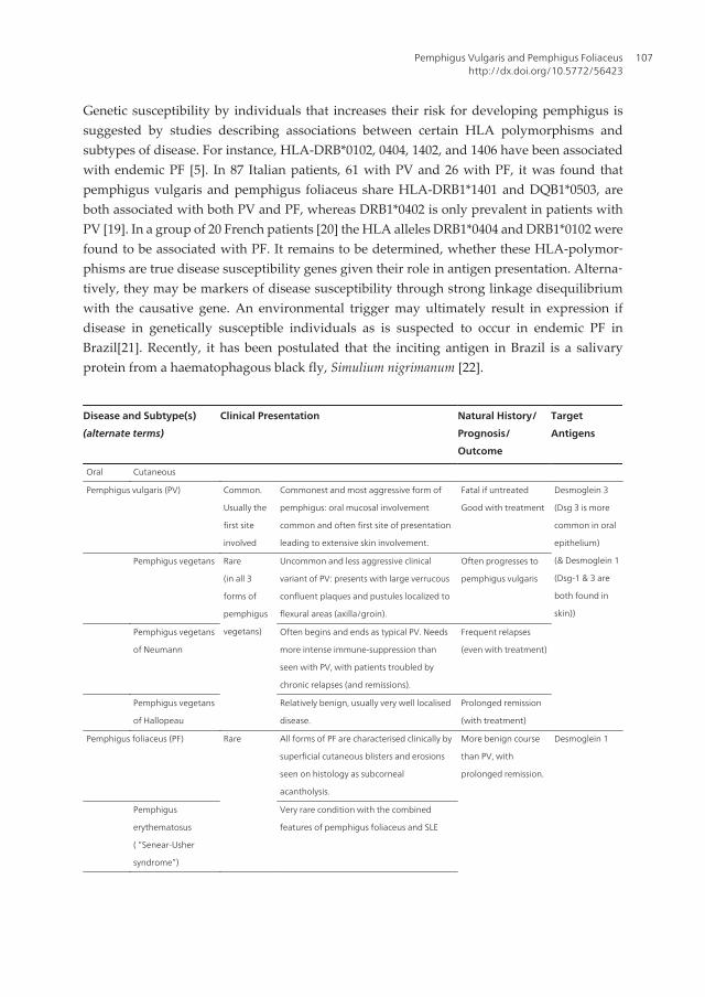
Genetic susceptibility by individuals that increases their risk for developing pemphigus issuggested by studies describing associations between certain HLA polymorphisms andsubtypes of disease. For instance, HLA-DRB*0102, 0404, 1402, and 1406 have been associatedwith endemic PF [5]. In 87 Italian patients, 61 with PV and 26 with PF, it was found thatpemphigus vulgaris and pemphigus foliaceus share HLA-DRB1*1401 and DQB1*0503, areboth associated with both PV and PF, whereas DRB1*0402 is only prevalent in patients withPV [19]. In a group of 20 French patients [20] the HLA alleles DRB1*0404 and DRB1*0102 werefound to be associated with PF. It remains to be determined, whether these HLA-polymor‐phisms are true disease susceptibility genes given their role in antigen presentation. Alterna‐tively, they may be markers of disease susceptibility through strong linkage disequilibriumwith the causative gene. An environmental trigger may ultimately result in expression ifdisease in genetically susceptible individuals as is suspected to occur in endemic PF inBrazil[21]. Recently, it has been postulated that the inciting antigen in Brazil is a salivaryprotein from a haematophagous black fly, Simulium nigrimanum [22].
Disease and Subtype(s)
(alternate terms)
Clinical Presentation Natural History/
Prognosis/
Outcome
Target
Antigens
Oral Cutaneous
Pemphigus vulgaris (PV) Common.
Usually the
first site
involved
Commonest and most aggressive form of
pemphigus: oral mucosal involvement
common and often first site of presentation
leading to extensive skin involvement.
Fatal if untreated
Good with treatment
Desmoglein 3
(Dsg 3 is more
common in oral
epithelium)
(& Desmoglein 1
(Dsg-1 & 3 are
both found in
skin))
Pemphigus vegetans Rare
(in all 3
forms of
pemphigus
vegetans)
Uncommon and less aggressive clinical
variant of PV: presents with large verrucous
confluent plaques and pustules localized to
flexural areas (axilla/groin).
Often progresses to
pemphigus vulgaris
Pemphigus vegetans
of Neumann
Often begins and ends as typical PV. Needs
more intense immune-suppression than
seen with PV, with patients troubled by
chronic relapses (and remissions).
Frequent relapses
(even with treatment)
Pemphigus vegetans
of Hallopeau
Relatively benign, usually very well localised
disease.
Prolonged remission
(with treatment)
Pemphigus foliaceus (PF) Rare All forms of PF are characterised clinically by
superficial cutaneous blisters and erosions
seen on histology as subcorneal
acantholysis.
More benign course
than PV, with
prolonged remission.
Desmoglein 1
Pemphigus
erythematosus
( “Senear-Usher
syndrome”)
Very rare condition with the combined
features of pemphigus foliaceus and SLE
Pemphigus Vulgaris and Pemphigus Foliaceushttp://dx.doi.org/10.5772/56423
107
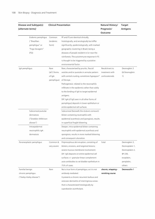
Disease and Subtype(s)
(alternate terms)
Clinical Presentation Natural History/
Prognosis/
Outcome
Target
Antigens
Endemic pemphigus
( “Brazillian
pemphigus” or
“Fogo Sevagem”
(FS))
Common
(endemic
form)
PF and FS are identical clinically,
histologically, and serologically but differ
significantly, epidemiologically, with marked
geographic clustering in Brazil, being a
diseases of people resident in/or near the
rainforests. The autoimmune response in FS
is thought to be triggered by a putative
environmental factor.
IgA pemphigus Rare
(all 3 forms
of IgA
pemphigus)
Rare, characterised by pruritic, flaccid
vesicles and/or pustules in annular pattern
with central crusting, sometimes hypopyon*
of the eye.
Pathogenesis: related to the neutrophilic
infiltrate in the epidermis rather than solely
to the binding of IgA to target epidermal
antigens.
DIF: IgA (cf IgG seen in all other forms of
pemphigus) deposits in lower epithelium or
entire epidermal cell surfaces
Recalcitrant to
treatment with
corticosteroids
Desmoglein 3
(& Desmoglein
1)
Subcorneal pustular
dermatosis
(“Sneddon-Wilkinson
disease”)
Subcorneal (beneath the stratum corneum)
blister containing neutrophils with
epidermal acanthosis and spongiosis, results
in superficial fragile blistering.
Intraepidermal
neutrophilic IgA
dermatosis
Deeper, intra-epidermal blister containing
neutrophils with epidermal acanthosis and
spongiosis, results in more marked blistering
and consequent ulceration.
Paraneoplastic pemphigus Common &
very severe
Polymorphous skin eruption, consisting of
blisters, erosions, and targetoid lesions;
severe mucous membrane involvement.
DIF: IgG deposits on entire epidermal cell
surfaces +/- granular-linear complement
auto-antibodies to rat bladder epithelium in
75% of cases.
Fatal Desmoglein 3,
Desmoplakin 1,
Desmoplakin 2,
BP 230,
evoplakin,
periplakin,
others
Familial benign
chronic pemphigus
(“Hailey-Hailey disease”)
Rare Not a true form of pemphigus, as it is not
antibody mediated.
It presents a chronic recurrent bullous and
vesicular dermatitis of intertriginous areas
that is characterized histologically by
suprabasilar acantholysis.
chronic, relapsing–
remitting course
Desmocollin 1chronic, relapsing–
remitting course
Desmocollin 1chronic, relapsing–
remitting course
Desmocollin 1
Skin Biopsy - Diagnosis and Treatment108
Disease and Subtype(s)
(alternate terms)
Clinical Presentation Natural History/
Prognosis/
Outcome
Target
Antigens
Pathogenesis: heterozygous mutations of
the ATP2C1 gene leads to a malfunction of
the encoded protein hPMR1 - hPMR1 being
a high-affinity calcium transport ATPase
pump of the Golgi complex. A low level of
intracellular Ca2+ induces premature
keratinocyte proliferation, which leads to
dysfunctional desmosomal proteins and
thus abnormal keratinocyte adhesion.
PV = pemphigus vulgaris; PF = pemphigus foliaceous, SLE = systemic lupus erythematosus, FS = Fogo Sevagem, DIF =direct immune-fluorescence,
cf = in contrast, BP = bullous pemphigoid, AD = autosomal dominant inheritance, Ca2+ = calcium ion
*hypopyon= sterile leukocytic exudate, seen in the anterior chamber of the eye
Table 1. Clinical and Immunohistochemical Variants of Pemphigus
4. Clinical features
4.1. Pemphigus vulgaris
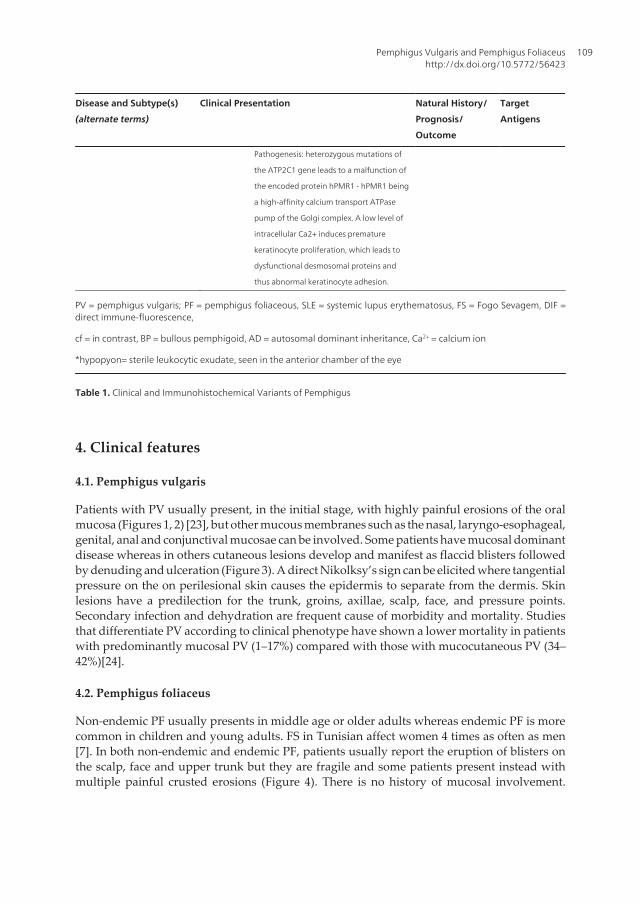
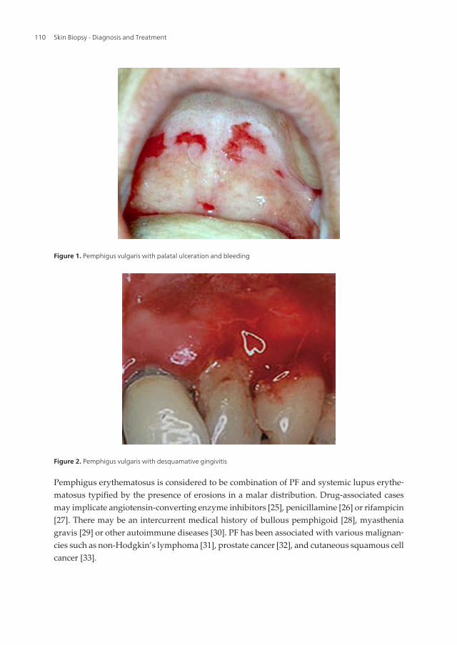
Patients with PV usually present, in the initial stage, with highly painful erosions of the oralmucosa (Figures 1, 2) [23], but other mucous membranes such as the nasal, laryngo-esophageal,genital, anal and conjunctival mucosae can be involved. Some patients have mucosal dominantdisease whereas in others cutaneous lesions develop and manifest as flaccid blisters followedby denuding and ulceration (Figure 3). A direct Nikolksy’s sign can be elicited where tangentialpressure on the on perilesional skin causes the epidermis to separate from the dermis. Skinlesions have a predilection for the trunk, groins, axillae, scalp, face, and pressure points.Secondary infection and dehydration are frequent cause of morbidity and mortality. Studiesthat differentiate PV according to clinical phenotype have shown a lower mortality in patientswith predominantly mucosal PV (1–17%) compared with those with mucocutaneous PV (34–42%)[24].
4.2. Pemphigus foliaceus
Non-endemic PF usually presents in middle age or older adults whereas endemic PF is morecommon in children and young adults. FS in Tunisian affect women 4 times as often as men[7]. In both non-endemic and endemic PF, patients usually report the eruption of blisters onthe scalp, face and upper trunk but they are fragile and some patients present instead withmultiple painful crusted erosions (Figure 4). There is no history of mucosal involvement.
Pemphigus Vulgaris and Pemphigus Foliaceushttp://dx.doi.org/10.5772/56423
109
Pemphigus erythematosus is considered to be combination of PF and systemic lupus erythe‐matosus typified by the presence of erosions in a malar distribution. Drug-associated casesmay implicate angiotensin-converting enzyme inhibitors [25], penicillamine [26] or rifampicin[27]. There may be an intercurrent medical history of bullous pemphigoid [28], myastheniagravis [29] or other autoimmune diseases [30]. PF has been associated with various malignan‐cies such as non-Hodgkin’s lymphoma [31], prostate cancer [32], and cutaneous squamous cellcancer [33].
Figure 1. Pemphigus vulgaris with palatal ulceration and bleeding
Figure 2. Pemphigus vulgaris with desquamative gingivitis
Skin Biopsy - Diagnosis and Treatment110
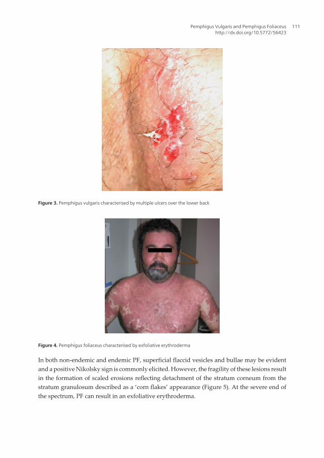
Figure 4. Pemphigus foliaceus characterised by exfoliative erythroderma
In both non-endemic and endemic PF, superficial flaccid vesicles and bullae may be evidentand a positive Nikolsky sign is commonly elicited. However, the fragility of these lesions resultin the formation of scaled erosions reflecting detachment of the stratum corneum from thestratum granulosum described as a ‘corn flakes’ appearance (Figure 5). At the severe end ofthe spectrum, PF can result in an exfoliative erythroderma.
Figure 3. Pemphigus vulgaris characterised by multiple ulcers over the lower back
Pemphigus Vulgaris and Pemphigus Foliaceushttp://dx.doi.org/10.5772/56423
111
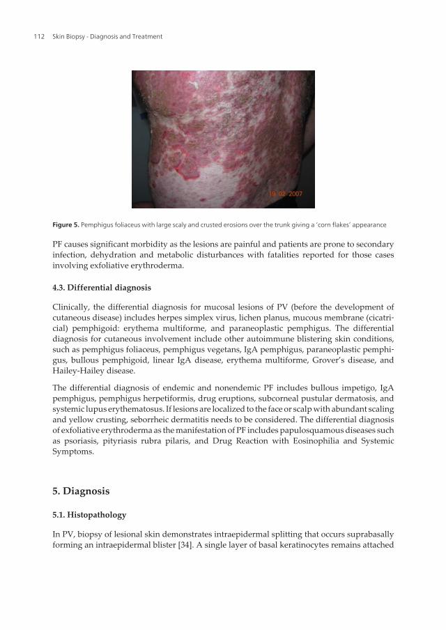
Figure 5. Pemphigus foliaceus with large scaly and crusted erosions over the trunk giving a ‘corn flakes’ appearance
PF causes significant morbidity as the lesions are painful and patients are prone to secondaryinfection, dehydration and metabolic disturbances with fatalities reported for those casesinvolving exfoliative erythroderma.
4.3. Differential diagnosis
Clinically, the differential diagnosis for mucosal lesions of PV (before the development ofcutaneous disease) includes herpes simplex virus, lichen planus, mucous membrane (cicatri‐cial) pemphigoid: erythema multiforme, and paraneoplastic pemphigus. The differentialdiagnosis for cutaneous involvement include other autoimmune blistering skin conditions,such as pemphigus foliaceus, pemphigus vegetans, IgA pemphigus, paraneoplastic pemphi‐gus, bullous pemphigoid, linear IgA disease, erythema multiforme, Grover’s disease, andHailey-Hailey disease.
The differential diagnosis of endemic and nonendemic PF includes bullous impetigo, IgApemphigus, pemphigus herpetiformis, drug eruptions, subcorneal pustular dermatosis, andsystemic lupus erythematosus. If lesions are localized to the face or scalp with abundant scalingand yellow crusting, seborrheic dermatitis needs to be considered. The differential diagnosisof exfoliative erythroderma as the manifestation of PF includes papulosquamous diseases suchas psoriasis, pityriasis rubra pilaris, and Drug Reaction with Eosinophilia and SystemicSymptoms.
5. Diagnosis
5.1. Histopathology
In PV, biopsy of lesional skin demonstrates intraepidermal splitting that occurs suprabasallyforming an intraepidermal blister [34]. A single layer of basal keratinocytes remains attached
Skin Biopsy - Diagnosis and Treatment112
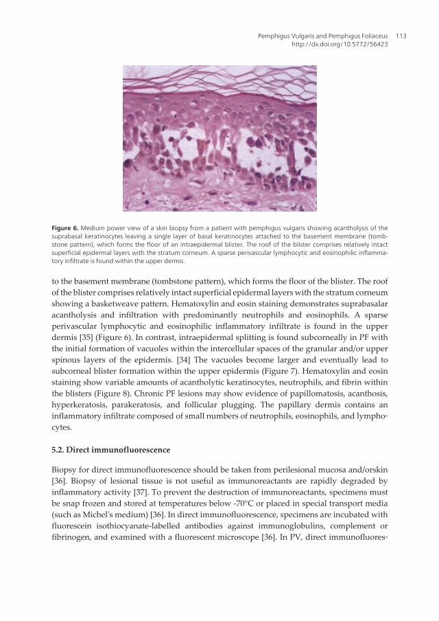
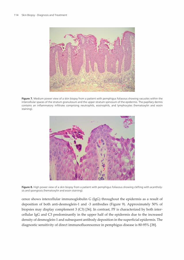
to the basement membrane (tombstone pattern), which forms the floor of the blister. The roofof the blister comprises relatively intact superficial epidermal layers with the stratum corneumshowing a basketweave pattern. Hematoxylin and eosin staining demonstrates suprabasalaracantholysis and infiltration with predominantly neutrophils and eosinophils. A sparseperivascular lymphocytic and eosinophilic inflammatory infiltrate is found in the upperdermis [35] (Figure 6). In contrast, intraepidermal splitting is found subcorneally in PF withthe initial formation of vacuoles within the intercellular spaces of the granular and/or upperspinous layers of the epidermis. [34] The vacuoles become larger and eventually lead tosubcorneal blister formation within the upper epidermis (Figure 7). Hematoxylin and eosinstaining show variable amounts of acantholytic keratinocytes, neutrophils, and fibrin withinthe blisters (Figure 8). Chronic PF lesions may show evidence of papillomatosis, acanthosis,hyperkeratosis, parakeratosis, and follicular plugging. The papillary dermis contains aninflammatory infiltrate composed of small numbers of neutrophils, eosinophils, and lympho‐cytes.
5.2. Direct immunofluorescence
Biopsy for direct immunofluorescence should be taken from perilesional mucosa and/orskin[36]. Biopsy of lesional tissue is not useful as immunoreactants are rapidly degraded byinflammatory activity [37]. To prevent the destruction of immunoreactants, specimens mustbe snap frozen and stored at temperatures below -70°C or placed in special transport media(such as Michel's medium) [36]. In direct immunofluorescence, specimens are incubated withfluorescein isothiocyanate-labelled antibodies against immunoglobulins, complement orfibrinogen, and examined with a fluorescent microscope [36]. In PV, direct immunofluores‐
Figure 6. Medium power view of a skin biopsy from a patient with pemphigus vulgaris showing acantholysis of thesuprabasal keratinocytes leaving a single layer of basal keratinocytes attached to the basement membrane (tomb‐stone pattern), which forms the floor of an intraepidermal blister. The roof of the blister comprises relatively intactsuperficial epidermal layers with the stratum corneum. A sparse perivascular lymphocytic and eosinophilic inflamma‐tory infiltrate is found within the upper dermis.
Pemphigus Vulgaris and Pemphigus Foliaceushttp://dx.doi.org/10.5772/56423
113
cence shows intercellular immunoglobulin G (IgG) throughout the epidermis as a result ofdeposition of both anti-desmoglein-1 and -3 antibodies (Figure 9). Approximately 50% ofbiopsies may display complement 3 (C3) [36]. In contrast, PF is characterized by both inter‐cellular IgG and C3 predominantly in the upper half of the epidermis due to the increaseddensity of desmoglein-1 and subsequent antibody deposition in the superficial epidermis. Thediagnostic sensitivity of direct immunofluorescence in pemphigus disease is 80-95% [38].
Figure 7. Medium power view of a skin biopsy from a patient with pemphigus foliaceus showing vacuoles within theintercellular spaces of the stratum granulosum and the upper stratum spinosum of the epidermis. The papillary dermiscontains an inflammatory infiltrate comprising neutrophils, eosinophils, and lymphocytes (hematoxylin and eosinstaining).
Figure 8. High power view of a skin biopsy from a patient with pemphigus foliaceus showing clefting with acantholy‐sis and spongiosis (hematoxylin and eosin staining).
Skin Biopsy - Diagnosis and Treatment114
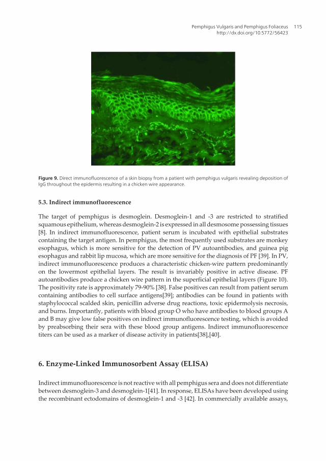
Figure 9. Direct immunofluorescence of a skin biopsy from a patient with pemphigus vulgaris revealing deposition ofIgG throughout the epidermis resulting in a chicken wire appearance.
5.3. Indirect immunofluorescence
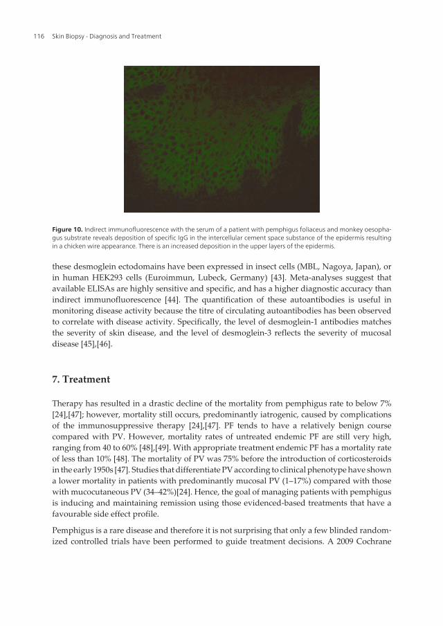
The target of pemphigus is desmoglein. Desmoglein-1 and -3 are restricted to stratifiedsquamous epithelium, whereas desmoglein-2 is expressed in all desmosome possessing tissues[8]. In indirect immunofluorescence, patient serum is incubated with epithelial substratescontaining the target antigen. In pemphigus, the most frequently used substrates are monkeyesophagus, which is more sensitive for the detection of PV autoantibodies, and guinea pigesophagus and rabbit lip mucosa, which are more sensitive for the diagnosis of PF [39]. In PV,indirect immunofluorescence produces a characteristic chicken-wire pattern predominantlyon the lowermost epithelial layers. The result is invariably positive in active disease. PFautoantibodies produce a chicken wire pattern in the superficial epithelial layers (Figure 10).The positivity rate is approximately 79-90% [38]. False positives can result from patient serumcontaining antibodies to cell surface antigens[39]; antibodies can be found in patients withstaphylococcal scalded skin, penicillin adverse drug reactions, toxic epidermolysis necrosis,and burns. Importantly, patients with blood group O who have antibodies to blood groups Aand B may give low false positives on indirect immunofluorescence testing, which is avoidedby preabsorbing their sera with these blood group antigens. Indirect immunofluorescencetiters can be used as a marker of disease activity in patients[38],[40].
6. Enzyme-Linked Immunosorbent Assay (ELISA)
Indirect immunofluorescence is not reactive with all pemphigus sera and does not differentiatebetween desmoglein-3 and desmoglein-1[41]. In response, ELISAs have been developed usingthe recombinant ectodomains of desmoglein-1 and -3 [42]. In commercially available assays,
Pemphigus Vulgaris and Pemphigus Foliaceushttp://dx.doi.org/10.5772/56423
115
these desmoglein ectodomains have been expressed in insect cells (MBL, Nagoya, Japan), orin human HEK293 cells (Euroimmun, Lubeck, Germany) [43]. Meta-analyses suggest thatavailable ELISAs are highly sensitive and specific, and has a higher diagnostic accuracy thanindirect immunofluorescence [44]. The quantification of these autoantibodies is useful inmonitoring disease activity because the titre of circulating autoantibodies has been observedto correlate with disease activity. Specifically, the level of desmoglein-1 antibodies matchesthe severity of skin disease, and the level of desmoglein-3 reflects the severity of mucosaldisease [45],[46].
7. Treatment
Therapy has resulted in a drastic decline of the mortality from pemphigus rate to below 7%[24],[47]; however, mortality still occurs, predominantly iatrogenic, caused by complicationsof the immunosuppressive therapy [24],[47]. PF tends to have a relatively benign coursecompared with PV. However, mortality rates of untreated endemic PF are still very high,ranging from 40 to 60% [48],[49]. With appropriate treatment endemic PF has a mortality rateof less than 10% [48]. The mortality of PV was 75% before the introduction of corticosteroidsin the early 1950s [47]. Studies that differentiate PV according to clinical phenotype have showna lower mortality in patients with predominantly mucosal PV (1–17%) compared with thosewith mucocutaneous PV (34–42%)[24]. Hence, the goal of managing patients with pemphigusis inducing and maintaining remission using those evidenced-based treatments that have afavourable side effect profile.
Pemphigus is a rare disease and therefore it is not surprising that only a few blinded random‐ized controlled trials have been performed to guide treatment decisions. A 2009 Cochrane
Figure 10. Indirect immunofluorescence with the serum of a patient with pemphigus foliaceus and monkey oesopha‐gus substrate reveals deposition of specific IgG in the intercellular cement space substance of the epidermis resultingin a chicken wire appearance. There is an increased deposition in the upper layers of the epidermis.
Skin Biopsy - Diagnosis and Treatment116
review [50] assessed interventions for PV and PF and concluded that there is inadequateinformation available to ascertain optimal therapy for pemphigus. They ascertained that thequality of most studies was not high and the majority examine patients with newly diagnosedor active disease. Another consideration in the evaluation of data is lack of generally accepteddefinitions and measurements for the clinical evaluation of patients with pemphigus and thedefinitions of disease control and remission. A recent consensus statement has been releasedto assist with future trials enabling improved comparisons to be made [51].
As high-dose systemic corticosteroids, followed by alternate immune-suppressive agents,serves as the mainstay of initial therapy for PV, there is the need to exclude underlying latentinfectious diseases that can be reactivated by the corticosteroids (e.g.: HIV, Hepatitis B and C,and tuberculosis). In addition, screening for the diseases initiated or exacerbated by high-dose‘steroids, such as hypertension, diabetes mellitus and osteoporosis is prudent.
8. Systemic corticosteroids
Systemic corticosteroids are currently the mainstay of treatment as they have a rapid onset ofaction and are effective in controlling disease and improving prognosis [52]. Howeversignificant side effects such as diabetes, osteoporosis, adrenal suppression, peptic ulceration,weight gain, increased susceptibility to infection, mood changes, proximal myopathy,Cushing’s syndrome, and cataracts limit their usefulness. Adjuvant treatments have thereforebeen introduced as steroid sparing agents. Various corticosteroid regimens are used to treatpemphigus, the most common of which is a gradual reduction of an oral formulation [47]. Innewly diagnosed patients, an initial daily dose of 0.5 mg/kg of prednisone/prednisolone, (orequivalent) appears preferable to 1 mg/kg. A randomized trial compared these two regimensin 19 patients with PV and 3 with PF followed for 5 years, with remission defined as less than15 mg of corticosteroids per day to maintain disease control [53]. No difference was observedin remission or the incidence of complications between the high and low dose regimens. In arandomized trial that included only patients with newly diagnosed PV, pulsed oral dexame‐thasone provided no additional benefit to the combination of oral prednisone and azathioprinewith remission defined as cessation of systemic treatment [54]. Furthermore, there were anincreased number of adverse events in those participants receiving pulsed dexamethasone.However, the possible benefit of high dose pulsed intravenous corticosteroids in achievingdisease control and maintaining remission was suggested in a small case-controlled retro‐spective study of patients with PV initially unresponsive to low dose of prednisone (less than40 mg daily) [55] and an open study of new diagnosed PV patients [56]. There have no studiesto date examining the effects of high dose pulsed intravenous corticosteroids in PF.
Currently no optimal regimen for corticosteroid therapy has been defined for the treatment ofpemphigus despite its proven benefits. Hence in routine practice, a tailored regime is recom‐mended. A starting dose of prednisolone 0.5 mg/kg daily is prudent that may need to beincreased, until no new blister formation is observed. Such higher doses of corticosteroidsincluding pulsed therapy may be warranted in newly diagnosed severe disease and recalci‐trant disease but this remains to be substantiated in randomized studies.
Pemphigus Vulgaris and Pemphigus Foliaceushttp://dx.doi.org/10.5772/56423
117
9. Adjuvant treatment of pemphigus
Immunosuppressive therapy for pemphigus includes azathioprine, mycophenolate mofetil,methotrexate, cyclophosphamide, cyclosporin and dapsone. Adjuvant agents with immuno‐modulatory activity that have also been used in pemphigus include calcineurin inhibitors,epidermal derived growth factor and tetracycline antibiotics. The main role of these immu‐nosuppressive medications is to function as a steroid-sparing agent. As they generally have aslow mode of onset, approximately 4-6 weeks, they are used in maintenance therapy ratherthan in the initiation of disease control. However, their role needs to be further elucidated asthere has been only limited number of randomized controlled trials with most of the literaturederived from case series reports.
9.1. Azathioprine
Azathioprine is a purine antimetabolite that is cleaved to 6-mercaptopurine, which in turn isconverted to additional metabolites that inhibit de novo purine synthesis. Cell proliferation isinhibited and as a consequence a variety of lymphocyte functions are impaired. Azathioprineis commonly used to treat pemphigus; a survey of dermatologists in 2003 showed it was themost commonly prescribed adjuvant agent used to treat PV [57]. It has even been used asmonotherapy in mild cases [58]. In a recent randomized trial conducted by Chams-Davatchiet al, 120 new patients with PV were treated for over one year with one of four regimens. Theseregimes were prednisolone alone, prednisolone plus azathioprine, prednisolone plus intrave‐nous cyclophosphamide and prednisolone plus mycophenolate mofetil [59]. Azathioprinereduced the cumulative dose of prednisolone compared with prednisolone alone howeverremission rates were similar. Side effects were similar between the two groups. Thus in thistrial, which included only patients with PV, azathioprine reduced the cumulative corticoste‐roid dose but not the rate of remission. More recently, a non-randomized study compared highdose oral prednisone daily (1.5 mg/kg/daily) versus low dose oral prednisone (40 mg onalternate days) plus azathioprine (100 mg/daily) in 36 patients with oral PV [60]. Both treat‐ments resulted in high rates of clinical remission; the monotherapy group showed a reducedmean time to remission but this group was associated with an increased rate of treatment-associated adverse events Other non-randomized trials using azathioprine are have generallyshown favourable outcomes in PV [61],[62].
9.2. Mycophenolate mofetil
Mycophenolate mofetil is a prodrug and its active drug, mycophenolic acid, inhibits inosinemonophosphate dehydrogenase, an important enzyme in guanine nucleotide synthesis.Lymphocytes are highly dependent on this pathway and are selectively inhibited by myco‐phenolate mofetil [63]. In the randomized trial by Chams-Davatchi et al, no difference inremission was observed for mycophenolate mofetil when compared to prednisolone alone[59]. In this same study no difference in remission was demonstrated between azathioprineand mycophenolate mofetil. The steroid sparing effect of mycophenolate mofetil was inferiorto azathioprine. Beissert et al. compared oral methylprednisolone plus azathioprine with
Skin Biopsy - Diagnosis and Treatment118
mycophenolate mofetil in 33 patients with PV and 7 patients with PF [64]. The primary outcomewas complete healing of all lesions. This study concluded that mycophenolate mofetil andazathioprine demonstrated similar efficacy. Safety profiles were similar as was corticosteroid-sparing effects. There were less severe side effects observed in the mycophenolate group butthis was not statistically significant. Many non-randomized trials have supported the use ofmycophenolate mofetil in the treatment of pemphigus [65]-[69]. The majority of patients inthese trials had PV. Mycophenolate mofetil is generally well tolerated; lymphopenia, gastro‐intestinal symptoms and infections are the most common side effects. Currently it is a relativelyexpensive medication, often precluding its off-label use. The drug is usually commenced atdose of 1 g per day in adult patients, and if required, increased in 500-mg increments up todoses of 2-3 g per day [70].
9.3. Cyclophosphamide
Cyclophosphamide is an alkylating agent that disturbs DNA synthesis and cell division.Cyclophosphamide interferes with DNA integrity and function inducing cell death in rapidlyproliferating tissues including lymphocytes. This provides the basis for their therapeutic andtoxic properties. Several randomized trials have assessed cyclophosphamide in treatment ofpemphigus. Chrysomallis et al. used oral cyclophosphamide in patients with PV whose diseasewas limited to oral involvement [71]. Twenty-eight patients were divided into 3 groups andgiven corticosteroids alone, corticosteroids with cyclophosphamide or cyclosporine. Nodifference in remission was seen when cyclophosphamide was compared with corticosteroidsalone, and at 5 years, all patients had their disease controlled with a low dose corticosteroidregimen. The more recent study by Chams-Davatchi et al. described above and comprisingentirely of patients with PV concluded that there was no difference in remission rates followingpulsed intravenous cyclophosphamide therapy [59]. A randomized control trial that included6 patients with PF as well as 16 with PV compared pulsed cyclophosphamide with dexame‐thasone and daily cyclophosphamide with methylprednisolone plus azathioprine [72]. Nodifference in disease control was observed for the cyclophosphamide group compared to theazathioprine group after 2 years. Several non-randomized case series have utilized pulsecyclophosphamide with variable outcomes [73]-[75].
Given the lack of superiority of cyclophosphamide over other regimes in randomized trialsand its well-described serious side effect profile, the authors recommend that its use berestricted for the treatment of severe or refractory cases of PF, where alternative agents suchas rituximab or IVIg are not available.
9.4. Cyclosporin
Cyclosporin is a calcineurin inhibitor that prevents dephosphorylation of nuclear factor ofactivated T cells (NFAT) preventing its translocation into nucleus and as a consequence the Tcells fail to respond to specific antigenic stimulation. Cyclosporin also increases expression ofTGF- beta, a potent inhibitor of IL-2–stimulated T-cell proliferation. A randomized trialcompared oral methylprednisolone alone with oral methylprednisolone plus cyclosporin in33 newly diagnosed patients, 29 with PV and 4 with PF [76]. The patients were followed for
Pemphigus Vulgaris and Pemphigus Foliaceushttp://dx.doi.org/10.5772/56423
119
4-6 years and the investigators concluded that the combination regimen of corticosteroids andcyclosporin provided no additional benefit over corticosteroids alone. Side effects, includinghypertrichosis, hypertension and renal dysfunction, were more common in the cyclosporingroup. The randomized controlled study by Chrysomallis et al. in newly diagnosed PV limitedto oral involvement found no difference in remission or relapse rates between the cyclophos‐phamide and cyclosporine (5 mg/kg) groups [71]. Again, adverse events were more commonin the cyclosporin group and included hypertrichosis and renal impairment. A case seriesreported successful the successful treatment of 6 patients with recalcitrant PV [77].
The randomized trials have not supported the use of cyclosporine at a dose of 5 mg/kg for thetreatment of new onset pemphigus and side effects are relatively common. Further random‐ized controlled trials are required to determine its benefit in recalcitrant disease.
9.5. Dapsone
Dapsone has anti-inflammatory and antimicrobial actions. Its immunomodulatory action isincompletely understood but several actions have been described including prevention of therespiratory burst from myeloperoxidase, suppression of neutrophil migration by blockingintegrin-mediated adherence, inhibition of adherence of antibodies to neutrophils, andreduction of eicosanoid release. Most studies utilising dapsone have been performed inpatients with PV. A randomized controlled trial performed in 19 patients, all with PV,compared dapsone with placebo [78]. Patients were in the maintenance phase after glucocor‐ticoids and/or cytotoxic agents (azathioprine, mycophenolate or methotrexate) were used toachieve remission. Doses of dapsone were increased to 150 mg per day and then to a further200 mg per day if tolerated. The trial was performed over 1 year, and the main outcomemeasured was reduction of prednisolone to doses of 7.5mg/day or less. Five of the 9 patientsin the placebo group achieved the main outcome compared with 3 out of 10 in the placebogroup. The difference was not statistically significant although there was a trend favouringdapsone as a steroid sparing agent. A retrospective study in 9 patients with PV suggested thatdapsone reduced steroid dependence in these patients [79]. Another study reported improve‐ment in 5 of 9 cases of superficial pemphigus treated with dapsone [80]. A recent meta-analysiscomprising 55 patients with pemphigus revealed that 32 patients with PV and 14 patients withPF responded to dapsone [81].
The side effects of dapsone observed in these studies include methaemoglobinaemia, haemol‐ysis and agranulocytosis. [81],[82] Patients should be tested for glucose-6-phosphate dehy‐drogenase deficiency prior to commencing this agent. Dapsone at best may have a role as asteroid sparing agent in the maintaining remission in pemphigus but cannot be recommendedin treatment of acute disease.
9.6. Methotrexate
The antimetabolite methotrexate is a folic acid analogue that competitively inhibits dihydro‐folate reductase and has multiple immunosuppressive actions including suppressing lym‐phocytes in the skin [83],[84]. A small number of non-randomized trials have investigated the
Skin Biopsy - Diagnosis and Treatment120
role of methotrexate in treatment of pemphigus. The most recent study [83] treated 9 patientswith chronic active PV, who were unable to successfully wean their prednisolone dose. Theywere treated with a mean dose of 12.5 mg per week of methotrexate. Prednisolone wasdiscontinued in 6 of the 9 patients within 6 months of commencing methotrexate. There wereminimal adverse effects reported in the study. A recent review of the English literaturerevealed that 111 (82%) of 136 pemphigus patients responded to methotrexate [85]. However,meaningful conclusions are limited by the lack of randomized trials, varying doses andschedules of treatment, and insufficient information on clinical progress including the lack ofconsistency of the length of follow up.
Thus methotrexate may be useful as a steroid sparing agent but further trials are requiredbefore recommending methotrexate as an initial steroid-sparing agent.
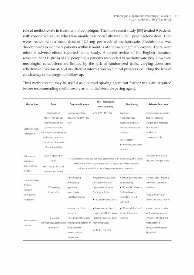
Medication Dose Contra-indicationsPre-Therapeutic
InvestigationsMonitoring Adverse Reactions
Corticosteroids
(“steroids”)
prednisolone:
(1) ½-1 mg per kg/
bodyweight of the
patient for 4 days
(or longer in pemphigus)
with rapid taper until
clinical remission occurs
(2) < 7.5 mg/daily
∙ Active infectious
diseases: Tb, HIV, HBV,
HCV
∙ Tb, HIV, HBV, HCV Systemic:
∙ hypertension,
psychosis, diabetes
mellitus, weight gain,
cataracts
hypertension, psychosis,
diabetes mellitus,
weight gain, cataracts
Oral Mucosa:
candidiasis,
mucosal atrophyOral Mucosa:
∙ candidiasis, mucosal
atrophy
Calcineurin-
Inhibitors:
pimecrolimus
(Elidel®)
Topical Application
Only:A causal relationship has not been established with malignancy. Skin cancer
and lymphoma have been reported in patients treated with topical
calcineurin inhibitors, including pimecrolimus 1% cream.
irritation, pruritis and
erythema on application
thin layer to affected
mucosa twice daily
Lysosomotropic
Amines:
hydroxyl-
chloroquine
(Plaquenil®)
200-400 mg
(once daily)
∙ pre-existing
retinopathy
∙ psoriasis
∙ porphyria
∙ G6PD deficiency
∙ baseline visual acuity
testing (for macular
degeneration) by an
Ophthalmologist
∙ annual baseline visual
acuity testing
∙ FBC and LFT's weekly
for first 4 weeks;
thereafter, only if
indicated
UV (sun) light–initiated
lichenoid cutaneous
reactions
∙ FBC; G6DP levels; LFT’s
Note: slow onset of
action of up to 3 months
azathioprine
(Imuran®)
1.0-2.0 mg
per kg bodyweight/daily
(once daily)
∙ recent use of live
vaccines
∙ pregnancy (Category
D) (including partner of
male patient)
∙ concomitant
allopurinol
∙ thiopurine methyl-
transferase (TPMT) assay
(determines risk of bone
marrow aplasia)
∙ FBC weekly for first 8
weeks; thereafter
monthly
severe adverse reaction
with xanthine oxidase
inhibitors of which the
most potent is
allopurinol (Progout®
Zyloprim®)∙ FBC; LFT’s; E/U/C
Pemphigus Vulgaris and Pemphigus Foliaceushttp://dx.doi.org/10.5772/56423
121

mycophenolate
(CellCept®,
Myfortic®)
max 2 g/day
(once daily or
divided dose)
∙ pregnancy (Category
D)
∙ HIV, HBV, HCV ∙ FBC and LFT’s weekly
for first 4 weeks;
thereafter, only if
indicated
malignancy risk eg skin
cancer, lymphoma;
infection; progressive
multifocal leuco-
encephalopathy; bone
marrow depression
dapsone
(Dapsone®)
maintenance dose:
50-100 mg daily
(≥ 300 mg daily
∙ FBC, G6DP levels, LFT’s
∙ HIV, HBV, HCV
∙ FBC, LFT’s weekly for
the first month, monthly
for six months and semi-
annually thereafter
dose related haemolysis,
especially in G6DP
deficient patients;
agranulocytosis; toxic
hepatitis and cholestatic
jaundice
methotrexate
(Methoblastin®)
10 to 25 mg/ ∙ pregnancy (Category
D)
∙ liver/renal
impairment
∙ HIV, HBV, HCV
∙ immune-deficiency;
∙ concomitant
retinoinds
FBC, LFT’s, E/U/C
HIV, HBV, HCV
∙ FBC weekly for first 8
weeks, thereafter
monthly
hepato/ nephrotoxicity;
ulcerative stomatitis;
bone marrow
depression; immune-
suppression
WEEKLY ONLY
until adequate response
is achieved
Rituximab
(Mabthera®)
375 mg/m2 (body
surface area) once
weekly
∙ Murine(mouse)
protein hypersensitivity
∙ Tb, HIV, HBV, HCV ∙ infection progressive multifocal
leucoencephalopathy
(PML)
for 4 weeks
or 2x 1.0g -500 mg ivi
infusions over two
weeks
(A)
Medication Actions
corticosteroids
(“ ‘steroids)
Profound, generalised inhibitory effects on inflammatory processes and cells achieved by the control of protein synthesis by
reacting with certain corticosteroid responsive genes of sensitive cells in many tissues:
⇩ production of acute inflammatory mediators, especially eicosonoids, postaglndins and leukotrienes. (corticosteroids increase
the production of a polypeptide – lipocortin, that in turn inhibits phospholipase A2 the enzyme responsible for the mobilising
arachniodonic acid from cell membranes)
⇩ production and numbers of circulating immune-competent cells: e.g.: neutrophils, macrophages, T and B lymphocytes
⇩ Complement activation
⇩ activity of macrophages and fibroblasts involved in the chronic stages of inflammation – leading to decreased inflammation
and healing
cyclosporine
tacrolimus
Calcineurin inhibitors:
Skin Biopsy - Diagnosis and Treatment122
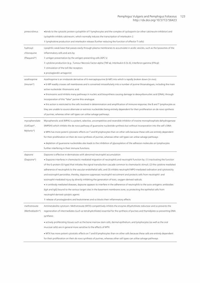
pimecrolimus ∙binds to the cytosolic protein cyclophilin of T-lymphocytes and the complex of cyclosporin (or other calcineurin-inhibitor) and
cyclophilin inhibits calcineurin, which normally induces the transcription of interleukin-2.
⇩ lymphokine production and interleukin release (further reducing the function of effector T-cells)
hydroxyl-
chloroquine
(Plaquenil®)
Lipophilic weak base that passes easily through plasma membranes to accumulate in acidic vesicles, such as the lysosomes of the
inflammatory cells and acts by:
⇩ antigen presentation by the antigen-presenting cells (APC’s)
⇩ cytokine production (e.g.: Tumour Necrosis Factor-alpha (TNF-α), Interleukin-6 (IL-6), Interferon-gamma (IFN-γ))
⇩ stimulation of the toll-like receptors
∙ prostaglandin antagonist
azathioprine
(Imuran®)
Azathioprine is an imidazole derivative of 6-mercaptopurine (6-MP) into which is rapidly broken down (in vivo).
∙ 6-MP readily crosses cell membranes and is converted intracellularly into a number of purine thioanalogues, including the main
active nucleotide: thioinosinic acid.
∙ thioinosinic acid inhibits many pathways in nucleic acid biosynthesis causing damage to deoxyribonucleic acid (DNA), through
incorporation of this “false” purine thio-analogue.
∙ this action is restricted to the cells involved in determination and amplification of immune response; the B and T lymphocytes as
they are unable to source alternate or extrinsic nucleotides being entirely dependent for their proliferation on de novo synthesis
of purines, whereas other cell types can utilise salvage pathways.
mycophenolate
(CellCept®,
Myfortic®)
Mycophenolic acid (MPA) is a potent, selective, uncompetitive and reversible inhibitor of inosine monophosphate dehydrogenase
(IMPDH) which inhibits the de novo pathway of guanosine nucleotide synthesis but without incorporation into the cell’s DNA:
∙ MPA has more potent cytostatic effects on T and B lymphocytes than on other cells because these cells are entirely dependent
for their proliferation on their de novo synthesis of purines, whereas other cell types can utilise salvage pathways
∙ depletion of guanosine nucleotides also leads to the inhibition of glycosylation of the adhesion molecules on lymphocytes
further interfering in their immune functions
dapsone
(Dapsone®)
Dapsone is effective in dermatoses with abnormal neutrophil accumulation
∙ Dapsone interferes in chemotactic-mediated migration of neutrophils and neutrophil function by: (1) inactivating the function
of the G-protein (Gi type) that initiates the signal transduction cascade common to chemotactic stimuli; (2) the cytokine mediated
adherence of neutrophils to the vascular endothelial cells; and (3) inhibits neutrophil MPO-mediated iodination and cytotoxicity
and eosinophil peroxidise, thereby, dapsone suppresses neutrophil recruitment and protects cells from neutrophil- and
eosinophil-mediated injury by directly inhibiting the generation of toxic, oxygen-derived radicals.
∙ in antibody mediated diseases, dapsone appears to interfere in the adherence of neutrophils to the auto-antigenic antibodies
(IgA and IgG) bound to the various target sites in the basement membrane zone, so protecting the epithelial cells from
neutrophil-derived cytolytic agents
⇩ release of prostaglandins and leukotrienes and so blocks their inflammatory effects
methotrexate
(Methoblastin®)
Antimetabolite cytotoxin. Methotrexate (MTX) competitively inhibits the enzyme dihydrofolate reductase and so prevents the
regeneration of intermediates (such as tetrahydrofolate) essential for the synthesis of purines and thymidylate so preventing DNA
synthesis:
∙ actively proliferating tissues such as the bone marrow stem cells, dermal epithelium, and lymphocytes (as well as the oral
mucosal cells) are in general more sensitive to the effects of MTX
∙ MTX has more potent cytostatic effects on T and B lymphocytes than on other cells because these cells are entirely dependent
for their proliferation on their de novo synthesis of purines, whereas other cell types can utilise salvage pathways
Pemphigus Vulgaris and Pemphigus Foliaceushttp://dx.doi.org/10.5772/56423
123
Rituximab
(Mabthera®)
Rituximab is a genetically engineered chimeric murine/human monoclonal antibody that binds specifically to the antigen CD20, a
transmembrane molecule located on pre-B and mature B-lymphocytes, only. This non-glycosylated phosphoprotein is found on
both normal (and malignant B cells), but not on haemopoietic stem cells, pro-B cells, normal plasma cells or other normal tissues:
∙ inhibits CD20 which regulates the early steps in the activation process for B-cell cycle initiation and differentiation
∙ the depletion of circulating autoreactive B cells (for up to 12 months) and , presumably specific downregulation of dsg3-specific
CD4(+ve) T-lymphocytes and the associated release of proinflammatory cytokines
mayre-establish immune homeostasis and tolerance
(B)
A: Tb = tuberculosis, HIV = human immune-deficiency virus, HBV = hepatitis B virus, HCV = hepatitis C virus, G6DP = glucose-6-phosphate dehydrogen‐
ase, FBC – full blood count, LFT’s = liver function tests, UV = ultraviolet light, E/U/C = electrolytes/urea/creatinine
B: ⇩ = decreased or reduced
Table 2. (A) Therapeutic Agents Useful in the treatment of Pemphigus, (B) Therapeutic Agents Useful in Treating ofPemphigus
9.7. Gold
Auad [86] performed a blinded placebo-controlled randomized trial on the utility of auranofinin 30 patients with PF. Nearly one third of the patients withdrew from the active treatmentarm due to side effects. In the placebo group, a reduction in the mean corticosteroid steroiddose was evident. Other case series comprising patients with PV and/or PF have describedsimilar findings [87]-[89]. The most recent study published showed that 62% of patients withPV achieved remission or halve their dose of prednisone during a period of 10 years ofintramuscular gold therapy [90]. However, the mean time to halve the dose of prednisone was3 months and 42% developed side effects blood dyscrasia, proteinuria and nephrotic syn‐drome, cutaneous reactions and dizziness. These adverse effects in addition to the relativelylong time to take effect as a steroid-sparing agent preclude its use in favour of other steroid-sparing agents in the treatment of treatment of pemphigus.
9.8. Tetracycline antibiotics and nicotinamide
Tetracycline antibiotics with and without nicotinamide have been combined with otheradjuvant agents as treatment for pemphigus. Several small non-randomized trials have shownvarying results [91]-[93]. Minocycline given at a dose of 100 mg per day allowed the reductionof prednisolone in 6 out of 10 patients with pemphigus [91]. Tetracycline at a dose of 2 g perday for 1 month reducing to 1 g per day for 4 weeks enabled more rapid tapering of cortico‐steroids in 13 patients with PV [93]. However the study by Alspoy found that a combinationof tetracycline (2 g/d) and nicotinamide (1.5 g/d) for 2 months was not an effective alternativeto the classic forms of therapy in 14 patients with pemphigus [92]. Further trials are neededbefore recommending these agents.
Skin Biopsy - Diagnosis and Treatment124
9.9. Topical agents
9.9.1. Epidermal growth factor
A double-blind randomized controlled study investigated the use of epidermal growth factor(EGF) on skin lesions of 20 patients with PV [94]. Topical epidermal growth factor 10 ug/g in0.1% silver sulfadiazine cream was applied to skin lesions daily until lesions had healed andcompared to the effect of applying 0.1% silver sulfadiazine (SSD) cream alone. Topical EGFappeared to hasten lesion healing by a median of 6 days compared to SSD.
9.9.2. Topical corticosteroids (Clobetasol)
A small study comprised of 4 patients with mild PF and 3 with mild PV were treated with thevery potent topical corticosteroid, Clobetasol propionate 0.05%, as a sole agent [95]. The creamwas applied to mucosal lesions and involved skin twice a day for at least 15 days, thenprogressively tapered. Control of disease was defined as healing of lesions was obtained, witha 75% decrease in the number of new lesions per week without the addition of systemictreatment. Disease control was achieved in all 7 patients, with cutaneous remission attainedin 15 days, although mucosal regression occurring more slowly. In 4 patients, remission wasmaintained with topical corticosteroid alone for a mean 19-month follow up. In 3 patients,relapse occurred after 2-11 months, requiring systemic treatment. Another study is in progresscomparing the effect of Clobetosol with placebo in pemphigus vulgaris.
9.9.3. Topical calcineurin inhibitors
Tacrolimus and Pimecrolimus are non-steroidal immunomodulatory macrobactams thatinhibit the enzyme, calcineurin, impairing the production and IL-2 and subsequent T-cellactivation and proliferation [96]. A small number of case reports and case series suggest auseful role for Tacrolimus in mild PV and PF but further randomized controlled trials arerequired to clarify its role in this setting [97]-[99]. A recent double blind study of 11 patientswith PV refractory to azathioprine and corticosteroids showed a marked response to pime‐crolimus 1% by day 15 when compared to placebo using the epithelialization index [100].
10. Novel therapies and strategies
10.1. Biological agents
10.1.1. Rituximab
Rituximab is a chimeric (human/murine) monoclonal antibody directed against CD20, a cellsurface molecule specific to B cells. Although it was initially approved for use in B-cell non-Hodgkin’s lymphoma, a growing number of reports have described the efficacy of rituximabfor B-cell depletion in the treatment of autoimmune diseases [101]. The mode of action ofrituximab in autoimmune diseases includes the removal of precursors of autoantibody-
Pemphigus Vulgaris and Pemphigus Foliaceushttp://dx.doi.org/10.5772/56423
125
producing plasma cells, and impairment of autoantigen presentation to CD4 cells [102]. In PV,Eming et al, show that rituximab not also causes marked reduction of anti-desmoglein 3antibodies but also depletion of desmoglein-specific CD4 cells. In contrast, tetanus toxoid-specific CD4 cells were not affected nor were the overall number of CD4 cells [103]. Tetaustoxoid has been used in as an antigen in the assessment of memory CD4 cell responses [104].Eming et al, speculate that this specific effect of rituximab on autoreactive rather than patho‐gen-specific T cells is that the latter do not require CD20 B cells as antigen-presenting cells. Anumber of case reports and series have demonstrated the benefit on the use of rituximab inover 40 patients with either PV or PF [40],[105]-[115]. The largest case series to date comprising42 patients, 37 with PV and 5 with PF, utilized the rheumatoid arthritis protocol where two 1ginfusions of rituximab are administered 15 days apart [116]. Patients were followed for up to5 years and 36 of the 42 patients achieved a complete response and were able to cease corti‐costeroids within 6 months from induction therapy. Twenty patients experienced relapses withthe time to relapse ranging from 8 to 64 months. Relapses were treated with rituximab (500mg) without corticosteroids resulting in a new complete response. Importantly, no seriousadverse events were observed.
The largest case series to date using the lymphoma protocol where rituximab is administeredweekly (375 mg/m2) for 4 weeks induced remission in 12 of 14 patients with PV and 6 of 7patients with PF within 3 months [117]. These patients had previously not responded to first-line immunosuppressive agents or had contraindications to corticosteroid therapy. Thetreatment was generally well tolerated; however, two cases were complicated by severeinfection, one resulting in death. Similarly, Canchini et al, showed that the administration ofrituximab, 375 mg/m2 once weekly for 4 weeks, induced remission in all 10 patients with PVand both patients with PF [118]. Another study comprising 11 patients with refractory PV,evaluated the effect of combination therapy consisting of 10 infusions of rituximab (375mg/m2) and 6 infusions of intravenous immunoglobulin (IVIg) (2 g/kg body weight) admin‐istered over a 6-month period [119]. Remission was induced in 9 patients for a period of 22 to37 months following treatment and there were no reports of serious adverse events.
More recently, Kasperkiewicz et al reported use of rituximab in combination with immunoad‐sorption, pulsed dexamethasone and azathioprine or mycophenolate mofetil in 23 patients.[120] IA was performed at initially 3- and later 4-week intervals until lesions healed by 90%;1 g rituximab was given at weeks 1 and 3, and intravenous dexamethasone pulses wereadministered at first every 3 weeks and then at increasing intervals in addition to dailyazathioprine or mycophenolate mofetil. All patients demonstrated clinical improvementwithin the first few weeks of therapy accompanies by a concomitant rapid fall in anti-desmo‐glein antibody levels. However, two patients had non -atal severe adverse events; onedeveloped Staphylococcus aureus sepsis from a central intravenous line followed by spinalhaemorrhage and transient paraplegia, and another developed extensive herpes simplexinfection
The high frequency of treatment failure with corticosteroids and first line immunosuppressivetherapies has raised the issue of whether rituximab should be implemented earlier in thetreatment of PV. Horvath et al conducted a study comprising 15 patients (12 with PV, 3 with
Skin Biopsy - Diagnosis and Treatment126
PF) who were treated with two infusions of rituximab (500 mg each) at an interval of 2 weeks[121]. All 15 patients responded to therapy. Eight patients achieved complete remission in amedian period of 5 weeks. Seven patients achieved partial remission in a median period of 345 weeks. Relapses (40%) were seen between 53 and 103 weeks after start of therapy.
In the majority of these studies, abrogation of peripheral B cells and concomitant reduction inthe level of circulating antipemphigus autoantibodies for 6 to 12 months occurs with only twoto four infusions of rituximab. Interestingly, clinical remission in both PV and PF is oftensustained beyond B cell recovery. The reason as provided Mouquet et al, is that the phenotypeof restored B cells following rituximab treatment is that of a naïve B cell with a diverserepertoire rather than a primed autoreactive B cell [122].
Accumulation of the data from case reports and series reveals that 16% of patients with PVdeveloped the serious complications of bacterial sepsis, fatal Pneumocystis jirovecii pneumonia,persistent hypogammaglobulinemia or pulmonary embolism [123]. Concerns have also beenraised regarding the role of rituximab and other biological agents used in patients with otherimmune-mediated diseases such as rheumatoid arthritis developing Progressive MultifocalLeukoencephalopathy (PML) [124]. PML is an inevitably fatal demyelinating disease of thecentral nervous system, that occurs almost exclusively in immunosuppressed individuals dueto reactivation of the polyomavirus JC (JCV) [125]. This is in contrast to rarity of adverse eventsobserve in patients treated for non-Hodgkin’s lymphoma [126]. Although, the question hasbeen raised of the use of rituximab as a first line agent due to the impressive rates of remission[127], the incidence of serious side effects may at present preclude its role in initial therapy.Ongoing surveillance of patients treated with rituximab for pemphigus and other autoimmunediseases is required to monitor for long-term complications.
There is enough evidence to suggest that rituximab should be the therapy of choice for patientswith pemphigus who have refractory disease or contraindications to first-line immunosup‐pressive therapy although randomized controlled trials have not been performed and as such,there is no uniform protocol on its administration. The value of adjunctive therapies such asIVIg and IA in patients treated with rituximab for pemphigus needs to be also furtherelucidated. However, one of the most important questions to address is whether it is costeffective and safe to administer rituximab for PV and severe PF as a first line agent.
10.1.2. TNF-antagonists
Studies have demonstrated that TNF released by keratinocytes plays a role in acantholysis inPV. Human keratinocytes pretreated with anti-TNF antibodies, are resistant to the acantholyticeffect of anti-desmoglein 3 antibodies [128],[129]. Furthermore, TNF-deficient mice are moresusceptible to blister formation after injection with anti-desmoglein 3 antibodies [128]. The roleof TNF in PF has not been as extensively studied. Etanercept [130]-[133], infliximab [134],[135],and adalimumab [136], have all demonstrated benefit in a small number of patients withrefractory PV. Randomized controlled trials of infliximab and etanercept in refractory PV arecurrently in progress. However, the effect of these agents in refractory PF has not been reportedto date.
Pemphigus Vulgaris and Pemphigus Foliaceushttp://dx.doi.org/10.5772/56423
127
10.2. Intravenous immunoglobulin
IVIg is a fractionated and purified blood product derived from the pooled plasma of up to 15,000 healthy donors. Hence it has a high concentration of IgG with a broad range of specificitiesagainst various antigens [137]. The mode of action of IVIg in autoimmune disease has not beenclearly defined but is probably multifactorial and includes provision of anti-idiotypic anti‐bodies, modulation of expression and function of Fc receptors thereby neutralizing the effectof pathogenic antibodies, blocking of complement activation, reduced secretion of pro-inflammatory cytokines through modulation of dendritic, T and B-cell activation [138],[139].IVIg upregulates endogenous caspase inhibitors protecting keratinocytes from proapoptoticmolecules and thereby inhibits acantholysis [140]. In PV, IVIg causes a selective and rapiddecline in serum levels of pathogenic antibodies, specifically IgG1 and IgG4 anti-desmoglein-1and anti-desmoglein-3 antibodies without affecting total serum IgG levels [141],[142]. Areduction in anti-desmoglein-1 antibodies also occurs in PF [143]. FcRn receptors, whichnormally function to protect serum IgG from degredation, are saturated following IVIgresulting in catabolism of all IgG molecules including autoreactive antibodies [144]. However,pathogenic autoantibodies are selectively reduced because catabolized normal antibodies arereplaced by those present in the IVIg preparation [141].
Three case series and 1 retrospective analysis comprising 54 patients with refractory PVdocumented the induction of clinical remission following IVIg in all but 2 patients [145]-[148].Two case series involving a total of 15 patients with refractory PF all responded to IVIg [149],[150]. One of these studies featuring 7 patients revealed a prolonged mean remission time of18.6 months following discontinuation of IVIg [150]. Two retrospective analyses that included17 patients with refractory PV and 2 patients with refractory PF, however, demonstrated amuch less favourable response with the majority of patients harbouring active diseasefollowing IVIg [151],[152]. Recently, a double blind randomized study investigated the effectof a 5 day course of IVIg at varying doses (400, 200 or 0 mg/kg/day) in 40 patients with PV and21 patients with PF resistant to doses of steroids greater that 20 mg daily [153]. The study didnot specify whether these patients had previously been treated with or were currentlyreceiving corticosteroid-sparing immunosuppressive agents. The patients were maintained ontheir study entry dose of corticosteroids for the duration of the trial. The primary end pointwas the time to escape from the protocol, which was defined as the length of period that thepatient remained on the protocol, without any additional treatment. Patients that showed noimprovement after 2 weeks or developed fresh lesions necessitating an increase in corticoste‐roids or additional immunosuppression were considered as having escaped from the protocol.The study demonstrated a significantly longer time to escape the protocol, and a lower diseaseactivity index accompanied by a fall in anti-desmoglein antibody levels in the 400 mg/kg/daygroup at days 43 and 85 for both PV and PF patients. There were no significant differences inthe side effects observed between the groups. Adverse events that were reported in a minorityof patients included fever, headache, palpitations, hypertension, gastrointestinal bleeding,increased creatinine, abnormal liver function tests, and anemia. One patient in the 200 mg/kg/day died as a result of liver failure from exacerbation of pre-existing chronic hepatitis C. Hence,this study does provide useful evidence for the efficacy of IVIg in both PV and PF. Cessation
Skin Biopsy - Diagnosis and Treatment128
of IVIg may result in new synthesis of autoantibodies exceeding that initially present [154] andthis rebound in antibody levels may minimized by concurrent cytotoxic therapy.
Comparative trials in refractory disease with other modalities such as biological agents andextracorporeal treatments still need to be performed. The considerable expense of IVIgwarrants clarification of the optimal dose, frequency and duration of therapy. Further studiesare required to determine whether IVIg can be ceased once remission is achieved, and thepatient maintained on conventional first line immunosuppressive agents to minimize reboundsynthesis of pathogenic antibodies.
10.3. Extracorporeal treatments
10.3.1. Plasmapheresis
Plasmapheresis results in the potential removal of pathogenic antibodies from the pa‐tient’s plasma. Forty patients with pemphigus were recruited in a multicentre study andrandomized to receive prednisone alone or prednisone and plasma exchange, whichconsisted of 10 treatments over 4 weeks [155]. No adjuvant immunosupressive therapy wasused for any of the patients. No difference was observed between the 2 groups. Fourpatients in each group did not achieve disease control. Four patients in the plasmaphere‐sis group died from either sepsis or thromboembolism. The lack of response is surprisingas Nagasaka et al, demonstrated in 15 patients with PV and 1 patient with PF that onecentrifugal plasmapheresis treatment eliminates 15% of the IgG autoantibodies as meas‐ured in the effluents and this is reflected in serum measurements performed one day later[156]. Numerous case series have demonstrated benefit in severe or recalcitrant pemphi‐gus when plasmapheresis is combined not only with corticosteroids but other immunosup‐pressive agents as well [157]-[161]. Many of these patients experienced side effects includingthrombocytopenia, hypocalcemia, urticaria, fever, hypotension, acute hepatitis, nausea,dizziness and leg cramps. The beneficial response observed in these case series studies incontrast to the randomized controlled study of Guillaume et al, may therefore be ex‐plained by the concurrent use of immunosuppressive agents in order to prevent therebound production of autoantibodies. Hence further randomized control studies areneeded to clarify the value of plasmapheresis combined with immunosuppressive therapy.
10.3.2. Immunoadsorption
Immunoadsorption (IA) is an extracorporeal treatment for the selective removal of antibodiesand circulating immune complexes from plasma. This differs from plasmapheresis, whichnonspecifically removes plasma proteins including clotting factors, hormones and albumin,thus requiring substitution of fresh frozen plasma or albumin. The Food and Drug Adminis‐tration approved IA for the treatment of rheumatoid arthritis, idiopathic thrombocytopenicpurpure and hemophilia with inhibitors. It has also been used off label for the treatment ofvarious autoimmune mediated conditions including dilated cardiomyopathy, systemic lupuserythematosus, myasthenia gravis and autoimmune bullous disorders [162].
Pemphigus Vulgaris and Pemphigus Foliaceushttp://dx.doi.org/10.5772/56423
129
Initially, 4 case series and 2 case reports totalling 31 patients with PV and 5 patients with PFreported efficacy for IA in combination with immunosuppressive therapies in the treatmentof recalcitrant disease [163]-[167]. The treatment schedule generally consisted of 3 initial cycleson consecutive days, a fourth cycle on day 8, followed by 19 cycles in incrementally prolongedintervals of 1 to 4 weeks. However, relapses are common once IA is discontinued and concur‐rent immunosuppressive therapy tapered [167]. More recently, a small case series comprising7 patients with refractory PV demonstrated that 23 cycles of IA administered 40 weeks, asdescribed above, in combination with rituximab (375 mg/m2 weekly for 4 weeks) and con‐comitant conventional therapy resulted in complete remission in 3 patients for a period ofbetween 13 and 30 months with minimal or no maintenance immunosupresion [168]. Onepatient attained partial remission but required significantly less dose of corticosteroids thanprior to IA and rituximab. The remaining 3 patients relapsed following the completion oftreatment. Two of these patients achieved remission when IVIg (2 g/kg body weight every 4weeks) was administered and the other only partially responded to IVIg. A retrospective studyon refractory PV compared the efficacy of IA in 6 patients with rituximab in 5 patients andshowed remission in all patients at 6 months [169]. This remission was sustained in all patientswho had received rituximab compared to half of those that received IA. Randomized trials arerequired to compare the efficacy of rituximab or IVIg with IA and determine any additionalbenefit from a combination of these modalities.
The treatment is generally well tolerated. Rare adverse events that have been reported includecatheter related sepsis, Pneumocystis jerovecii pneumonia, mild hypotension, bradycardia andin relation to anticoagulant use, hypocalcemia and paraesthesia. The trials by Schmidt et al,and Shiminovich et al, showed that the combination of IA and immunosuppression resultedin anemia in 30% of patients [164],[167].
10.3.3. Extracorporeal Photochemotherapy (ECP)
In ECP, a patient’s leukocytes are collected, exposed to 8-methoxypsoralen, irradiated withultraviolet-A light and reinfused into the patient. The principle of ECP is to induce apoptosisof leukocytes with ultraviolet-A radiation after their presentation by psoralens [170]. Earlyapoptotic cells produce anti-inflammatory cytokines such as IL-10 and TGF-beta, whichstimulates their engulfment by macrophages and immature dendritic cells. The furtherproduction of IL-10 and TGF-beta by these antigen presenting cells with subsequent downregulation of proinflammatory cytokines such as TNF, IL-1 and IL-12 results in immunosu‐pression and absence of co-stimulation of effector T cells [171]. A deficiency of apoptotic cellclearance may contribute to the pathogenesis of autoimmune diseases including pemphigusand therefore ECP may enhance clearance of autoreactive cells and the reduce formation ofpathogenic autoantibodies by B cells [172],[173]. Collectively, 9 patients, 8 with PV and 1 withPF, originating from a small number of case studies and series, received ECP for refractorydisease in conjunction with their baseline immunosuprressive agents. [174]-[177]. In contrastto the patients with PV, the lone patient with PF achieved only partial remission and long termimmunosuppression was unable to be weaned successfully [177]. ECP was well tolerated inthese patients with no adverse effects reported.
Skin Biopsy - Diagnosis and Treatment130
10.4. Cholinergic agonists
Studies have suggested that acetylcholine and its receptors are involved in the acantholysis ofpemphigus. Approximately 85% of patients with pemphigus have antibodies against acetyl‐choline receptors on keratinocytes [178]. Cholinergic antagonists mediate similar acantholyticeffects on keratinocytes as PV IgG [179]. Acantholytic antibodies can recognize the alpha-9acetylcholine receptor [180] and pemphaxin [181], which can function as an acetylcholinereceptor. Finally, cholinergic agonists can prevent acantholysis in vivo [182] and reverse theprocess in vitro [179].
Grando demonstrated a response in 3 of 6 patients with PV treated with pyridostigminebromide and conventional immunosuppression with 2 responders ultimately able to controltheir disease with pyridostigmine bromide alone [183]. A recent double blind placebo con‐trolled study comprising 3 PV patients showed a superior epithelialisation effect from 4%pilocarpine gel compared with placebo [184]. The use of these agents in PF has not beenreported.
10.5. Peptide immunotherapy
Immunization with intravenous desmoglein-3 peptides was developed to suppressproduction of anti-desmoglein-3 antibodies through inactivation of disease specific CD4cells. A phase I clinical trial in PV patients found no significant change in anti-desmo‐glein-3 antibodies following administration of intravenous desmoglein-3 peptides [185].Additional studies utilizing higher doses and longer treatment are in progress. It remainsto be determined whether peptide immunotherapy with desmoglein-1 peptides will havea beneficial effect in PF.
10.5.1. Inhibitors of intracellular signalling and apoptosis
Studies performed by Berkowitz’s group have demonstrated the role of p38 mitogen-activatedprotein kinase (p38MAPK) in the pathogenesis of pemphigus. Human keratinocytes treatedwith PV IgG show a time and dose dependent increase in levels of p38MAPK and heat shockprotein 27 (HSP27) proteins involved in regulating cytoskeletal components such as keratinintermediate filaments [186]. Inhibitors of MAPK signalling blocked phosphorylation ofHSP27 following PV IgG stimulation of human keratinocytes and importantly preventedkeratin filament retraction, an early change evident in acantholysis [186]. Inhibition ofp38MAPK in murine and cell culture models of pemphigus vulgaris also prevented blisterformation [187],[188]. The same group also recently demonstrated p38MAPK inhibition andprevention of blister formation in a murine model of PF [189]. An open labelled uncontrolledstudy is currently in progress to determine the safety and efficacy of the oral allostericp38MAPK inhibitor, KC706 (Kémia, Inc), in refractory PV.
Activation of protein kinase C (PKC) followed by plakoglobin dislocation and subsequentdissociation of desmogleins from desmosomes also appear important in the pathogenesisof pemphigus [190]-[194]. Inhibitors of PKC and plakoglobin/c-myc proto-oncogene axishave been shown to inhibit PV IgG induced blister formation in the neonatal passive
Pemphigus Vulgaris and Pemphigus Foliaceushttp://dx.doi.org/10.5772/56423
131
transfer murine model of pemphigus vulgaris [195]. It has been demonstrated that p53knockout mice are protected from PV IgG induced disease [196]. Neonatal mice pre-treated with p53 inhibitor pifithrin-alpha were resistant to both PV and PF IgG mediatedblister formation [197].
As shown by the studies of Waschke et al, PV autoantibodies directly block desmoglein-3transinteraction in contrast to anti-desmoglein antibodies found in PF, which disruptdesmoglein-1 transinteraction via cellular signalling events rather than by direct inhibi‐tion [198]-[200]. Hence targeting these signalling proteins in PF may provide a more specifictarget of therapy as compared to immunosuppressive or biological agents. It remains to bedetermined whether targeting these signalling proteins results in mediating diseaseremission in humans. Although they target specific areas of pemphigus pathogenesis, theseproteins mediate numerous cellular functions and hence the outcome of early safety studiesis eagerly awaited.
11. Elimination of triggering antigens
The elimination or avoidance of an antigen in a genetically susceptible individual mayprevent the onset of disease or reduce disease activity. Cessation of offending medica‐tions such as penicillamine or captopril results in remission of drug-induced PF [25],[201],[202]. Similarly, patients with endemic pemphigus, who relocate from their native ruralendemic environment to a more industrialized area experience clinical and immunologicdisease regression [203]. Hence, the clear identification of a triggering environmentalantigen such as arthropod protein, microorganism or otherwise, will not only enhance ourunderstanding of disease pathogenesis but also significantly enhance therapies in endem‐ic pemphigus and possibly non-endemic PF [5].
12. Conclusion
PV and PF are debilitating and potentially life-threatening conditions that are thereforeimportant to promptly recognize clinically and then confirm through their characteristicfeatures on histology and direct immunofluorescence. The detection of serum autoantibodiesby indirect immunofluorescence or ELISA does not obviate the need for a tissue diagnosis butmight be useful is assessing disease severity and activity.
Although, corticosteroids remain the cornerstone of therapy for pemphigus, the morbidityassociated with its use restricts its value as a long term treatment option. This is complicatedby the fact that steroid-sparing agents are also associated with serious adverse events and thereare only few randomized controlled trials demonstrating a beneficial response from the use ofthese agents. This has been further compounded, until very recently, by the inconsistentparameters of disease activity used in different studies. Azathioprine and mycophenolatemofetil appear to be the most feasible first line adjunctive agents in terms of inducing and
Skin Biopsy - Diagnosis and Treatment132

maintaining remission and having a comparatively favourable side effect profile. An enhancedunderstanding of the pathogenesis of pemphigus has resulted in the implementation of anumber of novel agents. These therapies have also been mainly studies through case seriesreports, are expensive and difficult to access in some centres, and are associated with a numberof deleterious side effects. Rituximab, has emerged as the therapy of choice in severe refractorydisease and is now being explored as a first line agent. We eagerly await further studies on theeffects and safety profiles of more specific agents, especially those targeting signallingmolecules involved in the pathogenesis of pemphigus.
We have formulated guidelines on the treatment of pemphigus as suggested by the currentlevel of evidence [204]. Corticosteroids remain the mainstay of treatment and should beinitiated at a dose of 0.5 mg /kg of oral prednisone per day and continued at this dose untildisease control is obtained. This is defined as the time at which new lesions cease to form andexisting lesions begin to heal and in responsive patients, this usually occurs within weeks. Atthe end of the consolidation phase defined as the point in time where no new lesions haveoccurred after 2 weeks and 80% of established lesions have healed, the corticosteroid dose istapered. Unless disease is mild we would recommend adding an adjuvant agent. Eithermycophenolate mofetil or azathioprine could be utilized at this stage. For severe or recalcitrantdisease rituximab is recommended given the current level of evidence. Other biologic agents,extracorporeal therapies or cytotoxic agents can be considered if rituximab is unavailable orcontraindicated.
Author details
Suran L. Fernando1,2, Jamma Li1 and Mark Schifter3,4
1 Department of Clinical Immunology and Allergy, Royal North Shore Hospital, Sydney,Australia
2 Sydney Medical School – Northern, Sydney University, Sydney, Australia
3 Department of Oral Medicine, Oral Pathology and Special Care Dentistry, Westmead Hos‐pital, (Western) Sydney, Australia
4 Faculty of Dentistry, University of Sydney, Sydney, Australia
References
[1] Chams-Davatchi C, Valikhani M, Daneshpazhooh M, et al. Pemphigus: analysis of1209 cases. Int J Dermatol 2005;44:470-6.
Pemphigus Vulgaris and Pemphigus Foliaceushttp://dx.doi.org/10.5772/56423
133
[2] Uzun S, Durdu M, Akman A, et al. Pemphigus in the Mediterranean region of Tur‐key: a study of 148 cases. Int J Dermatol 2006;45:523-8.
[3] Krain LS. Pemphigus. Epidemiologic and survival characteristics of 59 patients,1955-1973. Arch Dermatol 1974;110:862-5.
[4] Aboobaker J, Morar N, Ramdial PK, Hammond MG. Pemphigus in South Africa. IntJ Dermatol 2001;40:115-9.
[5] Culton DA, Qian Y, Li N, et al. Advances in pemphigus and its endemic pemphigusfoliaceus (Fogo Selvagem) phenotype: a paradigm of human autoimmunity. J Auto‐immun 2008;31:311-24.
[6] Abreu-Velez AM, Hashimoto T, Bollag WB, et al. A unique form of endemic pemphi‐gus in northern Colombia. J Am Acad Dermatol 2003;49:599-608.
[7] Bastuji-Garin S, Souissi R, Blum L, et al. Comparative epidemiology of pemphigus inTunisia and France: unusual incidence of pemphigus foliaceus in young Tunisianwomen. J Invest Dermatol 1995;104:302-5.
[8] Amagai M, Tsunoda K, Zillikens D, Nagai T, Nishikawa T. The clinical phenotype ofpemphigus is defined by the anti-desmoglein autoantibody profile. J Am Acad Der‐matol 1999;40:167-70.
[9] Stanley JR, Koulu L, Thivolet C. Distinction between epidermal antigens bindingpemphigus vulgaris and pemphigus foliaceus autoantibodies. J Clin Invest1984;74:313-20.
[10] Koulu L, Kusumi A, Steinberg MS, Klaus-Kovtun V, Stanley JR. Human autoantibod‐ies against a desmosomal core protein in pemphigus foliaceus. J Exp Med1984;160:1509-18.
[11] Gniadecki R. Desmoglein autoimmunity in the pathogenesis of pemphigus. Autoim‐munity 2006;39:541-7.
[12] Anhalt G, Labib K, Vorhees J, Beals T, Diaz L. Induction of pemphigus in neonatalmice by passive transfer of IgG from patients with the disease. N Engl J Med1982;306:1189-92.
[13] Nishifuji K, Amagai M, Kuwana M, Iwasaki T, Nishukawa T. Detection of antigen-specific B cells in patients with pemphigus vulgaris by enzyme-linked immunospotassay: Requirement of T cell collaboration for autoantibody production. J Invest Der‐matol 2000;114:88-94.
[14] Hertl M. Humoral and Cellular Autoimmunity in Autoimmune Bullous Skin Disor‐ders. Int Arch Allergy Immunol 2000;122:91-100.
[15] Bhol K, Ahmed A, Aoki V, Mohimen A, Nagarwalla N, Natarajan K. Correlation ofpeptide specificity and IgG subclass with pathogenic and non pathogenic autoanti‐
Skin Biopsy - Diagnosis and Treatment134
bodies in pemphigus vulgaris: A model for autoimmunity. Proc Natl Acad Sci USA1995;92:5239-43.
[16] Giordano C, Sinha A. Cytokine networks in Pemphigus vulgaris: An integratedviewpoint. Autoimmunity 2012;45:427-39.
[17] Satyam A, Khandpur S, Sharma V, Sharma A. Involvement of Th1/Th2 cytokines inthe pathogenesis of autoimmune skin disease - Pemphigus vulgaris. Immunol Invest2009;38:498-509.
[18] D'Auria L, Bonifati C, Mussi A, et al. Cytokines in the sera of patients with pemphi‐gus vulgaris: interleukin-6 and tumour necrosis factor-alpha levels are significantlyincreased as compared to healthy subjects and correlate with disease activity. EurCytokine Netw 1997;8:383-7.
[19] Lombardi ML, Mercuro O, Ruocco V, et al. Common human leukocyte antigen al‐leles in pemphigus vulgaris and pemphigus foliaceus Italian patients. J Invest Der‐matol 1999;113:107-10.
[20] Loiseau P, Lecleach L, Prost C, et al. HLA class II polymorphism contributes to speci‐fy desmoglein derived peptides in pemphigus vulgaris and pemphigus foliaceus. JAutoimmun 2000;15:67-73.
[21] Aoki V, Millikan RC, Rivitti EA, et al. Environmental risk factors in endemic pemphi‐gus foliaceus (fogo selvagem). J Investig Dermatol Symp Proc 2004;9:34-40.
[22] Ribeiro JM, Valenzuela JG, Pham VM, et al. An insight into the sialotranscriptome ofSimulium nigrimanum, a black fly associated with fogo selvagem in South America.The American journal of tropical medicine and hygiene 2010;82:1060-75.
[23] Sirois DA, Fatahzadeh M, Roth R, Ettlin D. Diagnostic patterns and delays in pem‐phigus vulgaris: experience with 99 patients. Arch Dermatol 2000;136:1569-70.
[24] Mourellou O, Chaidemenos GC, Koussidou T, Kapetis E. The treatment of pemphi‐gus vulgaris. Experience with 48 patients seen over an 11-year period. Br J Dermatol1995;133:83-7.
[25] Kaplan RP, Potter TS, Fox JN. Drug-induced pemphigus related to angiotensin-con‐verting enzyme inhibitors. J Am Acad Dermatol 1992;26:364-6.
[26] Penas PF, Buezo GF, Carvajal I, Dauden E, Lopez A, Diaz LA. D-penicillamine-in‐duced pemphigus foliaceus with autoantibodies to desmoglein-1 in a patient withmixed connective tissue disease. Journal of the American Academy of Dermatology1997;37:121-3.
[27] Lee CW, Lim JH, Kang HJ. Pemphigus foliaceus induced by rifampicin. The Britishjournal of dermatology 1984;111:619-22.
Pemphigus Vulgaris and Pemphigus Foliaceushttp://dx.doi.org/10.5772/56423
135
[28] Ishiko A, Hashimoto T, Shimizu H, et al. Combined features of pemphigus foliaceusand bullous pemphigoid: immunoblot and immunoelectron microscopic studies. Ar‐chives of dermatology 1995;131:732-4.
[29] Imamura S, Takigawa M, Ikai K, Yoshinaga H, Yamada M. Pemphigus foliaceus,myasthenia gravis, thymoma and red cell aplasia. A case report and indirect immu‐nofluorescence study on 38 patients with myasthenia gravis. Clinical and experimen‐tal dermatology 1978;3:285-91.
[30] Ng PP, Ng SK, Chng HH. Pemphigus foliaceus and oral lichen planus in a patientwith systemic lupus erythematosus and thymoma. Clinical and experimental derma‐tology 1998;23:181-4.
[31] Cowley NC, Neill SM, Staughton RC. Pemphigus foliaceus and non-Hodgkin's lym‐phoma. International journal of dermatology 1994;33:510-1.
[32] Ota M, Sato-Matsumura KC, Matsumura T, Tsuji Y, Ohkawara A. Pemphigus folia‐ceus and figurate erythema in a patient with prostate cancer. The British journal ofdermatology 2000;142:816-8.
[33] Inaoki M, Kaji K, Furuse S, et al. Pemphigus foliaceus developing after metastasis ofcutaneous squamous cell carcinoma to regional lymph nodes. Journal of the Ameri‐can Academy of Dermatology 2001;45:767-70.
[34] Mihai S, Sitaru C. Immunopathology and molecular diagnosis of autoimmune bul‐lous diseases. J Cell Mol Med 2007;11:462-81.
[35] Kneisel A, Hertl M. Autoimmune bullous skin diseases. Part 2: diagnosis and thera‐py. J Dtsch Dermatol Ges 2011;9:927-47.
[36] Morrison L. Direct immunofluorescence microscopy in the diagnosis of autoimmunebullous dermatoses. Clinics in Dermatology 2001;19:607-13.
[37] Kirtschig G, Wojnarowska F. Autoimmune blistering diseases: an up-date of diag‐nostic methods and investigations. Clinical and Experimental Dermatology1994;19:97-112.
[38] Harrist TJ, Mihm MC. Cutaneous immunopathology. The diagnostic use of directand indirect immunofluorescence techniques in dermatologic disease. Human path‐ology 1979;10:625-53.
[39] Kanitakis J. Indirect Immunofluorescence Microscopy for the Serological Diagnosisof Autoimmune Blistering Skin Diseases: A Review. Clinics in Dermatology2001;19:614-21.
[40] Fernando SL, O'Connor KS. Treatment of severe pemphigus foliaceus with rituxi‐mab. Med J Aust 2008;189:289-90.
Skin Biopsy - Diagnosis and Treatment136
[41] Schmidt E, Dahnrich C, Rosemann A, et al. Novel ELISA systems for antibodies todesmoglein 1 and 3: correlation of disease activity with serum autoantibody levels inindividual pemphigus patients. Experimental Dermatology 2010;19:458-63.
[42] Ishii K, Amagal M, Hall R, et al. Characterization of autoantibodies in pemphigus us‐ing antigen-specific enzyme linked immunosorbent assays with baculovirus-ex‐pressed recombinant desmogleins. J Immunol 1997;159:2010-7.
[43] Schmidt E, Zillikens D. Modern diagnosis of autoimmune blistering skin diseases.Autoimmunity Reviews 2010;10:84-9.
[44] Tampoia M, Giavarina D, Di Giorgio C, Bizzaro N. Diagnostic accuracy of enzyme-linked immunosorbent assays (ELISA) to detect anti-skin autoantibodies in autoim‐mune blistering skin diseases: A systematic review and meta-analysis.Autoimmunity Reviews 2012;12:121-6.
[45] Harman K, Seed P, Gratian M, Bhogal B, Challacombe S, Black M. The severity of cu‐taneous and oral pemphigus is related to desmoglein 1 and 3 antibody levels. BritishJournal of Dermatology 2001;144:775-80.
[46] Hallaji Z, Mortazavi H, Lajevardi V, et al. Serum and salivary desmoglein 1 and 3 en‐zyme-linked immunosorbent assay in pemphigus vulgaris: correlation with pheno‐type and severity. J Eur Acad Dermatol Venereol 2010;24:275-80.
[47] Bystryn JC, Steinman NM. The adjuvant therapy of pemphigus. An update. ArchDermatol 1996;132:203-12.
[48] Castro RM, Roscoe JT, Sampaio SA. Brazilian pemphigus foliaceus. Clin Dermatol1983;1:22-41.
[49] Diaz LA, Sampaio SA, Rivitti EA, et al. Endemic pemphigus foliaceus (fogo selva‐gem). I. Clinical features and immunopathology. J Am Acad Dermatol1989;20:657-69.
[50] Martin LK, Werth V, Villanueva E, Segall J, Murrell DF. Interventions for pemphigusvulgaris and pemphigus foliaceus. Cochrane Database Syst Rev 2009:CD006263.
[51] Murrell DF, Dick S, Ahmed AR, et al. Consensus statement on definitions of disease,end points, and therapeutic response for pemphigus. J Am Acad Dermatol2008;58:1043-6.
[52] Bystryn JC. Adjuvant therapy of pemphigus. Arch Dermatol 1984;120:941-51.
[53] Ratnam KV, Phay KL, Tan CK. Pemphigus therapy with oral prednisolone regimens.A 5-year study. Int J Dermatol 1990;29:363-7.
[54] Mentink LF, Mackenzie MW, Toth GG, et al. Randomized controlled trial of adjuvantoral dexamethasone pulse therapy in pemphigus vulgaris: PEMPULS trial. Arch Der‐matol 2006;142:570-6.
Pemphigus Vulgaris and Pemphigus Foliaceushttp://dx.doi.org/10.5772/56423
137
[55] Werth VP. Treatment of pemphigus vulgaris with brief, high-dose intravenous glu‐cocorticoids. Arch Dermatol 1996;132:1435-9.
[56] Femiano F, Gombos F, Scully C. Pemphigus vulgaris with oral involvement: evalua‐tion of two different systemic corticosteroid therapeutic protocols. J Eur Acad Der‐matol Venereol 2002;16:353-6.
[57] Mimouni D, Nousari CH, Cummins DL, Kouba DJ, David M, Anhalt GJ. Differencesand similarities among expert opinions on the diagnosis and treatment of pemphigusvulgaris. J Am Acad Dermatol 2003;49:1059-62.
[58] Roenigk HH, Jr., Deodhar S. Pemphigus treatment with azathioprine. Clinical andimmunologic correlation. Arch Dermatol 1973;107:353-7.
[59] Chams-Davatchi C, Esmaili N, Daneshpazhooh M, et al. Randomized controlledopen-label trial of four treatment regimens for pemphigus vulgaris. J Am Acad Der‐matol 2007;57:622-8.
[60] Chaidemenos G, Apalla Z, Koussidou T, Papagarifallou I, Ioannides D. High dose or‐al prednisone vs. prednisone plus azathioprine for the treatment of oral pemphigus:a retrospective, bi-centre, comparative study. J Eur Acad Dermatol Venereol2011;25:206-10.
[61] Aberer W, Wolff-Schreiner EC, Stingl G, Wolff K. Azathioprine in the treatment ofpemphigus vulgaris. A long-term follow-up. J Am Acad Dermatol 1987;16:527-33.
[62] Benoit Corven C, Carvalho P, Prost C, et al. [Treatment of pemphigus vulgaris byazathioprine and low doses of prednisone (Lever scheme)]. Ann Dermatol Venereol2003;130:13-5.
[63] Krensky AM. Immunosuppressants, Tolerogens, and Immunostimulants. In: BruntonLL, ed. Goodman & Gilman's The Pharmacological Basis of Therapeutics, 11th Edi‐tion. 11th ed: McGraw-Hill; 2006.
[64] Beissert S, Werfel T, Frieling U, et al. A comparison of oral methylprednisolone plusazathioprine or mycophenolate mofetil for the treatment of pemphigus. Arch Derma‐tol 2006;142:1447-54.
[65] Enk AH, Knop J. Mycophenolate is effective in the treatment of pemphigus vulgaris.Arch Dermatol 1999;135:54-6.
[66] Mimouni D, Anhalt GJ, Cummins DL, Kouba DJ, Thorne JE, Nousari HC. Treatmentof pemphigus vulgaris and pemphigus foliaceus with mycophenolate mofetil. ArchDermatol 2003;139:739-42.
[67] Esmaili N, Chams-Davatchi C, Valikhani M, Farshidfar F, Parvaneh N, Tamizifar B.Treatment of pemphigus vulgaris with mycophenolate mofetil as a steroid-sparingagent. Eur J Dermatol 2008;18:159-64.
Skin Biopsy - Diagnosis and Treatment138
[68] Enk AH, Knop J. Treatment of pemphigus vulgaris with mycophenolate mofetil.Lancet 1997;350:494.
[69] Powell AM, Albert S, Al Fares S, et al. An evaluation of the usefulness of mycophe‐nolate mofetil in pemphigus. Br J Dermatol 2003;149:138-45.
[70] Orvis AK, Wesson SK, Breza TS, Jr., Church AA, Mitchell CL, Watkins SW. Myco‐phenolate mofetil in dermatology. J Am Acad Dermatol 2009;60:183-99; quiz 200-2.
[71] Chrysomallis F, Ioannides D, Teknetzis A, Panagiotidou D, Minas A. Treatment oforal pemphigus vulgaris. Int J Dermatol 1994;33:803-7.
[72] Rose E, Wever S, Zilliken D, Linse R, Haustein UF, Brocker EB. Intravenous dexame‐thasone-cyclophosphamide pulse therapy in comparison with oral methylpredniso‐lone-azathioprine therapy in patients with pemphigus: results of a multicenterprospectively randomized study. J Dtsch Dermatol Ges 2005;3:200-6.
[73] Kaur S, Kanwar AJ. Dexamethasone-cyclophosphamide pulse therapy in pemphigus.Int J Dermatol 1990;29:371-4.
[74] Pasricha JS, Khaitan BK, Raman RS, Chandra M. Dexamethasone-cyclophosphamidepulse therapy for pemphigus. Int J Dermatol 1995;34:875-82.
[75] Fleischli ME, Valek RH, Pandya AG. Pulse intravenous cyclophosphamide therapyin pemphigus. Arch Dermatol 1999;135:57-61.
[76] Ioannides D, Chrysomallis F, Bystryn JC. Ineffectiveness of cyclosporine as an adju‐vant to corticosteroids in the treatment of pemphigus. Arch Dermatol2000;136:868-72.
[77] Mobini N, Padilla T, Jr., Ahmed AR. Long-term remission in selected patients withpemphigus vulgaris treated with cyclosporine. J Am Acad Dermatol 1997;36:264-6.
[78] Werth VP, Fivenson D, Pandya AG, et al. Multicenter randomized, double-blind, pla‐cebo-controlled, clinical trial of dapsone as a glucocorticoid-sparing agent in mainte‐nance-phase pemphigus vulgaris. Arch Dermatol 2008;144:25-32.
[79] Heaphy MR, Albrecht J, Werth VP. Dapsone as a glucocorticoid-sparing agent inmaintenance-phase pemphigus vulgaris. Arch Dermatol 2005;141:699-702.
[80] Basset N, Guillot B, Michel B, Meynadier J, Guilhou JJ. Dapsone as initial treatmentin superficial pemphigus. Report of nine cases. Arch Dermatol 1987;123:783-5.
[81] Gurcan HM, Ahmed AR. Efficacy of dapsone in the treatment of pemphigus andpemphigoid: analysis of current data. Am J Clin Dermatol 2009;10:383-96.
[82] Pfeiffer C, Wozel G. Dapsone and sulfones in dermatology: overview and update. JAm Acad Dermatol 2003;48:308-9.
[83] Smith TJ, Bystryn JC. Methotrexate as an adjuvant treatment for pemphigus vulgaris.Arch Dermatol 1999;135:1275-6.
Pemphigus Vulgaris and Pemphigus Foliaceushttp://dx.doi.org/10.5772/56423
139
[84] Mashkilleyson N, Mashkilleyson AL. Mucous membrane manifestations of pemphi‐gus vulgaris. A 25-year survey of 185 patients treated with corticosteroids or withcombination of corticosteroids with methotrexate or heparin. Acta Derm Venereol1988;68:413-21.
[85] Gurcan HM, Ahmed AR. Analysis of current data on the use of methotrexate in thetreatment of pemphigus and pemphigoid. Br J Dermatol 2009;161:723-31.
[86] Auad A, Auad T, Auad. A., P. A. Coadjuvant therapy for the treatment of SouthAmerican pemphigus foliaceus with auraofin, A double-blind study.. Anais Brasi‐leiros de Dermatologia 1986;61:131-4.
[87] Penneys NS, Eaglstein WH, Frost P. Management of pemphigus with gold com‐pounds: a long-term follow-up report. Arch Dermatol 1976;112:185-7.
[88] Poulin Y, Perry HO, Muller SA. Pemphigus vulgaris: results of treatment with goldas a steroid-sparing agent in a series of thirteen patients. J Am Acad Dermatol1984;11:851-7.
[89] Pandya AG, Dyke C. Treatment of pemphigus with gold. Arch Dermatol1998;134:1104-7.
[90] Lange D, Meiss F, Fiedler E, Treiber K, Marsch WC, Fischer M. [Gold. Effective thera‐py for mucosal lesions of pemphigus vulgaris]. Der Hautarzt; Zeitschrift fur Derma‐tologie, Venerologie, und verwandte Gebiete 2007;58:142-5.
[91] Gaspar ZS, Walkden V, Wojnarowska F. Minocycline is a useful adjuvant therapy forpemphigus. Australas J Dermatol 1996;37:93-5.
[92] Alpsoy E, Yilmaz E, Basaran E, Yazar S, Cetin L. Is the combination of tetracyclineand nicotinamide therapy alone effective in pemphigus? Arch Dermatol1995;131:1339-40.
[93] Calebotta A, Saenz AM, Gonzalez F, Carvalho M, Castillo R. Pemphigus vulgaris:benefits of tetracycline as adjuvant therapy in a series of thirteen patients. Int J Der‐matol 1999;38:217-21.
[94] Tabrizi MN, Chams-Davatchi C, Esmaeeli N, et al. Accelerating effects of epidermalgrowth factor on skin lesions of pemphigus vulgaris: a double-blind, randomized,controlled trial. J Eur Acad Dermatol Venereol 2007;21:79-84.
[95] Dumas V, Roujeau JC, Wolkenstein P, Revuz J, Cosnes A. The treatment of mildpemphigus vulgaris and pemphigus foliaceus with a topical corticosteroid. Br J Der‐matol 1999;140:1127-9.
[96] Nghiem P, Pearson G, Langley RG. Tacrolimus and pimecrolimus: from clever pro‐karyotes to inhibiting calcineurin and treating atopic dermatitis. J Am Acad Derma‐tol 2002;46:228-41.
Skin Biopsy - Diagnosis and Treatment140
[97] Hall VC, Liesegang TJ, Kostick DA, Lookingbill DP. Ocular mucous membrane pem‐phigoid and ocular pemphigus vulgaris treated topically with tacrolimus ointment.Arch Dermatol 2003;139:1083-4.
[98] Termeer CC, Technau K, Augustin M, Simon JC. Topical tacrolimus (protopic) for thetreatment of a localized pemphigus foliaceus. J Eur Acad Dermatol Venereol2004;18:636-7.
[99] Cassano N, Coviello C, Scoppio BM, Miracapillo A, Vena GA. Recalcitrant lesions ofpemphigus foliaceous at a surgical site: successful treatment with tacrolimus 0.1%ointment. Eur J Dermatol 2006;16:443-4.
[100] Iraji F, Asilian A, Siadat AH. Pimecrolimus 1% cream in the treatment of cutaneouslesions of pemphigus vulgaris: a double-blind, placebo-controlled clinical trial. Jour‐nal of drugs in dermatology : JDD 2010;9:684-6.
[101] Browning JL. B cells move to centre stage: novel opportunities for autoimmune dis‐ease treatment. Nat Rev Drug Discov 2006;5:564-76.
[102] Edwards JC, Cambridge G. Prospects for B-cell-targeted therapy in autoimmune dis‐ease. Rheumatology (Oxford) 2005;44:151-6.
[103] Eming R, Nagel A, Wolff-Franke S, Podstawa E, Debus D, Hertl M. Rituximab exertsa dual effect in pemphigus vulgaris. J Invest Dermatol 2008;128:2850-8.
[104] Cellerai C, Harari A, Vallelian F, Boyman O, Pantaleo G. Functional and phenotypiccharacterization of tetanus toxoid-specific human CD4+ T cells following re-immuni‐zation. Eur J Immunol 2007;37:1129-38.
[105] Herrmann G, Hunzelmann N, Engert A. Treatment of pemphigus vulgaris with anti-CD20 monoclonal antibody (rituximab). Br J Dermatol 2003;148:602-3.
[106] Goebeler M, Herzog S, Brocker EB, Zillikens D. Rapid response of treatment-resistantpemphigus foliaceus to the anti-CD20 antibody rituximab. Br J Dermatol2003;149:899-901.
[107] Dupuy A, Viguier M, Bedane C, et al. Treatment of refractory pemphigus vulgariswith rituximab (anti-CD20 monoclonal antibody). Arch Dermatol 2004;140:91-6.
[108] Espana A, Fernandez-Galar M, Lloret P, Sanchez-Ibarrola A, Panizo C. Long-termcomplete remission of severe pemphigus vulgaris with monoclonal anti-CD20 anti‐body therapy and immunophenotype correlations. J Am Acad Dermatol2004;50:974-6.
[109] Morrison LH. Therapy of refractory pemphigus vulgaris with monoclonal anti-CD20antibody (rituximab). J Am Acad Dermatol 2004;51:817-9.
[110] Wenzel J, Bauer R, Bieber T, Tuting T. Successful rituximab treatment of severe pem‐phigus vulgaris resistant to multiple immunosuppressants. Acta Derm Venereol2005;85:185-6.
Pemphigus Vulgaris and Pemphigus Foliaceushttp://dx.doi.org/10.5772/56423
141
[111] Schmidt E, Herzog S, Brocker EB, Zillikens D, Goebeler M. Long-standing remissionof recalcitrant juvenile pemphigus vulgaris after adjuvant therapy with rituximab. BrJ Dermatol 2005;153:449-51.
[112] Arin MJ, Engert A, Krieg T, Hunzelmann N. Anti-CD20 monoclonal antibody (rituxi‐mab) in the treatment of pemphigus. Br J Dermatol 2005;153:620-5.
[113] Kong HH, Prose NS, Ware RE, Hall RP, 3rd. Successful treatment of refractory child‐hood pemphgus vulgaris with anti-CD20 monoclonal antibody (rituximab). PediatrDermatol 2005;22:461-4.
[114] El Tal AK, Posner MR, Spigelman Z, Ahmed AR. Rituximab: a monoclonal antibodyto CD20 used in the treatment of pemphigus vulgaris. J Am Acad Dermatol2006;55:449-59.
[115] Serrao VV, Martins A, Paris FR, Lopes MP. Successful treatment of recalcitrant pem‐phigus foliaceus with rituximab. J Eur Acad Dermatol Venereol 2008;22:768-70.
[116] Cianchini G, Lupi F, Masini C, Corona R, Puddu P, De Pita O. Therapy with rituxi‐mab for autoimmune pemphigus: results from a single-center observational study on42 cases with long-term follow-up. Journal of the American Academy of Dermatolo‐gy 2012;67:617-22.
[117] Joly P, Mouquet H, Roujeau JC, et al. A single cycle of rituximab for the treatment ofsevere pemphigus. N Engl J Med 2007;357:545-52.
[118] Cianchini G, Corona R, Frezzolini A, Ruffelli M, Didona B, Puddu P. Treatment ofsevere pemphigus with rituximab: report of 12 cases and a review of the literature.Arch Dermatol 2007;143:1033-8.
[119] Ahmed AR, Spigelman Z, Cavacini LA, Posner MR. Treatment of pemphigus vulga‐ris with rituximab and intravenous immune globulin. N Engl J Med 2006;355:1772-9.
[120] Kasperkiewicz M, Shimanovich I, Meier M, et al. Treatment of severe pemphiguswith a combination of immunoadsorption, rituximab, pulsed dexamethasone andazathioprine/mycophenolate mofetil: a pilot study of 23 patients. The British journalof dermatology 2012;166:154-60.
[121] Horvath B, Huizinga J, Pas HH, Mulder AB, Jonkman MF. Low-dose rituximab is ef‐fective in pemphigus. The British journal of dermatology 2012;166:405-12.
[122] Mouquet H, Musette P, Gougeon ML, et al. B-cell depletion immunotherapy in pem‐phigus: effects on cellular and humoral immune responses. J Invest Dermatol2008;128:2859-69.
[123] Schmidt E, Brocker EB, Goebeler M. Rituximab in treatment-resistant autoimmuneblistering skin disorders. Clin Rev Allergy Immunol 2008;34:56-64.
Skin Biopsy - Diagnosis and Treatment142
[124] Molloy ES, Calabrese LH. Progressive multifocal leukoencephalopathy associatedwith immunosuppressive therapy in rheumatic diseases: evolving role of biologictherapies. Arthritis and rheumatism 2012;64:3043-51.
[125] Padgett BL, Walker DL, ZuRhein GM, Eckroade RJ, Dessel BH. Cultivation of papo‐va-like virus from human brain with progressive multifocal leucoencephalopathy.Lancet 1971;1:1257-60.
[126] Cohen Y, Solal-Celigny P, Polliack A. Rituximab therapy for follicular lymphoma: acomprehensive review of its efficacy as primary treatment, treatment for relapseddisease, re-treatment and maintenance. Haematologica 2003;88:811-23.
[127] Hertl M, Eming R, Borradori L. Rituximab (anti-CD20 monoclonal antibody)--ulti‐mate or first choice in pemphigus? Dermatology 2007;214:275-7.
[128] Feliciani C, Toto P, Amerio P, et al. In vitro and in vivo expression of interleukin-1al‐pha and tumor necrosis factor-alpha mRNA in pemphigus vulgaris: interleukin-1al‐pha and tumor necrosis factor-alpha are involved in acantholysis. J Invest Dermatol2000;114:71-7.
[129] Lopez-Robles E, Avalos-Diaz E, Vega-Memije E, et al. TNFalpha and IL-6 are media‐tors in the blistering process of pemphigus. Int J Dermatol 2001;40:185-8.
[130] Berookhim B, Fischer HD, Weinberg JM. Treatment of recalcitrant pemphigus vulga‐ris with the tumor necrosis factor alpha antagonist etanercept. Cutis 2004;74:245-7.
[131] Lin MH, Hsu CK, Lee JY. Successful treatment of recalcitrant pemphigus vulgarisand pemphigus vegetans with etanercept and carbon dioxide laser. Arch Dermatol2005;141:680-2.
[132] Gubinelli E, Bergamo F, Didona B, Annessi G, Atzori F, Raskovic D. Pemphigus folia‐ceus treated with etanercept. J Am Acad Dermatol 2006;55:1107-8.
[133] Shetty A, Marcum CB, Glass LF, Carter JD. Successful treatment of pemphigus vul‐garis with etanercept in four patients. Journal of drugs in dermatology : JDD2009;8:940-3.
[134] Jacobi A, Shuler G, Hertl M. Rapid control of therapy-refractory pemphigus vulgarisby treatment with the tumour necrosis factor-alpha inhibitor infliximab. Br J Derma‐tol 2005;153:448-9.
[135] Pardo J, Mercader P, Mahiques L, Sanchez-Carazo JL, Oliver V, Fortea JM. Infliximabin the management of severe pemphigus vulgaris. Br J Dermatol 2005;153:222-3.
[136] Howell SM, Bessinger GT, Altman CE, Belnap CM. Rapid response of IgA pemphi‐gus of the subcorneal pustular dermatosis subtype to treatment with adalimumaband mycophenolate mofetil. J Am Acad Dermatol 2005;53:541-3.
[137] Jolles S, Sewell WA, Misbah SA. Clinical uses of intravenous immunoglobulin. ClinExp Immunol 2005;142:1-11.
Pemphigus Vulgaris and Pemphigus Foliaceushttp://dx.doi.org/10.5772/56423
143
[138] Kazatchkine MD, Kaveri SV. Immunomodulation of autoimmune and inflammatorydiseases with intravenous immune globulin. N Engl J Med 2001;345:747-55.
[139] Zhu KY, Feferman T, Maiti PK, Souroujon MC, Fuchs S. Intravenous immunoglobu‐lin suppresses experimental myasthenia gravis: immunological mechanisms. J Neu‐roimmunol 2006;176:187-97.
[140] Arredondo J, Chernyavsky AI, Karaouni A, Grando SA. Novel mechanisms of targetcell death and survival and of therapeutic action of IVIg in Pemphigus. Am J Pathol2005;167:1531-44.
[141] Bystryn JC, Jiao D. IVIg selectively and rapidly decreases circulating pathogenic au‐toantibodies in pemphigus vulgaris. Autoimmunity 2006;39:601-7.
[142] Green MG, Bystryn JC. Effect of intravenous immunoglobulin therapy on serum lev‐els of IgG1 and IgG4 antidesmoglein 1 and antidesmoglein 3 antibodies in pemphi‐gus vulgaris. Arch Dermatol 2008;144:1621-4.
[143] Sami N, Bhol KC, Ahmed AR. Influence of IVIg therapy on autoantibody titers todesmoglein 1 in patients with pemphigus foliaceus. Clin Immunol 2002;105:192-8.
[144] Li N, Zhao M, Hilario-Vargas J, et al. Complete FcRn dependence for intravenous Igtherapy in autoimmune skin blistering diseases. J Clin Invest 2005;115:3440-50.
[145] Ahmed AR. Intravenous immunoglobulin therapy in the treatment of patients withpemphigus vulgaris unresponsive to conventional immunosuppressive treatment. JAm Acad Dermatol 2001;45:679-90.
[146] Bystryn JC, Jiao D, Natow S. Treatment of pemphigus with intravenous immunoglo‐bulin. J Am Acad Dermatol 2002;47:358-63.
[147] Sami N, Qureshi A, Ruocco E, Ahmed AR. Corticosteroid-sparing effect of intrave‐nous immunoglobulin therapy in patients with pemphigus vulgaris. Arch Dermatol2002;138:1158-62.
[148] Baum S, Scope A, Barzilai A, Azizi E, Trau H. The role of IVIg treatment in severepemphigus vulgaris. J Eur Acad Dermatol Venereol 2006;20:548-52.
[149] Sami N, Qureshi A, Ahmed AR. Steroid sparing effect of intravenous immunoglobu‐lin therapy in patients with pemphigus foliaceus. Eur J Dermatol 2002;12:174-8.
[150] Ahmed AR, Sami N. Intravenous immunoglobulin therapy for patients with pemphi‐gus foliaceus unresponsive to conventional therapy. J Am Acad Dermatol2002;46:42-9.
[151] Wetter DA, Davis MD, Yiannias JA, et al. Effectiveness of intravenous immunoglobu‐lin therapy for skin disease other than toxic epidermal necrolysis: a retrospective re‐view of Mayo Clinic experience. Mayo Clin Proc 2005;80:41-7.
Skin Biopsy - Diagnosis and Treatment144
[152] Segura S, Iranzo P, Martinez-de Pablo I, et al. High-dose intravenous immunoglobu‐lins for the treatment of autoimmune mucocutaneous blistering diseases: evaluationof its use in 19 cases. J Am Acad Dermatol 2007;56:960-7.
[153] Amagai M, Ikeda S, Shimizu H, et al. A randomized double-blind trial of intravenousimmunoglobulin for pemphigus. J Am Acad Dermatol 2009;60:595-603.
[154] Bystryn JC, Schenkein I, Uhr JW. A model for regulation of antibody synthesis by se‐rum antibody. Prog Immunol 1971;1:627-36.
[155] Guillaume JC, Roujeau JC, Morel P, et al. Controlled study of plasma exchange inpemphigus. Arch Dermatol 1988;124:1659-63.
[156] Nagasaka T, Fujii Y, Ishida A, et al. Evaluating efficacy of plasmapheresis for pa‐tients with pemphigus using desmoglein enzyme-linked immunosorbent assay. Br JDermatol 2008;158:685-90.
[157] Roujeau JC, Andre C, Joneau Fabre M, et al. Plasma exchange in pemphigus. Uncon‐trolled study of ten patients. Arch Dermatol 1983;119:215-21.
[158] Ruocco V, Astarita C, Pisani M. Plasmapheresis as an alternative or adjunctive thera‐py in problem cases of pemphigus. Dermatologica 1984;168:219-23.
[159] Blaszczyk M, Chorzelski TP, Jablonska S, Daszynski J, Beutner EH. Indications for fu‐ture studies on the treatment of pemphigus with plasmapheresis. Arch Dermatol1989;125:843-4.
[160] Tan-Lim R, Bystryn JC. Effect of plasmapheresis therapy on circulating levels of pem‐phigus antibodies. J Am Acad Dermatol 1990;22:35-40.
[161] Sondergaard K, Carstens J, Zachariae H. The steroid-sparing effect of long-term plas‐mapheresis in pemphigus: An update. Ther Apher 1997;1:155-8.
[162] Eming R, Hertl M. Immunoadsorption in pemphigus. Autoimmunity 2006;39:609-16.
[163] Ogata K, Yasuda K, Matsushita M, Kodama H. Successful treatment of adolescentpemphigus vulgaris by immunoadsorption method. J Dermatol 1999;26:236-9.
[164] Schmidt E, Klinker E, Opitz A, et al. Protein A immunoadsorption: a novel and effec‐tive adjuvant treatment of severe pemphigus. Br J Dermatol 2003;148:1222-9.
[165] Luftl M, Stauber A, Mainka A, Klingel R, Schuler G, Hertl M. Successful removal ofpathogenic autoantibodies in pemphigus by immunoadsorption with a tryptophan-linked polyvinylalcohol adsorber. Br J Dermatol 2003;149:598-605.
[166] Frost N, Messer G, Fierlbeck G, Risler T, Lytton SD. Treatment of pemphigus vulga‐ris with protein A immunoadsorption: case report of long-term history showing fa‐vorable outcome. Ann N Y Acad Sci 2005;1051:591-6.
Pemphigus Vulgaris and Pemphigus Foliaceushttp://dx.doi.org/10.5772/56423
145
[167] Shimanovich I, Herzog S, Schmidt E, et al. Improved protocol for treatment of pem‐phigus vulgaris with protein A immunoadsorption. Clin Exp Dermatol2006;31:768-74.
[168] Shimanovich I, Nitschke M, Rose C, Grabbe J, Zillikens D. Treatment of severe pem‐phigus with protein A immunoadsorption, rituximab and intravenous immunoglo‐bulins. Br J Dermatol 2008;158:382-8.
[169] Pfutze M, Eming R, Kneisel A, Kuhlmann U, Hoyer J, Hertl M. Clinical and immuno‐logical follow-up of pemphigus patients on adjuvant treatment with immunoadsorp‐tion or rituximab. Dermatology 2009;218:237-45.
[170] Hivelin M, Siemionow M, Grimbert P, Lantieri L. Extracorporeal photopheresis:From solid organs to face transplantation. Transpl Immunol 2009.
[171] Voll RE, Herrmann M, Roth EA, Stach C, Kalden JR, Girkontaite I. Immunosuppres‐sive effects of apoptotic cells. Nature 1997;390:350-1.
[172] Wolf R, Matz H, Ruocco E, Ruocco V. The putative role of apoptosis in the inductionof pemphigus. Med Hypotheses 2005;64:44-5.
[173] Yeh SW, Sami N, Ahmed RA. Treatment of pemphigus vulgaris: current and emerg‐ing options. Am J Clin Dermatol 2005;6:327-42.
[174] Rook AH, Jegasothy BV, Heald P, et al. Extracorporeal photochemotherapy for drug-resistant pemphigus vulgaris. Ann Intern Med 1990;112:303-5.
[175] Liang G, Nahass G, Kerdel FA. Pemphigus vulgaris treated with photopheresis. J AmAcad Dermatol 1992;26:779-80.
[176] Gollnick HP, Owsianowski M, Taube KM, Orfanos CE. Unresponsive severe general‐ized pemphigus vulgaris successfully controlled by extracorporeal photopheresis. JAm Acad Dermatol 1993;28:122-4.
[177] Wollina U, Lange D, Looks A. Short-time extracorporeal photochemotherapy in thetreatment of drug-resistant autoimmune bullous diseases. Dermatology1999;198:140-4.
[178] Nguyen VT, Arredondo J, Chernyavsky AI, Pittelkow MR, Kitajima Y, Grando SA.Pemphigus vulgaris acantholysis ameliorated by cholinergic agonists. Arch Dermatol2004;140:327-34.
[179] Grando SA, Dahl, M.V. Activation of keratinocyte muscarinic acetylcholine receptorsreverses pemphigus acantholysis. J Eur Acad Dermatol Venereol 1993;2:72-86.
[180] Nguyen VT, Ndoye A, Grando SA. Novel human alpha9 acetylcholine receptor regu‐lating keratinocyte adhesion is targeted by Pemphigus vulgaris autoimmunity. Am JPathol 2000;157:1377-91.
Skin Biopsy - Diagnosis and Treatment146
[181] Nguyen VT, Ndoye A, Grando SA. Pemphigus vulgaris antibody identifies pem‐phaxin. A novel keratinocyte annexin-like molecule binding acetylcholine. J BiolChem 2000;275:29466-76.
[182] Nguyen VT, Ndoye A, Shultz LD, Pittelkow MR, Grando SA. Antibodies against ker‐atinocyte antigens other than desmogleins 1 and 3 can induce pemphigus vulgaris-like lesions. J Clin Invest 2000;106:1467-79.
[183] Grando SA. New approaches to the treatment of pemphigus. J Investig DermatolSymp Proc 2004;9:84-91.
[184] Iraji F, Yoosefi A. Healing effect of Pilocarpine gel 4% on skin lesions of pemphigusvulgaris. Int J Dermatol 2006;45:743-6.
[185] Anhalt GJ, Werth VP, Strober B. An open-label phase I clinical study to assess thesafety of PI-0824 in patients with pemphigus vulgaris. J Invest Dermatol2005;125:1088.
[186] Berkowitz P, Hu P, Liu Z, et al. Desmosome signaling. Inhibition of p38MAPK pre‐vents pemphigus vulgaris IgG-induced cytoskeleton reorganization. J Biol Chem2005;280:23778-84.
[187] Berkowitz P, Hu P, Warren S, Liu Z, Diaz LA, Rubenstein DS. p38MAPK inhibitionprevents disease in pemphigus vulgaris mice. Proc Natl Acad Sci U S A2006;103:12855-60.
[188] Berkowitz P, Diaz LA, Hall RP, Rubenstein DS. Induction of p38MAPK and HSP27phosphorylation in pemphigus patient skin. J Invest Dermatol 2008;128:738-40.
[189] Berkowitz P, Chua M, Liu Z, Diaz LA, Rubenstein DS. Autoantibodies in the autoim‐mune disease pemphigus foliaceus induce blistering via p38 mitogen-activated pro‐tein kinase-dependent signaling in the skin. Am J Pathol 2008;173:1628-36.
[190] Kitajima Y, Aoyama Y, Seishima M. Transmembrane signaling for adhesive regula‐tion of desmosomes and hemidesmosomes, and for cell-cell datachment induced bypemphigus IgG in cultured keratinocytes: involvement of protein kinase C. J InvestigDermatol Symp Proc 1999;4:137-44.
[191] Aoyama Y, Kitajima Y. Pemphigus vulgaris-IgG causes a rapid depletion of desmo‐glein 3 (Dsg3) from the Triton X-100 soluble pools, leading to the formation of Dsg3-depleted desmosomes in a human squamous carcinoma cell line, DJM-1 cells. J InvestDermatol 1999;112:67-71.
[192] Aoyama Y, Owada MK, Kitajima Y. A pathogenic autoantibody, pemphigus vulga‐ris-IgG, induces phosphorylation of desmoglein 3, and its dissociation from plako‐globin in cultured keratinocytes. Eur J Immunol 1999;29:2233-40.
Pemphigus Vulgaris and Pemphigus Foliaceushttp://dx.doi.org/10.5772/56423
147
[193] Caldelari R, de Bruin A, Baumann D, et al. A central role for the armadillo proteinplakoglobin in the autoimmune disease pemphigus vulgaris. J Cell Biol2001;153:823-34.
[194] Lo Muzio L, Pannone G, Staibano S, et al. A possible role of catenin dyslocalization inpemphigus vulgaris pathogenesis. J Cutan Pathol 2001;28:460-9.
[195] Williamson L, Raess NA, Caldelari R, et al. Pemphigus vulgaris identifies plakoglo‐bin as key suppressor of c-Myc in the skin. EMBO J 2006;25:3298-309.
[196] Li N, Liu, Z., Park, M., Diaz, L.A. p53-knockout mice are resistant to the induction ofskin blisters and acantholysis by pemphigus autoantibodies. In: Annual meeting SID;2008; Japan; 2008.
[197] Li N, Liu, Z., Wang, J., Warren, S.J.P., Diaz, L.A. Inhibition of experimental pemphi‐gus by the p53 inhibitor pifithrin-alpha. J Invest Dermatol 2007;127:Abstract 64.
[198] Heupel WM, Zillikens D, Drenckhahn D, Waschke J. Pemphigus vulgaris IgG direct‐ly inhibit desmoglein 3-mediated transinteraction. J Immunol 2008;181:1825-34.
[199] Heupel WM, Muller T, Efthymiadis A, Schmidt E, Drenckhahn D, Waschke J. Pepti‐des Targeting the Desmoglein 3 Adhesive Interface Prevent Autoantibody-inducedAcantholysis in Pemphigus. J Biol Chem 2009;284:8589-95.
[200] Waschke J, Bruggeman P, Baumgartner W, Zillikens D, Drenckhahn D. Pemphigusfoliaceus IgG causes dissociation of desmoglein 1-containing junctions withoutblocking desmoglein 1 transinteraction. J Clin Invest 2005;115:3157-65.
[201] Bialy-Golan A, Brenner S. Penicillamine-induced bullous dermatoses. J Am AcadDermatol 1996;35:732-42.
[202] Nagao K, Tanikawa A, Yamamoto N, Amagai M. Decline of anti-desmoglein 1 IgGELISA scores by withdrawal of D-penicillamine in drug-induced pemphigus folia‐ceus. Clin Exp Dermatol 2005;30:43-5.
[203] Diaz LA, Sampaio SA, Rivitti EA, et al. Endemic pemphigus foliaceus (Fogo Selva‐gem): II. Current and historic epidemiologic studies. J Invest Dermatol 1989;92:4-12.
[204] Fernando SL, Broadfoot AJ. Treatment options for pemphigus foliaceus. Giornaleitaliano di dermatologia e venereologia : organo ufficiale, Societa italiana di dermato‐logia e sifilografia 2009;144:363-77.
Skin Biopsy - Diagnosis and Treatment148