peer reviewed feature 2 cpd points … assessment of spondyloarthritis inter national society (asas)...
TRANSCRIPT
Ankylosing spondylitis (AS) is the prototypic form of spondyloarthritis (SpA). Historically, the term SpA has referred to a group
of chronic systemic, inflammatory diseases that include AS, psoriatic arthritis, arthritis related to inflammatory bowel disease, reactive arthritis, undifferentiated SpA and a subgroup of juvenile idiopathic arthritis.1 These diseases share overlapping features, such as sacroiliitis, extraarticular manifestations (e.g. acute anterior uveitis, psoriasis and inflammatory bowel disease), human leucocyte antigen (HLA)B27
positivity and familial aggregation.2 In the past decade, major progress has
been made in the understanding, recognition and treatment of SpA. As a result, the Assessment of SpondyloArthritis International Society (ASAS) has developed new classification criteria for SpA.3 The ASAS system characterises SpA as either axial (affecting the spine and sacroiliac joints) or peripheral (affecting mainly peripheral joints), according to the predominant articular features at presentation, although these groups overlap and one may progress to the other. Axial SpA
includes AS and nonradiographic axial SpA (Box 1).4
A characteristic feature of SpA is enthesitis, defined as inflammation at the site of attachment of tendons, ligaments, joint capsule or fascia to bone.2 The enthesis is thought to be the major target of the immune response in SpA and thus the primary site for its immunopathology.2 The different forms of SpA are associated
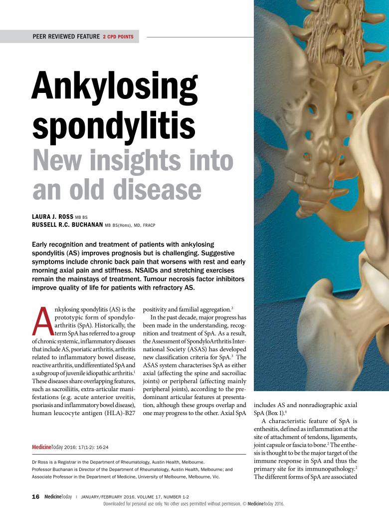
Ankylosing spondylitisNew insights into an old diseaseLAURA J. ROSS MB BS
RUSSELL R.C. BUCHANAN MB BS(Hons), MD, FRACP
Early recognition and treatment of patients with ankylosing spondylitis (AS) improves prognosis but is challenging. Suggestive symptoms include chronic back pain that worsens with rest and early morning axial pain and stiffness. NSAIDs and stretching exercises remain the mainstays of treatment. Tumour necrosis factor inhibitors improve quality of life for patients with refractory AS.
MedicineToday 2016; 17(1-2): 16-24
Dr Ross is a Registrar in the Department of Rheumatology, Austin Health, Melbourne.
Professor Buchanan is Director of the Department of Rheumatology, Austin Health, Melbourne; and
Associate Professor in the Department of Medicine, University of Melbourne, Melbourne, Vic.
PEER REVIEWED FEATURE 2 CPD POINTS
16 MedicineToday ❙ JANUARY/FEBRUARY 2016, VOLUME 17, NUMBER 1-2
Downloaded for personal use only. No other uses permitted without permission. © MedicineToday 2016.
����������������������������������������������
with characteristic extraarticular manifestations, which can be useful to distinguish SpA from other types of inflammatory arthritis and assist with prognostication.5
This article will address the contemporary definition of AS, early recognition in general practice of patients with AS and recent advances in diagnosis and management.
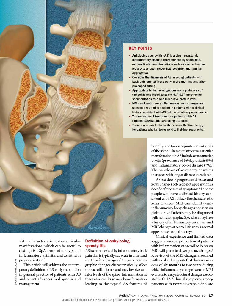
Definition of ankylosing spondylitisAS is characterised by inflammatory back pain that is typically subacute in onset and starts before the age of 45 years. Radiographic changes characteristically affect the sacroiliac joints and may involve variable levels of the spine. Inflammation at these sites results in new bone formation leading to the typical AS features of
bridging and fusion of joints and ankylosis of the spine. Characteristic extraarticular manifestations in AS include acute anterior uveitis (prevalence of 26%), psoriasis (9%) and inflammatory bowel disease (7%).5 The prevalence of acute anterior uveitis increases with longer disease duration.5
AS is a slowly progressive disease, and xray changes often do not appear until a decade after onset of symptoms.6 In some people who have a clinical history consistent with AS but lack the characteristic xray changes, MRI can identify early inflammatory bony changes not seen on plain xray.7 Patients may be diagnosed with nonradiographic SpA when they have a history of inflammatory back pain and MRI changes of sacroiliitis with a normal appearance on plain xrays.
Clinical experience and limited data suggest a sizeable proportion of patients with inflammation of sacroiliac joints on MRI will go on to develop xray changes.6 A review of the MRI changes associated with axial SpA suggests that there is a window of six months to two years during which inflammatory changes seen on MRI evolve into early structural changes associated with AS.8 Clinical symptoms among patients with nonradiographic SpA are
KEY POINTS
• Ankylosing spondylitis (AS) is a chronic systemic inflammatory disease characterised by sacroiliitis, extra-articular manifestations such as uveitis, human leucocyte antigen (HLA)-B27 positivity and familial aggregation.
• Consider the diagnosis of AS in young patients with back pain and stiffness early in the morning and after prolonged sitting.
• Appropriate initial investigations are a plain x-ray of the pelvis and blood tests for HLA-B27, erythrocyte sedimentation rate and C-reactive protein level.
• MRI can identify early inflammatory bony changes not seen on x-ray and is prudent in patients with a clinical history consistent with AS but a normal x-ray appearance.
• The mainstay of treatment for patients with AS remains NSAIDs and stretching exercises.
• Tumour necrosis factor inhibitors are effective therapy for patients who fail to respond to first-line treatments.
© J
OH
N W
. K
AR
APE
LOU
, C
MI
MedicineToday ❙ JANUARY/FEBRUARY 2016, VOLUME 17, NUMBER 1-2 17Downloaded for personal use only. No other uses permitted without permission. © MedicineToday 2016.
����������������������������������������������
comparable with those among patients with xrayproven AS.1
ASAS has validated classification criteria for axial SpA, including AS and nonradiographic axial SpA (Box 2).3 Criteria include the presence of inflammatory back pain, extraarticular manifestations of SpA and HLAB27 positivity, with or without xray changes of sacroiliitis.
Diagnosis Early diagnosis of AS remains a challenge and is typically delayed up to eight to 10 years after symptom onset.9 AS remains a clinical diagnosis based on symptoms and signs. Treatment response is generally better in patients with short disease duration and good functional status.
HistoryPatients with AS account for 5% of patients with chronic low back pain.10 Identifying patients with inflammatory back pain is the key to diagnosing AS and requires a targeted history.7 Useful questions to ask patients are listed in Box 3.11 Important features of inflammatory back pain include significant morning stiffness, gel phenomenon (stiffness following sitting or other inactivity) and awakening in the second half of the night with spinal stiffness. In
contrast, mechanical back pain is often intermittent, exacerbated by activity and better with rest (Table).
Important additional clues to identifying patients with inflammatory back pain include a history of peripheral inflammatory arthritis, a family history of AS and a good response of the pain to NSAIDs. The presence of alternating buttock pain, commonly radiating into the posterior thighs, is highly suggestive of sacroiliac joint pain.
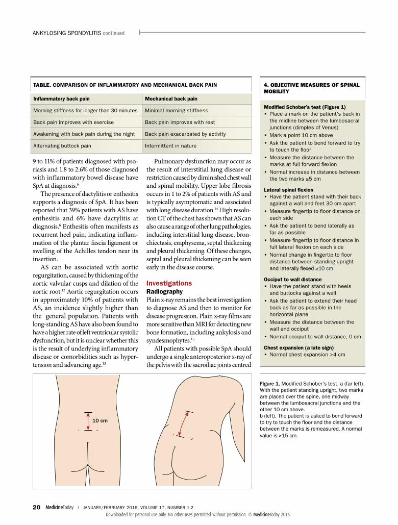
ExaminationThe characteristic examination finding in patients with AS is a reduced range of spinal movement. Lateral spinal flexion is often the first movement to be affected. Important objective measures of spinal mobility are described in Box 4 and Figure 1.
Extra-articular manifestationsExtraarticular manifestations that support the diagnosis of AS or other forms of SpA include anterior uveitis, psoriasis and inflammatory bowel disease. Around
1. CLASSIFICATION OF THE SPONDYLOARTHROPATHIES*
Predominantly axial SpA
• Ankylosing spondylitis
• Nonradiographic axial SpA
Predominantly peripheral SpA
• Arthritis with inflammatory bowel disease
• Psoriatic arthritis
• Reactive arthritis
• Undifferentiated SpA
Abbreviation: SpA=spondyloarthritis.
* Adapted from ASAS (Assessment of SpondyloArthritis International Society). Slide-educational kit. Available online at: http://www.asas-group.org (accessed February 2016).4
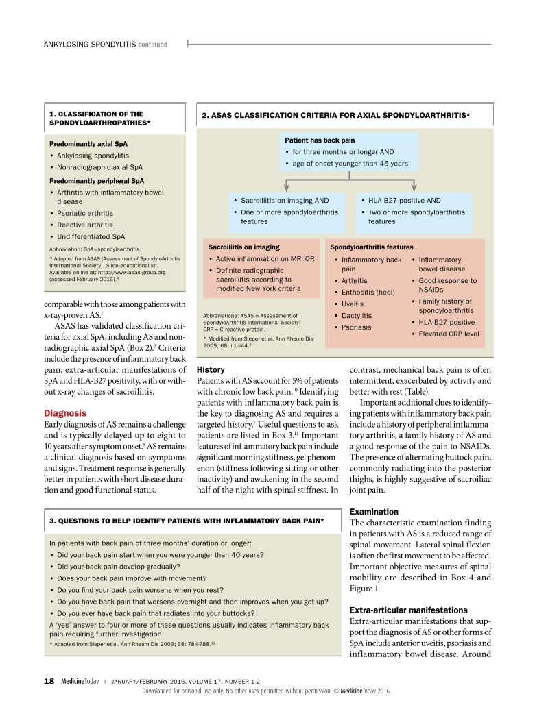
2. ASAS CLASSIFICATION CRITERIA FOR AXIAL SPONDYLOARTHRITIS*
3. QUESTIONS TO HELP IDENTIFY PATIENTS WITH INFLAMMATORY BACK PAIN*
In patients with back pain of three months’ duration or longer:
• Did your back pain start when you were younger than 40 years?
• Did your back pain develop gradually?
• Does your back pain improve with movement?
• Do you find your back pain worsens when you rest?
• Do you have back pain that worsens overnight and then improves when you get up?
• Do you ever have back pain that radiates into your buttocks?
A ‘yes’ answer to four or more of these questions usually indicates inflammatory back pain requiring further investigation.* Adapted from Sieper et al. Ann Rheum Dis 2009; 68: 784-788.11
ANKYLOSINg SPONDYLItIS continued
Abbreviations: ASAS = Assessment of SpondyloArthritis International Society; CRP = C-reactive protein.
* Modified from Sieper et al. Ann Rheum Dis 2009; 68: ii1-ii44.3
Patient has back pain
• for three months or longer AND
• age of onset younger than 45 years
Spondyloarthritis features
• Inflammatory back pain
• Arthritis
• Enthesitis (heel)
• Uveitis
• Dactylitis
• Psoriasis
• Inflammatory bowel disease
• good response to NSAIDs
• Family history of spondyloarthritis
• HLA-B27 positive
• Elevated CRP level
Sacroiliitis on imaging
• Active inflammation on MRI OR
• Definite radiographic sacroiliitis according to modified New York criteria
• HLA-B27 positive AND
• two or more spondylo arthritis features
• Sacroiliitis on imaging AND
• One or more spondylo arthritis features
18 MedicineToday ❙ JANUARY/FEBRUARY 2016, VOLUME 17, NUMBER 1-2
Downloaded for personal use only. No other uses permitted without permission. © MedicineToday 2016.
����������������������������������������������
9 to 11% of patients diagnosed with psoriasis and 1.8 to 2.6% of those diagnosed with inflammatory bowel disease have SpA at diagnosis.6
The presence of dactylitis or enthesitis supports a diagnosis of SpA. It has been reported that 39% patients with AS have enthesitis and 6% have dactylitis at diagnosis.6 Enthesitis often manifests as recurrent heel pain, indicating inflammation of the plantar fascia ligament or swelling of the Achilles tendon near its insertion.
AS can be associated with aortic regurgitation, caused by thickening of the aortic valvular cusps and dilation of the aortic root.12 Aortic regurgitation occurs in approximately 10% of patients with AS, an incidence slightly higher than the general population. Patients with longstanding AS have also been found to have a higher rate of left ventricular systolic dysfunction, but it is unclear whether this is the result of underlying inflammatory disease or comorbidities such as hypertension and advancing age.13
Pulmonary dysfunction may occur as the result of interstitial lung disease or restriction caused by diminished chest wall and spinal mobility. Upper lobe fibrosis occurs in 1 to 2% of patients with AS and is typically asymptomatic and associated with long disease duration.14 High resolution CT of the chest has shown that AS can also cause a range of other lung pathologies, including interstitial lung disease, bronchiectasis, emphysema, septal thickening and pleural thickening. Of these changes, septal and pleural thickening can be seen early in the disease course.
InvestigationsRadiographyPlain xray remains the best investigation to diagnose AS and then to monitor for disease progression. Plain xray films are more sensitive than MRI for detecting new bone formation, including ankylosis and syndesmophytes.15
All patients with possible SpA should undergo a single anteroposterior xray of the pelvis with the sacroiliac joints centred
TABLE. COMPARISON OF INFLAMMATORY AND MECHANICAL BACK PAIN
Inflammatory back pain Mechanical back pain
Morning stiffness for longer than 30 minutes Minimal morning stiffness
Back pain improves with exercise Back pain improves with rest
Awakening with back pain during the night Back pain exacerbated by activity
Alternating buttock pain Intermittent in nature
Figure 1. Modified Schober’s test. a (far left). With the patient standing upright, two marks are placed over the spine, one midway between the lumbosacral junctions and the other 10 cm above.b (left). the patient is asked to bend forward to try to touch the floor and the distance between the marks is remeasured. A normal value is ≥15 cm.
4. OBJECTIVE MEASURES OF SPINAL MOBILITY
Modified Schober’s test (Figure 1) • Place a mark on the patient's back in
the midline between the lumbosacral junctions (dimples of Venus)
• Mark a point 10 cm above• Ask the patient to bend forward to try
to touch the floor• Measure the distance between the
marks at full forward flexion• Normal increase in distance between
the two marks ≥5 cm
Lateral spinal flexion • Have the patient stand with their back
against a wall and feet 30 cm apart• Measure fingertip to floor distance on
each side• Ask the patient to bend laterally as
far as possible• Measure fingertip to floor distance in
full lateral flexion on each side• Normal change in fingertip to floor
distance between standing upright and laterally flexed ≥10 cm
Occiput to wall distance • Have the patient stand with heels
and buttocks against a wall• Ask the patient to extend their head
back as far as possible in the horizontal plane
• Measure the distance between the wall and occiput
• Normal occiput to wall distance, 0 cm
Chest expansion (a late sign) • Normal chest expansion >4 cm
10 cm
ANKYLOSINg SPONDYLItIS continued
20 MedicineToday ❙ JANUARY/FEBRUARY 2016, VOLUME 17, NUMBER 1-2
Downloaded for personal use only. No other uses permitted without permission. © MedicineToday 2016.
����������������������������������������������
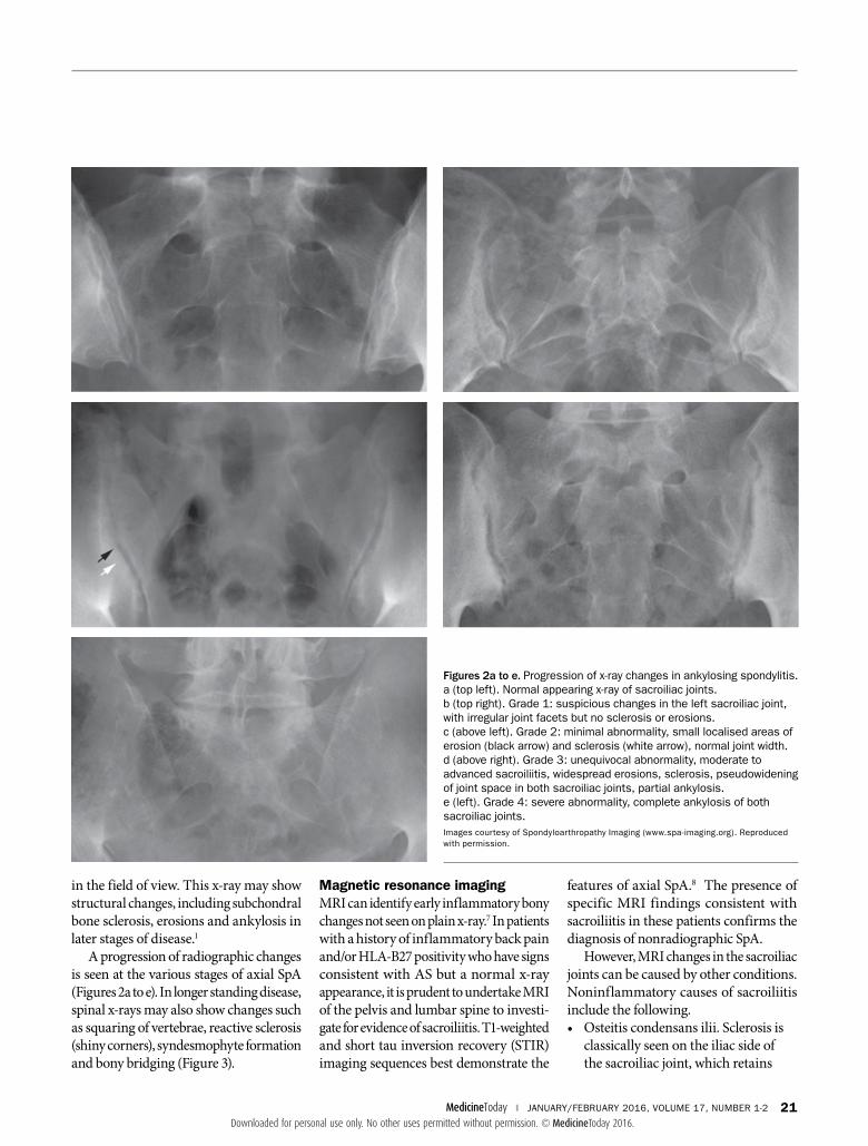
in the field of view. This xray may show structural changes, including subchondral bone sclerosis, erosions and ankylosis in later stages of disease.1
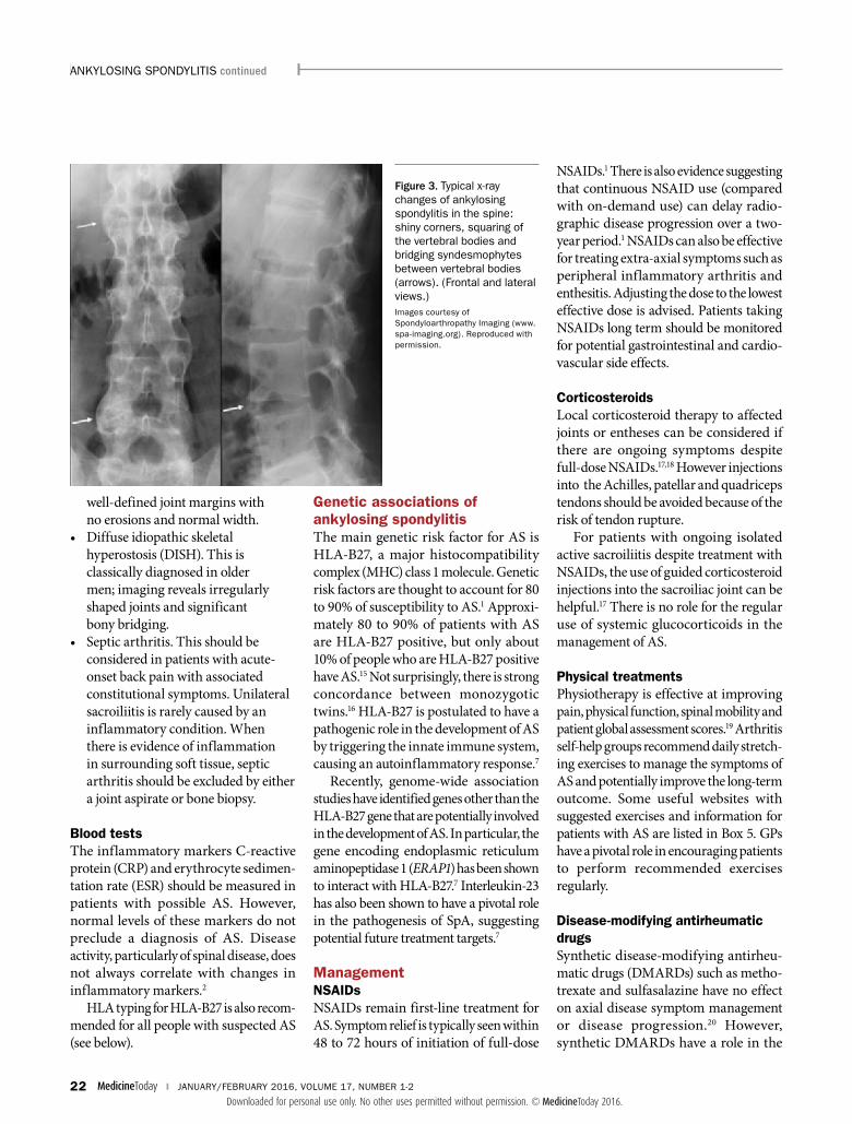
A progression of radiographic changes is seen at the various stages of axial SpA (Figures 2a to e). In longer standing disease, spinal xrays may also show changes such as squaring of vertebrae, reactive sclerosis (shiny corners), syndesmophyte formation and bony bridging (Figure 3).
Magnetic resonance imagingMRI can identify early inflammatory bony changes not seen on plain xray.7 In patients with a history of inflammatory back pain and/or HLAB27 positivity who have signs consistent with AS but a normal xray appearance, it is prudent to undertake MRI of the pelvis and lumbar spine to investigate for evidence of sacroiliitis. T1weighted and short tau inversion recovery (STIR) imaging sequences best demonstrate the
features of axial SpA.8 The presence of specific MRI findings consistent with sacroiliitis in these patients confirms the diagnosis of nonradiographic SpA.
However, MRI changes in the sacroiliac joints can be caused by other conditions. Noninflammatory causes of sacroiliitis include the following.• Osteitis condensans ilii. Sclerosis is
classically seen on the iliac side of the sacroiliac joint, which retains
Figures 2a to e. Progression of x-ray changes in ankylosing spondylitis. a (top left). Normal appearing x-ray of sacroiliac joints. b (top right). grade 1: suspicious changes in the left sacroiliac joint, with irregular joint facets but no sclerosis or erosions. c (above left). grade 2: minimal abnormality, small localised areas of erosion (black arrow) and sclerosis (white arrow), normal joint width. d (above right). grade 3: unequivocal abnormality, moderate to advanced sacroiliitis, widespread erosions, sclerosis, pseudowidening of joint space in both sacroiliac joints, partial ankylosis. e (left). grade 4: severe abnormality, complete ankylosis of both sacroiliac joints.Images courtesy of Spondyloarthropathy Imaging (www.spa-imaging.org). Reproduced with permission.
MedicineToday ❙ JANUARY/FEBRUARY 2016, VOLUME 17, NUMBER 1-2 21Downloaded for personal use only. No other uses permitted without permission. © MedicineToday 2016.
����������������������������������������������
welldefined joint margins with no erosions and normal width.
• Diffuse idiopathic skeletal hyperostosis (DISH). This is classically diagnosed in older men; imaging reveals irregularly shaped joints and significant bony bridging.
• Septic arthritis. This should be considered in patients with acuteonset back pain with associated constitutional symptoms. Unilateral sacroiliitis is rarely caused by an inflammatory condition. When there is evidence of inflammation in surrounding soft tissue, septic arthritis should be excluded by either a joint aspirate or bone biopsy.
Blood testsThe inflammatory markers Creactive protein (CRP) and erythrocyte sedimentation rate (ESR) should be measured in patients with possible AS. However, normal levels of these markers do not preclude a diagnosis of AS. Disease activity, particularly of spinal disease, does not always correlate with changes in inflammatory markers.2
HLA typing for HLAB27 is also recommended for all people with suspected AS (see below).
Genetic associations of ankylosing spondylitisThe main genetic risk factor for AS is HLAB27, a major histocompatibility complex (MHC) class 1 molecule. Genetic risk factors are thought to account for 80 to 90% of susceptibility to AS.1 Approximately 80 to 90% of patients with AS are HLAB27 positive, but only about 10% of people who are HLAB27 positive have AS.15 Not surprisingly, there is strong concordance between monozygotic twins.16 HLAB27 is postulated to have a pathogenic role in the development of AS by triggering the innate immune system, causing an autoinflammatory response.7
Recently, genomewide association studies have identified genes other than the HLAB27 gene that are potentially involved in the development of AS. In particular, the gene encoding endoplasmic reticulum aminopeptidase 1 (ERAP1) has been shown to interact with HLAB27.7 Interleukin23 has also been shown to have a pivotal role in the pathogenesis of SpA, suggesting potential future treatment targets.7
ManagementNSAIDsNSAIDs remain firstline treatment for AS. Symptom relief is typically seen within 48 to 72 hours of initiation of fulldose
NSAIDs.1 There is also evidence suggesting that continuous NSAID use (compared with ondemand use) can delay radiographic disease progression over a twoyear period.1 NSAIDs can also be effective for treating extraaxial symptoms such as peripheral inflammatory arthritis and enthesitis. Adjusting the dose to the lowest effective dose is advised. Patients taking NSAIDs long term should be monitored for potential gastrointestinal and cardiovascular side effects.
CorticosteroidsLocal corticosteroid therapy to affected joints or entheses can be considered if there are ongoing symptoms despite fulldose NSAIDs.17,18 However injections into the Achilles, patellar and quadriceps tendons should be avoided because of the risk of tendon rupture.
For patients with ongoing isolated active sacroiliitis despite treatment with NSAIDs, the use of guided corticosteroid injections into the sacroiliac joint can be helpful.17 There is no role for the regular use of systemic glucocorticoids in the management of AS.
Physical treatmentsPhysiotherapy is effective at improving pain, physical function, spinal mobility and patient global assessment scores.19 Arthritis selfhelp groups recommend daily stretching exercises to manage the symptoms of AS and potentially improve the longterm outcome. Some useful websites with suggested exercises and information for patients with AS are listed in Box 5. GPs have a pivotal role in encouraging patients to perform recommended exercises regularly.
Disease-modifying antirheumatic drugsSynthetic diseasemodifying antirheumatic drugs (DMARDs) such as methotrexate and sulfasalazine have no effect on axial disease symptom management or disease progression.20 However, synthetic DMARDs have a role in the
Figure 3. typical x-ray changes of ankylosing spondylitis in the spine: shiny corners, squaring of the vertebral bodies and bridging syndesmophytes between vertebral bodies (arrows). (Frontal and lateral views.)Images courtesy of Spondyloarthropathy Imaging (www.spa-imaging.org). Reproduced with permission.
ANKYLOSINg SPONDYLItIS continued
22 MedicineToday ❙ JANUARY/FEBRUARY 2016, VOLUME 17, NUMBER 1-2
Downloaded for personal use only. No other uses permitted without permission. © MedicineToday 2016.
����������������������������������������������
management of patients with coexisting peripheral joint inflammatory arthritis.
Tumour necrosis factor inhibitors Tumour necrosis factor alpha (TNFα) inhibitors such as etanercept, infliximab, golimumab and adalimumab have revolutionised treatment of AS. These biological DMARDs have improved the quality of life for more than twothirds of patients with AS who do not respond to firstline therapy.21
PBS criteria stipulate a threemonth trial of exercise and daily use of two different NSAIDs before TNF inhibitor therapy may be considered. Currently patients qualify for PBSsubsidised TNF inhibitor therapy only when there is radiographic evidence of sacroiliitis (bilateral grade 2 or unilateral grade 3 sacroiliitis changes on xray). However, there is evidence to suggest that early treatment can reduce radiographic progression.7 Current ASAS guidelines support the use of TNF inhibitor therapy in clinically active nonradiographic SpA.17
Specialist referralDelay to diagnosis remains a key challenge in the management of patients with AS.
Patients should be referred to a rheumatologist for assessment if the clinical history is consistent with inflammatory back pain or there is evidence of sacroiliitis on xray or MRI. These patients can be commenced on NSAIDs and given physical exercises while they await assessment by a rheumatologist.
ComplicationsAllcause mortality is increased in patients with AS. A Swedish nationwide cohort study found that predictors of death include lower socioeconomic status, increased general medical comorbidities and previous hip replacement.22 Cardiovascular disease was the most common medical comorbidity, and patients had a higher baseline comorbidity than matched healthy control subjects. Common causes of death of AS patients were infection and cardiovascular disease.
Patients with AS are considered to have an increased risk of cardiovascular disease compared with healthy control subjects, with epidemiological data suggesting that ischaemic heart disease is a greater problem in young patients.23 This higher risk may be due to inflammatory disease, traditional cardiovascular risk factors or NSAID use. It highlights the importance of longterm optimisation of modifiable cardiovascular risk factors.
Patients with AS have an increased prevalence of both osteopenia and osteoporosis. High disease activity, generally detected by increased ESR and CRP level, predicts increased bone loss.24 Patient screening with dual emission xray absorptiometry (DXA) scanning of both the spine and hip is recommended.17 It is important to consider spine bone mineral density results carefully, as patients with AS have an increased rate of spinal fracture and spinal cord injury.
ConclusionEarly recognition of signs and symptoms of AS and prompt referral to a rheumatologist remain a challenge in this condition. AS should be considered if symptoms
consistent with inflammatory back pain are noted. These include waking in the second half of the night with back pain, axial early morning stiffness and alternating buttock pain.
The mainstay of treatment remains NSAIDs and stretching exercises. The quality of life for patients with refractory axial SpA has improved significantly with the introduction of antiTNFα therapy.
Ongoing research into the genetics and pathophysiology of this condition has improved our understanding of this disease, leading to better classification and diagnostic testing. This also leads to much optimism for future management of patients with AS. MT
Acknowledgementthe authors are grateful for the helpful comments
from Dr Lionel Schachna, Austin Spondylitis Clinic,
Austin Health, Melbourne, Vic, in the preparation of
this article.
ReferencesA list of references is included in the website version
(www.medicinetoday.com.au) of this article.
COMPEtINg INtEREStS: None.
ANKYLOSINg SPONDYLItIS continued
ONLINE CPD JOURNAL PROGRAM
What is the characteristic physical examination finding in patients with ankylosing spondylitis?
Review your knowledge of this topic and earn CPD points by taking part in MedicineToday’s Online CPD Journal Program. Log in to www.medicinetoday.com.au/cpd
© S
tOC
KD
EVIL
/IS
tOC
KPH
OtO
5. USEFUL WEBSITES FOR PATIENTS WITH ANKYLOSING SPONDYLITIS
Arthritis Australia http://www.arthritisaustralia.com.au
Arthritis and Osteoporosis New South Wales http://arthritisnsw.org.au
Arthritis Victoria http://www.arthritisvic.org.au
Arthritis Research UKhttp://www.arthritisresearchuk.org
Exercises for patients with ankylosing spondylitis
• http://www.arthritisaustralia.com.au/images/stories/Michael_Slater/MS_exercisesheet_V4%20FINAL-red.pdf
• http://www.arthritisresearchuk.org/arthritis-information/conditions/ankylosing-spondylitis/self-help-and-daily-living/exercise.aspx
24 MedicineToday ❙ JANUARY/FEBRUARY 2016, VOLUME 17, NUMBER 1-2
Downloaded for personal use only. No other uses permitted without permission. © MedicineToday 2016.
����������������������������������������������
MedicineToday 2016; 17(1-2): 16-24
Ankylosing spondylitisNew insights into an
old diseaseLAURA J. ROSS MB BS; RUSSELL R.C. BUCHANAN MB BS(Hons), MD, FRACP
References
1. Dougados M, Baeten D. Spondyloarthritis. Lancet 2011; 177: 2127-2137.
2. Sieper J, Braun J, Rudwaleit M, Boonen A, Zink A. Ankylosing spondylitis:
an overview. Ann Rheum Dis 2002; 61 Suppl III: iii8-iii18.
3. Sieper J, Rudwaleit M, Baraliakos X, et al. the Assessment of
SpondyloArthritis International Society (ASAS) handbook: a guide to assess
spondyloarthritis. Ann Rheum Dis 2009; 68: ii1-ii44.
4. ASAS (Assessment of SpondyloArthritis International Society). Slide-
educational kit. Available online at: http://www.asas-group.org (accessed
January 2016).
5. Stolwijk C, van tubergen A, Castillo-Ortiz JD, Boonen A. Prevalence of
extra-articular manifestations in patients with ankylosing spondylitis:
a systematic review and meta-analysis. Ann Rheum Dis 2015; 74: 65-73.
6. Rudwaleit M, Haibel H, Baraliakos X, et al. the early disease stage in axial
spondylarthritis: results from the german spondyloarthritis inception cohort.
Arthritis Rheum 2009; 60: 717-727.
7. Rudwaleit M, Metter A, Listing J, Sieper J, Braun J. Inflammatory back
pain in ankylosing spondylitis: a reassessment of the clinical history for
application as classification and diagnostic criteria. Arthritis Rheum 2006;
54: 569-578.
8. Hermann Kg, Baraliakos X, van der Heijde D, et al. Descriptions of spinal
MRI lesions and definition of a positive MRI of the spine in axial spondyloarthritis:
a consensual approach by the ASAS/OMERACt MRI study group. Ann Rheum
Dis 2012; 71: 1278-1288.
9. Feldtkeller E, Khan M, van der Heijde D, van der Linden S, Braun J. Age at
disease onset and diagnosis delay in HLA-B27 negative vs. positive patients
with ankylosing spondylitis. Rheumatol Int 2003; 23: 61-66.
10. Poddubnyy D, Rudwaleit M. Early spondyloarthritis. Rheum Dis Clin N Am
2012; 38: 387-403.
11. Sieper J, van der Heijde D, Landewé R, et al. New criteria for inflammatory
back pain in patients with chronic back pain: a real patient exercise by experts
from the Assessment of SpondyloArthritis International Society (ASAS). Ann
Rheum Dis 2009; 68: 784-788.
12. Vinsonneau U, Brondex A, Mansourati J, et al. Cardiovascular disease in
patient with spondyloarthritis. Joint Bone Spine 2008; 75: 18-21.
13. Brunner F, Kunz A, Weber U, Kissling R. Ankylosing spondylitis and heart
abnormalities: do cardiac conduction disorders, valve regurgitation and
diastolic dysfunction occur more often in male patients diagnosed with
ankylosing spondylitis for over 15 years than in the normal population? Clin
Rheumatol 2006; 25: 24-29.
14. Quismorio F Jr. Pulmonary involvement in ankylosing spondylitis. Curr Opin
Pulm Med 2006; 12: 342-345.
15. Robinson P, Benham H. Advances in classification, basic mechanisms and
clinical science in ankylosing spondylitis and axial spondyloarthritis. Intern
Med J 2015; 45: 127-133.
16. Dougados M, Baeten D. Spondyloarthritis. Lancet 2011; 177: 2127-2137.
17. Ward M, Deodhar A, Akl E, et al. American College of Rheumatology/
Spondylitis Association of America/Spondyloarthritis Research and treatment
Network 2015 recommendations for the treatment of ankylosing spondylitis
and nonradiographic axial spondyloarthritis. Arthritis Rheumatol 2015 Sep 24;
epub ahead of print (doi: 10.1002/art.39298).
18. Braun J, van den Berg R, Baraliakos X, et al. 2010 update of the ASAS/
EULAR recommendations for the management of ankylosing spondylitis. Ann
Rheum Dis 2011; 70: 896-904.
19. Dagfinrud H, Kvien tK, Hagen KB. Physiotherapy interventions for
ankylosing spondylitis. Cochrane Database Syst Rev 2008; (1): CD002822.
20. van den Berg R, Baraliakos X, Braun J, van der Heijde D. First update of the
current evidence for the management of ankylosing spondylitis with non-
pharmacological treatment and non-biologic drugs: a systematic literature
review for the ASAS/EULAR management recommendations in ankylosing
spondylitis. Rheumatology (Oxford) 2012; 51: 1388-1396.
21. van der Heijde D, Sieper J, Maksymowych W, et al. 2010 update of the
international ASAS recommendations for the use of anti-tNF agents in patients
with axial spondyloarthritis. Ann Rheum Dis 2011; 70: 905-908.
22. Exarchou S, Lie E, Lindstrom U, et al. Mortality in ankylosing spondylitis:
results from a nation-wide population-based study. Ann Rheum Dis 2015 Sep 2.
pii: epub ahead of print (annrheumdis-2015-207688).
23. Huang YP, Wang YH, Pan SL. Increased risk of ischaemic heart disease in
young patients with newly diagnosed ankylosing spondylitis – a population-
based longitudinal follow-up study. PLoS One 2013; 8: e64155.
24. Wang DM, Zeng QY, Chen SB, gong Y, Hou ZD, Ziao ZY. Prevalence and risk
factors of osteoporosis in patients with ankylosing spondylitis: a 5-year follow
up study of 504 cases. Clin Exp Rheumatol 2015; 33: 465-470.
Downloaded for personal use only. No other uses permitted without permission. © MedicineToday 2016.
����������������������������������������������