patrolling monocytes promote intravascular … monocytes promote intravascular neutrophil activation...
TRANSCRIPT
Patrolling monocytes promote intravascular neutrophilactivation and glomerular injury in the acutelyinflamed glomerulusMichaela Finsterbuscha, Pam Halla, Anqi Lia, Sapna Devia, Clare L. V. Westhorpea, A. Richard Kitchinga,b,c,and Michael J. Hickeya,1
aCentre for Inflammatory Diseases, Monash University Department of Medicine, Monash Medical Centre, Clayton, VIC 3168, Australia; bDepartment ofNephrology, Monash Medical Centre, Clayton, VIC 3168, Australia; and cDepartment of Paediatric Nephrology, Monash Medical Centre, Clayton, VIC 3168,Australia
Edited by Paul Kubes, University of Calgary, Calgary, AB, Canada, and accepted by Editorial Board Member Carl F. Nathan July 5, 2016 (received for reviewApril 20, 2016)
Nonclassical monocytes undergo intravascular patrolling in bloodvessels, positioning them ideally to coordinate responses to inflam-matory stimuli. Under some circumstances, the actions of monocyteshave been shown to involve promotion of neutrophil recruitment.However, the mechanisms whereby patrolling monocytes controlthe actions of neutrophils in the circulation are unclear. Here, weexamined the contributions ofmonocytes to antibody- and neutrophil-dependent inflammation in a model of in situ immune complex-mediated glomerulonephritis. Multiphoton and spinning disk confocalintravital microscopy revealed that monocytes patrol both un-inflamed and inflamed glomeruli using β2 and α4 integrins andCX3CR1. Monocyte depletion reduced glomerular injury, demonstrat-ing that these cells promote inappropriate inflammation in this set-ting. Monocyte depletion also resulted in reductions in neutrophilrecruitment and dwell time in glomerular capillaries and in reactiveoxygen species (ROS) generation by neutrophils, suggesting a rolefor cross-talk between monocytes and neutrophils in inductionof glomerulonephritis. Consistent with this hypothesis, patrollingmonocytes and neutrophils underwent prolonged interactions inglomerular capillaries, with the duration of these interactions in-creasing during inflammation. Moreover, neutrophils that inter-acted with monocytes showed increased retention and a greaterpropensity for ROS generation in the glomerulus. Also, renal patrol-ling monocytes, but not neutrophils, produced TNF during inflam-mation, and TNF inhibition reduced neutrophil dwell time and ROSproduction, as well as renal injury. These findings show that mono-cytes and neutrophils undergo interactions within the glomeru-lar microvasculature. Moreover, evidence indicates that, in responseto an inflammatory stimulus, these interactions allow monocytesto promote neutrophil recruitment and activation within theglomerular microvasculature, leading to neutrophil-dependenttissue injury.
monocyte–neutrophil interaction | inflammation | glomerulonephritis |reactive oxygen species | tumor necrosis factor
Neutrophil recruitment is a crucial event in the response totissue injury and host defense against pathogens. However,
if dysregulated, it can also cause significant tissue injury. In-appropriate recruitment and activation of neutrophils are re-cognized as major contributors to organ damage in numerousinflammatory diseases, including inflammatory arthritis, acuterespiratory distress syndrome, systemic lupus erythematosus, andacute glomerulonephritis (1–4). Although our understanding ofthe molecular basis of neutrophil recruitment is well-developed,less is known about the interactions of neutrophils with othercirculating immune cells during the development of the innateinflammatory response. However, over the last decade, evidencehas emerged that neutrophil recruitment and function duringinflammation can be influenced by another circulating immunecell—the monocyte (5–10).
Monocytes exist in two major populations, termed classicaland nonclassical, as defined by expression of specific surfacemarkers. In mice, these populations are CC chemokine receptor2 (CCR2)hi CX3C chemokine receptor 1 (CX3CR1)lo Ly6C+
(classical) and CX3CR1hi CCR2− Ly6C− (nonclassical), with thecorresponding populations in humans being CD14hi CD16− andCD14lo CD16hi, respectively. A third low abundance population,termed intermediate, also exists, characterized in mice by in-termediate Ly6C expression and in humans as CD14hi CD16+
(11–13). Classical monocytes are selectively recruited to in-flamed tissues and lymph nodes partly in a CCR2-dependentmanner, where they produce inflammatory and antimicrobialfactors and contribute to local and systemic inflammation (12–16). In contrast, CX3CR1hi Ly6C− monocytes lack CCR2 and arepresent in both resting and inflammatory tissues (12, 15). Evi-dence indicates that nonclassical monocytes perform a special-ized form of immune homeostasis, serving as initiators of acuteinflammatory responses and contributing to tissue remodeling(5, 6, 13, 17–19). A unique aspect of the function of these cells istheir capacity to undergo extensive intravascular crawling or“patrolling,” a constitutive phenomenon that has now been ob-served in a wide range of organs (5, 6, 20–22). A growing body ofevidence indicates that this form of immune surveillance allows
Significance
Nonclassical monocytes patrol the microvascular lumen in nu-merous organs in behavior thought to represent a form ofimmune surveillance. However, the mechanisms whereby theypromote inflammatory responses are unclear. Here, we showusing in vivo imaging that, in the unique microvasculature of theglomerulus in the kidney, monocytes constitutively undergointeractions with intravascular migratory neutrophils. Upon in-duction of glomerular inflammation, neutrophils that interactwith monocytes show increased retention in glomerular capil-laries and increased propensity to generate reactive oxygenspecies, leading to renal injury. These findings of immune cellinteractions occurring within the glomerular microvasculatureindicate that cell–cell contact between neutrophils and non-classical monocytes is a previously unrecognized intravascularinflammatory mechanism underpinning glomerular injury.
Author contributions: M.F., A.R.K., and M.J.H. designed research; M.F., P.H., A.L., S.D., andC.L.V.W. performed research; M.F., P.H., A.L., S.D., and C.L.V.W. analyzed data; and M.F.,A.R.K., and M.J.H. wrote the paper.
The authors declare no conflict of interest.
This article is a PNAS Direct Submission. P.K. is a guest editor invited by the EditorialBoard.1To whom correspondence should be addressed. Email: [email protected].
This article contains supporting information online at www.pnas.org/lookup/suppl/doi:10.1073/pnas.1606253113/-/DCSupplemental.
E5172–E5181 | PNAS | Published online August 15, 2016 www.pnas.org/cgi/doi/10.1073/pnas.1606253113
nonclassical monocytes to respond rapidly to inflammatory stimuliand to direct the responses of other immune cells in the circu-lation, particularly neutrophils (5, 6). However, the nature andmechanisms of this response are not fully understood. Moreover,the mode of action of nonclassical monocytes in specializedmicrovascular beds, in which the mechanisms of leukocyte re-cruitment do not follow the conventional paradigm, such as theglomerulus (20, 23), has not been investigated.The glomerulus in the kidney is a key target of damaging in-
flammation in a wide range of conditions (2). Indeed, glo-merulonephritis is a leading cause of end-stage renal failure.Furthermore, although capillaries are typically not major sites ofleukocyte recruitment, leukocyte recruitment to the glomerularcapillaries plays a central role in glomerular injury and renaldysfunction (23, 24). We recently used multiphoton imaging todemonstrate that leukocyte recruitment to the glomerulus occursvia a distinct paradigm (20). In the absence of inflammation,neutrophils underwent constitutive intravascular retention andcrawling within glomerular capillaries. Upon induction of glo-merular inflammation, the major response of the neutrophil wasto significantly increase the duration of its retention in the glo-merular capillaries (referred to as dwell time), which, however,did not subsequently progress to transendothelial migration. Incontrast, while retained intravascularly, neutrophils generatedreactive oxygen species (ROS) that were responsible for glomer-ular injury and renal dysfunction. In parallel, CX3CR1hi mono-cytes were also observed to patrol the glomerular capillaries and tosignificantly increase their dwell time during glomerular in-flammation. However, while this study showed that both theseleukocyte subsets patrol the glomerular capillaries, their functionalrelationships and whether these cells undergo interactions withinthe glomerular microvasculature have not been investigated. There-fore, the aim of this study was to investigate the contribution ofpatrolling monocytes to neutrophil-dependent glomerulonephritis.These experiments show that patrolling monocytes interact withneutrophils within the glomerular microvasculature and, duringglomerular inflammation, promote neutrophil retention andactivation that underlie glomerular dysfunction.
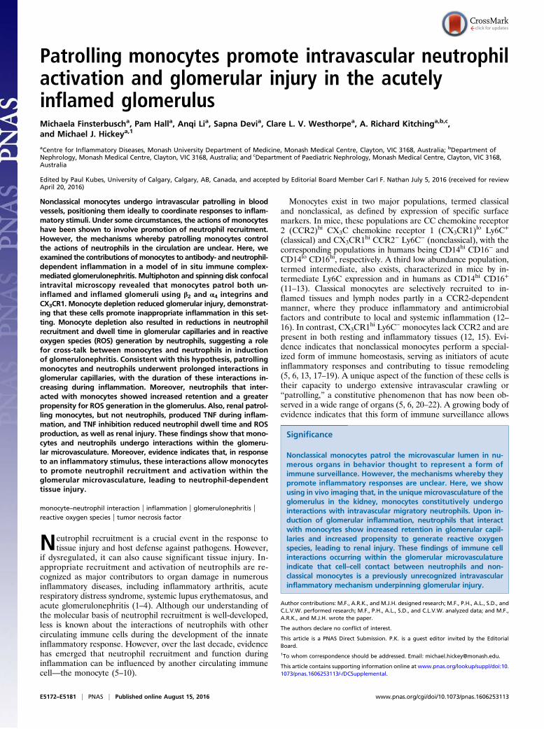
ResultsMonocytes Patrol Uninflamed Glomerular Capillaries via CX3CR1 andβ2 and α4 Integrins. Monocytes constitutively migrate within glo-merular capillaries in vivo (20). To determine the mechanism ofmonocyte migration within the glomerular microvasculature,Cx3cr1GFP/+ and Cx3cr1GFP/GFP mice, in which monocytes arevisible via GFP expression (5, 25), were examined by multiphotonmicroscopy. Similar to previously published data, in Cx3cr1GFP/+
mice, monocytes were found to undergo extended periods of in-travascular retention and migration in glomerular capillaries in theabsence of inflammatory stimulation (Fig. 1A and Movie S1). Themajority of monocytes retained in glomeruli were migratory, andtheir duration of retention averaged ∼15 min (Fig. 1 B and C). Inmice, the two major populations of monocytes are the Ly6C+
classical and the Ly6C− nonclassical. To clarify which populationof monocytes underwent migration in the glomerular capillaries,we performed parallel experiments using spinning disk confocalmicroscopy (SDCM), using anti–Gr-1 in Cx3Cr1GFP/+ mice.Anti–Gr-1 detects both Ly6C and the neutrophil-restrictedantigen Ly6G, allowing the differentiation of classical (GFP+,Gr-1+) and nonclassical (GFP+, Gr-1−) monocytes. In theseexperiments, Gr-1+ GFP+ monocytes were very rare, beingdetected at the rate of ∼0.1 per glomerulus per hour. In con-trast, the vast majority of monocytes, migrating through glo-meruli at a rate of ∼5 per glomerulus per hour, were Gr-1−.These findings indicate that the monocytes crawling in the glo-merular capillaries were almost exclusively Ly6C− nonclassicalpatrolling monocytes.Mice deficient in CX3CR1 (Cx3cr1GFP/GFP mice) showed a
significant reduction in the number of retained monocytes,compared with heterozygous Cx3cr1GFP/+ control animals, solelyvia reduction in the number of crawling cells (Fig. 1B). This ob-servation may have stemmed from a reduction in the number ofnonclassical monocytes in the blood of Cx3cr1GFP/GFP mice as pre-viously reported (26, 27). Nevertheless, the dwell time of recruitedmonocytes was also significantly reduced in Cx3cr1GFP/GFP mice(Fig. 1 A and C and Movie S1), providing clear evidence of a rolefor CX3CR1 in retention of monocytes in glomeruli. Inhibition of
Fig. 1. Monocyte patrolling in uninflamed glomerularcapillaries is dependent on CX3CR1 and β2 and α4integrins. The role of surface receptors in monocytetrafficking within uninflamed glomerular capillaries wasinvestigated in Cx3cr1GFP mice using intravital multi-photon microscopy. (A) Representative multiphotonimage sequences of an untreated Cx3cr1GFP/+ mouse(Top) and Cx3cr1GFP/GFP mouse (Bottom) illustrating mi-gration of a GFP+ monocyte (green) within glomerularcapillaries (blue; labeled with Qtracker-655). Asterisksindicate starting position, and arrows indicate the pathof migration. Time stamp is shown above the images.(Scale bar: 10 μm.) See also Movie S1. (B and C) Thenumber of monocytes adhering in glomeruli per hourshown for total, crawling, and static cells (B) andmonocyte dwell time (of total cells) (C) was assessed inuntreated Cx3cr1GFP/+ (n = 8) and Cx3cr1GFP/GFP mice (n =5). (D) Monocyte dwell timewas compared in Cx3cr1GFP/+
mice pretreated with anti-CD18 and anti-CD49d block-ing antibodies together (n = 4), or the respectiveisotype controls (n = 6). Data are presented asmean ± SEM; *P < 0.05 vs. corresponding controlgroup.
Finsterbusch et al. PNAS | Published online August 15, 2016 | E5173
IMMUNOLO
GYAND
INFLAMMATION
PNASPL
US
CD18 (integrin subunit β2) or CD49d (integrin subunit α4) didnot reduce monocyte trafficking during steady-state conditions(Fig. S1). In contrast, blocking both β2 and α4 integrins signifi-cantly reduced monocyte dwell time (Fig. 1D). These data in-dicate that patrolling monocytes use CX3CR1 and both β2 and α4integrins to undergo constitutive intravascular retention andmigration in the glomerulus.
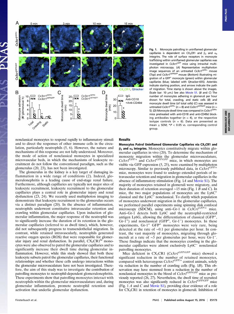
CX3CR1, LFA-1, and Mac-1 Mediate Distinct Steps of GlomerularMonocyte Trafficking During Inflammation. We previously demon-strated that monocyte dwell time increases with inflammation(20), and this finding is replicated in the present study in thatdwell time was found to increase from ∼20 min to ∼40 min duringthe anti-GBM Ab response (compare Fig. 1C with Fig. 2A).Therefore, we next used multiphoton microscopy to decipher themechanisms of glomerular monocyte trafficking under inflamma-tory conditions. Glomerular inflammation was induced using theanti-glomerular basement membrane antibody (anti-GBM Ab)model of in situ immune complex-mediated inflammation. In-hibition of LFA-1 resulted in a significant reduction in monocyteattachment to glomerular capillaries 1–2 h after anti-GBM Ab ad-ministration. However, LFA-1 inhibition did not alter the dwelltime of the remaining monocytes (Fig. 2A). In contrast, inhibition ofMac-1 had no effect on the number of adherent monocytes butdiminished monocyte dwell time by ∼50% (Fig. 2B). These dataindicate that LFA-1, but not Mac-1, is required for initial monocyteattachment whereas Mac-1 is more important for intravascularmonocyte retention during inflammation.The actions of CX3CR1 varied according to the phase of the
response. In the first hour of inflammation, there were fewermonocytes recruited to glomerular capillaries in Cx3cr1GFP/GFP
mice compared with Cx3cr1GFP/+ control animals whereasmonocyte dwell time remained unchanged (Fig. 2C). In contrast,in the second hour of inflammation (1–2 h after anti-GBMAb injection), monocyte dwell time was markedly reduced inCx3cr1GFP/GFP mice whereas the change in number of adherentmonocytes was not significant (Fig. 2D). Together, these data indicatethat the initial attachment of monocytes to glomerular capillariesduring inflammation requires LFA-1 and CX3CR1, but not Mac-1,whereas Mac-1 and CX3CR1 were crucial for prolonged dwell timesof monocyte patrolling in the second hour of the response.
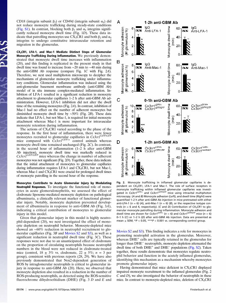
Monocytes Contribute to Acute Glomerular Injury by ModulatingNeutrophil Responses. To investigate the functional role of mono-cytes in acute glomerulonephritis, we assessed the effect ofclodronate liposome-mediated monocyte depletion on pathologicalalbuminuria, a clinically relevant marker of functional glomer-ular injury. Notably, monocyte depletion prevented develop-ment of albuminuria in response to anti-GBM Ab (Fig. 3A),indicating a critical contribution of monocytes to glomerularinjury in this model.Given that glomerular injury in this model is highly neutro-
phil-dependent (20), we next investigated the effect of mono-cyte depletion on neutrophil behavior. Monocyte-depleted miceshowed an ∼60% reduction in neutrophil recruitment to glo-merular capillaries (Fig. 3B and Movies S2 and S3), as well as asignificant reduction in neutrophil dwell time (Fig. 3C). Theseresponses were not due to an unanticipated effect of clodronateon the proportion of circulating neutrophils because neutrophilnumbers in the blood were not reduced in clodronate-treatedmice (control, 18 ± 3% vs. clodronate, 22 ± 5%; n = 3 pergroup), consistent with previous reports (28, 29). We have alsopreviously demonstrated that Nox2-dependent generation ofROS by intraglomerular neutrophils is critical to glomerular in-jury in response to anti-GBM Ab (20). Here, we observed thatmonocyte depletion also resulted in a reduction in the number ofROS-producing neutrophils, as detected using the ROS-sensitivefluorochrome dihydroethidium (DHE) (Fig. 3 D and E and
Movies S2 and S3). This finding indicates a role for monocytes inpromoting neutrophil activation in the glomerulus. Moreover,whereas DHE+ cells are typically retained in the glomerulus forlonger than DHE− neutrophils, monocyte depletion attenuated thedwell time of both DHE+ and DHE− populations (Fig. S2). Takentogether, these results demonstrate that monocytes regulate neutro-phil behavior and function in the acutely inflamed glomerulus,identifying this mechanism as a mechanism whereby monocytespromote glomerular injury.Having demonstrated that mice deficient in CX3CR1 displayed
impaired monocyte recruitment to the inflamed glomerulus (Fig. 2C and D), we also investigated the behavior of neutrophils in thesemice. In contrast to monocyte-depleted mice, deletion of CX3CR1
Fig. 2. Monocyte trafficking in inflamed glomerular capillaries is de-pendent on CX3CR1, LFA-1 and Mac-1. The role of surface receptors inmonocyte trafficking within inflamed glomerular capillaries was investi-gated in Cx3cr1GFP/+ and Cx3cr1GFP/GFP mice using intravital multiphotonmicroscopy. (A and B) Monocyte adhesion (Left), and dwell time (Right) werequantified 1–2 h after anti-GBM Ab injection in mice pretreated with eitheranti-LFA-1 (n = 6) (A), anti-Mac-1 (n = 6) (B), or the respective isotype con-trols (n = 6 and 4, respectively). (C and D) Contribution of CX3CR1 to glo-merular monocyte patrolling during inflammation. Monocyte adhesion anddwell time are shown for Cx3cr1GFP/+ (n = 6) and Cx3cr1GFP/GFP mice (n = 6)0–1 h (C) or 1–2 h (D) after anti-GBM Ab injection. Data are presented asmean ± SEM; *P < 0.05, ***P < 0.001 vs. corresponding control group.
E5174 | www.pnas.org/cgi/doi/10.1073/pnas.1606253113 Finsterbusch et al.
did not reduce neutrophil adhesion or dwell time after inductionof inflammation, compared with Cx3cr1GFP/+ mice (Fig. S3 A andB). However, the percentage of ROS-producing neutrophils wasreduced in Cx3cr1GFP/GFP mice (Fig. S3C), and this effect cor-related with reduced renal injury as determined by albuminuria(Fig. S3D). Therefore, absence of CX3CR1 and consequent re-duced monocyte retention were also associated with reducedneutrophil activation in the acutely-inflamed glomerulus.
Monocytes and Neutrophils Interact in Glomerular Capillaries, andInflammation Increases the Duration of These Interactions. To in-terrogate the mechanism whereby monocytes regulate neutrophilfunction during glomerular inflammation, we next investigatedthe possibility that monocytes and neutrophils interact in theglomerular microvasculature. For these experiments, we usedSDCM to facilitate simultaneous imaging of GFP+ monocytesand phycoerythrin (PE) or Dylight650 anti–Gr-1–labeled neu-trophils in Cx3cr1GFP/+ mice. Imaging of Cx3cr1GFP/+ mice via
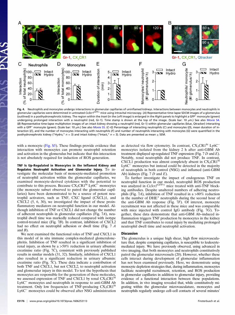
SDCM showed similar neutrophil and monocyte numbers and dwelltimes under control and inflammatory conditions, compared withour findings obtained with multiphoton microscopy (Fig. S4 andref. 20). In the absence of inflammation, neutrophils (exclusivelyGr-1+ cells) formed contacts with monocytes (exclusively GFP+)present in glomerular capillaries. This behavior was observed inboth posthydronephrotic and intact kidneys (Fig. 4 A and B andMovies S4 and S5), indicating that this behavior was unrelated toinduction of hydronephrosis. Analysis of monocyte–neutrophilinteractions demonstrated that ∼35% of all neutrophils (Fig. 4C)and ∼50% of all monocytes (Fig. 4D) retained in glomerularcapillaries underwent interactions with the other cell type. Inuninflamed glomeruli, these interactions lasted on average for3 min (Fig. 4E). Neutrophils typically underwent physical contactwith one monocyte during their retention in glomerular capil-laries whereas each monocyte interacted with approximately twoneutrophils (Fig. 4 F and G).We next investigated whether these interactions were altered
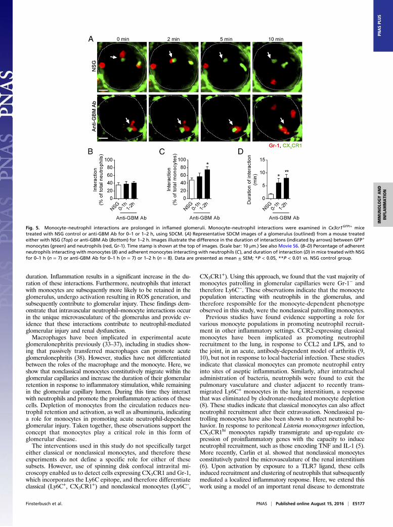
during glomerular inflammation, comparing control, normal sheepglobulin (NSG)-treated, and anti-GBM Ab–treated mice. As seenin untreated mice, monocyte–neutrophil interactions were fre-quent in NSG-treated mice (Fig. 5 A–C), with a typical duration of2–3 min (Fig. 5 A and D). During inflammation, the duration ofinteractions between neutrophils and monocytes increased toover 6 or 7 min, 0–1 h, or 1–2 h after injection of anti-GBM Ab,respectively (Fig. 5 A and D and Movie S6). Although thepercentage of neutrophils interacting with monocytes withinglomerular capillaries was similar under control and inflammatoryconditions (Fig. 5B), a greater proportion of monocytes underwentinteractions with neutrophils 1–2 h after anti-GBM Ab adminis-tration (Fig. 5C). Finally, we determined the timing of these inter-actions relative to the entry of the neutrophil into the glomerulus,which revealed that, in inflamed glomeruli, neutrophils form con-tacts with monocytes mainly, but not exclusively, early in their re-tention in the glomerulus (Fig. S5).
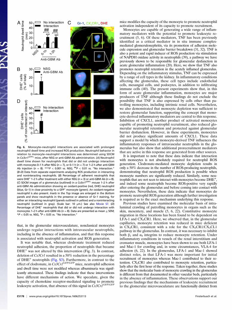
Monocyte–Neutrophil Interactions Are Associated with ProlongedNeutrophil Dwell Time and Neutrophil Activation. The above ob-servations raised the possibility of a relationship betweenmonocyte–neutrophil interactions and neutrophil activation dur-ing acute glomerulonephritis. To assess this hypothesis, we com-pared the responses of neutrophils that underwent interactionwith monocytes with the responses of neutrophils that did not.Under resting conditions (NSG), dwell time was comparablebetween those that had and had not undergone interaction with amonocyte (Fig. 6A). In contrast, during inflammation, neutro-phils that interacted with monocytes showed significantly pro-longed dwell times 0–1 and 1–2 h after anti-GBM Ab injectionwhereas noninteracting neutrophils displayed similar dwell timesto basal levels. These results are consistent with the hypothesisthat interaction with monocytes regulates neutrophil responsesin acute glomerulonephritis.To examine this hypothesis further, we performed additional
experiments to compare ROS generation by interacting andnoninteracting neutrophils. One to 2 h after anti-GBM Ab ad-ministration, ∼15% of intraglomerular neutrophils producedROS as assessed by DHE-derived fluorescence (Fig. 6B), con-sistent with previous observations (20). Interestingly, neutrophilsthat underwent interactions with monocytes were fivefold morelikely to produce ROS, compared with neutrophils that did notinteract with monocytes (Fig. 6 C and D and Movie S7). In ad-dition, comparison of monocyte/neutrophil interactions betweenDHE+ and DHE− neutrophils revealed that the mean dura-tion of the interactions was significantly greater for DHE+ cellscompared with DHE− cells (Fig. S6). However, analysis of thetiming of initiation of ROS production by neutrophils after en-tering the glomerulus indicated that, in ∼50% of cases, neutro-phils became DHE+ before undergoing a detectable interaction
Fig. 3. Monocyte depletion diminishes renal injury and neutrophil re-sponses. C57BL/6 WT mice were pretreated with clodronate liposomes (clod)for monocyte depletion or control liposomes (control) and subsequentlyunderwent the anti-GBM Ab model. (A) Urinary albumin/creatinine ratio inclodronate (n = 6) or control liposome-treated mice (n = 5) after anti-GBMAb administration. (B and C) Neutrophil adhesion (B) and dwell time (C) inmonocyte-depleted (clodronate, n = 5) and control mice (n = 6–7, re-spectively) 1–2 h after anti-GBM Ab injection as determined using multi-photon intravital microscopy. (D) Representative multiphoton images ofcontrol liposome (Top) or clodronate liposome-treated mice (Bottom) 1–2 hafter anti-GBM Ab administration, illustrating neutrophils (green, Gr-1)within glomerular capillaries (blue, Qtracker) being oxidant-positive (red,DHE) (denoted by arrowheads) or oxidant-negative (denoted by asterisks).(Insets) Highlighted neutrophils in the absence of Gr-1 staining. (Scale bar:10 μm.) See also Movies S2 and S3. (E) Number of DHE+ neutrophils in controlliposome-treated (n = 5) and clodronate liposome-treated mice (n = 6) 1–2 hafter anti-GBM Ab administration. Data are presented as mean ± SEM; *P <0.05, **P < 0.01, ***P < 0.001 vs. corresponding control group.
Finsterbusch et al. PNAS | Published online August 15, 2016 | E5175
IMMUNOLO
GYAND
INFLAMMATION
PNASPL
US
with a monocyte (Fig. S5). These findings provide evidence thatinteraction with monocytes can promote neutrophil retentionand activation in the glomerulus but indicate that this interactionis not absolutely required for induction of ROS generation.
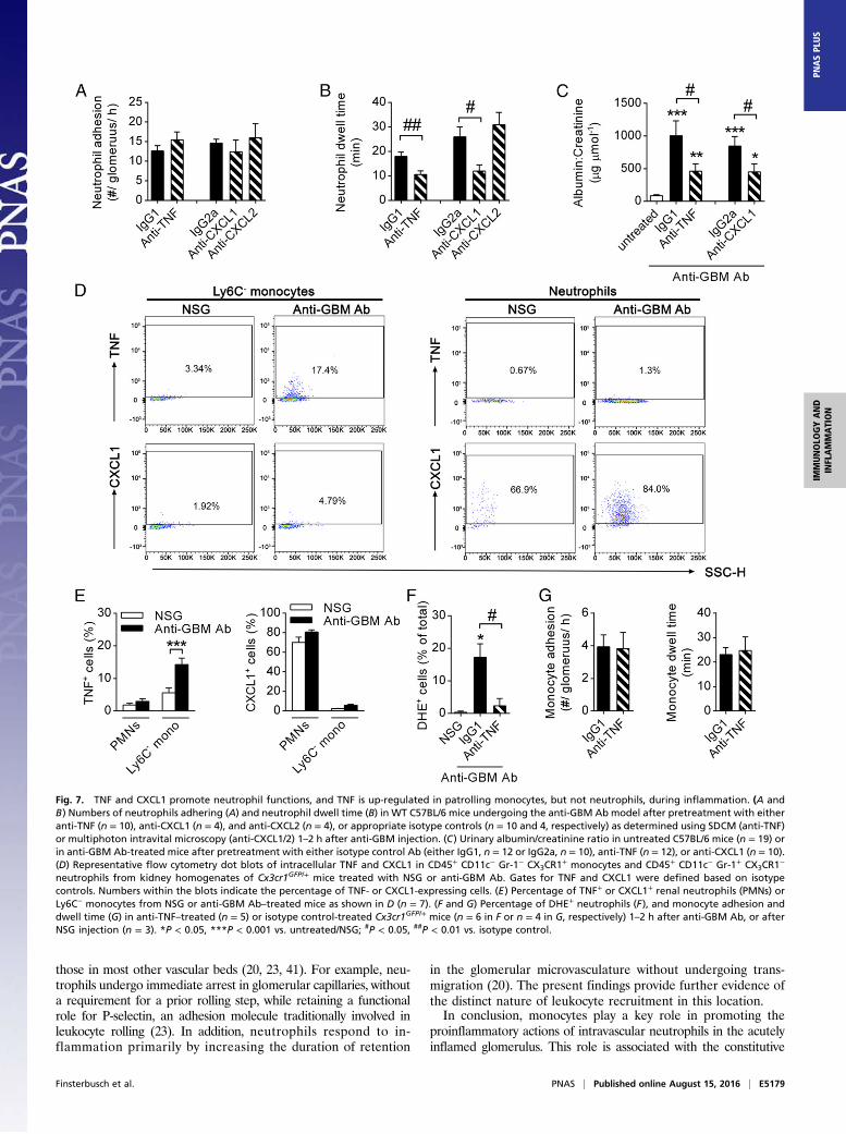
TNF Is Up-Regulated in Monocytes in the Inflamed Kidney andRegulates Neutrophil Activation and Glomerular Injury. To in-vestigate the molecular basis of monocyte-mediated promotionof neutrophil activation within the glomerular capillaries, weexamined monocyte-derived cytokines with the potential tocontribute to this process. Because CX3CR1hi Ly6C− monocytes(the monocyte subset observed to patrol the glomerular capil-laries) have been demonstrated to be a source of potent neu-trophil activators, such as TNF, CXC ligand (CXCL)1, andCXCL2 (5, 6, 30), we investigated the impact of these proin-flammatory mediators on neutrophil function in our model. Al-though inhibition of TNF or CXCL1 did not change the numberof adherent neutrophils in glomerular capillaries (Fig. 7A), neu-trophil dwell time was markedly reduced compared with isotypecontrol-treated mice (Fig. 7B). In contrast, inhibition of CXCL2had no effect on neutrophil adhesion or dwell time (Fig. 7 Aand B).We next examined the functional roles of TNF and CXCL1 in
this model of in situ immune complex-mediated glomerulone-phritis. Inhibition of TNF resulted in a significant inhibition ofrenal injury, as shown by a >50% reduction in urinary albumin:creatinine ratio (Fig. 7C), consistent with previously publishedresults in similar models (31, 32). Similarly, inhibition of CXCL1also resulted in a significant reduction in urinary albumin:creatinine ratio (Fig. 7C). These data indicate a contribution ofboth TNF and CXCL1, but not CXCL2, to neutrophil activationand glomerular injury in this model. To test the hypothesis thatmonocytes are responsible for the generation of these molecules,we assessed expression of TNF and CXCL1 by renal CX3CR1hi
Ly6C− monocytes and neutrophils in response to anti-GBM Abtreatment. Only low frequencies of TNF-producing CX3CR1hi
Ly6C− monocytes could be observed after NSG administration
as detected via flow cytometry. In contrast, CX3CR1hi Ly6C−
monocytes isolated from the kidney 2 h after anti-GBM Abtreatment displayed up-regulated TNF expression (Fig. 7 D and E).Notably, renal neutrophils did not produce TNF. In contrast,CXCL1 production was almost completely absent in CX3CR1hi
Ly6C− monocytes but instead could be detected in the majorityof neutrophils in both control (NSG) and inflamed (anti-GBMAb) kidneys (Fig. 7 D and E).To further investigate the impact of endogenous TNF on
neutrophil function in our model, neutrophil ROS productionwas analyzed in Cx3cr1GFP/+ mice treated with anti-TNF block-ing antibodies. Despite unaltered numbers of adhering neutro-phils (Fig. 7A), inhibition of TNF resulted in a >80% reductionin the number of DHE+ neutrophils during the second hour ofthe anti-GBM Ab response (Fig. 7F). Of interest, monocyterecruitment was not affected in these mice and was comparablewith mice injected with control IgG antibody (Fig. 7G). To-gether, these data demonstrate that anti-GBM Ab–induced in-flammation triggers TNF production by monocytes in the kidneyand that TNF contributes to renal injury by mediating prolongedneutrophil dwell time and neutrophil activation.
DiscussionThe glomerulus is a unique high shear, high flow microvascula-ture that, despite comprising capillaries, is susceptible to leukocyte-mediated injury. We have previously observed, using advanced invivo imaging, that both monocytes and neutrophils constitutivelypatrol the glomerular microvessels (20). However, whether thesecells interact during development of glomerular inflammationhas not been examined previously. Here, we demonstrate usingmonocyte depletion strategies that, during inflammation, monocytesfacilitate neutrophil recruitment, retention, and ROS productionin glomerular capillaries in addition to glomerular injury, providingevidence of a functional interaction between these populations.In addition, in vivo imaging revealed that, while constitutively mi-grating within the glomerular microvasculature, monocytes andneutrophils regularly undergo cell–cell contacts of several minutes’
Fig. 4. Neutrophils and monocytes undergo interactions in glomerular capillaries of uninflamed kidneys. Interactions between monocytes and neutrophils inglomerular capillaries were determined in untreated Cx3cr1GFP/+ mice using intravital microscopy. (A) Representative time-lapse SDCM images of a glomerulus(outlined) in a posthydronephrotic kidney. The region within the Inset (in the Left image) is enlarged in the Right panels to highlight a GFP+ monocyte (green)undergoing prolonged interaction with a neutrophil (red, Gr-1). Time stamp is shown at the top of the image. (Scale bar: 10 μm.) See also Movie S4.(B) Representative time-lapse multiphoton images of an intact kidney showing a neutrophil (red, Gr-1) within glomerular capillaries (blue, Qtracker) interactingwith a GFP+ monocyte (green). (Scale bar: 10 μm.) See also Movie S5. (C–G) Percentage of interacting neutrophils (C) and monocytes (D), mean duration of in-teraction (E), and the number of monocytes interacting with neutrophils (F) and number of neutrophils interacting with monocytes (G) were quantified in theposthydronephrotic kidney (“Hydro,” n = 3) and intact kidney (“Intact,” n = 3). Data are presented as mean ± SEM.
E5176 | www.pnas.org/cgi/doi/10.1073/pnas.1606253113 Finsterbusch et al.
duration. Inflammation results in a significant increase in the du-ration of these interactions. Furthermore, neutrophils that interactwith monocytes are subsequently more likely to be retained in theglomerulus, undergo activation resulting in ROS generation, andsubsequently contribute to glomerular injury. These findings dem-onstrate that intravascular neutrophil–monocyte interactions occurin the unique microvasculature of the glomerulus and provide ev-idence that these interactions contribute to neutrophil-mediatedglomerular injury and renal dysfunction.Macrophages have been implicated in experimental acute
glomerulonephritis previously (33–37), including in studies show-ing that passively transferred macrophages can promote acuteglomerulonephritis (38). However, studies have not differentiatedbetween the roles of the macrophage and the monocyte. Here, weshow that nonclassical monocytes constitutively migrate within theglomerular capillaries and increase the duration of their glomerularretention in response to inflammatory stimulation, while remainingin the glomerular capillary lumen. During this time they interactwith neutrophils and promote the proinflammatory actions of thesecells. Depletion of monocytes from the circulation reduces neu-trophil retention and activation, as well as albuminuria, indicatinga role for monocytes in promoting acute neutrophil-dependentglomerular injury. Taken together, these observations support theconcept that monocytes play a critical role in this form ofglomerular disease.The interventions used in this study do not specifically target
either classical or nonclassical monocytes, and therefore theseexperiments do not define a specific role for either of thesesubsets. However, use of spinning disk confocal intravital mi-croscopy enabled us to detect cells expressing CX3CR1 and Gr-1,which incorporates the Ly6C epitope, and therefore differentiateclassical (Ly6C+, CX3CR1
+) and nonclassical monocytes (Ly6C−,
CX3CR1+). Using this approach, we found that the vast majority of
monocytes patrolling in glomerular capillaries were Gr-1− andtherefore Ly6C−. These observations indicate that the monocytepopulation interacting with neutrophils in the glomerulus, andtherefore responsible for the monocyte-dependent phenotypeobserved in this study, were the nonclassical patrolling monocytes.Previous studies have found evidence supporting a role for
various monocyte populations in promoting neutrophil recruit-ment in other inflammatory settings. CCR2-expressing classicalmonocytes have been implicated as promoting neutrophilrecruitment to the lung, in response to CCL2 and LPS, and tothe joint, in an acute, antibody-dependent model of arthritis (9,10), but not in response to local bacterial infection. These studiesindicate that classical monocytes can promote neutrophil entryinto sites of aseptic inflammation. Similarly, after intratrachealadministration of bacteria, neutrophils were found to exit thepulmonary vasculature and cluster adjacent to recently trans-migrated Ly6C+ monocytes in the lung interstitium, a responsethat was eliminated by clodronate-mediated monocyte depletion(8). These studies indicate that classical monocytes can also affectneutrophil recruitment after their extravasation. Nonclassical pa-trolling monocytes have also been shown to affect neutrophil be-havior. In response to peritoneal Listeria monocytogenes infection,CX3CR1
hi monocytes rapidly transmigrate and up-regulate ex-pression of proinflammatory genes with the capacity to induceneutrophil recruitment, such as those encoding TNF and IL-1 (5).More recently, Carlin et al. showed that nonclassical monocytesconstitutively patrol the microvasculature of the renal interstitium(6). Upon activation by exposure to a TLR7 ligand, these cellsinduced recruitment and clustering of neutrophils that subsequentlymediated a localized inflammatory response. Here, we extend thiswork using a model of an important renal disease to demonstrate
Fig. 5. Monocyte–neutrophil interactions are prolonged in inflamed glomeruli. Monocyte–neutrophil interactions were examined in Cx3cr1GFP/+ micetreated with NSG control or anti-GBM Ab for 0–1 or 1–2 h, using SDCM. (A) Representative SDCM images of a glomerulus (outlined) from a mouse treatedeither with NSG (Top) or anti-GBM Ab (Bottom) for 1–2 h. Images illustrate the difference in the duration of interactions (indicated by arrows) between GFP+
monocytes (green) and neutrophils (red, Gr-1). Time stamp is shown at the top of images. (Scale bar: 10 μm.) See also Movie S6. (B–D) Percentage of adherentneutrophils interacting with monocytes (B) and adherent monocytes interacting with neutrophils (C), and duration of interaction (D) in mice treated with NSGfor 0–1 h (n = 7) or anti-GBM Ab for 0–1 h (n = 7) or 1–2 h (n = 8). Data are presented as mean ± SEM; *P < 0.05, **P < 0.01 vs. NSG control group.
Finsterbusch et al. PNAS | Published online August 15, 2016 | E5177
IMMUNOLO
GYAND
INFLAMMATION
PNASPL
US
that, in the glomerular microvasculature, nonclassical monocytesundergo regular interactions with intravascular neutrophils,including in the absence of inflammation, and that this responseis associated with neutrophil activation and ROS generation.It was notable that, whereas clodronate treatment reduced
neutrophil adhesion, the proportion of neutrophils that becameDHE+ was not altered by this intervention (Fig. 3). In contrast,deletion of Cx3Cr1 resulted in a 50% reduction in the percentageof DHE+ neutrophils (Fig. S3). Furthermore, in contrast to theeffect of clodronate, in Cx3Cr1GFP/GFP mice, neutrophil adhesionand dwell time were not modified whereas albuminuria was signif-icantly attenuated. These findings indicate that these interventionshave different mechanisms of action. We speculate, given thecapacity of chemokine receptor-mediated signaling to promoteleukocyte activation, that absence of this signal in Cx3Cr1GFP/GFP
mice modifies the capacity of the monocyte to promote neutrophilactivation independent of its capacity to promote recruitment.Monocytes are capable of generating a wide range of inflam-
matory mediators with the potential to promote leukocyte re-cruitment (5, 6). Of these mediators, TNF has been previouslyidentified as a critical mediator in in situ immune complex-mediated glomerulonephritis, via its promotion of adhesion mole-cule expression and glomerular barrier breakdown (31, 32). TNF isalso a potent and rapid inducer of ROS production via stimulationof NAPDH oxidase activity in neutrophils (39), a pathway we havepreviously shown to be responsible for glomerular dysfunction inacute glomerular inflammation (20). Here, we show that TNF alsopromotes neutrophil retention in the acutely inflamed glomerulus.Depending on the inflammatory stimulus, TNF can be expressedby a range of cell types in the kidney. In inflammatory conditionsaffecting the glomerulus, these cell types include endothelialcells, mesangial cells, and podocytes, in addition to infiltratingimmune cells (40). The present experiments show that, in thisform of acute glomerular inflammation, monocytes are majorproducers of TNF although these findings do not exclude thepossibility that TNF is also expressed by cells other than pa-trolling monocytes, including intrinsic renal cells. Nevertheless,we also demonstrated that monocyte depletion was sufficient torestore glomerular function, supporting the concept that mono-cyte-derived inflammatory mediators are central to this response.Inhibition of CXCL1, another product of activated monocytescapable of promoting neutrophil recruitment, also reduced glo-merular neutrophil retention and protected against glomerularbarrier dysfunction. However, in these experiments, monocytesdid not produce significant amounts of CXCL1. These datasupport a model by which monocyte-derived TNF influences theinflammatory responses of intravascular neutrophils in the glo-merulus but also show that additional prorecruitment mediatorsthat are active in this response are generated from other sources.It is important to note that these data indicate that interaction
with monocytes is not absolutely required for neutrophil ROSgeneration. Clodronate-mediated monocyte depletion results inonly a 50% decrease in the number of ROS-producing neutrophils,demonstrating that neutrophil ROS production is possible whenmonocyte numbers are significantly reduced. Similarly, some neu-trophils that are not seen to interact with monocytes become DHE+,and indeed some neutrophils become ROS-positive very rapidlyafter entering the glomerulus and before coming into contact withmonocytes. Nevertheless, these data indicate that monocytes dopromote neutrophil ROS generation although further investigationis required as to the exact mechanism underlying this response.Previous studies have examined the molecular basis of intra-
luminal crawling of patrolling monocytes in organs such as theskin, mesentery, and muscle (5, 6, 22). Constitutive monocytemigration in these locations has been found to be dependent onLFA-1 and CX3CR1. Here, we observed that, in the glomerularcapillaries, monocyte retention was reduced in mice deficientin CX3CR1, consistent with a role for the CX3CR1/CX3CL1pathway in the glomerulus. In contrast, it was necessary to inhibitboth β2 and α4 integrins to reduce monocyte retention. Underinflammatory conditions in vessels of the renal interstitium andcremaster muscle, monocytes have been shown to use both LFA-1and Mac-1 for crawling and, in some circumstances, VLA-4 foradhesion (6, 22). In the glomerulus, LFA-1 and Mac-1 showeddistinct roles, in that LFA-1 was more important for initialrecruitment of monocytes whereas Mac-1 contributed to their re-tention. CX3CR1 also contributed to monocyte retention, particu-larly after the first hour of the response. Taken together, these studiesshow that the molecular basis of monocyte crawling in the glomerulusis different from that documented in other vascular beds, particularlyin the absence of inflammation. These observations support ourprevious findings that the mechanisms of leukocyte recruitmentto the glomerular microvasculature are functionally distinct from
Fig. 6. Monocyte–neutrophil interactions are associated with prolongedneutrophil dwell time and increased ROS production. Neutrophil behavior inrelation to monocyte–neutrophil interactions was determined using SDCMin Cx3cr1GFP/+ mice, after NSG or anti-GBM Ab administration. (A) Neutrophildwell time shown for neutrophils that did or did not undergo interactionwith monocytes 0–1 h after NSG (n = 7), or 0–1 h (n = 7) or 1–2 h after anti-GBMAb injection (n = 8). ***P < 0.001 vs. NSG; ##P < 0.01 vs. “No interaction.”(B–D) Data from separate experiments analyzing ROS production in interactingand noninteracting neutrophils. (B) Percentage of adherent neutrophils thatwere DHE+ 1–2 h after treatment with either NSG (n = 3) or anti-GBM Ab (n = 8).(C) SDCM images of a glomerulus (outlined) in a Cx3cr1GFP/+ mouse 1–2 h afteranti-GBM Ab administration showing an oxidant-positive (red, DHE) neutrophil(blue, Gr-1) in close proximity to a GFP+ monocyte (green). An oxidant-negativeneutrophil is also present. Insets in the Top image are enlarged in the Bottompanels and show neutrophils in the presence or absence of Gr-1 staining, foreither an interacting neutrophil (panels outlined in yellow) and a noninteractingneutrophil (outlined in gray). (Scale bar: 10 μm.) See also Movie S7. (D)Percentage of DHE+ neutrophils that did or did not undergo interaction withmonocytes 1–2 h after anti-GBM Ab (n = 8). Data are presented as mean ± SEM;*P < 0.05 vs. NSG, #P < 0.05 vs. “No interaction.”
E5178 | www.pnas.org/cgi/doi/10.1073/pnas.1606253113 Finsterbusch et al.
those in most other vascular beds (20, 23, 41). For example, neu-trophils undergo immediate arrest in glomerular capillaries, withouta requirement for a prior rolling step, while retaining a functionalrole for P-selectin, an adhesion molecule traditionally involved inleukocyte rolling (23). In addition, neutrophils respond to in-flammation primarily by increasing the duration of retention
in the glomerular microvasculature without undergoing trans-migration (20). The present findings provide further evidence ofthe distinct nature of leukocyte recruitment in this location.In conclusion, monocytes play a key role in promoting the
proinflammatory actions of intravascular neutrophils in the acutelyinflamed glomerulus. This role is associated with the constitutive
Fig. 7. TNF and CXCL1 promote neutrophil functions, and TNF is up-regulated in patrolling monocytes, but not neutrophils, during inflammation. (A andB) Numbers of neutrophils adhering (A) and neutrophil dwell time (B) in WT C57BL/6 mice undergoing the anti-GBM Ab model after pretreatment with eitheranti-TNF (n = 10), anti-CXCL1 (n = 4), and anti-CXCL2 (n = 4), or appropriate isotype controls (n = 10 and 4, respectively) as determined using SDCM (anti-TNF)or multiphoton intravital microscopy (anti-CXCL1/2) 1–2 h after anti-GBM injection. (C) Urinary albumin/creatinine ratio in untreated C57BL/6 mice (n = 19) orin anti-GBM Ab-treated mice after pretreatment with either isotype control Ab (either IgG1, n = 12 or IgG2a, n = 10), anti-TNF (n = 12), or anti-CXCL1 (n = 10).(D) Representative flow cytometry dot blots of intracellular TNF and CXCL1 in CD45+ CD11c− Gr-1− CX3CR1
+ monocytes and CD45+ CD11c− Gr-1+ CX3CR1−
neutrophils from kidney homogenates of Cx3cr1GFP/+ mice treated with NSG or anti-GBM Ab. Gates for TNF and CXCL1 were defined based on isotypecontrols. Numbers within the blots indicate the percentage of TNF- or CXCL1-expressing cells. (E) Percentage of TNF+ or CXCL1+ renal neutrophils (PMNs) orLy6C− monocytes from NSG or anti-GBM Ab–treated mice as shown in D (n = 7). (F and G) Percentage of DHE+ neutrophils (F), and monocyte adhesion anddwell time (G) in anti-TNF–treated (n = 5) or isotype control-treated Cx3cr1GFP/+ mice (n = 6 in F or n = 4 in G, respectively) 1–2 h after anti-GBM Ab, or afterNSG injection (n = 3). *P < 0.05, ***P < 0.001 vs. untreated/NSG; #P < 0.05, ##P < 0.01 vs. isotype control.
Finsterbusch et al. PNAS | Published online August 15, 2016 | E5179
IMMUNOLO
GYAND
INFLAMMATION
PNASPL
US
migration of both of these populations in the glomerular capillaries,behaviors that allow these cells to regularly encounter each other ona random basis. Under physiological conditions, monocyte patrol-ling may be important in maintaining homeostasis in the glomeru-lus. However, the current studies demonstrate that this ongoingimmune surveillance by monocytes also underpins inappropriateglomerular inflammation in response to nephritogenic stimuli, suchas immune complexes, and mediates glomerular dysfunction underthese conditions.
Materials and MethodsMice. Cx3cr1GFP/+ and Cx3cr1GFP/GFP mice, expressing GFP under the CX3CR1promotor (25), on a C57BL/6 background were bred in-house. C57BL/6 WT micewere obtained from Monash Animal Services. Mice were maintained on astandard chow diet. Male mice were used for most experiments. For imagingthe intact kidney, both male and female mice were used. All experiments wereapproved by the Monash Medical Centre B Animal Ethics Committee.
Antibodies and Reagents. Polyclonal sheep anti-mouse GBM Abs were pre-pared as described previously (23). Normal sheep globulin (NSG) as a controlwas prepared from nonimmune sheep serum using an identical protocol.Anti-mouse Gr-1 (clone RB6-8C5) was grown from hybridoma or purchasedfrom eBioscience and conjugated to Alexa-488, E450, or PE. CD11c-PECy7(N418) was also from eBioscience. Anti-mouse CD45.2-PerCPC5.5 (104) and theblocking antibodies anti-CD18 (GAME-46), anti-LFA-1 (M17/4), and anti-Mac-1(5C6) were purchased from BD Bioscience. Anti-α4 integrin (PS/2) was grown in-house; anti-CXCL1 antibody (48415) and anti-CXCL2 antibody (40605) were fromR&D Systems; and purified and PE-conjugated anti-TNF antibody (MP6-XT22)was from eBioscience. Purified (Bio X Cell) or PE-conjugated (eBioscience) ratIgG1, rat IgG2a, and rat IgG2b were used as control antibodies. Purified anti-mouse Gr-1, anti-mouse CXCL1, and rat IgG2a were conjugated to DyLight-650using a commercially available kit (Thermo Fisher Scientific). DHE and Qtrackernontargeted Quantum Dots-655 were from Thermo Fisher Scientific. Formonocyte depletion experiments, clodronate liposomes and control PBS lipo-somes were purchased from clodronateliposomes.com. Brefeldin A (Sigma-Aldrich) was used for intracellular cytokine detection. Collagenase D andDNase I were purchased from Roche Life Sciences.
Induction of Glomerular Inflammation. To induce glomerulonephritis, anti-GBM Ab (15 mg in 300 μL of saline) was administered intravenously. Ascontrol for these experiments, NSG was used at the same dose. For imagingexperiments, kidneys were examined either 0–1 or 1–2 h after anti-GBMAb administration.
Renal Intravital Imaging.Male mice subjected to unilateral ureteric ligation aspreviously described (23) were used between 16 and 18 wk of age, 12 wkafter undergoing ureteric ligation. For intravital imaging, the posthydrone-phrotic kidney was prepared as previously described (20, 23, 42). Briefly, micewere anesthetized by initial i.p. injection of ketamine hydrochloride (150 mg/kg;Troy Laboratories) and xylazine (10 mg/kg; Pfizer) in saline, and the leftjugular vein was catheterized for administration of further anesthetic,blocking antibodies, fluorochromes, and reagents. Mice were maintained on aheated platform at a constant temperature of 37 °C. The kidney was exteri-orized, drained of urine, and extended over a heated viewing platform using4/0 silk tied to the kidney capsule. The kidney was superfused with saline andcovered with a coverslip. To visualize neutrophils, mice received anti–Gr-1(2 μg; conjugated to Alexa-488, E450, PE, or DyLight 650) i.v. immediatelybefore imaging. For multiphoton microscopy, the vasculature was labeledwith Qtracker nontargeted Quantum Dots-655 (10 μL of 1:10 dilution i.v.).
Preparation of the Intact Kidney for in Vivo Imaging. To image intact kidneys,3- to 4-wk-old Cx3cr1GFP/+ mice were prepared as previously described (20).Mice were anesthetized, catheterized, and maintained on a heated platformas described above. The left kidney was exteriorized through a dorsal inci-sion and immobilized in a heated well incorporated into a custom-builtstage, where it was covered with saline and a coverslip. Superficial glomerulilocated 50–100 μm below the surface of the kidney were visualized with thevascular label Qtracker-655 (10 μL of 1:10 dilution i.v.). To visualize neutro-phils, mice received 2 μg of anti–Gr-1-PE i.v. immediately before imaging viamultiphoton microscopy.
Multiphoton Microscopy. In experiments involving the analysis of eithermonocytes or neutrophils, or imaging of the intact kidney, the renal
microvasculature was examined using a Leica SP5 multiphoton confocalmicroscope (Leica Microsystems), incorporating a 20× water-dipping objec-tive (N.A. 1.0) and a Spectra Physics MaiTai pulsed infrared laser (NewSpec)as previously described (20). Z-stack images of at least three glomeruli permouse (within one field of view) were captured for 1 h. Images were col-lected every 30 s at a resolution of 512 × 512 pixels and a 6-μm step size. Inexperiments involving GFP (Cx3cr1GFP/+ or Cx3cr1GFP/GFP mice), images wereacquired at 900 nm excitation; otherwise, 810-nm excitation was used. Emittedfluorescence was detected by nondescanned detectors with 432–482 nm, 500–550 nm, 575–605 nm, and 625–675 nm emission filters. Predefined settings forlaser power and detector gain were used for all experiments.
Spinning Disk Confocal Microscopy. Most experiments involving the simulta-neous analysis of monocytes and neutrophils were performed using anUltraVIEW VoX spinning disk microscope system (PerkinElmer) mounted onan upright intravital microscope (Axioplan 2 Imaging; Carl Zeiss) equippedwith a 20× water-dipping objective (N.A. 0.5) (43). The preparation wasexcited with 488-, 561-, and 640-nm lasers. Images of at least three glomeruliper mouse (within one field of view) were collected every 30 s for 1 h using a512 × 512-pixel electron-multiplying charge-coupled device camera (C9100-13;Hamamatsu). Exposure times between 0.05 and 0.4 seconds for each channelwere used. Data acquisition was controlled via Volocity (PerkinElmer).
Integrin, CXCL1, CXCL2, and TNF Inhibition. For examination of the function ofadhesion molecules or chemokines/cytokines, antibodies against CD18 (1 mg/kg),LFA-1 (5.6 mg/kg), Mac-1 (3.3 mg/kg), α4 integrin (2mg/kg), TNF (3mg/kg), CXCL1(3.3 mg/kg), or CXCL2 (3.3 mg/kg) or the same dose of the respective isotypecontrols was injected i.v. 5 min before administration of anti-GBM Ab or,if untreated, 5 min before imaging.
Monocyte Depletion. WT or Cx3cr1GFP/+ mice were depleted of circulatingmonocytes using clodronate as previously described (44). Mice were injectedwith 10 μL/g of clodronate liposomes i.v. into the tail vein 24 h before inductionof inflammation. Control mice received the same dose of PBS liposomes. Mul-tiphoton microscopy of the kidney of Cx3cr1GFP/+ mice demonstrated a >80%reduction in patrolling monocytes in glomerular capillaries after clodronatetreatment (control, 6.4 ± 1.8 monocytes per glomerulus per hour; clodronate,1.1 ± 1.5 monocytes per glomerulus per hour; n = 4 and 3, respectively, P < 0.01).
Detection of Reactive Oxygen Species. Generation of ROS in leukocytes in theglomerulus was investigated using the oxidant-sensitive fluorochromeDHE inimaging experiments, as previously described (20). DHE allows detection ofthe intracellular superoxide anion (45). DHE stock was prepared as 15.7 mgin 1.5 mL of DMSO and stored at −20 °C. For experiments, 2 mg/kg (multi-photon microscopy) or 4 mg/kg (spinning disk microscopy) was used. DHEwas diluted in 150 μL of saline warmed up to 60 °C and immediately injectedi.v. into mice prepared for renal intravital microscopy. ROS production wasexamined 20 min after DHE injection and 1–2 h after anti-GBM Ab or NSG ad-ministration. In some experiments, mice were pretreated with clodronate lipo-somes or anti-TNF blocking Ab before anti-GBM Ab and DHE administration.
Image Analysis. Image sequences captured by multiphoton (maximum pro-jections) or spinning disk confocal microscopy were analyzed offline usingIMARIS (Bitplane). The number of monocytes and neutrophils recruited toglomerular capillaries, as well as their duration of retention in the glomerulus(referred to as “dwell time”), were assessed as described previously (20).Briefly, monocytes and neutrophils were defined as adherent if they remainedarrested in the glomerulus for at least two consecutive frames (30 s). Leukocytedwell time was determined based on the number of frames leukocytes persistedin the glomerulus. Leukocytes were also characterized as either static or crawl-ing, according to their behavior within the glomerulus. Neutrophils or mono-cytes were defined as interacting if they were in contact with the other cell typefor at least two consecutive frames (30 s). In addition, the number and per-centage of adherent neutrophils also positive for DHE staining were determined.Three to five glomeruli per mouse were analyzed and averaged, and datawere plotted as mean per mouse (unless stated otherwise).
Assessment of Renal Injury. Renal function was assessed by measuring albu-min and creatinine levels in urine collections using a mouse albumin ELISA kit(Bethyl Laboratories) and an alkaline picric acid method and autoanalyzer,respectively. Urine was collected overnight commencing 2 h after anti-GBMAb administration. Tomeasure albuminuria/creatinine in monocyte-depletedmice, anti-GBM Ab was given 5 h after clodronate or control liposome in-jection. Data were expressed as albumin:creatinine ratio (μg/μmol).
E5180 | www.pnas.org/cgi/doi/10.1073/pnas.1606253113 Finsterbusch et al.
Renal Leukocyte Isolation and Flow Cytometry. For ex vivo analysis of renalcytokine/chemokine production, kidneys of Cx3cr1GFP/+ mice were removed2 h after anti-GBM or NSG treatment (15 mg i.v.). Tissues were finely mincedwith a scalpel and digested for 25 min in collagenase D (1 mg/mL) and DNaseI (100 μg/mL) in RPMI medium containing Brefeldin A (10 μg/mL) at 37 °Cunder agitation. Kidneys were further disrupted mechanically. Suspensionswere then filtered through 100-μm nylon mesh, and red blood cellswere lysed (46). For ex vivo restimulation, isolated cells were cultured at1 × 106 cells per milliliter in the presence of Brefeldin A (10 μg/mL) in RPMIsupplemented with 10% (vol/vol) FCS, penicillin, streptomycin, L-glutamine,and 50 mM β-mercaptoethanol at 37 °C (47). Three hours after restimulation,cells were washed and treated with mouse BD Fc Block (BD Biosciences)before being stained for the surface markers CD45, CD11c, and Gr-1.Subsequently, cells were fixed and permeabilized with a BD Cytofix/Cytoperm kit (BD Biosciences) and stained for intracellular TNF andCXCL1, or using the appropriate isotype controls rat IgG1 and rat IgG2a,respectively.
Flow cytometric data were acquired on a FACSFortessa (BD Biosciences)and analyzed using FlowJo software (Tree Star Inc.). Patrolling monocytes
were defined as CD45+ CD11b+ CD11c− Gr-1− CX3CR1+ cells, and neutrophils
as CD45+ CD11b+ CD11c− Gr-1+ CX3CR1− cells.
Statistics. Data are presented as mean ± SEM of n observations, where nrepresents the number of animals studied. Statistical differences betweengroups were analyzed using Student’s t test or ANOVA followed by Dunnettor Bonferroni multiple comparison tests using GraphPad Prism 6.0 (Graph-Pad Software). P values of <0.05 were considered significant.
Data in this paper will be made available on request after contacting thesenior author via email.
ACKNOWLEDGMENTS. We thank Dr. Camden Lo (Monash Micro Imaging)for assistance with imaging and image analysis and Dr. Sarah Snelgrove(Monash University) for training in multiphoton imaging of the intact kidney.This study was supported by National Health and Medical Research Council(NHMRC), Australia Grants 1045165 and 1064112 (to M.J.H. and A.R.K.),the Rebecca L. Cooper Medical Research Foundation, and ANZ Trustees. M.J.H.is an NHMRC Senior Research Fellow (Grant 1042775). M.F. was supported byan Erwin Schroedinger Fellowship from the Austrian Science Fund (ProjectJ 3752-B28).
1. Grommes J, Soehnlein O (2011) Contribution of neutrophils to acute lung injury. MolMed 17(3-4):293–307.
2. Kurts C, Panzer U, Anders HJ, Rees AJ (2013) The immune system and kidney disease:Basic concepts and clinical implications. Nat Rev Immunol 13(10):738–753.
3. Segel GB, HaltermanMW, LichtmanMA (2011) The paradox of the neutrophil’s role intissue injury. J Leukoc Biol 89(3):359–372.
4. Villanueva E, et al. (2011) Netting neutrophils induce endothelial damage, infiltratetissues, and expose immunostimulatory molecules in systemic lupus erythematosus.J Immunol 187(1):538–552.
5. Auffray C, et al. (2007) Monitoring of blood vessels and tissues by a population ofmonocytes with patrolling behavior. Science 317(5838):666–670.
6. Carlin LM, et al. (2013) Nr4a1-dependent Ly6C(low) monocytes monitor endothelialcells and orchestrate their disposal. Cell 153(2):362–375.
7. Grainger JR, et al. (2013) Inflammatory monocytes regulate pathologic responses tocommensals during acute gastrointestinal infection. Nat Med 19(6):713–721.
8. Kreisel D, et al. (2010) In vivo two-photon imaging reveals monocyte-dependentneutrophil extravasation during pulmonary inflammation. Proc Natl Acad Sci USA107(42):18073–18078.
9. Wang B, et al. (2012) In vivo imaging implicates CCR2(+) monocytes as regulators ofneutrophil recruitment during arthritis. Cell Immunol 278(1-2):103–112.
10. Maus UA, et al. (2003) Monocytes are potent facilitators of alveolar neutrophil emi-gration during lung inflammation: Role of the CCL2-CCR2 axis. J Immunol 170(6):3273–3278.
11. Ziegler-Heitbrock L, et al. (2010) Nomenclature of monocytes and dendritic cells inblood. Blood 116(16):e74–e80.
12. Sunderkötter C, et al. (2004) Subpopulations of mouse blood monocytes differ inmaturation stage and inflammatory response. J Immunol 172(7):4410–4417.
13. Geissmann F, Jung S, Littman DR (2003) Blood monocytes consist of two principalsubsets with distinct migratory properties. Immunity 19(1):71–82.
14. Palframan RT, et al. (2001) Inflammatory chemokine transport and presentation inHEV: A remote control mechanism for monocyte recruitment to lymph nodes in in-flamed tissues. J Exp Med 194(9):1361–1373.
15. Tacke F, et al. (2007) Monocyte subsets differentially employ CCR2, CCR5, and CX3CR1to accumulate within atherosclerotic plaques. J Clin Invest 117(1):185–194.
16. Karlmark KR, et al. (2009) Hepatic recruitment of the inflammatory Gr1+ monocytesubset upon liver injury promotes hepatic fibrosis. Hepatology 50(1):261–274.
17. Hamers AA, et al. (2012) Bone marrow-specific deficiency of nuclear receptor Nur77enhances atherosclerosis. Circ Res 110(3):428–438.
18. Hanna RN, et al. (2012) NR4A1 (Nur77) deletion polarizes macrophages toward aninflammatory phenotype and increases atherosclerosis. Circ Res 110(3):416–427.
19. Nahrendorf M, et al. (2007) The healing myocardium sequentially mobilizes twomonocyte subsets with divergent and complementary functions. J Exp Med 204(12):3037–3047.
20. Devi S, et al. (2013) Multiphoton imaging reveals a new leukocyte recruitment par-adigm in the glomerulus. Nat Med 19(1):107–112.
21. Li W, et al. (2012) Intravital 2-photon imaging of leukocyte trafficking in beatingheart. J Clin Invest 122(7):2499–2508.
22. Sumagin R, Prizant H, Lomakina E, Waugh RE, Sarelius IH (2010) LFA-1 and Mac-1define characteristically different intralumenal crawling and emigration patterns formonocytes and neutrophils in situ. J Immunol 185(11):7057–7066.
23. Kuligowski MP, Kitching AR, Hickey MJ (2006) Leukocyte recruitment to the inflamedglomerulus: A critical role for platelet-derived P-selectin in the absence of rolling.J Immunol 176(11):6991–6999.
24. Kitching AR, Holdsworth SR, Hickey MJ (2008) Targeting leukocytes in immune glo-merular diseases. Curr Med Chem 15(5):448–458.
25. Jung S, et al. (2000) Analysis of fractalkine receptor CX(3)CR1 function by targeteddeletion and green fluorescent protein reporter gene insertion. Mol Cell Biol 20(11):4106–4114.
26. Landsman L, et al. (2009) CX3CR1 is required for monocyte homeostasis and ath-
erogenesis by promoting cell survival. Blood 113(4):963–972.27. Auffray C, et al. (2009) CX3CR1+ CD115+ CD135+ common macrophage/DC precur-
sors and the role of CX3CR1 in their response to inflammation. J Exp Med 206(3):
595–606.28. Misharin AV, et al. (2014) Nonclassical Ly6C(-) monocytes drive the development of
inflammatory arthritis in mice. Cell Reports 9(2):591–604.29. Qian Q, Jutila MA, Van Rooijen N, Cutler JE (1994) Elimination of mouse splenic
macrophages correlates with increased susceptibility to experimental disseminated
candidiasis. J Immunol 152(10):5000–5008.30. Schiwon M, et al. (2014) Crosstalk between sentinel and helper macrophages permits
neutrophil migration into infected uroepithelium. Cell 156(3):456–468.31. Karkar AM, et al. (1995) Modulation of antibody-mediated glomerular injury in vivo
by IL-1ra, soluble IL-1 receptor, and soluble TNF receptor. Kidney Int 48(6):1738–1746.32. Mulligan MS, et al. (1993) Requirements for leukocyte adhesion molecules in neph-
rotoxic nephritis. J Clin Invest 91(2):577–587.33. Schreiner GF, Cotran RS, Pardo V, Unanue ER (1978) A mononuclear cell component in
experimental immunological glomerulonephritis. J Exp Med 147(2):369–384.34. Holdsworth SR, Neale TJ, Wilson CB (1981) Abrogation of macrophage-dependent
injury in experimental glomerulonephritis in the rabbit: Use of an antimacrophage
serum. J Clin Invest 68(3):686–698.35. Thaiss F, et al. (1986) Mediator systems in a passive model of in situ immune complex
glomerulonephritis: Role for complement, polymorphonuclear granulocytes and
monocytes. Lab Invest 54(6):624–635.36. Tang WW, Qi M, Warren JS (1996) Monocyte chemoattractant protein 1 mediates
glomerular macrophage infiltration in anti-GBM Ab GN. Kidney Int 50(2):665–671.37. Huang XR, Kitching AR, Tipping PG, Holdsworth SR (2000) Interleukin-10 inhibits
macrophage-induced glomerular injury. J Am Soc Nephrol 11(2):262–269.38. Ikezumi Y, Hurst LA, Masaki T, Atkins RC, Nikolic-Paterson DJ (2003) Adoptive transfer
studies demonstrate that macrophages can induce proteinuria and mesangial cell
proliferation. Kidney Int 63(1):83–95.39. Morgan MJ, Kim YS, Liu ZG (2008) TNFalpha and reactive oxygen species in necrotic
cell death. Cell Res 18(3):343–349.40. Ernandez T, Mayadas TN (2009) Immunoregulatory role of TNFalpha in inflammatory
kidney diseases. Kidney Int 76(3):262–276.41. Hickey MJ, Westhorpe CL (2013) Imaging inflammatory leukocyte recruitment in
kidney, lung and liver: Challenges to the multi-step paradigm. Immunol Cell Biol
91(4):281–289.42. Devi S, et al. (2010) Platelet recruitment to the inflamed glomerulus occurs via an
alphaIIbbeta3/GPVI-dependent pathway. Am J Pathol 177(3):1131–1142.43. Abeynaike LD, et al. (2014) Regulatory T cells dynamically regulate selectin ligand
function during multiple challenge contact hypersensitivity. J Immunol 193(10):
4934–4944.44. Tacke F, et al. (2006) Immature monocytes acquire antigens from other cells in the
bone marrow and present them to T cells after maturing in the periphery. J Exp Med
203(3):583–597.45. Zhao H, et al. (2005) Detection and characterization of the product of hydroethidine
and intracellular superoxide by HPLC and limitations of fluorescence. Proc Natl Acad
Sci USA 102(16):5727–5732.46. Chan AJ, et al. (2014) Innate IL-17A-producing leukocytes promote acute kidney
injury via inflammasome and Toll-like receptor activation. Am J Pathol 184(5):
1411–1418.47. Honda T, et al. (2014) Tuning of antigen sensitivity by T cell receptor-dependent
negative feedback controls T cell effector function in inflamed tissues. Immunity
40(2):235–247.
Finsterbusch et al. PNAS | Published online August 15, 2016 | E5181
IMMUNOLO
GYAND
INFLAMMATION
PNASPL
US