patric lindqvist-reis - diva portal8740/fulltext01.pdf · patric lindqvist-reis doctoral thesis...
TRANSCRIPT
Structure of solvated metal ions Solution and crystal structure of Ga3+, In3+, Sc3+, Y3+, La3+ and
Ca2+ ions with water and non-aqueous oxygen donor solvents
Patric Lindqvist-Reis
Doctoral Thesis Department of Chemistry Stockholm 2000

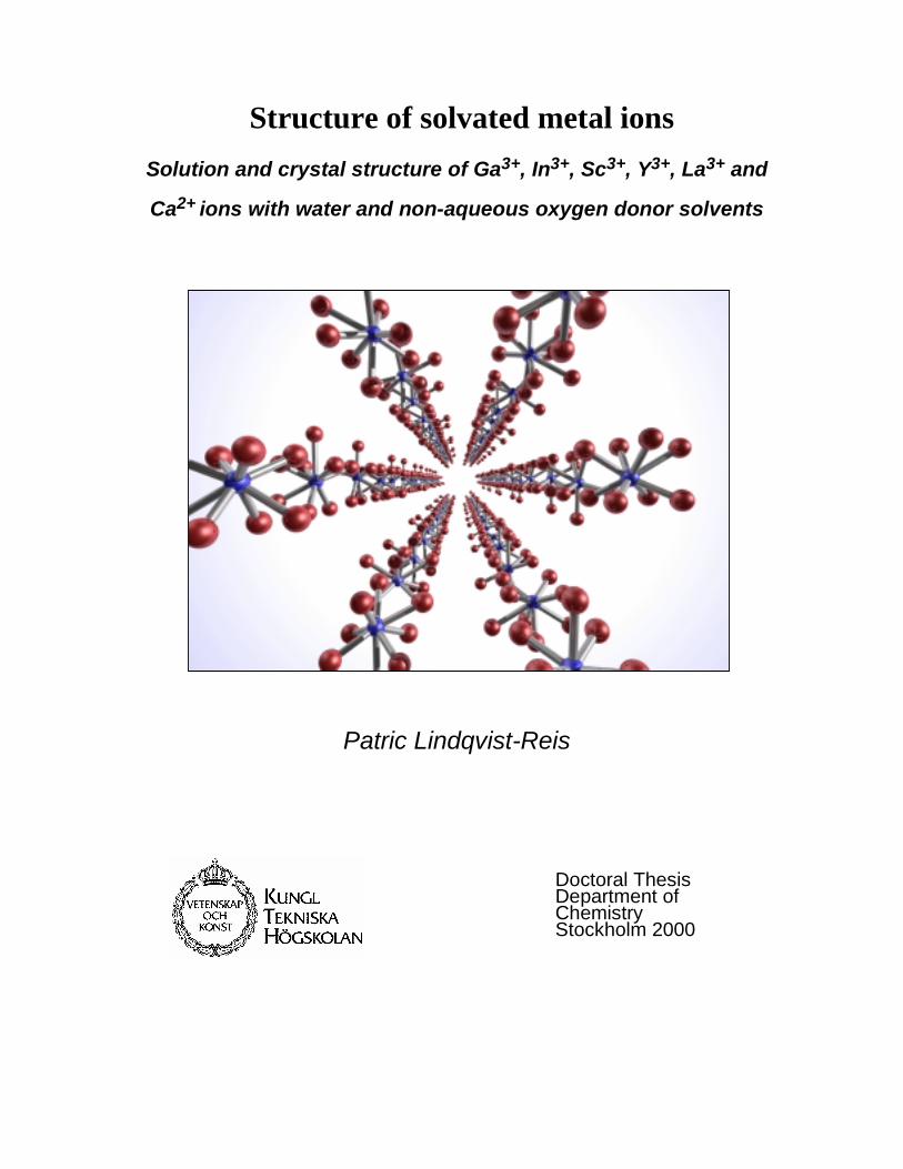
Patric Lindqvist-Reis Department of Chemistry Inorganic Chemistry Royal Institute of Technology S-100 44 Stockholm Sweden Printed by Kista Snabbtryck AB ON THE COVER: Sc(H2O)8
3+ ions in the crystal structure of [Sc(H2O)8](CF3SO3)3
ISBN 91-7170-569-4 ISSN 0348-825X TRITA-OOK-1060
STRUCTURE OF SOLVATED METAL IONS Solution and crystal structure of Ga3+, In3+, Sc3+, Y3+, La3+ and Ca2+ ions with water and non-aqueous oxygen donor solvents Patric Lindqvist-Reis Akademisk avhandling som med tillstånd av Kungliga Tekniska Högskolan i Stockholm framlägges till offentlig granskning för avläggande av filosofie doktorsexamen i oorganisk kemi, onsdagen den 14 juni, kl. 10.00 i kollegiesalen, administrationsbyggnaden, KTH, Vallhallavägen 79, Stockholm Avhandlingen försvaras på engelska.
ROYAL INSTITUTE OF TECHNOLOGY
ii
Abstract The structure of the solvated group 3 ions scandium(III), yttrium(III) and lanthanum(III), has been determined in aqueous solution and in some oxygen donor solvents, and compared with the hydrated group 13 ions gallium(III) and indium(III). A combination of X-ray absorption fine structure (XAFS), large angle X-ray scattering (LAXS), crystallography and vibration spectroscopy has been used for the structure studies of the hydrated ions and their dimethylsulfoxide and N,N´-dimethylpropylene urea solvates in solution and in the solid state. For the hexahydrated gallium(III) and indium(III) ions in solution, the metal-oxygen distances were found to be 1.959(6) Å and 2.131(7) Å to the first hydration shell, and 4.05(1) and 4.13(1) Å, respectively, to fairly well-defined second hydration shells containing about twelve water molecules. The hydration structure of the calcium(II) ion was studied by combining EXAFS and LAXS studies and Molecular Dynamics simulations. The biochemically important calcium ion was found to be octahydrated in aqueous solution with a broad and asymmetric distribution of the Ca-O bond distances around the mean value 2.46(1) Å. Also the yttrium(III) ion was found to be octahydrated in aqueous solution with an asymmetric distribution of the Y-O bond distances around a mean value of 2.366(5) Å. The mean Y-O distance in the eight-coordinated compound ([(H2O)4Y(µ-OSO2CH3)2]CH3SO3)n is 2.35 Å. The hydration of the lanthanum(III) ion was consistent with a coordination of nine water molecules with the La-O distances 6×2.52(2) and 3×2.65(3) Å, probably with the oxygen atoms forming a tricapped trigonal prism. Also for the hydrated scandium(III) ion in aqueous solution, a capped trigonal prismatic coordination figure, with the mean Sc-O bond distance 2.17(1) Å to the water molecules in the prism, gives the best description of the structure. There seems to be 2 or 3 capping water molecules, but the Sc-O distances in the range 2.3-2.5 Å are not well-defined. The crystal structure of [Sc(H2O)8](CF3SO3)3, which at ambient temperature is isomorphous with the [M(H2O)9](CF3SO3)3 compounds with a series of nonahydrated tricapped trigonal prismatic lanthanide(III) ions, was studied at temperatures down to 100 K. One capping water molecule was found to be missing in the [Sc(H2O)8](CF3SO3)3 structure, causing a displacement of the scandium ion about 0.2 Å from the centre of the trigonal prism. A redetermination of the isomorphous [Yb(H2O)n](CF3SO3)3 structure was made, resulting in n ≈ 8.6 for the slightly larger Yb(III) ion. Octahedrally coordinated scandium(III) ions were found in the crystal structures of [Sc(H2O)6][Sc(CH3SO3)6] and trans-[Sc(H2O)4Cl2]Cl⋅2H2O, with mean Sc-O distances of 2.08 and 2.11 Å, respectively. The scandium(III) ion was found to be octahedrally six-coordinated in dimethylsulfoxide solution, with the mean Sc-O distance 2.09 Å, and in the solid [Sc(dmso)6]I3 compound with Sc-O 2.075(3) Å. The larger yttrium(III) ion is eight-coordinated in solution with the mean Y-O distance 2.36(1) Å. In the crystal structure of [Y(dmso)8]I3 the mean Y-O distance is 2.36 Å, while the Y-O distance to the two dimethylsulfoxide ligands in [Y(H2O)6(dmso)2]Cl3 is only 2.27 Å and the mean Y-Owater distance 2.37 Å. Also lanthanum(III) in dimethylsulfoxide solution was found to form an eight-coordinated complex with the mean La-O distance 2.50(2) Å. The N,N´-dimethylpropylene urea solvated scandium(III), yttrium(III) and lanthanum(III) ions were found to have the coordination number six, six and seven in solution, respectively, showing that steric ligand-ligand repulsion effects are controlling the solvation numbers in aprotic solvents for these ions with noble gas electron configuration.

iii
To my Father and Mother, Sister and Brother
iv
Preface This thesis summarises the papers (I-IX) listed below. Four of these (I-IV) are published, papers V and VI are accepted for publication, VIII is submitted for publication, and papers VII and IX are in manuscript. My contribution to most of these papers is substantial except for paper VI where I contributed less. Paper VIII is a collaboration, where my role was initiation; Daniel Spångberg and I worked out how to present the results from the EXAFS/molecular dynamic ‘interface’. Daniel was always computing and Farideh Jalilehvand worked with the experimental EXAFS spectra. The common theme in these papers is the coordination chemistry of the solvated Sc3+, Y3+, La3+ and Ca2+ (d0 ions) and Ga3+ and In3+ (d10 ions) metal ions in aqueous and non-aqueous solutions. Several crystal structure determinations of the solvated metal ions are also included. Most of these compounds have been very useful for our understanding of the solvated ions in solution. Crystal data collection has mainly been performed at Structural Chemistry at Stockholm University and at Department of Chemistry, SLU, in Uppsala. All EXAFS data were collected at SSRL, Stanford, USA. I EXAFS study of the hydration structure of Ga3+ aqueous solution. Comparison of data
from two laboratories Munoz-Paez, A.; Diaz-Moreno, S.; Sanchez Marcos, E.; Martinez, J.M.; Pappalardo,
R.R.; Persson, I.; Sandström, M.; Pattanaik, S.; Lindqvist-Reis, P. J. Phys. IV France 1997, 7, C2-647. II The structure of the hydrated gallium(III), indium(III) and chromium(III) ions in
aqueous Solution. A large angle X-ray scattering and EXAFS study. Lindqvist-Reis, P.; Muñoz-Páez, A.; Díaz-Moreno, S.; Pattanaik, S.; Persson, I.;
Sandström, M. Inorg. Chem. 1998, 37, 6675. III The structure of hexaaqua bis(dimethylsulphoxide)yttrium(III) chloride. Kristiansson, O.; Lindqvist-Reis, P. Acta Crystallogr. Sect. C 1999, 56, 163. IV Hydration of the yttrium(III) ion in aqueous solutions. An X-ray diffraction and XAFS
study. Lindqvist-Reis, P.; Lamble, K.; Pattanaik, S.; Sandström, M.; Persson, I. J. Phys. Chem. B 2000, 104, 402. V Structure of the solvated yttrium(III) ion in the oxygen donor solvents
dimethylsulfoxide, N,N-dimethylformamide and N,N´-dimethylpropylene urea, and crystal structures of [Y(OSMe2 )8]I3 and [Y(OCN2Me2 (CH2 )3 )6]I3.
Lindqvist-Reis, P.; Näslund, J.; Sandström, M.; Persson, I. J. Chem. Soc., Dalton Trans., accepted for publication.
v
VI Steric effects control the structure of the solvated lanthanum(III) ion in aqueous, dimethylsulfoxide and N,N´-dimethylpropylene urea solution. An EXAFS and large angle X-ray scattering study.
Näslund, J.; Lindqvist-Reis, P.; Persson, I.; Sandström, M. Inorg. Chem. accepted for publication. VII Structure of the Solvated Scandium(III) Ion in Aqueous, Dimethylsulfoxide,
N,N´-Dimethylpropylene Urea and Methanol Solution by XAFS and Raman Spectroscopic and X-Ray Diffraction Methods. Crystal Structures of Tetraaquadi-cloroscandium(III) Chloride Dihydrate, Hexakis(dimethylsulfoxide) scandium(III) Iodide and Hexakis(N,N'-dimethylpropylene urea) scandium(III) Iodide
. Lindqvist-Reis, P.; Kristiansson, O.; Pattanaik, S.; Sandström, M.; Persson, I. in manuscript. VIII The Hydration of the Calcium Ion. An EXAFS, Large Angle X-Ray Scattering and
Molecular Dynamics Simulation Study. Jalilehvand, F.; Spångberg, D.; Lindqvist-Reis, P; Hermansson, K.; Persson, I.
Sandström, M. J. Am. Chem. Soc. submitted.
IX The flexible scandium(III) ion with hydration numbers between six and nine in ordered
and disordered crystal hydrates. An X-Ray diffraction and EXAFS study. Lindqvist-Reis, P.; Eriksson, L.; Sandström, M.; Lidin, S.; Persson, I.
in manuscript.
___________________________________________________________________________ Abbreviations
EXAFS Extended X-ray Absorption Fine Structure FT Fourier Transform IR Infrared absorption LAXS Large Angle X-ray Scattering MD Molecular Dynamics RDF Radial Distribution Function XAFS X-ray Absorption Fine Structure XANES X-ray Absorption Near Edge Structure XAS X-ray Absorption Spectroscopy XRD X-Ray Diffraction Triflate Trifluoromethane sulfonate, CF3SO3
-
vi
Table of content
PATRIC LINDQVIST-REIS ...................................................................................................................................1
PREFACE ..........................................................................................................................................................IV
INTRODUCTION ............................................................................................................................................... 1
1.1 GENERAL ..................................................................................................................................................1 1.2 SOLVENTS .................................................................................................................................................2
1.2.1 Water and non-aqueous solvents. ....................................................................................................... 2 1.3 ION HYDRATION / SOLVATION ...................................................................................................................3 1.4 TECHNIQUES FOR STRUCTURE DETERMINATION OF SOLUTIONS .................................................................5
1.4.1 Large angle X-ray scattering (LAXS). ................................................................................................ 5 1.4.2 X-ray absorption spectroscopy (XAS)............................................................................................ 5 1.4.3 IR and Raman spectroscopy. ......................................................................................................... 7
SOLVATED IONS IN CRYSTALLINE COMPOUNDS ................................................................................ 8
2.1 CRYSTALLINE HEXA, HEPTA AND OCTA HYDRATES ...................................................................................8 2.1.1 Hexahydrates. ................................................................................................................................ 8 2.1.2 Heptahydrates................................................................................................................................ 9 2.1.3 Hydrolysis complexes. ................................................................................................................... 9 2.1.4 Octahydrates.................................................................................................................................. 9
2.1 THE ISOMORPHOUS TRIFLATE SERIES ......................................................................................................10 2.1.1 [M(H2O)9](CF3SO3)3. .................................................................................................................. 10 2.1.2 False 9-coordination. .................................................................................................................. 10 2.1.3 Dehydration ⇔ hydration. .......................................................................................................... 14
2.4 SYSTEMS WITH COORDINATING ANIONS ..................................................................................................15 2.4.1 Mesylate complexes. .................................................................................................................... 15 2.4.2 Chloride complexes. .................................................................................................................... 15
2.5 NON-AQUEOUS SOLVATES.......................................................................................................................18 2.6 CONFIGURATIONAL DISORDER.................................................................................................................18
2.6.1 Octaaquascandium(III). .............................................................................................................. 18 2.6.2 Octakis(dimethylsulfoxide)yttrium(III). ....................................................................................... 18
HYDRATED IONS IN SOLUTION ................................................................................................................ 19
3.1 WATER STRUCTURE IN IONIC SOLUTIONS ...............................................................................................19 3.1.1 Double difference IR spectra (OD-region) ....................................................................................... 19
3.2 M-O DISTANCE VS. COORDINATION NUMBER...........................................................................................22 3.3 FIRST AND SECOND HYDRATION SHELLS OF TRIVALENT IONS ..................................................................23
3.3.1 Second hydration shell or multiple scattering? ........................................................................... 25 3.4 COORDINATION GEOMETRY ........................................................................................................................26
3.4.1 M-O stretching vibration. ............................................................................................................ 26
vii
3.4.2 XANES and EXAFS. .................................................................................................................... 26 3.4.3 Evidence against Oh geometry for the hydrated scandium(III) ion. ............................................ 27
3.5 COORDINATION SHELL ASYMMETRY .......................................................................................................28
NON-AQUEOUS SOLUTIONS ....................................................................................................................... 30
4.1 MULTIPLE SCATTERING IN EXAFS .............................................................................................................30 4.1.1 Multiple scattering in the Y-O-C entity........................................................................................ 32
CONCLUSIONS................................................................................................................................................ 33
ACKNOWLEDGEMENTS .............................................................................................................................. 35
REFERENCES .................................................................................................................................................. 36
Chapter 1
Introduction 1.1 General
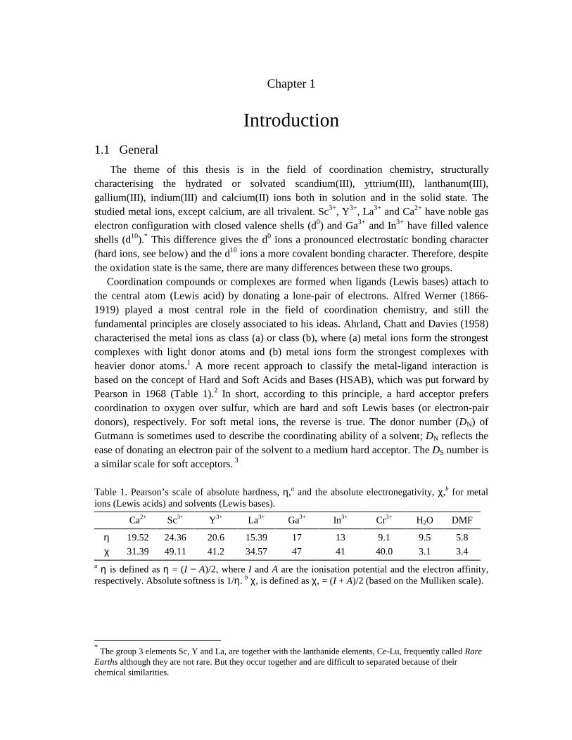
The theme of this thesis is in the field of coordination chemistry, structurally characterising the hydrated or solvated scandium(III), yttrium(III), lanthanum(III), gallium(III), indium(III) and calcium(II) ions both in solution and in the solid state. The studied metal ions, except calcium, are all trivalent. Sc3+, Y3+, La3+ and Ca2+ have noble gas electron configuration with closed valence shells (d0) and Ga3+ and In3+ have filled valence shells (d10).* This difference gives the d0 ions a pronounced electrostatic bonding character (hard ions, see below) and the d10 ions a more covalent bonding character. Therefore, despite the oxidation state is the same, there are many differences between these two groups. Coordination compounds or complexes are formed when ligands (Lewis bases) attach to the central atom (Lewis acid) by donating a lone-pair of electrons. Alfred Werner (1866-1919) played a most central role in the field of coordination chemistry, and still the fundamental principles are closely associated to his ideas. Ahrland, Chatt and Davies (1958) characterised the metal ions as class (a) or class (b), where (a) metal ions form the strongest complexes with light donor atoms and (b) metal ions form the strongest complexes with heavier donor atoms.1 A more recent approach to classify the metal-ligand interaction is based on the concept of Hard and Soft Acids and Bases (HSAB), which was put forward by Pearson in 1968 (Table 1).2 In short, according to this principle, a hard acceptor prefers coordination to oxygen over sulfur, which are hard and soft Lewis bases (or electron-pair donors), respectively. For soft metal ions, the reverse is true. The donor number (DN) of Gutmann is sometimes used to describe the coordinating ability of a solvent; DN reflects the ease of donating an electron pair of the solvent to a medium hard acceptor. The DS number is a similar scale for soft acceptors. 3
Table 1. Pearson’s scale of absolute hardness, η,a and the absolute electronegativity, χ,b for metal ions (Lewis acids) and solvents (Lewis bases).
Ca2+ Sc3+ Y3+ La3+ Ga3+ In3+ Cr3+ H2O DMF
η 19.52 24.36 20.6 15.39 17 13 9.1 9.5 5.8 χ 31.39 49.11 41.2 34.57 47 41 40.0 3.1 3.4
a η is defined as η = (I − A)/2, where I and A are the ionisation potential and the electron affinity, respectively. Absolute softness is 1/η. b χ, is defined as χ, = (I + A)/2 (based on the Mulliken scale).
* The group 3 elements Sc, Y and La, are together with the lanthanide elements, Ce-Lu, frequently called Rare Earths although they are not rare. But they occur together and are difficult to separated because of their chemical similarities.
2
1.2 Solvents 1.2.1 Water and non-aqueous solvents. Although consisting of only two elements the structure of liquid water is very complex. The key to the many anomalous properties of liquid water is the hydrogen bonding, and water is in this regard special being both hydrogen donor and acceptor, O−H⋅⋅⋅O.* In the late 1960s Narten and Levy performed X-ray experiments on water at different temperatures and pressures, 4 and their studies showed that there are approximately four nearest neighbours to each water molecule and the distance between them (O⋅⋅⋅O) is about 2.9 Å.5 The radial distribution function was also found to contain two broad peaks at around 4.5-5.3 and 6.4-7.8 Å. These results indicate that at least part of the bulk structure of liquid water is similar to ice I, and the coordination is approximately tetrahedral. The coordination geometry of one water molecule is also approximately tetrahedral (including the lone pairs) with the angle H-O-H 104.5° and the O-H distances 0.957 Å in the gas phase. A similar model, i.e. where water molecules are hydrogen bonded to four nearest neighbours in an idealised tetrahedral geometry, is often used in diffraction experiments to account for the aqueous bulk structure, although it is a vast simplification of the structure of liquid water. However, all models of liquid water presented so far, both experimental and theoretical, are limited in the sense that they do not explain some of the important properties such as the familiar density anomaly: just above the melting point temperature, warming the liquid causes it to shrink. Alcohols and liquid ammonia are also polar hydrogen-bonded (protic) solvents like water. Dimethylsulfoxide (DMSO), N,N-dimethylformamide (DMF) and N,N´-dimethylpropylene urea (DMPU) are examples of polar, so-called aprotic solvents. The solvent-solvent interactions in the latter class are mostly dipole-dipole in nature, and the bulk structure is much less ordered than for water. However, it has recently been reported that even for some aprotic solvent (e.g. DMSO and DMF) weak hydrogen bonds of the type C-H…O between the solute and solvent can give noticeable effects and influence the structure of the solution. 6 Important physical properties of a solvent are the dipole moment (µ) and the permittivity (ε). Solvents with relatively high µ and ε values dissolve ionic compounds well (Table 2).7 Solvent effects may influence or even control a chemical reaction, for example the rates of dissociative and associative (SN1 and SN2) ligand exchange reactions.
Table 2. Some physical properties of oxygen donor solvents used in this study.a
Solvent Formula Mp / °C Bp / °C ε µ / D DS
water H2O 0.0 100.0 78.5 1.85 17 DMSO (CH3)2SO 18.5 189.0 46.4 3.96 27.5 DMF (CH3)2NCHO -60 150 36.7 3.91 24 DMPU (CH2)3(CH3)2N2CO -38.8 246.5 36.1 4.23 34
a Melting point (Mp), boiling point (Bp), permittivity (ε), dipole moment (µ) and DN number. The DS number is defined as the difference between the Hg-Br stretching vibration of a HgBr2 molecule in the gaseous phase and in the solvent under study.
* In their standard manifestation, hydrogen bonds result from the approach of a proton donor molecule toward an acceptor, forming a bridge of the sort D−H⋅⋅⋅A. The donor D atom is mostly very electronegative, e.g. oxygen, nitrogen or fluorine.
3
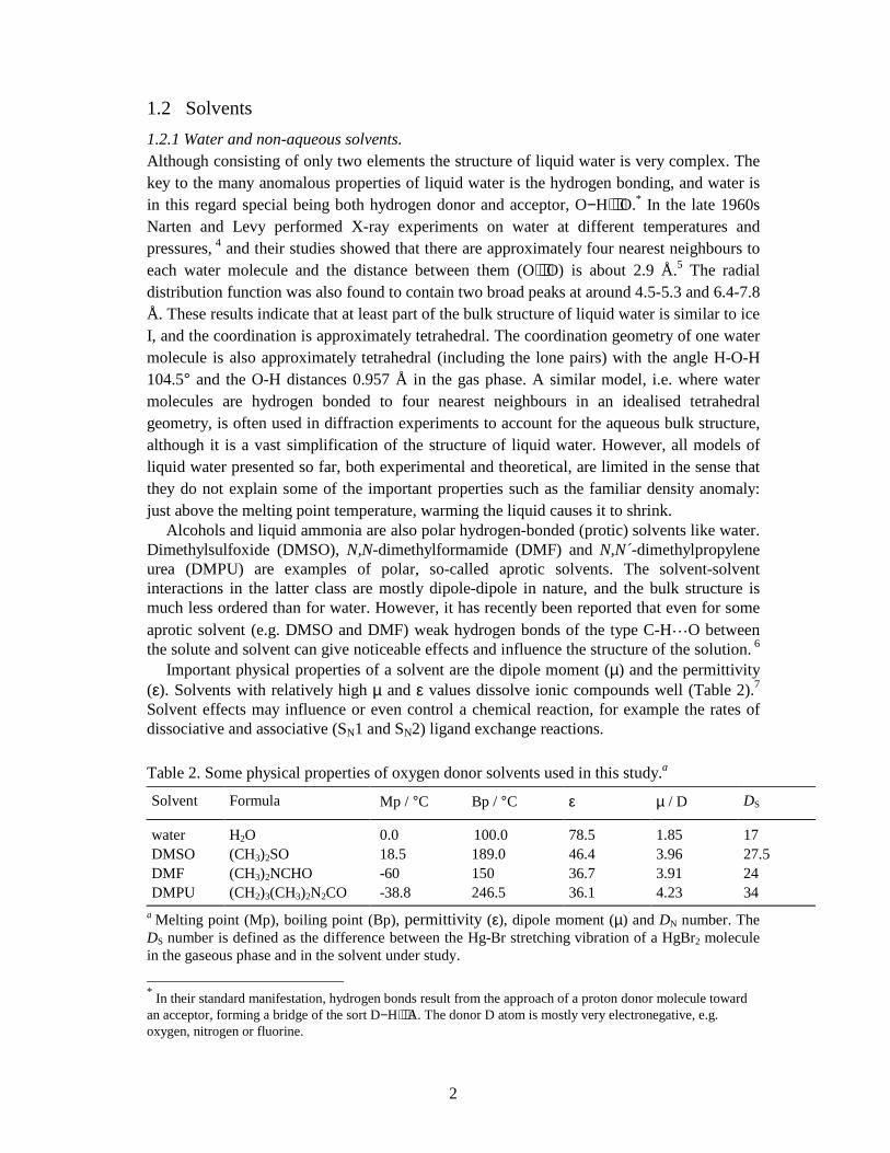
1.3 Ion hydration / solvation In the vicinity of the hydrated/solvated cations some perturbation of the solvent structure will occur. In aqueous solution, the metal ions polarise the surrounding water and form a water layer or hydration shell (Figure 1).8 The polarising effect of a cation is mainly determined by its charge/radius ratio. Highly charged metal ions, such as the trivalent ions of group 3 and 13, have also well-defined second hydration shells, formed by water molecules hydrogen bonded to those in the first shell. A third hydration shell is in most cases too diffuse to be distinguished from the aqueous bulk structure, and for more weakly polarising ions already the second hydration shell is not very distinct. The coordination of water may either be trigonal or pyramidal, i.e. in the former case the angle between the metal and the plane of the water molecule is about zero. Trigonal geometry is considered to be the most important for strongly polarising cations, and pyramidal for the ions with weaker polarisation ability. The pyramidal type of orientation, where a lone pair of the oxygen atom is donated to the metal ion, is most similar to the geometry of a structure unit of liquid water, in which both of the lone pairs are engaged in hydrogen bonding. Anions (e.g. X- = ClO4
-, CF3SO3-,
Cl-, etc.) are hydrated by forming hydrogen bonds to the water molecules X-⋅⋅⋅⋅⋅⋅⋅⋅⋅⋅⋅⋅H-O-H (Figure.2)
Figure 1. Hydration of a metal ion in water showing the formation of hydration shells. Highly charged metal ions (e.g. M3+ of group 3 and 13 ) have also well-defined second hydration shells. Note how the water molecules bind (coordinate) to the metal ion M, forming hydrogen bonds, O-H⋅⋅⋅O). Trigonal orientation of coordinated water molecules is most likely to occur in the inner shell.
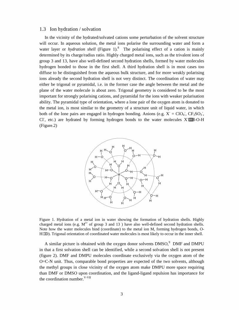
A similar picture is obtained with the oxygen donor solvents DMSO,9 DMF and DMPU in that a first solvation shell can be identified, while a second solvation shell is not present (figure 2). DMF and DMPU molecules coordinate exclusively via the oxygen atom of the O=C-N unit. Thus, comparable bond properties are expected of the two solvents, although the methyl groups in close vicinity of the oxygen atom make DMPU more space requiring than DMF or DMSO upon coordination, and the ligand-ligand repulsion has importance for the coordination number.V-VII
M
H HO H
HO
H
H
OH
H O
HHO
H
H
OH
HO
H
H
O
HHO
HH
OHH
O
H
H
O
HH O
HH
OH
H O
H
HO
H
HO
H
H
O
H
HO
H
H
O
HHO
4
Figure 2. Left: The coordination of water to a chloride ion (from the crystal structure of [Sc(H2O)4Cl2]Cl⋅2H2O, see Paper VIII). Right: Coordination of DMSO to metal ion in [Sc(OS(Me2)6]I3 (Paper VIII, hydrogen atoms are omitted for clarity). The central iodide ion is in contact with the DMSO methyl groups, with the closest C-H…I- distance 3.98 Å. This is in the range of weak attractive C-H…I- interactions (ref. 6, Chapter 3.3), but also the attraction to the distant Sc3+ ion (Sc3+…I- 5.82 Å) may be of importance for the structural arrangement.
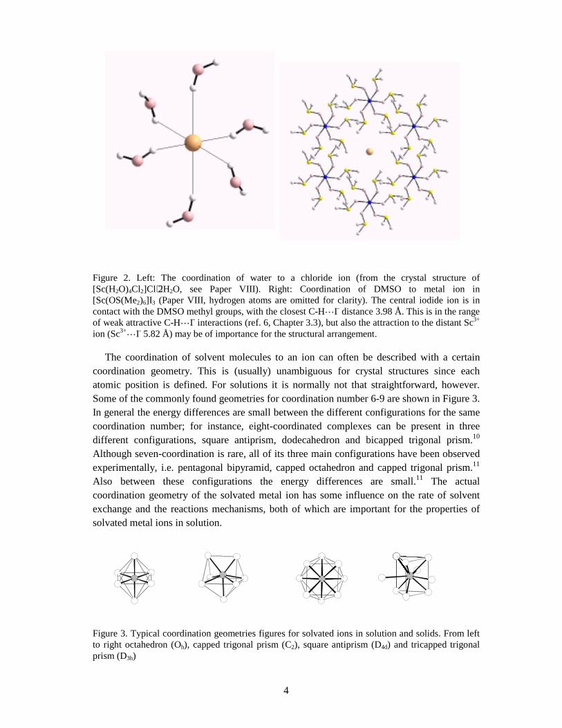
The coordination of solvent molecules to an ion can often be described with a certain coordination geometry. This is (usually) unambiguous for crystal structures since each atomic position is defined. For solutions it is normally not that straightforward, however. Some of the commonly found geometries for coordination number 6-9 are shown in Figure 3. In general the energy differences are small between the different configurations for the same coordination number; for instance, eight-coordinated complexes can be present in three different configurations, square antiprism, dodecahedron and bicapped trigonal prism.10 Although seven-coordination is rare, all of its three main configurations have been observed experimentally, i.e. pentagonal bipyramid, capped octahedron and capped trigonal prism.11 Also between these configurations the energy differences are small.11 The actual coordination geometry of the solvated metal ion has some influence on the rate of solvent exchange and the reactions mechanisms, both of which are important for the properties of solvated metal ions in solution.
Figure 3. Typical coordination geometries figures for solvated ions in solution and solids. From left to right octahedron (Oh), capped trigonal prism (C2), square antiprism (D4d) and tricapped trigonal prism (D3h)
5
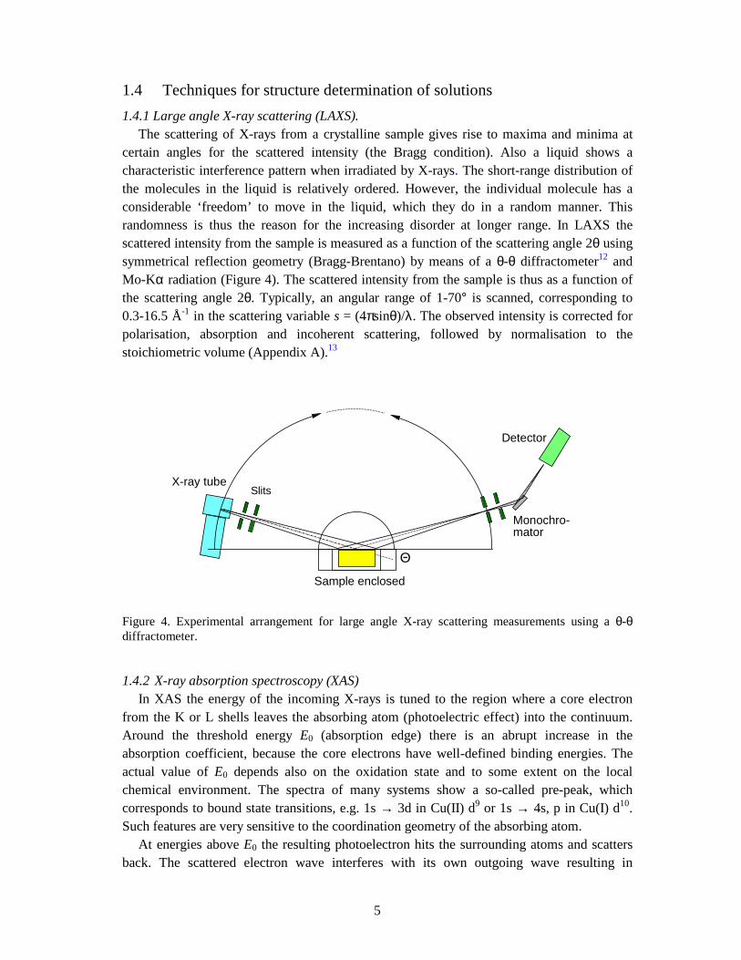
1.4 Techniques for structure determination of solutions 1.4.1 Large angle X-ray scattering (LAXS). The scattering of X-rays from a crystalline sample gives rise to maxima and minima at certain angles for the scattered intensity (the Bragg condition). Also a liquid shows a characteristic interference pattern when irradiated by X-rays. The short-range distribution of the molecules in the liquid is relatively ordered. However, the individual molecule has a considerable ‘freedom’ to move in the liquid, which they do in a random manner. This randomness is thus the reason for the increasing disorder at longer range. In LAXS the scattered intensity from the sample is measured as a function of the scattering angle 2θ using symmetrical reflection geometry (Bragg-Brentano) by means of a θ-θ diffractometer12 and Mo-Kα radiation (Figure 4). The scattered intensity from the sample is thus as a function of the scattering angle 2θ. Typically, an angular range of 1-70° is scanned, corresponding to 0.3-16.5 Å-1 in the scattering variable s = (4πsinθ)/λ. The observed intensity is corrected for polarisation, absorption and incoherent scattering, followed by normalisation to the stoichiometric volume (Appendix A).13
Figure 4. Experimental arrangement for large angle X-ray scattering measurements using a θ-θ diffractometer.
1.4.2 X-ray absorption spectroscopy (XAS) In XAS the energy of the incoming X-rays is tuned to the region where a core electron from the K or L shells leaves the absorbing atom (photoelectric effect) into the continuum. Around the threshold energy E0 (absorption edge) there is an abrupt increase in the absorption coefficient, because the core electrons have well-defined binding energies. The actual value of E0 depends also on the oxidation state and to some extent on the local chemical environment. The spectra of many systems show a so-called pre-peak, which corresponds to bound state transitions, e.g. 1s → 3d in Cu(II) d9 or 1s → 4s, p in Cu(I) d10. Such features are very sensitive to the coordination geometry of the absorbing atom. At energies above E0 the resulting photoelectron hits the surrounding atoms and scatters back. The scattered electron wave interferes with its own outgoing wave resulting in
SlitsX-ray tube
Θ
Sample enclosed
Detector
Monochro-mator
6
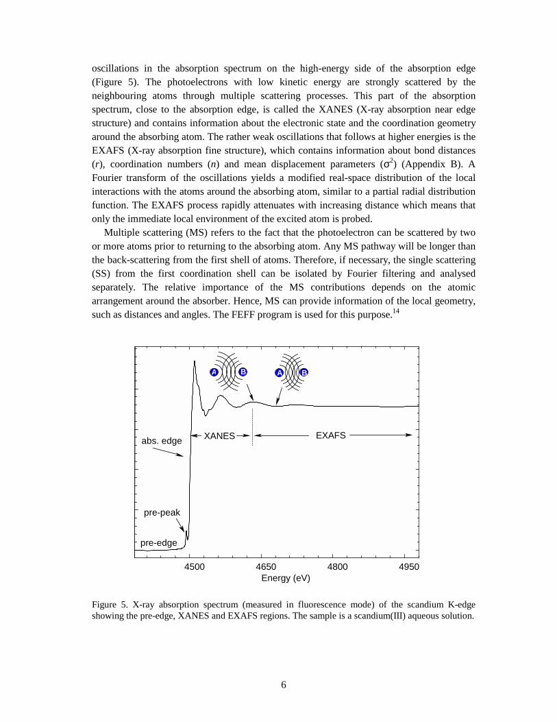
oscillations in the absorption spectrum on the high-energy side of the absorption edge (Figure 5). The photoelectrons with low kinetic energy are strongly scattered by the neighbouring atoms through multiple scattering processes. This part of the absorption spectrum, close to the absorption edge, is called the XANES (X-ray absorption near edge structure) and contains information about the electronic state and the coordination geometry around the absorbing atom. The rather weak oscillations that follows at higher energies is the EXAFS (X-ray absorption fine structure), which contains information about bond distances (r), coordination numbers (n) and mean displacement parameters (σ2) (Appendix B). A Fourier transform of the oscillations yields a modified real-space distribution of the local interactions with the atoms around the absorbing atom, similar to a partial radial distribution function. The EXAFS process rapidly attenuates with increasing distance which means that only the immediate local environment of the excited atom is probed. Multiple scattering (MS) refers to the fact that the photoelectron can be scattered by two or more atoms prior to returning to the absorbing atom. Any MS pathway will be longer than the back-scattering from the first shell of atoms. Therefore, if necessary, the single scattering (SS) from the first coordination shell can be isolated by Fourier filtering and analysed separately. The relative importance of the MS contributions depends on the atomic arrangement around the absorber. Hence, MS can provide information of the local geometry, such as distances and angles. The FEFF program is used for this purpose.14
Figure 5. X-ray absorption spectrum (measured in fluorescence mode) of the scandium K-edge showing the pre-edge, XANES and EXAFS regions. The sample is a scandium(III) aqueous solution.
4500 4650 4800 4950Energy (eV)
pre-edge
pre-peak
abs. edge EXAFSXANES
B BBA BA BB
7
1.4.3 IR and Raman spectroscopy. In theory, symmetry information of a solvated complex can be obtained by combining infrared and Raman spectroscopy.15 In practice, however, this is difficult since many of the bands representing the fundamental vibrations are usually weak and broad and thereby difficult to observe. This is particularly true for hydrated ions in aqueous solution. For such studies high salt concentrations are required in order to make the signal strong enough. Infrared spectroscopy is particularly sensitive for detecting water; i.e. the absorption of the OH stretching vibration in water (∼ 3500 cm-1) is very intense. The change in the OH frequency of the water molecules coordinated to a metal ion, caused by the polarisation effect of the hydrated ion, could then be studied if this absorption can be separated from that of the bulk water, which is the major component. The problem is thus to separate a weak signal of coordinated H2O from that of a large signal originating from the free water molecules. Other problems for interpretation the spectrum are the interaction between the two stretching modes, ν1 and ν2, and the first overtone of the bending vibration, 2ν2, and the combination of these frequencies However, the double difference method seems to overcome these problems.16 In this method one determines the OD or OH vibration spectra of HDO molecules present in the hydration shells of the hydrated ions in isotopically diluted solutions, by subtracting the dominant spectral contribution from HDO molecules present in the bulk water (Appendix C). The presence of HDO in H2O changes the combination band about 2000 cm-1, which makes a double difference necessary. However, the effect of the ions on this combination band is assumed to be negligible. Provided that the subtractions of the spectra can eliminate the contributions from the bulk water structure, the final difference spectrum gives a picture of the distributions of OD (or OH) oscillators differing from the bulk water.
8
Chapter 2
Solvated Ions in Crystalline Compounds A salt, which is crystallized from a solution with strong solute-solvent interactions, often remains solvated in the crystal structure. Such compounds, which incorporate solvated metal ions, can be used as model structures for the local environment of the solvated ions in solution. However, for such comparisons it is necessary that the solvated ions have the same coordination number and similar configuration in solid and solution, even though the second coordination shells are usually rather different. Factors such as packing effects or hydrogen bond arrangement may often influence the coordination geometry of hydrated metal ions in crystalline hydrates; for instance, the local structure of the hydrated scandium ion depends on the property (e.g. size and “shape”) of the counter ion. Thus, scandium crystallises with six water molecules in octahedral coordination geometry with perchlorate as counter ion in the [Sc(H2O)6](ClO4)3 compound, while it is octahydrated with bicapped trigonal prismatic geometry in the triflate salt, [Sc(H2O)8](CF3SO3)3. The d10 ions of group 13 seem in this respect less flexible in changing coordination numbers. For example thallium(III), which has comparable ionic radius to yttrium(III),17 is six-coordinated in the triflate salt, Tl(H2O)3(CF3SO3)3,18 showing that it is unfavourable to exceed coordination number six. There are no examples of hydrated/solvated trivalent ions of group 13 with a coordination number higher than six. In crystal structures where the metal ions are ligated with e.g. DMSO or DMPU solvent molecules, the interaction between the CH3 or CH2 groups of the ligands and the counter ions in the crystal lattice are weak. Ligand-ligand repulsion effects will therefore become important for controlling the coordination number of the metal ions.
2.1 Crystalline hexa, hepta and octa hydrates 2.1.1 Hexahydrates. Structural information of value for modelling the structure (e.g. M-O bond distance) of hexahydrated metal ions in aqueous solution is gained from the structure of the crystalline alum salts, CsI[MIII(H2O)6](SO4)2⋅6H2O (the anion can also be SeO4
2-), where M is a trivalent metal ion in octahedral environment. There are two main modifications of the alum structures, namely the α and β types.19,20,21 The difference between these is the value of the tilt angle of the water molecule with respect to the M-O bond. For β alums θ is ∼ 0° and in the α types ∼ 35° ± 10°, which corresponds to trigonal planar and trigonal pyramidal coordination geometry of the water oxygen atom, respectively (cf. Figure 16). The former geometry appears to be favourable for promoting strong hydrogen bonds to the second coordination shell. The metal ions belonging to the α alums are Co, Rh and Ir and to the β alums Al, Ga, In, Ti, V, Cr, Mo, Mn, Fe and Ru. However, as was shown for the hydrated Ga3+, In3+ and Cr3+ ions, they have tilt angles of about 35° in concentrated aqueous solutions,
9
although the M-O bond distances are in good agreement with those of the corresponding alums.II 2.1.2 Heptahydrates. The heptahydrated scandium(III) ion has been identified in the crystal structure of [Sc(H2O)7][C(SO2CF3)3]3⋅H2O, which is the first and, yet, the only example of the isolated heptahydrated scandium(III) ion.22 The coordination geometry is a distorted capped trigonal prism with an average Sc-O bond distance of 2.17 Å. The isomorphous ytterbium structure ([Yb(H2O)8][C(SO2CF3)3]3⋅H2O) is an octahydrate with distorted square antiprismatic coordination.22 Unfortunately, the crystal data of the scandium structure is of rather low quality, and the water oxygen atoms are disordered. Heptahydrated scandium(III) ions with pentagonal bipyramidal coordination geometry have been suggested to be a stable species in aqueous solution, but a capped prism seems to be more reasonable for [Sc(H2O)7]3+ ions (as above). Ab-initio calculations have shown that the isolated heptahydrated scandium ion has C2 symmetry, i.e. a monocapped trigonal prism (cf. Figure 3).23,24,25
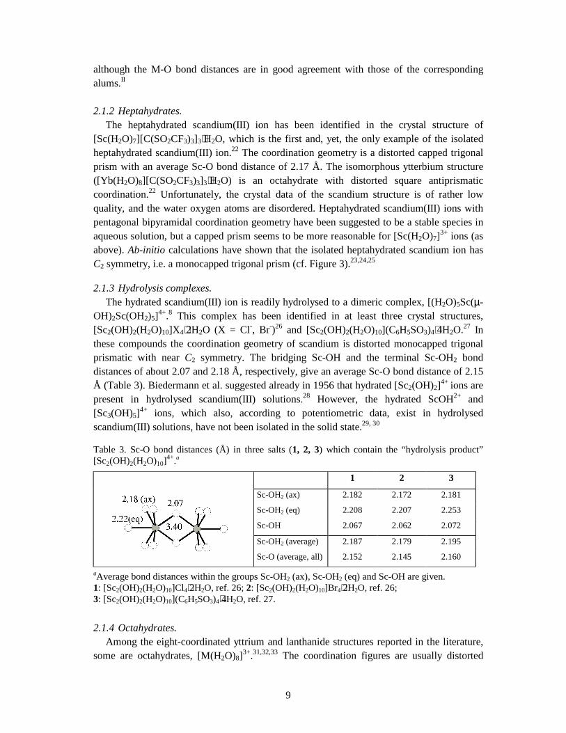
2.1.3 Hydrolysis complexes. The hydrated scandium(III) ion is readily hydrolysed to a dimeric complex, [(H2O)5Sc(µ-OH)2Sc(OH2)5]4+.8 This complex has been identified in at least three crystal structures, [Sc2(OH)2(H2O)10]X4⋅2H2O (X = Cl-, Br-)26 and [Sc2(OH)2(H2O)10](C6H5SO3)4⋅4H2O.27 In these compounds the coordination geometry of scandium is distorted monocapped trigonal prismatic with near C2 symmetry. The bridging Sc-OH and the terminal Sc-OH2 bond distances of about 2.07 and 2.18 Å, respectively, give an average Sc-O bond distance of 2.15 Å (Table 3). Biedermann et al. suggested already in 1956 that hydrated [Sc2(OH)2]4+ ions are present in hydrolysed scandium(III) solutions.28 However, the hydrated ScOH2+ and [Sc3(OH)5]4+ ions, which also, according to potentiometric data, exist in hydrolysed scandium(III) solutions, have not been isolated in the solid state.29, 30
Table 3. Sc-O bond distances (Å) in three salts (1, 2, 3) which contain the “hydrolysis product” [Sc2(OH)2(H2O)10]4+.a
1 2 3
Sc-OH2 (ax) 2.182 2.172 2.181
Sc-OH2 (eq) 2.208 2.207 2.253
Sc-OH 2.067 2.062 2.072
Sc-OH2 (average) 2.187 2.179 2.195 Sc-O (average, all) 2.152 2.145 2.160
aAverage bond distances within the groups Sc-OH2 (ax), Sc-OH2 (eq) and Sc-OH are given. 1: [Sc2(OH)2(H2O)10]Cl4⋅2H2O, ref. 26; 2: [Sc2(OH)2(H2O)10]Br4⋅2H2O, ref. 26; 3: [Sc2(OH)2(H2O)10](C6H5SO3)4⋅4H2O, ref. 27.
2.1.4 Octahydrates. Among the eight-coordinated yttrium and lanthanide structures reported in the literature, some are octahydrates, [M(H2O)8]3+.31,32,33 The coordination figures are usually distorted
10
with a relatively large spread of M-O distances, Figure 1 in Paper IV. One type of such structures are those containing crown ethers, e.g. [M(H2O)8]Cl3⋅(15-crown-5), (M = Y, Gd). The crown ethers stabilise the hydration shell of the metal ion by hydrogen bonding, and thus mimic a second hydration shell in solution. No structures of this kind are known for hepta or nonahydrated ions.
2.1 The Isomorphous triflate series 2.1.1 [M(H2O)9](CF3SO3)3. This series of isomorphous compounds are all crystallising in the space group P63/m where the metal atom of the hydrated complex lies on a 6 site and the oxygen atoms at the vertices of tricapped trigonal prism, i.e. a nine-coordination geometry. Almost all of the lanthanides and yttrium have been structurally characterised and an isomorphous bismuth structure is also known.34,35,36 The tricapped trigonal prismatic coordination geometry comprises two groups of M-O bond distances, where the six within the prism are shorter than to those in the capping positions; the difference increases with decreasing ionic size. There are two other similar series of isomorphous structures containing nonahydrated lanthanide ions in tricapped trigonal prismatic geometry, namely [M(H2O)9](EtOSO3)3 (M = Y, Yb, Ho, Er, Pr) and [M(H2O)9](BrO3)3 (M = Yb, Sm, Pr).37 These are, together with the triflate series, well established and are often referred to in the discussion of coordination geometries of the lanthanide(III) ions in aqueous solution.
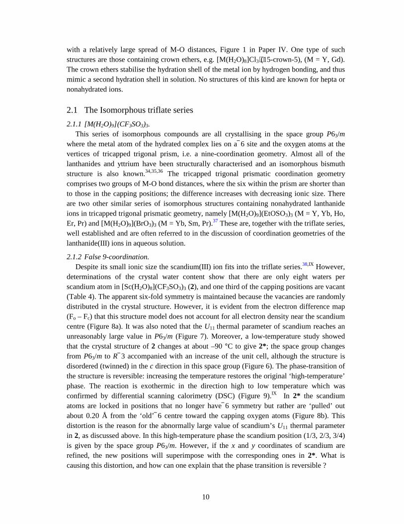
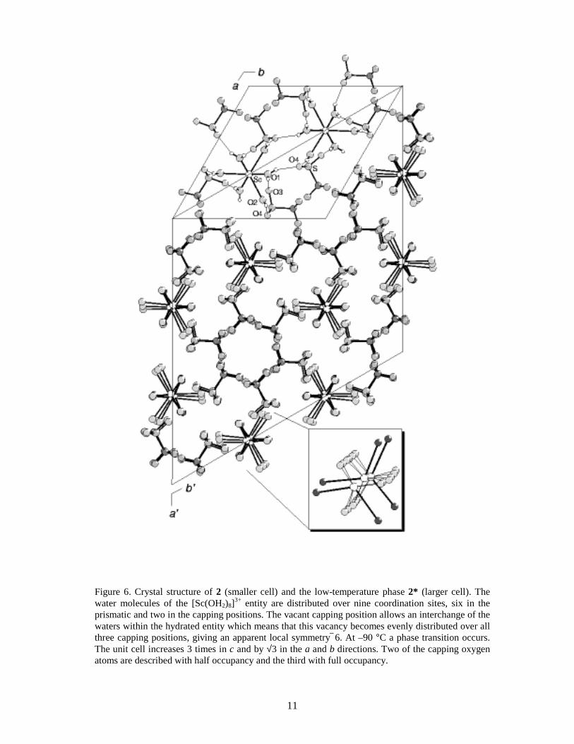
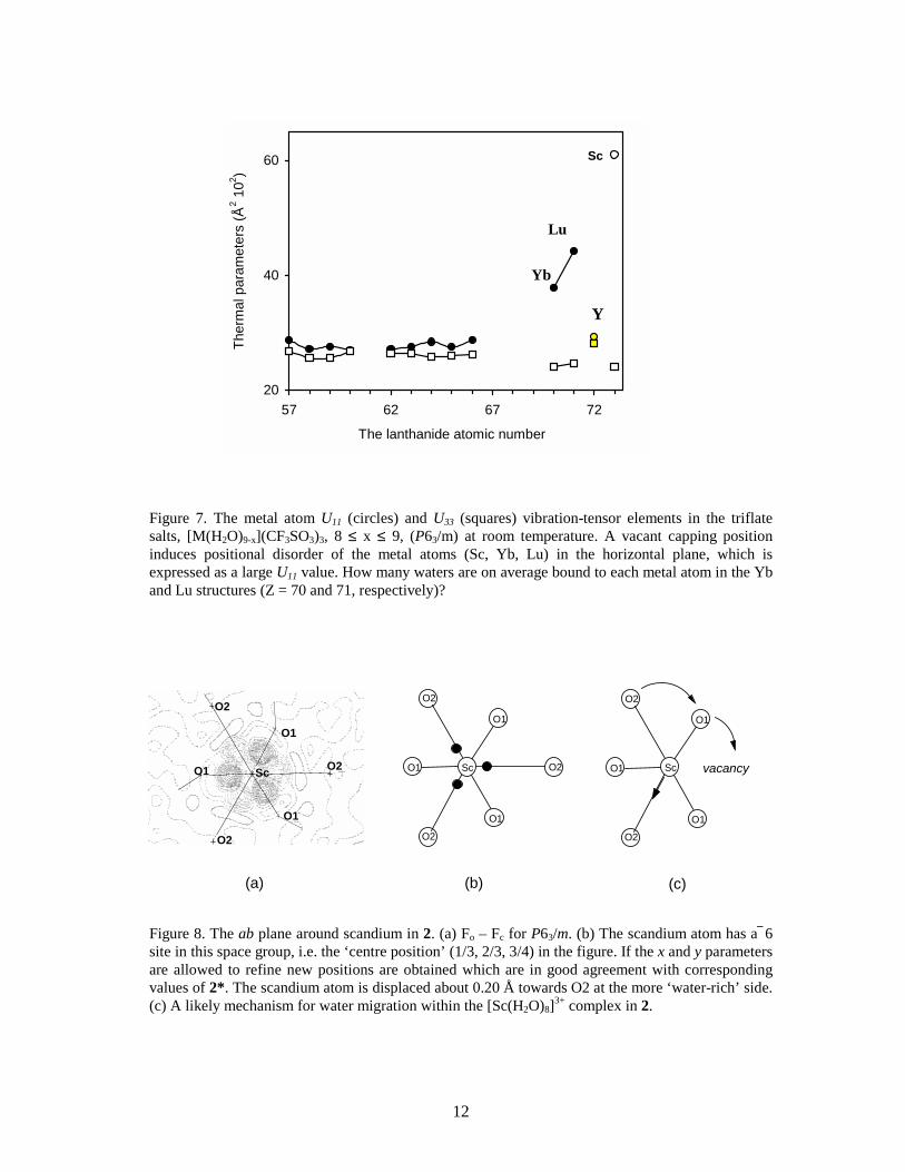
2.1.2 False 9-coordination. Despite its small ionic size the scandium(III) ion fits into the triflate series.38,IX However, determinations of the crystal water content show that there are only eight waters per scandium atom in [Sc(H2O)8](CF3SO3)3 (2), and one third of the capping positions are vacant (Table 4). The apparent six-fold symmetry is maintained because the vacancies are randomly distributed in the crystal structure. However, it is evident from the electron difference map (Fo – Fc) that this structure model does not account for all electron density near the scandium centre (Figure 8a). It was also noted that the U11 thermal parameter of scandium reaches an unreasonably large value in P63/m (Figure 7). Moreover, a low-temperature study showed that the crystal structure of 2 changes at about –90 °C to give 2*; the space group changes from P63/m to R 3 accompanied with an increase of the unit cell, although the structure is disordered (twinned) in the c direction in this space group (Figure 6). The phase-transition of the structure is reversible: increasing the temperature restores the original ‘high-temperature’ phase. The reaction is exothermic in the direction high to low temperature which was confirmed by differential scanning calorimetry (DSC) (Figure 9).IX In 2* the scandium atoms are locked in positions that no longer have 6 symmetry but rather are ‘pulled’ out about 0.20 Å from the ‘old’ 6 centre toward the capping oxygen atoms (Figure 8b). This distortion is the reason for the abnormally large value of scandium’s U11 thermal parameter in 2, as discussed above. In this high-temperature phase the scandium position (1/3, 2/3, 3/4) is given by the space group P63/m. However, if the x and y coordinates of scandium are refined, the new positions will superimpose with the corresponding ones in 2*. What is causing this distortion, and how can one explain that the phase transition is reversible ?
11
Figure 6. Crystal structure of 2 (smaller cell) and the low-temperature phase 2* (larger cell). The water molecules of the [Sc(OH2)8]3+ entity are distributed over nine coordination sites, six in the prismatic and two in the capping positions. The vacant capping position allows an interchange of the waters within the hydrated entity which means that this vacancy becomes evenly distributed over all three capping positions, giving an apparent local symmetry 6. At –90 °C a phase transition occurs. The unit cell increases 3 times in c and by √3 in the a and b directions. Two of the capping oxygen atoms are described with half occupancy and the third with full occupancy.
12
Figure 7. The metal atom U11 (circles) and U33 (squares) vibration-tensor elements in the triflate salts, [M(H2O)9-x](CF3SO3)3, 8 ≤ x ≤ 9, (P63/m) at room temperature. A vacant capping position induces positional disorder of the metal atoms (Sc, Yb, Lu) in the horizontal plane, which is expressed as a large U11 value. How many waters are on average bound to each metal atom in the Yb and Lu structures (Z = 70 and 71, respectively)?
Figure 8. The ab plane around scandium in 2. (a) Fo – Fc for P63/m. (b) The scandium atom has a 6 site in this space group, i.e. the ‘centre position’ (1/3, 2/3, 3/4) in the figure. If the x and y parameters are allowed to refine new positions are obtained which are in good agreement with corresponding values of 2*. The scandium atom is displaced about 0.20 Å towards O2 at the more ‘water-rich’ side. (c) A likely mechanism for water migration within the [Sc(H2O)8]3+ complex in 2.
20
40
60
57 62 67 72
The lanthanide atomic number
Ther
mal
par
amet
ers
(Å2 1
02 )
Sc
Yb
Lu
Y
O1
O1
O1
O2
O2
O2Sc
(a) (b) (c)
vacancy
O2
O1
O2
O1
O2
O1
O2
O1
O1
O2
O1Sc Sc
13
A logical consequence of the reversibility of these structural changes is that the water molecules must readily and continuously be changing positions in order to fill the vacancy in the high temperature form. Two migration routes for water molecules are possible: capped → prism → capped → prism… and capped → capped → capped…, where the latter seems less likely since each subsequent pathway is longer than those in the former (Figure 8c). Such ‘water migration’ is manifested in an abnormally large value for the U11 thermal parameter of scandium, expressing substantial positional disordered rather than the normal relatively small disorder caused by thermal vibration. Also the lutetium and ytterbium salts show this behaviour, although less pronounced (Figure 7), most probably for the similar reason, i.e. due to some deficiency of water in the coordination shell. A fractional coordination number is feasible in these structures, and any number between eight and nine would result in an abnormally high U11 value. A more correct molecular formula for these triflate salts would then be [M(H2O)9-x](CF3SO3)3, 8 ≤ x ≤ 9. This trend, with decreasing water content for heavier (thus smaller) lanthanide ions, is consistent with the view that the ions in the end of the lanthanide series are hydrated by eight water molecules in aqueous solution, whereas the early lanthanides are hydrated by nine waters.39
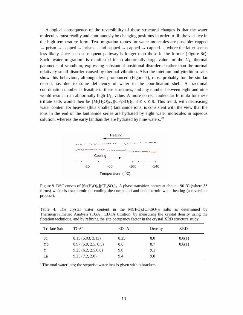
Figure 9. DSC curves of [Sc(H2O)8](CF3SO3)3. A phase transition occurs at about – 90 °C (where 2* forms) which is exothermic on cooling the compound and endothermic when heating (a reversible process).
Table 4. The crystal water content in the M(H2O)x(CF3SO3)3 salts as determined by Thermogravimetric Analysis (TGA), EDTA titration, by measuring the crystal density using the flotation technique, and by refining the site occupancy factor in the crystal XRD structure study.
Triflate Salt TGAa EDTA Density XRD
Sc 8.15 (5.03, 3.13) 8.25 8.0 8.0(1) Yb 8.97 (5.9, 2.5, 0.5) 8.6 8.7 8.6(1) Y 9.25 (6.2, 2.5,0.6) 9.0 9.1 La 9.25 (7.2, 2.0) 9.4 9.0
a The total water loss; the stepwise water loss is given within brackets.
-140-100-60-20
Temperature ( OC)
Cooling
Heating
14
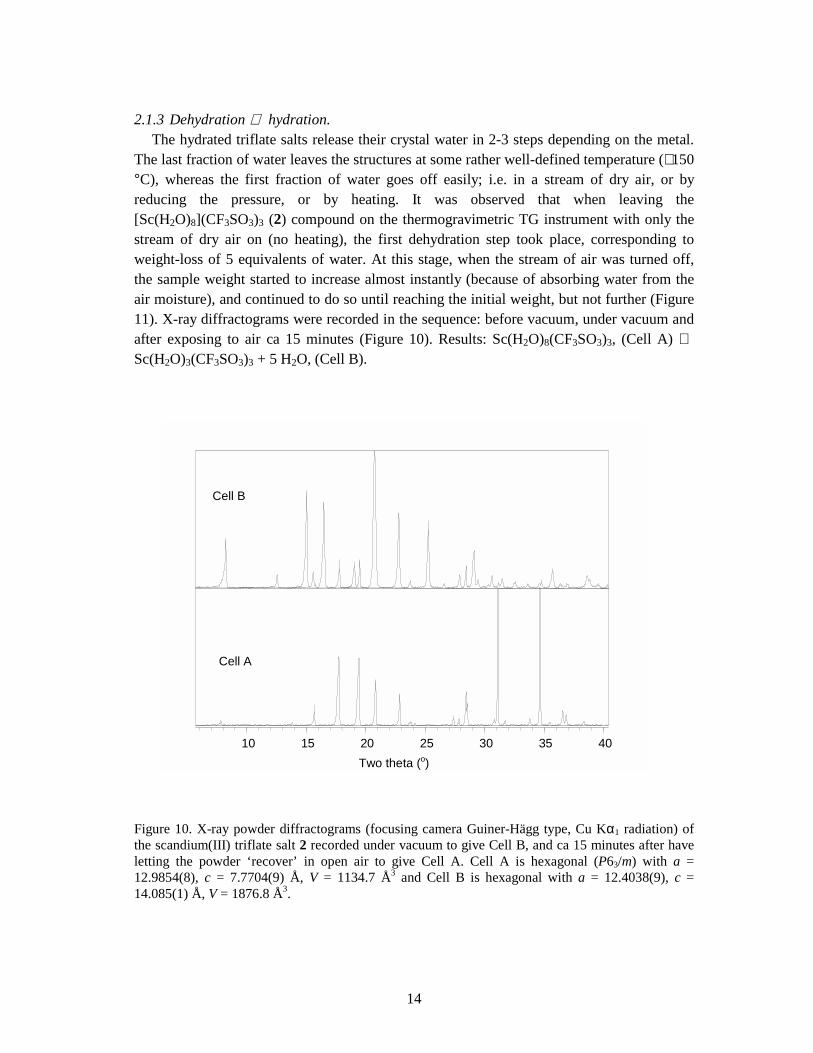
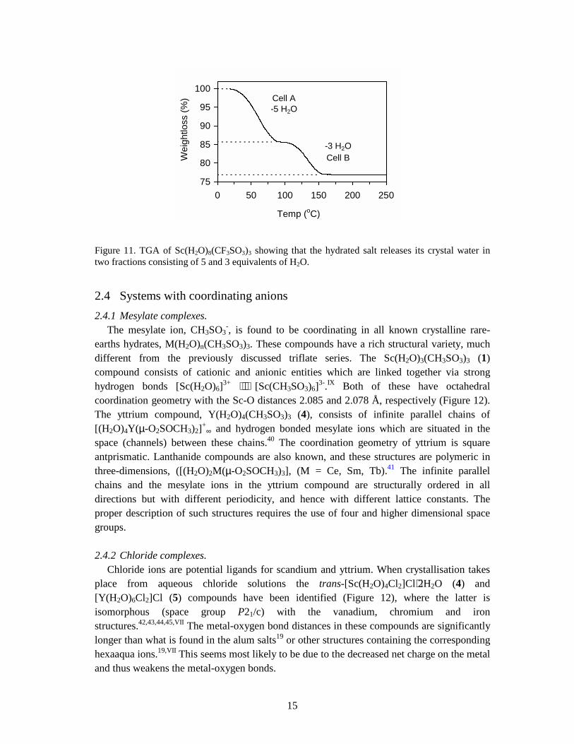
2.1.3 Dehydration ⇔ hydration. The hydrated triflate salts release their crystal water in 2-3 steps depending on the metal. The last fraction of water leaves the structures at some rather well-defined temperature (∼ 150 °C), whereas the first fraction of water goes off easily; i.e. in a stream of dry air, or by reducing the pressure, or by heating. It was observed that when leaving the [Sc(H2O)8](CF3SO3)3 (2) compound on the thermogravimetric TG instrument with only the stream of dry air on (no heating), the first dehydration step took place, corresponding to weight-loss of 5 equivalents of water. At this stage, when the stream of air was turned off, the sample weight started to increase almost instantly (because of absorbing water from the air moisture), and continued to do so until reaching the initial weight, but not further (Figure 11). X-ray diffractograms were recorded in the sequence: before vacuum, under vacuum and after exposing to air ca 15 minutes (Figure 10). Results: Sc(H2O)8(CF3SO3)3, (Cell A) ⇔ Sc(H2O)3(CF3SO3)3 + 5 H2O, (Cell B).
Figure 10. X-ray powder diffractograms (focusing camera Guiner-Hägg type, Cu Kα1 radiation) of the scandium(III) triflate salt 2 recorded under vacuum to give Cell B, and ca 15 minutes after have letting the powder ‘recover’ in open air to give Cell A. Cell A is hexagonal (P63/m) with a = 12.9854(8), c = 7.7704(9) Å, V = 1134.7 Å3 and Cell B is hexagonal with a = 12.4038(9), c = 14.085(1) Å, V = 1876.8 Å3.
10 15 20 25 30 35 40Two theta (o)
Cell B
Cell A
15
Figure 11. TGA of Sc(H2O)8(CF3SO3)3 showing that the hydrated salt releases its crystal water in two fractions consisting of 5 and 3 equivalents of H2O.
2.4 Systems with coordinating anions 2.4.1 Mesylate complexes. The mesylate ion, CH3SO3
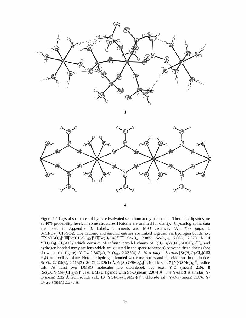
-, is found to be coordinating in all known crystalline rare-earths hydrates, M(H2O)n(CH3SO3)3. These compounds have a rich structural variety, much different from the previously discussed triflate series. The Sc(H2O)3(CH3SO3)3 (1) compound consists of cationic and anionic entities which are linked together via strong hydrogen bonds [Sc(H2O)6]3+ ⋅⋅⋅ [Sc(CH3SO3)6]3-.IX Both of these have octahedral coordination geometry with the Sc-O distances 2.085 and 2.078 Å, respectively (Figure 12). The yttrium compound, Y(H2O)4(CH3SO3)3 (4), consists of infinite parallel chains of [(H2O)4Y(µ-O2SOCH3)2]+
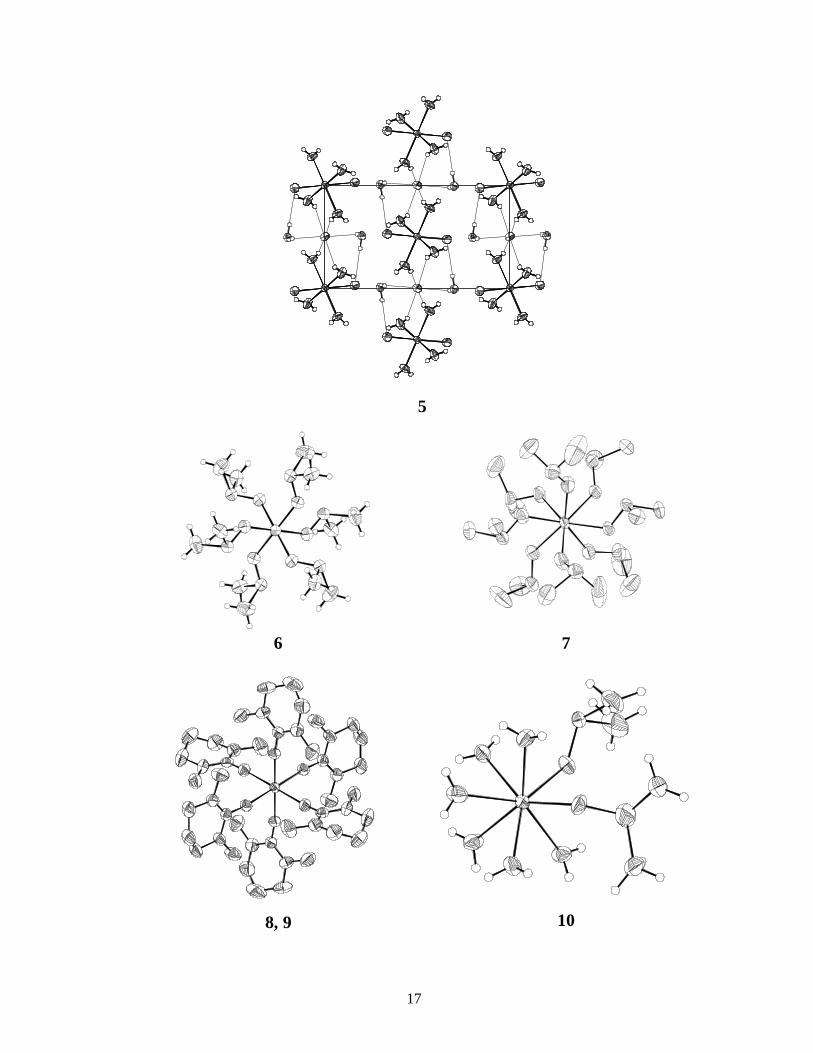
∞ and hydrogen bonded mesylate ions which are situated in the space (channels) between these chains.40 The coordination geometry of yttrium is square antprismatic. Lanthanide compounds are also known, and these structures are polymeric in three-dimensions, ([(H2O)2M(µ-O2SOCH3)3], (M = Ce, Sm, Tb).41 The infinite parallel chains and the mesylate ions in the yttrium compound are structurally ordered in all directions but with different periodicity, and hence with different lattice constants. The proper description of such structures requires the use of four and higher dimensional space groups. 2.4.2 Chloride complexes. Chloride ions are potential ligands for scandium and yttrium. When crystallisation takes place from aqueous chloride solutions the trans-[Sc(H2O)4Cl2]Cl⋅2H2O (4) and [Y(H2O)6Cl2]Cl (5) compounds have been identified (Figure 12), where the latter is isomorphous (space group P21/c) with the vanadium, chromium and iron structures.42,43,44,45,VII The metal-oxygen bond distances in these compounds are significantly longer than what is found in the alum salts19 or other structures containing the corresponding hexaaqua ions.19,VII This seems most likely to be due to the decreased net charge on the metal and thus weakens the metal-oxygen bonds.
75
80
85
90
95
100
0 50 100 150 200 250
Temp (oC)
Wei
ghtlo
ss (%
) Cell A-5 H2O
-3 H2OCell B
16
1
4
Figure 12. Crystal structures of hydrated/solvated scandium and yttrium salts. Thermal ellipsoids are at 40% probability level. In some structures H-atoms are omitted for clarity. Crystallographic data are listed in Appendix D. Labels, comments and M-O distances (Å). This page: 1 Sc(H2O)3)(CH3SO3). The cationic and anionic entities are linked together via hydrogen bonds, i.e. ⋅⋅⋅[Sc(H2O)6]3+⋅⋅⋅[Sc(CH3SO3)6]3-⋅⋅⋅[Sc(H2O)6]3+⋅⋅⋅. Sc-OW 2.085, Sc-OMES 2.085, 2.078 Å. 4 Y(H2O)4(CH3SO3)3 which consists of infinite parallel chains of [(H2O)4Y(µ-O2SOCH3)2
+]+∞ and
hydrogen bonded mesylate ions which are situated in the space (channels) between these chains (not shown in the figure). Y-OW 2.367(4), Y-OMES 2.332(4) Å. Next page. 5 trans-[Sc(H2O)4Cl2]Cl⋅2 H2O, unit cell bc-plane. Note the hydrogen bonded water molecules and chloride ions in the lattice. Sc-OW 2.109(3), 2.113(3), Sc-Cl 2.429(1) Å. 6 [Sc(OSMe2)6]3+, iodide salt. 7 [Y(OSMe2)8]3+, iodide salt. At least two DMSO molecules are disordered, see text. Y-O (mean) 2.36. 8 [Sc(OCN2Me2(CH2)3)6]3+, i.e. DMPU ligands with Sc-O(mean) 2.074 Å. The Y-salt 9 is similar, Y-O(mean) 2.22 Å from iodide salt. 10 [Y(H2O)6(OSMe2)2]3+, chloride salt. Y-OW (mean) 2.376, Y-ODMSO (mean) 2.273 Å.
17
5
6 7
8, 9 10
18
2.5 Non-aqueous solvates Scandium and yttrium iodide crystallise from dimethylsulfoxide (DMSO = Me2SO) solution to form [Sc(dmso)6]I3 (6) and [Y(dmso)8]I3 (7), respectively. The iodide ion interacts weakly with the methyl protons of dimethylsulfoxide. Both scandium and yttrium are solvated by six N,N´-dimethylpropylene urea solvent molecules, both in solution and in the solid state, i.e. [M(dmpu)6]I3 (8, 9).V,VII Thus, the coordination number of yttrium decreases from eight in dimethylsulfoxide to six in N,N´-dimethylpropylene urea, whereas scandium remains hexasolvated in both solvents despite that the solvent molecules of the latter are relatively large and bulky. The structures of 8 and 9 are isomorphous. Preliminary determinations of the crystal structures of [Ca(dmpu)6]X2 (X = ClO4
-, CF3SO3-) have been
performed but not yet reported.46 In the mixed water/dimethylsulfoxide solvated compound [Y(H2O)6(dmso)2]Cl3 (10) the Y-O bond distances to the dimethylsulfoxide ligands are significantly shorter than those to the water (Figure 12).III It is interesting to compare this structure with that of the hydrated yttrium chloride, [Y(H2O)6Cl2]Cl which is similar to 10, with the chloride ions at the corresponding dimethylsulfoxide positions.42
2.6 Configurational disorder 2.6.1 Octaaquascandium(III). A direct consequence of the change in the positions of the water molecules in 2, is a synchronised movement of the scandium atom, although its exact pathway is not known. The crystal structure model requires the scandium atom to be in the centre (on the 6 axis), which only is the average position in the crystal structure. However, the EXAFS method does not depend on the crystallographic symmetry, and positional disorder of the atoms does not affect the evaluation of the coordination distances. EXAFS can therefore provide a more reliable mean Sc-O bond distance in such cases. The refined mean Sc-O distance in EXAFS (using the cumulant expansion method) is about 2.20 Å, which is substantially shorter than the apparent value from the atomic positions in the crystal structure, 2.27 Å.IX
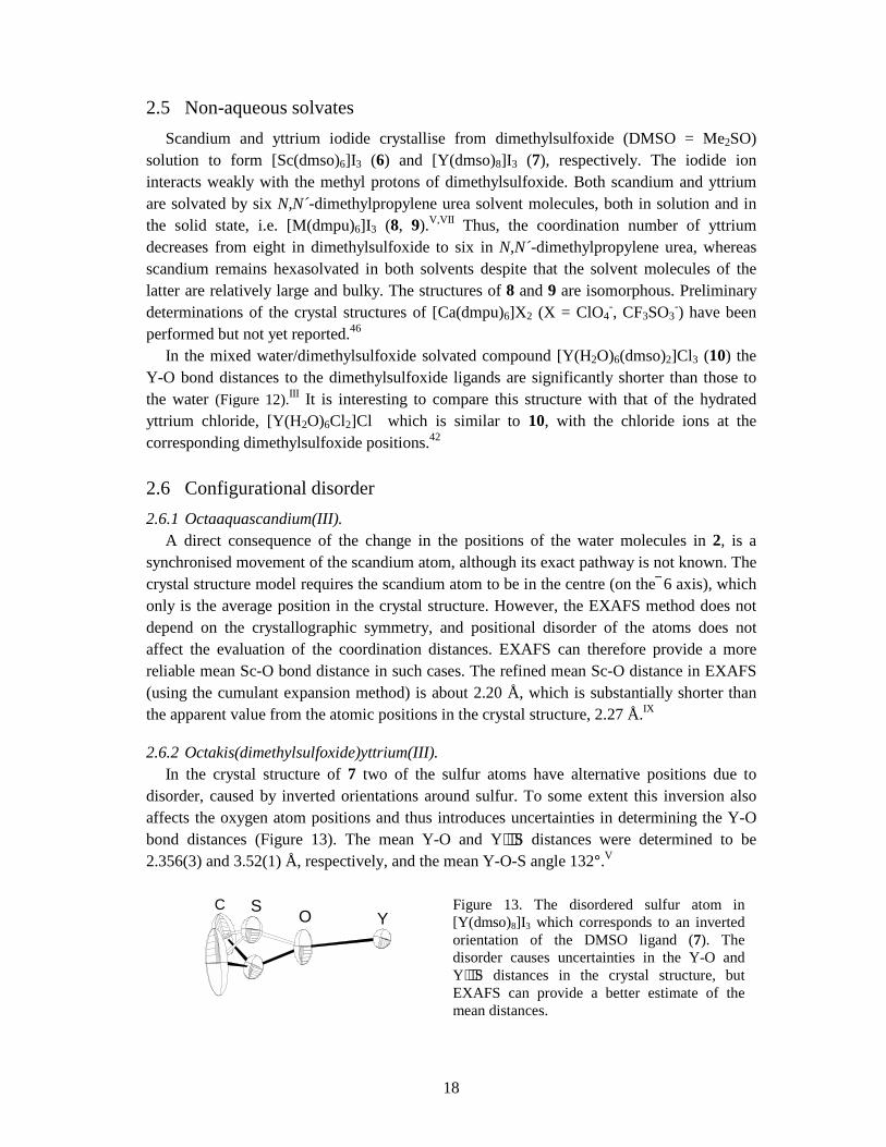
2.6.2 Octakis(dimethylsulfoxide)yttrium(III). In the crystal structure of 7 two of the sulfur atoms have alternative positions due to disorder, caused by inverted orientations around sulfur. To some extent this inversion also affects the oxygen atom positions and thus introduces uncertainties in determining the Y-O bond distances (Figure 13). The mean Y-O and Y⋅⋅⋅S distances were determined to be 2.356(3) and 3.52(1) Å, respectively, and the mean Y-O-S angle 132°.V
Figure 13. The disordered sulfur atom in [Y(dmso)8]I3 which corresponds to an inverted orientation of the DMSO ligand (7). The disorder causes uncertainties in the Y-O and Y⋅⋅⋅S distances in the crystal structure, but EXAFS can provide a better estimate of the mean distances.
COS
Y
19
Chapter 3
Hydrated Ions in Solution
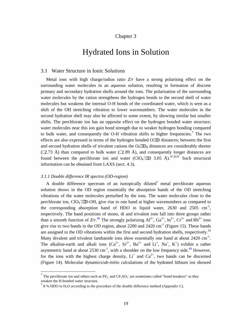
3.1 Water Structure in Ionic Solutions Metal ions with high charge/radius ratio Z/r have a strong polarising effect on the surrounding water molecules in an aqueous solution, resulting in formation of discrete primary and secondary hydration shells around the ions. The polarisation of the surrounding water molecules by the cation strengthens the hydrogen bonds to the second shell of water molecules but weakens the internal O-H bonds of the coordinated water, which is seen as a shift of the OH stretching vibration to lower wavenumbers. The water molecules in the second hydration shell may also be affected to some extent, by showing similar but smaller shifts. The perchlorate ion has an opposite effect on the hydrogen bonded water structure; water molecules near this ion gain bond strength due to weaker hydrogen bonding compared to bulk water, and consequently the O-H vibration shifts to higher frequencies.* The two effects are also expressed in terms of the hydrogen bonded O⋅⋅⋅O distances; between the first and second hydration shells of trivalent cations the OI⋅⋅⋅OII distances are considerably shorter (∼ 2.73 Å) than compared to bulk water (∼ 2.89 Å), and consequently longer distances are found between the perchlorate ion and water (ClO4
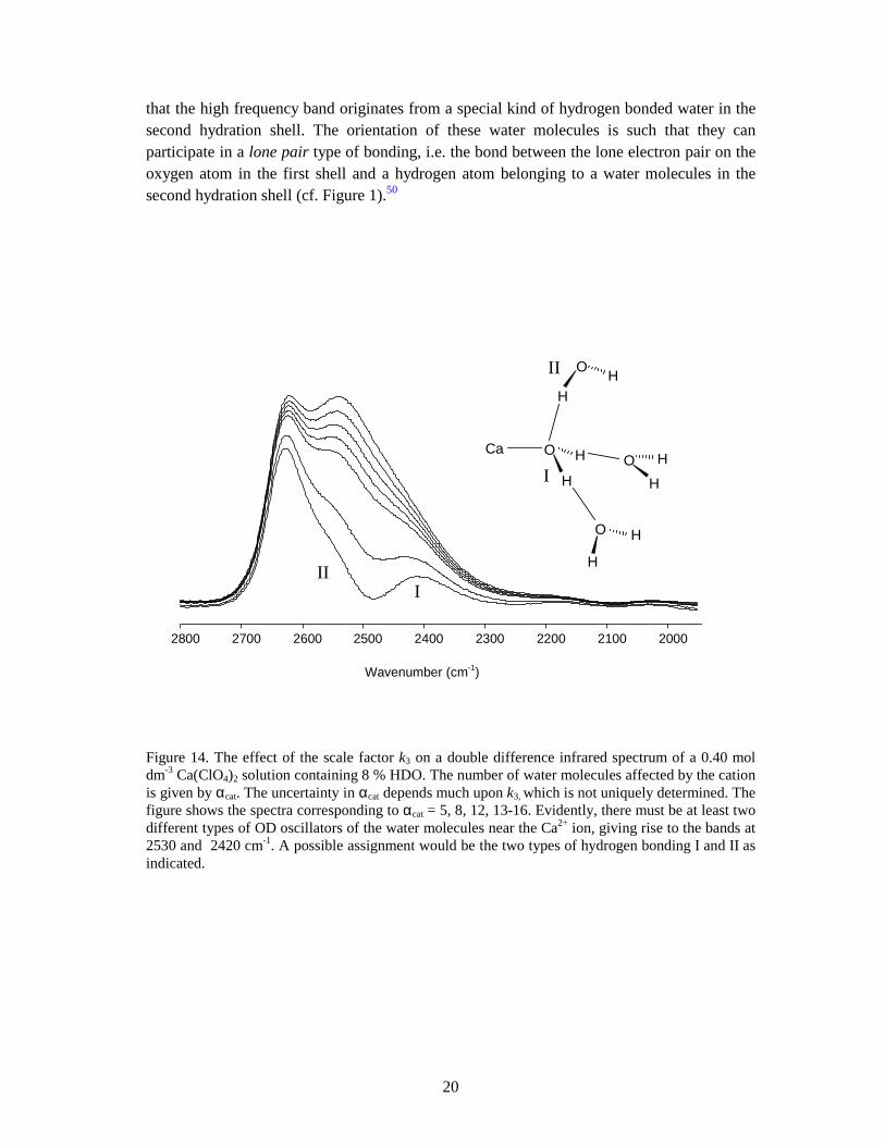
-⋅⋅⋅O 3.05 Å).47,II,IV Such structural information can be obtained from LAXS (sect. 4.3). 3.1.1 Double difference IR spectra (OD-region) A double difference spectrum of an isotopically diluted† metal perchlorate aqueous solution shows in the OD region essentially the absorption bands of the OD stretching vibrations of the water molecules perturbed by the ions. The water molecules close to the perchlorate ion, ClO4
-⋅⋅⋅D-OH, give rise to one band at higher wavenumbers as compared to the corresponding absorption band of HDO in liquid water, 2630 and 2505 cm-1, respectively. The band positions of mono, di and trivalent ions fall into three groups rather than a smooth function of Z/r.48 The strongly polarising Al3+, Ga3+, In2+, Cr3+ and Rh3+ ions give rise to two bands in the OD region, about 2200 and 2420 cm-1 (Figure 15). These bands are assigned to the OD vibrations within the first and second hydration shells, respectively.16 Many divalent and trivalent lanthanide ions show essentially one band at about 2420 cm-1. The alkaline-earth and alkali ions (Ca2+, Sr2+, Ba2+ and Li+, Na+, K+) exhibit a rather asymmetric band at about 2530 cm-1, with a shoulder on the low frequency side.49 However, for the ions with the highest charge density, Li+ and Ca2+, two bands can be discerned (Figure 14). Molecular dynamics/ab-initio calculations of the hydrated lithium ion showed
* The perchlorate ion and others such as PF6
- and CF3SO3- are sometimes called “bond breakers” as they
weaken the H-bonded water structure. † 8 % HDO in H2O according to the procedure of the double difference method (Appendix C).
20
that the high frequency band originates from a special kind of hydrogen bonded water in the second hydration shell. The orientation of these water molecules is such that they can participate in a lone pair type of bonding, i.e. the bond between the lone electron pair on the oxygen atom in the first shell and a hydrogen atom belonging to a water molecules in the second hydration shell (cf. Figure 1).50
Figure 14. The effect of the scale factor k3 on a double difference infrared spectrum of a 0.40 mol dm-3 Ca(ClO4)2 solution containing 8 % HDO. The number of water molecules affected by the cation is given by αcat. The uncertainty in αcat depends much upon k3, which is not uniquely determined. The figure shows the spectra corresponding to αcat = 5, 8, 12, 13-16. Evidently, there must be at least two different types of OD oscillators of the water molecules near the Ca2+ ion, giving rise to the bands at 2530 and 2420 cm-1. A possible assignment would be the two types of hydrogen bonding I and II as indicated.
2800 2700 2600 2500 2400 2300 2200 2100 2000
Wavenumber (cm-1)
III
Ca O
H
H
H
O
O H
H
O H
H
H
I
II
21
Figure 15. Double difference IR spectra (combined OH and OD-regions) of 0.40 mol dm-3 M(ClO4)3 aqueous solutions containing 8 % HDO. From the top to the bottom M = La, Y, Sc, In, Ga, Al.
17502250275032503750
Wavenumber, cm-1
La3+
Y3+
Sc3+
In3+
Ga3+
Al3+
22
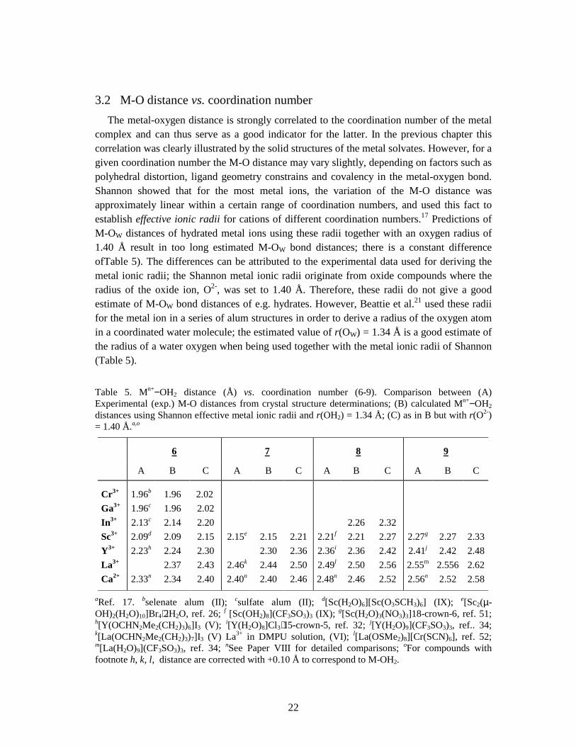
3.2 M-O distance vs. coordination number The metal-oxygen distance is strongly correlated to the coordination number of the metal complex and can thus serve as a good indicator for the latter. In the previous chapter this correlation was clearly illustrated by the solid structures of the metal solvates. However, for a given coordination number the M-O distance may vary slightly, depending on factors such as polyhedral distortion, ligand geometry constrains and covalency in the metal-oxygen bond. Shannon showed that for the most metal ions, the variation of the M-O distance was approximately linear within a certain range of coordination numbers, and used this fact to establish effective ionic radii for cations of different coordination numbers.17 Predictions of M-OW distances of hydrated metal ions using these radii together with an oxygen radius of 1.40 Å result in too long estimated M-OW bond distances; there is a constant difference ofTable 5). The differences can be attributed to the experimental data used for deriving the metal ionic radii; the Shannon metal ionic radii originate from oxide compounds where the radius of the oxide ion, O2-, was set to 1.40 Å. Therefore, these radii do not give a good estimate of M-OW bond distances of e.g. hydrates. However, Beattie et al.21 used these radii for the metal ion in a series of alum structures in order to derive a radius of the oxygen atom in a coordinated water molecule; the estimated value of r(OW) = 1.34 Å is a good estimate of the radius of a water oxygen when being used together with the metal ionic radii of Shannon (Table 5). Table 5. Mn+−OH2 distance (Å) vs. coordination number (6-9). Comparison between (A) Experimental (exp.) M-O distances from crystal structure determinations; (B) calculated Mn+−OH2 distances using Shannon effective metal ionic radii and r(OH2) = 1.34 Å; (C) as in B but with r(O2-) = 1.40 Å.a,o
6 7 8 9
A B C A B C A B C A B C
Cr3+ 1.96b 1.96 2.02 Ga3+ 1.96c 1.96 2.02 In3+ 2.13c 2.14 2.20 2.26 2.32 Sc3+ 2.09d 2.09 2.15 2.15e 2.15 2.21 2.21f 2.21 2.27 2.27g 2.27 2.33Y3+ 2.23h 2.24 2.30 2.30 2.36 2.36i 2.36 2.42 2.41j 2.42 2.48La3+ 2.37 2.43 2.46k 2.44 2.50 2.49l 2.50 2.56 2.55m 2.556 2.62Ca2+ 2.33n 2.34 2.40 2.40n 2.40 2.46 2.48n 2.46 2.52 2.56n 2.52 2.58
aRef. 17. bselenate alum (II); csulfate alum (II); d[Sc(H2O)6][Sc(O3SCH3)6] (IX); e[Sc2(µ-OH)2(H2O)10]Br4⋅2H2O, ref. 26; f [Sc(OH2)8](CF3SO3)3 (IX); g[Sc(H2O)3(NO3)3]18-crown-6, ref. 51; h[Y(OCHN2Me2(CH2)3)6]I3 (V); i[Y(H2O)8]Cl3⋅15-crown-5, ref. 32; j[Y(H2O)9](CF3SO3)3, ref.. 34; k[La(OCHN2Me2(CH2)3)7]I3 (V) La3+ in DMPU solution, (VI); l[La(OSMe2)8][Cr(SCN)6], ref. 52; m[La(H2O)9](CF3SO3)3, ref. 34; nSee Paper VIII for detailed comparisons; oFor compounds with footnote h, k, l, distance are corrected with +0.10 Å to correspond to M-OH2.
23
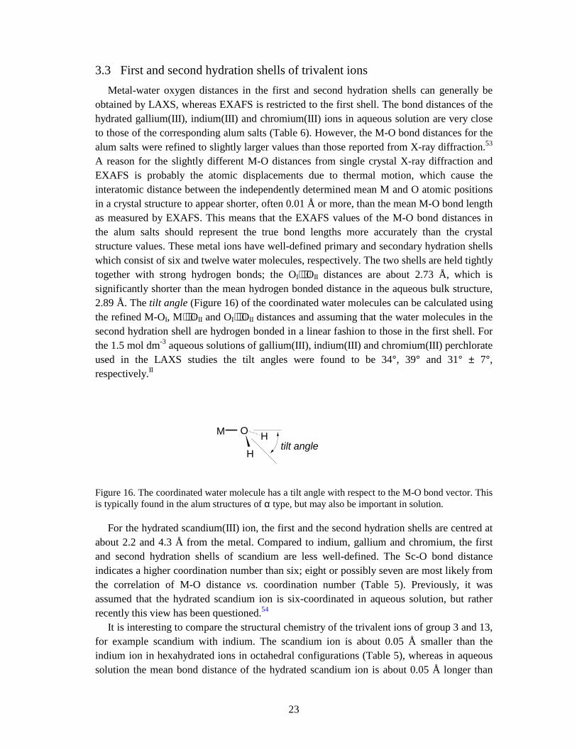
3.3 First and second hydration shells of trivalent ions Metal-water oxygen distances in the first and second hydration shells can generally be obtained by LAXS, whereas EXAFS is restricted to the first shell. The bond distances of the hydrated gallium(III), indium(III) and chromium(III) ions in aqueous solution are very close to those of the corresponding alum salts (Table 6). However, the M-O bond distances for the alum salts were refined to slightly larger values than those reported from X-ray diffraction.53 A reason for the slightly different M-O distances from single crystal X-ray diffraction and EXAFS is probably the atomic displacements due to thermal motion, which cause the interatomic distance between the independently determined mean M and O atomic positions in a crystal structure to appear shorter, often 0.01 Å or more, than the mean M-O bond length as measured by EXAFS. This means that the EXAFS values of the M-O bond distances in the alum salts should represent the true bond lengths more accurately than the crystal structure values. These metal ions have well-defined primary and secondary hydration shells which consist of six and twelve water molecules, respectively. The two shells are held tightly together with strong hydrogen bonds; the OI⋅⋅⋅OII distances are about 2.73 Å, which is significantly shorter than the mean hydrogen bonded distance in the aqueous bulk structure, 2.89 Å. The tilt angle (Figure 16) of the coordinated water molecules can be calculated using the refined M-OI, M⋅⋅⋅OII and OI⋅⋅⋅OII distances and assuming that the water molecules in the second hydration shell are hydrogen bonded in a linear fashion to those in the first shell. For the 1.5 mol dm-3 aqueous solutions of gallium(III), indium(III) and chromium(III) perchlorate used in the LAXS studies the tilt angles were found to be 34°, 39° and 31° ± 7°, respectively.II
Figure 16. The coordinated water molecule has a tilt angle with respect to the M-O bond vector. This is typically found in the alum structures of α type, but may also be important in solution.
For the hydrated scandium(III) ion, the first and the second hydration shells are centred at about 2.2 and 4.3 Å from the metal. Compared to indium, gallium and chromium, the first and second hydration shells of scandium are less well-defined. The Sc-O bond distance indicates a higher coordination number than six; eight or possibly seven are most likely from the correlation of M-O distance vs. coordination number (Table 5). Previously, it was assumed that the hydrated scandium ion is six-coordinated in aqueous solution, but rather recently this view has been questioned.54 It is interesting to compare the structural chemistry of the trivalent ions of group 3 and 13, for example scandium with indium. The scandium ion is about 0.05 Å smaller than the indium ion in hexahydrated ions in octahedral configurations (Table 5), whereas in aqueous solution the mean bond distance of the hydrated scandium ion is about 0.05 Å longer than
M O
H
Htilt angle
24
that of indium which remains hexahydrated also in solution (Table 6). A similar comparison of octahedral yttrium(III) and thallium(III) complexes, e.g. the perchlorate salts,55 shows that thallium remains hexahydrated in aqueous solution with Tl-O distance of 2.21 Å,60 while for yttrium the Y-O distance increases from about 2.24 to 2.37 Å corresponding to a change in coordination number from six to eight at hydration in aqueous solution.IV
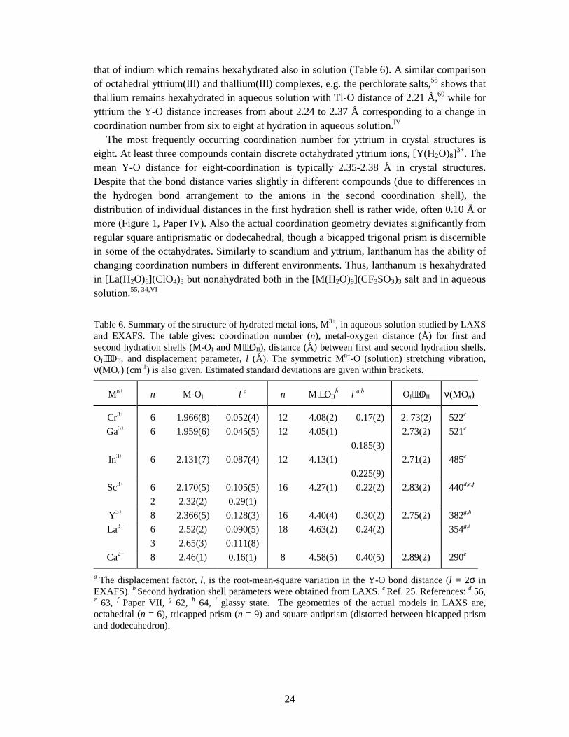
The most frequently occurring coordination number for yttrium in crystal structures is eight. At least three compounds contain discrete octahydrated yttrium ions, [Y(H2O)8]3+. The mean Y-O distance for eight-coordination is typically 2.35-2.38 Å in crystal structures. Despite that the bond distance varies slightly in different compounds (due to differences in the hydrogen bond arrangement to the anions in the second coordination shell), the distribution of individual distances in the first hydration shell is rather wide, often 0.10 Å or more (Figure 1, Paper IV). Also the actual coordination geometry deviates significantly from regular square antiprismatic or dodecahedral, though a bicapped trigonal prism is discernible in some of the octahydrates. Similarly to scandium and yttrium, lanthanum has the ability of changing coordination numbers in different environments. Thus, lanthanum is hexahydrated in [La(H2O)6](ClO4)3 but nonahydrated both in the [M(H2O)9](CF3SO3)3 salt and in aqueous solution.55, 34,VI Table 6. Summary of the structure of hydrated metal ions, M3+, in aqueous solution studied by LAXS and EXAFS. The table gives: coordination number (n), metal-oxygen distance (Å) for first and second hydration shells (M-OI and M⋅⋅⋅OII), distance (Å) between first and second hydration shells, OI⋅⋅⋅OII, and displacement parameter, l (Å). The symmetric Mn+-O (solution) stretching vibration, ν(MOn) (cm-1) is also given. Estimated standard deviations are given within brackets.
Mn+ n M-OI l a n M⋅⋅⋅OIIb l a,b OI⋅⋅⋅OII ν(MOn)
Cr3+ 6 1.966(8) 0.052(4) 12 4.08(2) 0.17(2) 2. 73(2) 522c
Ga3+ 6 1.959(6) 0.045(5) 12 4.05(1) 0.185(3)
2.73(2) 521c
In3+ 6 2.131(7) 0.087(4) 12 4.13(1) 0.225(9)
2.71(2) 485c
Sc3+ 6
2 2.170(5) 2.32(2)
0.105(5) 0.29(1)
16 4.27(1) 0.22(2) 2.83(2) 440d,e,f
Y3+ 8 2.366(5) 0.128(3) 16 4.40(4) 0.30(2) 2.75(2) 382g,h
La3+ 6
3 2.52(2) 2.65(3)
0.090(5) 0.111(8)
18 4.63(2) 0.24(2) 354g,i
Ca2+ 8 2.46(1) 0.16(1) 8 4.58(5) 0.40(5) 2.89(2) 290e
a The displacement factor, l, is the root-mean-square variation in the Y-O bond distance (l = 2σ in EXAFS). b Second hydration shell parameters were obtained from LAXS. c Ref. 25. References: d 56, e 63, f Paper VII, g 62, h 64, i glassy state. The geometries of the actual models in LAXS are, octahedral (n = 6), tricapped prism (n = 9) and square antiprism (distorted between bicapped prism and dodecahedron).
25
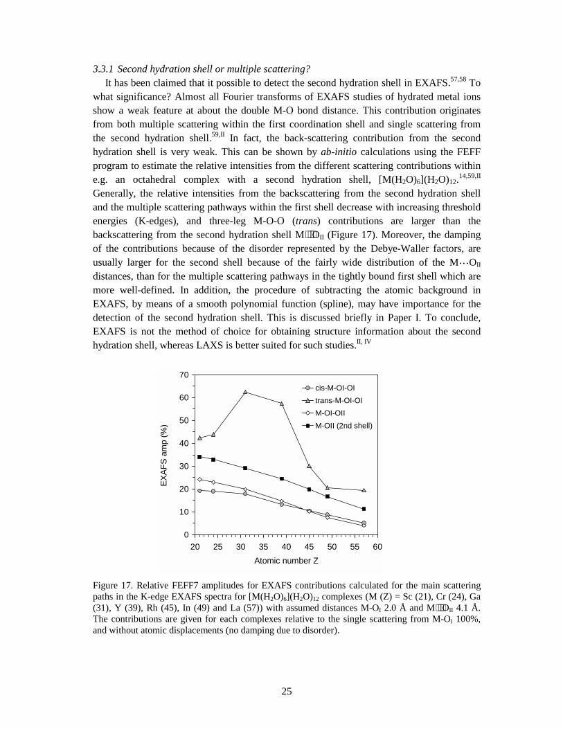
3.3.1 Second hydration shell or multiple scattering? It has been claimed that it possible to detect the second hydration shell in EXAFS.57,58 To what significance? Almost all Fourier transforms of EXAFS studies of hydrated metal ions show a weak feature at about the double M-O bond distance. This contribution originates from both multiple scattering within the first coordination shell and single scattering from the second hydration shell.59,II In fact, the back-scattering contribution from the second hydration shell is very weak. This can be shown by ab-initio calculations using the FEFF program to estimate the relative intensities from the different scattering contributions within e.g. an octahedral complex with a second hydration shell, [M(H2O)6](H2O)12.14,59,II Generally, the relative intensities from the backscattering from the second hydration shell and the multiple scattering pathways within the first shell decrease with increasing threshold energies (K-edges), and three-leg M-O-O (trans) contributions are larger than the backscattering from the second hydration shell M⋅⋅⋅OII (Figure 17). Moreover, the damping of the contributions because of the disorder represented by the Debye-Waller factors, are usually larger for the second shell because of the fairly wide distribution of the M…OII distances, than for the multiple scattering pathways in the tightly bound first shell which are more well-defined. In addition, the procedure of subtracting the atomic background in EXAFS, by means of a smooth polynomial function (spline), may have importance for the detection of the second hydration shell. This is discussed briefly in Paper I. To conclude, EXAFS is not the method of choice for obtaining structure information about the second hydration shell, whereas LAXS is better suited for such studies.II, IV
Figure 17. Relative FEFF7 amplitudes for EXAFS contributions calculated for the main scattering paths in the K-edge EXAFS spectra for [M(H2O)6](H2O)12 complexes (M (Z) = Sc (21), Cr (24), Ga (31), Y (39), Rh (45), In (49) and La (57)) with assumed distances M-OI 2.0 Å and M⋅⋅⋅OII 4.1 Å. The contributions are given for each complexes relative to the single scattering from M-OI 100%, and without atomic displacements (no damping due to disorder).
0
10
20
30
40
50
60
70
20 25 30 35 40 45 50 55 60Atomic number Z
EXAF
S am
p (%
)
cis-M-OI-OItrans-M-OI-OIM-OI-OIIM-OII (2nd shell)
26
3.4 Coordination geometry 3.4.1 M-O stretching vibration. The coordination symmetry of a solvated complex in solution can in principle be obtained by assigning the fundamental bands in the IR and Raman spectra and applying the selection rules from group theory. Practically, this is a rather difficult task because of too weak and broad bands,60 although the symmetric M-O stretching vibration, ν1, is usually obtained without difficulty in the Raman spectrum. Since the metal atom is stationary in the symmetric stretching (breathing) mode, the frequency of this band is proportional to the square root of the force constant K of the M-O stretching: ν1 = ½πc(K/m)1/2, where m is the mass of the water molecule (assuming the interactions between oxygen atoms to be negligible). For a linear ion-dipole type of bond the energy is inversely proportional to the square root of the M-O distance, whereas a covalent bond is more sensitive to changes in the distance. Thus, ν1 for the hexahydrated scandium and yttrium ions in the crystalline [M(H2O)6](ClO4)3 compounds were found to be very similar to those of the aqueous solutions, 435 ± 5 and 383 ± 5 for scandium and yttrium, respectively, despite that scandium and yttrium have considerably longer M-O distances in solution (Table 5 and Table 6).61,62,63,64,VIII
3.4.2 XANES and EXAFS. Coordination geometries of high symmetry (e.g. octahedral) give characteristic multiple scattering features which can be identified in the EXAFS spectrum. Such features are small for compounds with distorted coordination figures and irregular geometry, since the individual signals partially cancel due to slightly different scattering pathways. The phase corrected Fourier transform of hydrated yttrium(III) ions in aqueous solutions shows only a major peak at 2.37 Å corresponding to the Y-O bond distance of the octahydrated ions.65,IV The XANES region contains more information about coordination geometry than the EXAFS region, but is usually (at present) difficult to describe theoretically,66 although several modern program codes, e.g. FFFF8,14 provide calculation of theoretical XANES spectra. In order to assign a feasible coordination figure for the hydrated scandium(III), yttrium(III) and lanthanum(III) ions in aqueous solution, the XANES and EXAFS spectra of several crystalline hydrates, with different types of coordination geometries, were analysed and compared to those of the aqueous solutions. The spectra of the aqueous scandium(III) solutions are most similar to that of the crystalline compound [Sc(H2O)8](CF3SO3)3 (2), which has bicapped trigonal prismatic coordination geometryVII The yttrium ion in the [Y(H2O)8]Cl3⋅(15-crown-5) compound is also best described in this geometry, which gives similar spectra as the hydrated yttrium ion in aqueous solution.IV Similarly the spectra of [La(H2O)9](CF3SO3)3 and aqueous lanthanum(III) solutions are also in very good agreement.VI
27
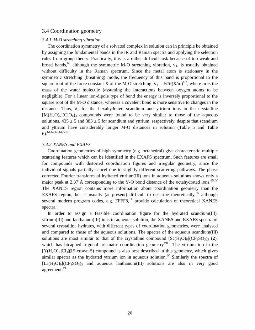
3.4.3 Evidence against Oh geometry for the hydrated scandium(III) ion. X-ray absorption K-edge spectra of first-row transition metals have weak pre-edge features due to excitation of the 1s electron to the valence 3d orbitals.67,* This 1s → 3d transition is also seen for scandium(III) and calcium(II) complexes (Sc3+ and Ca2+ are d0 ions). The split pre-peak for the octahedral complexes [Sc(H2O)6](ClO4)3, [Sc(H2O)6][Sc(CH3SO3)6] (1) and [Sc(H2O)4(C7H7SO3)2]C7H7SO3 ⋅2H2O, corresponds to the two groups of 3d orbitals of t2g and eg symmetry in an octahedral ligand field, which are separated by ∆o ∼ 1.5 eV (Figure 18). However, the eight and seven coordinated [Sc(H2O)8](CF3SO3)3 (2) and [Sc2(µ-OH)2(H2O)10]Cl4⋅2H2O compounds do not show split pre-peaks. The pre-edge feature and the XANES of the aqueous solution is most similar to that of 2. Thus, the absence of a split pre-peak of the aqueous solution indicates that the coordination geometry is not octahedral, but more close to a bicapped trigonal prism, as in 2.
Figure 18. Sc K-edges (XANES region) of a scandium(III) aqueous solution (L1, dashed line) compared with the solid samples (solid lines): S1 [Sc(H2O)8](CF3SO3)3, bicapped trigonal prism coordination geometry; S2 [Sc(H2O)6](ClO4)3, octahedral; S3 [Sc2(OH)2(H2O)10]Br4⋅2H2O, monocapped trigonal prism cf. Table 3. Note the difference in the pre-peak due to 1s → 3d transition, see text.
* The 1s → 3d transition is electric dipole forbidden for centrosymmmetric complexes by parity considerations. A weak pre-edge feature is still observable for such complexes, however, and electronic quadrupole coupling is most likely the mechanism to gain some intensity into the spectrum. Non-centrosymmetric complexes have normally larger pre-peaks, which has been attributed to metal 4p mixing into the 3d orbitals, thus providing some electric dipole allowed 1s → 4p character (ref. 67). According to crystal field theory, in an octahedral ligand field the degenerate metal d orbital splits into two sets, t2g and eg, and the energy difference between these is the ligand field parameter ∆o.
28
3.5 Coordination shell asymmetry A distribution of the distances between the central atom and the ligands always reduces the amplitudes of the oscillations in EXAFS, and consequently the magnitude of its Fourier transform. This is becomes most evident by comparing the magnitudes of the Fourier transforms of similar solid structures which only differ slightly in the degree of positional disorder (i.e. the degree of distortion of the coordination shell). The yttrium atom is eight-coordinated in both of the crystalline salts [Y(H2O)8]Cl3⋅(15-crown-5) and [Y(H2O)6(C7H7SO3)2](C7H7SO3)⋅2H2O, where the former has slightly larger spread of Y-O bond distances. Yttrium is nine-coordinated in [Y(H2O)9](CF3SO3)3, but here the Y-O distances are distributed in two groups, 6×2.34 Å and 3×2.52 Å (Figure 1, Paper IV). These differences give these salt somewhat different intensities of their Y-O peak in the Fourier transform (Figure 3, Paper IV). The smallest Y-O peak is seen for the nine-coordinated structure, which probably is a result of destructive interference between the two back-scattering contributions from the six prismatic and the three capping oxygen atoms.V The largest peak is addressed to the compound with the smallest distribution of Y-O bond distances; i.e. [Y(H2O)6(C7H7SO3)2](C7H7SO3)⋅2H2O. Thermal vibrations of atoms result in dynamic disorder. Presumably the dynamic disorder is of similar magnitude for the yttrium compounds in this example. The disorder (displacement) parameter σ gives the width of the distribution of the bond lengths as the root-mean-square displacement of the atoms from their equilibrium positions. Coordination shell disorder follows the relation: σ2 = σdynamic
2 + σstatic
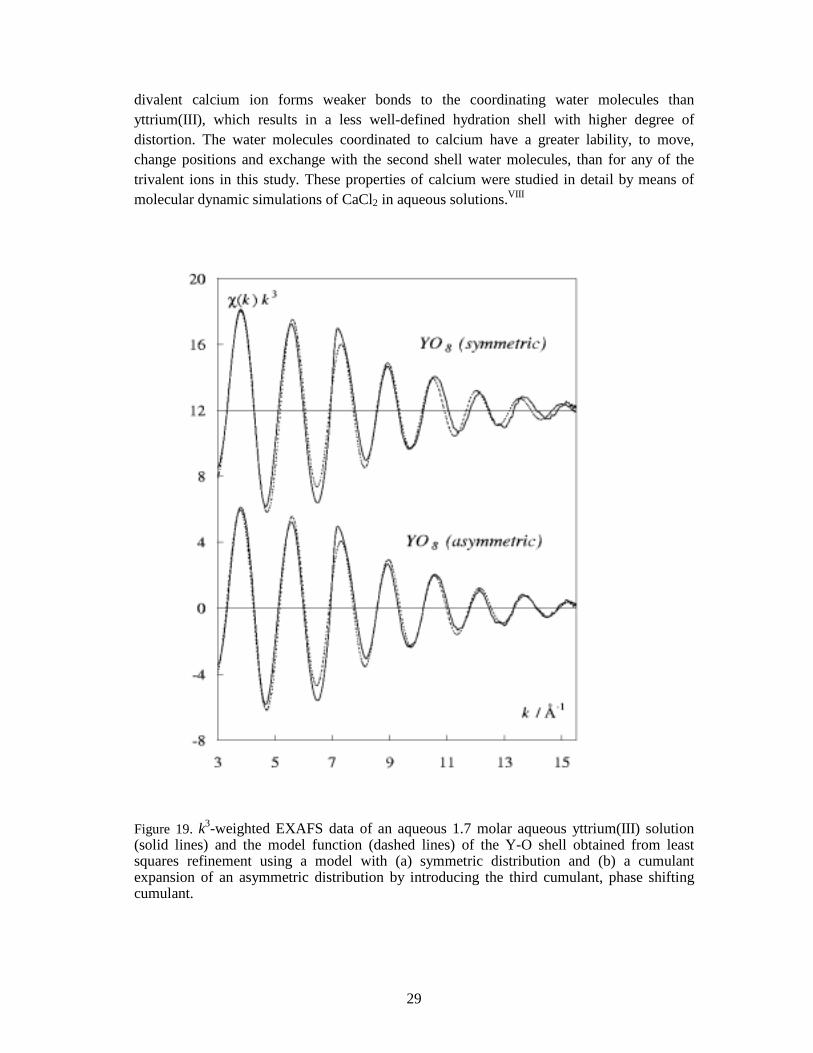
2 for symmetric (Gaussian) distributions. From a single EXAFS spectrum one cannot separate the two components, simply because of the scattering process itself which is faster (∼ 103) than the molecular vibrations. The EXAFS spectrum is thus an average of all configurations of the vibrating atoms. For samples with high site symmetry and well-defined bonds (harmonic vibrations), the distribution of distances is approximately Gaussian, which means that the damping of the EXAFS amplitude is given by the Debye-Waller factor, exp (-2σdynamic
2k2) (Appendix B). The coordination shell of the hydrated yttrium ion in aqueous solution is distorted from the ideal square antiprismatic geometry in a similar way as in the [Y(OH2)8]Cl3.(15-crown-5) salt, which has an asymmetric distribution of the Y-O distances. Applying a Gaussian (symmetric) model for an asymmetric distribution of distances does not give a satisfactory fit to the experimental data since this model leaves a phase shift at high k values in the EXAFS function (Figure 19), which often result in shorter mean bond distance.IV The coordination shell asymmetry may be static as in the [Y(OH2)8]Cl3.(15-crown-5) salt, which would explain the observed phase shift. Another possibility for asymmetry could be conformational disorder between different coordination geometries, or perhaps a frequently occurring water exchange between an eight and nine coordinated yttrium ion. A similar situation has been suggested for the ions in the middle of the lanthanide series, with a hydration number of about 8.5 on the average.10,68 The asymmetry of the distribution of metal-oxygen distances seems to be even more severe for the hydrated calcium(II) ion in aqueous solution. As for yttrium, the EXAFS spectrum of the hydrated calcium ion shows a relatively large phase shift at high k values when assuming a symmetric distribution of bond distances (Figure 1c, Paper VIII). The
29
divalent calcium ion forms weaker bonds to the coordinating water molecules than yttrium(III), which results in a less well-defined hydration shell with higher degree of distortion. The water molecules coordinated to calcium have a greater lability, to move, change positions and exchange with the second shell water molecules, than for any of the trivalent ions in this study. These properties of calcium were studied in detail by means of molecular dynamic simulations of CaCl2 in aqueous solutions.VIII
Figure 19. k3-weighted EXAFS data of an aqueous 1.7 molar aqueous yttrium(III) solution (solid lines) and the model function (dashed lines) of the Y-O shell obtained from least squares refinement using a model with (a) symmetric distribution and (b) a cumulant expansion of an asymmetric distribution by introducing the third cumulant, phase shifting cumulant.
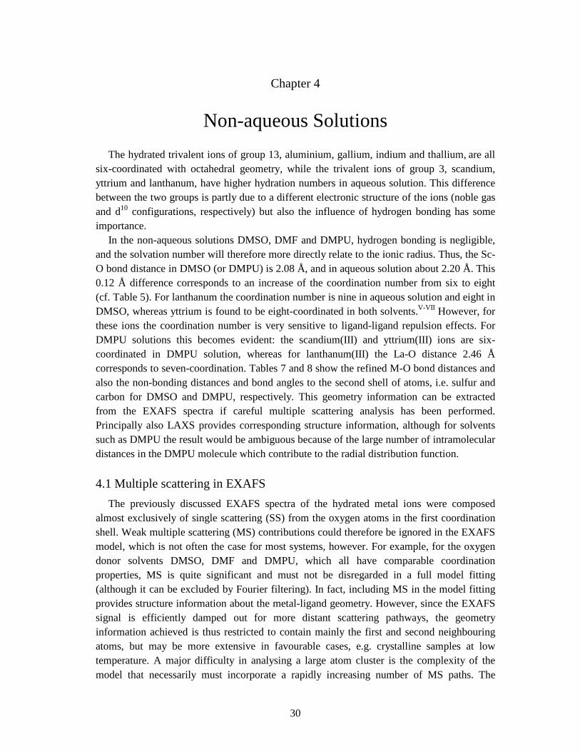
30
Chapter 4
Non-aqueous Solutions The hydrated trivalent ions of group 13, aluminium, gallium, indium and thallium, are all six-coordinated with octahedral geometry, while the trivalent ions of group 3, scandium, yttrium and lanthanum, have higher hydration numbers in aqueous solution. This difference between the two groups is partly due to a different electronic structure of the ions (noble gas and d10 configurations, respectively) but also the influence of hydrogen bonding has some importance. In the non-aqueous solutions DMSO, DMF and DMPU, hydrogen bonding is negligible, and the solvation number will therefore more directly relate to the ionic radius. Thus, the Sc-O bond distance in DMSO (or DMPU) is 2.08 Å, and in aqueous solution about 2.20 Å. This 0.12 Å difference corresponds to an increase of the coordination number from six to eight (cf. Table 5). For lanthanum the coordination number is nine in aqueous solution and eight in DMSO, whereas yttrium is found to be eight-coordinated in both solvents.V-VII However, for these ions the coordination number is very sensitive to ligand-ligand repulsion effects. For DMPU solutions this becomes evident: the scandium(III) and yttrium(III) ions are six-coordinated in DMPU solution, whereas for lanthanum(III) the La-O distance 2.46 Å corresponds to seven-coordination. Tables 7 and 8 show the refined M-O bond distances and also the non-bonding distances and bond angles to the second shell of atoms, i.e. sulfur and carbon for DMSO and DMPU, respectively. This geometry information can be extracted from the EXAFS spectra if careful multiple scattering analysis has been performed. Principally also LAXS provides corresponding structure information, although for solvents such as DMPU the result would be ambiguous because of the large number of intramolecular distances in the DMPU molecule which contribute to the radial distribution function.
4.1 Multiple scattering in EXAFS The previously discussed EXAFS spectra of the hydrated metal ions were composed almost exclusively of single scattering (SS) from the oxygen atoms in the first coordination shell. Weak multiple scattering (MS) contributions could therefore be ignored in the EXAFS model, which is not often the case for most systems, however. For example, for the oxygen donor solvents DMSO, DMF and DMPU, which all have comparable coordination properties, MS is quite significant and must not be disregarded in a full model fitting (although it can be excluded by Fourier filtering). In fact, including MS in the model fitting provides structure information about the metal-ligand geometry. However, since the EXAFS signal is efficiently damped out for more distant scattering pathways, the geometry information achieved is thus restricted to contain mainly the first and second neighbouring atoms, but may be more extensive in favourable cases, e.g. crystalline samples at low temperature. A major difficulty in analysing a large atom cluster is the complexity of the model that necessarily must incorporate a rapidly increasing number of MS paths. The
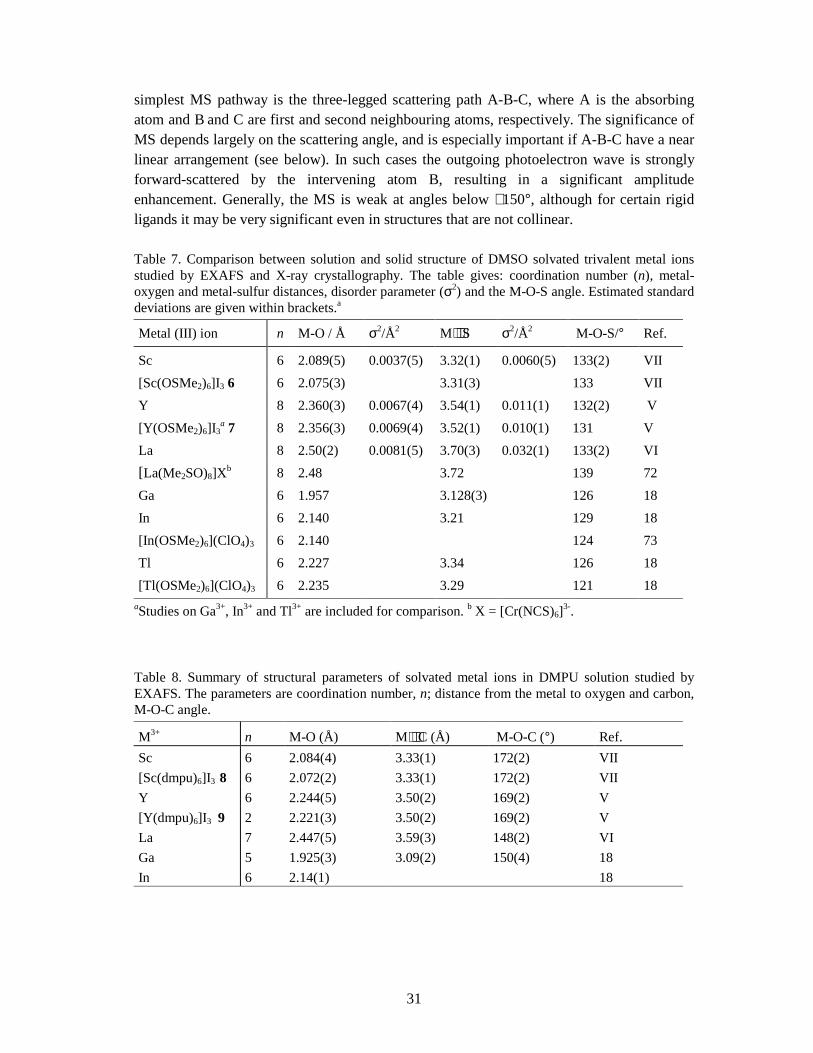
31
simplest MS pathway is the three-legged scattering path A-B-C, where A is the absorbing atom and B and C are first and second neighbouring atoms, respectively. The significance of MS depends largely on the scattering angle, and is especially important if A-B-C have a near linear arrangement (see below). In such cases the outgoing photoelectron wave is strongly forward-scattered by the intervening atom B, resulting in a significant amplitude enhancement. Generally, the MS is weak at angles below ∼ 150°, although for certain rigid ligands it may be very significant even in structures that are not collinear.
Table 7. Comparison between solution and solid structure of DMSO solvated trivalent metal ions studied by EXAFS and X-ray crystallography. The table gives: coordination number (n), metal-oxygen and metal-sulfur distances, disorder parameter (σ2) and the M-O-S angle. Estimated standard deviations are given within brackets.a
Metal (III) ion n M-O / Å σ2/Å2 M⋅⋅⋅S σ2/Å2 M-O-S/° Ref.
Sc 6 2.089(5) 0.0037(5) 3.32(1) 0.0060(5) 133(2) VII [Sc(OSMe2)6]I3 6 6 2.075(3) 3.31(3) 133 VII Y 8 2.360(3) 0.0067(4) 3.54(1) 0.011(1) 132(2) V [Y(OSMe2)6]I3
a 7 8 2.356(3) 0.0069(4) 3.52(1) 0.010(1) 131 V La 8 2.50(2) 0.0081(5) 3.70(3) 0.032(1) 133(2) VI [La(Me2SO)8]Xb 8 2.48 3.72 139 72 Ga 6 1.957 3.128(3) 126 18 In 6 2.140 3.21 129 18 [In(OSMe2)6](ClO4)3 6 2.140 124 73 Tl 6 2.227 3.34 126 18 [Tl(OSMe2)6](ClO4)3 6 2.235 3.29 121 18
aStudies on Ga3+, In3+ and Tl3+ are included for comparison. b X = [Cr(NCS)6]3-.
Table 8. Summary of structural parameters of solvated metal ions in DMPU solution studied by EXAFS. The parameters are coordination number, n; distance from the metal to oxygen and carbon, M-O-C angle.
M3+ n M-O (Å) M⋅⋅⋅C (Å) M-O-C (°) Ref. Sc 6 2.084(4) 3.33(1) 172(2) VII [Sc(dmpu)6]I3 8 6 2.072(2) 3.33(1) 172(2) VII Y 6 2.244(5) 3.50(2) 169(2) V [Y(dmpu)6]I3 9 2 2.221(3) 3.50(2) 169(2) V La 7 2.447(5) 3.59(3) 148(2) VI Ga 5 1.925(3) 3.09(2) 150(4) 18 In 6 2.14(1) 18
32
4.1.1 Multiple scattering in the Y-O-C entity The DMF and DMPU solvent molecules coordinate via an O=C-N group where the carbon atom has sp2 hybridisation. The O=C-N group is rigid but the C=O bond may have different angles to a metal ion upon coordination. The solid [Sc(dmpu)6]I3 and [Y(dmpu)6]I3 compounds have relatively large M-O-C angles, whereas DMF solvated metal ions represent the former, where the angle M-O-C is found to be about 135° in several structures, e.g. [Nd(dmf)8]n{[W4Ag5S16]}n.69,70,71 The M-O-C angle of solvated metal ions in DMF and DMPU solutions can be determined by simulating the EXAFS spectra of these solutions using the FEFF code and including MS. The Fourier transform of the DMF solution shows a small peak at about 3.4 Å, which is close to the Y⋅⋅⋅ C distance of the solvated [Y(dmf)8]3+ complex, whereas in the DMPU solution this peak is considerably larger, despite the lower coordination number for [Y(dmpu)6]3+ (Figure 20). This reflects in fact the angle dependence of the 3 and 4-legged MS. The FEFF program allows us not only to estimate the individual scattering contributions, but also to simulate the EXAFS. In Paper V this procedure was used to determine the Y-O-C angle for solvated [Y(dmpu)6]3+ is solution (Fig 7 in Paper V). Moreover, the M-O-C angle seems to be correlated to the size of the metal ion in hexasolvated complexes; the trend is a decreasing M-O-C angle with increasing ionic radius. For the crystalline DMPU solvates of scandium, yttrium and calcium one finds: Sc-O 2.075 Å ⇔ Sc-O-C 172°; Y-O 2.22 Å ⇔ Y-O-C 169°; Ca-O 2.32 Å ⇔ Ca-O-C 164°.V,VII.72
Figure 20. Fourier transforms of solvated yttrium(III) in DMF (top) and DMPU (bottom) solutions, illustrating the importance of multiple scattering in EXAFS. A large Y-O-C angle results in enhancement of the 3 and 4-legged Y-O-C and Y-O-C-O MS, respectively. The back-scattering contribution from the carbon atoms is not angle dependent, and thus is largest for DMF because of two more back-scattering carbon atoms (if one assumes similar Debye-Waller factors).
33
Conclusions The structural chemistry of several solvated ions with noble gas (d0) and with d10 electronic configuration has been investigated for aqueous and some non-aqueous oxygen donor systems. The filled d shell for the group 13 ions gallium(III) and indium(III), gives a more covalent bonding character than for the group 3 ions calcium, scandium(III), yttrium(III) and lanthanum(III), without d-electrons in the valence shell. A preference for octahedral coordination has been found for gallium(III) and indium(III) for the oxygen donor ligands water and dimethylsulfoxide, both in solution and in the solid state, while steric ligand-ligand repulsion effects seem to control the coordination numbers for the group 3 ions. In the crystal structures of [M(H2O)]CF3SO3, the hydrogen bond arrangement stabilises coordination of nine water molecules in a tricapped trigonal prism around the metal ion. All trivalent lanthanide ions and yttrium fit into this isomorphous series of compounds, even scandium despite its small ionic radius. However, the smallest ions in this series, scandium(III), lutetium(III) and ytterbium(III), are found not to be fully hydrated in these structures. Scandium is eight-coordinated and ytterbium has a fractional coordination number, ∼ 8.6. The crystal symmetry (P63/m) can still be maintained because of a random distribution of the vacant coordination site. The aqueous chemistry of scandium(III) was found to differ from the view given in most text books, where the hydrated ion has been proposed to be hexahydrated in aqueous solution, a hydration number that could be expected from its small ionic size. The ionic radius of the scandium(III) ion in six-coordination is about 0.05 Å smaller than that of the hexahydrated indium(III) in octahedral geometry. However, the mean Sc-O distance of the hydrated scandium(III) ion is considerably larger than that for the hexahydrated indium(III) ion in aqueous solution, and is consistent with a capped trigonal prismatic configuration, but the exact hydration number cannot the determined due to the very weak bonds to the water molecules in the capping positions. The hydrated scandium(III) ion is readily hydrolysed. At pH about 1-3 the dominating hydrolysed species in aqueous solution is the dimer, [Sc2(µ-OH)2(OH2)10]4+, in which the scandium(III) ion is seven-coordinated. This hydrolysis complex has been isolated and its structure determined in several crystal structures. The yttrium(III) ion is larger and less acidic, and resembles the heavier lanthanide ions. It is hydrated by eight water molecules in aqueous solution, most probably with a distorted bicapped trigonal prismatic geometry. The hydrated lanthanum(III) ion is well-described in a nine-coordinated tricapped trigonal prismatic coordination geometry. The divalent calcium ion is relatively weakly hydrated, yet it has a high coordination number. In the present study, it is found that the calcium ion is hydrated by eight waters in aqueous solution, while most text books propose the calcium ion to be six-coordinated. The reason for such different results is probably the weak Ca-O bonding in water, which results in some disorder of the
34
water molecules in its hydration shell, and thus a coordination figure which is not very well-defined. In such disordered systems asymmetric distributions of the M-O bond distances can become important. The degree of asymmetry can be estimated from EXAFS studies. If not too severe, asymmetric distributions can be accounted for by means of the cumulant expansion method in the EXAFS data analysis. The use of crystalline model compounds in the data analysis, e.g. hydrates and solvates, is important for selecting probable structural models for the solvated metal ions in solution. Both the XANES and EXAFS parts of the XAS spectra are useful in achieving such information. In particular, for the yttrium(III) ion in aqueous solution it was possible to deduce its hydration number and also to estimate the degree of coordination shell asymmetry by such comparisons. The pre-peak of the scandium(III) K-edge is very sensitive for differences in the coordination geometry, and thus can be used to qualitatively indicate the geometry of solvated scandium complexes. The combination of MD simulations and calculation of theoretical EXAFS spectra using the FEFF code is very useful for the understanding of complicated structural features of ions in amorphous materials. This is used for the studies of calcium(II) ions in CaCl2(aq) systems in order to get a more detailed picture of the coordination of water molecules to calcium. These results were compared with experimental EXAFS and LAXS data of similar calcium solutions. Multiple scattering (MS) in EXAFS is quite important for coordination compounds with rigid polyatomic ligands (i.e. for DMSO and DMPU but not for H2O), especially for near linear coordination geometries. The bulky DMPU molecule coordinates with a large M-O-C angle in the six-coordinated solvates (e.g. Sc, Y, Ca) which gives strong MS effects. In such cases the use of theoretically calculated EXAFS spectra (using the FEFF program) can provide useful structure information, for example a good geometrical description of the coordination geometry of the two first neighbouring atoms, i.e. oxygen and sulphur in DMSO, and oxygen and carbon in DMPU. These solvated ions retain their coordination figures in crystal structures, even though some difference may occur due to crystal packing effects. Iodide salts can be used to crystallise non-aqueous solvated ions of group 3, e.g. [M(dmpu)6]I3, whereas the perchlorate salts should be better for the group 13 ions, since the ‘soft’ iodide ion may coordinate to the softer metal ions of group 13.
35
Acknowledgements I would like to express my sincere gratitude to my supervisor Prof. Magnus Sandström for supporting me during these years. I’m grateful for the ‘scientific freedom’ Magnus has given me, and to his open-minded attitude, which in my opinion has meant many unexpected and interesting results. Prof. Ingmar Persson, my co-supervisor in Uppsala (SLU), has also been a great support in most of my chemistry work. He has taught me EXAFS and much more. I would like to thank you both for all I have learned from you. Olof Kristiansson (SLU) has meant a lot to me. I learned a lot about IR spectroscopy from you. Later we switched fields and tried crystallography and then we came on the cover to Acta Cryst! Thank you Olof! I am most grateful for the collaboration I’ve had with Lars Eriksson at Stockholm University. Lars is an excellent crystallographer and teacher. We have mostly been looking for the “missing water”, and sometimes for beer. Sidhartha Pattanaik helped me with LAXS calculations and crystal structure determinations. We worked a lot together, and it was of great importance to me. We used to think quite hard about how many waters coordinate to scandium in an aqueous solution. I would like to express my gratitude to Mikhail Skripkin who has visited us during some summers. I appreciate our friendship. It is a pleasure to have colleagues and friends like Mikhail Maliarik, who’s always helpful in all situations. Many thanks for sharing so much of your knowledge. I wish to thank Zoltán Szabó, Imre Tóth, János & Lili Mink, Éva Bencze, Peter, Ildiko and Andrea, who all have kindly helped me so much during this time. I am honoured to know so nice people. Thank you Annette, Guibin, Ali, Åsa and Misha, you made my office a nice working place. Janne Näslund, it has been a pleasure always to work with you. Kersti Hermansson (Uppsala University) and Daniel Spångberg (UU), I wish to say how much I liked a good time on a good project. Thank you both! Per Persson in Umeå, Sven Lidin and Rolf Norrestam in Stockholm University, thank you for helping my scientifically and for a good time. Special thanks to Rolf who gave me the opportunity to use the single crystal diffractometer. Ignasi, Horst Borrman, Ulrich Häussermann, Sabina Abmrent, Kristina Axe (Thuresson), Lars Göthe, Ernst Hansen, Malin Olofsson have all helped me in many ways. Thank you so much! Prof. Lars Kloo and Prof. Ingmar Grenthe, I appreciate your help. I especially want to thank you from the Plymouth League (Dawn, Ana...) for all the good times. To Father John, Susie, Juicy, Elaine, and most deeply to Princess K (Kathryn), I want to thank you for a wonderful time in England. I am grateful to all friends, and guests at Hostel P, Andrew Young, Stina Nordenstam (especially for Dynamite) and Skinny Puppy Finally I like to thank my family for all their support and loving.. 73
36
References 1 Ahrland, S.; Chatt, J.; Davies, N.R. Quart. Rev. Chem. Soc. 1958, 12, 265. 2 Pearson, R.G. J. Chem. Educ. 1968, 45, 643; Inorg. Chem. 1988, 27, 734. 3 Persson, I.; Sandström, M. Goggin, P.L. Inorg. Chim. Acta 1987, 129, 15. 4 Levy, H.A.; Danford, M.D.; Narten, A.H. Data Collection and Evaluation with X-ray Diffractometer Designed for the Study of Liquid Structure, Report ORNL-3960, Oak Ridge National Laboratory: Oak Ridge, TN, 1966. 5 Articles about liquid water are for example: Stanely, H.E.; Buldyrev, S.V.; Canpolat, M.;
Mishima, O.; Sadr-Lahijany, M.R.; Scala, A.; Starr, F.W. Phys. Chem. Chem. Phys. 2000, 2, 1551; Angell, C.A.; Bressel, R.D.; Hemmati, M.; Sare, E.J.; Tucker, J.C. Phys. Chem. Chem. Phys. 2000, 2, 1559; Walrafen, G.E. J. Chem. Phys. 1967, 47, 114; Walrafen, G.E.; Yang, W.-H., Chu, Y.C.; Hokmabadi, M.S. J. Phys. Chem. 1996, 100, 1381; Maréchal, Y., J. Mol. Struct. 1994, 322, 105; Gorbaty, Yu. E.; Kalinichev, A.G. J. Phys. Chem. 1995, 99, 5336; Othaki, H.; Radnai, T.; Yamaguchi, T. Chem. Soc. Rev. 1997, 42. Cho, C.H., Singh, S. Robinson, G.W. Faraday Discuss. 1996, 103, 19.
6 Desiraju, G. R.; Steiner, T. The Weak Hydrogen Bond, IUCr Monographs on Crystallography, No. 9. Oxford University Press, New York, 1999, Chapters 2.2.12, and 3.3.
7 Burgess, J., Metal Ions in Solution, John Wiley & Sons Limited, Chichester, U.K., 1978. 8 Richens, D. T. The Chemistry of Aqua Ions, Wiley, Chichester, U.K., 1997. 9 Calligaris, M.; Carugo, O. Coord. Chem. Rev. 1996, 153, 83; Walrafen, G.E. J. Chem. Phys.
1967, 47, 114. Walrafen, G.E.; Yang, W.-H., Chu, Y.C.; Hokmabadi, M.S. J. Phys. Chem. 1996, 100, 1381; Maréchal, Y. J. Mol. Struct. 1994, 322, 105. Gorbaty, Yu. E.; Kalinichev, A.G. J. Phys. Chem. 1995, 99, 5336. Othaki, H.; Radnai, T.; Yamaguchi, T. Chem. Soc. Rev. 1997, 42. Cho, C.H., Singh, S. Robinson, G.W. Faraday Discuss. 1996, 103, 19.
10 Kowall, Th.; Foglia, F.; Helm, L.; Merbach, A. E. J. Phys. Chem. 1995, 99, 13078. 11 (a) Kepert, D. L. Inorganic Stereochemistry, Springer Verlag: Berlin, 1982. (b) Kepert, D. L. In ‘Comprehensive Coordination Chemistry’, Wilkinson, G., Gillard, R. D.,
McCleverty, J. A., Eds.; Pergamon Press: Oxford, U.K., 1986; Vol. 1, p. 31. 12 Johansson, G. Acta Chem. Scand. 1966, 20, 553; 1971, 25, 2787. 13 Johansson, G.; Sandström, M. Chem. Scr. 1973, 4, 195. 14 FEFF code for ab initio calculations of XAFS: Zabinsky, S. I.; Rehr, J. J.; Ankudinov, A.; Albers,
R. C.; Eller, M. J. Phys. Rev. B 1995, 52, 2995; Ankudinov, A. Ph. D. Thesis (University of Washington 1996).
15 Nakamoto, K., Infrared and Raman Spectra of Inorganic and Coordination Compounds, Wiley-Interscience: New York, 1986, 4th edition.
16 Bergström, P.-Å.; Lindgren, J.; Read, M. C.; Sandström, M. J. Phys. Chem. 1991, 95, 7650. 17 Shannon, R. D. Acta Crystallogr., Ser. A 1976, 32, 751. 18 Näslund, J. Solvated Trivalent Metal Ions in Solution, Ph.D. Thesis, Swedish University of
Agricultural Sciences, Uppsala, Sweden 2000. 19 Beattie, J. K; Best, S. P. Coord. Chem. Rev. 1997, 166, 391.
37
20 Armstrong, R.S.; Beattie, J.K.; Best, S.P.; Braithwaite, G.P.; Favero, P.; Skelton B.W.; White,
A.H.J. Aust. J. Chem. 1990, 43, 393. 21 Beattie, J.K.; Best, S.P.; Skelton B.W.; White, A.H. J. J. Chem. Soc., Dalton Trans. 1981, 2105. 22 Waller, F.J.; Barret, A.G.M.; Braddock, D.C.; Ramprasad, D.; McKinnel, R.M.; White, A.J.P.;
Williams, D.J.; Duckray, R. J. Org. Chem. 1999, 64, 2910. 23 Rotzinger, F. P. J. Am. Chem. Soc. 1997, 119, 5230. 24 Tsutsui, Y.; Wasada, H; Funahashi, S. J. Molecular Sctrut. (Teochem) 1999, 461-462, 379. 25 Åkesson, R.; Pettersson, L.G.M.; Sandström, M.; Wahlgren, U. J. Am. Chem. Soc. 1994, 16,
8691, 8705. 26 Ilyukhin, A.B.; Petrosyants, S.P. Zh. Neorg. Khim. 1994, 39, 1517. 27 Fumiko, M.; Ohki, Y.; Suzuki, Y.; Ouchi, A. Bull. Chem. Soc. Jpn. 1989, 62, 2081. 28 Biedermann, G.; Kilpatrick, M.; Pokras, L.; Sillén, L.-G. Acta Chem. Scand. 1956, 10, 1327. 29 Aveston, J. J. Chem. Soc. (A) 1966, 1599. 30 Cotton, S.A. Polyhedron 1999, 18, 1691. 31 Bukowska-Strzyzewska, M.; Tosik, A. Acta Crystallogr., Sect. B 1982, 38, 950. 32 Rogers, R.D.; Kurihara, L. K. Inorg. Chim. Acta 1986, 116, 171; 1987, 129, 277. 33 Cotton, F.A.; Davison, A.; Day, V.W.; Fredrich, M.F.; Orvig, C.; Swanson, R. Inorg. Chem.
1982, 21, 1211. 34 Harrowfield, J. M.; Kepert, D. L.; Patrick, J. M.; White, A. H. Aust. J. Chem. 1983, 36, 483. 35 Chatterjee, A.; Maslen, E. N.; Watson, K. J. Acta Crystallogr. Ser. B 1988, 44, 381, 386. 36 Frank, W.; Reiss, G.J.; Schneider, J. Angew. Chem. Int. Ed. Engl. 1995, 34, 2416. 37 Albertsson, J.; Elding, I. Acta Crystallogr., Sect. B 1977, 33, 1460; Broach, R.W., Williams, J.M.
Acta Crystallogr., Sect. B 1979, 35, 2317. 38 Bisi Castellani, C; Carugo, O.; Giusti, M.; Sardone, N. Eur. J. Solid State Inorg. Chem. 1995, 32, 1089. 39 Habenschuss, H.; Spedding, F.H. J. Chem. Phys. 1979, 70, 2797, 3758; 1980, 73, 442. 40 Lindqvist-Reis, P; Eriksson, L., unpublished material. 41 Aricó, E.M.; Zinner, L.B.; Apostolidis, C.; Dornberger, E. Kanellakopulos, B. Rebizant, J. J.
Alloys Compounds 1997, 249, 111. 42 Bell, A.M.T; Smith, A.J. Acta Crystallogr., Sect. C 1990, 46, 960. 43 Donovan, W.F.; Smith, P.W. J. Chem. Soc., Dalton Trans. 1975, 894. 44 Morsin, B. Acta Crystallogr., Sect. A 1966, 21, 280. 45 Lind, M.D. J. Chem. Phys. 1967, 47, 990. 46 Kristiansson, O., private communication. 47 Bergström, P.-Å.; Kristiansson, O.; Lindgren, J. J. Phys. Chem. 1988, 92, 2680. 48 Kristiansson, O. Hydration of ions in aqueous solution studied by infrared spectroscopy,
Doctoral Thesis, ISBN 91-554-2356-6, University of Uppsala, 1989. 49 Lindgren, J.; Kristiansson, O.; Paluszkiewicz, C. Interactions of Water in Ionic and Nonionic
Hydrates, Kleeberg, H. (Ed.), Springer-Verlag Berlin Heilderberg, 1987. 50 Lindgren, J.; Hermansson, K.; Wojcik, M. J. J. Phys. Chem. 1993, 97, 5254. 51 Jin, Z.-S.; Liu, Y.-S.; Zhang, S.-G.; Yu, F.-L.; Ni, J.-Z. Huaxue Xueboo (Acta Chim. Sin.) 1987,
45, 1048.
38
52 Cherkasova, T. G. Zh. Neorg. Khim. 1994, 39, 1316. 53 Stålhandske, C. M. V.; Persson, I.; Sandström, M.; Kamienska-Piotrowicz, E. Inorg. Chem. 1997,
36, 3174. 54 Smirnov, P.; Wakita, H.; Yamaguchi, T. J. Phys. Chem. B 1998, 102, 4802; Yamaguchi, T.;
Niihara, M.; Takamuku, T.; Wakita, H.; Kanno, H. J. Chem. Phys. Lett. 1997, 274, 485. 55 Glaser, J.; Johansson, G. Acta Chem. Scand., Ser A 1981, 35, 639. 56 Yamaguchi, T.; Niihara, M.; Takamuku, T.; Wakita, H.; Kanno, H. J. Chem. Phys. Lett. 1997,
274, 485. 57 Muñoz-Páez, A; Pappalardo, R.R; Sánchez Marcos, E. J. Am. Chem. Soc. 1995, 117, 11710. 58 Sakane, H.; Muñoz-Páez, A.; Díaz-Moreno, S.; Martínez, J.M.; Pappalardo, R.R; Sánchez
Marcos, E. J. Am. Chem. Soc. 1998, 120, 10397. 59 Lindqvist-Reis, P.; Munoz-Paez, A.; Diaz-Moreno, S.; Pattanaik, S.; Persson, I.;
Sandström M. Inorg. Chem. 1998, 37, 6675. 60 Blixt, J.; Glaser, J.; Mink, J.; Persson, I.; Persson, P.; Sandström, M. J. Am. Chem. Soc. 1995,
117, 5089. 61 Sipachev, V. A.; Grigorev, A. I. Zh. Neorg. Khim. 1970, 15, 1762. 62 Kanno, H.; Yamaguchi, T.; Ohtaki, H. J. Phys. Chem. 1989, 93, 1695. 63 Rudolph, W. W.; Pye, P. J. Phys. Chem. 2000, 104, 1627. 64 de Barros Marques, M. I.; Alves Marques, M.; Resina Rodrigues, J. M. J. Phys. Condens. Matter
1992, 4, 7679. 65 Lindquist-Reis, P.; Lamble, K.; Pattanaik, S.; Persson, I.; Sandström, M. J. Phys. Chem. B 2000,
104, 402. 66 Díaz-Moreno, S.; Muñoz-Páez, A.; Caboy, J. J. Phys. Chem. A, 2000, 104(6); 1278. 67 Westre, T.E.; Kennepohl, P; DeWitt, J.G.; Hedman, B.; Hodgson, K.O.; Solomon, E.I. J. Am.
Chem. Soc. 1997, 119, 6297. 68 Kowall, Th.; Foglia, F.; Helm, L.; Merbach, A. E. Chem. Eur. J. 1996, 2, 285. 69 Huang, Q.; Wu, X.; Wang, Q.; Sheng, T.; Lu, J. Angew. Chem. Int. Ed. Engl. 1996, 35, 868. 70 W. Levason, W.; Quirk, J.J.; Reid, G. Acta Crystallogr. Sect. C. 1997, 53, 1224. 71 Suzuki, H.; Ishiguro, S. Acta Crystallogr., Sect. C 1998, 54, 586. 72 Cherkasova, T. G. Zh. Neorg. Khim, 1994, 39, 1316. 73 Harrowfield, J.M; Skelton, B.W.; White, A.H. Aust. J Chem, 1990, 43, 759.
40
Appendices
Appendix A LAXS Large angle X-ray scattering measurements were performed using a θ-θ diffractometer with semi-focusing Bragg-Brentano geometry and Mo-Kα radiation, λ = 0.7107 Å. Intensity data are normally collected for 400 discrete values between 2 and 140° in the scattering angle 2θ, which corresponds to a range of 0.3 - 16.5 Å-1 of the scattering variable s = (4πsinθ)/λ. The structure-sensitive part of the experimental scattered intensities, i(s) is obtained by subtracting the calculated structure-independent part, and convert to a modified radial distribution function (RDF) by a Fourier transformation
ssrsMsisrrrD d ))sin(()( )4 - )( ⋅/(2↓=↓2 πρπ ο (integrated over smin to smax)
where M(s) is a modification function and ρ0 the average bulk electronic density. The experimental intensities, i(s), are corrected for low frequency features by removing the spurious peaks below 1 Å in the D(r) function by a Fourier back-transformation procedure. Calculated intensities for well-defined short-range atom-pair interaction between the atoms p and q in the assumed model were obtained from the expression
)21exp()/()sin()( 22 slsdsdffnsi pqpqpqqppqcalc −=
where dpq is the average distance within each atom-pair dpq is the variation in the distance as described by the Debye-Waller factor, exp(-½ l2
pqs2), and npq is the frequency factor or coordination number. Least-squares refinements of some model parameters for the atom-pair contributions are performed by means of the STEPLR program. Refinements are carried out in different s-ranges to assess the influence of systematic deviations from the model functions, in particular to test the lower s-limit above which the diffuse long-range interatomic interactions can be neglected. The final model calculations are performed in s space, usually within the s-range 5.0-16.0 Å-1 by minimising the function U(s) = [si(s)exp − si(s)calc]2
41
Appendix B EXAFS The energy of the photoelectron is given by p2/2me = E – E0 where p, me, E and E0 are the momentum and the mass of the photoelectron, the incident photon energy and the threshold core electron energy (E0 is conventionally defined as the first inflection point of the absorption edge), respectively. The deBroglie equation gives the wavelength of the photoelectron λ = h/p where h is Planck’s constant. A general expression for the EXAFS is χ(k) = Σ NjAj(k)S0
2(kRj2)-1 exp(-2Rj/λ(k)) exp(-2σj
2k2)⋅sin[2kRj + φj(k)] where χ(k) is the fractional modulation in the absorption coefficient µ of the absorbing atom relative a smooth background coefficient µs, normalised by µ0 of the free atomχ(k) = (µ − µs)/ µ0 (in practice µ0 is approximated by µspline), Nj is the number of scattering atoms at distance Rj, k = (2m(E – E0)/η2)-1/2 is the photoelectron wavevector, Aj(k) is the backscattering amplitude, φj(k) is the phase shift, λ(k) is the mean free path. χ(k) is damped as k increases, which is given by the two factors exp(-2Rj/λ(k) and exp(-2σj
2k2). The latter is called Debye-Waller factor and accounts for thermal vibrations (assuming harmonic vibrations) and static disorder (assuming Gaussian pair distribution). The phase and amplitude parameters is nowadays calculated ab-initio, i.e. using the FEFF program. This program code does also contain an efficient algorithm for evaluating all possible MS scattering paths in EXAFS. As can be seen from the EXAFS equation, there are two damping factors in within the expression, exp(-2Rj/λ(k)) and exp(-2σj
2k2), which both are functions of k, meaning that the EXAFS modulations are efficiently damped at higher k values.
Appendix C Double difference technique Scheme for preparing isotopically dilute HDO in water Four IR spectra A-D are recorded: (A) x M salt in y mol % HDO in H2O; (B) x M salt in H2O; (C) y mol % in H2O; (D) H2O, where x is around 0.4 M for a perchlorate salt and y = 8.00. A corresponding route may be followed for preparing isotopically dilute HDO in D2O. The difference spectrum is calculated as (Aobs – k1Bobs) – k3(Cobs – k2Dobs) where Aobs, Bobs, Cobs and Dobs are the spectra corresponding to the solutions A, B, C and D, respectively. The scale factors k1 and k2 are determined as k1 = [H2O (A)] / [H2O (B)] k2 = [H2O (C)] / [H2O (D)] where [H2O ()] is the water concentration in the different solutions. The scale factor k3 is determined from k3 = ([H2O (B)] - α[salt]) / [H2O (D)] where α = am + bn/2 is the total number of affected water molecules of the hydrated salt Ma
q+Xb(aq/b)-, and m and n can be approximated to the coordination number of the cation and
anion, respectively. k3 may in some cases be obtained from band shape analysis which was described previously.
42
Appendix D Crystal data
Sc(H2O)3(CH3SO3)3
1 [Sc(H2O)8](CF3SO3)3
2 [Sc(H2O)8](CF3SO3)3
2*
[Yb(H2O)8.6](CF3SO3)3 3
Y(H2O)4(CH3SO3)3 4
[Sc(H2O)4Cl2]Cl⋅2H2O 5
formula C3H15O12S3Sc. C3H16F9O17S3Sc C3H16F9O17S3Sc C3H16F9O17.6S3Yb C3H17O13S3Y Cl3H12O6Sc fw 384.22 636.30 636.30 772.67 446.26 259.41 Temp [K] 293(3) 293(3) 150(3) 293(3) 293(3) 293(3) Crystal system Trigonal Hexagonal Hexagonal Hexagonal Orthorhombic Monoclinic Space group R 3 P63/m R 3 P63/m Cmma P21/c a [Å] 14.019(2) 12.964(2 22.497(3) 13.290(3) 12.307(3) 6.511(4) b [Å] 14.019(2) 12.964(2 22.497(3) 13.290(3) 20.027(4) 6.622(4) c [Å] 25.380(5) 7.723(3) 23.091(3) 7.7810(10) 6.0530(12) 12.023(7) α [°] 90 90 90 90 90 90 β [°] 90 90 90 90 90 98.701(10) γ [°] 120 120 120 120 90 90 V [Å3] 4319.7(12) 1123.9(5) 10121(2) 1190.2(4) 1491.9 (5) 512.4(5) Z 12 2 18 2 4 2 ρ [g⋅cm-3] 1.773 1.880 1.986 2.156 1.987 1.689 Cryst. size [mm] 0.8 × 0.1 × 0.02 µ [mm-1] 0.997 0.750 0.758 4.331 4.387 1.488 F(000) 2376 664 6264 747 904 264 θ range 1.86 - 20.05 3.14 - 25.95 2.05-25.90 3.07 – 26.08 2.03 - 23.97 3.17 - 27.98 h -13 < h < 11 -15 < h < 15 -23 < h < 0 -16 < h < 16 -13 < h < 14 -7 < h < 8 k 0 < k < 13 -15 < k < 15 0 < k < 27 -16 < k < 16 -21 < k < 22 -6 < k < 12 l 0 < l < 24 -9 < l < 9 0 < l < 28 -9 < l < 9 -6 < l < 6 -12 < l < 15 Collected data 1398 8757 9299 4303 1148 Unique data 883 787 2237 844 615 416 No. of parames 134 60 309 60 66 74 Compl to θmax 0.323 0.990 0.994 0.988 0.45 0.932 GOF (F2) 1.183 1.044 1.129 1.12 1.085 0.466 Rint 0.034 0.046 0.036 0.050 R1 (I > 2σ (I)) 0.038 0.047 0.082 0.030 0.040 0.047 wR2 (F2) 0.086 0.099 0.223 0.063 0.114 0.075 ρ max, ρmin [eÅ-3] 0.32, -0.36 1.02, -1.24 0.72, -0.52 1.39, -1.90 1.30, -0.83 0.45, - 0.43
a R1 = Fo− Fc / Fo , wR (F2) = [ [w(Fo2 - Fc
2) 2]/ [w(Fo2) 2]] 1/2 ; w = 1 / [σ2 (Fo2) + (0.0538 P) 2] where P = (Fo
2 + 2Fc2/3)
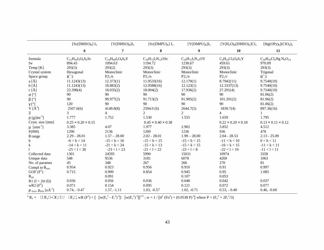
43
[Sc(DMSO)6] I3
6
[Y(DMSO)8]I3
7
[Sc(DMPU)6] I3
8
[Y(DMPU)6]I3
9
[Y(H2O)6(DMSO)2]Cl3
10
[Hg(OPy)6](ClO4)2
11
formula C12H36I3O6S6Sc C16H48I3O8S8Y C36H72I3N12OSc C36H72I3N12OY C4H24Cl3O8S2Y C30H30Cl2Hg N6O14 fw 894.43 1094.63 1194.72 1238.67 459.61 970.09 Temp [K] 293(3) 293(2) 293(3) 293(3) 293(3) 293(3) Crystal system Hexagonal Monoclinic Monoclinic Monoclinic Monoclinic Trigonal Space group R 3 P21/n P21/n P21/n P21/c R 3 a [Å] 11.1243(13) 12.373(1) 11.9533(16) 12.170(1) 8.7942(11) 9.7540(10) b [Å] 11.1243(13) 18.883(2) 12.0588(16) 12.123(1) 12.3337(13) 9.7540(10) c [Å] 23.398(4) 18.035(2) 18.004(2) 17.936(2) 27.291(4) 9.7540(10) α [°] 90 90 90 90 90 81.06(2) β [°] 90 99.977(2) 91.713(2) 91.905(2) 101.201(2) 81.06(2) γ [°] 120 90 90 90 90 81.06(2) V [Å3] 2507.6(6) 4149.8(8) 2594.0 (6) 2644.7(5) 1839.7(4) 897.36(16) Z 3 4 2 2 4 1 ρ [g⋅cm-3] 1.777 1.752 1.530 1.555 1.659 1.795 Cryst. size [mm] 0.25 × 0.20 × 0.15 0.45 × 0.40 × 0.38 0.22 × 0.20 × 0.18 0.13 × 0.11 × 0.12 µ [mm-1] 3.385 4.07 1.977 2.902 3.852 4.512 F(000) 1296 2136 1200 1236 936 478 θ range 2.29 - 28.01 1.57 – 28.00 2.02 - 28.01 1.99 – 28.00 2.04 - 28.53 2.13 - 25.09 h -6 < h < 14 -15 < h < 16 -15 < h < 15 -15 < h < 15 -11 < h < 10 0 < h < 11 k -14 < k < 11 -21 < k < 24 -15 < k < 13 -15 < k < 15 -16 < k < 15 -11 < k < 11 l -25 < l < 30 -23 < l < 23 -21 < l < 22 -23 < l < 8 -22 < l < 19 -11 < l < 11 Collected data 1301 24595 5990 15031 10974 3358 Unique data 548 9536 3181 6078 4269 1063 No. of parames 45 348 267 266 270 81 Compl to θmax 0.954 0.923 0.956 0.910 0.91 0.997 GOF (F2) 0.715 0.999 0.854 0.945 0.95 1.085 Rint 0.091 0.107 0.053 R1 (I > 2σ (I)) 0.036 0.056 0.036 0.048 0.042 0.037 wR2 (F2) 0.071 0.154 0.095 0.121 0.072 0.077 ρ max, ρmin [eÅ-3] 0.74, - 0.47 1.57, -1.11 1.03, -0.57 1.02, -0.71 0.53, - 0.40 0.46, -0.60
a R1 = Fo− Fc / Fo , wR (F2) = [ [w(Fo2 - Fc
2) 2]/ [w(Fo2) 2]] 1/2 ; w = 1 / [σ2 (Fo2) + (0.0538 P) 2] where P = (Fo
2 + 2Fc2/3)
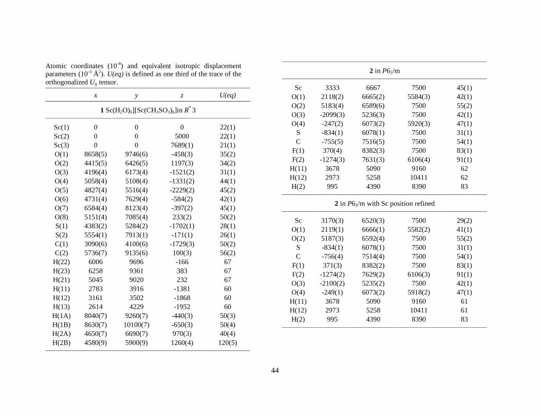
44
Atomic coordinates (10-4) and equivalent isotropic displacement parameters (10-3 Å2). U(eq) is defined as one third of the trace of the orthogonalized Uij tensor.
x y z U(eq)
1 Sc(H2O)6][Sc(CH3SO3)6]in R 3
Sc(1) 0 0 0 22(1) Sc(2) 0 0 5000 22(1) Sc(3) 0 0 7689(1) 21(1) O(1) 8658(5) 9746(6) -458(3) 35(2) O(2) 4415(5) 6426(5) 1197(3) 34(2) O(3) 4196(4) 6173(4) -1521(2) 31(1) O(4) 5058(4) 5108(4) -1331(2) 44(1) O(5) 4827(4) 5516(4) -2229(2) 45(2) O(6) 4731(4) 7629(4) -584(2) 42(1) O(7) 6584(4) 8123(4) -397(2) 45(1) O(8) 5151(4) 7085(4) 233(2) 50(2) S(1) 4383(2) 5284(2) -1702(1) 28(1) S(2) 5554(1) 7913(1) -171(1) 26(1) C(1) 3090(6) 4100(6) -1729(3) 50(2) C(2) 5736(7) 9135(6) 100(3) 56(2)
H(22) 6006 9696 -166 67 H(23) 6258 9361 383 67 H(21) 5045 9020 232 67 H(11) 2783 3916 -1381 60 H(12) 3161 3502 -1868 60 H(13) 2614 4229 -1952 60 H(1A) 8040(7) 9260(7) -440(3) 50(3) H(1B) 8630(7) 10100(7) -650(3) 50(4) H(2A) 4650(7) 6690(7) 970(3) 40(4) H(2B) 4580(9) 5900(9) 1260(4) 120(5)
2 in P63/m
Sc 3333 6667 7500 45(1) O(1) 2118(2) 6665(2) 5584(3) 42(1) O(2) 5183(4) 6589(6) 7500 55(2) O(3) -2099(3) 5236(3) 7500 42(1) O(4) -247(2) 6073(2) 5920(3) 47(1)
S -834(1) 6078(1) 7500 31(1) C -755(5) 7516(5) 7500 54(1)
F(1) 370(4) 8382(3) 7500 83(1) F(2) -1274(3) 7631(3) 6106(4) 91(1)
H(11) 3678 5090 9160 62 H(12) 2973 5258 10411 62 H(2) 995 4390 8390 83
2 in P63/m with Sc position refined
Sc 3170(3) 6520(3) 7500 29(2) O(1) 2119(1) 6666(1) 5582(2) 41(1) O(2) 5187(3) 6592(4) 7500 55(2)
S -834(1) 6078(1) 7500 31(1) C -756(4) 7514(4) 7500 54(1)
F(1) 371(3) 8382(2) 7500 83(1) F(2) -1274(2) 7629(2) 6106(3) 91(1) O(3) -2100(2) 5235(2) 7500 42(1) O(4) -249(1) 6073(2) 5918(2) 47(1)
H(11) 3678 5090 9160 61 H(12) 2973 5258 10411 61 H(2) 995 4390 8390 83
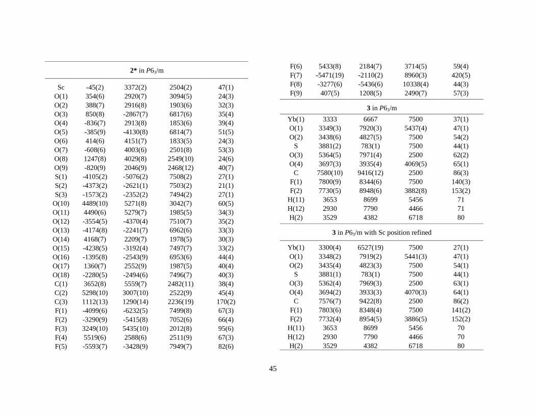
45
2* in P63/m
Sc -45(2) 3372(2) 2504(2) 47(1) O(1) 354(6) 2920(7) 3094(5) 24(3) O(2) 388(7) 2916(8) 1903(6) 32(3) O(3) 850(8) -2867(7) 6817(6) 35(4) O(4) -836(7) 2913(8) 1853(6) 39(4) O(5) -385(9) -4130(8) 6814(7) 51(5) O(6) 414(6) 4151(7) 1833(5) 24(3) O(7) -608(6) 4003(6) 2501(8) 53(3) O(8) 1247(8) 4029(8) 2549(10) 24(6) O(9) -820(9) 2046(9) 2468(12) 40(7) S(1) -4105(2) -5076(2) 7508(2) 27(1) S(2) -4373(2) -2621(1) 7503(2) 21(1) S(3) -1573(2) -2352(2) 7494(2) 27(1)
O(10) 4489(10) 5271(8) 3042(7) 60(5) O(11) 4490(6) 5279(7) 1985(5) 34(3) O(12) -3554(5) -4370(4) 7510(7) 35(2) O(13) -4174(8) -2241(7) 6962(6) 33(3) O(14) 4168(7) 2209(7) 1978(5) 30(3) O(15) -4238(5) -3192(4) 7497(7) 33(2) O(16) -1395(8) -2543(9) 6953(6) 44(4) O(17) 1360(7) 2552(9) 1987(5) 40(4) O(18) -2280(5) -2494(6) 7496(7) 40(3) C(1) 3652(8) 5559(7) 2482(11) 38(4) C(2) 5298(10) 3007(10) 2522(9) 45(4) C(3) 1112(13) 1290(14) 2236(19) 170(2) F(1) -4099(6) -6232(5) 7499(8) 67(3) F(2) -3290(9) -5415(8) 7052(6) 66(4) F(3) 3249(10) 5435(10) 2012(8) 95(6) F(4) 5519(6) 2588(6) 2511(9) 67(3) F(5) -5593(7) -3428(9) 7949(7) 82(6)
F(6) 5433(8) 2184(7) 3714(5) 59(4) F(7) -5471(19) -2110(2) 8960(3) 420(5) F(8) -3277(6) -5436(6) 10338(4) 44(3) F(9) 407(5) 1208(5) 2490(7) 57(3)
3 in P63/m Yb(1) 3333 6667 7500 37(1) O(1) 3349(3) 7920(3) 5437(4) 47(1) O(2) 3438(6) 4827(5) 7500 54(2)
S 3881(2) 783(1) 7500 44(1) O(3) 5364(5) 7971(4) 2500 62(2) O(4) 3697(3) 3935(4) 4069(5) 65(1)
C 7580(10) 9416(12) 2500 86(3) F(1) 7800(9) 8344(6) 7500 140(3) F(2) 7730(5) 8948(6) 3882(8) 153(2)
H(11) 3653 8699 5456 71 H(12) 2930 7790 4466 71 H(2) 3529 4382 6718 80
3 in P63/m with Sc position refined
Yb(1) 3300(4) 6527(19) 7500 27(1) O(1) 3348(2) 7919(2) 5441(3) 47(1) O(2) 3435(4) 4823(3) 7500 54(1)
S 3881(1) 783(1) 7500 44(1) O(3) 5362(4) 7969(3) 2500 63(1) O(4) 3694(2) 3933(3) 4070(3) 64(1)
C 7576(7) 9422(8) 2500 86(2) F(1) 7803(6) 8348(4) 7500 141(2) F(2) 7732(4) 8954(5) 3886(5) 152(2)
H(11) 3653 8699 5456 70 H(12) 2930 7790 4466 70 H(2) 3529 4382 6718 80
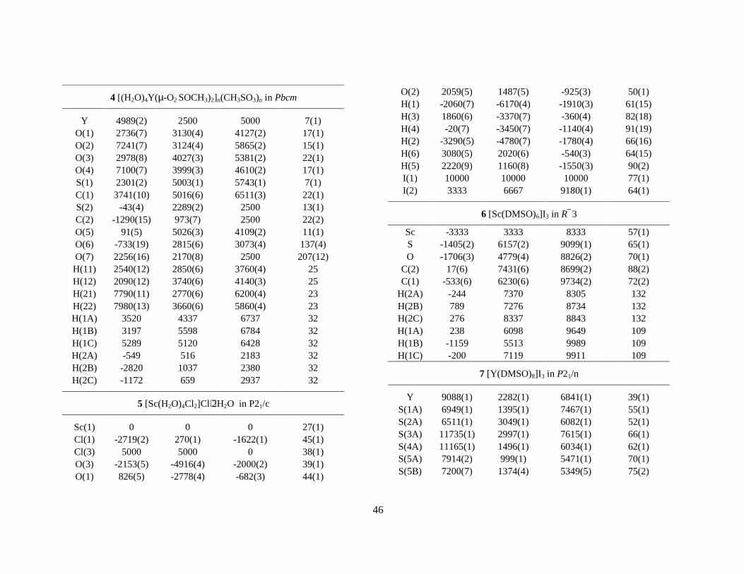
46
4 [(H2O)4Y(µ-O2 SOCH3)2]n(CH3SO3)n in Pbcm
Y 4989(2) 2500 5000 7(1) O(1) 2736(7) 3130(4) 4127(2) 17(1) O(2) 7241(7) 3124(4) 5865(2) 15(1) O(3) 2978(8) 4027(3) 5381(2) 22(1) O(4) 7100(7) 3999(3) 4610(2) 17(1) S(1) 2301(2) 5003(1) 5743(1) 7(1) C(1) 3741(10) 5016(6) 6511(3) 22(1) S(2) -43(4) 2289(2) 2500 13(1) C(2) -1290(15) 973(7) 2500 22(2) O(5) 91(5) 5026(3) 4109(2) 11(1) O(6) -733(19) 2815(6) 3073(4) 137(4) O(7) 2256(16) 2170(8) 2500 207(12) H(11) 2540(12) 2850(6) 3760(4) 25 H(12) 2090(12) 3740(6) 4140(3) 25 H(21) 7790(11) 2770(6) 6200(4) 23 H(22) 7980(13) 3660(6) 5860(4) 23 H(1A) 3520 4337 6737 32 H(1B) 3197 5598 6784 32 H(1C) 5289 5120 6428 32 H(2A) -549 516 2183 32 H(2B) -2820 1037 2380 32 H(2C) -1172 659 2937 32
5 [Sc(H2O)4Cl2]Cl⋅2H2O in P21/c
Sc(1) 0 0 0 27(1) Cl(1) -2719(2) 270(1) -1622(1) 45(1) Cl(3) 5000 5000 0 38(1) O(3) -2153(5) -4916(4) -2000(2) 39(1) O(1) 826(5) -2778(4) -682(3) 44(1)
O(2) 2059(5) 1487(5) -925(3) 50(1) H(1) -2060(7) -6170(4) -1910(3) 61(15) H(3) 1860(6) -3370(7) -360(4) 82(18) H(4) -20(7) -3450(7) -1140(4) 91(19) H(2) -3290(5) -4780(7) -1780(4) 66(16) H(6) 3080(5) 2020(6) -540(3) 64(15) H(5) 2220(9) 1160(8) -1550(3) 90(2) I(1) 10000 10000 10000 77(1) I(2) 3333 6667 9180(1) 64(1)
6 [Sc(DMSO)6]I3 in R 3
Sc -3333 3333 8333 57(1) S -1405(2) 6157(2) 9099(1) 65(1) O -1706(3) 4779(4) 8826(2) 70(1)
C(2) 17(6) 7431(6) 8699(2) 88(2) C(1) -533(6) 6230(6) 9734(2) 72(2)
H(2A) -244 7370 8305 132 H(2B) 789 7276 8734 132 H(2C) 276 8337 8843 132 H(1A) 238 6098 9649 109 H(1B) -1159 5513 9989 109 H(1C) -200 7119 9911 109
7 [Y(DMSO)8]I3 in P21/n
Y 9088(1) 2282(1) 6841(1) 39(1) S(1A) 6949(1) 1395(1) 7467(1) 55(1) S(2A) 6511(1) 3049(1) 6082(1) 52(1) S(3A) 11735(1) 2997(1) 7615(1) 66(1) S(4A) 11165(1) 1496(1) 6034(1) 62(1) S(5A) 7914(2) 999(1) 5471(1) 70(1) S(5B) 7200(7) 1374(4) 5349(5) 75(2)
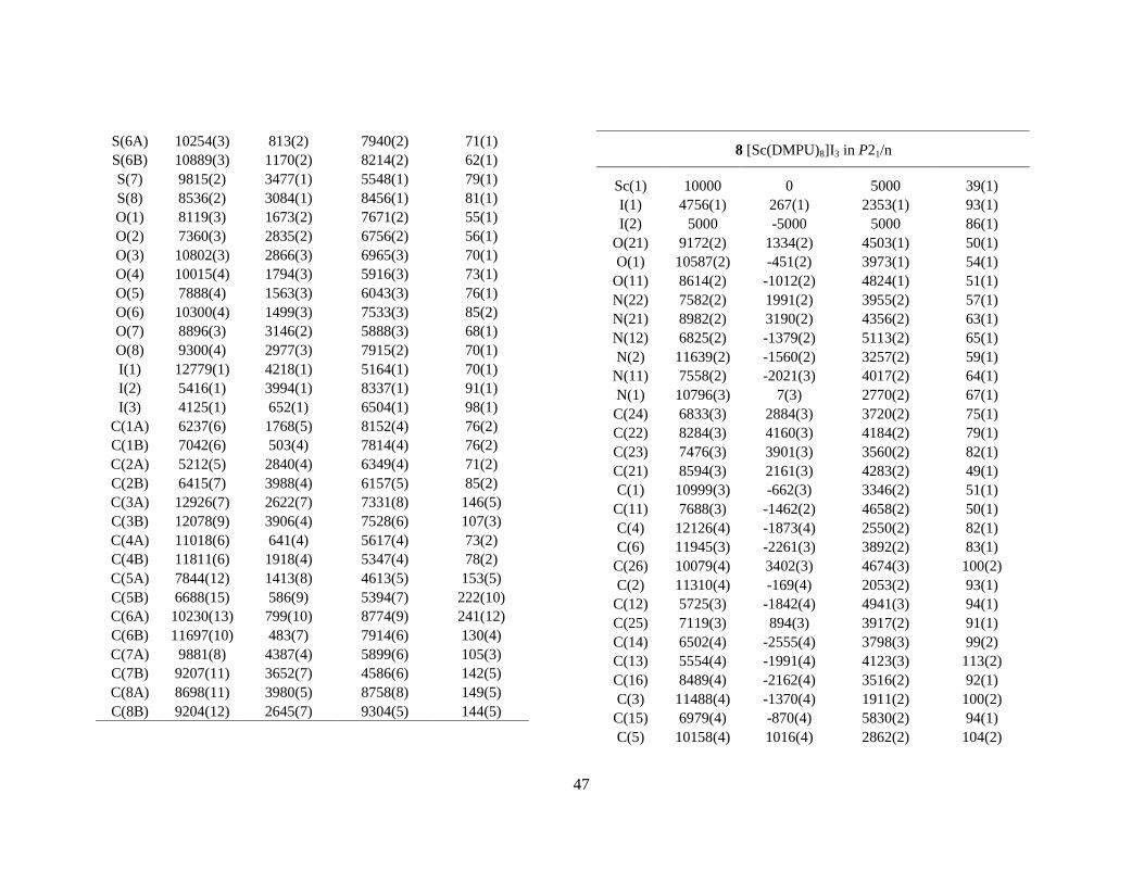
47
S(6A) 10254(3) 813(2) 7940(2) 71(1) S(6B) 10889(3) 1170(2) 8214(2) 62(1) S(7) 9815(2) 3477(1) 5548(1) 79(1) S(8) 8536(2) 3084(1) 8456(1) 81(1) O(1) 8119(3) 1673(2) 7671(2) 55(1) O(2) 7360(3) 2835(2) 6756(2) 56(1) O(3) 10802(3) 2866(3) 6965(3) 70(1) O(4) 10015(4) 1794(3) 5916(3) 73(1) O(5) 7888(4) 1563(3) 6043(3) 76(1) O(6) 10300(4) 1499(3) 7533(3) 85(2) O(7) 8896(3) 3146(2) 5888(3) 68(1) O(8) 9300(4) 2977(3) 7915(2) 70(1) I(1) 12779(1) 4218(1) 5164(1) 70(1) I(2) 5416(1) 3994(1) 8337(1) 91(1) I(3) 4125(1) 652(1) 6504(1) 98(1)
C(1A) 6237(6) 1768(5) 8152(4) 76(2) C(1B) 7042(6) 503(4) 7814(4) 76(2) C(2A) 5212(5) 2840(4) 6349(4) 71(2) C(2B) 6415(7) 3988(4) 6157(5) 85(2) C(3A) 12926(7) 2622(7) 7331(8) 146(5) C(3B) 12078(9) 3906(4) 7528(6) 107(3) C(4A) 11018(6) 641(4) 5617(4) 73(2) C(4B) 11811(6) 1918(4) 5347(4) 78(2) C(5A) 7844(12) 1413(8) 4613(5) 153(5) C(5B) 6688(15) 586(9) 5394(7) 222(10) C(6A) 10230(13) 799(10) 8774(9) 241(12) C(6B) 11697(10) 483(7) 7914(6) 130(4) C(7A) 9881(8) 4387(4) 5899(6) 105(3) C(7B) 9207(11) 3652(7) 4586(6) 142(5) C(8A) 8698(11) 3980(5) 8758(8) 149(5) C(8B) 9204(12) 2645(7) 9304(5) 144(5)
8 [Sc(DMPU)8]I3 in P21/n
Sc(1) 10000 0 5000 39(1) I(1) 4756(1) 267(1) 2353(1) 93(1) I(2) 5000 -5000 5000 86(1)
O(21) 9172(2) 1334(2) 4503(1) 50(1) O(1) 10587(2) -451(2) 3973(1) 54(1)
O(11) 8614(2) -1012(2) 4824(1) 51(1) N(22) 7582(2) 1991(2) 3955(2) 57(1) N(21) 8982(2) 3190(2) 4356(2) 63(1) N(12) 6825(2) -1379(2) 5113(2) 65(1) N(2) 11639(2) -1560(2) 3257(2) 59(1)
N(11) 7558(2) -2021(3) 4017(2) 64(1) N(1) 10796(3) 7(3) 2770(2) 67(1) C(24) 6833(3) 2884(3) 3720(2) 75(1) C(22) 8284(3) 4160(3) 4184(2) 79(1) C(23) 7476(3) 3901(3) 3560(2) 82(1) C(21) 8594(3) 2161(3) 4283(2) 49(1) C(1) 10999(3) -662(3) 3346(2) 51(1)
C(11) 7688(3) -1462(2) 4658(2) 50(1) C(4) 12126(4) -1873(4) 2550(2) 82(1) C(6) 11945(3) -2261(3) 3892(2) 83(1)
C(26) 10079(4) 3402(3) 4674(3) 100(2) C(2) 11310(4) -169(4) 2053(2) 93(1)
C(12) 5725(3) -1842(4) 4941(3) 94(1) C(25) 7119(3) 894(3) 3917(2) 91(1) C(14) 6502(4) -2555(4) 3798(3) 99(2) C(13) 5554(4) -1991(4) 4123(3) 113(2) C(16) 8489(4) -2162(4) 3516(2) 92(1) C(3) 11488(4) -1370(4) 1911(2) 100(2)
C(15) 6979(4) -870(4) 5830(2) 94(1) C(5) 10158(4) 1016(4) 2862(2) 104(2)
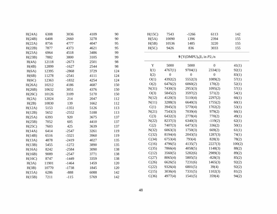
48
H(24A) 6308 3036 4109 90 H(24B) 6408 2660 3278 90 H(22A) 8756 4777 4047 95 H(22B) 7877 4373 4621 95 H(23A) 6964 4518 3486 99 H(23B) 7882 3800 3105 99 H(4A) 12118 -2673 2501 98 H(4B) 12899 -1627 2544 98 H(6A) 12395 -2868 3729 124 H(6B) 11278 -2541 4111 124 H(6C) 12363 -1832 4254 124
H(26A) 10212 4186 4687 150 H(26B) 10632 3051 4376 150 H(26C) 10126 3109 5170 150 H(2A) 12024 214 2047 112 H(2B) 10830 139 1662 112
H(12A) 5153 -1351 5126 113 H(12B) 5652 -2552 5188 113 H(25A) 6393 920 3675 137 H(25B) 7052 605 4410 137 H(25C) 7603 425 3639 137 H(14A) 6414 -2547 3261 119 H(14B) 6516 -3321 3960 119 H(13A) 4878 -2419 4027 135 H(13B) 5455 -1272 3890 135 H(16A) 8242 -2584 3090 138 H(16B) 9089 -2547 3771 138 H(16C) 8747 -1449 3359 138 H(3A) 11901 -1464 1459 120 H(3B) 10770 -1738 1845 120
H(15A) 6286 -888 6088 142 H(15B) 7211 -115 5769 142
H(15C) 7543 -1266 6113 142 H(5A) 10090 1396 2394 155 H(5B) 10536 1485 3220 155 H(5C) 9426 836 3033 155
9 [Y(DMPU)8]I3 in P21/n
Y 5000 5000 0 41(1) I(1) 4767(1) 9704(1) 2334(1) 92(1) I(2) 0 0 0 83(1) O(1) 4392(2) 5552(3) 1089(2) 57(1) O(2) 6476(2) 6060(2) 170(2) 52(1)
N(31) 7430(3) 2953(3) 1095(2) 57(1) O(3) 5845(2) 3597(2) 571(2) 54(1)
N(12) 4120(3) 5110(4) 2297(2) 66(1) N(11) 3288(3) 6640(3) 1755(2) 60(1) C(1) 3945(3) 5770(4) 1702(2) 53(1)
N(21) 7543(3) 7039(4) 979(2) 66(1) C(3) 6432(3) 2778(4) 770(2) 49(1)
N(22) 8237(3) 6340(3) -119(2) 62(1) C(2) 7407(3) 6473(3) 336(2) 50(1)
N(32) 6063(3) 1750(3) 669(2) 61(1) C(32) 8194(4) 2043(5) 1287(3) 74(1) C(34) 6753(4) 793(4) 828(3) 78(2) C(16) 4796(5) 4135(7) 2227(3) 100(2) C(35) 7866(4) 4058(5) 1148(3) 88(2) C(12) 3560(5) 5282(6) 2989(3) 89(2) C(27) 8065(4) 5805(5) -828(3) 85(2) C(26) 6626(5) 7231(6) 1465(3) 92(2) C(22) 9326(4) 6801(5) 39(4) 92(2) C(15) 3036(4) 7331(5) 1102(3) 81(2) C(36) 4977(4) 1541(5) 359(4) 94(2)
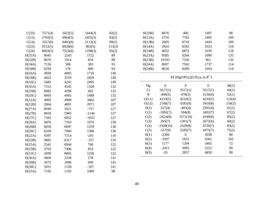
49
C(33) 7571(4) 1022(5) 1444(3) 82(2) C(13) 2765(5) 6964(5) 2455(3) 83(2) C(14) 3357(6) 6493(6) 3113(3) 99(2) C(23) 9512(5) 6928(6) 863(5) 113(3) C(24) 8603(5) 7523(6) 1198(3) 95(2)
H(32A) 8645 2243 1722 89 H(32B) 8676 1914 876 89 H(34A) 7136 588 383 93 H(34B) 6294 176 966 93 H(16A) 4930 4005 1710 149 H(16B) 4422 3510 2429 149 H(16C) 5483 4242 2495 149 H(35A) 7312 4545 1328 132 H(35B) 8082 4298 665 132 H(35C) 8493 4065 1488 132 H(12A) 4005 4989 3402 107 H(12B) 2864 4891 2971 107 H(27A) 8040 5021 -757 127 H(27B) 8659 5985 -1146 127 H(27C) 7383 6052 -1055 127 H(26A) 6876 7163 1976 138 H(26B) 6059 6697 1359 138 H(26C) 6339 7960 1380 138 H(22A) 9387 7514 -201 110 H(22B) 9881 6317 -157 110 H(15A) 2541 6944 766 122 H(15B) 3703 7496 853 122 H(15C) 2699 8005 1258 122 H(36A) 4600 2228 278 141 H(36B) 4575 1096 699 141 H(36C) 5031 1158 -107 141 H(33A) 7195 1105 1909 98
H(33B) 8076 406 1497 98 H(13A) 2759 7762 2495 100 H(13B) 2009 6710 2443 100 H(14A) 2924 6592 3553 118 H(14B) 4052 6872 3195 118 H(23A) 9585 6204 1090 135 H(23B) 10193 7326 961 135 H(24A) 8697 7502 1737 114 H(24B) 8618 8289 1043 114
11 [Hg(OPy)6](ClO4)3 in R 3
Hg 0 0 0 48(1) Cl 3517(1) 3517(1) 3517(1) 64(1) N -499(5) 478(5) 3150(4) 52(1)
O(11) 4233(5) 4233(5) 4233(5) 123(4) O(12) 2186(7) 4281(8) 3410(8) 134(2) O(1) 217(4) -493(4) 2391(4) 61(1) C(1) -1892(7) 594(8) 3402(7) 67(2) C(2) -2624(8) 1571(10) 4189(8) 85(2) C(3) 205(7) 1301(7) 3672(6) 60(2) C(4) -1928(10) 2420(8) 4720(7) 83(2) C(5) -517(9) 2285(7) 4475(7) 75(2) H(1) -2366 6 3038 80 H(2) -3597 1655 4361 102 H(3) 1177 1209 3492 72 H(4) -2421 3095 5252 99 H(5) -35 2857 4850 90