pathophysiology of the post-polio syndrome and persistence ......pathophysiology of the post-polio...
TRANSCRIPT

Andreina Baj1, Giuseppe Maccari1, Giorgio Bono2,
Oscar Diaz-Horta1, Antonio Toniolo1
Institute of Microbiology1 and Institute of Neurology2,
University of Insubria Medical School, Varese, Italy
Pathophysiology of the post-polio
syndrome
and persistence of poliovirus
genomes in polio survivors

The “invisible group” of polio survivors
Polio survivors form the largest single group of people
with physical disabilities.
Polio survivors are “invisible” in the community.
Knowledge about the late consequences of polio
(LEOP), and their impact upon the lives of polio survivors
and their families, is almost non existent amongst the
medical profession, policy makers, the community at
large, and indeed the polio survivors themselves.
The LEOP have a dramatic impact on the ability of those polio survivors
affected to maintain their mobility and independence and successfully
undertake the activities of daily living.
There is a high cost to both polio survivors and the health system in trying to
get a diagnosis and adequate treatments.
Thus, the need for in-depth knowledge about the causes and
pathogenesis with the aim of developing effective therapies.

Interest in PoliovirusWe investigate viruses as infectious agents that cause disease in their
host.
Our subjects are the etiology of viral diseases and mechanisms of viral
pathogenensis.
We are interested in Picornaviruses (RNA viruses) whose prototype is
Poliovirus (3 types are known to exist).
Picornaviruses (over 100 types) are estimated to infect billions of humans
per year, causing a vast array of disease syndromes (paralysis,
meningitis, heart disease, hepatitis, common cold, etc.).
Picornaviruses contain a plus-stranded RNA genome that functions as
mRNA as soon as the viral particle enters the cell. The viral proteins,
which are synthesized, recruit cellular factors. Together, they provide a
menu for genome replication and genome encapsidation (i.e. the
formation of new viral particles).
From: Eckard Wimmer, Stony Brook University, NY - http://www.mgm.stonybrook.edu/wimmer/index.shtml

Poliovirus


Family Genus Species Serotypes
Picornaviridae Enterovirus
(>100
serotypes)
Human enterovirus A (17) CVA-2, 3, 4, 5, 6, 7, 8, 10, 12, 14, 16;
Enterovirus-71, 76, 89, 90, 91, 92
Human enterovirus B (58) CBV-1, 2, 3, 4, 5 (incl. SVDV), 6; CVA9;
Echovirus-1, 2, 3, 4, 5, 6, 7, 9, 11, 12, 13, 14, 15,
16, 17, 18, 19, 20, 21, 24, 25, 26, 27, 29, 30, 31, 32,
33;
Enterovirus-69, 73, 74, 75, 77, 78, 79, 80, 81, 82,
83, 84, 85, 86, 87, 88, 93, 97, 98, 100, 101, 106, 107
Human enterovirus C (19) CVA-1, 11, 13, 17, 19, 20, 21, 22, 24; Enterovirus-
95, 96, 99, 102, 104, 105, 109
Poliovirus-1, 2, 3
Human enterovirus D (3) Enterovirus-68, 70, 94
Parechovirus Human parechovirus (14)
Ljungan virus (4)
HPeV-1, 2, 3, 5, 6, 7, 8, 9, 10, 11, 12, 13, 14
Kobuvirus Human Aichi virus (1)
Bovine Kobuvirus
AiV
Cardiovirus Encephalomyocarditis virus (1)
Theilovirus (12)
animal EMC;
Theiler‘s murine encephalomyelitis virus;
Rat Theravirus;
Human Vilyuisk encephalomyelitis virus;
Human Saffold virus (9)
Picornavirus classification

Pathogenesis of poliomyelitisIn virology, the term “Pathogenesis” refers to mechanisms by which a virus
causes disease in a host organism.
Pathogenetic mechanisms are complex and multi level.
Poliovirus infects humans only, but disease can also be produced
experimentally in primates and transgenic mice.
The human receptor for poliovirus is a protein designed as CD155. It is the
key for pathogenesis as it allows viral entry into cells.
Poliovirus replicates in the oro-gastro-intestinal tract (tonsils, Peyer's
patches?) from which it can migrate to the central nervous system where it
targets motor neurons.
Destruction of motor neurons causes irreversible paralysis or death, a
disease called poliomyelitis.
From: Eckard Wimmer, Stony Brook University, NY - http://www.mgm.stonybrook.edu/wimmer/index.shtml

Poliovirus receptor
Outer
cellular
space
Cell
membrane
Cytoplasm
CD155 receptor CD155 bound to poliovirus
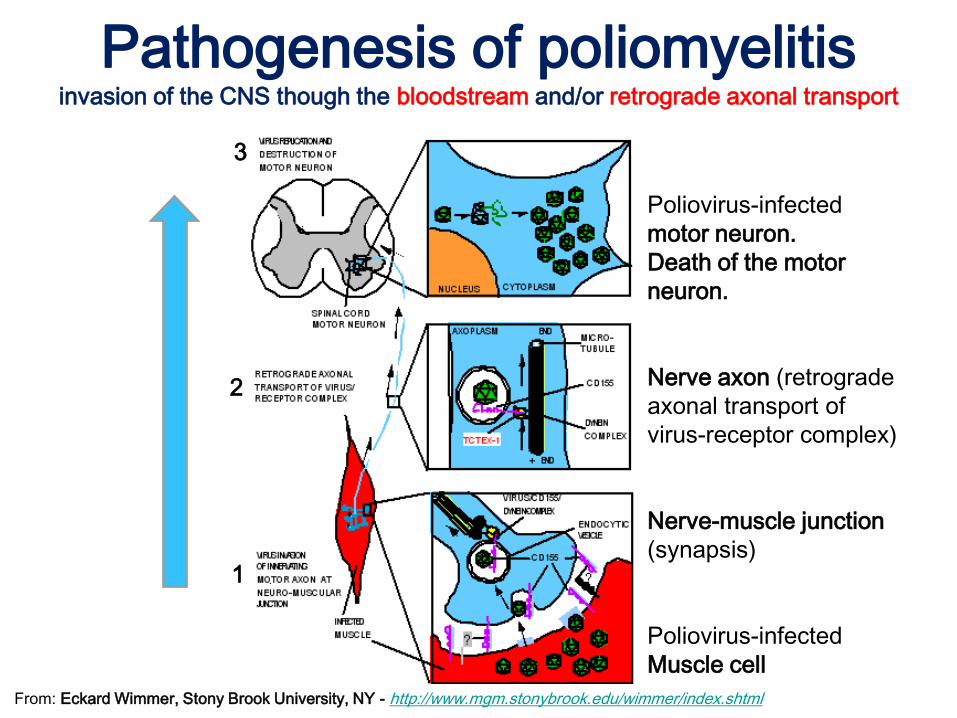
Pathogenesis of poliomyelitisinvasion of the CNS though the bloodstream and/or retrograde axonal transport
3
2
1
Poliovirus-infected
motor neuron.
Death of the motor
neuron.
Nerve axon (retrograde
axonal transport of
virus-receptor complex)
Nerve-muscle junction
(synapsis)
Poliovirus-infected
Muscle cell
From: Eckard Wimmer, Stony Brook University, NY - http://www.mgm.stonybrook.edu/wimmer/index.shtml

Viruses reside at the threshold between dead and living matter.
Poliovirus is a chemical with a life cycle. Its empirical formula is:
C332,652 H492,388 N98,245 O131,196 P7,501 S2,340
Inside the cells, poliovirus expresses the hallmarks of a living entity:
1. Multiplication,
2. Heredity,
3. Variation (mutation of the genome sequence),
4. Recombination (i.e., exchange of genomic sequences with related
viruses such as members of Picornaviridae).
Genetic elements of the viral genome influence neurovirulence.
We will discuss poliovirus multiplication, heredity and variation
with regard to the Post-Polio Syndrome
Pathogenesis of polio
From: Eckard Wimmer, Stony Brook University, NY - http://www.mgm.stonybrook.edu/wimmer/index.shtml

Poliovirus RNA genome
From: Eckard Wimmer, Stony Brook University, NY - http://www.mgm.stonybrook.edu/wimmer/index.shtml

Replication of Poliovirus Genome
3D
RNA polymerase(critical enzyme for
virus replication)
From: Eckard Wimmer, Stony Brook University, NY - http://www.mgm.stonybrook.edu/wimmer/index.shtml

Acute/symptomatic enterovirus infections are widely
recognized. Examples include paralytic polio, meningitis,
myocarditis, etc.
Persistent enterovirus infections are documented in
agammaglobulinemic patients. Examples are chronic excretion
of poliovirus after natural infection or vaccination with live
attenuated virus.
Recently, it has been been shown that the incubation period of
polio can span several years.
In fact, polio developed in an immunodeficient woman
who remained chronically infected for 12 years after
vaccinating her child (N Engl J Med. 2011 Jun 16;
364:2316-23).

N Engl J Med 2011; 364:2316-2323 June 16, 2011
Vaccine-Derived Poliomyelitis 12 Years after Infection in Minnesota
Aaron S. DeVries et al.
A 44-year-old woman with long-standing common variable immunodeficiency who
was receiving intravenous immune globulin suddenly had paralysis of all four limbs
and the respiratory muscles, resulting in death. Type 2 vaccine-derived poliovirus
was isolated from stool. The viral capsid protein VP1 region had diverged from the
vaccine strain at 12.3% of nucleotide positions, and the two attenuating substitutions
had reverted to the wild-type sequence. Infection probably occurred 11.9 years
earlier (95% confidence interval [CI], 10.9 to 13.2), when her child received the oral
poliovirus vaccine. No secondary cases were identified among close contacts or
2038 screened health care workers. Patients with common variable
immunodeficiency can be chronically infected with poliovirus, and poliomyelitis can
develop despite treatment with intravenous immune globulin.
We thank the following persons for their efforts during this project: Richard Danila, Kristen Ehresmann, Sara Lowther, Claudia
Miller, and Elly Pretzel at the Minnesota Department of Health; Gregory Armstrong, Jane Iber, Olen Kew, Eric Mast, Steven
Oberste, Mark Pallansch, and Jane Seward at the Centers for Disease Control and Prevention; Vicki Carlson, Karen Ferrara,
Gary Kravitz, and Doris Nordbye at the United Hospital and Clinic; Anita Guelcher, Chris Hendrickson, and Lisa Ide at the
University of Minnesota Medical Center, Fairview; and John Modlin at the Dartmouth–Hitchcock Medical Center.

Pathogenesis of PPS
In contrast to paralytic polio, the origin of PPS is
unclear.
Different factors have been blamed for: the aging
process, the distal degeneration of the residual
enlarged motor neurons that are proper of polio,
and chronic inflammation.
It has been proposed (but not proven) that
persistence of mutated PVs may cause the
progressive neuromuscular damage seen in
PPS.

Poliomyelitis: CNS areas infected by Polioviruses

Late effects of polio: progression from poliomyelitis to PPS
Residual enlarged spinal motor
neuron that is innervating
increased numbers of muscle cells
Death of spinal motor
neurons following
infection with poliovirus
Distal degeneration of
axonal sprouts or loss
of entire motor units

The Post- Polio Syndrome: multiple factors in pathogenesis
We investigated whether
persistent poliovirus infections
were associated with
the late consequences of polio

METHODS

Bioinformatics: Phylogenetic tree of PVs and polio-like EVs

Different genomic regions have been investigated:
• 5’UTR (cloverleaf and entry into ribosomes)
• 5’UTR-VP2 (entry into ribosomes-capsid proteins)
• 3Dpol (RNA polymerase)
Bioinformatics: design of primers for detecting PVs
RNA
genome
Primers
PVs: %
sequence
similarity

Methods: amplification of the 3Dpol region
Amplification of the 3 poliovirus types
Primers
for the
3Dpol
region

Antibody
Cell cultures infected with different enterovirus types
CB
V1
CB
V2
CB
V3
CB
V4
CB
V5
CB
V6
CA
V2
CA
V4
CA
V9
ech
o3
ech
o7
ech
o1
1
EV
71
PV
1
PV
2
PV
3
Pan-
Enterovirus
9-D5
(Chemicon)
+++ +++ +++ +++ +++ ++ ++ +++ ++ +++ +++ ++ +++ +++ +++ +++
CBV4-356.1 - - - +++ - - - - - - - - - - - -
Poliovirus
Blend
(Chemicon)
- - - - - - - - - - - - - ++ ++ ++
Immunofluorescence of polioviruses using different antibodies

Saliv
aU
rin
eC
SF
RT-
PC
R
Leu
cocy
tes
co-culture withhuman cell lines
leukocyteseparation
Ne
rve
Mu
scle
Pri
mar
yce
llcu
ltu
re
Du
od
en
um
Procedures for detecting polioviruses in post-polio patients
RT-PCR, CPE, antigen expression, cytokines
CAN POLIOVIRUS BE DETECTED
IN PPS PATIENTS?

Male/FemaleAge
(years, M ± SD)
Years from APP
(years, M ± SD)
0.36 57.4 ± 7.3 53 ± 7.0
PPS patients (n = 81)
Male/FemaleAge
(years, M ± SD)
0.67 39.7 ± 13.4
Controls (n = 76)Blood donors (n=26); neurologic patients with non-infectious, autoimmune,or neoplastic disease (n=11); family members of PPS patients (n=39)

1930
1932
1934
1936
1938
1940
1942
1944
1946
1948
1950
1952
1954
1956
1958
1960
1962
1964
1966
1968
1970
1972
1974
1976
1978
1980
1982
1984
1986
1988
1990
1992
1994
1996
1998
2000
2002
VA
PP
CA
SE
S
Year
of
Acu
te P
ara
lytic P
olio
mye
litis
Yea
r o
f A
cute
Pa
raly
tic
Po
liom
yelit
is

0.0
1.0
10
20
30
40
2 months
4 months
6 months
8 months
Ag
e a
t th
e o
nset
of
AP
P
Age at onset of Acute Paralytic Poliomyelitis

40
45
50
55
60
65
70
75
80
Ag
e o
f P
PS
pati
en
ts (
years
)
Age of the investigated PPS patients

PV1 PV2 PV3 ND Neg0
10
20
30
40
50
No
. o
f p
ati
en
tsPV genome fragment in PPS patients (n=81)
69/81 Positive (84%)
65%
14%
7%12%
16%

Pathologic controls (1/11)
Family members of PPS patients (2/39)
Blood donors (0/26)
Poliovirus genome fragments in control subjects (n=76)
No. positive: 3/76 (1,3%)

MU
SC
LE
FR
AG
ME
NT
(R
R)
ISC
HIA
TIC
NE
RV
E
FR
AG
ME
NT
(C
M)
Primary culture of surgical samples:virus detection after >30 yrs from the acute event
PV detection in primary cultures from PPS patients: skeletal muscle & nerve
PV antigen

PATIENT LL:
detection of poliovirus genome fragment in duodenal cells (DC)
acute infection, 1933; virus detection, 2009
DC1 DC2 PV1
LL patient (Chat strain)
3D pol (630 bp)Polio-13Dpol fragment
PV detection in primary cultures from PPS patients: duodenal epithelial cells

PV antigens in the AV3 cell line co-cultured with leukocytes of PPS patients
Uninfected cells Reference PV1 (Chat strain)
PPS (RR strain) PPS (LL strain)
PV antigen

PATIENT LL1933: acute paralytic polio
2007: detection of PV1 in CSF and leukocytes
Detection of PV-1 in a PPS patient 74 years after APP

PVs obtained from PPS patients: sequenced genome regions
Multiple mutations and deletions have been detected in the 5`UTRand VP1 regions of PVs obtained from PPS patients.THE 3D POL REGION IS INSTEAD CONSERVED.

3D
po
l REG
ION

Since previous literature could only show “genomic
fragments” of polioviruses in PPS patients,
we investigated whether fragments from all over the
poliovirus genome could be obtained.
To this end, a collaboration with the Institute of Human
Virology (Baltimore, Maryland) was established.

740 900 1746 2480 3385 3832 4124 5111 5382 5438 5989 7000
VPg 5’UTR VP4 VP2 VP3 VP1 2A 2B 2C 3A 3B 3C 3D 3’UTR AA
Full-lenght amplification of persistent PV isolates from PPS patients
166 602
453 1196
1176 1811
1788 2202
2185 2580
2558 3031
3011 3416
3395 3782
3765 4068
4047 4488
4465 4721
4699 5280
5256 5958
5941 6372
5941 6377
6349 6856
6349 6946
6349 7045
6836 7396
6929 7396
7023 7396
I round
II round
I round
II round
P03LLVR: 2580bp fragment
PV-specific primer pairs
P03LLVR and P19RRVA: 1248bp fragment
Large fragments obtained so far

It came out that fragments spanning the entire PV
genome can be amplified in samples of PPS patients.
However - due to the extremely small amounts of
poliovirus in samples from PPS patients – difficulties
were encountered in sequencing the whole genome
of persisting poliovirus.

Together with our previous results, the data
produced in Baltimore demonstrate that amplified
fragments of the poliovirus can be obtained from
almost all genome regions (red boxes).
The 2A and 2B regions have not been detected yet.

IS THERE ANY RESIDUAL BIOLOGICAL ACTIVITY IN
PERSISTENT POLIOVIRUS OF PPS PATIENTS?

Cytopathic effect and expression of poliovirus antigen in cell lines exposed to persistent poliovirus of PPS patients
PV antigen

Cytokines elicited by persistent poliovirus of PPS patients
TNF
IL1B IL
6IL
12
IL17
AIL
8
MCP-1
RANTE
S
IP-1
0M
IG
TARC
IFNa
0
50
100
150
200
250
300
FO
LD
CH
AN
GE
OV
ER
UN
IN
FE
CT
ED
CO
NT
RO
LS
TNF
IL1B IL
6IL
12
IL17
AIL
8
MCP-1
RANTES
IP-1
0M
IG
TARC
IFNa
-5
0
5
10
15
20
FO
LD
CH
AN
GE
OV
ER
UN
IN
FE
CT
ED
CO
NT
RO
LS
Per
sist
ent
po
liovi
rus
fro
m P
PS
pa
tien
ts (
n=1
8)
ACUTE PV1 INFECTION (Chat strain, 30 hr p.i.)
multi-analyte ELISA array

Thus, persistent Poliovirus from PPS patients produces the following effects in human cell lines:1) slight cytopathic effect;2) expression of viral antigens within infected cells;3) stimulation of cytokine production, especially MCP-1.
MCP1 is a chemokine (chemotactic factor for monocytes and basophils, but not neutrophils or eosinophils) that has been implicated in chronic diseases with monocytic infiltrates (e.g., psoriasis, rheumatoid arthritis, atherosclerosis).
For comparison, infection with vaccine Poliovirus-1 causes:1) strong cytopathic effect;2) expression of viral antigens within infected cells;3) stimulation of cytokine production, especially IL-12 and RANTES.
IL12 stimulates IFN-γ and TNF-α production and reduces IL4 mediated suppression of IFN-γ. It enhances the cytotoxic activity of NK cells and CD8+ T lymphocytes. RANTES is a chemoattractant for monocytes, memory T-helper cells and eosinophils.

POSSIBLE PPS TREATMENTS

I. Post-polio patients have increased cytokine levels in CSF (CNS inflammation)
II. Inflammation is possibly down-modulated by normal Hu immunoglobulin
III. Down-modulated inflammation is associated with increased muscle strength and better quality of life
IV. Unfortunately, results are only temporary and repeated treatment may be required
Intravenous mmunoglobulin

Intravenous mmunoglobulin

Neutralizing Poliovirus antibody
J Virol. 2011. 85:4354-62Chimpanzee-human monoclonal antibodies for treatment of
chronic poliovirus excretors and emergency postexposure
prophylaxis.Chen Z, Chumakov K, Dragunsky E, Kouiavskaia D, Makiya M, Neverov A, Rezapkin G, Sebrell A, Purcell R.
National Institute of Allergy and Infectious Diseases, National Institutes of Health, Bethesda, Maryland
Six poliovirus-neutralizing Fabs were recovered from a combinatorial Fab phage display library constructed
from bone marrow-derived lymphocytes of immunized chimpanzees. The chimeric chimpanzee-human full-
length IgGs (hereinafter called monoclonal antibodies [MAbs]) were generated by combining a chimpanzee
IgG light chain and a variable domain of heavy chain with a human constant Fc region. The six MAbs
neutralized vaccine strains and virulent strains of poliovirus. Five MAbs were serotype specific, while one
MAb cross-neutralized serotypes 1 and 2.
Epitope mapping performed by selecting and sequencing antibody-resistant viral variants indicated that the
cross-neutralizing MAb bound between antigenic sites 1 and 2, thereby covering the canyon region
containing the receptor-binding site.
Another serotype 1-specific MAb recognized a region located between antigenic sites 2 and 3 that included
parts of capsid proteins VP1 and VP3. Both serotype 2-specific antibodies recognized antigenic site 1. No
escape mutants to serotype 3-specific MAbs could be generated. The administration of a serotype 1-specific
MAb to transgenic mice susceptible to poliovirus at a dose of 5 μg/mouse completely protected them from
paralysis after challenge with a lethal dose of wild-type poliovirus. Moreover, MAb injection 6 or 12 h after
virus infection provided significant protection.
The MAbs described here could be tested in clinical trials to determine whether they might be
useful for treatment of immunocompromised chronic virus excretors and for emergency
protection of contacts of a paralytic poliomyelitis case.

Antiviral drugs
Emerging Infectious Diseases • www.cdc.gov/eid • 14, April 2008
Potential Use of Antiviral Agents in Polio EradicationArmando M. De Palma,* Gerhard Pürstinger,† Eva Wimmer,† Amy K.
Patick,‡ Koen Andries,§ Bart Rombaut,¶ Erik De Clercq,* and Johan Neyts*
In 1988, the World Health Assembly launched the Global Polio Eradication
Initiative, which aimed to use largescale vaccination with the oral vaccine to
eradicate polio worldwide by the year 2000. Although important progress has
been made, polio remains endemic in several countries.
Also, the current control measures will likely be inadequate to deal with
problems that may arise in the postpolio era.
A panel convoked by the National Research Council concluded that the use
of antiviral drugs may be essential in the polio eradication strategy. We here
report on a comparative study of the antipoliovirus activity of a selection of
molecules that have previously been reported to be inhibitors of picornavirus
replication and discuss their potential use, alone or in combination, for the
treatment or prophylaxis of poliovirus infection.

PNAS 2010. 107:22505–22510
Structural basis for active site closure by the poliovirus RNA-dependent
RNA polymerasePeng Gong and Olve B. Peersen
Positive-strand RNA viruses include a large number of human and animal pathogens whose essential RNA-dependent RNA
polymerases (RdRPs) share a structurally homologous core with an encircled active site. RdRPs are targets for antiviral drug
development, but these efforts are hindered by limited structural information about the RdRP catalytic cycle. To further our
understanding of RdRP function, we assembled, purified, and then crystallized poliovirus elongation complexes after multiple
rounds of nucleotide incorporation. Here we present structures capturing the active polymerase and its nucleotide triphosphate
complexes in four distinct states, leading us to propose a six-state catalytic cycle involving residues that are highly conserved
among positive-strand RNA virus RdRPs. The structures indicate that RdRPs use a fully prepositioned templating base for
nucleotide recognition and close their active sites for catalysis using a novel structural rearrangement in the palm domain. The
data also suggest that translocation by RDRPs may not be directly linked to the conformational changes responsible for active
site closure and reopening.

Post-Polio Health InternationalSaint Louis, Missouri, USA
THE FIFTH AWARD (2009)
PHI award to a team from University of Insubria (Varese, Italy) led by AntonioToniolo, MD. The study, Persisting Noninfectious Fragments of Poliovirus inPPS Patients: Virus Detection and Susceptibility to Antiviral Drugs, will set upmethods for detecting polioviruses in PPS patients and sequencing theirgenome.

SUMMARY AND CONCLUSIONS

We detected genomic fragments of PVs in 69/81 PPS patients: 84%
Persisting PVs carry multiple mutations and deletions
Average documented persistence of PVs in PPS patients: 53 yrs
Thus, mutated PVs with modest ability to replicate can persist for decades in polio survivors
Persisting polioviruses are endowed with residual biologic activity expressed, for instance, as inflammatory stimuli
Persisting PVs were not found in family members of PPS patients. This result demonstrates that transmission of these “defective viruses” does not occur within the families of PPS patients
Therefore, these mutated polioviruses are not transmissible
SUMMARY

In polio survivors, the persistence of residual polioviralactivity can be associated with chronic inflammation
Viral diagnosis may pave the way to treating patients with antibodies and/or antiviral drugs in order to stop the progression of virus-associated cell damage
Current experiments in vitro show that neutralizing antibodies and antiviral drugs under development are effective against polioviruses
Polio meetings like this one in Copenhagen will help collecting ideas and will promote the development of effective treatments for PPS patients.
CONCLUSIONS

Thanks to the colleagues who introduced us to this area of research:
Abner L. Notkins (NIH, Bethesda, Maryland)Marinos Dalakas (NIH , Bethesda, Maryland)Takashi Onodera, Akio Nomoto (University of Tokyo, Japan)Salvatore Monaco, Luisa Bertolasi, Gianluigi Zanusso (University of Verona, Italy)Frans Nollet (University of Amsterdam, The Netherlands)Franco Molteni (Ospedale Valduce, Como, Italy)Luisa Arrondini (Fondazione Don Gnocchi, Milano, Italy)Merja Roivainen (Center for Poliovirus Surveillance and Enterovirus Research, Helsinki, Finland)Jean Carr (Institute of Human Virology, Baltimore, Maryland)Eckard Wimmer (Stony Brook University, New York)

We gratefully acknowledge the generous support of:• Post Polio Health International, St. Louis, MO• Regione Lombardia, Milan, IT• Asociación Afectados de Polio y Síndrome Post-Polio, Madrid, ES• Cariplo Foundation, Milan, IT
Our deepest appreciation goes to the many patients and their family members whose enthusiasm, patience, and suggestions made these investigations possible.