pathological study on a case of bladder ......pathological study on a case of bladder carcinoma...
TRANSCRIPT

Instructions for use
Title PATHOLOGICAL STUDY ON A CASE OF BLADDER CARCINOMA ACCOMPANIED BY CHRONICCYSTITIS IN COW
Author(s) OHSHIMA, Kan-ichi; ONO, Hitoshi
Citation Japanese Journal of Veterinary Research, 5(1), 19-26
Issue Date 1957-03-25
DOI 10.14943/jjvr.5.1.19
Doc URL http://hdl.handle.net/2115/1703
Type bulletin (article)
File Information KJ00002373102.pdf
Hokkaido University Collection of Scholarly and Academic Papers : HUSCAP

PATHOLOGICAL STUDY ON A CASE OF BLADDER
CARCINOMA ACCOMPANIED BY
CHRONIC CYSTITIS IN COW
Kan-ichi OHSHIMA'~' and Hitoshi ONO**
(Received for publication, Jan. 18, 1957)
PREFACE
It is said that tumors of the bladder are quite often observed in domestic animals. With respect to carcinoma, LUBARSCH and OSTERTAG (1896) and JOEST
(1924) pointed out its frequent occurrence in horses, cattle and dogs. Particularly in horses, STOLZ (1886), SCHLEGEL (1903), LYDING (1909), VomIN (1910), MONTPELLIER
and NYKA (1930) and RUSCHER (1931) and others have reported carcinoma cases,
and RUSCHER reported 25 cases of bladder carcinoma in horses in past literature.
On the other hand, reports on such cases in cattle are hardly known except the
one made by FRANKE (1909). However STICKER, in 1901, reported 9 cases (11.5%) of bladder carcinoma out of 78 primary carcinoma cases; MIYAMOTO (1928) observed
9 cases of carcinoma out of 24 bladder tumor cases, as well as WAKE and GOTO (1940) 46 out of 161, studying on hematuria in Formosa yellow cattle. GOTO et
al. (1954) stated that they found on the bladders of Formosa yellow cattle 6 tumor
cases containing carcinoma among 219 hematuria cases.
In human medicine, with regard to the bladder carcinoma as well as the other general cancer, workers have been concerned about the pathogenesis of the
case which has great significance for the determination of clinical prognosis,
which can easily be explained by looking over the report by IcHIKAWA et aL (1955).
In this case, a carcinoma as well as an adenoma with chronic cystitis was observed on the bladder mucosa of a 6 year old slaughtered cow, which is con
sidered to have much interest in connection with the pathogenesis of tumors.
MATERIALS AND METHODS
E 1792, a cross-bred Holstein-Friesian cow, 6 years old, habitat Nemuro-Shibetsu, Hokkaido, was slaughtered on July 22, 1955 at the Sapporo slaughter house. A meat
inspector of the slaughter house detected a neoplasm on the bladder, and brought it to the writers' laboratory for further pathological examination. Materials were bladder,
.)<0 Department of Veterinary Pathology, Facu1ty of Veterinary 111edicin, Hokkaido University, Sapporo, Japan.
** Federation of Hukkaido Agricultural Mutual Rel'ief Associations, Sapporo.
JAP. 1. VET. RES., VOL. 5, No.1, 1957
20 OHSHIMA, K. AND H. ONO
both sides of ureters, kidneys and suprarenal glands and several lymphonodi in pelvic region, and a part of liver and colon. It is said that there were no neoplasms detected on the other part of the body at the time of meat inspection.
Detail observations were carried out on the fresh materials, and touch preparation as well as smear were made from some pieces of tumors. MA. y-GIEMSA staining was adopted for cytological observation. The materials were fixed with 10% formalin. Paraffin sections were prepared adopting hematoxylin-eosin and thionine staining as well as VAN GIESON's method for pathological examination.
RESULTS
1. Macroscopic Findings
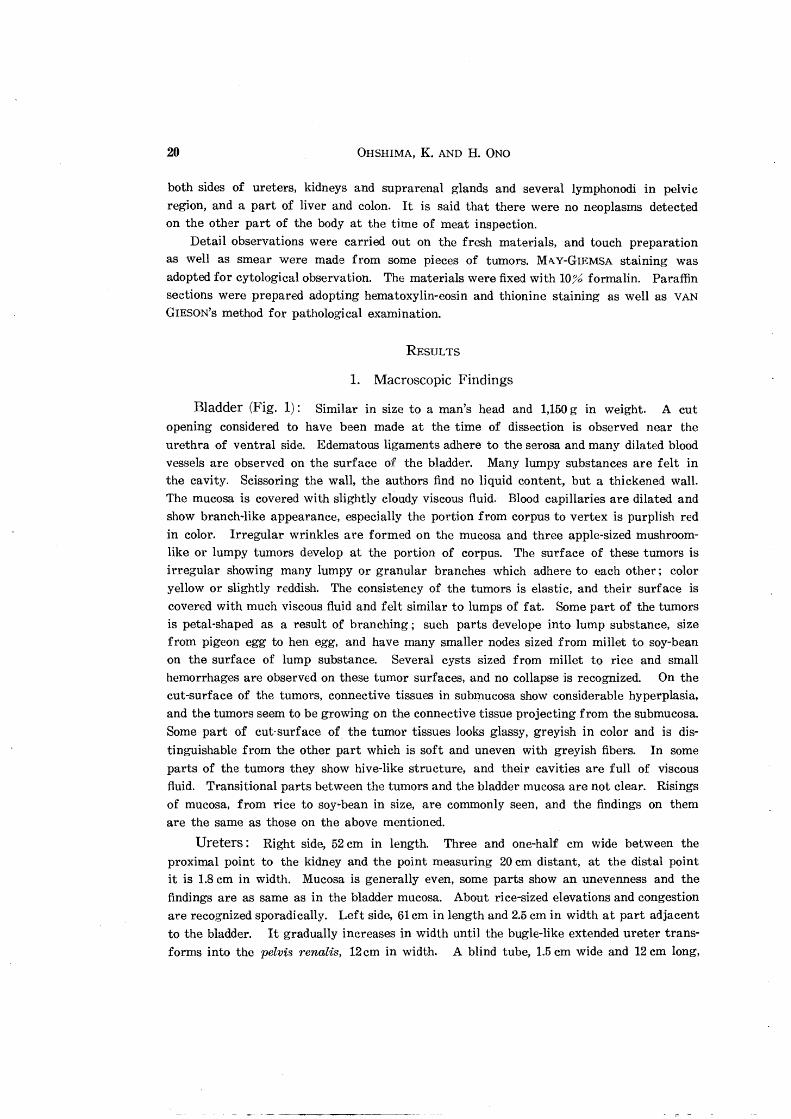
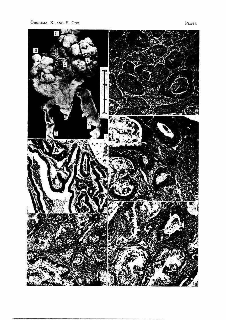
Bladder (Fig. 1): Similar in size to a man's head and 1,150 g in weight. A cut opening considered to have been made at the time of dissection is observed near the urethra of ventral side. Edematous ligaments adhere to the serosa and many dilated blood vessels are observed on the surface of the bladder. Many lumpy substances are felt in the cavity. Scissoring the wall, the authors find no liquid content, but a thickened wall. The mucosa is covered with slightly cloudy viscous fluid. Blood capillaries are dilated and show branch-like appearance, especially the portion from corpus to vertex is purplish red in color. Irregular wrinkles are formed on the mucosa and three apple-sized mushroomlike or lumpy tumors develop at the portion of corpus. The surface of these tumors is irregular showing many lumpy or granular branches which adhere to each other; color yellow or slightly reddish. The consistency of the tumors is elastic, and their surface is
covered with much viscous fluid and felt similar to lumps of fat. Some part of the tumors is petal-shaped as a result of branching; such parts develope into lump substance, size from pigeon egg to hen egg, and have many smaller nodes sized from millet to soy-bean on the surface of lump substance. Several cysts sized from millet to rice and small hemorrhages are observed on these tumor surfaces, and no collapse is recognized. On the cut-surface of the tumors, connective tissues in submucosa show considerable hyperplasia, and the tumors seem to be growing on the connective tissue projecting from the submucosa. Some part of cut-surface of the tumor tissues looks glassy, greyish in color and is distinguishable from the other part which is soft and uneven with greyish fibers. In some
parts of the tumors they show hive-like structure. and their cavities are full of viscous fluid. Transitional parts between the tumors and the bladder mucosa are not clear. Risings of mucosa, from rice to soy-bean in size, are commonly seen, and the findings on them are the same as those on the above mentioned.
Ureters: Right side, 52 cm in length. Three and one-half cm wide between the proximal point to the kidney and the point measuring 20 ern distant, at the distal point it is 1.8 em in width. Mucosa is generally even, some parts show an unevenness and the
findings are as same as in the bladder mucosa. About rice-sized elevations and congestion are recognized sporadically. Left side, 61 cm in length and 2.5 cm in width at part adjacent to the bladder. It gradually increases in width until the bugle-like extended ureter transforms into the pelvis renalis, 12 cm in width. A blind tube, 1.5 cm wide and 12 cm long,
Bladder Carcinoma Accompanied by Chronic Cystitis in Cow 21
is branched from a point 20 cm from the bladder. The greater part of the mucosa is covered with necrotic pseudomembranous substance, and granular risings are felt here and there. Severe congestion is also observed. The periureteric tissue located adjacent
to the pelvis is considerably edematous forming a cuff with a thickness of 3-4 em.
Kidneys: Right side, 1,200 g in weight, measure 25.5 em by 12 cm by 6 em. Perirenal
fatty tissue is poorly developed and yellowish in color. It is difficult to remove it from
the capsula renis which is attached to the parenchyma of kidney with fibrosis. Lobular formation of the kidney is normal. The surface shows greyish-white foci from a rice grain to red bean in size; irregular concave lines on surface with greyish color are commonly
observed. Consistency is elastic and the greyish portions are tight. Fluctuation is felt in some parts of the kidney. Upon incision in kidney most striking lesions are found to appear in the tissues. The renal pelvis is intensely dilated, and the calyces are frequently
developed into cysts containing cloudy purplish red fluid; some of the cysts have also sand
like urinary calculi colored yellowish brown. The wall is edematous, eroded, dirty grey
in appearance and is often bleeding. Left side, 1,500 g in weight, 25 cm by 13 cm by 8.5 cm. The lesions are slightly more severe than those of the right kidney with the exception of
the qualities.
Other organs: Any of the lymphonodi in pelvic cavity are edematous enlarged
from broad bean to pigeon egg size; no lesions are detected. The suprarenal glands,
liver, lumbar artery, intestine etc. show no changes macroscopically.
Anatomical diagnosis: Growing neoplasms on the bladder mucosa with chronic
inflammatory changes of the bladder and the ureters as well as purulent pyelonephritis containing renal calculi.
2. Risto-Pathological Findings
Bladder: In regard to tumor I (ef. Fig. 1) which grew on the ventral posterior
portion of corpus, the greater part of the mucosa is thickened and deeply stained by hematoxylin. In this portio~, transitional epithelial cell carcinoma are formed, which
consist of cubic or cylindrical cells cons1dered to be bladder epithelium and it shows islandlike structure in appearance. Slightly developed stroma, which contains many infiltrated
round cells mainly consisting of plasma cells showing metachromasia by thionine staining, mixed with a few histiocytic elements and no separated tumor cells. The tumor cells in the parenchyma are closely in contact with each other forming irregular or sometimes
no borders. The nuclei are poor in chromatin having one or two to several nucleoli, and
the cells become larger in the center than in the margin of the islands. Occasionally
recognized hyaline substances or debris of nuclei in the center of the parenchyma islands
(l!'ig. 4). Connective tissues are intensely increased in the submucosa, and perivascular
plasma cellular foci exist here and there but no infiltrative development of the tumor
c.ells is recognized. Muscle tissues in the muscular layer are scattered by strongly developed connective tissues. Near the carcinoma tissue, increased adenomatous epithelial tissues
are observed and several parts of the adenomatous tissues show transitional figures becoming carcinoma. That is, the epithelial cells which should cover the surface of the mucosa are

22 OHSHIMA, K. AND H. ONO
almost removed, and large and small glandular cavities are formed. The walls of these
cavities are lined with uniform cylindrical epithelial cells assumed to secret mucus intensely, and the cavities contain degenerate and desquamated cells as well as debris. The stroma
is comparatively well developed) and plasma cellular infiltration is commonly observed. Adjacent to the carcinoma tissue in the adenomatous cavities, the cell layers lining the
walls become gradually poly·layer, more eosinophilic and then the spaces smaller (Figs.
5 & 6).
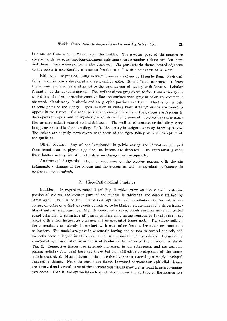
With respect to tumor II which developed on the ventral anterior portion of the bladder,
it should be named a cystadenoma; the epithelial cells successively increase, and the cavities are irre;?;ularly expanded. The increased cells hardly show polymorphous and they are almost all cylindrical cells arranged as mono~layer. Vacuolization is recognized in the protoplasm in the most of the epithelial cells. Most of the glandular cavities contain tissue debris of mucous substances. The stroma is poorly developed and is slightly recognized together with fine fibers consisting of nbroblasts. Infiltration by round cells in the
stroma is considerably severe (Fig. 2). Regarding tumor III observed on the dexter portion of the corpus, the epithelial cells
are adenomatously increased forming complex processes supported by well-developed con .. nective tissue (Fig. 3). Most of the epithelial cells are cylindrical and some of them become more eosinophilic, cubic and occasionally showing poly-layer arrangern,ent where the figure is assumed to show a transitional portion in process of forming carcinoma lesion. The lymph apparatus is well developed in the submucosa and the infiltration of these cells as well as plasma cells is noticeable.
On the stained preparation which contains the rising recognized dorsal portion of the corpus, it resembles the appearance of tumor III, the epithelial cell layer reverse int.o
the submucosa following its development. In addition, the original epithelial cells have desquamated already and cellular infiltration in the submucosa is also severe, blood vessels are dilated and full of blood, showing chronic catarrhal inflammatory changes.
Ureters: Several sections from both sides of the ureters are prepared and observed. Some portions of the left ureters are keratose and .thickened on the surface showing necrosis; commonly collection of polymorphonuclear leucocytes is recognized. In some portion of the mucosa, the epithelial cells are completely desquamated and in the submucosa,
fibrous hyperplasia, dilation of blood vessels and congestion, intense plasma cellular and lymphocytic infiltration and a few neutrophil leucocytes are recognized. These changes show purulent and necrotic uretedtis. On the other hand, as to the right side, fibrous
hyperplasia in the submucosa is rather mild, few epithelial cells of the mucosa are recog
nized and superficial necrosis is clearly observed.
Kidneys: Both kidneys show nearly same changes. The greater part of the kidney
tissue is occupied by increased fibrous tissue, thickening of BOWMAN's capsules and remarkable appearance of fibroblasts, plasma c::lls and lymphocytes. The glomeruli show
rather edematous enlargement, the tubules frequently disappear, the tubular epithelium
becomes flat and cysts result. Sometimes cellular casts in the tubules are recognized,
and many of the nuclei of the epithelial cells show pycnosis or other changes. At the renal papilla, the epithelial cells fl,re almost desquamated, superficial necrosis, the shadows
;

Bladder Carcinoma Accompanied by Chronic Cystitis in Cow 23
of intensely infiltrated round cells and collections of leucocytes are observed here and there. That is chronic purulent pyelonephritis.
Lymphonodi: The lymphonodi of the renal portal regjons and pelvic cavity are observed microscopically; no metastasis is detected but active RES at the medullary cord and catarrh of the lymph sinuses are recognized.
No obvious changes are recognized in the other examined tissues.
DISCUSSION
A bladder tumor case of a 6 year old cow was subjected to pathological study. Lesions were observed on the kidneys, ureters and bladder which showed severe
chronic purulent and necrotic inflammation; three apple-sized neoplasms grew
on the mucosa of the corpus vesicae. One of these tumors as well as other
general mucosa of the bladder showed adenomatous growth of the epithelial cells,
the 2nd one was diagnosed as a cystadenoma and the last as transitional epithelial
cell carcinoma. The histological view of these tumors assumes them to be in
a state of transition from one to the other. They seem outwardly to present
a complicated appearance. However, from the chronic cystitis to carcinoma
formation, furthermore the continual changes through the ureters to pelvis and
parenchyma of the kidneys have very initimate relation with each other. There
has hitherto been no report made on such a case as this with continual changes.
After pathological examinations carried out to the many hematuria cases of
Formosa yellow cattle, MIYAMOTO (1928), WAKE and GOTO (1940) and GOTO et al.
(1954) considered that the chronic cystitis frequently builds tumors and tends
gradually to become malignant. Furthermore, it has been formerly reported that
the continuous stimulus to the bladder mucosa by administration of naphthylamin results in the development of tumorsl~, n.
The authors' case had been reared at Nemuro-Shibetsu, known as a district
with a frequent occurrence of bovine hematuria; pathological examinations of
the sickness were carried out by SUGANO (1950), FUJIMOTO et al. (1952) and
OHBA YASHI et al. It is interesting to know that they have pointed out that the
mucosa suffering from cystitis freq uently develop into adenoma or adenomatous
lesions. Clinical data of the authors' case are quite unknown, but it is not difficult
to consider that the cow frequently hematurinated, became more and more
emaciated and then was sent to the slaughter house. Histologically it can be
shown that chronic inflammation of the bladder mucosa causes the development
of adenoma, and then transforms to carcinoma. Furthermore it is easily considered
that, before long, mechanical closing of the urethra occurs. the bladder becomes
full of urine, the ureters and the pelvis expand, the case become pyelonephritis,
and then the appearance of urinary calculi develops. SCHLEGEL reported a case
24 OHSHIMA, K. AND H. ONO
of a bladder carcinoma in a horse which, like to the writers' present case showing an expansion of the ureters and the pelvis and calculi formation on the pelvis. The authors previously supposed in this case that the adenoma transformed into the carcinoma; since these tumors are developed from the same tissue, it can be considered that they may grew individually and also at the same time. However, in this case, the infiltrative development of the tumor cells, which may be regarded as the initial lesion of a carcinoma, cannot be found, but it should Le noted that there is a formation of poly-layer consisting of the increased epithelial cells in the adenomatous tissue. For the reasons above mentioned, it is assumed that the carcinoma has developed from an adenoma.
In relation to the malignancy of a carcinoma, especially in human medicine, histological examinations have been carried out in detail for the sake of judgement
for the prognosis together with that the metastasis formed or not. !eHIKA W A
et al. (1955) made pathological studies of 115 cases of human bladder carcinoma and reported that the 95 cases (83%7) were transitional epithelial cell carcinoma, about the 80% of these had worsened from papillary tumor. They attemped to propose the standards of classifying the malignancy of tumors by the cell differentiation and the degree of the infiltration. In the case discussed in the present paper the malignancy is rather mild because the papillary structures are observed as mentioned above, and in the histological appearance of the growths, the differentiation of the cells is not severe and no infiltrative growths are recognized; the existence of the tumor cells is found just in the submucosa, etc. This fact has an intimate relation to the presence of metastasis; the authors are unable
to recognize either tumor embolus on preparations or metastasis elsewhere of the
body, so it seems proven that the case is not very malignant.
CONCLUSION
A bladder tumor case of a slaughtered cow was pathologically examined and discussion was offered concerning some former reports.
In the authors' case, chronic cystitis is the beginning of the disease, and adenomatous growth of the mucosa and at length a transitional epithelial cell carcinoma were developed. Following the growths the urethra was mechanically closed, the dilation of the ureters and pelvis occurred and then the animal suffered from ureteropyelonephritis accompanied with the formation of calculi. In this examination the authors could inquire to sOme degree into the process of the pathogenesis and came to the opinion that the morphology of the tumors is very interesting in respect to pathogenesis.
Bladder Carcinoma Accompanied by Chronic Cystitis in Cow 25
Finally, the authors wish to extend their hearty thanks to Prof. YAMA.GIWA for
reviewing this paper and to the staff of the Sapporo slaughter house for their cooperation
in supplying the materials.
REFERENCES
1) BONSER, G. M., D. B. CLAYSON & J. W. JULL (1951): Lancet, 261, 286 [Vet. Bull., Weybr'l:dge, 22, 87 (1952)].
2) FRANKE (1909): Viiif. Jber. Beamt. T. Preuss., 2, f. [JOESTti)].
3) FUJIMOTO, Y., S. SUGANO & T. KOBAYASHI (1952): Mem. Far:. Agr. Holelcaido Un'iv., 1, 159 (in Japanese with English summary).
4) GOTO, J., T. KATO & N. HOSl:IIKAWA (1954): .rap. J. vet. SC'i., 16, 209 (in Japane3e
with English summary).
5) ICHIKAWA, T., S. ISHIYAMA & I. TSUJI (1955): Nissin Igaleu, 42, 201 (in Japanese
with English summary).
6) JOEST, E. (1924): Spez. path. Anat. Haust., 3, SS. 398~401, Richard Scholtz, Berlin.
7) LUBARSCH, O. & R. OSTERTAG (1896): Ergebn. allg. Path. path. Anat., 3, 438,
Wiesbaden.
8) LYDING, F. (1909): Berl. tierib·ztl. Wschr., 26, 68.
9) MIYAMOTO, T. (1928): Jap. J. 'vet. Sm'., 7, 190.
10) MONTPELLIER & NYKA (1930): Bull. Assoc. Franq. ]?;t1tde Cane., 19, 132 [Jber.
VetN/ed., 50, 498 (1930)].
11) OHBAYASHI, M., S. SUGANO & Y. OHMURA (1952): Mem. Pac. Agr. Hokkaido Univ., 1, 167 (in Japanese with English summary).
12) RiJSCHER, W. (1931): Arch. wiss. prakt. Ti,erhlk., 64, 29.
13) SCHLEGEL, M. (1903): Berl. tieriirztl. Wschr., 20, 225.
14) SEHAR, W. (1930): T,e Cancer, 7, 205 [Jber. VetMed., 51, 447 (1931)J.
15) STICKER, A. (1901): Dtsch. Neriirztl. Wschr., 9, 433.
16) STOLZ, E. (1886): Al'ch. 'wiss. prakt. Tiel"heilk., 12, 288.
17) SUGANO, S. (1950): Jiii CMleusan 8hinpo, 33, 79 (in .Japanese).
18) VOIRIN, V. (1910): Berl. tieriir.ztl. Wsehr., 26, 349.
19) WAKE, 1. & J. GOTO (1940): Gann, 34, 127.
26 OHSHIMA, K. AND H. ONO
EXPLANATION OF PLATE
Macroscopic Photograph.
Fig. 1. Tumors developed on the bladder mucosa (I-III), and C-Corpus vesicae. U-Ureters.
Microphotographs, H.-E. ~taining, x 100.
Fig. 2. Preparati~n made from tumor II, forming a cystadenoma. Fig. 3. Preparation made from tumor III, adenomatous development of the
epithelialt;?~Us and cellular infiltration in the stroma. Fig. 4. Prepar.ationmade from tumor I, transitional epithelial cell carcinoma.
island-like .development of the carcinoma. some of which have debris in them.
Figs. 5 & 6. Show~ .the transformation from adenoma to carcinoma; note that the adenom:atous epithelial cells become poly-layer.
., r
., r
OHSHIMA, K. AND H. ONO PLATE