patent ductus arteriosus pda - university of...
TRANSCRIPT
1
Patent ductus arteriosus PDA
• Is connecting between the aortic end just distal to the origin of the LT sub clavian artery& the pulmonary artery at its bifurcation.
• Female/male ratio is 2:1 and it is more common in premature infants.
• PDA is present in 10% of other CHD.
2
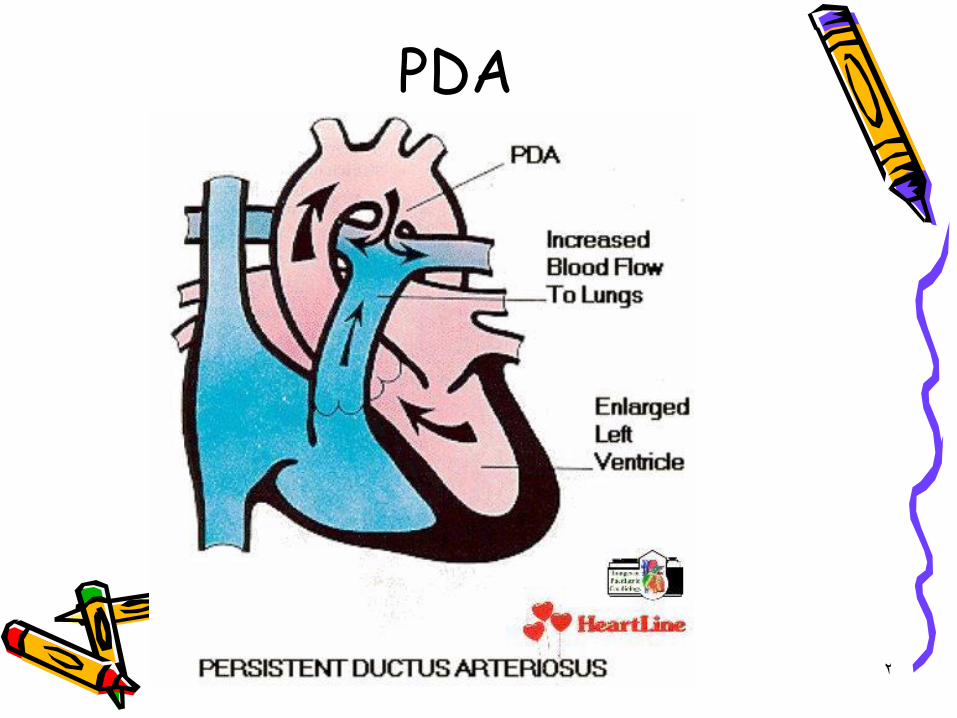
PDA
3
Pathophysiology of PDA
• It is basically similar to VSD and depend on the size of the defect.
4
Clinical manifestation of PDA
• Symptoms : are basically similar to VSD.• Signs :• 1.Wide pulse pressure with bounding pulse.• 2.Prominant apical impulse.• 3.Systolic or continuous murmur in the 2nd
intercostal space radiating toward the clavicle down the LT lower sternal border or apex.
• 4.Machinary murmur.• 5. Middiastolic murmur.
5
Investigation of PDA• In small PDA:CXR&ECG is normal.• In large PDA:CXR will show prominent pulmonary
a. with increase vascularity .• & cardiac size depend on the size of the shunt.• ECG : show increase LT atrial & LT ventricular
dimensions.• Scanning of suprasternal notch will allow
visualization of the ductus & color Doppler &pulsed Doppler will show the turbulent blood flow in the pulmonary a. .
6
Prognosis of PDA• 1.Spontaneous closure after infancy
is rare.
• 2.cardiac failure occur in large duct.
• 3.infective endocarditis .
• 4.Eisenmenger syndrome
• 5.Pulmonary hypertension.
7
Treatment of PDA• Surgical closure fatality rate is<1%
&is indicated in asymptomatic patients preferably before 1 year.
• Closure by catheterization.
• Medical treatment of congestive heart failure.
8
Differential diagnosis• 1.venous hum.
• 2.Aortico pulmonary window defect.
• 3.truncus arteriosus .
• 4.Coronary arteriovenous fistula.
9
Coarctation of aorta• Means constriction of the aorta at
any area from the aortic arch to the iliac bifurcation most commonly just below the origin of the LT subclavian a. in 98% of cases.
• Male Female ratio is 2:1.
10
Clinical manifestation
• 1.Mostly asymptomatic.• 2.The classical sign of coarctation is the
disparity in pulsation &Blood pressure of the arms & legs they are absent in 40% of cases ,the radial &femoral pulse should be examined simultaneously.
• 3.B.P in upper limbs is higher than lower limbs.• 4.Leg pain &weakness in children & adolescents.
11
Investigations of coarctation of aorta
• CXR : show the effect of hypertension on the heart &the effect of the collaterals on the lower border of the ribs with cardiomegally & LT ventricular enlargement.
• ECG:LT ventricular hypertrophy• Echo cardiography : by 2dimensional echo
& color Doppler .
12
Treatment of coarctation
• 1.Treatment of congestive heart failure.
• 2.Surgical repair.
13
Cyanotic congenital heart disease
Cyanotic CHD
1.Lesions associated with decreased pulmonary blood flow
14
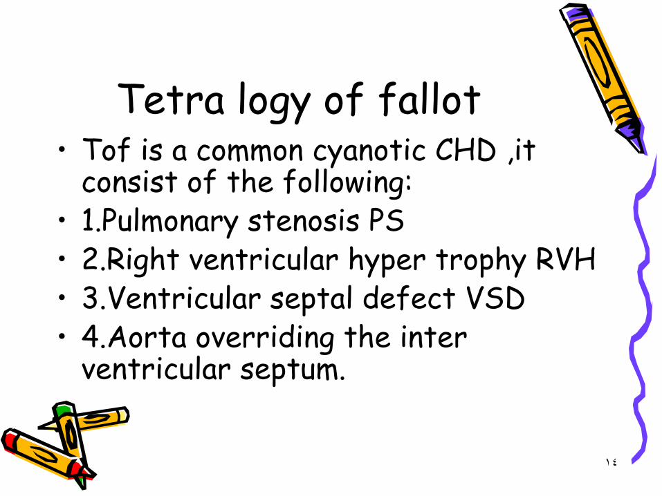
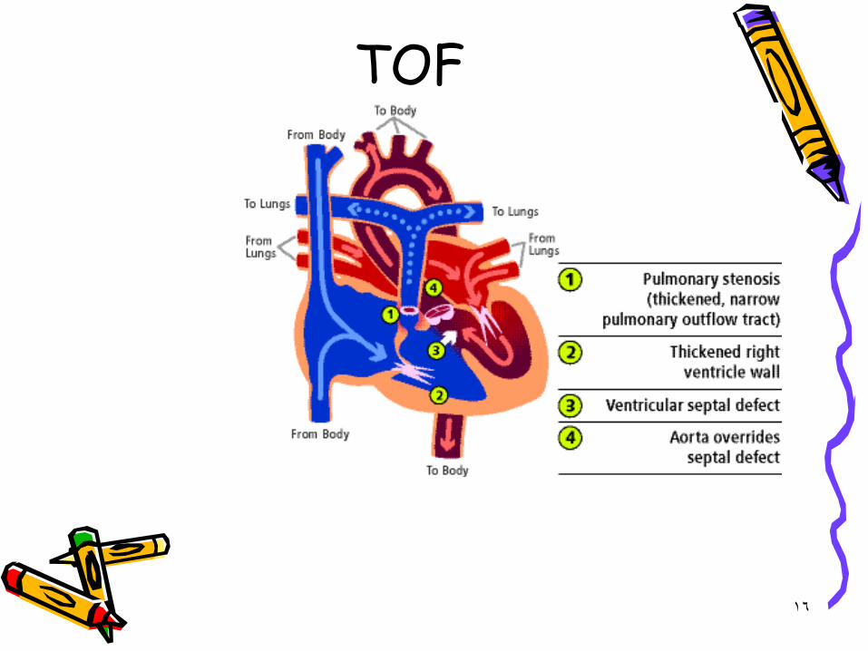
Tetra logy of fallot• Tof is a common cyanotic CHD ,it
consist of the following:• 1.Pulmonary stenosis PS• 2.Right ventricular hyper trophy RVH• 3.Ventricular septal defect VSD• 4.Aorta overriding the inter
ventricular septum.
15
Patho physiology of TOF• Systemic venous blood return to the RT atrium
&RT ventricle. During systole in the presence of PS blood is shunted across the VSD in to the aorta [RT to LT ] shunt ,so pulmonary blood flow may be by bronchial collateral circulation ,or in newborn baby by PDA.
• The degree of intra cardiac shunting is the function of outflow resistance which include:
• 1.Pulmonary resistance.• 2.Systemic arteriolar resistance.
16
TOF
17
Clinical manifestation of TOF
• Symptoms:• 1.Cyanosis usually start few monthes after
birth late in the first year.• 2.Dyspnoea on exertion.• 3.Growth failure.• 4.Paroxysmal hyper cyanotic attacks
[Hypoxic ,Blue ,tet] spells usually in the first 2 years of life with increase cyanosis ,gasping respiration.
18
TOFSigns
• 1.Central cyanosis &digital clubbing.• 2.Pulse is usually normal.• 3.The LT anterior hemi thorax may bulge anteriorly.• 4.Substernal RT ventricular impulse & systolic thrill along
the LT sternal border in the third & forth spaces.• 5.Soft S2 with loud harsh ejection systolic murmur at the
upper sternal border or it may be holosystolic murmur toward the lower sternal border.
• 6.During spells the murmur become softer.• 7.There may be continuous murmur of the collaterals.
19
TOFInvestigations
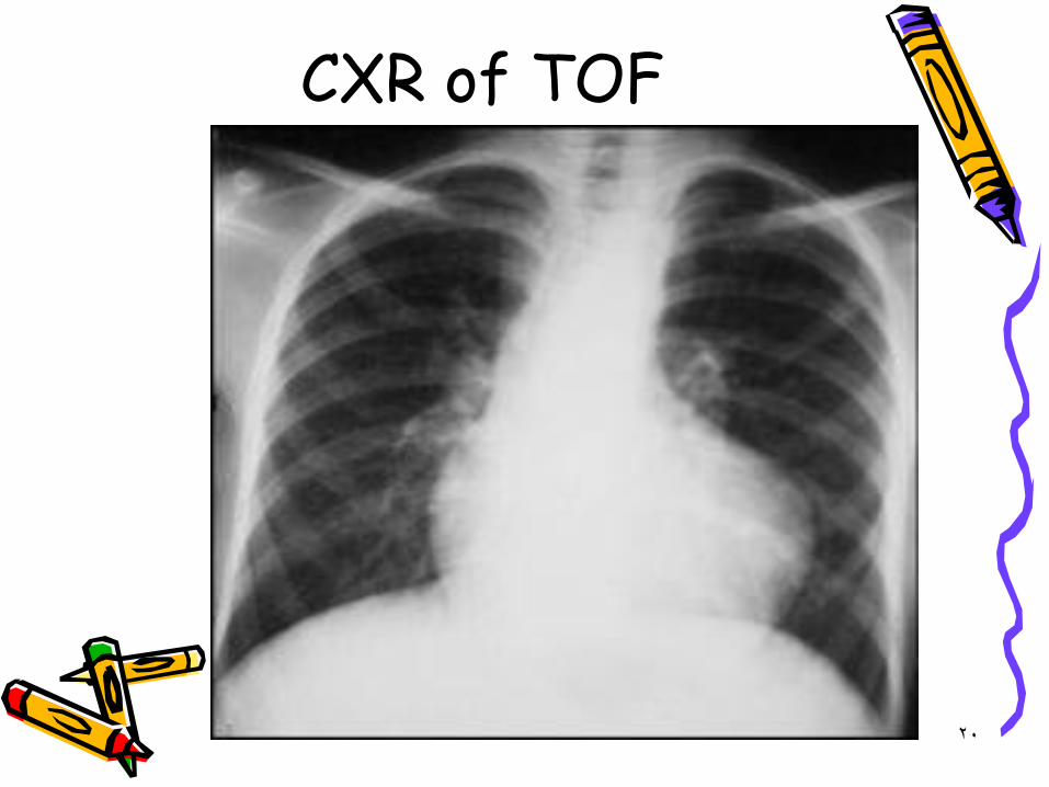
• CXR :the heart is not enlarged ,concavity is usually found in the area occupied by main pulmonary a. With boot shape heart & Oligaemic lung fields .
• ECG :will show RT axis deviation & RT ventricular hypertrophy ,P wave is tall & peaked.
• ECHO: will establish the diagnosis.
20
CXR of TOF
21
TOF Complications
• 1.Cerebrovascular accident due to thrombosis secondary to dehydration & polycythemia in the first two years of life .
• Brain abscess after 2 years .
• SBE.
22
TOFTreatment
• 1.Palliative surgery by Blalock Tausig shunt [Pulmonary to Systemic artery shunt].
• 2.Total corrective surgery under cardio pulmonary bypass .
• 3.Treatment of complications like Hypoxic [tet] spells by:
• 1.Position• 2.Oxygen• 3.Morphine S.C 0.2mg/kg .• 4.Propranolol 0.1mg/kg I.V .
23
Tricuspid Atresia• There is no outlet from RT atrium to RT ventricle
so LT ventricle push blood to RT ventricle via VSD & severity of cyanosis depend on size of VSD & severity of pulmonary stenosis .
• Clinically : cyanosis present since birth, with increase LT ventricular impulse, with holosystolic murmur on LT lower sternal border, with single S2 ,associated with easy fatigability ,exertional dyspnoea &occasional hypoxic episodes ,
24
Diagnosis & treatment of Tricuspid Atresia
• CXR :Oligaemic lung.• ECG: LT axis deviation < ventricular
hypertrophy .• ECHO : 2 D echo will diagnose the condition.• Treatment is by:• 1.Blalock-Taussig procedure.• 2.Modified Fontan procedure by anastomosing RT
atrium to the pulmonary a. Done at 1.5_3 years of age .
25
Ebstein anomaly• There is downward displacement of an
abnormal tricuspid valve into the RT ventricle with remaining poorly functioning RT ventricle which fail to pump blood to the pulmonary a. Producing functional pulmonary atresia so there will be RT atrial to LT atrial shunt through foramen ovale to produce cyanosis.
26
2.Cyanotic CHD with increase pulmonary blood flow
Transposition of great arteries TGA
• TGA is common CHD form about 5% of all CHD.
• In this anomaly the aorta arisesfrom RT ventricle & pulmonary a.Arises from LT ventricle sounsaturated blood will be pumpedagain via aorta to all the body sosurvival depend on presence offoramen ovale & ductus arteriosus& 50% of patients will have VSD ,itis more common in infants ofdiabetic mothers & in males 3:1female.
27
TGA with intact septumClinical manifestation
• Cyanosis & tachypnea are recognized within the first hours or days after birth if not treated most patient will not survive the neonatal period ,hypoxia is severe &heart failure is less common it is a medical emergency.
• O \E:1.central cyanosis since birth .• 2.normal apex beat or there may be parasternal
heave .• 3.2nd heart sound is usually single &loud .• 4.murmurs may be absent or systolic murmur at
the mid left sternal border .
28
Investigation &Treatment of TGA
• CXR :mild Cardiomegally ,narrow mediastinum ,increase pulmonary blood flow .
• ECG :show normal neonatal RT dominant pattern.• The arterial PO2 is low even after 100% Oxygen saturation
[hypoxia test] .• ECHO :will confirm the diagnosis .• Cardiac catheterization .• Treatment :when TGA is expected the patient infused with
Prostaglandin E1 immediately to maintain the patency of ductus arteriosus & to improve oxygenation ,the dose is [0.05-20 microgram\Kg\minute] with keeping the patient worm to prevent hypothermia & acidosis also to correct hypoglycemia .
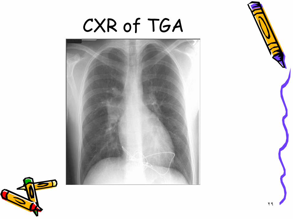
29
CXR of TGA
30
TGA Treatment• In patients with severe hypoxia in spite of
prostaglandin will need [Rashkind Balloon Atrial Septostomy] .
• The atrial switch [Jatene procedure] is the surgical treatment of choice ,can be performed in the first 2 weeks of life .
31
TGA with large VSD• Will be manifested by history of heart
failure & cyanosis is subtle or present later & the murmur is holosystolic murmur.
• CXR :Cardiomegally ,narrow mediastinum &increase pulmonary vascularity.
• ECHO :will confirm the diagnosis .• Treatment :Arterial switch procedure
+VSD closure .
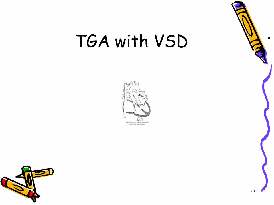
TGA with VSD •
32