part 5: adult basic life support : 2010 american heart ... adulti 2010 (aha).pdf · part 5: adult...
TRANSCRIPT

Robert A. SworCave, Mary Fran Hazinski, E. Brooke Lerner, Thomas D. Rea, Michael R. Sayre and Robert A. Berg, Robin Hemphill, Benjamin S. Abella, Tom P. Aufderheide, Diana M.
for Cardiopulmonary Resuscitation and Emergency Cardiovascular CarePart 5: Adult Basic Life Support : 2010 American Heart Association Guidelines
ISSN: 1524-4539 Copyright © 2010 American Heart Association. All rights reserved. Print ISSN: 0009-7322. Online
72514Circulation is published by the American Heart Association. 7272 Greenville Avenue, Dallas, TX
doi: 10.1161/CIRCULATIONAHA.110.9709392010, 122:S685-S705Circulation
http://circ.ahajournals.org/content/122/18_suppl_3/S685located on the World Wide Web at:
The online version of this article, along with updated information and services, is
http://circ.ahajournals.org/http://circ.ahajournals.org/content/124/15/e402.full.pdf An erratum has been published regarding this article. Please see the attached page for:
http://www.lww.com/reprintsReprints: Information about reprints can be found online at
[email protected]. E-mail:
Fax:Kluwer Health, 351 West Camden Street, Baltimore, MD 21202-2436. Phone: 410-528-4050. Permissions: Permissions & Rights Desk, Lippincott Williams & Wilkins, a division of Wolters
http://circ.ahajournals.org//subscriptions/Subscriptions: Information about subscribing to Circulation is online at
by guest on December 9, 2011http://circ.ahajournals.org/Downloaded from
Part 5: Adult Basic Life Support2010 American Heart Association Guidelines for Cardiopulmonary
Resuscitation and Emergency Cardiovascular Care
Robert A. Berg, Chair; Robin Hemphill; Benjamin S. Abella; Tom P. Aufderheide; Diana M. Cave;Mary Fran Hazinski; E. Brooke Lerner; Thomas D. Rea; Michael R. Sayre; Robert A. Swor
Basic life support (BLS) is the foundation for saving livesfollowing cardiac arrest. Fundamental aspects of BLS
include immediate recognition of sudden cardiac arrest(SCA) and activation of the emergency response system,early cardiopulmonary resuscitation (CPR), and rapid de-fibrillation with an automated external defibrillator (AED).Initial recognition and response to heart attack and stroke arealso considered part of BLS. This section presents the 2010adult BLS guidelines for lay rescuers and healthcare provid-ers. Key changes and continued points of emphasis from the2005 BLS Guidelines include the following:
● Immediate recognition of SCA based on assessing unre-sponsiveness and absence of normal breathing (ie, thevictim is not breathing or only gasping)
● “Look, Listen, and Feel” removed from the BLS algorithm● Encouraging Hands-Only (chest compression only) CPR
(ie, continuous chest compression over the middle of thechest) for the untrained lay-rescuer
● Sequence change to chest compressions before rescuebreaths (CAB rather than ABC)
● Health care providers continue effective chest compres-sions/CPR until return of spontaneous circulation (ROSC)or termination of resuscitative efforts
● Increased focus on methods to ensure that high-qualityCPR (compressions of adequate rate and depth, allowingfull chest recoil between compressions, minimizing inter-ruptions in chest compressions and avoiding excessiveventilation) is performed
● Continued de-emphasis on pulse check for health careproviders
● A simplified adult BLS algorithm is introduced with therevised traditional algorithm
● Recommendation of a simultaneous, choreographed ap-proach for chest compressions, airway management, rescuebreathing, rhythm detection, and shocks (if appropriate) byan integrated team of highly-trained rescuers in appropriatesettings
Despite important advances in prevention, SCA continuesto be a leading cause of death in many parts of the world.1
SCA has many etiologies (ie, cardiac or noncardiac causes),circumstances (eg, witnessed or unwitnessed), and settings(eg, out-of-hospital or in-hospital). This heterogeneity sug-gests that a single approach to resuscitation is not practical,but a core set of actions provides a universal strategy forachieving successful resuscitation. These actions are termedthe links in the “Chain of Survival.” For adults they include
● Immediate recognition of cardiac arrest and activation ofthe emergency response system
● Early CPR that emphasizes chest compressions● Rapid defibrillation if indicated● Effective advanced life support● Integrated post–cardiac arrest care
When these links are implemented in an effective way,survival rates can approach 50% following witnessed out-of-hospital ventricular fibrillation (VF) arrest.2 Unfortunatelysurvival rates in many out-of-hospital and in-hospital settingsfall far short of this figure. For example, survival ratesfollowing cardiac arrest due to VF vary from approximately5% to 50% in both out-of-hospital and in-hospital settings.3,4
This variation in outcome underscores the opportunity forimprovement in many settings.
Recognition of cardiac arrest is not always straightforward,especially for laypersons. Any confusion on the part of a rescuercan result in a delay or failure to activate the emergency responsesystem or to start CPR. Precious time is lost if bystanders are tooconfused to act. Therefore, these adult BLS Guidelines focus onrecognition of cardiac arrest with an appropriate set of rescueractions. Once the lay bystander recognizes that the victim isunresponsive, that bystander must immediately activate (or sendsomeone to activate) the emergency response system. Once thehealthcare provider recognizes that the victim is unresponsivewith no breathing or no normal breathing (ie, only gasping) thehealthcare provider will activate the emergency response sys-tem. After activation, rescuers should immediately begin CPR.
Early CPR can improve the likelihood of survival, and yetCPR is often not provided until the arrival of professionalemergency responders.5 Chest compressions are an especiallycritical component of CPR because perfusion during CPR
The American Heart Association requests that this document be cited as follows: Berg RA, Hemphill R, Abella BS, Aufderheide TP, Cave DM,Hazinski MF, Lerner EB, Rea TD, Sayre MR, Swor RA. Part 5: Adult basic life support: 2010 American Heart Association Guidelines forCardiopulmonary Resuscitation and Emergency Cardiovascular Care. Circulation. 2010;122(suppl 3):S685–S705.
(Circulation. 2010;122[suppl ]:S685–S705.)© 2010 American Heart Association, Inc.
Circulation is available at http://circ.ahajournals.org DOI: 10.1161/CIRCULATIONAHA.110.970939
S685 by guest on December 9, 2011http://circ.ahajournals.org/Downloaded from

depends on these compressions. Therefore, chest compres-sions should be the highest priority and the initial action whenstarting CPR in the adult victim of sudden cardiac arrest. Thephrase “push hard and push fast” emphasizes some of thesecritical components of chest compression. High-quality CPRis important not only at the onset but throughout the course ofresuscitation. Defibrillation and advanced care should beinterfaced in a way that minimizes any interruption in CPR.6
Rapid defibrillation is a powerful predictor of successfulresuscitation following VF SCA.7,8 Efforts to reduce theinterval from collapse to defibrillation can potentially im-prove survival in both out-of-hospital and in-hospital settings.8,9
Depending on the setting and circumstances, earlier defibril-lation may be achieved by a variety of strategies that includerescuers who are laypersons, nontraditional first responders,police, emergency medical services (EMS) professionals, andhospital professionals.9–12 One of these strategies is the use ofan AED. The AED correctly assesses heart rhythm, enablinga rescuer who is not trained in heart rhythm interpretation toaccurately provide a potentially lifesaving shock to a victimof SCA.13
Immediate recognition and activation, early CPR, andrapid defibrillation (when appropriate) are the first threeBLS links in the adult Chain of Survival. BLS care in theout-of-hospital setting is often provided by laypersons whomay be involved in a resuscitation attempt only once in theirlives. Thus, creating an effective strategy to translate BLSskills to real-world circumstances presents a challenge. Thissection updates the adult BLS guidelines with the goal ofincorporating new scientific information while acknowledg-ing the challenges of real-world application. Everyone, re-gardless of training or experience, can potentially be alifesaving rescuer.
The rest of this chapter is organized in sections that addressthe emergency response system, adult BLS sequence, adultBLS skills, use of an AED, special resuscitation situations,and the quality of BLS. The “Adult BLS Sequence” sectionprovides an overview and an abridged version of the BLSsequence. The “Adult BLS Skills” section provides greaterdetail regarding individual CPR skills and more informationabout Hands-Only (compression-only) CPR. The “SpecialResuscitation Situations” section addresses acute coronarysyndromes, stroke, hypothermia, and foreign body airwayobstruction. Because of increasing interest in monitoring andensuring the quality of CPR, the last section focuses on thequality of BLS.
Activating the Emergency Response SystemEmergency medical dispatch is an integral component of theEMS response.14 Bystanders (lay responders) should immedi-ately call their local emergency number to initiate a responseanytime they find an unresponsive victim. Because dispatcherCPR instructions substantially increase the likelihood of by-stander CPR performance and improve survival from cardiacarrest, all dispatchers should be appropriately trained to providetelephone CPR instructions (Class I, LOE B).15–21
When dispatchers ask bystanders to determine if breathingis present, bystanders often misinterpret agonal gasps orabnormal breathing as normal breathing. This erroneous
information can result in failure by 911 dispatchers to instructbystanders to initiate CPR for a victim of cardiac arrest.19,22–26
To help bystanders recognize cardiac arrest, dispatchersshould inquire about a victim’s absence of consciousness andquality of breathing (normal versus not normal). Dispatchersshould be specifically educated in recognition of abnormalbreathing in order to improve recognition of gasping andcardiac arrest (Class I, LOE B). Notably, dispatchers shouldbe aware that brief generalized seizures may be the firstmanifestation of cardiac arrest.26,27 Dispatchers should rec-ommend CPR for unresponsive victims who are not breathingnormally because most are in cardiac arrest and the frequencyof serious injury from chest compressions in the nonarrestgroup is very low (Class I, LOE B).28 In summary, in additionto activating professional emergency responders, the dis-patcher should ask straightforward questions about whetherthe patient is conscious and breathing normally in order toidentify patients with possible cardiac arrest. The dispatchershould also provide CPR instructions to help bystandersinitiate CPR when cardiac arrest is suspected.
Because it is easier for rescuers receiving telephone CPRinstructions to perform Hands-Only (compression-only) CPRthan conventional CPR (compressions plus rescue breathing),dispatchers should instruct untrained lay rescuers to provideHands-Only CPR for adults with SCA (Class I, LOE B).29
While Hands-Only CPR instructions have broad applicability,instances remain when rescue breaths are critically important.Dispatchers should include rescue breathing in their tele-phone CPR instructions to bystanders treating adult andpediatric victims with a high likelihood of an asphyxial causeof arrest (eg, drowning).30
The EMS system quality improvement process, includingreview of the quality of dispatcher CPR instructions providedto specific callers, is considered an important component of ahigh-quality lifesaving program (Class IIa, LOE B).31–33
Adult BLS SequenceThe steps of BLS consist of a series of sequential assessmentsand actions, which are illustrated in the new simplified BLSalgorithm (Figure 1). The intent of the algorithm is to presentthe steps of BLS in a logical and concise manner that is easyfor all types of rescuers to learn, remember and perform.These actions have traditionally been presented as a sequenceof distinct steps to help a single rescuer prioritize actions.However, many workplaces and most EMS and in-hospitalresuscitations involve teams of providers who should performseveral actions simultaneously (eg, one rescuer activates theemergency response system while another begins chest com-pressions, and a third either provides ventilations or retrievesthe bag-mask for rescue breathing, and a fourth retrieves andsets up a defibrillator).
Immediate Recognition and Activation of theEmergency Response SystemIf a lone rescuer finds an unresponsive adult (ie, no move-ment or response to stimulation) or witnesses an adult whosuddenly collapses, after ensuring that the scene is safe, therescuer should check for a response by tapping the victim onthe shoulder and shouting at the victim. The trained or
S686 Circulation November 2, 2010
by guest on December 9, 2011http://circ.ahajournals.org/Downloaded from
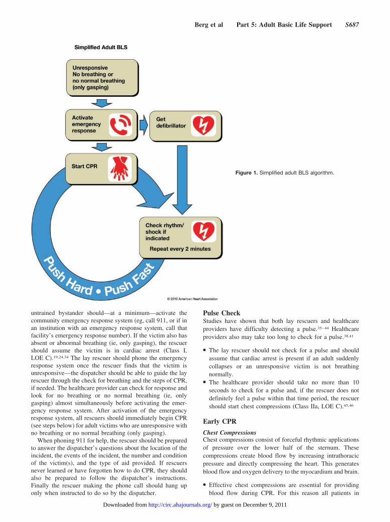
untrained bystander should—at a minimum—activate thecommunity emergency response system (eg, call 911, or if inan institution with an emergency response system, call thatfacility’s emergency response number). If the victim also hasabsent or abnormal breathing (ie, only gasping), the rescuershould assume the victim is in cardiac arrest (Class I,LOE C).19,24,34 The lay rescuer should phone the emergencyresponse system once the rescuer finds that the victim isunresponsive—the dispatcher should be able to guide the layrescuer through the check for breathing and the steps of CPR,if needed. The healthcare provider can check for response andlook for no breathing or no normal breathing (ie, onlygasping) almost simultaneously before activating the emer-gency response system. After activation of the emergencyresponse system, all rescuers should immediately begin CPR(see steps below) for adult victims who are unresponsive withno breathing or no normal breathing (only gasping).
When phoning 911 for help, the rescuer should be preparedto answer the dispatcher’s questions about the location of theincident, the events of the incident, the number and conditionof the victim(s), and the type of aid provided. If rescuersnever learned or have forgotten how to do CPR, they shouldalso be prepared to follow the dispatcher’s instructions.Finally the rescuer making the phone call should hang uponly when instructed to do so by the dispatcher.
Pulse CheckStudies have shown that both lay rescuers and healthcareproviders have difficulty detecting a pulse.35–44 Healthcareproviders also may take too long to check for a pulse.38,41
● The lay rescuer should not check for a pulse and shouldassume that cardiac arrest is present if an adult suddenlycollapses or an unresponsive victim is not breathingnormally.
● The healthcare provider should take no more than 10seconds to check for a pulse and, if the rescuer does notdefinitely feel a pulse within that time period, the rescuershould start chest compressions (Class IIa, LOE C).45,46
Early CPR
Chest CompressionsChest compressions consist of forceful rhythmic applicationsof pressure over the lower half of the sternum. Thesecompressions create blood flow by increasing intrathoracicpressure and directly compressing the heart. This generatesblood flow and oxygen delivery to the myocardium and brain.
● Effective chest compressions are essential for providingblood flow during CPR. For this reason all patients in
Figure 1. Simplified adult BLS algorithm.
Berg et al Part 5: Adult Basic Life Support S687
by guest on December 9, 2011http://circ.ahajournals.org/Downloaded from
cardiac arrest should receive chest compressions (Class I,LOE B).47–51
● To provide effective chest compressions, push hard andpush fast. It is reasonable for laypersons and healthcareproviders to compress the adult chest at a rate of at least100 compressions per minute (Class IIa, LOE B) with acompression depth of at least 2 inches/5 cm (Class IIa,LOE B). Rescuers should allow complete recoil of thechest after each compression, to allow the heart to fillcompletely before the next compression (Class IIa, LOE B).
● Rescuers should attempt to minimize the frequency andduration of interruptions in compressions to maximize thenumber of compressions delivered per minute (Class IIa, LOEB). A compression-ventilation ratio of 30:2 is recommended(Class IIa, LOE B).
Rescue BreathsA change in the 2010 AHA Guidelines for CPR and ECC isto recommend the initiation of compressions before ventila-tions. While no published human or animal evidence demon-strates that starting CPR with 30 compressions rather than 2ventilations leads to improved outcomes, it is clear that bloodflow depends on chest compressions. Therefore, delays in,and interruptions of, chest compressions should be minimizedthroughout the entire resuscitation. Moreover, chest compres-sions can be started almost immediately, while positioningthe head, achieving a seal for mouth-to-mouth rescue breath-ing, and getting a bag-mask apparatus for rescue breathing alltake time. Beginning CPR with 30 compressions rather than2 ventilations leads to a shorter delay to first compression(Class IIb, LOE C).52–54
Once chest compressions have been started, a trainedrescuer should deliver rescue breaths by mouth-to-mouth orbag-mask to provide oxygenation and ventilation, as follows:
● Deliver each rescue breath over 1 second (Class IIa, LOE C).● Give a sufficient tidal volume to produce visible chest rise
(Class IIa, LOE C).55
● Use a compression to ventilation ratio of 30 chest com-pressions to 2 ventilations.
Early Defibrillation With an AEDAfter activating the emergency response system the lonerescuer should next retrieve an AED (if nearby and easilyaccessible) and then return to the victim to attach and use theAED. The rescuer should then provide high-quality CPR.When 2 or more rescuers are present, one rescuer shouldbegin chest compressions while a second rescuer activates theemergency response system and gets the AED (or a manualdefibrillator in most hospitals) (Class IIa, LOE C). The AEDshould be used as rapidly as possible and both rescuers shouldprovide CPR with chest compressions and ventilations.
Defibrillation Sequence
● Turn the AED on.● Follow the AED prompts.● Resume chest compressions immediately after the shock
(minimize interruptions).
Rescuer Specific CPR Strategies: Putting ItAll TogetherThis section summarizes the sequence of CPR interventionsthat should be performed by 3 prototypical rescuers after theyactivate the emergency response system. The specific stepsthat rescuers should take (Hands-Only CPR, conventionalCPR with rescue breathing, CPR and AED use) are deter-mined by the rescuer’s level of training.
Untrained Lay RescuerIf a bystander is not trained in CPR, then the bystander shouldprovide Hands-Only (chest compression only) CPR, with anemphasis on “push hard and fast,” or follow the directions ofthe emergency medical dispatcher. The rescuer should con-tinue Hands-Only CPR until an AED arrives and is ready foruse or healthcare providers take over care of the victim (ClassIIa, LOE B).
Trained Lay RescuerAll lay rescuers should, at a minimum, provide chest com-pressions for victims of cardiac arrest. In addition, if thetrained lay rescuer is able to perform rescue breaths, he or sheshould add rescue breaths in a ratio of 30 compressions to 2breaths. The rescuer should continue CPR until an AEDarrives and is ready for use or EMS providers take over careof the victim (Class I, LOE B).
Healthcare ProviderOptimally all healthcare providers should be trained in BLS.In this trained population it is reasonable for both EMS andin-hospital professional rescuers to provide chest compres-sions and rescue breaths for cardiac arrest victims (Class IIa,LOE B). This should be performed in cycles of 30 compres-sions to 2 ventilations until an advanced airway is placed;then continuous chest compressions with ventilations at a rateof 1 breath every 6 to 8 seconds (8 to 10 ventilations perminute) should be performed. Care should be taken tominimize interruptions in chest compressions when placing,or ventilating with, an advanced airway. In addition, exces-sive ventilation should be avoided.
It is reasonable for healthcare providers to tailor the sequenceof rescue actions to the most likely cause of arrest. For example,if a lone healthcare provider sees an adolescent suddenlycollapse, the provider may assume that the victim has suffered asudden cardiac arrest and call for help (phone 911 or theemergency response number), get an AED (if nearby), andreturn to the victim to attach and use the AED and then provideCPR. If a lone healthcare provider aids an adult drowning victimor a victim of foreign body airway obstruction who becomesunconscious, the healthcare provider may give about 5 cycles(approximately 2 minutes) of CPR before activating the emer-gency response system (Class IIa, LOE C).
Adult BLS SkillsThe sequence of BLS skills for the healthcare provider isdepicted in the BLS Healthcare Provider Algorithm (seeFigure 2).
Recognition of Arrest (Box 1)The necessary first step in the treatment of cardiac arrest isimmediate recognition. Bystanders may witness the sudden
S688 Circulation November 2, 2010
by guest on December 9, 2011http://circ.ahajournals.org/Downloaded from
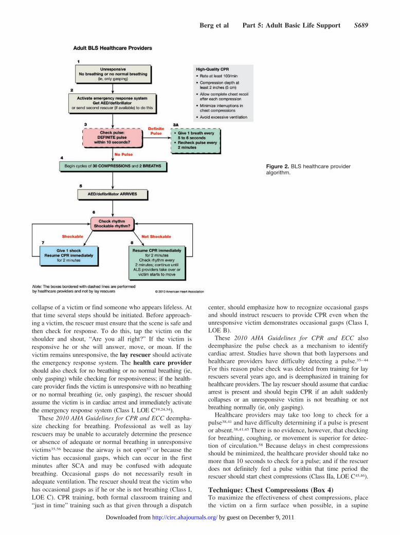
collapse of a victim or find someone who appears lifeless. Atthat time several steps should be initiated. Before approach-ing a victim, the rescuer must ensure that the scene is safe andthen check for response. To do this, tap the victim on theshoulder and shout, “Are you all right?” If the victim isresponsive he or she will answer, move, or moan. If thevictim remains unresponsive, the lay rescuer should activatethe emergency response system. The health care providershould also check for no breathing or no normal breathing (ie,only gasping) while checking for responsiveness; if the health-care provider finds the victim is unresponsive with no breathingor no normal breathing (ie, only gasping), the rescuer shouldassume the victim is in cardiac arrest and immediately activatethe emergency response system (Class I, LOE C19,24,34).
These 2010 AHA Guidelines for CPR and ECC deempha-size checking for breathing. Professional as well as layrescuers may be unable to accurately determine the presenceor absence of adequate or normal breathing in unresponsivevictims35,56 because the airway is not open57 or because thevictim has occasional gasps, which can occur in the firstminutes after SCA and may be confused with adequatebreathing. Occasional gasps do not necessarily result inadequate ventilation. The rescuer should treat the victim whohas occasional gasps as if he or she is not breathing (Class I,LOE C). CPR training, both formal classroom training and“just in time” training such as that given through a dispatch
center, should emphasize how to recognize occasional gaspsand should instruct rescuers to provide CPR even when theunresponsive victim demonstrates occasional gasps (Class I,LOE B).
These 2010 AHA Guidelines for CPR and ECC alsodeemphasize the pulse check as a mechanism to identifycardiac arrest. Studies have shown that both laypersons andhealthcare providers have difficulty detecting a pulse.35–44
For this reason pulse check was deleted from training for layrescuers several years ago, and is deemphasized in training forhealthcare providers. The lay rescuer should assume that cardiacarrest is present and should begin CPR if an adult suddenlycollapses or an unresponsive victim is not breathing or notbreathing normally (ie, only gasping).
Healthcare providers may take too long to check for apulse38,41 and have difficulty determining if a pulse is presentor absent.38,41,45 There is no evidence, however, that checkingfor breathing, coughing, or movement is superior for detec-tion of circulation.58 Because delays in chest compressionsshould be minimized, the healthcare provider should take nomore than 10 seconds to check for a pulse; and if the rescuerdoes not definitely feel a pulse within that time period therescuer should start chest compressions (Class IIa, LOE C45,46).
Technique: Chest Compressions (Box 4)To maximize the effectiveness of chest compressions, placethe victim on a firm surface when possible, in a supine
Figure 2. BLS healthcare provideralgorithm.
Berg et al Part 5: Adult Basic Life Support S689
by guest on December 9, 2011http://circ.ahajournals.org/Downloaded from
position with the rescuer kneeling beside the victim’s chest(eg, out-of-hospital) or standing beside the bed (eg, in-hospital).59 Because hospital beds are typically not firm andsome of the force intended to compress the chest results inmattress displacement rather than chest compression, we havetraditionally recommended the use of a backboard despiteinsufficient evidence for or against the use of backboardsduring CPR.60–63 If a backboard is used, care should be takento avoid delays in initiation of CPR, to minimize interruptionsin CPR, and to avoid line/tube displacement.61 Air-filledmattresses should be deflated when performing CPR.64,65
The rescuer should place the heel of one hand on the center(middle) of the victim’s chest (which is the lower half of thesternum) and the heel of the other hand on top of the first so thatthe hands are overlapped and parallel (Class IIa, LOE B66–69).
Correct performance of chest compressions requires sev-eral essential skills. The adult sternum should be depressed atleast 2 inches (5 cm) (Class IIa, LOE B70–73), with chestcompression and chest recoil/relaxation times approximatelyequal (Class IIb, LOE C74,75). Allow the chest to completelyrecoil after each compression (Class IIa, LOE B76–80). Inhuman studies of CPR in out-of-hospital81 and in-hospitalsettings,78–80 incomplete chest wall recoil was common,particularly when rescuers were fatigued.78,81 Incompleterecoil during BLS CPR is associated with higher intrathoracicpressures and significantly decreased hemodynamics, includ-ing decreased coronary perfusion, cardiac index, myocardialblood flow, and cerebral perfusion.76,82 Importantly, theincidence of incomplete chest wall recoil can be reducedduring CPR by using electronic recording devices that pro-vide real-time feedback.80 Manikin studies suggest that liftingthe heel of the hand slightly, but completely, off the chest canimprove chest recoil.77,81
The total number of chest compressions delivered to thevictim is a function of the chest compression rate and theproportion of time that chest compressions are deliveredwithout interruption. The compression rate refers to the speedof compressions, not the actual number of compressionsdelivered per minute. The actual number of chest compres-sions delivered per minute is determined by the rate of chestcompressions and the number and duration of interruptions toopen the airway, deliver rescue breaths, and allow AEDanalysis.83,84 The number of chest compressions delivered perminute is an important determinant of return of spontaneouscirculation (ROSC) and neurologically intact survival.6,85
One study of in-hospital cardiac arrest patients85 showed thatdelivery of �80 compressions/min was associated withROSC. Extrapolation of data from an out-of-hospital obser-vational study6 showed improved survival to hospital dis-charge when at least 68 to 89 chest compressions per minutewere delivered; the study also demonstrated that improvedsurvival occurred with chest compression rates as high as120/min. It is therefore reasonable for lay rescuers andhealthcare providers to perform chest compressions for adultsat a rate of at least 100 compressions per minute (Class IIa,LOE B).
The term “duty cycle” refers to the time spent compressingthe chest as a proportion of the time between the start of 1cycle of compression and the start of the next. Coronary
blood flow is determined partly by the duty cycle (reducedcoronary perfusion is associated with a duty cycle of �50%)and partly by how fully the chest is relaxed at the end of eachcompression.86 Although duty cycles ranging between 20%and 50% can result in adequate coronary and cerebralperfusion,87–90 a duty cycle of 50% is recommended becauseit is easy to achieve with practice (Class IIb, LOE C75).
In 2005 3 human observational studies91–93 showed thatinterruptions of chest compressions were common, averaging24% to 57%85,91–93 of the total arrest time.
The preponderance of efficacy data94,95 suggests that lim-iting the frequency and duration of interruptions in chestcompressions may improve clinically meaningful outcomesin cardiac arrest patients. Data are now accumulating regard-ing the effectiveness of these interventions in “the realworld.”2,96–102 Therefore, despite some data to the contrary,103
it is reasonable for rescuers to minimize interruption of chestcompressions for checking the pulse, analyzing rhythm, orperforming other activities throughout the entire resuscita-tion, particularly in the period immediately before and after ashock is delivered (Class IIa, LOE B94–98).
Additional evidence of the importance of minimizinginterruptions in chest compressions comes from nonrandom-ized studies suggesting that survival from out-of-hospitalcardiac arrest may be improved by the initial EMS providerdelivery of continuous chest compressions without initialassisted ventilations,97,98 or by EMS providers using a highercompression-to-ventilation ratio (50:2).96 Notably, in each ofthese studies, the airway was opened, oxygen insufflationswere provided, and assisted ventilation was recommended atsome point during the EMS resuscitation. Other EMS systemshave noted significant improvement in survival from out-of-hospital arrest with use of compressions-plus-ventilationswith emphases on improved quality of compressions andminimization of hands-off time.2,99 At this time there isinsufficient evidence to support the removal of ventilationsfrom CPR provided by EMS professionals.
Rescuer fatigue may lead to inadequate compression ratesor depth.104–106 Significant fatigue and shallow compressionsare common after 1 minute of CPR, although rescuers maynot recognize that fatigue is present for �5 minutes.105 When2 or more rescuers are available it is reasonable to switchchest compressors approximately every 2 minutes (or afterabout 5 cycles of compressions and ventilations at a ratio of30:2) to prevent decreases in the quality of compressions(Class IIa, LOE B). Consider switching compressors duringany intervention associated with appropriate interruptions inchest compressions (eg, when an AED is delivering a shock).Every effort should be made to accomplish this switch in �5seconds. If the 2 rescuers are positioned on either side of thepatient, 1 rescuer will be ready and waiting to relieve the“working compressor” every 2 minutes.
Interruptions of chest compressions to palpate for a spon-taneous pulse or to otherwise check for return of spontaneouscirculation (ROSC) can compromise vital organ perfu-sion.2,94–99 Accordingly lay rescuers should not interruptchest compressions to palpate pulses or check for ROSC(Class IIa, LOE C). In addition lay rescuers should continue
S690 Circulation November 2, 2010
by guest on December 9, 2011http://circ.ahajournals.org/Downloaded from
CPR until an AED arrives, the victim wakes up, or EMSpersonnel take over CPR (Class IIa, LOE B).
Healthcare providers should interrupt chest compressionsas infrequently as possible and try to limit interruptions to nolonger than 10 seconds, except for specific interventions suchas insertion of an advanced airway or use of a defibrillator(Class IIa, LOE C). Because of difficulties with pulseassessments, interruptions in chest compressions for a pulsecheck should be minimized during the resuscitation, even todetermine if ROSC has occurred.
Because of the difficulty in providing effective chestcompressions while moving the patient during CPR, theresuscitation should generally be conducted where the patientis found (Class IIa, LOE C). This may not be possible if theenvironment is dangerous.
Compression-Ventilation Ratio (Box 4)A compression-ventilation ratio of 30:2 is reasonable inadults, but further validation of this guideline is needed (ClassIIb, LOE B83,107–111). This 30:2 ratio in adults is based on aconsensus among experts and on published case series.2,99–102
Further studies are needed to define the best method forcoordinating chest compressions and ventilations during CPRand to define the best compression-ventilation ratio in termsof survival and neurologic outcome in patients with orwithout an advanced airway in place.
Once an advanced airway is in place, 2 rescuers no longerneed to pause chest compressions for ventilations. Instead,the compressing rescuer should give continuous chest com-pressions at a rate of at least 100 per minute without pausesfor ventilation (Class IIa, LOE B). The rescuer deliveringventilation can provide a breath every 6 to 8 seconds (whichyields 8 to 10 breaths per minute).
Hands-Only CPROnly about 20% to 30% of adults with out-of-hospital cardiacarrests receive any bystander CPR.29,48–51,112,113 Hands-Only(compression-only) bystander CPR substantially improvessurvival following adult out-of-hospital cardiac arrests com-pared with no bystander CPR.29,48–51 Observational studies ofadults with cardiac arrest treated by lay rescuers showedsimilar survival rates among victims receiving Hands-OnlyCPR versus conventional CPR with rescue breaths.29,48–51 Ofnote, some healthcare providers114–116 and laypersons116,117
indicate that reluctance to perform mouth-to-mouth ventila-tion for victims of cardiac arrest is a theoretical and potentialbarrier to performing bystander CPR. When actual bystanderswere interviewed, however, such reluctance was not ex-pressed; panic was cited as the major obstacle to laypersonsperformance of bystander CPR.118 The simpler Hands-Onlytechnique may help overcome panic and hesitation to act.
How can bystander CPR be effective without rescuebreathing? Initially during SCA with VF, rescue breaths arenot as important as chest compressions because the oxygenlevel in the blood remains adequate for the first severalminutes after cardiac arrest. In addition, many cardiac arrestvictims exhibit gasping or agonal gasps, and gas exchangeallows for some oxygenation and carbon dioxide (CO2)elimination.110,111,119 If the airway is open, passive chestrecoil during the relaxation phase of chest compressions can
also provide some air exchange.19,110,111,119–122 However, atsome time during prolonged CPR, supplementary oxygenwith assisted ventilation is necessary. The precise interval forwhich the performance of Hands-Only CPR is acceptable isnot known at this time.110,111,119,123–126
Laypersons should be encouraged to provide chest com-pressions (either Hands-Only or conventional CPR, includingrescue breaths) for anyone with a presumed cardiac arrest(Class I, LOE B). No prospective study of adult cardiac arresthas demonstrated that layperson conventional CPR providesbetter outcomes than Hands-Only CPR when provided beforeEMS arrival. A recent large study of out-of-hospital pediatriccardiac arrests showed that survival was better when conven-tional CPR (including rescue breaths) as opposed to Hands-Only CPR was provided for children in cardiac arrest due tononcardiac causes.30 Because rescue breathing is an importantcomponent for successful resuscitation from pediatric arrests(other than sudden, witnessed collapse of adolescents), fromasphyxial cardiac arrests in both adults and children (eg,drowning, drug overdose) and from prolonged cardiac arrests,conventional CPR with rescue breathing is recommended forall trained rescuers (both in hospital and out of hospital) forthose specific situations (Class IIa, LOE C109,123,127–129).
Managing the AirwayAs previously stated, a significant change in these Guidelinesis to recommend the initiation of chest compressions beforeventilations (CAB rather than ABC). This change reflects thegrowing evidence of the importance of chest compressionsand the reality that setting up airway equipment takes time.The ABC mindset may reinforce the idea that compressionsshould wait until ventilations have begun. This mindset canoccur even when more than 1 rescuer is present because“airway and breathing before ventilations” is so ingrained inmany rescuers. This new emphasis on CAB helps clarify thatairway maneuvers should be performed quickly and effi-ciently so that interruptions in chest compressions are mini-mized and chest compressions should take priority in theresuscitation of an adult.
Open the Airway: Lay RescuerThe trained lay rescuer who feels confident that he or she canperform both compressions and ventilations should open theairway using a head tilt–chin lift maneuver (Class IIa, LOE B).For the rescuer providing Hands-Only CPR, there is insuffi-cient evidence to recommend the use of any specific passiveairway (such as hyperextending the neck to allow passiveventilation).
Open the Airway: Healthcare ProviderA healthcare provider should use the head tilt–chin liftmaneuver to open the airway of a victim with no evidence ofhead or neck trauma. Although the head tilt–chin lift tech-nique was developed using unconscious, paralyzed adultvolunteers and has not been studied in victims with cardiacarrest, clinical130 and radiographic evidence131,132 and a caseseries133 have shown it to be effective (Class IIa, LOE B).
Between 0.12 and 3.7% of victims with blunt trauma havea spinal injury,134–136 and the risk of spinal injury is increasedif the victim has a craniofacial injury,137,138 a Glasgow Coma
Berg et al Part 5: Adult Basic Life Support S691
by guest on December 9, 2011http://circ.ahajournals.org/Downloaded from
Scale score of �8,139,140 or both.138,139 For victims withsuspected spinal injury, rescuers should initially use manualspinal motion restriction (eg, placing 1 hand on either side ofthe patient’s head to hold it still) rather than immobilizationdevices (Class IIb, LOE C141,142). Spinal immobilizationdevices may interfere with maintaining a patent airway,143,144
but ultimately the use of such a device may be necessary tomaintain spinal alignment during transport.
If healthcare providers suspect a cervical spine injury, theyshould open the airway using a jaw thrust without head exten-sion (Class IIb, LOE C133). Because maintaining a patent airwayand providing adequate ventilation are priorities in CPR (Class I,LOE C), use the head tilt–chin lift maneuver if the jaw thrustdoes not adequately open the airway.
Rescue Breathing (Box 3A, 4)The 2010 AHA Guidelines for CPR and ECC make many ofthe same recommendations regarding rescue breathing as in2005:
● Deliver each rescue breath over 1 second (Class IIa, LOE C).● Give a sufficient tidal volume to produce visible chest rise
(Class IIa, LOE C).55
● Use a compression to ventilation ratio of 30 chest com-pressions to 2 ventilations.
● When an advanced airway (ie, endotracheal tube, Combi-tube, or laryngeal mask airway [LMA]) is in place during2-person CPR, give 1 breath every 6 to 8 seconds withoutattempting to synchronize breaths between compressions(this will result in delivery of 8 to 10 breaths/minute).There should be no pause in chest compressions fordelivery of ventilations (Class IIb, LOE C).
Studies in anesthetized adults (with normal perfusion)suggest that a tidal volume of 8 to 10 mL/kg maintainsnormal oxygenation and elimination of CO2. During CPR,cardiac output is �25% to 33% of normal, so oxygen uptakefrom the lungs and CO2 delivery to the lungs are alsoreduced. As a result, a low minute ventilation (lower thannormal tidal volume and respiratory rate) can maintaineffective oxygenation and ventilation.55,110,111,119 For thatreason during adult CPR tidal volumes of approximately 500to 600 mL (6 to 7 mL/kg) should suffice (Class IIa, LOEB).145–147 This is consistent with a tidal volume that producesvisible chest rise.
Patients with airway obstruction or poor lung compliancemay require high pressures to be properly ventilated (to makethe chest visibly rise). A pressure-relief valve on a resuscita-tion bag-mask may prevent the delivery of a sufficient tidalvolume in these patients.148 Ensure that the bag-mask deviceallows you to bypass the pressure-relief valve and use highpressures, if necessary, to achieve visible chest expansion.149
Excessive ventilation is unnecessary and can cause gastricinflation and its resultant complications, such as regurgitationand aspiration (Class III, LOE B150–152). More important,excessive ventilation can be harmful because it increasesintrathoracic pressure, decreases venous return to the heart,and diminishes cardiac output and survival.152 In summary,rescuers should avoid excessive ventilation (too many breathsor too large a volume) during CPR (Class III, LOE B).
During CPR the primary purpose of assisted ventilation isto maintain adequate oxygenation; the secondary purpose isto eliminate CO2. However, the optimal inspired oxygenconcentration, tidal volume and respiratory rate to achievethose purposes are not known. As noted above, during thefirst minutes of sudden VF cardiac arrest, rescue breaths arenot as important as chest compressions29,108,153 because theoxygen content in the noncirculating arterial blood remainsunchanged until CPR is started; the blood oxygen contentthen continues to be adequate during the first several minutesof CPR. In addition, attempts to open the airway and giverescue breaths (or to access and set up airway equipment)may delay the initiation of chest compressions.154 Theseissues support the CAB approach of the 2010 AHA Guide-lines for CPR and ECC (ie, starting with Chest Compressionsprior to Airway and Breathing).
For victims of prolonged cardiac arrest both ventilationsand compressions are important because over time oxygen inthe blood is consumed and oxygen in the lungs is depleted(although the precise time course is unknown). Ventilationsand compressions are also important for victims of asphyxialarrest, such as children and drowning victims, because theyare hypoxemic at the time of cardiac arrest.30,109
Mouth-to-Mouth Rescue BreathingMouth-to-mouth rescue breathing provides oxygen and ven-tilation to the victim.155 To provide mouth-to-mouth rescuebreaths, open the victim’s airway, pinch the victim’s nose,and create an airtight mouth-to-mouth seal. Give 1 breathover 1 second, take a “regular” (not a deep) breath, and givea second rescue breath over 1 second (Class IIb, LOE C).Taking a regular rather than a deep breath prevents therescuer from getting dizzy or lightheaded and preventsoverinflation of the victim’s lungs. The most common causeof ventilation difficulty is an improperly opened airway,57 soif the victim’s chest does not rise with the first rescue breath,reposition the head by performing the head tilt–chin lift againand then give the second rescue breath.
If an adult victim with spontaneous circulation (ie, strongand easily palpable pulses) requires support of ventilation, thehealthcare provider should give rescue breaths at a rate ofabout 1 breath every 5 to 6 seconds, or about 10 to 12 breathsper minute (Class IIb, LOE C). Each breath should be givenover 1 second regardless of whether an advanced airway is inplace. Each breath should cause visible chest rise.
Mouth-to–Barrier Device BreathingSome healthcare providers114–116 and lay rescuers state that theymay hesitate to give mouth-to-mouth rescue breathing and preferto use a barrier device. The risk of disease transmission throughmouth to mouth ventilation is very low, and it is reasonable toinitiate rescue breathing with or without a barrier device. Whenusing a barrier device the rescuer should not delay chestcompressions while setting up the device.
Mouth-to-Nose and Mouth-to-Stoma VentilationMouth-to-nose ventilation is recommended if ventilationthrough the victim’s mouth is impossible (eg, the mouth isseriously injured), the mouth cannot be opened, the victim isin water, or a mouth-to-mouth seal is difficult to achieve
S692 Circulation November 2, 2010
by guest on December 9, 2011http://circ.ahajournals.org/Downloaded from

(Class IIa, LOE C). A case series suggests that mouth-to-noseventilation in adults is feasible, safe, and effective.156
Give mouth-to-stoma rescue breaths to a victim with atracheal stoma who requires rescue breathing. A reasonablealternative is to create a tight seal over the stoma with around, pediatric face mask (Class IIb, LOE C). There is nopublished evidence on the safety, effectiveness, or feasibilityof mouth-to-stoma ventilation. One study of patients withlaryngectomies showed that a pediatric face mask created abetter peristomal seal than a standard ventilation mask.157
Ventilation With Bag and MaskRescuers can provide bag-mask ventilation with room air oroxygen. A bag-mask device provides positive-pressure ven-tilation without an advanced airway; therefore a bag-maskdevice may produce gastric inflation and its complications.
The Bag-Mask DeviceA bag-mask device should have the following158: a nonjaminlet valve; either no pressure relief valve or a pressure reliefvalve that can be bypassed; standard 15-mm/22-mm fittings;an oxygen reservoir to allow delivery of high oxygen con-centrations; a nonrebreathing outlet valve that cannot beobstructed by foreign material and will not jam with anoxygen flow of 30 L/min; and the capability to functionsatisfactorily under common environmental conditions andextremes of temperature.
Masks should be made of transparent material to allowdetection of regurgitation. They should be capable of creatinga tight seal on the face, covering both mouth and nose. Masksshould be fitted with an oxygen (insufflation) inlet and havea standard 15-mm/22-mm connector.159 They should beavailable in one adult and several pediatric sizes.
Bag-Mask VentilationBag-mask ventilation is a challenging skill that requiresconsiderable practice for competency.160,161 Bag-mask venti-lation is not the recommended method of ventilation for alone rescuer during CPR. It is most effective when providedby 2 trained and experienced rescuers. One rescuer opens theairway and seals the mask to the face while the other squeezesthe bag. Both rescuers watch for visible chest rise.160,162
The rescuer should use an adult (1 to 2 L) bag to deliverapproximately 600 mL tidal volume163–165 for adult victims.This amount is usually sufficient to produce visible chest riseand maintain oxygenation and normocarbia in apneic patients(Class IIa, LOE C145–147). If the airway is open and a good,tight seal is established between face and mask, this volumecan be delivered by squeezing a 1-L adult bag about twothirds of its volume or a 2-L adult bag about one third of itsvolume. As long as the patient does not have an advancedairway in place, the rescuers should deliver cycles of 30compressions and 2 breaths during CPR. The rescuer deliversventilations during pauses in compressions and delivers eachbreath over 1 second (Class IIa, LOE C). The healthcareprovider should use supplementary oxygen (O2 concentration�40%, at a minimum flow rate of 10 to 12 L/min) whenavailable.
Ventilation With a Supraglottic AirwaySupraglottic airway devices such as the LMA, theesophageal-tracheal combitube and the King airway device,
are currently within the scope of BLS practice in a number ofregions (with specific authorization from medical control).Ventilation with a bag through these devices provides anacceptable alternative to bag-mask ventilation for well-trainedhealthcare providers who have sufficient experience to use thedevices for airway management during cardiac arrest (Class IIa,LOE B166–171). It is not clear that these devices are any more orless complicated to use than a bag and mask; training is neededfor safe and effective use of both the bag-mask device and eachof the advanced airways. These devices are discussed in greaterdetail in Part 8.1 of these Guidelines.
Ventilation With an Advanced AirwayWhen the victim has an advanced airway in place duringCPR, rescuers no longer deliver cycles of 30 compressionsand 2 breaths (ie, they no longer interrupt compressions todeliver 2 breaths). Instead, continuous chest compressions areperformed at a rate of at least 100 per minute without pausesfor ventilation, and ventilations are delivered at the rate of 1breath about every 6 to 8 seconds (which will deliverapproximately 8 to 10 breaths per minute).
Passive Oxygen Versus Positive-Pressure OxygenDuring CPRAlthough many studies describe outcomes after compression-only CPR, these studies infrequently address additional tech-niques to improve ventilation or oxygenation. Two compar-ative studies97,172 and 2 post hoc analysis studies98,173 ofpassive ventilation airway techniques during cardiac arrestused the same protocol. The protocol included insertion of anoral airway and administration of oxygen with a nonre-breather mask, with interposed ventilations versus passiveinsufflation of oxygen during minimally interrupted chestcompressions. These studies did not demonstrate a significantoverall improvement in outcome measures. However, sub-group analysis showed better survival with passive oxygeninsufflation among patients with witnessed VF cardiac arrest.For layperson Hands-Only CPR, evidence is insufficient tosupport recommending the use of any specific passive airwayor ventilation technique.
Cricoid PressureCricoid pressure is a technique of applying pressure to thevictim’s cricoid cartilage to push the trachea posteriorly andcompress the esophagus against the cervical vertebrae. Cri-coid pressure can prevent gastric inflation and reduce the riskof regurgitation and aspiration during bag-mask ventilation,but it may also impede ventilation. Seven randomized,controlled studies demonstrated that cricoid pressure candelay or prevent the placement of an advanced airway andthat aspiration can occur despite application of pressure.174–180
Additional manikin studies181–194 found training in the ma-neuver to be difficult for both expert and nonexpert rescuers.Neither expert nor nonexpert rescuers demonstrated masteryof the technique, and the applied pressure was frequentlyinconsistent and outside of effective limits. Cricoid pressuremight be used in a few special circumstances (eg, to aid inviewing the vocal cords during tracheal intubation). How-ever, the routine use of cricoid pressure in adult cardiac arrestis not recommended (Class III, LOE B).
Berg et al Part 5: Adult Basic Life Support S693
by guest on December 9, 2011http://circ.ahajournals.org/Downloaded from
AED Defibrillation (Box 5, 6)All BLS providers should be trained to provide defibrillationbecause VF is a common and treatable initial rhythm in adultswith witnessed cardiac arrest.195 For victims with VF, sur-vival rates are highest when immediate bystander CPR isprovided and defibrillation occurs within 3 to 5 minutesof collapse.4,5,10,11,196,197 Rapid defibrillation is the treatmentof choice for VF of short duration, such as for victims ofwitnessed out-of-hospital cardiac arrest or for hospitalizedpatients whose heart rhythm is monitored (Class I, LOE A).
In swine, microvascular blood flow is markedly reducedwithin 30 seconds of the onset of VF; chest compressionsrestore some of the diminished microvascular blood flowwithin 1 minute.198 Performing chest compressions whileanother rescuer retrieves and charges a defibrillator improvesthe probability of survival.6 After about 3 to 5 minutes ofuntreated VF, some animal models suggest that a period ofchest compressions before defibrillation may be beneficial.199
In 2 randomized controlled trials in adults with out-of-hospital VF/pulseless ventricular tachycardia (VT), a periodof 1 1⁄2 to 3 minutes of CPR by EMS before defibrillation didnot improve ROSC or survival rates regardless of EMSresponse interval.200,201 A third randomized controlled trial202
and a cohort clinical trial with historic controls203 also foundno overall differences in outcomes. However, in two of thesestudies subgroups of patients with the EMS response intervalintervals longer than 4 to 5 minutes showed increasedsurvival to hospital discharge with a period of CPR prior todefibrillation.202, 203
There is insufficient evidence to recommend for or againstdelaying defibrillation to provide a period of CPR for patientsin VF/pulseless VT out-of-hospital cardiac arrest. In settingswith lay rescuer AED programs (AED onsite and available)and for in-hospital environments, or if the EMS rescuerwitnesses the collapse, the rescuer should use the defibrillatoras soon as it is available (Class IIa, LOE C). When more thanone rescuer is available, one rescuer should provide chestcompressions while another activates the emergency responsesystem and retrieves the defibrillator. Defibrillation is dis-cussed in further detail in Part 6: “Electrical Therapies.”
Recovery PositionThe recovery position is used for unresponsive adult victimswho clearly have normal breathing and effective circulation.This position is designed to maintain a patent airway andreduce the risk of airway obstruction and aspiration. Thevictim is placed on his or her side with the lower arm in frontof the body.
There are several variations of the recovery position, eachwith its own advantages. No single position is perfect for allvictims.204,205 The position should be stable, near a true lateralposition, with the head dependent and with no pressure on thechest to impair breathing (Class IIa, LOE C). Studies innormal volunteers206 show that extending the lower armabove the head and rolling the head onto the arm, whilebending both legs, may be feasible for victims with known orsuspected spinal injury.207
Special Resuscitation SituationsAcute Coronary SyndromesIn the United States coronary heart disease was responsiblefor 1 of every 6 hospital admissions in 2005 and 1 in every 6deaths in 2006.208 The American Heart Association estimatesthat in 2010, 785 000 Americans will have a new coronaryattack and 470 000 will have a recurrent attack.208 Approxi-mately 70% of deaths from acute myocardial infarction(AMI) occur outside of the hospital, most within the first 4hours after the onset of symptoms.208,209
Early recognition, diagnosis, and treatment of AMI canimprove outcome by limiting damage to the heart,210 buttreatment is most effective if provided within a few hours ofthe onset of symptoms.211 Patients at risk for acute coronarysyndromes (ACS) and their families should be taught torecognize the symptoms of ACS and to immediately activatethe EMS system when symptoms appear, rather than delayingcare by contacting family, calling a physician, or drivingthemselves to the hospital.
The classic symptoms associated with ACS are chestdiscomfort, discomfort in other areas of the upper body,shortness of breath, sweating, nausea, and lightheadedness.The symptoms of AMI characteristically last more than 15minutes. Atypical symptoms of ACS may be more commonin the elderly, women, and diabetic patients, but any patientmay present with atypical signs and symptoms.212–214 Signsand symptoms cannot be used to confirm or exclude thediagnosis of ACS because reported sensitivity ranges from35% to 92% and specificity ranges from 28% of 91%.Numerous studies do not support the use of any clinical signsand symptoms independent of electrocardiograph (ECG)tracings, cardiac biomarkers, or other diagnostic tests to rulein or rule out ACS in prehospital or emergency department(ED) settings.215–228
To improve ACS outcome, all dispatchers and EMSproviders must be trained to recognize ACS symptoms, evenif atypical. It is reasonable for dispatchers to advise patientswith potential cardiac symptoms to chew an aspirin (160 to325 mg), providing the patient has no history of aspirinallergy and no signs of active or recent gastrointestinalbleeding (Class IIa, LOE C).229–233
EMS providers should obtain a 12-lead ECG, determineonset of ACS symptoms, and provide prearrival notificationto the destination hospital.229,234 Clinical trials have shownimproved outcomes in ST-segment elevation myocardialinfarction (STEMI) patients transported by EMS directly to apercutaneous coronary intervention (PCI)–capable hospi-tal.235–237 If the patient has a STEMI on ECG and if PCI is thechosen method of reperfusion, it is reasonable to transport thepatient directly to a PCI facility, bypassing closer emergencydepartments as necessary, in systems where time intervalsbetween first medical contact and balloon times are less than90 minutes, and transport times are relatively short (ie, lessthan 30 minutes), or based on regional EMS protocols (ClassIIa, LOE B).
Common practice has been for basic EMT’s to administeroxygen during the initial assessment of patients with sus-pected ACS. However, there is insufficient evidence to
S694 Circulation November 2, 2010
by guest on December 9, 2011http://circ.ahajournals.org/Downloaded from
‘support or refute oxygen use in uncomplicated ACS. If thepatient is dyspneic, hypoxemic, has obvious signs of heartfailure, or an oxyhemoglobin saturation �94%, providersshould administer oxygen and titrate therapy to provide thelowest administered oxygen concentration that will maintainthe oxyhemoglobin saturation �94% (Class I, LOE C).238 Ifthe patient has not taken aspirin and has no history of aspirinallergy and no evidence of recent gastrointestinal bleeding,EMS providers should give the patient nonenteric aspirin(160 to 325 mg) to chew (Class I, LOE C).229,234,239,240
EMS providers can administer nitroglycerin for patientswith chest discomfort and suspected ACS. Although it isreasonable to consider the early administration of nitroglyc-erin in select hemodynamically stable patients, insufficientevidence exists to support or refute the routine administrationof nitroglycerin in the ED or prehospital setting in patientswith a suspected ACS (Class IIb, LOE B).241–243 Nitrates inall forms are contraindicated in patients with initial systolicblood pressure �90 mm Hg or �30 mm Hg below baselineand in patients with right ventricular infarction (see Part 10).Caution is advised in patients with known inferior wallSTEMI, and a right-sided ECG should be performed toevaluate right ventricular infarction. Administer nitrates withextreme caution, if at all, to patients with inferior STEMI andsuspected RV involvement because these patients requireadequate RV preload. Nitrates are contraindicated whenpatients have taken a phosphodiesterase-5 (PDE-5) inhibitorwithin 24 hours (48 hours for tadalafil).
For patients diagnosed with STEMI in the prehospitalsetting, EMS providers should administer appropriate anal-gesics, such as intravenous morphine, for persistent chestpain (Class IIa, LOE C). EMS providers may consideradministering intravenous morphine for undifferentiatedchest pain unresponsive to nitroglycerin (Class IIb, LOE C).However, morphine should be used with caution in unstableangina (UA)/NSTEMI due to an association with increasedmortality in a large registry.
Additional information about the assessment and treatmentof the patient with ACS and STEMI is included in Part 10:“Acute Coronary Syndromes.”
StrokeAlmost 800 000 people suffer stroke each year in the UnitedStates, and stroke is a leading cause of severe, long-termdisability and death.245 Fibrinolytic therapy administered withinthe first hours of the onset of symptoms limits neurologicalinjury and improves outcome in selected patients with acuteischemic stroke.246–249 The window of opportunity is extremelylimited, however. Effective therapy requires early detection ofthe signs of stroke, prompt activation of the EMS system anddispatch of EMS personnel; appropriate triage to a stroke center;prearrival notification; rapid triage, evaluation, and managementin the ED; and rapid delivery of fibrinolytic therapy to eligiblepatients. For additional information about these steps, see theAHA/American Stroke Association (ASA) Guidelines for themanagement of acute ischemic stroke and Part 11: “AdultStroke.”250,251
Patients at high risk for stroke, their family members, andBLS providers should learn to recognize the signs and symptoms
of stroke and to call EMS as soon as any signs of stroke arepresent (Class I, LOE C). The signs and symptoms of stroke aresudden numbness or weakness of the face, arm, or leg, especiallyon one side of the body; sudden confusion, trouble speaking orunderstanding; sudden trouble seeing in one or both eyes;sudden trouble walking, dizziness, loss of balance or coordina-tion; and sudden severe headache with no known cause.252,253
Community and professional education is essential to improvestroke recognition and early EMS activation.254–256
EMS dispatchers should be trained to suspect stroke andrapidly dispatch emergency responders. EMS personnelshould be able to perform an out-of-hospital stroke assess-ment (Class I, LOE B257–259), establish the time of symptomonset when possible, provide cardiopulmonary support, andnotify the receiving hospital that a patient with possiblestroke is being transported.260–262 EMS systems should haveprotocols that address triaging the patient when possibledirectly to a stroke center (Class I, LOE B261,263,264). It may beimportant for a family member to accompany the patientduring transport to verify the time of symptom onset andprovide consent for interventional therapy.
Patients with acute stroke are at risk for respiratorycompromise, and the combination of poor perfusion andhypoxemia will exacerbate and extend ischemic brain injuryleading to worse outcomes.265 Both out-of-hospital and in-hospital medical personnel should administer supplementaryoxygen to hypoxemic (ie, oxygen saturation �94%) strokepatients (Class 1, LOE C) or those with unknown oxygensaturation. There are no data to support initiation of hyper-tension intervention in the prehospital environment. Unlessthe patient is hypotensive (systolic blood pressure�90 mm Hg), prehospital intervention for blood pressure isnot recommended (Class III, LOE C). Additional informationabout the assessment of stroke using stroke scales and themanagement of stroke is included in Part 11: “Adult Stroke.”
DrowningDrowning is a preventable cause of death for more than 3500Americans annually.266 Over the last 25 years, the incidenceof fatal drowning has declined significantly from 3.8 deathsper 100 000 population in 1970 to 1.2 in 2006.266 Theduration and severity of hypoxia sustained as a result ofdrowning is the single most important determinant of out-come.267,268 Rescuers should provide CPR, particularly rescuebreathing, as soon as an unresponsive submersion victim isremoved from the water (Class I, LOE C). When rescuing adrowning victim of any age, it is reasonable for the lonehealthcare provider to give 5 cycles (about 2 minutes) of CPRbefore leaving the victim to activate the EMS system.
Mouth-to-mouth ventilation in the water may be helpfulwhen administered by a trained rescuer (Class IIb, LOE C269).Chest compressions are difficult to perform in water; theymay not be effective and they could potentially cause harm toboth the rescuer and the victim. There is no evidence thatwater acts as an obstructive foreign body. Maneuvers torelieve foreign-body airway obstruction (FBAO) are notrecommended for drowning victims because such maneuversare not necessary and they can cause injury, vomiting,aspiration, and delay of CPR.270
Berg et al Part 5: Adult Basic Life Support S695
by guest on December 9, 2011http://circ.ahajournals.org/Downloaded from
Rescuers should remove drowning victims from the water bythe fastest means available and should begin resuscitation asquickly as possible. Spinal cord injury is rare among fataldrowning victims.271 Victims with obvious clinical signs ofinjury, alcohol intoxication, or a history of diving into shallowwater are at a higher risk of spinal cord injury, and health careproviders may consider stabilization and possible immobiliza-tion of the cervical and thoracic spine for these victims.272
HypothermiaIn an unresponsive victim with hypothermia, assessments ofbreathing and pulse are particularly difficult because heart rateand breathing may be very slow, depending on the degree ofhypothermia.
If the victim is unresponsive with no normal breathing, layrescuers should begin chest compressions immediately (see Part12: “Cardiac Arrest in Special Situations”). If the adult victim isunresponsive with no breathing or no normal breathing (ie, onlygasping), healthcare providers can check for a pulse, but shouldstart CPR if a pulse is not definitely felt within 10 seconds. Donot wait to check the victim’s temperature and do not wait untilthe victim is rewarmed to start CPR. To prevent further heat loss,remove wet clothes from the victim; insulate or shield the victimfrom wind, heat, or cold; and if possible, ventilate the victimwith warm, humidified oxygen.
Avoid rough movement, and transport the victim to ahospital as soon as possible. If VF is detected, emergencypersonnel should deliver shocks using the same protocolsused for the normothermic cardiac arrest victim (see Part 12:“Cardiac Arrest in Special Situations”).
For the hypothermic patient in cardiac arrest, continueresuscitative efforts until the patient is evaluated by advancedcare providers. In the out-of-hospital setting, passive warm-ing can be used until active warming is available.
Foreign-Body Airway Obstruction (Choking)FBAO is an uncommon, but preventable, cause of death.273
Most reported cases of FBAO occur in adults while they areeating.274 Most reported episodes of choking in infants andchildren occur during eating or play when parents or child-care providers are present. The choking event is thereforecommonly witnessed, and the rescuer usually interveneswhile the victim is still responsive. Treatment is usuallysuccessful, and survival rates can exceed 95%.275
Recognition of Foreign-Body Airway ObstructionBecause recognition of FBAO is the key to successful outcome,it is important to distinguish this emergency from fainting, heartattack, seizure, or other conditions that may cause suddenrespiratory distress, cyanosis, or loss of consciousness.
Foreign bodies may cause either mild or severe airwayobstruction. The rescuer should intervene if the choking victimshows signs of severe airway obstruction. These include signs ofpoor air exchange and increased breathing difficulty, such as asilent cough, cyanosis, or inability to speak or breathe. Thevictim may clutch the neck, demonstrating the universal chokingsign. Quickly ask, “Are you choking?” If the victim indicates“yes” by nodding his head without speaking, this will verify thatthe victim has severe airway obstruction.
Relief of Foreign-Body Airway ObstructionWhen FBAO produces signs of severe airway obstruction,rescuers must act quickly to relieve the obstruction. If mildobstruction is present and the victim is coughing forcefully, donot interfere with the patient’s spontaneous coughing and breath-ing efforts. Attempt to relieve the obstruction only if signs ofsevere obstruction develop: the cough becomes silent, respira-tory difficulty increases and is accompanied by stridor, or thevictim becomes unresponsive. Activate the EMS system quicklyif the patient is having difficulty breathing. If more than onerescuer is present, one rescuer should phone 911 while the otherrescuer attends to the choking victim.
The clinical data about effectiveness of maneuvers torelieve FBAO are largely retrospective and anecdotal. Forresponsive adults and children �1 year of age with severeFBAO, case reports show the feasibility and effectiveness ofback blows or “slaps,”276–278 abdominal thrusts,275–277,279,280
and chest thrusts.276,281 In 1 case series of 513 chokingepisodes for which EMS was summoned,275 approximately50% of the episodes of airway obstruction were relieved priorto arrival of EMS. EMS intervention with abdominal thrustssuccessfully relieved the obstruction in more than 85% of theremaining cases. The few patients with persistent obstructionusually responded to suction or the use of Magill forceps.Less than 4% died.275
Although chest thrusts, back slaps, and abdominal thrusts arefeasible and effective for relieving severe FBAO in conscious(responsive) adults and children �1 year of age, for simplicity intraining it is recommended that abdominal thrusts be applied inrapid sequence until the obstruction is relieved (Class IIb, LOEB). If abdominal thrusts are not effective, the rescuer mayconsider chest thrusts (Class IIb, LOE B). It is important to notethat abdominal thrusts are not recommended for infants �1 yearof age because thrusts may cause injuries.
Chest thrusts should be used for obese patients if therescuer is unable to encircle the victim’s abdomen. If thechoking victim is in the late stages of pregnancy, the rescuershould use chest thrusts instead of abdominal thrusts.
If the adult victim with FBAO becomes unresponsive, therescuer should carefully support the patient to the ground,immediately activate (or send someone to activate) EMS, andthen begin CPR. The healthcare provider should carefullylower the victim to the ground, send someone to activate theemergency response system and begin CPR (without a pulsecheck). After 2 minutes, if someone has not already done so,the healthcare provider should activate the emergency re-sponse system. A randomized trial of maneuvers to open theairway in cadavers282 and 2 prospective studies in anesthe-tized volunteers281,283 showed that higher sustained airwaypressures can be generated using the chest thrust rather thanthe abdominal thrust. Each time the airway is opened duringCPR, the rescuer should look for an object in the victim’smouth and if found, remove it. Simply looking into the mouthshould not significantly increase the time needed to attemptthe ventilations and proceed to the 30 chest compressions.
No studies have evaluated the routine use of the fingersweep to clear an airway in the absence of visible airwayobstruction. The recommendation to use the finger sweep inpast guidelines was based on anecdotal reports that suggested
S696 Circulation November 2, 2010
by guest on December 9, 2011http://circ.ahajournals.org/Downloaded from
that it was helpful for relieving an airway obstruc-tion.276,277,284 However, case reports have also documentedharm to the victim236,285,286 or rescuer.
The Quality of BLSThe quality of unprompted CPR in both in-hospital and out-of–hospital cardiac arrest events is often poor, and methods shouldbe developed to improve the quality of CPR delivered to victimsof cardiac arrest.73,91–93,287 Several studies have demonstratedimprovement in chest compression rate, depth, chest recoil,ventilation rate, and indicators of blood flow such as end-tidalCO2 (PETCO2) when real-time feedback or prompt devices areused to guide CPR performance.72,73,80,288–293 However, there areno studies to date that demonstrate a significant improvement inpatient survival related to the use of CPR feedback devicesduring actual cardiac arrest events. Other CPR feedback deviceswith accelerometers may overestimate compression depth whencompressions are performed on a soft surface such as a mattressbecause the depth of sternal movement may be partly due tomovement of the mattress rather than anterior-posterior (AP)compression of the chest.62,294 Nevertheless, real-time CPRprompting and feedback technology such as visual and auditoryprompting devices can improve the quality of CPR (Class IIa,LOE B).
SummaryThe critical lifesaving steps of BLS are
● Immediate Recognition and Activation of the emergencyresponse system
● Early CPR and● Rapid Defibrillation for VF
When an adult suddenly collapses, whoever is nearbyshould activate the emergency system and begin chest com-pressions (regardless of training). Trained lay rescuers whoare able and healthcare providers should provide compres-sions and ventilations. Contrary to the belief of too many inthis situation, CPR is not harmful. Inaction is harmful andCPR can be lifesaving. However, the quality of CPR iscritical. Chest compressions should be delivered by pushinghard and fast in the center of the chest (ie, chest compressionsshould be of adequate rate and depth). Rescuers should allowcomplete chest recoil after each compression and minimizeinterruptions in chest compressions. They should also avoidexcessive ventilation. If and when available, an AED shouldbe applied and used without delaying chest compressions. Withprompt and effective provision of these actions, lives are savedevery day.
Disclosures
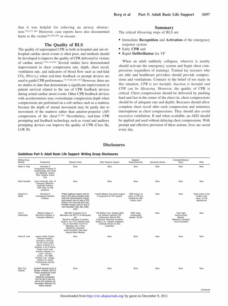
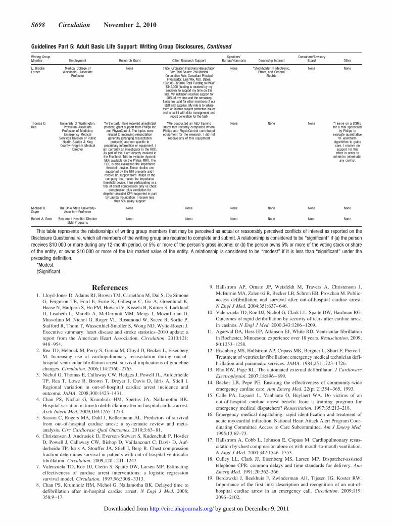
Guidelines Part 5: Adult Basic Life Support: Writing Group Disclosures
Writing GroupMember Employment Research Grant Other Research Support
Speakers’Bureau/Honoraria Ownership Interest
Consultant/AdvisoryBoard Other
Robert A. Berg University ofPennsylvania–Professor ofAnesthesiology and Critical
Care Medicine, DivisionChief, Pediatric Critical
Care
None None None None None None
Robin Hemphill Emory University, Dept. ofEmergency Medicine–
Associate Professor*Paid writer for AHA
guidelines
None None None None None None
Benjamin S.Abella
University ofPennsylvania–Assistant
Professor
†Philips Healthcare-research grant forstudy of CPR during inhospital cardiacarrest AHA Clinical Research Programgrant-research grant for study of CPRtraining in the community Doris DukeFoundation-research grant for study ofpost resuscitation injury after cardiac
arrest
*Laerdal Medical Corp-inkind supportof equipment for CPR research
*CME lectures ontopics of CPR andhypothermia after
cardiac arrest
None None *legal review of twocardiac arrestcases, no trialappearances
Tom P.Aufderheide
Medical College ofWisconsin–Professor ofEmergency Medicine
†NIH-ROC Consortium-PI ofMilwaukee site NETT-PI of Milwaukee
site*ResQTrial (Advanced Circulatory
Systems, Inc.)-PI of Oshkosh studysite, In Kind NHLBI Trial-PI for
Milwaukee site, In KindMedtronics-Consultant
JoLife-Consultant Take HeartAmerica-Board Member
*Zoll Medical Corp.-Supplied AEDsand software capturing CPRperformance data for ROC
Consortium Advanced CirculatorySystems, Inc.-Supplied impedance
threshold devices for ROCConsortium
*EMS Today-Compensated
speaker, $2,000
None *Take Heart*America-Board
Member*Medtronic-Consultant
*JoLife-Consultant
None
Diana M. Cave Legacy Health System,Emanuel Hospital,
Emergency Services:Not-for-profit healthsystem consists of 5
hospitals in the Portland,Oregon metro area.
Emanuel Hospital is aLevel I Trauma
Center.– RN, MSN;Portland Com. College,
Institute for HealthProfessionals–Faculty/
Instructor
None None None None None None
Mary FranHazinski
Vanderbilt University School ofNursing—Professor; AHA ECCProduct Development–Senior
Science Editor†Significant compensationfrom the AHA to write and
edit the AHA Guidelines andresuscitation statements and
training materials
None None None None None None
(Continued)
Berg et al Part 5: Adult Basic Life Support S697
by guest on December 9, 2011http://circ.ahajournals.org/Downloaded from
References1. Lloyd-Jones D, Adams RJ, Brown TM, Carnethon M, Dai S, De Simone
G, Ferguson TB, Ford E, Furie K, Gillespie C, Go A, Greenlund K,Haase N, Hailpern S, Ho PM, Howard V, Kissela B, Kittner S, LacklandD, Lisabeth L, Marelli A, McDermott MM, Meigs J, Mozaffarian D,Mussolino M, Nichol G, Roger VL, Rosamond W, Sacco R, Sorlie P,Stafford R, Thom T, Wasserthiel-Smoller S, Wong ND, Wylie-Rosett J.Executive summary: heart disease and stroke statistics–2010 update: areport from the American Heart Association. Circulation. 2010;121:948–954.
2. Rea TD, Helbock M, Perry S, Garcia M, Cloyd D, Becker L, EisenbergM. Increasing use of cardiopulmonary resuscitation during out-of-hospital ventricular fibrillation arrest: survival implications of guidelinechanges. Circulation. 2006;114:2760–2765.
3. Nichol G, Thomas E, Callaway CW, Hedges J, Powell JL, AufderheideTP, Rea T, Lowe R, Brown T, Dreyer J, Davis D, Idris A, Stiell I.Regional variation in out-of-hospital cardiac arrest incidence andoutcome. JAMA. 2008;300:1423–1431.
4. Chan PS, Nichol G, Krumholz HM, Spertus JA, Nallamothu BK.Hospital variation in time to defibrillation after in-hospital cardiac arrest.Arch Intern Med. 2009;169:1265–1273.
5. Sasson C, Rogers MA, Dahl J, Kellermann AL. Predictors of survivalfrom out-of-hospital cardiac arrest: a systematic review and meta-analysis. Circ Cardiovasc Qual Outcomes. 2010;3:63–81.
6. Christenson J, Andrusiek D, Everson-Stewart S, Kudenchuk P, HostlerD, Powell J, Callaway CW, Bishop D, Vaillancourt C, Davis D, Auf-derheide TP, Idris A, Stouffer JA, Stiell I, Berg R. Chest compressionfraction determines survival in patients with out-of-hospital ventricularfibrillation. Circulation. 2009;120:1241–1247.
7. Valenzuela TD, Roe DJ, Cretin S, Spaite DW, Larsen MP. Estimatingeffectiveness of cardiac arrest interventions: a logistic regressionsurvival model. Circulation. 1997;96:3308–3313.
8. Chan PS, Krumholz HM, Nichol G, Nallamothu BK. Delayed time todefibrillation after in-hospital cardiac arrest. N Engl J Med. 2008;358:9–17.
9. Hallstrom AP, Ornato JP, Weisfeldt M, Travers A, Christenson J,McBurnie MA, Zalenski R, Becker LB, Schron EB, Proschan M. Public-access defibrillation and survival after out-of-hospital cardiac arrest.N Engl J Med. 2004;351:637–646.
10. Valenzuela TD, Roe DJ, Nichol G, Clark LL, Spaite DW, Hardman RG.Outcomes of rapid defibrillation by security officers after cardiac arrestin casinos. N Engl J Med. 2000;343:1206–1209.
11. Agarwal DA, Hess EP, Atkinson EJ, White RD. Ventricular fibrillationin Rochester, Minnesota: experience over 18 years. Resuscitation. 2009;80:1253–1258.
12. Eisenberg MS, Hallstrom AP, Copass MK, Bergner L, Short F, Pierce J.Treatment of ventricular fibrillation: emergency medical technician defi-brillation and paramedic services. JAMA. 1984;251:1723–1726.
13. Rho RW, Page RL. The automated external defibrillator. J CardiovascElectrophysiol. 2007;18:896–899.
14. Becker LB, Pepe PE. Ensuring the effectiveness of community-wideemergency cardiac care. Ann Emerg Med. 22(pt 2):354–365, 1993.
15. Calle PA, Lagaert L, Vanhaute O, Buylaert WA. Do victims of anout-of-hospital cardiac arrest benefit from a training program foremergency medical dispatchers? Resuscitation. 1997;35:213–218.
16. Emergency medical dispatching: rapid identification and treatment ofacute myocardial infarction. National Heart Attack Alert Program Coor-dinating Committee Access to Care Subcommittee. Am J Emerg Med.1995;13:67–73.
17. Hallstrom A, Cobb L, Johnson E, Copass M. Cardiopulmonary resus-citation by chest compression alone or with mouth-to-mouth ventilation.N Engl J Med. 2000;342:1546–1553.
18. Culley LL, Clark JJ, Eisenberg MS, Larsen MP. Dispatcher-assistedtelephone CPR: common delays and time standards for delivery. AnnEmerg Med. 1991;20:362–366.
19. Berdowski J, Beekhuis F, Zwinderman AH, Tijssen JG, Koster RW.Importance of the first link: description and recognition of an out-of-hospital cardiac arrest in an emergency call. Circulation. 2009;119:2096–2102.
Guidelines Part 5: Adult Basic Life Support: Writing Group Disclosures, Continued
Writing GroupMember Employment Research Grant Other Research Support
Speakers’Bureau/Honoraria Ownership Interest
Consultant/AdvisoryBoard Other
E. BrookeLerner
Medical College ofWisconsin– Associate
Professor
None †Title: Circulation Improving ResuscitationCare Trial Source: Zoll Medical
Corporation Role: Consultant PrincipalInvestigator: Lars Wik, M.D. Dates:
12/2006–8/2010 Total Funding to MCW:$345,000 (funding is received by myemployer to support my time on this
trial. My institution receives support for20% of my time and the remaining
funds are used for other members of ourstaff and supplies. My role is to advise
them on human subject protection issuesand to assist with data management and
report generation for the trial)
None *Stockholder in Medtronic,Pfizer, and General
Electric
None None
Thomas D.Rea
University of Washington:Physician–Associate
Professor of Medicine;Emergency Medical
Services Division of PublicHealth-Seattle & King
County–Program MedicalDirector
*In the past, I have received unrestricted(modest) grant support from Philips Inc
and PhysioControl. The topics wererelated to improving resuscitationgenerally (changing resuscitation
protocols) and not specific toproprietary information or equipment. I
am currently an investigator in the ROC.As part of this, I am directly involved inthe Feedback Trial to evaluate dynamicfdbk available on the Philips MRX. TheROC is also evaluating the impedancethreshold device. These studies aresupported by the NIH primarily and I
receive no support from Philips or thecompany that makes the impedance
threshold device. I am participating in atrial of chest compression only vs chest
compression plus ventilation fordispatch-assisted CPR-supported in part
by Laerdal Foundation. I receive lessthan 5% salary support
*We conducted an AED trainingstudy that recently completed wherePhilips and PhysioControl contributedequipment for the research. I did not
receive any of this equipment
None None None *I serve on a DSMBfor a trial sponsored
by Philips toevaluate quantitative
VF waveformalgorhithm to guidecare. I receive nosupport for this
effort in order tominimize (eliminate)
any conflict
Michael R.Sayre
The Ohio State University-Associate Professor
None None None None None None
Robert A. Swor Beaumont Hospital–DirectorEMS Programs
None None None None None None
This table represents the relationships of writing group members that may be perceived as actual or reasonably perceived conflicts of interest as reported on theDisclosure Questionnaire, which all members of the writing group are required to complete and submit. A relationship is considered to be “significant” if (a) the personreceives $10 000 or more during any 12-month period, or 5% or more of the person’s gross income; or (b) the person owns 5% or more of the voting stock or shareof the entity, or owns $10 000 or more of the fair market value of the entity. A relationship is considered to be “modest” if it is less than “significant” under thepreceding definition.
*Modest.†Significant.
S698 Circulation November 2, 2010
by guest on December 9, 2011http://circ.ahajournals.org/Downloaded from
20. Kuisma M, Boyd J, Vayrynen T, Repo J, Nousila-Wiik M, HolmstromP. Emergency call processing and survival from out-of-hospital ven-tricular fibrillation. Resuscitation. 2005;67:89–93.
21. Rea TD, Eisenberg MS, Culley LL, Becker L. Dispatcher-assisted car-diopulmonary resuscitation and survival in cardiac arrest. Circulation.2001;104:2513–2516.
22. Hauff SR, Rea TD, Culley LL, Kerry F, Becker L, Eisenberg MS.Factors impeding dispatcher-assisted telephone cardiopulmonary resus-citation. Ann Emerg Med. 2003;42:731–737.
23. Vaillancourt C, Verma A, Trickett J, Crete D, Beaudoin T, Nesbitt L,Wells GA, Stiell IG. Evaluating the effectiveness of dispatch-assistedcardiopulmonary resuscitation instructions. Acad Emerg Med. 2007;14:877–883.
24. Bohm K, Rosenqvist M, Hollenberg J, Biber B, Engerstrom L, SvenssonL. Dispatcher-assisted telephone-guided cardiopulmonary resuscitation:an underused lifesaving system. Eur J Emerg Med. 2007;14:256–259.
25. Hallstrom AP, Cobb LA, Johnson E, Copass MK. Dispatcher assisted CPR:implementation and potential benefit. A 12-year study. Resuscitation. 2003;57:123–129.
26. Nurmi J, Pettila V, Biber B, Kuisma M, Komulainen R, Castren M.Effect of protocol compliance to cardiac arrest identification byemergency medical dispatchers. Resuscitation. 2006;70:463–469.
27. Clawson J, Olola C, Heward A, Patterson B. Cardiac arrest predictabilityin seizure patients based on emergency medical dispatcher identificationof previous seizure or epilepsy history. Resuscitation. 2007;75:298–304.
28. White L, Rogers J, Bloomingdale M, Fahrenbruch C, Culley L, SubidoC, Eisenberg M, Rea T. Dispatcher-assisted cardiopulmonary resusci-tation: risks for patients not in cardiac arrest. Circulation.121:91–97.
29. Sayre MR, Berg RA, Cave DM, Page RL, Potts J, White RD.Hands-only (compression-only) cardiopulmonary resuscitation: a call toaction for bystander response to adults who experience out-of-hospitalsudden cardiac arrest: a science advisory for the public from theAmerican Heart Association Emergency Cardiovascular Care Com-mittee. Circulation. 2008;117:2162–2167.
30. Kitamura T, Iwami T, Kawamura T, Nagao K, Tanaka H, Nadkarni VM,Berg RA, Hiraide A. Conventional and chest-compression-only cardio-pulmonary resuscitation by bystanders for children who have out-of-hospital cardiac arrests: a prospective, nationwide, population-basedcohort study. Lancet. 2010.
31. Heward A, Damiani M, Hartley-Sharpe C. Does the use of the AdvancedMedical Priority Dispatch System affect cardiac arrest detection? EmergMed J. 2004;21:115–118.
32. Roppolo LP, Westfall A, Pepe PE, Nobel LL, Cowan J, Kay JJ, IdrisAH. Dispatcher assessments for agonal breathing improve detection ofcardiac arrest. Resuscitation. 2009;80:769–772.
33. Bohm K, Stalhandske B, Rosenqvist M, Ulfvarson J, Hollenberg J,Svensson L. Tuition of emergency medical dispatchers in the recog-nition of agonal respiration increases the use of telephone assisted CPR.Resuscitation. 2009;80:1025–1028.
34. Clawson J, Olola C, Scott G, Heward A, Patterson B. Effect of a MedicalPriority Dispatch System key question addition in the seizure/convulsion/fitting protocol to improve recognition of ineffective(agonal) breathing. Resuscitation. 2008;79:257–264.
35. Bahr J, Klingler H, Panzer W, Rode H, Kettler D. Skills of lay peoplein checking the carotid pulse. Resuscitation. 1997;35:23–26.
36. Brennan RT, Braslow A. Skill mastery in public CPR classes. Am JEmerg Med. 1998;16:653–657.
37. Chamberlain D, Smith A, Woollard M, Colquhoun M, Handley AJ,Leaves S, Kern KB. Trials of teaching methods in basic life support :comparison of simulated CPR performance after first training and at 6months, with a note on the value of re-training. Resuscitation. 2002;53:179–187.
38. Eberle B, Dick WF, Schneider T, Wisser G, Doetsch S, Tzanova I.Checking the carotid pulse check: diagnostic accuracy of firstresponders in patients with and without a pulse. Resuscitation. 1996;33:107–116.
39. Frederick K, Bixby E, Orzel MN, Stewart-Brown S, Willett K. Willchanging the emphasis from ‘pulseless’ to ‘no signs of circulation’improve the recall scores for effective life support skills in children?Resuscitation. 2002;55:255–261.
40. Lapostolle F, Le Toumelin P, Agostinucci JM, Catineau J, Adnet F.Basic cardiac life support providers checking the carotid pulse: per-formance, degree of conviction, and influencing factors. Acad EmergMed. 2004;11:878–880.
41. Moule P. Checking the carotid pulse: diagnostic accuracy in students ofthe healthcare professions. Resuscitation. 2000;44:195–201.
42. Nyman J, Sihvonen M. Cardiopulmonary resuscitation skills in nursesand nursing students. Resuscitation. 2000;47:179–184.
43. Owen CJ, Wyllie JP. Determination of heart rate in the baby at birth.Resuscitation. 2004;60:213–217.
44. Sarti A, Savron F, Ronfani L, Pelizzo G, Barbi E. Comparison of threesites to check the pulse and count heart rate in hypotensive infants.Paediatr Anaesth. 2006;16:394–398.
45. Ochoa FJ, Ramalle-Gomara E, Carpintero JM, Garcia A, Saralegui I.Competence of health professionals to check the carotid pulse.Resuscitation. 1998;37:173–175.
46. Mather C, O’Kelly S. The palpation of pulses. Anaesthesia. 1996;51:189–191.
47. Olasveengen TM, Wik L, Steen PA. Standard basic life support vs.continuous chest compressions only in out-of-hospital cardiac arrest.Acta Anaesthesiol Scand. 2008;52:914–919.
48. Ong ME, Ng FS, Anushia P, Tham LP, Leong BS, Ong VY, Tiah L, LimSH, Anantharaman V. Comparison of chest compression only andstandard cardiopulmonary resuscitation for out-of-hospital cardiac arrestin Singapore. Resuscitation. 2008;78:119–126.
49. Bohm K, Rosenqvist M, Herlitz J, Hollenberg J, Svensson L. Survival issimilar after standard treatment and chest compression only in out-of-hospital bystander cardiopulmonary resuscitation. Circulation. 2007;116:2908–2912.
50. Iwami T, Kawamura T, Hiraide A, Berg RA, Hayashi Y, Nishiuchi T,Kajino K, Yonemoto N, Yukioka H, Sugimoto H, Kakuchi H, Sase K,Yokoyama H, Nonogi H. Effectiveness of bystander-initiatedcardiac-only resuscitation for patients with out-of-hospital cardiac arrest.Circulation. 2007;116:2900–2907.
51. SOS-KANTO Study Group. Cardiopulmonary resuscitation bybystanders with chest compression only (SOS-KANTO): an observa-tional study. Lancet. 2007;369:920–926.
52. Assar D, Chamberlain D, Colquhoun M, Donnelly P, Handley AJ,Leaves S, Kern KB. Randomised controlled trials of staged teaching forbasic life support, 1: skill acquisition at bronze stage. Resuscitation.2000;45:7–15.
53. Heidenreich JW, Higdon TA, Kern KB, Sanders AB, Berg RA, NieblerR, Hendrickson J, Ewy GA. Single-rescuer cardiopulmonary resusci-tation: ‘two quick breaths’–an oxymoron. Resuscitation. 2004;62:283–289.
54. Kobayashi M, Fujiwara A, Morita H, Nishimoto Y, Mishima T, Nitta M,Hayashi T, Hotta T, Hayashi Y, Hachisuka E, Sato K. A manikin-basedobservational study on cardiopulmonary resuscitation skills at the OsakaSenri medical rally. Resuscitation. 2008;78:333–339.
55. Baskett P, Nolan J, Parr M. Tidal volumes which are perceived to beadequate for resuscitation. Resuscitation. 1996;31:231–234.
56. Ruppert M, Reith MW, Widmann JH, Lackner CK, Kerkmann R,Schweiberer L, Peter K. Checking for breathing: evaluation of thediagnostic capability of emergency medical services personnel, phy-sicians, medical students, and medical laypersons. Ann Emerg Med.1999;34:720–729.
57. Safar P, Escarraga LA, Chang F. Upper airway obstruction in theunconscious patient. J Appl Physiol. 1959;14:760–764.
58. Perkins GD, Stephenson B, Hulme J, Monsieurs KG. Birminghamassessment of breathing study (BABS). Resuscitation. 2005;64:109–113.
59. Handley AJ, Handley JA. Performing chest compressions in a confinedspace. Resuscitation. 2004;61:55–61.
60. Andersen LO, Isbye DL, Rasmussen LS. Increasing compression depthduring manikin CPR using a simple backboard. Acta AnaesthesiolScand. 2007;51:747–750.
61. Perkins GD, Smith CM, Augre C, Allan M, Rogers H, Stephenson B,Thickett DR. Effects of a backboard, bed height, and operator positionon compression depth during simulated resuscitation. Intensive CareMed. 2006;32:1632–1635.
62. Perkins GD, Kocierz L, Smith SC, McCulloch RA, Davies RP. Com-pression feedback devices over estimate chest compression depth whenperformed on a bed. Resuscitation. 2009;80:79–82.
63. Noordergraaf GJ, Paulussen IW, Venema A, van Berkom PF, WoerleePH, Scheffer GJ, Noordergraaf A. The impact of compliant surfaces onin-hospital chest compressions: effects of common mattresses and abackboard. Resuscitation. 2009;80:546–552.
Berg et al Part 5: Adult Basic Life Support S699
by guest on December 9, 2011http://circ.ahajournals.org/Downloaded from
64. Delvaux AB, Trombley MT, Rivet CJ, Dykla JJ, Jensen D, Smith MR,Gilbert RJ. Design and development of a cardiopulmonary resuscitationmattress. J Intensive Care Med. 2009;24:195–199.
65. Perkins GD, Benny R, Giles S, Gao F, Tweed MJ. Do different mat-tresses affect the quality of cardiopulmonary resuscitation? IntensiveCare Med. 2003;29:2330–2335.
66. Kundra P, Dey S, Ravishankar M. Role of dominant hand positionduring external cardiac compression. Br J Anaesth. 2000;84:491–493.
67. Nikandish R, Shahbazi S, Golabi S, Beygi N. Role of dominant versusnon-dominant hand position during uninterrupted chest compressionCPR by novice rescuers: a randomized double-blind crossover study.Resuscitation. 2008;76:256–260.
68. Shin J, Rhee JE, Kim K. Is the inter-nipple line the correct hand positionfor effective chest compression in adult cardiopulmonary resuscitation?Resuscitation. 2007;75:305–310.
69. Kusunoki S, Tanigawa K, Kondo T, Kawamoto M, Yuge O. Safety ofthe inter-nipple line hand position landmark for chest compression.Resuscitation. 2009;80:1175–1180.
70. Babbs CF, Kemeny AE, Quan W, Freeman G. A new paradigm forhuman resuscitation research using intelligent devices. Resuscitation.2008;77:306–315.
71. Edelson DP, Abella BS, Kramer-Johansen J, Wik L, Myklebust H, BarryAM, Merchant RM, Hoek TL, Steen PA, Becker LB. Effects of com-pression depth and pre-shock pauses predict defibrillation failure duringcardiac arrest. Resuscitation. 2006;71:137–145.
72. Kramer-Johansen J, Myklebust H, Wik L, Fellows B, Svensson L,Sorebo H, Steen PA. Quality of out-of-hospital cardiopulmonary resus-citation with real time automated feedback: a prospective interventionalstudy. Resuscitation. 2006;71:283–292.
73. Edelson DP, Litzinger B, Arora V, Walsh D, Kim S, Lauderdale DS,Vanden Hoek TL, Becker LB, Abella BS. Improving in-hospital cardiacarrest process and outcomes with performance debriefing. Arch InternMed. 2008;168:1063–1069.
74. Talley DB, Ornato JP, Clarke AM. Computer-aided characterization andoptimization of the Thumper compression waveform in closed-chestCPR. Biomed Instrum Technol. 1990;24:283–288.
75. Handley AJ, Handley SA. Improving CPR performance using an audiblefeedback system suitable for incorporation into an automated externaldefibrillator. Resuscitation. 2003;57:57–62.
76. Yannopoulos D, McKnite S, Aufderheide TP, Sigurdsson G, PirralloRG, Benditt D, Lurie KG. Effects of incomplete chest wall decom-pression during cardiopulmonary resuscitation on coronary and cerebralperfusion pressures in a porcine model of cardiac arrest. Resuscitation.2005;64:363–372.
77. Aufderheide TP, Pirrallo RG, Yannopoulos D, Klein JP, von Briesen C,Sparks CW, Deja KA, Kitscha DJ, Provo TA, Lurie KG. Incompletechest wall decompression: A clinical evaluation of CPR performance bytrained laypersons and an assessment of alternative manual chestcompression-decompression techniques. Resuscitation. 2006;71:341–351.
78. Sutton RM, Niles D, Nysaether J, Abella BS, Arbogast KB, Nishisaki A,Maltese MR, Donoghue A, Bishnoi R, Helfaer MA, Myklebust H,Nadkarni V. Quantitative analysis of CPR quality during in-hospitalresuscitation of older children and adolescents. Pediatrics. 2009;124:494–499.
79. Sutton RM, Maltese MR, Niles D, French B, Nishisaki A, Arbogast KB,Donoghue A, Berg RA, Helfaer MA, Nadkarni V. Quantitative analysisof chest compression interruptions during in-hospital resuscitation ofolder children and adolescents. Resuscitation. 2009;80:1259–1263.
80. Niles D, Nysaether J, Sutton R, Nishisaki A, Abella BS, Arbogast K,Maltese MR, Berg RA, Helfaer M, Nadkarni V. Leaning is commonduring in-hospital pediatric CPR, and decreased with automated cor-rective feedback. Resuscitation. 2009;80:553–557.
81. Aufderheide TP, Pirrallo RG, Yannopoulos D, Klein JP, von Briesen C,Sparks CW, Deja KA, Conrad CJ, Kitscha DJ, Provo TA, Lurie KG.Incomplete chest wall decompression: a clinical evaluation of CPRperformance by EMS personnel and assessment of alternative manualchest compression-decompression techniques. Resuscitation. 2005;64:353–362.
82. Zuercher M, Hilwig RW, Ranger-Moore J, Nysaether J, Nadkarni VM,Berg MD, Kern KB, Sutton R, Berg RA. Leaning during chest com-pressions impairs cardiac output and left ventricular myocardial bloodflow in piglet cardiac arrest. Crit Care Med. 2010;38:1141–1146.
83. Babbs CF, Kern KB. Optimum compression to ventilation ratios in CPRunder realistic, practical conditions: a physiological and mathematicalanalysis. Resuscitation. 2002;54:147–157.
84. Kern KB, Hilwig RW, Berg RA, Ewy GA. Efficacy of chestcompression-only BLS CPR in the presence of an occluded airway.Resuscitation. 1998;39:179–188.
85. Abella BS, Sandbo N, Vassilatos P, Alvarado JP, O’Hearn N, WigderHN, Hoffman P, Tynus K, Vanden Hoek TL, Becker LB. Chest com-pression rates during cardiopulmonary resuscitation are suboptimal: aprospective study during in-hospital cardiac arrest. Circulation. 2005;111:428–434.
86. Wolfe JA, Maier GW, Newton JR Jr, Glower DD, Tyson GS Jr, SprattJA, Rankin JS, Olsen CO. Physiologic determinants of coronary bloodflow during external cardiac massage. J Thorac Cardiovasc Surg. 1988;95:523–532.
87. Maier GW, Tyson GS Jr, Olsen CO, Kernstein KH, Davis JW, Conn EH,Sabiston DC Jr, Rankin JS. The physiology of external cardiac massage:high-impulse cardiopulmonary resuscitation. Circulation. 1984;70:86–101.
88. Feneley MP, Maier GW, Kern KB, Gaynor JW, Gall SA Jr, Sanders AB,Raessler K, Muhlbaier LH, Rankin JS, Ewy GA. Influence of compressionrate on initial success of resuscitation and 24 hour survival after prolongedmanual cardiopulmonary resuscitation in dogs. Circulation. 1988;77:240–250.
89. Halperin HR, Tsitlik JE, Guerci AD, Mellits ED, Levin HR, Shi AY,Chandra N, Weisfeldt ML. Determinants of blood flow to vital organsduring cardiopulmonary resuscitation in dogs. Circulation. 1986;73:539–550.
90. Handley AJ, Handley JA. The relationship between rate of chest com-pression and compression:relaxation ratio. Resuscitation. 1995;30:237–241.
91. Wik L, Kramer-Johansen J, Myklebust H, Sorebo H, Svensson L,Fellows B, Steen PA. Quality of cardiopulmonary resuscitation duringout-of-hospital cardiac arrest. JAMA. 2005;293:299–304.
92. Abella BS, Alvarado JP, Myklebust H, Edelson DP, Barry A, O’HearnN, Vanden Hoek TL, Becker LB. Quality of cardiopulmonary resusci-tation during in-hospital cardiac arrest. JAMA. 2005;293:305–310.
93. Valenzuela TD, Kern KB, Clark LL, Berg RA, Berg MD, Berg DD, HilwigRW, Otto CW, Newburn D, Ewy GA. Interruptions of chest compressionsduring emergency medical systems resuscitation. Circulation. 2005;112:1259–1265.
94. Berg RA, Hilwig RW, Berg MD, Berg DD, Samson RA, Indik JH, KernKB. Immediate post-shock chest compressions improve outcome fromprolonged ventricular fibrillation. Resuscitation. 2008;78:71–76.
95. Tang W, Snyder D, Wang J, Huang L, Chang YT, Sun S, Weil MH.One-shock versus three-shock defibrillation protocol significantly improvesoutcome in a porcine model of prolonged ventricular fibrillation cardiacarrest. Circulation. 2006;113:2683–2689.
96. Garza AG, Gratton MC, Salomone JA, Lindholm D, McElroy J, ArcherR. Improved patient survival using a modified resuscitation protocol forout-of-hospital cardiac arrest. Circulation. 2009;119:2597–2605.
97. Bobrow BJ, Clark LL, Ewy GA, Chikani V, Sanders AB, Berg RA,Richman PB, Kern KB. Minimally interrupted cardiac resuscitation byemergency medical services for out-of-hospital cardiac arrest. JAMA.2008;299:1158–1165.
98. Kellum MJ, Kennedy KW, Barney R, Keilhauer FA, Bellino M,Zuercher M, Ewy GA. Cardiocerebral resuscitation improves neurolog-ically intact survival of patients with out-of-hospital cardiac arrest. AnnEmerg Med. 2008;52:244–252.
99. Sayre MR, Cantrell SA, White LJ, Hiestand BC, Keseg DP, Koser S.Impact of the 2005 American Heart Association cardiopulmonary resus-citation and emergency cardiovascular care guidelines on out-of-hospitalcardiac arrest survival. Prehosp Emerg Care. 2009;13:469–477.
100. Steinmetz J, Barnung S, Nielsen SL, Risom M, Rasmussen LS.Improved survival after an out-of-hospital cardiac arrest using newguidelines. Acta Anaesthesiol Scand. 2008;52:908–913.
101. Aufderheide TP, Yannopoulos D, Lick CJ, Myers B, Romig LA,Stothert JC, Barnard J, Vartanian L, Pilgrim AJ, Benditt DG. Imple-menting the 2005 American Heart Association Guidelines ImprovesOutcomes after Out-of-Hospital Cardiac Arrest. Heart Rhythm. 2010.
102. Hinchey PR, Myers JB, Lewis R, De Maio VJ, Reyer E, Licatese D,Zalkin J, Snyder G. Improved Out-of-Hospital Cardiac Arrest SurvivalAfter the Sequential Implementation of 2005 AHA Guidelines for Com-pressions, Ventilations, and Induced Hypothermia: The Wake CountyExperience. Ann Emerg Med. 2010.
S700 Circulation November 2, 2010
by guest on December 9, 2011http://circ.ahajournals.org/Downloaded from
103. Jost D, Degrange H, Verret C, Hersan O, Banville IL, Chapman FW,Lank P, Petit JL, Fuilla C, Migliani R, Carpentier JP. DEFI 2005: arandomized controlled trial of the effect of automated external defi-brillator cardiopulmonary resuscitation protocol on outcome from out-of-hospital cardiac arrest. Circulation. 2010;121:1614–1622.
104. Sugerman NT, Edelson DP, Leary M, Weidman EK, Herzberg DL,Vanden Hoek TL, Becker LB, Abella BS. Rescuer fatigue during actualin-hospital cardiopulmonary resuscitation with audiovisual feedback: aprospective multicenter study. Resuscitation. 2009;80:981–984.
105. Manders S, Geijsel FE. Alternating providers during continuous chestcompressions for cardiac arrest: every minute or every two minutes?Resuscitation. 2009;80:1015–1018.
106. Heidenreich JW, Berg RA, Higdon TA, Ewy GA, Kern KB, SandersAB. Rescuer fatigue: standard versus continuous chest-compressioncardiopulmonary resuscitation. Acad Emerg Med. 2006;13:1020–1026.
107. Dorph E, Wik L, Stromme TA, Eriksen M, Steen PA. Oxygen deliveryand return of spontaneous circulation with ventilation:compression ratio2:30 versus chest compressions only CPR in pigs. Resuscitation. 2004;60:309–318.
108. Berg RA, Sanders AB, Kern KB, Hilwig RW, Heidenreich JW, PorterME, Ewy GA. Adverse hemodynamic effects of interrupting chestcompressions for rescue breathing during cardiopulmonary resuscitationfor ventricular fibrillation cardiac arrest. Circulation. 2001;104:2465–2470.
109. Berg RA, Hilwig RW, Kern KB, Ewy GA. “Bystander” chest com-pressions and assisted ventilation independently improve outcome frompiglet asphyxial pulseless “cardiac arrest.” Circulation. 2000;101:1743–1748.
110. Berg RA, Kern KB, Hilwig RW, Berg MD, Sanders AB, Otto CW, EwyGA. Assisted ventilation does not improve outcome in a porcine modelof single-rescuer bystander cardiopulmonary resuscitation. Circulation.1997;95:1635–1641.
111. Berg RA, Kern KB, Hilwig RW, Ewy GA. Assisted ventilation during‘bystander’ CPR in a swine acute myocardial infarction model does notimprove outcome. Circulation. 1997;96:4364–4371.
112. Vaillancourt C, Stiell IG, Wells GA. Understanding and improving lowbystander CPR rates: a systematic review of the literature. CJEM.2008;10:51–65.
113. Stiell IG, Wells GA, Field B, Spaite DW, Nesbitt LP, De Maio VJ,Nichol G, Cousineau D, Blackburn J, Munkley D, Luinstra-Toohey L,Campeau T, Dagnone E, Lyver M. Advanced cardiac life support inout-of-hospital cardiac arrest. N Engl J Med. 2004;351:647–656.
114. Ornato JP, Hallagan LF, McMahan SB, Peeples EH, Rostafinski AG.Attitudes of BCLS instructors about mouth-to-mouth resuscitationduring the AIDS epidemic. Ann Emerg Med. 1990;19:151–156.
115. Brenner BE, Van DC, Cheng D, Lazar EJ. Determinants of reluctance toperform CPR among residents and applicants: the impact of experienceon helping behavior. Resuscitation. 1997;35:203–211.
116. Hew P, Brenner B, Kaufman J. Reluctance of paramedics andemergency medical technicians to perform mouth-to-mouth resusci-tation. J Emerg Med. 1997;15:279–284.
117. Sirbaugh PE, Pepe PE, Shook JE, Kimball KT, Goldman MJ, Ward MA,Mann DM. A prospective, population-based study of the demographics,epidemiology, management, and outcome of out-of-hospital pediatriccardiopulmonary arrest. Ann Emerg Med. 1999;33:174–184.
118. Swor R, Khan I, Domeier R, Honeycutt L, Chu K, Compton S. CPRtraining and CPR performance: do CPR-trained bystanders performCPR? Acad Emerg Med. 2006;13:596–601.
119. Tang W, Weil MH, Sun S, Kette D, Gazmuri RJ, O’Connell F, BiseraJ. Cardiopulmonary resuscitation by precordial compression but withoutmechanical ventilation. Am J Respir Crit Care Med. 1994;150(6 pt1):1709–1713.
120. Bobrow BJ, Zuercher M, Ewy GA, Clark L, Chikani V, Donahue D,Sanders AB, Hilwig RW, Berg RA, Kern KB. Gasping during cardiacarrest in humans is frequent and associated with improved survival.Circulation. 2008;118:2550–2554.
121. Clark JJ, Larsen MP, Culley LL, Graves JR, Eisenberg MS. Incidence ofagonal respirations in sudden cardiac arrest. Ann Emerg Med. 1992;21:1464–1467.
122. Bang A, Herlitz J, Martinell S. Interaction between emergency medicaldispatcher and caller in suspected out-of-hospital cardiac arrest callswith focus on agonal breathing. A review of 100 tape recordings of truecardiac arrest cases. Resuscitation. 2003;56:25–34.
123. Becker LB, Berg RA, Pepe PE, Idris AH, Aufderheide TP, Barnes TA,Stratton SJ, Chandra NC. A reappraisal of mouth-to-mouth ventilation
during bystander-initiated cardiopulmonary resuscitation. A statementfor healthcare professionals from the Ventilation Working Group of theBasic Life Support and Pediatric Life Support Subcommittees,American Heart Association. Resuscitation. 1997;35:189–201.
124. Weil MH, Rackow EC, Trevino R, Grundler W, Falk JL, Griffel MI.Difference in acid-base state between venous and arterial blood duringcardiopulmonary resuscitation. N Engl J Med. 1986;315:153–156.
125. Sanders AB, Otto CW, Kern KB, Rogers JN, Perrault P, Ewy GA.Acid-base balance in a canine model of cardiac arrest. Ann Emerg Med.1988;17:667–671.
126. Steen-Hansen JE. Favourable outcome after 26 minutes of “Com-pression only” resuscitation: a case report. Scand J Trauma ResuscEmerg Med. 2010;18:19.
127. Berg RA, Hilwig RW, Kern KB, Babar I, Ewy GA. Simulated mouth-to-mouth ventilation and chest compressions (bystander cardiopul-monary resuscitation) improves outcome in a swine model of prehospitalpediatric asphyxial cardiac arrest. Crit Care Med. 1999;27:1893–1899.
128. Iglesias JM, Lopez-Herce J, Urbano J, Solana MJ, Mencia S, DelCastillo J. Chest compressions versus ventilation plus chest com-pressions in a pediatric asphyxial cardiac arrest animal model. IntensiveCare Med. 2010;36:712–716.
129. Idris AH, Becker LB, Fuerst RS, Wenzel V, Rush WJ, Melker RJ, OrbanDJ. Effect of ventilation on resuscitation in an animal model of cardiacarrest. Circulation. 1994;90:3063–3069.
130. Guildner CW. Resuscitation: opening the airway. A comparative studyof techniques for opening an airway obstructed by the tongue. JACEP.1976;5:588–590.
131. Greene DG, Elam JO, Dobkin AB, Studley CL. Cinefluorographic studyof hyperextension of the neck and upper airway patency. JAMA. 1961;176:570–573.
132. Ruben HM, Elam JO, al. e. Investigations of pharyngeal xrays andperfomance by laymen. Anesthesiology. 1961;22:271–279.
133. Elam JO, Greene DG, Schneider MA, Ruben HM, Gordon AS, HusteadRF, Benson DW, Clements JA, Ruben A. Head-tilt method of oralresuscitation. JAMA. 1960;172:812–815.
134. Rhee P, Kuncir EJ, Johnson L, Brown C, Velmahos G, Martin M, WangD, Salim A, Doucet J, Kennedy S, Demetriades D. Cervical spine injuryis highly dependent on the mechanism of injury following blunt andpenetrating assault. J Trauma. 2006;61:1166–1170.
135. Lowery DW, Wald MM, Browne BJ, Tigges S, Hoffman JR, MowerWR. Epidemiology of cervical spine injury victims. Ann Emerg Med.2001;38:12–16.
136. Milby AH, Halpern CH, Guo W, Stein SC. Prevalence of cervical spinalinjury in trauma. Neurosurg Focus. 2008;25:E10.
137. Mithani SK, St-Hilaire H, Brooke BS, Smith IM, Bluebond-Langner R,Rodriguez ED. Predictable patterns of intracranial and cervical spineinjury in craniomaxillofacial trauma: analysis of 4786 patients. PlastReconstr Surg. 2009;123:1293–1301.
138. Hackl W, Hausberger K, Sailer R, Ulmer H, Gassner R. Prevalence ofcervical spine injuries in patients with facial trauma. Oral Surg OralMed Oral Pathol Oral Radiol Endod. 2001;92:370–376.
139. Holly LT, Kelly DF, Counelis GJ, Blinman T, McArthur DL, Cryer HG.Cervical spine trauma associated with moderate and severe head injury:incidence, risk factors, and injury characteristics. J Neurosurg Spine.2002;96:285–291.
140. Demetriades D, Charalambides K, Chahwan S, Hanpeter D, Alo K,Velmahos G, Murray J, Asensio J. Nonskeletal cervical spine injuries:epidemiology and diagnostic pitfalls. J Trauma. 2000;48:724–727.
141. Majernick TG, Bieniek R, Houston JB, Hughes HG. Cervical spinemovement during orotracheal intubation. Ann Emerg Med. 1986;15:417–420.
142. Lennarson PJ, Smith DW, Sawin PD, Todd MM, Sato Y, Traynelis VC.Cervical spinal motion during intubation: efficacy of stabilizationmaneuvers in the setting of complete segmental instability. J NeurosurgSpine. 2001;94:265–270.
143. Hastings RH, Wood PR. Head extension and laryngeal view during laryn-goscopy with cervical spine stabilization maneuvers. Anesthesiology. 1994;80:825–831.
144. Gerling MC, Davis DP, Hamilton RS, Morris GF, Vilke GM, Garfin SR,Hayden SR. Effects of cervical spine immobilization technique andlaryngoscope blade selection on an unstable cervical spine in a cadavermodel of intubation. Ann Emerg Med. 2000;36:293–300.
145. Wenzel V, Keller C, Idris AH, Dorges V, Lindner KH, BrimacombeJR. Effects of smaller tidal volumes during basic life support venti-
Berg et al Part 5: Adult Basic Life Support S701
by guest on December 9, 2011http://circ.ahajournals.org/Downloaded from
lation in patients with respiratory arrest: good ventilation, less risk?Resuscitation. 1999;43:25–29.
146. Dorges V, Ocker H, Hagelberg S, Wenzel V, Idris AH, Schmucker P.Smaller tidal volumes with room-air are not sufficient to ensureadequate oxygenation during bag-valve-mask ventilation. Resuscitation.2000;44:37–41.
147. Dorges V, Ocker H, Hagelberg S, Wenzel V, Schmucker P. Optimi-sation of tidal volumes given with self-inflatable bags without additionaloxygen. Resuscitation. 2000;43:195–199.
148. Finer NN, Barrington KJ, Al-Fadley F, Peters KL. Limitations of self-inflating resuscitators. Pediatrics. 1986;77:417–420.
149. Hirschman AM, Kravath RE. Venting vs ventilating. A danger ofmanual resuscitation bags. Chest. 1982;82:369–370.
150. Berg MD, Idris AH, Berg RA. Severe ventilatory compromise due togastric distention during pediatric cardiopulmonary resuscitation.Resuscitation. 1998;36:71–73.
151. Garnett AR, Ornato JP, Gonzalez ER, Johnson EB. End-tidal carbondioxide monitoring during cardiopulmonary resuscitation. JAMA. 1987;257:512–515.
152. Aufderheide TP, Sigurdsson G, Pirrallo RG, Yannopoulos D, McKniteS, von Briesen C, Sparks CW, Conrad CJ, Provo TA, Lurie KG.Hyperventilation-induced hypotension during cardiopulmonary resusci-tation. Circulation. 2004;109:1960–1965.
153. Kern KB, Hilwig RW, Berg RA, Sanders AB, Ewy GA. Importanceof continuous chest compressions during cardiopulmonary resusci-tation: improved outcome during a simulated single lay-rescuerscenario. Circulation. 2002;105:645– 649.
154. Wang HE, Simeone SJ, Weaver MD, Callaway CW. Interruptions incardiopulmonary resuscitation from paramedic endotracheal intubation.Ann Emerg Med. 2009;54:645–652 e641.
155. Wenzel V, Idris AH, Banner MJ, Fuerst RS, Tucker KJ. The compo-sition of gas given by mouth-to-mouth ventilation during CPR. Chest.1994;106:1806–1810.
156. Ruben H. The immediate treatment of respiratory failure. Br J Anaesth.1964;36:542–549.
157. Bhalla RK, Corrigan A, Roland NJ. Comparison of two face masks usedto deliver early ventilation to laryngectomized patients. Ear Nose ThroatJ. 2004;83:414, 416.
158. Barnes TA. Emergency ventilation techniques and related equipment.Respir Care. 1992;37:673–690, discussion 690–674.
159. Johannigman JA, Branson RD, Davis K Jr, Hurst JM. Techniques ofemergency ventilation: a model to evaluate tidal volume, airwaypressure, and gastric insufflation. J Trauma. 1991;31:93–98.
160. Elam JO. Bag-valve-mask O2 ventilation. In: Safar P, Elam JO, eds.Advances in Cardiopulmonary Resuscitation: The Wolf Creek Con-ference on Cardiopulmonary Resuscitation. New York, NY: Springer-Verlag, Inc.; 1977:73–79.
161. Dailey R, Young G, Simon B, Stewart R. The Airway: EmergencyManagement: C.V. Mosby; 1992.
162. Elling R, Politis J. An evaluation of emergency medical technicians’ability to use manual ventilation devices. Ann Emerg Med. 1983;12:765–768.
163. von Goedecke A, Bowden K, Wenzel V, Keller C, Gabrielli A. Effectsof decreasing inspiratory times during simulated bag-valve-mask venti-lation. Resuscitation. 2005;64:321–325.
164. von Goedecke A, Bowden K, Keller C, Voelckel WG, Jeske HC,Wenzel V. [Decreased inspiratory time during ventilation of an unpro-tected airway. Effect on stomach inflation and lung ventilation in abench model.] Anaesthesist. 2005;54:117–122.
165. von Goedecke A, Paal P, Keller C, Voelckel WG, Herff H, Lindner KH,Wenzel V. [Ventilation of an unprotected airway: evaluation of a newpeak-inspiratory-flow and airway-pressure-limiting bag-valve-mask.]Anaesthesist. 2006;55:629–634.
166. Rumball CJ, MacDonald D. The PTL, Combitube, laryngeal mask, andoral airway: a randomized prehospital comparative study of ventilatorydevice effectiveness and cost-effectiveness in 470 cases of cardiorespi-ratory arrest. Prehosp Emerg Care. 1997;1:1–10.
167. Comparison of arterial blood gases of laryngeal mask airway and bag-valve-mask ventilation in out-of-hospital cardiac arrests. Circ J. 2009;73:490–496.
168. Stone BJ, Chantler PJ, Baskett PJ. The incidence of regurgitation duringcardiopulmonary resuscitation: a comparison between the bag valvemask and laryngeal mask airway. Resuscitation. 1998;38:3–6.
169. Atherton GL, Johnson JC. Ability of paramedics to use the Combitubein prehospital cardiac arrest. Ann Emerg Med. 1993;22:1263–1268.
170. Kette F, Reffo I, Giordani G, Buzzi F, Borean V, Cimarosti R,Codiglia A, Hattinger C, Mongiat A, Tararan S. The use of laryngealtube by nurses in out-of-hospital emergencies: Preliminary expe-rience. Resuscitation. 2005;66:21–25.
171. Timmermann A, Russo SG, Rosenblatt WH, Eich C, Barwing J,Roessler M, Graf BM. Intubating laryngeal mask airway for difficultout-of-hospital airway management: a prospective evaluation. Br JAnaesth. 2007;99:286–291.
172. Kellum MJ, Kennedy KW, Ewy GA. Cardiocerebral resuscitationimproves survival of patients with out-of-hospital cardiac arrest. Am JMed. 2006;119:335–340.
173. Bobrow BJ, Ewy GA, Clark L, Chikani V, Berg RA, Sanders AB,Vadeboncoeur TF, Hilwig RW, Kern KB. Passive oxygen insufflation issuperior to bag-valve-mask ventilation for witnessed ventricular fibril-lation out-of-hospital cardiac arrest. Ann Emerg Med. 2009;54:656–662e651.
174. McNelis U, Syndercombe A, Harper I, Duggan J. The effect of cricoidpressure on intubation facilitated by the gum elastic bougie. Anaes-thesia. 2007;62:456–459.
175. Harry RM, Nolan JP. The use of cricoid pressure with the intubatinglaryngeal mask. Anaesthesia. 1999;54:656–659.
176. Noguchi T, Koga K, Shiga Y, Shigematsu A. The gum elastic bougieeases tracheal intubation while applying cricoid pressure compared to astylet. Can J Anaesth. 2003;50:712–717.
177. Asai T, Murao K, Shingu K. Cricoid pressure applied after placement oflaryngeal mask impedes subsequent fibreoptic tracheal intubationthrough mask. Br J Anaesth. 2000;85:256–261.
178. Snider DD, Clarke D, Finucane BT. The “BURP” maneuver worsens theglottic view when applied in combination with cricoid pressure. Can JAnaesth. 2005;52:100–104.
179. Smith CE, Boyer D. Cricoid pressure decreases ease of tracheal intu-bation using fibreoptic laryngoscopy (WuScope System). Can JAnaesth. 2002;49:614–619.
180. Asai T, Barclay K, Power I, Vaughan RS. Cricoid pressure impedesplacement of the laryngeal mask airway and subsequent tracheal intu-bation through the mask. Br J Anaesth. 1994;72:47–51.
181. Domuracki KJ, Moule CJ, Owen H, Kostandoff G, Plummer JL.Learning on a simulator does transfer to clinical practice. Resuscitation.2009;80:346–349.
182. Beavers RA, Moos DD, Cuddeford JD. Analysis of the application ofcricoid pressure: implications for the clinician. J Perianesth Nurs. 2009;24:92–102.
183. Meek T, Gittins N, Duggan JE. Cricoid pressure: knowledge and per-formance amongst anaesthetic assistants. Anaesthesia. 1999;54:59–62.
184. Clark RK, Trethewy CE. Assessment of cricoid pressure application byemergency department staff. Emerg Med Australas. 2005;17:376–381.
185. Kopka A, Robinson D. The 50 ml syringe training aid should be utilizedimmediately before cricoid pressure application. Eur J Emerg Med.2005;12:155–158.
186. Flucker CJ, Hart E, Weisz M, Griffiths R, Ruth M. The 50-millilitresyringe as an inexpensive training aid in the application of cricoidpressure. Eur J Anaesthesiol. 2000;17:443–447.
187. Shimabukuro A, Kawatani M, Nagao N, Inoue Y, Hayashida M, HikawaY. [Training in application of cricoid pressure.] Masui. 2006;55:742–744.
188. Schmidt A, Akeson J. Practice and knowledge of cricoid pressure insouthern Sweden. Acta Anaesthesiol Scand. 2001;45:1210–1214.
189. Patten SP. Educating nurses about correct application of cricoidpressure. AORN J. 2006;84:449–461.
190. Koziol CA, Cuddeford JD, Moos DD. Assessing the force generatedwith application of cricoid pressure. AORN J. 2000;72:1018–1028,1030.
191. Clayton TJ, Vanner RG. A novel method of measuring cricoid force.Anaesthesia. 2002;57:326–329.
192. Owen H, Follows V, Reynolds KJ, Burgess G, Plummer J. Learning toapply effective cricoid pressure using a part task trainer. Anaesthesia.2002;57:1098–1101.
193. Kopka A, Crawford J. Cricoid pressure: a simple, yet effective bio-feedback trainer. Eur J Anaesthesiol. 2004;21:443–447.
194. Quigley P, Jeffrey P. Cricoid pressure: assessment of performance andeffect of training in emergency department staff. Emerg Med Australas.2007;19:218–222.
195. The Public Access Defibrillation Trial Investigators. Public-access defi-brillation and survival after out-of-hospital cardiac arrest. N Engl J Med.2004;351:637–646.
S702 Circulation November 2, 2010
by guest on December 9, 2011http://circ.ahajournals.org/Downloaded from
196. Rea TD, Cook AJ, Stiell IG, Powell J, Bigham B, Callaway CW, ChughS, Aufderheide TP, Morrison L, Terndrup TE, Beaudoin T, Wittwer L,Davis D, Idris A, Nichol G. Predicting survival after out-of-hospitalcardiac arrest: role of the Utstein data elements. Ann Emerg Med.2010;55:249–257.
197. Caffrey SL, Willoughby PJ, Pepe PE, Becker LB. Public use ofautomated external defibrillators. N Engl J Med. 2002;347:1242–1247.
198. Fries M, Tang W, Chang YT, Wang J, Castillo C, Weil MH. Micro-vascular blood flow during cardiopulmonary resuscitation is predictiveof outcome. Resuscitation. 2006;71:248–253.
199. Stiell IG, Callaway C, Davis D, Terndrup T, Powell J, Cook A,Kudenchuk PJ, Daya M, Kerber R, Idris A, Morrison LJ, Aufderheide T.Resuscitation Outcomes Consortium (ROC) PRIMED cardiac arrest trialmethods part 2: rationale and methodology for “Analyze Later vs.Analyze Early” protocol. Resuscitation. 2008;78:186–195.
200. Baker PW, Conway J, Cotton C, Ashby DT, Smyth J, Woodman RJ,Grantham H. Defibrillation or cardiopulmonary resuscitation first forpatients with out-of-hospital cardiac arrests found by paramedics to be inventricular fibrillation? A randomised control trial. Resuscitation. 2008;79:424–431.
201. Jacobs IG, Finn JC, Oxer HF, Jelinek GA. CPR before defibrillation inout-of-hospital cardiac arrest: a randomized trial. Emerg Med Australas.2005;17:39–45.
202. Wik L, Hansen TB, Fylling F, Steen T, Vaagenes P, Auestad BH, SteenPA. Delaying defibrillation to give basic cardiopulmonary resuscitationto patients with out-of-hospital ventricular fibrillation: a randomizedtrial. JAMA. 2003;289:1389–1395.
203. Cobb LA, Fahrenbruch CE, Walsh TR, Copass MK, Olsufka M, BreskinM, Hallstrom AP. Influence of cardiopulmonary resuscitation prior todefibrillation in patients with out-of-hospital ventricular fibrillation.JAMA. 1999;281:1182–1188.
204. Handley AJ. Recovery Position. Resuscitation. 1993;26:93–95.205. Turner S, Turner I, Chapman D, Howard P, Champion P, Hatfield J,
James A, Marshall S, Barber S. A comparative study of the 1992 and1997 recovery positions for use in the UK. Resuscitation. 1998;39:153–160.
206. Gunn BD, Eizenberg N, Silberstein M, McMeeken JM, Tully EA,Stillman BC, Brown DJ, Gutteridge GA. How should an unconsciousperson with a suspected neck injury be positioned? Prehospital DisasterMed. 1995;10:239–244.
207. Blake WE, Stillman BC, Eizenberg N, Briggs C, McMeeken JM. Theposition of the spine in the recovery position–an experimental com-parison between the lateral recovery position and the modified HAINESposition. Resuscitation. 2002;53:289–297.
208. WRITING GROUP MEMBERS, Lloyd-Jones D, Adams RJ, BrownTM, Carnethon M, Dai S, De Simone G, Ferguson TB, Ford E, Furie K,Gillespie C, Go A, Greenlund K, Haase N, Hailpern S, Ho PM, HowardV, Kissela B, Kittner S, Lackland D, Lisabeth L, Marelli A, McDermottMM, Meigs J, Mozaffarian D, Mussolino M, Nichol G, Roger VL,Rosamond W, Sacco R, Sorlie P, Stafford R, Thom T, Wasserthiel-Smoller S, Wong ND, Wylie-Rosett J. Committee obotAHAS, StrokeStatistics Subcommittee. Heart Disease and Stroke Statistics–2010Update: A Report From the American Heart Association. Circulation.2010;121:e46–e215.
209. Chiriboga D, Yarzebski J, Goldberg RJ, Gore JM, Alpert JS. Temporaltrends (1975 through 1990) in the incidence and case-fatality rates ofprimary ventricular fibrillation complicating acute myocardial infarc-tion: a communitywide perspective. Circulation. 1994;89:998–1003.
210. Anderson JL, Karagounis LA, Califf RM. Metaanalysis of five reportedstudies on the relation of early coronary patency grades with mortalityand outcomes after acute myocardial infarction. Am J Cardiol. 1996;78:1–8.
211. Raitt MH, Maynard C, Wagner GS, Cerqueira MD, Selvester RH,Weaver WD. Relation between symptom duration before thrombolytictherapy and final myocardial infarct size. Circulation. 1996;93:48–53.
212. Douglas PS, Ginsburg GS. The evaluation of chest pain in women.N Engl J Med. 1996;334:1311–1315.
213. Solomon CG, Lee TH, Cook EF, Weisberg MC, Brand DA, Rouan GW,Goldman L. Comparison of clinical presentation of acute myocardialinfarction in patients older than 65 years of age to younger patients: theMulticenter Chest Pain Study experience. Am J Cardiol. 1989;63:772–776.
214. Peberdy MA, Ornato JP. Coronary artery disease in women. Heart DisStroke. 1992;1:315–319.
215. Body R, Carley S, Wibberley C, McDowell G, Ferguson J,Mackway-Jones K. The value of symptoms and signs in the emergentdiagnosis of acute coronary syndromes. Resuscitation. 2010;81:281–286.
216. Goodacre SW, Angelini K, Arnold J, Revill S, Morris F. Clinicalpredictors of acute coronary syndromes in patients with undifferentiatedchest pain. QJM. 2003;96:893–898.
217. Goodacre S, Locker T, Morris F, Campbell S. How useful are clinicalfeatures in the diagnosis of acute, undifferentiated chest pain? AcadEmerg Med. 2002;9:203–208.
218. Everts B, Karlson BW, Wahrborg P, Hedner T, Herlitz J. Localization ofpain in suspected acute myocardial infarction in relation to finaldiagnosis, age and sex, and site and type of infarction. Heart Lung.1996;25:430–437.
219. McSweeney JC, Cody M, O’Sullivan P, Elberson K, Moser DK, GarvinBJ. Women’s early warning symptoms of acute myocardial infarction.Circulation. 2003;108:2619–2623.
220. Panju AA, BR Hemmelgarn, GG Guyatt, DL Simel. Is this patienthaving a myocardial infarction? JAMA. 1998;280:1256–1263.
221. Mant J, McManus RJ, Oakes RA, Delaney BC, Barton PM, Deeks JJ,Hammersley L, Davies RC, Davies MK, Hobbs FD. Systematic reviewand modelling of the investigation of acute and chronic chest painpresenting in primary care. Health Technol Assess. 2004;8:iii,1–158.
222. Berger JP, Buclin T, Haller E, Van Melle G, Yersin B. Right arminvolvement and pain extension can help to differentiate coronarydiseases from chest pain of other origin: a prospective emergency wardstudy of 278 consecutive patients admitted for chest pain. J Intern Med.1990;227:165–172.
223. Jonsbu J, Rollag A, Aase O, Lippestad CT, Arnesen KE, Erikssen J,Koss A. Rapid and correct diagnosis of myocardial infarction: stan-dardized case history and clinical examination provide important infor-mation for correct referral to monitored beds. J Intern Med. 1991;229:143–149.
224. Hargarten KM, Aprahamian C, Stueven H, Olson DW, Aufderheide TP,Mateer JR. Limitations of prehospital predictors of acute myocardialinfarction and unstable angina. Ann Emerg Med. 1987;16:1325–1329.
225. Herlitz J, Hansson E, Ringvall E, Starke M, Karlson BW, Waagstein L.Predicting a life-threatening disease and death among ambulance-transported patients with chest pain or other symptoms raising suspicionof an acute coronary syndrome. Am J Emerg Med. 2002;20:588–594.
226. Lee TH, Pearson SD, Johnson PA, Garcia TB, Weisberg MC, Guad-agnoli E, Cook EF, Goldman L. Failure of information as an interventionto modify clinical management. A time-series trial in patients with acutechest pain. Ann Intern Med. 1995;122:434–437.
227. Henrikson CA, Howell EE, Bush DE, Miles JS, Meininger GR, Fried-lander T, Bushnell AC, Chandra-Strobos N. Chest pain relief by nitro-glycerin does not predict active coronary artery disease. Ann Intern Med.2003;139:979–986.
228. Lee TH, Rouan GW, Weisberg MC, Brand DA, Acampora D, Stasi-ulewicz C, Walshon J, Terranova G, Gottlieb L, Goldstein-Wayne B, etal. Clinical characteristics and natural history of patients with acutemyocardial infarction sent home from the emergency room. Am JCardiol. 1987;60:219–224.
229. Freimark D, Matetzky S, Leor J, Boyko V, Barbash IM, Behar S, HodH. Timing of aspirin administration as a determinant of survival ofpatients with acute myocardial infarction treated with thrombolysis.Am J Cardiol. 2002;89:381–385.
230. Barbash IM, Freimark D, Gottlieb S, Hod H, Hasin Y, Battler A, CrystalE, Matetzky S, Boyko V, Mandelzweig L, Behar S, Leor J. Outcome ofmyocardial infarction in patients treated with aspirin is enhanced bypre-hospital administration. Cardiology. 2002;98:141–147.
231. Randomised trial of intravenous streptokinase, oral aspirin, both, orneither among 17,187 cases of suspected acute myocardial infarction:ISIS-2. ISIS-2 (Second International Study of Infarct Survival) Collab-orative Group. Lancet. 1988;2:349–360.
232. Casaccia M, Bertello F, De Bernardi A, Sicuro M, Scacciatella P.Prehospital management of acute myocardial infarct in an experimentalmetropolitan system of medical emergencies [in Italian]. G Ital Cardiol.1996;26:657–672.
233. Quan D, LoVecchio F, Clark B, Gallagher JV III. Prehospital use ofaspirin rarely is associated with adverse events. Prehosp Disaster Med.2004;19:362–365.
234. Verheugt FW, van der Laarse A, Funke-Kupper AJ, Sterkman LG,Galema TW, Roos JP. Effects of early intervention with low-dose
Berg et al Part 5: Adult Basic Life Support S703
by guest on December 9, 2011http://circ.ahajournals.org/Downloaded from
aspirin (100 mg) on infarct size, reinfarction and mortality in anteriorwall acute myocardial infarction. Am J Cardiol. 1990;66:267–270.
235. Le May MR, So DY, Dionne R, Glover CA, Froeschl MP, Wells GA,Davies RF, Sherrard HL, Maloney J, Marquis JF, O’Brien ER, TrickettJ, Poirier P, Ryan SC, Ha A, Joseph PG, Labinaz M. A citywide protocolfor primary PCI in ST-segment elevation myocardial infarction. N EnglJ Med. 2008;358:231–240.
236. Stenestrand U, Lindback J, Wallentin L. Long-term outcome of primarypercutaneous coronary intervention vs prehospital and in-hospitalthrombolysis for patients with ST-elevation myocardial infarction.JAMA. 2006;296:1749–1756.
237. Le May MR, Davies RF, Dionne R, Maloney J, Trickett J, So D, Ha A,Sherrard H, Glover C, Marquis JF, O’Brien ER, Stiell IG, Poirier P,Labinaz M. Comparison of early mortality of paramedic-diagnosedST-segment elevation myocardial infarction with immediate transport toa designated primary percutaneous coronary intervention center to thatof similar patients transported to the nearest hospital. Am J Cardiol.2006;98:1329–1333.
238. Wijesinghe M, Perrin K, Ranchord A, Simmonds M, Weatherall M,Beasley R. Routine use of oxygen in the treatment of myocardialinfarction: systematic review. Heart. 2009;95:198–202.
239. Haynes BE, Pritting J. A rural emergency medical technician withselected advanced skills. Prehosp Emerg Care. 1999;3:343–346.
240. Funk D, Groat C, Verdile VP. Education of paramedics regarding aspirinuse. Prehosp Emerg Care. 2000;4:62–64.
241. Bussmann WD, Passek D, Seidel W, Kaltenbach M. Reduction of CKand CK-MB indexes of infarct size by intravenous nitroglycerin.Circulation. 1981;63:615– 622.
242. Charvat J, Kuruvilla T, al Amad H. Beneficial effect of intravenousnitroglycerin in patients with non-Q myocardial infarction. Cardiologia.1990;35:49–54.
243. Jugdutt BI, Warnica JW. Intravenous nitroglycerin therapy to limitmyocardial infarct size, expansion, and complications. Effect of timing,dosage, and infarct location. Circulation. 1988;78:906–919.
244. Madsen JK, Chevalier B, Darius H, Rutsch W, Wojcik J, Schneider S,Allikmets K. Ischaemic events and bleeding in patients undergoingpercutaneous coronary intervention with concomitant bivalirudintreatment. EuroIntervention. 2008;3:610–616.
245. Lloyd-Jones DM, Hong Y, Labarthe D, Mozaffarian D, Appel LJ, VanHorn L, Greenlund K, Daniels S, Nichol G, Tomaselli GF, Arnett DK,Fonarow GC, Ho PM, Lauer MS, Masoudi FA, Robertson RM, Roger V,Schwamm LH, Sorlie P, Yancy CW, Rosamond WD. Defining andsetting national goals for cardiovascular health promotion and diseasereduction: the American Heart Association’s strategic Impact Goalthrough 2020 and beyond. Circulation. 2010;121:586–613.
246. Grotta JC, Chiu D, Lu M, Patel S, Levine SR, Tilley BC, Brott TG,Haley EC Jr, Lyden PD, Kothari R, Frankel M, Lewandowski CA,Libman R, Kwiatkowski T, Broderick JP, Marler JR, Corrigan J, Huff S,Mitsias P, Talati S, Tanne D. Agreement and variability in the interpre-tation of early CT changes in stroke patients qualifying for intravenousrtPA therapy. Stroke. 1999;30:1528–1533.
247. Ingall TJ, O’Fallon WM, Asplund K, Goldfrank LR, Hertzberg VS,Louis TA, Christianson TJ. Findings from the reanalysis of the NINDStissue plasminogen activator for acute ischemic stroke treatment trial.Stroke. 2004;35:2418–2424.
248. Hacke W, Kaste M, Bluhmki E, Brozman M, Davalos A, Guidetti D,Larrue V, Lees KR, Medeghri Z, Machnig T, Schneider D, von KummerR, Wahlgren N, Toni D. Thrombolysis with alteplase 3 to 4.5 hours afteracute ischemic stroke. N Engl J Med. 2008;359:1317–1329.
249. Hacke W, Donnan G, Fieschi C, Kaste M, von Kummer R, Broderick JP,Brott T, Frankel M, Grotta JC, Haley EC Jr, Kwiatkowski T, Levine SR,Lewandowski C, Lu M, Lyden P, Marler JR, Patel S, Tilley BC, AlbersG, Bluhmki E, Wilhelm M, Hamilton S. Association of outcome withearly stroke treatment: pooled analysis of ATLANTIS, ECASS, andNINDS rt-PA stroke trials. Lancet. 2004;363:768–774.
250. Alberts MJ, Latchaw RE, Selman WR, Shephard T, Hadley MN, BrassLM, Koroshetz W, Marler JR, Booss J, Zorowitz RD, Croft JB, MagnisE, Mulligan D, Jagoda A, O’Connor R, Cawley CM, Connors JJ,Rose-DeRenzy JA, Emr M, Warren M, Walker MD. Recommendationsfor comprehensive stroke centers: a consensus statement from the BrainAttack Coalition. Stroke. 2005;36:1597–1616.
251. Alberts MJ, Hademenos G, Latchaw RE, Jagoda A, Marler JR, MaybergMR, Starke RD, Todd HW, Viste KM, Girgus M, Shephard T, Emr M,Shwayder P, Walker MD. Recommendations for the establishment of
primary stroke centers. Brain Attack Coalition. JAMA. 2000;283:3102–3109.
252. Barsan WG, Brott TG, Olinger CP, Adams HP Jr, Haley EC Jr, LevyDE. Identification and entry of the patient with acute cerebral infarction.Ann Emerg Med. 1988;17:1192–1195.
253. Barsan WG, Brott TG, Broderick JP, Haley EC, Levy DE, Marler JR.Time of hospital presentation in patients with acute stroke. Arch InternMed. 1993;153:2558–2561.
254. Morgenstern LB, Bartholomew LK, Grotta JC, Staub L, King M, ChanW. Sustained benefit of a community and professional intervention toincrease acute stroke therapy. Arch Intern Med. 2003;163:2198–2202.
255. Scott PA. Enhancing community delivery of tissue plasminogen activator instroke through community-academic collaborative clinical knowledgetranslation. Emerg Med Clin North Am. 2009;27:115–136, ix.
256. Kleindorfer D, Khoury J, Broderick JP, Rademacher E, Woo D, FlahertyML, Alwell K, Moomaw CJ, Schneider A, Pancioli A, Miller R, KisselaBM. Temporal trends in public awareness of stroke: warning signs, riskfactors, and treatment. Stroke. 2009;40:2502–2506.
257. Smith WS, Isaacs M, Corry MD. Accuracy of paramedic identificationof stroke and transient ischemic attack in the field. Prehosp Emerg Care.1998;2:170–175.
258. Kidwell CS, Starkman S, Eckstein M, Weems K, Saver JL. Identifyingstroke in the field. Prospective validation of the Los Angeles prehospitalstroke screen (LAPSS). Stroke. 2000;31:71–76.
259. Smith WS, Corry MD, Fazackerley J, Isaacs SM. Improved paramedicsensitivity in identifying stroke victims in the prehospital setting.Prehosp Emerg Care. 1999;3:207–210.
260. Kim SK, Lee SY, Bae HJ, Lee YS, Kim SY, Kang MJ, Cha JK.Pre-hospital notification reduced the door-to-needle time for iv t-PA inacute ischaemic stroke. Eur J Neurol. 2009;16:1331–1335.
261. Quain DA, Parsons MW, Loudfoot AR, Spratt NJ, Evans MK, RussellML, Royan AT, Moore AG, Miteff F, Hullick CJ, Attia J, McElduff P,Levi CR. Improving access to acute stroke therapies: a controlled trial oforganised pre-hospital and emergency care. Med J Aust. 2008;189:429–433.
262. Abdullah AR, Smith EE, Biddinger PD, Kalenderian D, Schwamm LH.Advance hospital notification by EMS in acute stroke is associated withshorter door-to-computed tomography time and increased likelihood ofadministration of tissue-plasminogen activator. Prehosp Emerg Care.2008;12:426–431.
263. Gropen TI, Gagliano PJ, Blake CA, Sacco RL, Kwiatkowski T,Richmond NJ, Leifer D, Libman R, Azhar S, Daley MB. Qualityimprovement in acute stroke: the New York State Stroke Center Des-ignation Project. Neurology. 2006;67:88–93.
264. Gladstone DJ, Rodan LH, Sahlas DJ, Lee L, Murray BJ, Ween JE, PerryJR, Chenkin J, Morrison LJ, Beck S, Black SE. A citywide prehospitalprotocol increases access to stroke thrombolysis in Toronto. Stroke.2009;40:3841–3844.
265. Langhorne P, Tong BL, Stott DJ. Association between physiologicalhomeostasis and early recovery after stroke. Stroke. 2000;31:2518–2519.
266. National Center for Injury Prevention and Control Web-based InjuryStatistics Query and Reporting System (WISQARS). Centers forDisease Control and Prevention. Available at: http://www.cdc.gov/injury/wisqars/index.html.
267. Youn CS, Choi SP, Yim HW, Park KN. Out-of-hospital cardiac arrestdue to drowning: An Utstein Style report of 10 years of experience fromSt. Mary’s Hospital. Resuscitation. 2009;80:778–783.
268. Suominen P, Baillie C, Korpela R, Rautanen S, Ranta S, Olkkola KT.Impact of age, submersion time and water temperature on outcome innear-drowning. Resuscitation. 2002;52:247–254.
269. Perkins GD. In-water resuscitation: a pilot evaluation. Resuscitation.2005;65:321–324.
270. Rosen P, Stoto M, Harley J. The use of the Heimlich maneuver innear-drowning: Institute of Medicine report. J Emerg Med. 1995;13:397–405.
271. Watson RS, Cummings P, Quan L, Bratton S, Weiss NS. Cervical spineinjuries among submersion victims. J Trauma. 2001;51:658–662.
272. Hwang V, Shofer FS, Durbin DR, Baren JM. Prevalence of traumaticinjuries in drowning and near drowning in children and adolescents.Arch Pediatr Adolesc Med. 2003;157:50–53.
273. Fingerhut LA, Cox CS. Warner M International comparative analysis ofinjury mortality. Findings from the ICE on injury statistics. InternationalCollaborative Effort on Injury Statistics. Adv Data. 1998(303):1–20.
S704 Circulation November 2, 2010
by guest on December 9, 2011http://circ.ahajournals.org/Downloaded from
274. Dolkas L, Stanley C, Smith AM, Vilke GM. Deaths associated withchoking in San Diego county. J Forensic Sci. 2007;52:176–179.
275. Soroudi A, Shipp HE, Stepanski BM, Ray LU, Murrin PA, Chan TC,Davis DP, Vilke GM. Adult foreign body airway obstruction in theprehospital setting. Prehosp Emerg Care. 2007;11:25–29.
276. Redding JS. The choking controversy: critique of evidence on theHeimlich maneuver. Crit Care Med. 1979;7:475–479.
277. Vilke GM, Smith AM, Ray LU, Steen PJ, Murrin PA, Chan TC. Airwayobstruction in children aged less than 5 years: the prehospital expe-rience. Prehosp Emerg Care. 2004;8:196–199.
278. Ingalls TH. Heimlich versus a slap on the back. N Engl J Med. 1979;300:990.
279. Heimlich HJ, Hoffmann KA, Canestri FR. Food-choking and drowningdeaths prevented by external subdiaphragmatic compression. Physio-logical basis. Ann Thorac Surg. 1975;20:188–195.
280. Boussuges S, Maitrerobert P, Bost M. [Use of the Heimlich Maneuveron children in the Rhone-Alpes area.] Arch Fr Pediatr. 1985;42:733–736.
281. Guildner CW, Williams D, Subitch T. Airway obstructed by foreignmaterial: the Heimlich maneuver. JACEP. 1976;5:675–677.
282. Langhelle A, Sunde K, Wik L, Steen PA. Airway pressure with chestcompressions versus Heimlich manoeuvre in recently dead adults withcomplete airway obstruction. Resuscitation. 2000;44:105–108.
283. Ruben H, Macnaughton FI. The treatment of food-choking. Practitioner.1978;221:725–729.
284. Brauner DJ. The Heimlich maneuver: procedure of choice? J Am GeriatrSoc. 1987;35:78.
285. Hartrey R, Bingham RM. Pharyngeal trauma as a result of blind fingersweeps in the choking child. J Accid Emerg Med. 1995;12:52–54.
286. Kabbani M, Goodwin SR. Traumatic epiglottis following blind fingersweep to remove a pharyngeal foreign body. Clin Pediatr (Phila).1995;34:495–497.
287. Rea TD, Stickney RE, Doherty A, Lank P. Performance of chest com-pressions by laypersons during the Public Access Defibrillation Trial.Resuscitation. 2010;81:293–296.
288. Chiang WC, Chen WJ, Chen SY, Ko PC, Lin CH, Tsai MS, Chang WT,Chen SC, Tsan CY, Ma MH. Better adherence to the guidelines duringcardiopulmonary resuscitation through the provision of audio-prompts.Resuscitation. 2005;64:297–301.
289. Kern KB, Sanders AB, Raife J, Milander MM, Otto CW, Ewy GA. Astudy of chest compression rates during cardiopulmonary resuscitationin humans: the importance of rate-directed chest compressions. ArchIntern Med. 1992;152:145–149.
290. Berg RA, Sanders AB, Milander M, Tellez D, Liu P, Beyda D. Efficacyof audio-prompted rate guidance in improving resuscitator performanceof cardiopulmonary resuscitation on children. Acad Emerg Med. 1994;1:35–40.
291. Abella BS, Edelson DP, Kim S, Retzer E, Myklebust H, Barry AM,O’Hearn N, Hoek TL, Becker LB. CPR quality improvement duringin-hospital cardiac arrest using a real-time audiovisual feedback system.Resuscitation. 2007;73:54–61.
292. Fletcher D, Galloway R, Chamberlain D, Pateman J, Bryant G,Newcombe RG. Basics in advanced life support: a role for downloadaudit and metronomes. Resuscitation. 2008;78:127–134.
293. Gruben KG, Romlein J, Halperin HR, Tsitlik JE. System for mechanicalmeasurements during ardiopulmonary resuscitation in humans. IEEETrans Biomed Eng. 1990;37:204–210.
294. Nishisaki A, Nysaether J, Sutton R, Maltese M, Niles D, Donoghue A,Bishnoi R, Helfaer M, Perkins GD, Berg R, Arbogast K, Nadkarni V.Effect of mattress deflection on CPR quality assessment for olderchildren and adolescents. Resuscitation. 2009;80:540–545.
KEY WORDS: cardiacarrest � defibrillation � emergency
Berg et al Part 5: Adult Basic Life Support S705
by guest on December 9, 2011http://circ.ahajournals.org/Downloaded from
Correction
In the article by Berg et al, “Part 5: Adult Basic Life Support: 2010 American Heart AssociationGuidelines for Cardiopulmonary Resuscitation and Emergency Cardiovascular Care,” whichpublished ahead of print on October 18, 2010, and appeared with the November 2, 2010, issue ofthe journal Circulation (2010;122[suppl 3]:S685–S705), a correction was needed.
On page S696, right column, line 8 in the fifth paragraph under “Relief of Foreign-Body AirwayObstruction” read “After 2 minutes . . . activate the emeregency response system.” It has beenchanged to read, “After 2 minutes . . . activate the emergency response system.”
This correction has been made to the current online version of the article, which is available athttp://circ.ahajournals.org/cgi/content/full/122/18_suppl_3/S685.
DOI: 10.1161/CIR.0b013e318235d003
(Circulation. 2011;124:e402.)© 2011 American Heart Association, Inc.
Circulation is available at http://circ.ahajournals.org
e402 by guest on December 9, 2011http://circ.ahajournals.org/Downloaded from