paranasal sinus pathogens in children with cystic fibrosis: do they relate to lower respiratory...
TRANSCRIPT

www.elsevier.com/locate/jcfJournal of Cystic Fibrosis xx (2014) xxx–xxx
JCF-01032; No of Pages 6
Original Article
Paranasal sinus pathogens in children with cystic fibrosis:Do they relate to lower respiratory tract pathogens and is
eradication successful?☆
P. Wilson a,⁎, C. Lambert a, S.B. Carr b, C. Pao a
a Paediatric Cystic Fibrosis Team, Royal London Children's Hospital, Royal London Hospital, Whitechapel Road, London E1 1BB, United Kingdomb Paediatric Respiratory Department, Royal Brompton Hospital, Sydney Street, London SW3 6NP, United Kingdom
Received 22 October 2013; received in revised form 21 February 2014; accepted 11 March 2014
Abstract
Background: The study aims were to assess the association of microflora between the paranasal sinus and the lower airways of children attending aregional paediatric cystic fibrosis centre and to determine the performance of an eradication treatment protocol for positive paranasal sinus samples.Method: Paired nasal lavage and lower airway samples (cough swabs or sputum) were taken from 54 children with cystic fibrosis (median age11 years). Positive paranasal sinus samples received eradication treatment, using oral and sinonasal nebulised antibiotics.Results: A correlation between paranasal sinus and lower airways was detected in 33/54 paired timed samples (p b 0.02). Of 4/54 children whoreported sinus symptoms, only 2 had paranasal sinus positive samples. 28 positive nasal lavage samples cultured 8 Pseudomonas aeruginosa (PA),8 Staphylococcus aureus (SA) and 12 other bacterial pathogens. Eradication using sinonasal nebulised antibiotics and oral antibiotics showed asuccess of 14/21 (67%) treated paranasal sinus positive samples at 1 month & 3 months after treatment. Success rate was 75% in the PA group and71% in the SA group. Ongoing monitoring with nasal lavage will continue.Conclusion: There was agreement between pathogens or lack of them found in the paranasal sinus and lower airways. Paranasal infection is oftenasymptomatic in children with cystic fibrosis. The eradication protocol for paranasal sinus pathogens had a good success rate.© 2014 European Cystic Fibrosis Society. Published by Elsevier B.V. All rights reserved.
1. Introduction
The main cause of morbidity and mortality in cystic fibrosis(CF) patients is a progressive lung disease [1–3] caused bybronchiectasis secondary to chronic colonisation with opportunis-tic bacteria including Pseudomonas aeruginosa (PA), Staphylo-coccus aureus (SA) and Haemophilius influenzae (HI) [4–6]. Theparanasal sinuses and the nose have mucous membrane linings thatare identical to the lungs with the dysfunctional cystic fibrosistransmembrane receptor (CFTR) causing altered mucus rheology,therefore negatively affecting the function of mucociliary clearancein managing bacterial infection.
☆ Presented at the European Cystic Fibrosis Conference, Lisbon 2013.⁎ Corresponding author. Tel.: +44 20 359 40466.
E-mail address: [email protected] (P. Wilson).
http://dx.doi.org/10.1016/j.jcf.2014.03.0031569-1993/© 2014 European Cystic Fibrosis Society. Published by Elsevier B
Please cite this article as: Wilson P, et al, Paranasal sinus pathogens in chileradication..., J Cyst Fibros (2014), http://dx.doi.org/10.1016/j.jcf.2014.03.003
.V. A
dren w
The relationship between upper airway and lower airway (LA)microorganisms has been documented in several studies [5–8],revealing that the paranasal sinus provides an environment forchronic colonisation and subsequently a reservoir for bacteria toinfect LA, which impacts on the pulmonary status of CF patients.This has been shown to be of particular importance in thepost-transplant population [9–11]. Post-lung transplant patientshave been highlighted as high risk for re-infection of bacteria.Walter et al. [11] demonstrated the re-infection rate post-lungtransplant with identical PA strains as found within the paranasalsinuses of these patients, highlighting the reservoir effect of theparanasal sinuses. Hansen et al. [8] highlighted the potential forthe paranasal sinuses to provide protection for opportunisticbacteria, where evolution and resistance to antibiotics can occurbefore being transmitted to the LA. Berkhout et al. [6] highlightedthe importance of paranasal sinus cultures in the care and
ll rights reserved.
ith cystic fibrosis: Do they relate to lower respiratory tract pathogens and is

2 P. Wilson et al. / Journal of Cystic Fibrosis xx (2014) xxx–xxx
management of CF patients, which may impact on the eradicationtherapy provided for positive LA cultures, by allowing re-infection.
The 4 pairs of paranasal sinuses (i.e., maxillary, ethmoid,sphenoid, and frontal) develop at various points during childhood.The ethmoid and maxillary paranasal sinuses begin to developduring the fourth month of gestation, and continue to grow,reaching full size by early adolescence. The sphenoid paranasalsinus begins to appear at the age of two years, and mature fully byadolescence; and the frontal paranasal sinus tends to develop fromfour years old [12].
To date, there is limited evidence in the treatment of paranasalsinus infection in CF patients. Endoscopic sinus surgery (ESS)has been shown to have limited impact on paranasal sinusinfections and pulmonary function in the CF patient [13,14].Mainz and Koitschev [15] studied the improvement of primarysymptoms after ESS, including nasal discharge, nasal airwayobstruction and olfactory function; the improvement was short-lived and did not prevent re-occurrence of paranasal sinussymptoms. Leung et al. [10] compared participants undergoingsinus surgery pre-transplantation and the re-infection rate ofbacteria post-transplant, showing no significant difference.Derosiers and Salas-Prato [16] studied the effects of nebulisedtherapy in patients post-paranasal sinus surgery based onsymptoms, including pain as well as a quality of life (QOL)questionnaire. They found no differences in the outcomemeasures.When compared with the study of placebo of isotonic saline,against tobramycin treatment to the paranasal sinuses, there was noreference to the eradication of bacteriology identified at theinitiation of treatment. There are studies that advocate earlytreatment to eradicate microorganisms before chronic colonisa-tion occurs and deterioration of lung function [17,18] whichwould suggest that eradication is possible in the paranasal sinus inchildren with CF.
Inhalation therapy has been successful for the eradicationof microorganisms of lower airway infections [18], thereforeproviding data to support topical antibiotic treatment for sinuseradication. Möller et al. concluded that topical antibiotic treat-ment could be delivered to the sinuses using pulsating aerosoldevices, demonstrating an improvement in deposition in thesinuses in comparison to non-pulsating administered aerosol drugdelivery methods [19].
The potential correlation between paranasal sinus and lowerairway bacteriology was determined in children with cysticfibrosis in a single tertiary CF centre. A prospective audit wasthen performed to assess the feasibility and performance of astandardised local paranasal sinus pathogen eradication proto-col that had been newly introduced into this centre.
2. Materials and methods
2.1. Participants
CF paediatric patients attending outpatient clinic appoint-ments at the Royal London Children's Hospital (RLCH) wereinvited to participate, inclusion criterion was any child over5 years old, with the ability to understand and perform a nasallavage, following simple commands. In October 2012 the new
Please cite this article as: Wilson P, et al, Paranasal sinus pathogens in children weradication..., J Cyst Fibros (2014), http://dx.doi.org/10.1016/j.jcf.2014.03.003
nasal lavage sampling (Appendix 1) and treatment eradicationprotocols were introduced (see below), LA sampling and treat-ment protocols remained the same [20].
2.2. Primary symptoms
All participants were asked standardised questions regard-ing sino-nasal symptoms that are commonly experienced. Thiswas prior to microbiological samples being taken and includedquestions on sinus pain, nasal obstruction, excessive nasalsecretions or snoring [15].
2.3. Sampling
A sampling guideline was developed (Appendix 1), to collectthe paranasal sinus sample using nasal lavage [20,21]. Nasallavage was also used as part of a teaching session with the patientfor self-management of paranasal sinus symptoms, includingsnoring, nasal dripping or nasal obstruction. The nasal lavage wastaken using NeilMed sinus rinse kits® with isotonic salinesolutions with a minimum of 50 ml used. The participant wasasked to lean forward and perform neck side flexion in order toimprove sinus drainage. The nasal lavage was performed for aminimum of 5 s in each nostril whilst mouth breathing. Thecontents of the nasal lavage were collected in a sterile plasticcontainer. Nasal lavage is recommended as the preferred methodof paranasal sinus sampling in several studies [5,20,21]. Duringthe same clinic appointment a LA sample was taken —preferably a sputum sample, in the case of no sputum beingexpectorated a cough swab sample was obtained. The sampleswere all processed in the same laboratory using a CF approvedprotocol [22]. LA samples were always obtained first to avoidcontamination from the nasal lavage.
2.4. Treatment eradication protocols
Treatment protocols for eradication of PA and SA from theparanasal sinuses were adapted from the previously developedones for the eradication of LA bacterial growth. Three protocolswere written, the first was for the eradication of PA, the secondwas for the eradication of SA, and the final was for the eradicationof all other pathogens grown in the paranasal sinus. If participantsgrew the same pathogen in both their paranasal sinus and LAsamples, a combination of treatment was provided in order tooptimise the process of eradication. The treatment was guided bythe sensitivities of the organism found.
For the eradication of PA, the participant was providedwith oralantibiotics (3 weeks of ciprofloxacin) and nebulised antibiotics(28 days tobramycin solution for inhalation (150 mg nebulised byPari-sinus® [19] the remaining 150 mg inhaled via mouthpieceinto the lungs, twice a day) followed by 2 months of 1–2 megaunits of Colistimethate sodium) twice a day by mouth-piece. The sinonasal antibiotic solution would be administeredusing the Pari-sinus® nebuliser to optimise delivery with apulsating aerosol, providing improved deposition in comparisonto conventional inhalation therapy [19]. The eradication protocolfor SA was the use of oral antibiotics for 2 weeks and Mupirocin
ith cystic fibrosis: Do they relate to lower respiratory tract pathogens and is
3P. Wilson et al. / Journal of Cystic Fibrosis xx (2014) xxx–xxx
for 5 days. All other paranasal sinus positive cultures weretreated using 2 weeks of antibiotics specific to sensitivities andMupirocin for 5 days.
2.5. Data analysis
The audit was registered and approved as a prospective datacollection study looking at the guidelines that have been introducedto practice within the CF population at the RLCH (audit number1118), registered with the clinical effectiveness unit at the RoyalLondon Hospital. Descriptive analysis was used for identifyingthe participants, primary symptoms and eradication outcomes.Differences between paranasal sinus and LA bacteriology wereanalysed using the Wilcoxon signed-rank statistical test, with ap value b 0.05 being statistically significant of a positive cor-relation between nasal lavage and LA bacteria growth.
3. Results
3.1. Participants
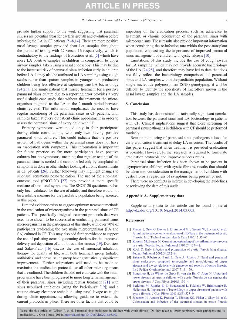
Of the 117 patients that attend CF clinics at the Royal LondonChildren's Hospital (RLCH), 54 patients (46%) provided nasallavage samples as well as routine LA samples obtained at eachclinic appointment, 36 (31%) were deemed too young to performnasal lavage sampling, and the other 27 patients (23%) refusedto participate in nasal lavage sampling (Flowchart 1). Table 1displays the characteristics of the 54 patients that suppliedparanasal samples. The median age was 11 years old (IQR 3).
3.2. Primary symptoms
Four patients complained of primary symptoms (they wereclassed as nasal dripping, snoring and paranasal sinus blockage).All other participants reported to have no paranasal sinussymptoms during clinic consultations. Two symptomatic partic-ipants, nasal dripping and snoring, had positive nasal lavagesamples, PA and SA respectively, which were treated as per theeradication protocol. These participants with reported nasaldripping and snoring did not eradicate on follow-up culture,however both have reported a reduction in symptoms withregular nasal lavage. The other two participants with a primary
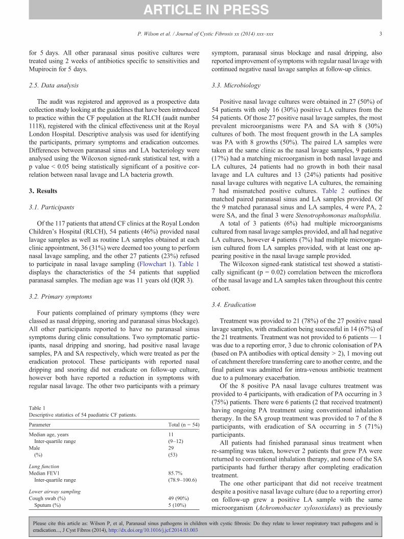
Table 1Descriptive statistics of 54 paediatric CF patients.
Parameter Total (n = 54)
Median age, yearsInter-quartile range
11(9–12)
Male(%)
29(53)
Lung functionMedian FEV1
Inter-quartile range85.7%(78.9–100.6)
Lower airway samplingCough swab (%)
Sputum (%)49 (90%)5 (10%)
Please cite this article as: Wilson P, et al, Paranasal sinus pathogens in children weradication..., J Cyst Fibros (2014), http://dx.doi.org/10.1016/j.jcf.2014.03.003
symptom, paranasal sinus blockage and nasal dripping, alsoreported improvement of symptomswith regular nasal lavage withcontinued negative nasal lavage samples at follow-up clinics.
3.3. Microbiology
Positive nasal lavage cultures were obtained in 27 (50%) of54 patients with only 16 (30%) positive LA cultures from the54 patients. Of those 27 positive nasal lavage samples, the mostprevalent microorganisms were PA and SA with 8 (30%)cultures of both. The most frequent growth in the LA sampleswas PA with 8 growths (50%). The paired LA samples weretaken at the same clinic as the nasal lavage samples, 9 patients(17%) had a matching microorganism in both nasal lavage andLA cultures, 24 patients had no growth in both their nasallavage and LA cultures and 13 (24%) patients had positivenasal lavage cultures with negative LA cultures, the remaining7 had mismatched positive cultures. Table 2 outlines thematched paired paranasal sinus and LA samples provided. Ofthe 9 matched paranasal sinus and LA samples, 4 were PA, 2were SA, and the final 3 were Stenotrophomonas maltophilia.
A total of 3 patients (6%) had multiple microorganismscultured from nasal lavage samples provided, and all had negativeLA cultures, however 4 patients (7%) had multiple microorgan-ism cultured from LA samples provided, with at least one ap-pearing positive in the nasal lavage sample provided.
The Wilcoxon signed-rank statistical test showed a statisti-cally significant (p = 0.02) correlation between the microfloraof the nasal lavage and LA samples taken throughout this centrecohort.
3.4. Eradication
Treatment was provided to 21 (78%) of the 27 positive nasallavage samples, with eradication being successful in 14 (67%) ofthe 21 treatments. Treatment was not provided to 6 patients— 1was due to a reporting error, 3 due to chronic colonisation of PA(based on PA antibodies with optical density N 2), 1 moving outof catchment therefore transferring care to another centre, and thefinal patient was admitted for intra-venous antibiotic treatmentdue to a pulmonary exacerbation.
Of the 8 positive PA nasal lavage cultures treatment wasprovided to 4 participants, with eradication of PA occurring in 3(75%) patients. There were 6 patients (2 that received treatment)having ongoing PA treatment using conventional inhalationtherapy. In the SA group treatment was provided to 7 of the 8participants, with eradication of SA occurring in 5 (71%)participants.
All patients had finished paranasal sinus treatment whenre-sampling was taken, however 2 patients that grew PA werereturned to conventional inhalation therapy, and none of the SAparticipants had further therapy after completing eradicationtreatment.
The one other participant that did not receive treatmentdespite a positive nasal lavage culture (due to a reporting error)on follow-up grew a positive LA sample with the samemicroorganism (Achromobacter xylosoxidans) as previously
ith cystic fibrosis: Do they relate to lower respiratory tract pathogens and is
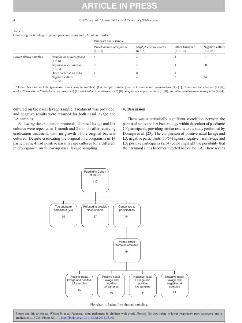
Table 2Comparing bacteriology of paired paranasal sinus and LA culture results.
Paranasal sinus sample
Pseudomonas aeruginosa(n = 8)
Staphylococcus aureus(n = 8)
Other bacteria a
(n = 12)Negative culture(n = 26)
Lower airway samples Pseudomonas aeruginosa(n = 8)
4 2 1 1
Staphylococcus aureus(n = 3)
0 2 1 0
Other bacteria a (n = 6) 1 0 4 1Negative culture(n = 37)
3 4 6 24
a Other bacteria include (paranasal sinus sample number) [LA sample number] — Achromobacter xylosoxidans (1) [1], Enterobacter cloacae (1) [0],methicillin-resistant Staphylococcus aureus (1) [1], Burkholderia multivorans (2) [0], Streptococcus pneumoniae (3) [0], and Stenotrophomonas maltophilia (4) [4].
4 P. Wilson et al. / Journal of Cystic Fibrosis xx (2014) xxx–xxx
cultured on the nasal lavage sample. Treatment was provided,and negative results were returned for both nasal lavage andLA samples.
Following the eradication protocols, all nasal lavage and LAcultures were repeated at 1 month and 3 months after receivingeradication treatment, with no growth of the original bacteriacultured. Despite eradicating the original microorganism in 14participants, 4 had positive nasal lavage cultures for a differentmicroorganism on follow-up nasal lavage sampling.
Paediatric Cohort at RLCH
117
Too young to participate (<5)
36
Refused to provide sinus sample
27
Copa
Pasamp
Positive nasal lavage and positive
LA samples
15
Positive nasal Lavage and
negative LA samples
13
Flowchart 1. Patient flow
Please cite this article as: Wilson P, et al, Paranasal sinus pathogens in children weradication..., J Cyst Fibros (2014), http://dx.doi.org/10.1016/j.jcf.2014.03.003
4. Discussion
There was a statistically significant correlation between theparanasal sinus and LA bacteriology within the cohort of paediatricCF participants, providing similar results to the study performed byDosanjh et al. [23]. The comparison of positive nasal lavage andLA negative participants (13/54) against negative nasal lavage andLA positive participants (2/54) could highlight the possibility thatthe paranasal sinus becomes infected before the LA. These results
nsented to rticipation
54
ired timed les obtained
54
Negative nasal lavage and negative LA
samples
24
Negative nasal Lavage and
positive LA samples
2
through sampling.
ith cystic fibrosis: Do they relate to lower respiratory tract pathogens and is
5P. Wilson et al. / Journal of Cystic Fibrosis xx (2014) xxx–xxx
provide further support to the work suggesting that paranasalsinuses are potential areas for bacteria growth and evolution beforeinfecting the LA in CF patients [5–8,14]. There are more positivenasal lavage samples provided than LA samples throughoutthe period of testing with 27 versus 16 respectively, which iscontradictory to the findings of Bonestroo et al. [5] which havemore LA positive samples in children in comparison to upperairway samples, taken using a nasal endoscopy. This may be dueto the increased risk of paranasal sinus infection early in childrenbefore LA. It may also be attributed to LA sampling using coughswabs rather than sputum samples in younger non-productivechildren being less effective at capturing true LA bacteriology[24,25]. The single patient that missed treatment for a positiveparanasal sinus culture due to a reporting error provides a veryuseful single case study that without the treatment the micro-organism migrated to the LA in the 2 month period betweenclinic reviews. This information emphasises the need to haveregular monitoring of the paranasal sinus in CF patients, withsamples taken at every outpatient clinic appointment in order toassess the paranasal sinus of every child with CF.
Primary symptoms were noted only in four participantsduring clinic consultations, with only two having positiveparanasal sinus cultures. This could indicate that the positivegrowth of pathogens within the paranasal sinus does not havean association with symptoms. This information is importantfor future practice as far more participants have positivecultures but no symptoms, meaning that regular testing of theparanasal sinus is needed and cannot be led only by complaints ofsymptoms as done in other studies looking at chronic rhinosinusitisin CF patients [26]. Further follow-up may highlight changes tosinonasal sensations post-eradication. The use of the sino-nasaloutcome tool (SNOT-20) [27] may provide a more specificmeasure of sino-nasal symptoms. The SNOT-20 questionnaire hasonly been validated for the use of adults, and therefore would notbe a reliable measure for the paediatric population being assessedin this paper.
Limited evidence exists to suggest optimum treatment methodsin the eradication of microorganisms in the paranasal sinus of CFpatients. The specifically designed treatment protocols that wereused have shown to be successful in eradicating paranasal sinusmicroorganisms in the participants of this study, with over 70% ofparticipants eradicating the two main microorganisms (PA andSA) cultured in CF. This may also add further evidence to supportthe use of pulsating aerosol generating devices for the improveddelivery and deposition of antibiotics to the sinuses [19]. Derosiersand Salas-Prato [16] discuss the use of sinonasal inhalationtherapy for quality of life; with both treatment group (inhaledantibiotics) and normal saline group having statistically significantimprovements. Further adaptations will need to be made tomaximise the eradication protocols for all other microorganismsthat are cultured. The children that did not eradicate with the initialprogramme have been progressed to more long term managementof their paranasal sinus, including regular treatment [21] withsinus nebulised antibiotics (using the Pari-sinus® [19]) and aroutine airway clearance technique using nasal lavage as taughtduring clinic appointments, allowing guidance to extend thecurrent protocols in place. There are other factors that could be
Please cite this article as: Wilson P, et al, Paranasal sinus pathogens in children weradication..., J Cyst Fibros (2014), http://dx.doi.org/10.1016/j.jcf.2014.03.003
impacting on the eradication process, such as adherence totreatment, or chronic colonisation of the paranasal sinus withmicroorganisms. These results also provide important informationwhen considering the re-infection rate within the post-transplantpopulation, emphasising the importance of improved paranasalsinus management of children with cystic fibrosis [10].
Limitations of this study include the use of cough swabsfor LA sampling, which may not provide accurate bacteriologyof the LA [24,25], and therefore may have led to data that doesnot fully reflect the bacteriology comparisons of paranasalsinus and LA samples within the paediatric population. Withoutsingle nucleotide polymorphism (SNP) genotyping, it will bedifficult to identify the specificity of microflora grown in thenasal lavage samples and the LA samples.
5. Conclusion
This study has demonstrated a statistically significant correla-tion between the paranasal sinus and LA bacteriology in patientswith CF. Clinical implications suggest that close screening ofparanasal sinus pathogens in children with CF should be performedroutinely.
Routine monitoring of paranasal sinus pathogens allows forearly eradication treatment to delay LA infection. The results ofthis paper suggest that when treatment is provided eradicationis possible. However, further research is required to formulateeradication protocols and improve success ratios.
Paranasal sinus infection has been shown to be present inasymptomatic children with cystic fibrosis, results that shouldbe taken into consideration in the management of children withcystic fibrosis regardless of symptoms being present or not.
There were no conflicts in interest in developing the guidelinesor reviewing the data of this audit.
Appendix A. Supplementary data
Supplementary data to this article can be found online athttp://dx.doi.org/10.1016/j.jcf.2014.03.003.
References
[1] Menzin J, Oster G, Davies L, Drummond MF, Greiner W, Lucioni C, et al.A multinational economic evaluation of rhDNase in the treatment of cysticfibrosis. Int J Technol Assess Health Care 1996;12:52–61.
[2] Konstan M, Berger M. Current understanding of the inflammatory processin cystic fibrosis. Pediatr Pulmonol 1997;24:137–42.
[3] Koch C. Early infection and progression of cystic fibrosis lung disease.Pediatr Pulmonol 2002;34:232–6.
[4] Sakano E, Ribeiro A, Barth L, Neto A, Ribeiro J. Nasal and paranasalsinus endoscopy, computed tomography and microbiology of upperairways and the correlations with genotype and severity of cystic fibrosis.Int J Pediatr Otorhinolaryngol 2007;71:41–50.
[5] Bonestroo H, de Winter-de Groot K, van der Ent C, Arets H. Upper andlower airways cultures in children with cystic fibrosis: do not neglect theupper airways. J Cyst Fibros 2010;9:130–4.
[6] Berkhout M, Rijntjes E, El Bouazzaoui L, Fokkens W, Brimicombe R,Heijerman H. Importance of bacteriology in upper airways of patients withcystic fibrosis. J Cyst Fibros 2013;12(5):525–9.
[7] Johansen H, Aanaes K, Pressler T, Nielsen KG, Fisker J, Skov M, et al.Colonisation and infection of the paranasal sinuses in cystic fibrosis
ith cystic fibrosis: Do they relate to lower respiratory tract pathogens and is
6 P. Wilson et al. / Journal of Cystic Fibrosis xx (2014) xxx–xxx
patients is accompanied by a reduced PMN response. J Cyst Fibros2012;11(6):525–31.
[8] Hansen S, Rau M, Johansen H, Ciofu O, Jelsbak L, Yang L, et al. Evolutionand diversification of Pseudomonas aeruginosa in the paranasal sinuses ofcystic fibrosis children have implications for chronic lung infection. ISME J2012;6:31–45.
[9] Vital D, Hofer M, Benden C, Holzmann D, Boehler A. Impact of sinussurgery on pseudomonal airway colonization, bronchiolitis obliteranssyndrome and survival in cystic fibrosis lung transplant recipients.Respiration 2013;86(1):25–31.
[10] Leung MK, Rachakonda L, Weill D, Hwang PH. Effects of sinus surgeryon lung transplantation outcomes in cystic fibrosis. Am J Rhinol2008;22(2):192–6.
[11] Walter S, Gudowius P, Boßhammer J, Römling U, Weißbrodt H,Schürmann W, et al. Epidemiology of chronic Pseudomonas aeruginosainfections in the airways of lung transplant recipients with cystic fibrosis.Thorax 1997;52(4):318–21.
[12] Rosin D. The sinus sourcebook. Los Angeles: Lowell House; 1998.[13] Osborn A, Leung R, Ratjen F, James A. Effects of endoscopic sinus
surgery on pulmonary function and microbial pathogens in a pediatricpopulation with cystic fibrosis. Arch Otolaryngol Head Neck Surg2011;6:542–54.
[14] Rihani J, McClay J. Impact of endoscopic sinus surgery on pulmonaryfunction in children with cystic fibrosis. Otolaryngol Head Neck Surg2011;145:108.
[15] Mainz J, Koitschev A. Management of chronic rhinosinusitis in CF. J CystFibros 2009;8:S10–4.
[16] Derosiers M, Salas-Prato M. Treatment of chronic rhinosinusitis refractoryto other treatments with topical antibiotic therapy delivered by means of alarge-particle nebulizer: results of a controlled trial. Otolaryngol HeadNeck Surg 2001;125:265–9.
[17] Taccetti G, Campana S, Festini F, Mascherini M, Döring G. Earlyeradication therapy against Pseudomonas aeruginosa in cystic fibrosispatients. Eur Respir J 2005;26:458–61.
Please cite this article as: Wilson P, et al, Paranasal sinus pathogens in children weradication..., J Cyst Fibros (2014), http://dx.doi.org/10.1016/j.jcf.2014.03.003
[18] Ratjen F, Döring G, Nikolaizik W. Effect of inhaled tobramycin on earlyPseudomonas aeruginosa colonisation in patients with cystic fibrosis.Lancet 2001;358:983–4.
[19] Möller W, Schuschnig U, Saba GK, Meyer G, Junge-Hülsing B, Keller M,et al. Pulsating aerosols for drug delivery to the sinuses in healthyvolunteers. Otolaryngol Head Neck Surg 2010;142(3):382–8.
[20] Mainz J, Naehrlich L, Schien M, Kading M, Schiller I, Mayr S, et al.Concordant genotype of upper and lower airways P aeruginosa and Saureus isolates in cystic fibrosis. Thorax 2009;64:535–40.
[21] PynnonenM,Mukerji S, KimH, AdamsM, Terrell J. Nasal saline for chronicsinonasal symptoms: a randomized controlled trial. Arch Otolaryngol HeadNeck Surg 2007;11:1115–20.
[22] Cystic Fibrosis Trust. Antibiotic treatment for cystic fibrosis. Report of theUK Cystic Fibrosis Trust Antibiotic Working Group. London: CysticFibrosis Trust; 2009 [Consensus document].
[23] Dosanjh A, Lakhani S, Elashoff D, Chin C, Hsu V, Hilman B. Acomparison of microbiologic flora of the sinuses and airway amongcystic fibrosis patients with maxillary antrostomies. Pediatr Transplant2000;4:182–5.
[24] Equi AC, Pike SE, Davies J, Bush A. Use of cough swabs in a cysticfibrosis clinic. Arch Dis Child 2001;85:438–9.
[25] Armstrong DS, Grimwood K, Carlin JB, Carzino R, Olinsky A, PhelanPD. Bronchoalveolar lavage of oropharyngeal cultures to identify lowerrespiratory pathogens in infants with cystic fibrosis. Pediatr Pulmonol1996;21:267–75.
[26] Rosbe K, Jones D, Rahbar R, Lahiri T, Auerbach A. Endoscopic sinussurgery in cystic fibrosis: do patients benefit from surgery? Int J PediatrOtorhinolaryngol 2001;61:113–9.
[27] Pynnonen M, Kim H, Terrell J. Validation of the Sino-Nasal OutcomeTest 20 (SNOT-20) domains in nonsurgical patients. Am J Rhinol Allergy2009;23:40–5.
ith cystic fibrosis: Do they relate to lower respiratory tract pathogens and is