paradoxical influence of hippocampal neurogenesis …dericbownds.net/uploaded_images/kandel.pdf ·...
TRANSCRIPT
Paradoxical influence of hippocampal neurogenesis on working memory
V. Sofroniew, Eric R. Kandel, and René Hen Michael D. Saxe, Gaël Malleret, Svetlana Vronskaya, Indira Mendez, A. Denise Garcia, Michael
doi:10.1073/pnas.0611718104 2007;104;4642-4646; originally published online Mar 5, 2007; PNAS
This information is current as of March 2007.
& ServicesOnline Information
www.pnas.org/cgi/content/full/104/11/4642etc., can be found at: High-resolution figures, a citation map, links to PubMed and Google Scholar,
Supplementary Material www.pnas.org/cgi/content/full/0611718104/DC1
Supplementary material can be found at:
References www.pnas.org/cgi/content/full/104/11/4642#BIBL
This article cites 27 articles, 6 of which you can access for free at:
www.pnas.org/cgi/content/full/104/11/4642#otherarticlesThis article has been cited by other articles:
E-mail Alerts. click hereat the top right corner of the article or
Receive free email alerts when new articles cite this article - sign up in the box
Rights & Permissions www.pnas.org/misc/rightperm.shtml
To reproduce this article in part (figures, tables) or in entirety, see:
Reprints www.pnas.org/misc/reprints.shtml
To order reprints, see:
Notes:
Paradoxical influence of hippocampal neurogenesison working memoryMichael D. Saxe*, Gael Malleret*, Svetlana Vronskaya*, Indira Mendez*, A. Denise Garcia†, Michael V. Sofroniew†,Eric R. Kandel*‡§, and Rene Hen*§
*Center for Neurobiology and Behavior and ‡Howard Hughes Medical Institute, Columbia University, 722 West 168th Street, New York, NY 10032;and †Department of Neurobiology and Brain Research Institute, University of California, Los Angeles, CA 90095
Contributed by Eric R. Kandel, January 3, 2007 (sent for review December 18, 2006)
To explore the function of adult hippocampal neurogenesis, weablated cell proliferation by using two independent and comple-mentary methods: (i) a focal hippocampal irradiation and (ii) aninducible and reversible genetic elimination of neural progenitorcells. Previous studies using these methods found a weakening ofcontextual fear conditioning but no change in spatial referencememory, suggesting a supportive role for neurogenesis in some,but not all, hippocampal-dependent memory tasks. In the presentstudy, we examined hippocampal-dependent and -independentworking memory using different radial maze tasks. Surprisingly,ablating neurogenesis caused an improvement of hippocampal-dependent working memory when repetitive information waspresented in a single day. These findings suggest that adult-borncells in the dentate gyrus have different, and in some cases,opposite roles in distinct types of memory.
hippocampus � irradiation � radial maze � interference
The fact that most mammals, including humans, continue toproduce new neurons in the hippocampus throughout adult-
hood has led to the suggestion that neurogenesis may serve animportant role in hippocampal-dependent memory processes(1–3). Consistent with this possibility, studies of adult rodents inwhich neurogenesis has been reduced, by systemic or whole-brain treatments or as a result of aging, suggest the involvementof these new neurons in some hippocampal-dependent tasks(4–7). Suppression of neurogenesis with the antimitotic agentMAM was shown to impair trace eyeblink and fear conditioning,whereas more restricted ablation strategies using irradiation orgenetically targeted blockade of neurogenesis resulted in aweakening of contextual fear conditioning and had no effect inspatial learning (5, 8). Notably, all reported changes in hip-pocampal-dependent memory tasks after loss of adult neuro-genesis have indicated either no role or a supportive role for thisprocess in memory function. However, it is still unknownwhether all types of hippocampal-dependent memory are simi-larly affected by the addition or presence of new neurons. Thisquestion is particularly relevant because the hippocampus isinvolved in a wide range of memory tasks.
For example, only a limited number of studies have begun toexamine the role of adult hippocampal neurogenesis in tests ofworking memory, a form of short-term memory that involvesboth the hippocampus and the prefrontal cortex (9, 10). In adultrats, performance in a water maze task that utilizes short-termnon-spatial memory was impaired after disruption of neurogen-esis by whole brain irradiation (7). In the present study, wesought to clarify the contribution of neurogenesis to workingmemory by using two different strategies that have been shownto eliminate new neurons from the adult hippocampus. The firstutilizes a focal x-irradiation procedure that results in a perma-nent loss of neurogenesis within the hippocampus of adult micebut spares neurogenesis in the subventricular zone and theolfactory bulb (11). The second is a complementary geneticapproach in which dividing glial acidic fibrillary protein(GFAP)-positive cells, known to be progenitors of new neurons,
are selectively eliminated by administering the drug ganciclovir(GCV) (12).
After both ablation procedures, we found a surprising im-provement of working memory performance, but only in taskswhere mice were required to discriminate highly similar cuespresented closely in time (within a single session). Also, thiseffect was limited to trials in which a long temporal delay (30�sec) was presented, consistent with previous observations that,in rodents, the hippocampus becomes crucial to working mem-ory only when the delay is �10 sec (13, 14). Because we havepreviously found that contextual fear conditioning is reducedand that spatial reference memory is unaffected after ablation ofneurogenesis using these same methods (8), our present studysuggests that the new neurons may have more than one functionand that their specific role in tests of hippocampal-dependentmemory may differ depending on the nature and cognitivedemands of the task.
ResultsOur experimental design used two independent and comple-mentary strategies to suppress neurogenesis in the hippocampusof adult mice. The first method targets the hippocampus withlow-dose x-irradiation using a small window in a protective leadshield (Fig. 1A Top) that blocks exposure to the body andremaining brain regions. This procedure produces a completeand lasting ablation of neurogenesis in the dentate gyrus, asassessed by doublecortin immunoreactivity (Fig. 1 A Middle andBottom). Mice were allowed to recover for 3 months beforebehavioral testing to avoid the transient inflammatory effects ofirradiation, such as microglial activation. We have previouslyshown that the number of activated microglia returns to basallevels by this time (15).
The second strategy used a transgenic mouse line in whichGFAP promoter drives the expression of herpes virus thymidinekinase (TK). Exogenous delivery of the pro-drug GCV inducesthe selective death of dividing GFAP-positive cells, whereas
Author contributions: M.D.S. and G.M. contributed equally to this work; M.D.S. and G.M.designed research; M.D.S., G.M., S.V., I.M., and A.D.G. performed research; M.D.S. andM.V.S. contributed new reagents/analytic tools; M.D.S., G.M., S.V., and I.M. analyzed data;and M.D.S., G.M., E.R.K., and R.H. wrote the paper.
Conflict of interest statement: E.R.K. declares a conflict of interest (such as defined by PNASpolicy). E.R.K. is one of four founders of Memory Pharmaceuticals and is Chairman of itsScientific Advisory Board. Memory Pharmaceuticals is concerned with developing drugs forage-related memory loss. Some of these drugs are also potentially useful in depression andschizophrenia. E.R.K.’s own laboratory is not involved in developing these drugs. E.R.K. isalso a consultant for BrainCells, Inc., which works on neurogenesis, an area in which he isnot directly involved.
Freely available online through the PNAS open access option.
Abbreviations: GCV, ganciclovir; GFAP, glial acidic fibrillary protein; HML, high-memoryload; LML, low-memory load; HI, high intertrial interference; LI, limited intertrial interfer-ence; NI, no intertrial interference; TK, thymidine kinase.
§To whom correspondence may be addressed. E-mail: [email protected] or [email protected].
This article contains supporting information online at www.pnas.org/cgi/content/full/0611718104/DC1.
© 2007 by The National Academy of Sciences of the USA
4642–4646 � PNAS � March 13, 2007 � vol. 104 � no. 11 www.pnas.org�cgi�doi�10.1073�pnas.0611718104
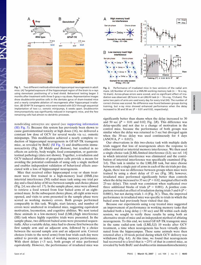
nondividing astrocytes are spared (see supporting information(SI) Fig. 5). Because this system has previously been shown tocause gastrointestinal toxicity at high doses (16), we delivered aconstant low dose of GCV for several weeks via s.c. osmoticminipumps. This modification achieved a nearly complete re-duction of hippocampal neurogenesis in GFAP-TK transgenicmice, as revealed by BrdU (SI Fig. 5) and doublecortin immu-noreactivity (Fig. 1B Middle and Bottom), but resulted in noeffects on activity, body weight, food consumption, or gastroin-testinal pathology (data not shown). Together, x-irradiation andGCV-induced ablation of progenitor cells provide a means foravoiding the potential confounds of using only a single methodand enables independent validation of behavioral effects asso-ciated with a loss of hippocampal neurogenesis.
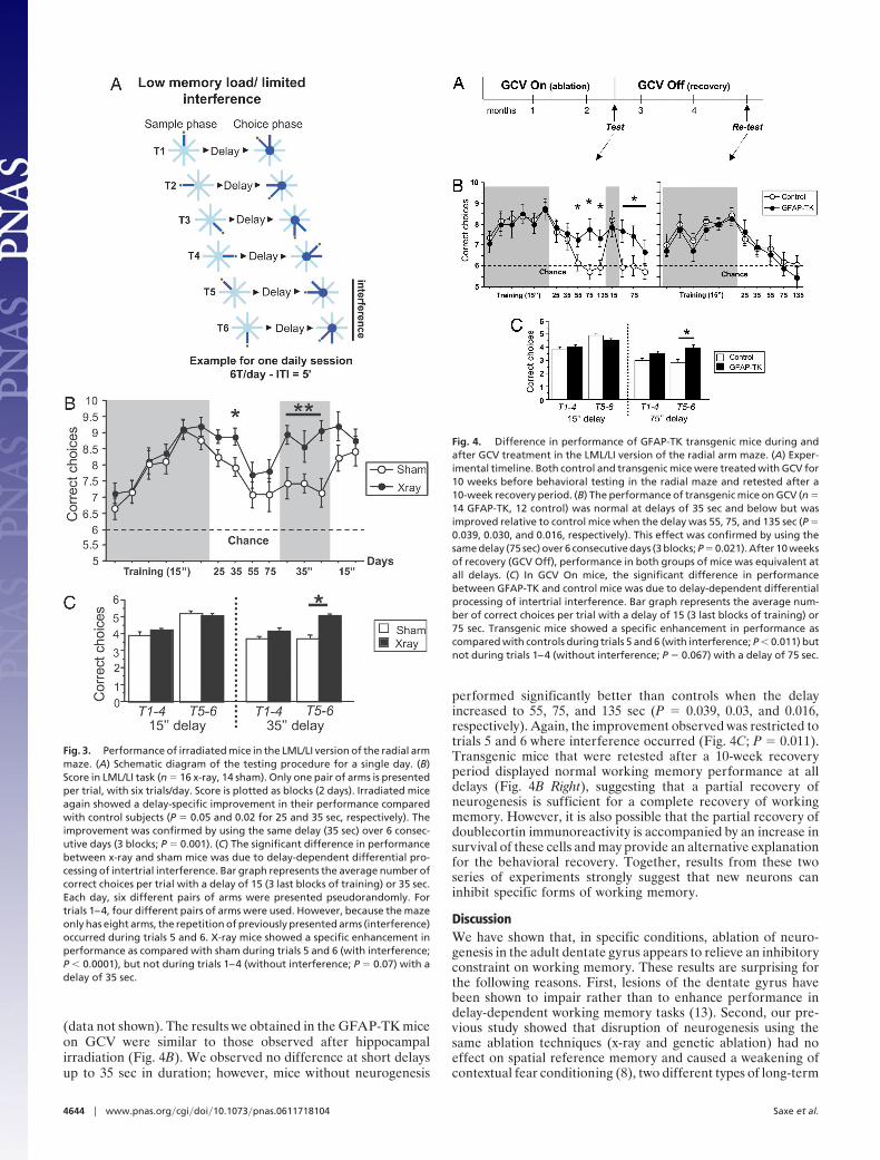
Mice that received either hippocampal x-ray or sham treat-ment were first trained in a high-memory load (HML)/nointertrial interference (NI) radial maze task using one trial perday and a fixed delay of 60 sec between sample and choice phases(Fig. 2A; see also ref. 17). In the sample phase, mice were allowedto retrieve a food reward from four baited arms of an eight-armed maze. In the subsequent choice phase, all eight arms wereopened, and visits to arms presented in the sample phase werescored as working memory errors. Both groups performedcomparably in this task. Weight, start latency, and number oferrors were unaltered in irradiated mice, suggesting equivalentmotivation, locomotion, and cognitive abilities. We next testedthese animals in a low-memory load (LML)/high interference(HI) task where highly repetitive trials were presented. In thesample phase, two different baited arms were presented sequen-tially. After a variable delay, mice had to choose first between thefirst sample arm and an adjacent arm, followed by a choicebetween the second sample arm and an adjacent arm. Correctchoices (visits to the novel arms) from four trials each day wereadded to provide an index of working memory performance.With short delays (�5 sec), both groups of mice performedequivalently. However, the performance of irradiated mice was
significantly better than shams when the delay increased to 30and 50 sec (P � 0.01 and 0.02; Fig. 2B). This difference wasdelay-specific and not due to a change of motivation in thecontrol mice, because the performance of both groups wassimilar when the delay was returned to 5 sec but diverged againwhen the 30-sec delay was used continuously for 4 days(ANOVA, P � 0.015).
Our observations in this two-choice task with multiple dailytrials suggest that loss of neurogenesis alters the response toeither intratrial or intertrial memory interference. We then useda single-choice task [LML/limited interference (LI); see ref. 14]in which intratrial interference was eliminated and the contri-bution of intertrial interference was specifically examined (Fig.3A). This task is similar to the LML/HI task, but mice choosebetween only a single pair of arms in each of the six trials per day.Again, there was no difference between groups when mice weretrained by using a short delay of 15 sec (Fig. 3B); however,irradiated mice performed significantly better than controlswhen the delay increased to 35 sec (P � 0.02, marginal effect with25-sec delay). This result was consistent when replicated overthree additional blocks of trials (P � 0.001). A posthoc com-parison revealed an effect of irradiation during trials 5 and 6 (P �0.0001), but not during trials 1–4 (Fig. 3C). Thus, the improvedperformance in irradiated mice was specific to trials in which thebaited arms had previously been visited that day.
Because our experiments using x-ray treated mice suggestedan improvement of performance in working memory tasks thatincluded both a long delay (�30 sec) and HI within each dailysession, we sought to verify these results by using both analternative strain of mice and an independent method of ablatingneurogenesis. To this end, we tested GFAP-TK transgenic micein the same radial-arm task (LML/LI) 10 weeks after GCVtreatment, a time when neurogenesis has been virtually elimi-nated from the hippocampus. These same animals were thenretested after a 10-week period during which GCV delivery waswithdrawn (Fig. 4A). At this time, we found that neurogenesishad recovered to a level that is �25% of that in control mice, asrevealed by both BrdU and doublecortin immunohistochemistry
Fig. 1. Two different methods eliminate hippocampal neurogenesis in adultmice. (A) Targeted exposure of the hippocampal region of the brain to x-raysusing stereotaxic positioning of a lead shield. Behavioral testing began 3months after treatment with three 5-gray x-ray doses. Representative imagesshow doublecortin-positive cells in the dentate gyrus of sham-treated miceand a nearly complete ablation of neurogenesis after hippocampal irradia-tion. (B) GFAP-TK transgenic mice were treated with GCV through sequentialimplantation of two s.c. osmotic minipumps, 6 weeks apart. Doublecortinimmunoreactivity was significantly reduced in transgenic mice, and the fewremaining cells had almost no dendritic processes.
Fig. 2. Performance of irradiated mice in two versions of the radial armmaze. (A) Number of errors in a HML/NI working memory task (n � 16 x-ray,16 sham). Across-phase errors were scored, and no significant effect of irra-diation was observed. (B) Score in an LML/HI task (n � 16 x-ray, 13 sham). Thesame two pairs of arms are used every day for every trial (HI). The number ofcorrect choices was scored. No difference was found between groups duringtraining, but x-ray mice showed enhanced performance when the delayincreased to 30 and 50 sec (P � 0.01 and 0.02, respectively).
Saxe et al. PNAS � March 13, 2007 � vol. 104 � no. 11 � 4643
NEU
ROSC
IEN
CE
(data not shown). The results we obtained in the GFAP-TK miceon GCV were similar to those observed after hippocampalirradiation (Fig. 4B). We observed no difference at short delaysup to 35 sec in duration; however, mice without neurogenesis
performed significantly better than controls when the delayincreased to 55, 75, and 135 sec (P � 0.039, 0.03, and 0.016,respectively). Again, the improvement observed was restricted totrials 5 and 6 where interference occurred (Fig. 4C; P � 0.011).Transgenic mice that were retested after a 10-week recoveryperiod displayed normal working memory performance at alldelays (Fig. 4B Right), suggesting that a partial recovery ofneurogenesis is sufficient for a complete recovery of workingmemory. However, it is also possible that the partial recovery ofdoublecortin immunoreactivity is accompanied by an increase insurvival of these cells and may provide an alternative explanationfor the behavioral recovery. Together, results from these twoseries of experiments strongly suggest that new neurons caninhibit specific forms of working memory.
DiscussionWe have shown that, in specific conditions, ablation of neuro-genesis in the adult dentate gyrus appears to relieve an inhibitoryconstraint on working memory. These results are surprising forthe following reasons. First, lesions of the dentate gyrus havebeen shown to impair rather than to enhance performance indelay-dependent working memory tasks (13). Second, our pre-vious study showed that disruption of neurogenesis using thesame ablation techniques (x-ray and genetic ablation) had noeffect on spatial reference memory and caused a weakening ofcontextual fear conditioning (8), two different types of long-term
Fig. 3. Performance of irradiated mice in the LML/LI version of the radial armmaze. (A) Schematic diagram of the testing procedure for a single day. (B)Score in LML/LI task (n � 16 x-ray, 14 sham). Only one pair of arms is presentedper trial, with six trials/day. Score is plotted as blocks (2 days). Irradiated miceagain showed a delay-specific improvement in their performance comparedwith control subjects (P � 0.05 and 0.02 for 25 and 35 sec, respectively). Theimprovement was confirmed by using the same delay (35 sec) over 6 consec-utive days (3 blocks; P � 0.001). (C) The significant difference in performancebetween x-ray and sham mice was due to delay-dependent differential pro-cessing of intertrial interference. Bar graph represents the average number ofcorrect choices per trial with a delay of 15 (3 last blocks of training) or 35 sec.Each day, six different pairs of arms were presented pseudorandomly. Fortrials 1–4, four different pairs of arms were used. However, because the mazeonly has eight arms, the repetition of previously presented arms (interference)occurred during trials 5 and 6. X-ray mice showed a specific enhancement inperformance as compared with sham during trials 5 and 6 (with interference;P � 0.0001), but not during trials 1–4 (without interference; P � 0.07) with adelay of 35 sec.
Fig. 4. Difference in performance of GFAP-TK transgenic mice during andafter GCV treatment in the LML/LI version of the radial arm maze. (A) Exper-imental timeline. Both control and transgenic mice were treated with GCV for10 weeks before behavioral testing in the radial maze and retested after a10-week recovery period. (B) The performance of transgenic mice on GCV (n �14 GFAP-TK, 12 control) was normal at delays of 35 sec and below but wasimproved relative to control mice when the delay was 55, 75, and 135 sec (P �0.039, 0.030, and 0.016, respectively). This effect was confirmed by using thesame delay (75 sec) over 6 consecutive days (3 blocks; P � 0.021). After 10 weeksof recovery (GCV Off), performance in both groups of mice was equivalent atall delays. (C) In GCV On mice, the significant difference in performancebetween GFAP-TK and control mice was due to delay-dependent differentialprocessing of intertrial interference. Bar graph represents the average num-ber of correct choices per trial with a delay of 15 (3 last blocks of training) or75 sec. Transgenic mice showed a specific enhancement in performance ascompared with controls during trials 5 and 6 (with interference; P � 0.011) butnot during trials 1–4 (without interference; P � 0.067) with a delay of 75 sec.
4644 � www.pnas.org�cgi�doi�10.1073�pnas.0611718104 Saxe et al.
memory that require the hippocampus. Similar results have beenreported after exposure of adult rats to gamma irradiation (7).This same study also reported an impairment of performance ina different working memory task (delayed-nonmatch-to-sample), contrasting with our results. However, it is unclearwhether the impairment observed was due specifically to inhi-bition of hippocampal neurogenesis as the irradiation procedureaffected the whole brain. In addition, there is no evidence forhippocampal dependency in this task. Thus, neurogenesis doesnot appear to have a unitary function in memory. Instead, theremoval of this small cell population appears to differentiallyinfluence diverse hippocampal-dependent behaviors.
Ablation of neurogenesis improved performance in workingmemory tasks, but only in situations where animals needed toignore or forget conflicting nonrelevant information from pre-vious trials (18). The altered performance in neurogenesis-deficient animals appears to indicate a lack of sensitivity tomemory interference. During multiple-trial radial maze tasks,interference-dependent reduction of performance was evidentin control animals, but only when the same pair of arms wasrepeated within a session and a long delay was used. At least twodifferent explanations for the observed enhancement of workingmemory performance in neurogenesis-deficient animals arepossible. The first is that blockade of neurogenesis reducesshort-term memory capacity by eliminating cells that are in-volved in the rapid encoding of memory traces, such as thoseformed during the sample phase of the radial arm maze task, thatrequire only a brief exposure to spatial configurations. Becauseworking memory performance was normal in the HML/NI task,where memory load is high but must only be stored for a periodof 60 sec, this interpretation suggests that defective retention isobserved only when memory traces are maintained for severalminutes. Indeed, an effect of ablation in the additional two taskswas only found when there was memory interference from trialsthat took place �30 min earlier. Thus, the enhancement ofperformance in later trials may be due to a lack of memory forearlier trials. However, in opposition to this argument areprevious findings that short-term memory in irradiated mice wasnormal in the Y-maze, a spatial task that used a 30-min intertrialdelay (8). An alternative explanation is that removal of neuro-genesis does not alter short-term memory in the radial maze butreduces the effects of interference by reducing the amount ofoverlap between the sets of neurons that represent spatialinformation during distinct trials. Central to this interpretationis the notion that the hippocampus, and the dentate gyrus inparticular, provides a means for separately encoding informationthat is highly similar but distinct in temporal or associativerelevance, a function referred to as pattern separation or or-thogonalization (19–21).
The fact that opposite effects on working memory were foundafter eliminating neurogenesis than after more complete lesionsof the dentate gyrus suggests that young dentate granule cellshave a different function than mature granule cells (19, 22).Recent computational models have suggested that the additionof new neurons to the stable network of the adult hippocampusmay impact memory processes such as information encoding,storage, or retrieval adversely. This could result from the re-placement of existing synapses by new ones (21, 23). Moreover,because young neurons are more excitable than mature granulecells (8, 24, 25), their response to related but distinct stimuli, suchas repetitive spatial representations, may contribute to memoryinterference or reduced discrimination but, at the same time,enhance encoding of highly distinct spatial information (21, 26).Indeed, our previous finding that ablation of neurogenesisimpairs contextual fear conditioning, where distinct and non-overlapping contextual cues must be discriminated, is consistentwith this hypothesis (8).
Irrespective of the mechanisms involved, our results suggestthat young neurons can have a negative influence on specificforms of working memory. Therefore, strategies aimed at stim-ulating hippocampal neurogenesis to elicit antidepressant orprocognitive effects will need to strike a fine balance betweenrestoring function and avoiding the potential negative conse-quences of an excess of neurogenesis.
Materials and MethodsAnimals. For all irradiation experiments, 10-week-old adult male129Sv/Ev mice were purchased from Taconic Farms (German-town, NY). Animals were maintained on a 12/12-h light/darkcycle throughout the course of the experiment. Behavioraltesting began 3 months after irradiation or sham treatment, andall tests were performed during the light phase. GFAP-TKtransgenic mice (line 7.1) were generated as described (16, 27).We transferred the GFAP-TK transgene onto a C57/BL6-BALB/c mixed background and used 12- to 20-week-old malelittermates derived from heterozygote crossings. Mice werehoused four or five per cage in a 12-h (06:00–18:00) light–darkcolony room at 22°C with freely available water. The proceduresdescribed herein were conducted in accordance with NationalInstitutes of Health regulations and approved by the InstitutionalAnimal Care and Use Committees of Columbia University andthe New York State Psychiatric Institute.
Drugs. GCV (Roche, Indianapolis, IN) was dissolved in sterilesaline at a concentration of 25 mg/ml and delivered throughosmotic minipumps (Alzet, Cupertino, CA) implanted s.c. underanesthesia. An average dose of 10 mg/kg per day was deliveredover a period of 10 weeks. Two pumps were implanted sequen-tially, lasting 4 weeks each, with 2 weeks in between implanta-tions. Control mice were also implanted with minipumps con-taining GCV.
Irradiation Procedure. The irradiation procedure was performedas described (11).
Histology and Stereology. To assess the effect of the irradiation orGCV treatments on the number of BrdU or doublecortin-positive cells, mice were deeply anesthetized with ketamine/xylazine (100 and 7 mg/kg, respectively), then transcardiallyperfused (cold saline, followed by 4% cold paraformaldehyde/0.1 M phosphate buffer), and brains were collected for immu-nohistochemistry. All brains were postfixed overnight in 4%paraformaldehyde at 4°C, then cryoprotected in 30% sucroseand stored at 4°C. Serial sections (35 �m) were cut through theentire hippocampus (corresponding to plates 41–61 of Franklinand Paxinos Atlas, 1997) on a cryostat, and stored in PBS with0.1% NaN3.
Sections were slide-mounted, and procedures for doublecortinconsisted of the following steps: 1 h incubation in 0.1 M TBS with0.5% Triton X-100 (Tx) and 10% normal donkey serum (NDS),followed by goat anti-doublecortin (1:3,500; Santa Cruz Bio-technology, Santa Cruz, CA) primary antibody in TBS/Tx for24 h at 4°C. The secondary antibody was biotinylated donkeyanti-goat (1:500) in TBS/NDS, followed by amplification usingan avidin–biotin complex, both for 1 h at room temperature.Sections were developed by using DAB, and bright field imageswere taken with an Axioplan-2 upright microscope (Zeiss,Thornwood, NY). The procedure for BrdU immunolabeling hasbeen described (11).
Double-labeling of GFAP and TK was done as described (12).Stereological quantification of labeled cells was performed byusing a Zeiss Axioplan-2 microscope and a CCD camera.Digitized images were collected and analyzed with Stereo In-vestigator software (Microbrightfield, Williston, VT). Using theoptical fractionator, the total number of GFAP/TK double-
Saxe et al. PNAS � March 13, 2007 � vol. 104 � no. 11 � 4645
NEU
ROSC
IEN
CE
labeled cells in the molecular layer of the dorsal dentate gyruswas estimated in transgenic animals given saline or GCV (n �3 and 5, respectively) by an investigator blind to the treatmentstatus. Both hemispheres were examined in every 12th sectionthroughout the entire dentate gyru. The molecular layer wasdefined as the region directly above the granule cell layer of thedentate gyrus and bounded dorsally by the hippocampal fissure.Contours were traced at 10� and cells were counted at 40� byusing DIC optics.
Eight-Arm Radial Maze. Habituation. Food-deprived males (85% ofad libitum weight) were habituated for 10 days to retrieve foodpellets in wells at the end of the eight baited arms of a radialmaze. The mice used distal visual cues located on the wallssurrounding the maze for spatial orientation.HML/NI task. This task was performed as described (17). To limitintertrial interference, the mice performed one trial per day,consisting of a sample and a test phase. During the sample phase,mice were allowed to visit four baited arms (HML) chosenrandomly each day. Mice were returned to the central platformfor a 60-sec delay before the test phase, where all eight arms wereopen, but only the previously blocked arms contained food. Amaximum of 5 min was allowed to retrieve the four remainingpellets. A visit to any arm from the sample phase was countedas a working memory error (Across phase error). Latency to firstentry, time to perform the task, weight and food regimen, rankof the first error, and ‘‘within phase error’’ were also recorded.LML/HI task. Mice were submitted to four trials per day, eachconsisting of a sample and a choice phase. In the sample phase,mice were first allowed to enter one randomly chosen baited armof a pair (pair-1), followed by a second baited arm from the
opposite pair (pair-2). Mice then returned to the platform for ashort delay of 1 sec (Training phase). During the choice phase,both pair-1 arms were opened and, after one arm was visited,both pair-2 arms were opened, and a second choice was made(two-choice task). Correct choices were visits to the two armsthat were blocked during the sample phase (max score for onetrial � 2; max score/day � 8). The same two pairs of arms wereused each day and trial (HI). After training, the delay betweensample and choice phase increased gradually to a maximum of70 sec.LML/LI task. This procedure was performed as described [see ref.13]. Mice were submitted to six trials per day. One pair of armswas used in each trial (one-choice task), but the procedure wassimilar to the LML/HI task (sample–delay–choice phase). Toavoid a postural mediation strategy (18), mice were removedfrom the maze after the sample phase and placed in a box for adelay of 3 sec (during training), then returned to the maze for thechoice phase (to remove and replace the mouse in the maze took�15 sec). After training, the time spent between the sample andchoice phase increased gradually to 75 sec. During this task, adifferent pair of arms was used for each trial (LI). However,because the maze only has eight arms, the repetition of previ-ously presented arms (interference) occurred during trials 5 and6. The number of correct choices was averaged per day. Thisnumber was also totaled per trial across days for a posthocanalysis.
Data Analysis. For all statistical analyses, ANOVAs were per-formed by using irradiation or genotype as a main factor. In theradial maze tasks, session, days, or trials were analyzed as mainfactors. Where ANOVA revealed a significant interaction be-tween factors, a posthoc analysis was performed.
1. Kempermann G (2002) J Neurosci 22:635–638.2. Gould E, Tanapat P, Hastings NB, Shors TJ (1999) Trends Cognit Sci
3:186–192.3. Leuner B, Gould E, Shors TJ (2006) Hippocampus 16:216–224.4. Raber J, Rola R, LeFevour A, Morhardt D, Curley J, Mizumatsu S, Vanden-
Berg SR, Fike JR (2004) Radiat Res 162:39–47.5. Shors TJ, Townsend DA, Zhao M, Kozorovitskiy Y, Gould E (2002) Hip-
pocampus 12:578–584.6. Snyder JS, Hong NS, McDonald RJ, Wojtowicz JM (2005) Neuroscience
130:843–852.7. Winocur G, Wojtowicz JM, Sekeres M, Snyder JS, Wang S (2006) Hippocampus
16:296–304.8. Saxe MD, Battaglia F, Wang JW, Malleret G, David DJ, Monckton JE, Garcia
AD, Sofroniew MV, Kandel ER, Santarelli L, et al. (2006) Proc Natl Acad SciUSA 103:17501–17506.
9. Jones MW (2002) Curr Mol Med 2:639–647.10. Wall PM, Messier C (2001) Behav Brain Res 127:99–117.11. Santarelli L, Saxe M, Gross C, Surget A, Battaglia F, Dulawa S, Weisstaub N,
Lee J, Duman R, Arancio O, et al. (2003) Science 301:805–809.
12. Garcia AD, Doan NB, Imura T, Bush TG, Sofroniew MV (2004) Nat Neurosci7:1233–1241.
13. Lee I, Kesner RP (2003) J Neurosci 23:1517–1523.14. Lee I, Kesner RP (2003) Behav Neurosci 117:1044–1053.15. Meshi D, Drew MR, Saxe M, Ansorge MS, David D, Santarelli L, Malapani
C, Moore H, Hen R (2006) Nat Neurosci 9:729–731.16. Bush TG, Savidge TC, Freeman TC, Cox HJ, Campbell EA, Mucke L, Johnson
MH, Sofroniew MV (1998) Cell 93:189–201.17. Floresco SB, Seamans JK, Phillips AG (1997) J Neurosci 17:1880–1890.18. Dudchenko PA (2004) Neurosci Biobehav Rev 28:699–709.19. Kesner RP, Lee I, Gilbert P (2004) Rev Neurosci 15:333–351.20. McClelland JL, Goddard NH (1996) Hippocampus 6:654–665.21. Becker S (2005) Hippocampus 15:722–738.22. Jarrard LE (1993) Behav Neurol Biol 60:9–26.23. Meltzer LA, Yabaluri R, Deisseroth K (2005) Trends Neurosci 28:653–660.24. Doetsch F, Hen R (2005) Curr Opin Neurobiol 15:121–128.25. Snyder JS, Kee N, Wojtowicz JM (2001) J Neurophysiol 85:2423–2431.26. Manev R, Manev H (2005) Med Hypotheses 64:114–117.27. Johnson WB, Ruppe MD, Rockenstein EM, Price J, Sarthy VP, Verderber LC,
Mucke L (1995) Glia 13:174–184.
4646 � www.pnas.org�cgi�doi�10.1073�pnas.0611718104 Saxe et al.