p aclitaxel-loaded plga nanoparticles: preparation, …lgouveia/sau_fcf2008/fonseca2002.pdf · p...
TRANSCRIPT
Journal of Controlled Release 83 (2002) 273–286www.elsevier.com/ locate/ jconrel
P aclitaxel-loaded PLGA nanoparticles: preparation,physicochemical characterization and in vitro anti-tumoral
activitya a,b , a,b*˜´ ´Cristina Fonseca , Sergio Simoes , Rogerio Gaspar
aCenter for Neuroscience and Cell Biology of Coimbra, University of Coimbra, 3000 Coimbra, PortugalbLaboratory of Pharmaceutical Technology, Faculty of Pharmacy, University of Coimbra, 3000 Coimbra, Portugal
Received 4 March 2002; accepted 30 July 2002
Abstract
The main objective of this study was to develop a polymeric drug delivery system for paclitaxel, intended to beintravenously administered, capable of improving the therapeutic index of the drug and devoid of the adverse effects of
Cremophor EL. To achieve this goal paclitaxel (Ptx)-loaded poly(lactic-co-glycolic acid) (PLGA) nanoparticles (Ptx-PLGA-Nps) were prepared by the interfacial deposition method. The influence of different experimental parameters on theincorporation efficiency of paclitaxel in the nanoparticles was evaluated. Our results demonstrate that the incorporationefficiency of paclitaxel in nanoparticles was mostly affected by the method of preparation of the organic phase and also bythe organic phase/aqueous phase ratio. Our data indicate that the methodology of preparation allowed the formation ofspherical nanometric (,200 nm), homogeneous and negatively charged particles which are suitable for intravenousadministration. The release behaviour of paclitaxel from the developed Nps exhibited a biphasic pattern characterised by aninitial fast release during the first 24 h, followed by a slower and continuous release. The in vitro anti-tumoral activity ofPtx-PLGA-Nps developed in this work was assessed using a human small cell lung cancer cell line (NCI-H69 SCLC) and
compared to the in vitro anti-tumoral activity of the commercial formulation Taxol . The influence of Cremophor EL oncell viability was also investigated. Exposure of NCI-H69 cells to 25mg/ml Taxol resulted in a steep decrease in cell
viability. Our results demonstrate that incorporation of Ptx in nanoparticles strongly enhances the cytotoxic effect of the drugas compared to Taxol , this effect being more relevant for prolonged incubation times.
2002 Elsevier Science B.V. All rights reserved.
Keywords: Nanoparticles; Poly(lactide-co-glycolide acid); Paclitaxel; Nanoprecipitation; Anti-tumoral activity
1 . Introduction activity against a variety of solid tumors, includingbreast cancer, advanced ovarian carcinoma, lung
Paclitaxel has been shown to exhibit a significant cancer, head and neck carcinomas, and acuteleukemias [1–4]. However, the success of its clinicalapplication is mainly limited by its low therapeutic*Corresponding author. Tel.:1351-239-859-927; fax:1351-index and low solubility in water as well as in many239-827-126.
˜E-mail address: [email protected](S. Simoes). other pharmaceutical solvents acceptable for in-
0168-3659/02/$ – see front matter 2002 Elsevier Science B.V. All rights reserved.PI I : S0168-3659( 02 )00212-2
274 C. Fonseca et al. / Journal of Controlled Release 83 (2002) 273–286
travascular (i.v) administration [5]. Presently, the patibility and biodegradation of the polyesters makesonly available formulation for clinical use (Taxol ) them suitable candidates for pharmaceutical purposes
consists of a solution of paclitaxel (6 mg/ml) in a [33]. Recently, some work on the use of PLGAvehicle composed of Cremophor EL and dehy- nanoparticles as carriers for paclitaxel has been
drated alcohol at a 50:50 (v/v) ratio. This vehicle has published [19,20,23]. Nevertheless, to our knowledgebeen associated with severe hypersensitivity re- there are no reports on the literature regarding theactions and has shown incompatibility with common incorporation of paclitaxel into PLGA nanoparticlesPVC intravenous administration sets [6–10]. In order using the nanoprecipitation method. Besides being
to eliminate the Cremophor based vehicle and in an the simplest method for Nps preparation, involvingattempt to increase the therapeutic efficacy of the only one step for dispersion of the non-toxic organicdrug, alternative dosage forms have been suggested, phase in the aqueous phase, thus avoiding anyincluding parenteral emulsions [11,12], liposomes purification procedure, it provides high yields of[13–15], nanoparticles [16–20] and microspheres encapsulation of hydrophobic compounds, and the[21–24]. formation of particles exhibiting adequate features
Among the new drug delivery systems, polymeric for i.v. administration. The resulting formulationsnanoparticles have been considered as promising were extensively characterized regarding their size,carriers for anticancer agents. In fact, it has been morphology and charge. In vitro drug release studiesdemonstrated that a significant improvement in drug were also performed using the most promisingspecificity of action can be reached upon its incorpo- formulation developed in this work. Finally, in vitroration into nanoparticles, this effect being mainly anti-tumoral activity of paclitaxel incorporated in theattributed to changes in tissue distribution and phar- nanoparticles was evaluated using a human small cellmacokinetics [25,26]. These modifications may con- lung cancer cell line (NCI-H69).sequently result in a reduction in the side-effects andtoxicity of the drug and in an increase in itstherapeutic efficacy. Furthermore, it has been demon- 2 . Materials and methodsstrated that nanoparticles can escape from the vas-culature through the leaky endothelial tissue that 2 .1. Materialssurrounds the tumor and thus accumulate in certainsolid tumors [27,28]. More recently, it was also Paclitaxel was a gift from Bristol Myers Squibbreported that nanoparticles can overcome the multi- (Portugal). PLGA copolymers (50/50; Resomer RGdrug resistance phenotype mediated by glycoprotein- 502H, MW 6000 and RG 502, MW 14 500) and theP leading to an increase in drug content inside the 75/25 PLGA copolymer (Resomer RG 755, MWneoplastic cells [29]. This finding is of great impor- 63 600) were purchased from Boheringer Ingelheim.
tance for the particular case of paclitaxel since Acetone pro analisi was supplied by Merck. Polox-acquired resistance to the drug has already been amer 188 (Symperonic F68) was obtained from ICI
reported [30]. Other important advantages associated (ICH, France). Dichloromethane (DCM), acetonitrilewith the use of nanoparticles include the ease of their and phosphoric acid (HPLC grade) were purchasedpreparation with well-defined biodegradable poly- from Merck. The tetrazolium dye 3-(4,5-di-mers (ex: PLGA) and their high stability in bio- methylthiazol-2-yl)-2,5-diphenyltetrazolium bromide
logical fluids and during storage [31]. (MTT) was obtained from Sigma. Cremophor ELHence, the main goal of this work was to develop was kindly provided by BASF (BASF, Portugal).
a polymeric drug delivery system for paclitaxel Water was purified by reverse osmosis (Milli-Q, aiming at avoiding the use of Cremophor EL and Milipore ).
improving the anti-tumoral efficacy of the drug.For this purpose nanoparticles of PLGA contain- 2 .2. Preparation of paclitaxel-loaded nanoparticles
ing paclitaxel were prepared by the interfacial depo-sition (nanoprecipitation) method [32]. PLGA was Nanoparticles were prepared by the interfacialused in this study since the demonstration of biocom- deposition method as previously described by Fessi
C. Fonseca et al. / Journal of Controlled Release 83 (2002) 273–286 275
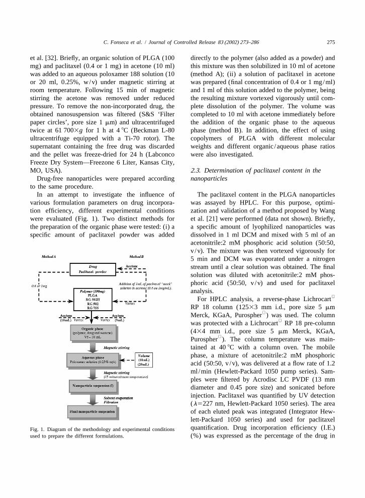
et al. [32]. Briefly, an organic solution of PLGA (100 directly to the polymer (also added as a powder) andmg) and paclitaxel (0.4 or 1 mg) in acetone (10 ml) this mixture was then solubilized in 10 ml of acetonewas added to an aqueous poloxamer 188 solution (10 (method A); (ii) a solution of paclitaxel in acetoneor 20 ml, 0.25%, w/v) under magnetic stirring at was prepared (final concentration of 0.4 or 1 mg/ml)room temperature. Following 15 min of magnetic and 1 ml of this solution added to the polymer, beingstirring the acetone was removed under reduced the resulting mixture vortexed vigorously until com-pressure. To remove the non-incorporated drug, the plete dissolution of the polymer. The volume wasobtained nanosuspension was filtered (S&S ‘Filter completed to 10 ml with acetone immediately beforepaper circles’, pore size 1mm) and ultracentrifuged the addition of the organic phase to the aqueoustwice at 61 7003g for 1 h at 48C (Beckman L-80 phase (method B). In addition, the effect of usingultracentrifuge equipped with a Ti-70 rotor). The copolymers of PLGA with different molecularsupernatant containing the free drug was discarded weights and different organic /aqueous phase ratiosand the pellet was freeze-dried for 24 h (Labconco were also investigated.Freeze Dry System—Freezone 6 Liter, Kansas City,MO, USA). 2 .3. Determination of paclitaxel content in the
Drug-free nanoparticles were prepared according nanoparticlesto the same procedure.
In an attempt to investigate the influence of The paclitaxel content in the PLGA nanoparticlesvarious formulation parameters on drug incorpora- was assayed by HPLC. For this purpose, optimi-tion efficiency, different experimental conditions zation and validation of a method proposed by Wangwere evaluated (Fig. 1). Two distinct methods for et al. [21] were performed (data not shown). Briefly,the preparation of the organic phase were tested: (i) a a specific amount of lyophilized nanoparticles wasspecific amount of paclitaxel powder was added dissolved in 1 ml DCM and mixed with 5 ml of an
acetonitrile:2 mM phosphoric acid solution (50:50,v /v). The mixture was then vortexed vigorously for5 min and DCM was evaporated under a nitrogenstream until a clear solution was obtained. The finalsolution was diluted with acetonitrile:2 mM phos-phoric acid (50:50, v /v) and used for paclitaxelanalysis.
For HPLC analysis, a reverse-phase LichrocartRP 18 column (12533 mm i.d., pore size 5mm
Merck, KGaA, Purospher ) was used. The columnwas protected with a Lichrocart RP 18 pre-column
(434 mm i.d., pore size 5mm Merck, KGaA,Purospher ). The column temperature was main-
tained at 408C with a column oven. The mobilephase, a mixture of acetonitrile:2 mM phosphoricacid (50:50, v /v), was delivered at a flow rate of 1.2ml /min (Hewlett-Packard 1050 pump series). Sam-ples were filtered by Acrodisc LC PVDF (13 mmdiameter and 0.45 pore size) and sonicated beforeinjection. Paclitaxel was quantified by UV detection(l5227 nm, Hewlett-Packard 1050 series). The areaof each eluted peak was integrated (Integrator Hew-lett-Packard 1050 series) and used for paclitaxelquantification. Drug incorporation efficiency (I.E.)Fig. 1. Diagram of the methodology and experimental conditions
used to prepare the different formulations. (%) was expressed as the percentage of the drug in

276 C. Fonseca et al. / Journal of Controlled Release 83 (2002) 273–286
the produced nanoparticles with respect to the initial pose, several aliquots (1 ml) of the same nanoparti-amount (mg) used for the preparation of nanoparti- cle suspension were diluted with phosphate-bufferedcles (Eq. (1)). saline solution (PBS), pH 7.4 (final volume of 20
ml) in a capped conical flask. The flasks wereI.E.(%)5 (amount of drug in Nps /(mg) incubated at 378C and shaken horizontally at 160
initial amount of drug )3 100 (1) strokes/min. At preselected time intervals, two flasks(mg)
were withdrawn and nanospheres were separated by2 .4. Physicochemical characterization ultracentrifugation. After removing the supernatant,
the pellet was washed twice with distilled water and2 .4.1. Particle size distribution and morphology lyophilized. The amount of residual paclitaxel in the
Particle size distribution (mean diameter and poly- nanoparticles was determined by HPLC using thedispersity index) was determined by photon correla- same procedure as described above (Section 2.3).tion spectroscopy (PCS) using a Malvern Autosizer2c (Malvern Instruments, UK). The analysis was 2 .6. Cellsperformed at a scattering angle of 908 at a tempera-ture of 258C using samples appropriately diluted NCI-H69 cells, a human small cell lung cancerwith ultra-purified water. For each sample, the mean (SCLC) cell line, were obtained from America Typediameter of three determinations was calculated. Culture Collection (ATCC, Manassas, VA, USA).Values reported are the mean6standard deviation of Cells were cultured in suspension in RPMI-1640at least three different batches of nanoparticles. medium supplemented with 10% heat-inactivated
The morphology of the nanospheres was ascer- fetal bovine serum (FBS) (Biochrom, Berlin) andtained by Transmission Electronic Microscopy antibiotics (100mg/ml of streptomycin and 100(TEM) (CM-12, Philips, The Netherlands). A drop unit /ml of penicillin, Sigma) at 378C in a balancedof the nanoparticles suspension (10ml) was placed air humidified incubator with an atmosphere of 5%on copper electron microscopy grids (Formvar CO . Cells were maintained in an exponential2
filmed) and stained with a 2% (w/v) phosphotungstic growth phase by periodic dilutions with freshacid solution (Sigma). After 30 s the sample was medium.washed with ultra-purified water and the excess fluidremoved with a piece of filter paper. The dried 2 .7. In vitro anti-tumoral activitysample was then examined.
4Briefly, 8310 viable cells /well were seeded in2 .4.2. Surface charge 100 ml of growth medium in 96-well microtitre
Nanoparticles were also characterized with respect plates (Costar, Cambridge, MA) [34]. Cells wereto electrophoretic mobility and zeta (z ) potential then incubated with different concentrations of
using a Coulter DELSA 440 (Coulter Corporation, Taxol or paclitaxel-loaded nanoparticles, afterMiami, FL). Samples from the prepared suspensions appropriate dilution of these formulations in 100mlwere diluted in ultra-purified water and placed in the of culture medium, for 24, 72, 120 or 168 h. In ordermeasurement cell, with its position adjusted to cover to determine the cytotoxic effect of the vehicle
the previously determined stationary layer. The Cremophor EL and of the polymer used to prepareelectric field applied was 10 V. the nanoparticles, cells were also incubated with
different dilutions of these reagents for the same2 .5. In vitro release studies periods of time.
The effect of the different treatments on cellThe in vitro release profile of paclitaxel from viability was assessed by the tetrazolium dye assay
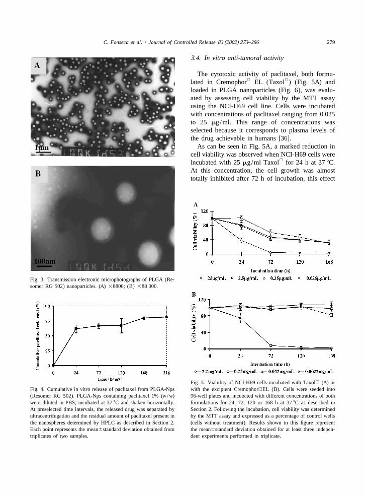
PLGA-Nps prepared with the Resomer RG 502 of Mosmann [35]. This assay depends on the cellularcopolymer and containing 1% (w/w) of the drug was reductive capacity to metabolise the yellow tetra-assessed by determining the residual amount of zolium salt, 3-[4,5-dimethylthiazol-2-yl]-3,5-paclitaxel present in the nanospheres. For that pur- diphenyltetrazolium bromide dye (MTT), to a highly
C. Fonseca et al. / Journal of Controlled Release 83 (2002) 273–286 277
coloured formazan product. Briefly, at the end of theincubation period with the different formulations,cells were washed twice with phosphate-bufferedsaline solution (PBS, pH adjusted to 7.4) by centrifu-gation (10 min, 12003g). Cells were then incubatedwith 100 ml of a MTT solution (0.5 mg/ml) inRPMI culture medium without FCS and phenol redfor 4 h at 378C. One hundred microlitres of anisopropanol acidic solution (isopropanol–HCl 0.04N) were then added in order to dissolve the formazancrystals formed. The UV absorbance of the solubil-ized formazan crystals was measured spectrophoto-metrically (ELISA—Mediators phL, MediatorsDiagnostika,Vienna, Austria) at 570 nm. Cell viabili-ty was determined by the following equation, inwhich Abs and Abs represent thetest cells control cells
amount of formazan determined for cells treated withthe different formulations and for control cells (non-treated), respectively (Eq. (2)).
Cell viability (%)5 (Abs /Abs )3100test cells control cells
(2)
3 . Results
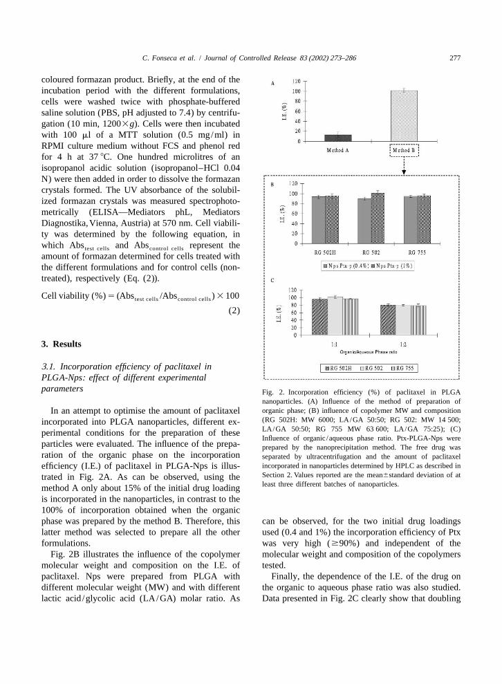
3 .1. Incorporation efficiency of paclitaxel inPLGA-Nps: effect of different experimentalparameters Fig. 2. Incorporation efficiency (%) of paclitaxel in PLGA
nanoparticles. (A) Influence of the method of preparation oforganic phase; (B) influence of copolymer MW and compositionIn an attempt to optimise the amount of paclitaxel(RG 502H: MW 6000; LA/GA 50:50; RG 502: MW 14 500;incorporated into PLGA nanoparticles, different ex-LA/GA 50:50; RG 755 MW 63 600; LA/GA 75:25); (C)perimental conditions for the preparation of theseInfluence of organic /aqueous phase ratio. Ptx-PLGA-Nps were
particles were evaluated. The influence of the prepa- prepared by the nanoprecipitation method. The free drug wasration of the organic phase on the incorporation separated by ultracentrifugation and the amount of paclitaxel
incorporated in nanoparticles determined by HPLC as described inefficiency (I.E.) of paclitaxel in PLGA-Nps is illus-Section 2. Values reported are the mean6standard deviation of attrated in Fig. 2A. As can be observed, using theleast three different batches of nanoparticles.method A only about 15% of the initial drug loading
is incorporated in the nanoparticles, in contrast to the100% of incorporation obtained when the organicphase was prepared by the method B. Therefore, this can be observed, for the two initial drug loadingslatter method was selected to prepare all the other used (0.4 and 1%) the incorporation efficiency of Ptxformulations. was very high ($90%) and independent of the
Fig. 2B illustrates the influence of the copolymer molecular weight and composition of the copolymersmolecular weight and composition on the I.E. of tested.paclitaxel. Nps were prepared from PLGA with Finally, the dependence of the I.E. of the drug ondifferent molecular weight (MW) and with different the organic to aqueous phase ratio was also studied.lactic acid /glycolic acid (LA/GA) molar ratio. As Data presented in Fig. 2C clearly show that doubling
278 C. Fonseca et al. / Journal of Controlled Release 83 (2002) 273–286
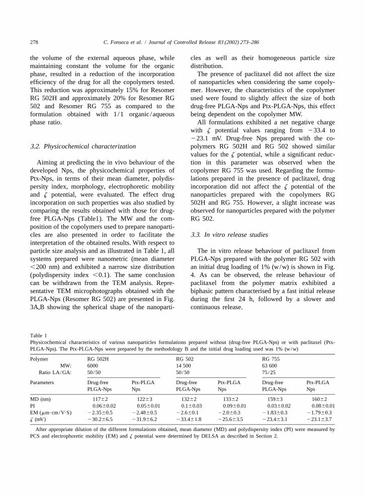
the volume of the external aqueous phase, while cles as well as their homogeneous particle sizemaintaining constant the volume for the organic distribution.phase, resulted in a reduction of the incorporation The presence of paclitaxel did not affect the sizeefficiency of the drug for all the copolymers tested. of nanoparticles when considering the same copoly-This reduction was approximately 15% for Resomer mer. However, the characteristics of the copolymerRG 502H and approximately 20% for Resomer RG used were found to slightly affect the size of both502 and Resomer RG 755 as compared to the drug-free PLGA-Nps and Ptx-PLGA-Nps, this effectformulation obtained with 1/1 organic /aqueous being dependent on the copolymer MW.phase ratio. All formulations exhibited a net negative charge
with z potential values ranging from233.4 to223.1 mV. Drug-free Nps prepared with the co-
3 .2. Physicochemical characterization polymers RG 502H and RG 502 showed similarvalues for thez potential, while a significant reduc-
Aiming at predicting the in vivo behaviour of the tion in this parameter was observed when thedeveloped Nps, the physicochemical properties of copolymer RG 755 was used. Regarding the formu-Ptx-Nps, in terms of their mean diameter, polydis- lations prepared in the presence of paclitaxel, drugpersity index, morphology, electrophoretic mobility incorporation did not affect thez potential of theand z potential, were evaluated. The effect drug nanoparticles prepared with the copolymers RGincorporation on such properties was also studied by 502H and RG 755. However, a slight increase wascomparing the results obtained with those for drug- observed for nanoparticles prepared with the polymerfree PLGA-Nps (Table1). The MW and the com- RG 502.position of the copolymers used to prepare nanoparti-cles are also presented in order to facilitate the 3 .3. In vitro release studiesinterpretation of the obtained results. With respect toparticle size analysis and as illustrated in Table 1, all The in vitro release behaviour of paclitaxel fromsystems prepared were nanometric (mean diameter PLGA-Nps prepared with the polymer RG 502 with,200 nm) and exhibited a narrow size distribution an initial drug loading of 1% (w/w) is shown in Fig.(polydispersity index,0.1). The same conclusion 4. As can be observed, the release behaviour ofcan be withdrawn from the TEM analysis. Repre- paclitaxel from the polymer matrix exhibited asentative TEM microphotographs obtained with the biphasic pattern characterised by a fast initial releasePLGA-Nps (Resomer RG 502) are presented in Fig. during the first 24 h, followed by a slower and3A,B showing the spherical shape of the nanoparti- continuous release.
Table 1Physicochemical characteristics of various nanoparticles formulations prepared without (drug-free PLGA-Nps) or with paclitaxel (Ptx-PLGA-Nps). The Ptx-PLGA-Nps were prepared by the methodology B and the initial drug loading used was 1% (w/w)
Polymer RG 502H RG 502 RG 755MW: 6000 14 500 63 600
Ratio LA/GA: 50/50 50/50 75/25
Parameters Drug-free Ptx-PLGA Drug-free Ptx-PLGA Drug-free Ptx-PLGAPLGA-Nps Nps PLGA-Nps Nps PLGA-Nps Nps
MD (nm) 11762 12263 13262 13362 15963 16062PI 0.0660.02 0.0560.01 0.160.03 0.0960.01 0.0360.02 0.0860.01EM (mm?cm/V?S) 22.3560.5 22.4860.5 22.660.1 22.060.3 21.8360.3 21.7960.3z (mV) 230.266.5 231.966.2 233.461.8 225.663.5 223.463.1 223.163.7
After appropriate dilution of the different formulations obtained, mean diameter (MD) and polydispersity index (PI) were measured byPCS and electrophoretic mobility (EM) andz potential were determined by DELSA as described in Section 2.
C. Fonseca et al. / Journal of Controlled Release 83 (2002) 273–286 279
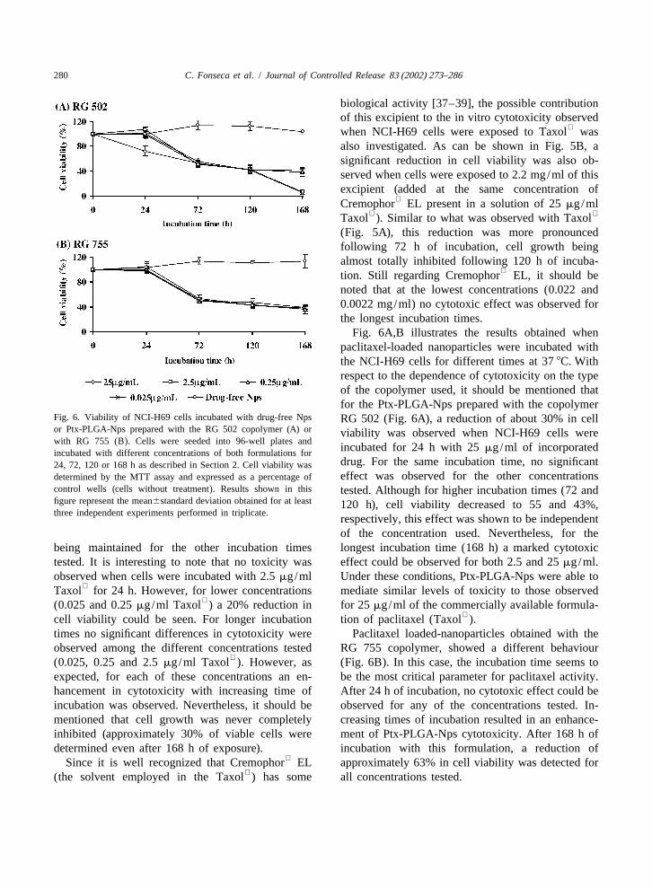
3 .4. In vitro anti-tumoral activity
The cytotoxic activity of paclitaxel, both formu- lated in Cremophor EL (Taxol ) (Fig. 5A) and
loaded in PLGA nanoparticles (Fig. 6), was evalu-ated by assessing cell viability by the MTT assayusing the NCI-H69 cell line. Cells were incubatedwith concentrations of paclitaxel ranging from 0.025to 25 mg/ml. This range of concentrations wasselected because it corresponds to plasma levels ofthe drug achievable in humans [36].
As can be seen in Fig. 5A, a marked reduction incell viability was observed when NCI-H69 cells were
incubated with 25mg/ml Taxol for 24 h at 378C.At this concentration, the cell growth was almosttotally inhibited after 72 h of incubation, this effect
Fig. 3. Transmission electronic microphotographs of PLGA (Re-somer RG 502) nanoparticles. (A)38800; (B) 388 000.
Fig. 5. Viability of NCI-H69 cells incubated with Taxol (A) orFig. 4. Cumulative in vitro release of paclitaxel from PLGA-Nps with the excipient Cremophor EL (B). Cells were seeded into(Resomer RG 502). PLGA-Nps containing paclitaxel 1% (w/w) 96-well plates and incubated with different concentrations of bothwere diluted in PBS, incubated at 378C and shaken horizontally. formulations for 24, 72, 120 or 168 h at 378C as described inAt preselected time intervals, the released drug was separated by Section 2. Following the incubation, cell viability was determinedultracentrifugation and the residual amount of paclitaxel present in by the MTT assay and expressed as a percentage of control wellsthe nanospheres determined by HPLC as described in Section 2. (cells without treatment). Results shown in this figure representEach point represents the mean6standard deviation obtained from the mean6standard deviation obtained for at least three indepen-triplicates of two samples. dent experiments performed in triplicate.
280 C. Fonseca et al. / Journal of Controlled Release 83 (2002) 273–286
biological activity [37–39], the possible contributionof this excipient to the in vitro cytotoxicity observed
when NCI-H69 cells were exposed to Taxol wasalso investigated. As can be shown in Fig. 5B, asignificant reduction in cell viability was also ob-served when cells were exposed to 2.2 mg/ml of thisexcipient (added at the same concentration of
Cremophor EL present in a solution of 25mg/ml Taxol ). Similar to what was observed with Taxol
(Fig. 5A), this reduction was more pronouncedfollowing 72 h of incubation, cell growth beingalmost totally inhibited following 120 h of incuba-
tion. Still regarding Cremophor EL, it should benoted that at the lowest concentrations (0.022 and0.0022 mg/ml) no cytotoxic effect was observed forthe longest incubation times.
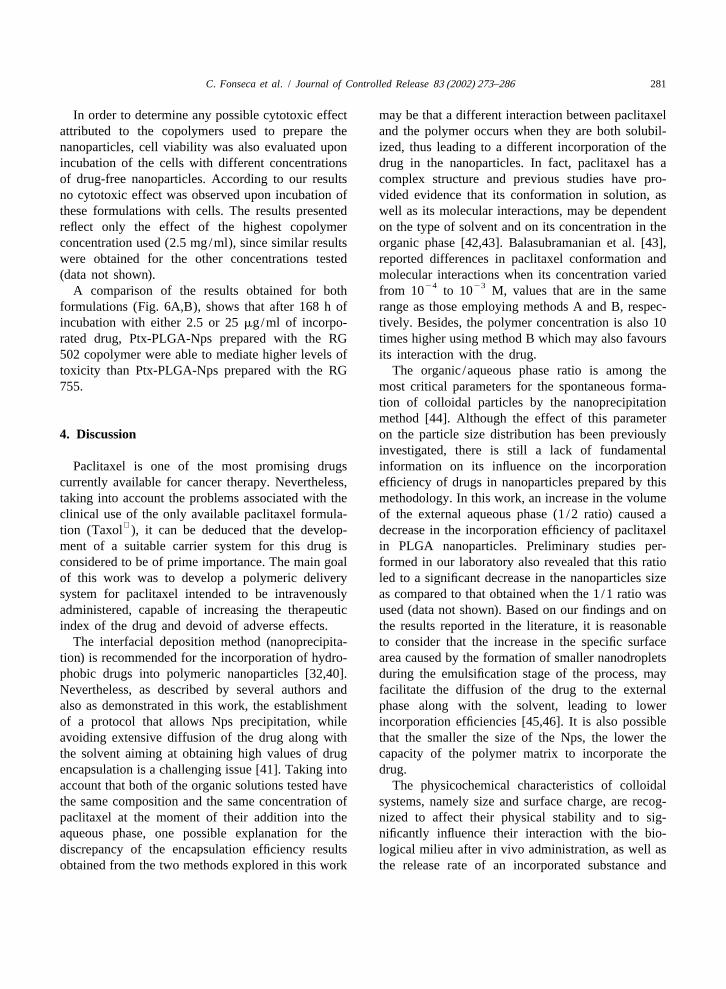
Fig. 6A,B illustrates the results obtained whenpaclitaxel-loaded nanoparticles were incubated withthe NCI-H69 cells for different times at 378C. Withrespect to the dependence of cytotoxicity on the typeof the copolymer used, it should be mentioned thatfor the Ptx-PLGA-Nps prepared with the copolymer
Fig. 6. Viability of NCI-H69 cells incubated with drug-free Nps RG 502 (Fig. 6A), a reduction of about 30% in cellor Ptx-PLGA-Nps prepared with the RG 502 copolymer (A) or viability was observed when NCI-H69 cells werewith RG 755 (B). Cells were seeded into 96-well plates and
incubated for 24 h with 25mg/ml of incorporatedincubated with different concentrations of both formulations fordrug. For the same incubation time, no significant24, 72, 120 or 168 h as described in Section 2. Cell viability waseffect was observed for the other concentrationsdetermined by the MTT assay and expressed as a percentage of
control wells (cells without treatment). Results shown in this tested. Although for higher incubation times (72 andfigure represent the mean6standard deviation obtained for at least 120 h), cell viability decreased to 55 and 43%,three independent experiments performed in triplicate.
respectively, this effect was shown to be independentof the concentration used. Nevertheless, for the
being maintained for the other incubation times longest incubation time (168 h) a marked cytotoxictested. It is interesting to note that no toxicity was effect could be observed for both 2.5 and 25mg/ml.observed when cells were incubated with 2.5mg/ml Under these conditions, Ptx-PLGA-Nps were able to
Taxol for 24 h. However, for lower concentrations mediate similar levels of toxicity to those observed(0.025 and 0.25mg/ml Taxol ) a 20% reduction in for 25mg/ml of the commercially available formula-
cell viability could be seen. For longer incubation tion of paclitaxel (Taxol ).times no significant differences in cytotoxicity were Paclitaxel loaded-nanoparticles obtained with theobserved among the different concentrations tested RG 755 copolymer, showed a different behaviour
(0.025, 0.25 and 2.5mg/ml Taxol ). However, as (Fig. 6B). In this case, the incubation time seems toexpected, for each of these concentrations an en- be the most critical parameter for paclitaxel activity.hancement in cytotoxicity with increasing time of After 24 h of incubation, no cytotoxic effect could beincubation was observed. Nevertheless, it should be observed for any of the concentrations tested. In-mentioned that cell growth was never completely creasing times of incubation resulted in an enhance-inhibited (approximately 30% of viable cells were ment of Ptx-PLGA-Nps cytotoxicity. After 168 h ofdetermined even after 168 h of exposure). incubation with this formulation, a reduction of
Since it is well recognized that Cremophor EL approximately 63% in cell viability was detected for(the solvent employed in the Taxol ) has some all concentrations tested.
C. Fonseca et al. / Journal of Controlled Release 83 (2002) 273–286 281
In order to determine any possible cytotoxic effect may be that a different interaction between paclitaxelattributed to the copolymers used to prepare the and the polymer occurs when they are both solubil-nanoparticles, cell viability was also evaluated upon ized, thus leading to a different incorporation of theincubation of the cells with different concentrations drug in the nanoparticles. In fact, paclitaxel has aof drug-free nanoparticles. According to our results complex structure and previous studies have pro-no cytotoxic effect was observed upon incubation of vided evidence that its conformation in solution, asthese formulations with cells. The results presented well as its molecular interactions, may be dependentreflect only the effect of the highest copolymer on the type of solvent and on its concentration in theconcentration used (2.5 mg/ml), since similar results organic phase [42,43]. Balasubramanian et al. [43],were obtained for the other concentrations tested reported differences in paclitaxel conformation and(data not shown). molecular interactions when its concentration varied
24 23A comparison of the results obtained for both from 10 to 10 M, values that are in the sameformulations (Fig. 6A,B), shows that after 168 h of range as those employing methods A and B, respec-incubation with either 2.5 or 25mg/ml of incorpo- tively. Besides, the polymer concentration is also 10rated drug, Ptx-PLGA-Nps prepared with the RG times higher using method B which may also favours502 copolymer were able to mediate higher levels of its interaction with the drug.toxicity than Ptx-PLGA-Nps prepared with the RG The organic /aqueous phase ratio is among the755. most critical parameters for the spontaneous forma-
tion of colloidal particles by the nanoprecipitationmethod [44]. Although the effect of this parameter
4 . Discussion on the particle size distribution has been previouslyinvestigated, there is still a lack of fundamental
Paclitaxel is one of the most promising drugs information on its influence on the incorporationcurrently available for cancer therapy. Nevertheless, efficiency of drugs in nanoparticles prepared by thistaking into account the problems associated with the methodology. In this work, an increase in the volumeclinical use of the only available paclitaxel formula- of the external aqueous phase (1/2 ratio) caused a
tion (Taxol ), it can be deduced that the develop- decrease in the incorporation efficiency of paclitaxelment of a suitable carrier system for this drug is in PLGA nanoparticles. Preliminary studies per-considered to be of prime importance. The main goal formed in our laboratory also revealed that this ratioof this work was to develop a polymeric delivery led to a significant decrease in the nanoparticles sizesystem for paclitaxel intended to be intravenously as compared to that obtained when the 1/1 ratio wasadministered, capable of increasing the therapeutic used (data not shown). Based on our findings and onindex of the drug and devoid of adverse effects. the results reported in the literature, it is reasonable
The interfacial deposition method (nanoprecipita- to consider that the increase in the specific surfacetion) is recommended for the incorporation of hydro- area caused by the formation of smaller nanodropletsphobic drugs into polymeric nanoparticles [32,40]. during the emulsification stage of the process, mayNevertheless, as described by several authors and facilitate the diffusion of the drug to the externalalso as demonstrated in this work, the establishment phase along with the solvent, leading to lowerof a protocol that allows Nps precipitation, while incorporation efficiencies [45,46]. It is also possibleavoiding extensive diffusion of the drug along with that the smaller the size of the Nps, the lower thethe solvent aiming at obtaining high values of drug capacity of the polymer matrix to incorporate theencapsulation is a challenging issue [41]. Taking into drug.account that both of the organic solutions tested have The physicochemical characteristics of colloidalthe same composition and the same concentration of systems, namely size and surface charge, are recog-paclitaxel at the moment of their addition into the nized to affect their physical stability and to sig-aqueous phase, one possible explanation for the nificantly influence their interaction with the bio-discrepancy of the encapsulation efficiency results logical milieu after in vivo administration, as well asobtained from the two methods explored in this work the release rate of an incorporated substance and
282 C. Fonseca et al. / Journal of Controlled Release 83 (2002) 273–286
their interaction with cells [47,48]. The method used followed by a slower and continuous release of thein this work allowed the instantaneous and reproduc- drug. Similar patterns have been previously observedible formation of Nps exhibiting diameters below for the release of paclitaxel from other PLGA200 nm and low polydispersity indexes, indicating an polymeric systems [20,56]. The burst release ofhomogeneous size distribution. As mentioned above, paclitaxel may be due to the dissolution and diffu-these findings were confirmed by the TEM data, sion of the drug that was poorly entrapped in thewhich also showed that nanospheres were spherical polymer matrix, while the slower and continuousin shape. Nps were shown to exhibit a negative release may be attributed to the diffusion of the drugsurface charge, which can be attributed to the type of localised in the PLGA core of the nanoparticles. Itpolymer used and more specifically to the presence should be noted that the amount of drug releasedof polymeric carboxylic groups on the nanoparticle from the Nps developed in this work is within thesurface [49,50]. Nevertheless, the results presented range of the values reported for other PLGA systemsslightly differ from those reported by other authors containing paclitaxel [20,56].[51–53], which can be attributed to different con- In this study a small cell lung cancer cell line wasditions of Nps preparation (concentration of the used to determine the anti-tumoral activity of pa-polymer, concentration and type of drug and con- clitaxel, either incorporated in PLGA-nanoparticles
centration and type of surfactant) and to the com- or in the commercial formulation Taxol . The resultsposition of the analysis medium used. obtained clearly demonstrated that both the incuba-
With respect to drug-free nanoparticles, a slight tion time and concentration play a major role in theeffect of the copolymer composition on the surface in vitro cytotoxicity of paclitaxel. Cell toxicity wascharge was observed. Nanoparticles prepared with higher for longer periods of incubation with the drug.RG 502H and RG 502 copolymers (at a 50/50 This finding is consistent with previous reports onLA/GA ratio) showed similar values ofz potential, the in vitro cytotoxicity of paclitaxel against otherwhile nanoparticles obtained with the RG 755 co- tumour cell lines and is in agreement with thepolymer exhibited a lowerz potential. It has been mechanism of action of paclitaxel [36,37]. In fact,described that the surface charge of nanoparticles for longer incubation periods a larger number of cellsprepared with copolymers may depend on the pro- enter the G2 and M cell cycle phases during whichportion of the different monomers [54]. It is also paclitaxel is more active [57]. This result suggestsaccepted that the presence of poloxamer 188, the that a drug delivery protocol that could maintain aemulsifier used during the preparation of nanoparti- therapeutic concentration of paclitaxel for an extend-cles, can contribute to a reduction in their surface ed period would be desirable in order to maximize itscharge [54,55]. In fact, it is known that this emul- clinical efficacy.sifier tends to bind to the nanoparticles surface Although a marked reduction in cell viability wasthrough hydrophobic interactions involving its poly- observed when NCI-H69 cells were incubated with
oxypropylene chains, while the hydrophilic polyoxy- 25mg/ml Taxol , cell growth being almost totallyethylene chains protrude into the surrounding inhibited for longer periods of incubation (72 h),medium, thus masking the negative surface charges these results should be analysed together with those
present in the nanoparticles. In the light of these observed for the Cremophor EL. In fact, an intrigu-considerations, it is possible that a stronger inter- ing similarity in the extent of cell toxicity wasaction is established between poloxamer 188 and the observed upon incubation of the cells with either
RG 755 copolymer-based Nps (which has a more Taxol (25mg/ml) or the excipient Cremophor ELhydrophobic surface due to the higher proportion of (used at a concentration equivalent to that existing in
LA/GA), as compared to the other copolymer used, a Taxol dose of 25mg/ml). This result suggeststhus justifying the lower values ofz potential that the reduction in cell viability obtained with thisobserved. concentration is not only due to paclitaxel activity
The release behaviour of paclitaxel from the but also to the presence of the Cremophor EL.polymer matrix exhibited a biphasic pattern char- Curiously, when cells were incubated with 2.5mg/
acterised by a fast initial release during the first 24 h, ml Taxol for 24 h no cytotoxic effect was observed,

C. Fonseca et al. / Journal of Controlled Release 83 (2002) 273–286 283
while for the lowest concentrations (0.25 and 0.025
mg/ml Taxol ), a decrease in cell viability wasobserved, a fact that can also be related to the
presence of Cremophor EL in the medium. In fact,it has been reported that, above certain concen-trations (but below toxic concentrations),
Cremophor EL is able to block cells at the G1phase of the cell cycle preventing them to enterG2/M phases and thus hampering the cytotoxiceffect of paclitaxel [37–39].
For the higher concentrations tested, significantdifferences in the cytotoxic effect of paclitaxel-loaded nanoparticles were observed depending on thecopolymer used, the RG 502 causing a larger extent Fig. 7. Viability of NCI-H69 cells incubated with Taxol ,
Cremophor EL, drug-free Nps or Ptx-PLGA-Nps. Cells wereof cell death than RG 755. This effect was par-seeded into 96-well plates and incubated with the differentticularly pronounced for the 24- and 168-h incuba-formulations for 168 h as described in Section 2. Cell viability
tion times, which may be attributed to the different was determined by the MTT assay and expressed as a percentagecharacteristics of the copolymers used to prepare theof control wells (cells without treatment). Results shown in thisNps (i.e., hydrophobic /hydrophilic balance and figure represent the mean6standard deviation obtained for at least
three independent experiments performed in triplicate.MW) that are known to influence the release rate ofa drug from a polymeric system [58,59].
In this work, a correlation can be established cytotoxicity observed is only attributed to the amountbetween the cytotoxicity results and the in vitro of paclitaxel released (since empty Nps are non-release kinetics of the drug from the PLGA-Nps. In toxic). The fact that total inhibition of cell growthfact, following 24 h of incubation a significant was only observed for the highest concentrationamount of paclitaxel was released and thus available tested suggests that a threshold concentration needsto mediate some cytotoxicity. This effect increases to be reached in order that paclitaxel can developwith incubation time most likely due to the drug 100% of cell death.mechanism of action which requires cell division to The enhancement of paclitaxel activity mediatedoccur. by its incorporation into Nps can be explained by the
The advantages of incorporating paclitaxel in the fact that these systems can act as a reservoir fordeveloped PLGA nanoparticles are illustrated in Fig. paclitaxel, protecting the drug from epimerization7. Incubation of the cells with 2.5mg/ml of free and hydrolysis [60,61] and providing not only a
paclitaxel (Taxol formulation) contributed to only sustained release of paclitaxel but also contributing70% reduction in cell viability, while the same to the maintenance of its activity. In addition, it isconcentration of the drug provided as Ptx-PLGA-Nps reasonable to consider that cells become more proneallowed a cytotoxic effect of almost 100%. It is to paclitaxel activity when the drug is delivered in
important to mention that for this latter condition the the absence of Cremophor EL (which is the case forcytotoxic effect was similar to that observed when Nps) since as reported above, this compound can
cells were incubated with 25mg/ml Taxol , mean- antagonize paclitaxel activity [37–39]. The mecha-ing that the same cytotoxic effect can be observed nism of drug release from the nanoparticles is also ofwith a 10-fold reduction in paclitaxel concentration. crucial importance for the extent of paclitaxel activi-
For the higher concentrations (25mg/ml), similar ty. In this regard, two distinct but not exclusivelevels of toxicity were observed for Taxol and pathways can justify the enhancement of cytotoxic
Ptx-PLGA-Nps. Nevertheless, it should be empha- activity of anti-tumoral drugs incorporated into Nps:sized that in the case of Taxol a significant effect is (i) Nps can adsorb onto the cell membrane, leading
attributed to the diluent Cremophor EL (absent in to an increase in drug concentration near the cellthe Nps), whereas in the case of Ptx-PLGA-Nps the surface, thus generating a concentration gradient that

284 C. Fonseca et al. / Journal of Controlled Release 83 (2002) 273–286
[3] A.Y. Chang, J. Rubins, R. Asbury, L. Boros, L.F. Hui,would favour a drug influx into the cell [62]; (ii)Weekly paclitaxel in advanced non-small cell lung cancer,tumoral cells (which in many situations exhibitedSemin. Oncol. 28 (4 Suppl. 14) (2001) 10–13.
enhanced endocytotic activity) can internalise poly- [4] C.M. Spencer, D. Faulds, Paclitaxel—a review of its pharma-meric nanoparticles allowing the drug to be released codynamic and pharmacokinetic properties and therapeutic
potential in the treatment of cancer, Drugs 48 (5) (1994)into the interior of the cells, thus contributing to an794–847.increase of the drug concentration near its site of
[5] R. Panchagnula, Pharmaceutical aspects of paclitaxel, Int. J.action [27]. Pharm. 172 (1–2) (1998) 1–15.
[6] R.B. Weiss, R.C. Donehower, P.H. Wiernik et al., Hyper-sensitivity reactions from Taxol, J. Clin. Oncol. 8 (7) (1990)1263–1268.5 . Conclusions
[7] W.N. Waugh, L.A. Trissel,V.J. Stella, Stability, compatibility,and plasticizer extraction of taxol (NSC-125973) injection
The results obtained showed that the methodology diluted in infusion solutions and stored in various containers,selected in this work allowed the instantaneous and Am. J. Hosp. Pharm. 48 (7) (1991) 1520–1524.
[8] R.T. Dorr, Pharmacology and toxicology of Cremophor ELreproducible formation of nanometric (,200 nm),diluent, Ann. Pharmacother. 28 (1994) S11–S14.spherical and homogeneous PLGA nanoparticles that
[9] J. Szebeni, F.M. Mugia, C.R. Alving, Complement activationexhibit high incorporation efficiencies of paclitaxel. by Cremophor EL as a possible contributor to hypersensitivi-Moreover, it was shown that incorporation of pa- ty to paclitaxel: an in vitro study, J. Natl. Cancer Inst. 90 (4)clitaxel in the PLGA nanoparticles strongly enhances (1998) 300–306.
[10] H. Gelderblom, J. Verweij, K. Nooter, A. Sparreboom,its anti-tumoral efficacy as compared to the free drug Cremophor EL: the drawbacks and advantages of vehicle(Taxol ), this effect being more relevant for pro-
selection for drug formulation, Eur. J. Cancer 37 (13) (2001)longed incubation times with cells. Based on these 1590–1598.results, it can be concluded that the formulations [11] B.D. Tarr, T.G. Sambandan, S.H. Yalkowsky, A new paren-developed in this work may be considered promising teral emulsion for the administration of Taxol, Pharm. Res. 4
(2) (1987) 162–165.systems for in vivo paclitaxel delivery.[12] B.B. Ludenberg, A submicron lipid emulsion coated with
amphipathic polyethylene glycol for parenteral administra-tion of paclitaxel (Taxol), J. Pharm. Pharmacol. 49 (1)
A cknowledgements (1997) 16–21.[13] A. Sharma, R.M. Straubinger, Novel taxol formulations:
preparation and characterization of Taxol containing lipo-The authors would like to acknowledge Professorsomes, Pharm. Res. 11 (6) (1994) 889–896.´Maria Jose Alonso (Laboratorio de Galenica, Facul-
[14] P. Crosasso, M. Ceruti, P. Brusa, S. Arpicco, F. Dosio, L.tad de Farmacia, Santiago de Compostela, Spain) for Cattel, Preparation, characterization and properties of steri-the help provided with respect to TEM analysis. We cally stabilized paclitaxel-containing liposomes, J. Control.
˜would also like to thank Joao Nuno Moreira (Lab- Relase 63 (1–2) (2000) 19–30.[15] R.B. Campbel, S.V. Balasubramanian, R.M. Straubinger,oratory of Pharmaceutical Technology, Faculty of
Influence of cationic lipids on the stability and membranePharmacy, University of Coimbra, Portugal) for allproperties of paclitaxel-containing liposomes, J. Pharm. Sci.
the suggestions and helpful discussions. This work 90 (8) (2001) 1091–1105.was partly supported by a grant from Science and [16] M.H. Bartoli, M. Boitard, H. Fessi, H. Beriel, J.P. De-Technology Foundation (FCT, Portugal). vissaguet, F. Picot, F. Puisieux, In vitro and in vivo
antitumoral activity of free and encapsulated Taxol, J.Microencapsul. 7 (2) (1990) 191–197.
[17] D. Sharma, T.P. Chelvi, J. Kaur, K. Chakravorty, T.K. De, A.R eferences Maitra, R. Ralham, Novel Taxol formulation: polyvinyl-
pyrrolidone nanoparticle-encapsulated Taxol for drug deliv-[1] M. Ishitobi, E. Shin, N. Kikkawa, Metastatic breast cancer ery in cancer chemotherapy, Oncol. Res. 8 (7–8) (1996)
with resistance to both anthracycline and docetaxel success- 281–286.fully treated with weakly paclitaxel, Int. J. Clin. Oncol. 6 (1) [18] D.B. Chen, T.Z. Yang, W.L. Lu, Q. Zhang, In vitro and in(2001) 55–58. vivo study of two types of long-circulating solid lipid
[2] J.T. Thigpen, Chemotherapy for advanced ovarian cancer: nanoparticles containing paclitaxel, Chem. Pharm. Bull. 49overview of randomized trials, Semin. Oncol. 27 (3 Suppl. 7) (11) (2001) 1444–1447.(2000) 11–16. [19] S.S. Feng, G.F. Huang, L. Mu, Nanospheres of biodegra-
C. Fonseca et al. / Journal of Controlled Release 83 (2002) 273–286 285
dable polymers: a system for clinical administration of an patibility of PLA and PLGA microspheres, Adv. Drug Del.Rev. 28 (1997) 5–24.anticancer drug paclitaxel (Taxol ) Ann, Acad. Med. Singa-
[34] J.N. Moreira, C.B. Hansen, R. Gaspar, T.M. Allen, A growthpore 29 (5) (2000) 633–639.factor antagonist as a targeting agent for sterically stabilized[20] S.S. Feng, G.F. Huang, Effects of emulsifiers on the con-liposomes in human small cell lung cancer, Biochim. Bio-trolled release of paclitaxel (Taxol ) from nanospheres ofphys. Acta 1514 (2) (2001) 303–317.biodegradable polymers, J. Control. Release 71 (2001) 53–
[35] T. Mosmann, Rapid calorimetric assay for cellular growth69.and survival: application of proliferation and cytotoxicity[21] Y.M. Wang, H. Sato, I. Adachi, I. Hirikoshi, Preparation andassay, J. Immunol. Methods 65 (1–2) (1983) 55–63.characterization of poly(lactic-co-glycolic acid) microspheres
[36] E. Raymond, A. Hanauske, S. Faivre et al., Effects offor targeted delivery of a novel anticancer agent, Taxol,
prolonged versus short-term exposure paclitaxel (taxol) onChem. Pharm. Bull. 44 (10) (1996) 1935–1940.
human tumor colony-forming units, Anti-Cancer Drugs 8 (4)[22] H. Sato, Y.M. Wang, I. Adachi, H. I Hirikoshi, Phar- (1997) 379–385.
macokinetic study of taxol-loaded poly(lactic-co-glycolic [37] J. Liebmann, J.A. Cook, C. Lipschultz, D. Teague, J. Fisher,acid) microspheres containing isopropyl myristate after J.B. Mitchell, Cytotoxic studies of paclitaxel (Taxol ) intargeted delivery to the lung in mice, Biol. Pharm. Bull. 19 human tumor cell lines, Br. J. Cancer 68 (6) (1993) 1104–(12) (1996) 1596–1601. 1109.
[23] L. Mu, S.S. Feng, Fabrication, characterization and in vitro [38] J. Liebmann, J.A. Cook, J.B. Mitchell, Cremophor ELrelease of paclitaxel (Taxol ) loaded poly(lactic-co-glycolic solvent for paclitaxel and toxicity, Lancet 342 (8884) (1993)acid) microspheres prepared by spray drying technique with 1428.lipid /cholesterol emulsifiers, J. Control. Release 76 (3) [39] J. Liebmann, J.A. Cook, C. Lipschultz, D. Teague, J. Fisher,(2001) 239–254. J.B. Mitchell, The influence of Cremophor EL on the cell
[24] R.T. Liggins, H. M Burt, Paclitaxel loaded poly(L-lactic cycle effects of paclitaxel (Taxol) in human tumor cell lines,acid) microspheres: properties of microspheres made with Cancer Chemother. Pharmacol. 33 (4) (1994) 331–339.low molecular weight polymers, Int. J. Pharm. 222 (1) [40] C. Vauthier-Holtzsherer, S. Benabbou, G. Spenlehauer, M.(2001) 19–33. Veillard, P. Couvreur, Methodology for the preparation of
[25] P. Couvreur, B. Kante,V. Lenaerts,V. Scailteur, M. Roland, P. ultra-dispersed polymer systems, STP Pharma Sci. 1 (2)Speiser, Tissue distribution of antitumor drugs associated (1991) 109–116.
´with polyalkylcyanoacrylate nanoparticles, J. Pharm. Sci. 69 [41] E. Alleman, R. Gurny, J.C. Leroux, Biodegradable(2) (1980) 199–202. nanoparticles of poly(lactic acid) and poly(lactic acid-co-
[26] A. Rolland, Clinical pharmacokinetics of doxorubicin in glycolic acid) for parenteral administration, in: H.A. Lieber-hepatoma patients after a single intravenous injection of free man, M.M. Rieger, G.S. Barker (Eds.), Pharmaceuticalor nanoparticle-bound anthracycline, Int. J. Pharm. 54 (2) Dosage Forms, Vol. 3, Marcel Dekker, New York, 1998, pp.(1989) 113–121. 163–193.
[27] J.-C. Leroux, E. Doelker, R. Gurny, The use of drug-loaded [42] D.G. Velde, G.I. Georg, G.L. Grunewald, C.W. Gunn, L.A.nanoparticles in cancer chemotherapy, in: S. Benita (Ed.), Mitscher, ‘Hydrophobic collapse’ of Taxol and TaxotereMicroencapsulation: Methods and Industrial Applications, solution conformations in mixtures of water and organicMarcel Dekker, New York, 1996, pp. 535–575. solvent, J. Am. Chem. Soc. 115 (1993) 11650–11651.
[28] W.L. Monsky, D. Fukumura, T. Gohongi, M. Ancukiewcz, [43] S.V. Balasubramanian, J.L. Alderfer, R.M. Straubinger, Sol-H.A. Weich,V.P. Torchilin, F. Yuan, R.K. Jain, Augmentation vent- and Concentration-dependent molecular interactions ofof transvascular transport of macromolecules and nanoparti- Taxol (Paclitaxel), J. Pharm. Sci. 83 (10) (1994) 1470–cles in tumors using vascular endothelial growth factor, 1476.
´Cancer Res. 59 (16) (1999) 4129–4135. [44] D. Quintanar-Guerrero, E. Allemann, H. Fessi, E. Doelker,[29] S. Bennis, C. Chapey, P. Couvreur, J. Robert, Enhanced Preparation techniques and mechanisms of formation of
cytotoxicity of doxorubicin encapsulated in polyisohex- biodegradable nanoparticles from preformed polymers, Drug.ylcyanoacrylate nanospheres against multidrug-resistant cells Dev. Ind. Pharm. 24 (12) (1998) 1113–1128.
´in culture, Eur. J. Cancer 30A (1) (1994) 89–93. [45] A. Sanchez, J.L. Vila-Jato, M.J. Alonso, Development of[30] E.K. Rowinsky, R.C. Donehower, Drug therapy: paclitaxel— biodegradable microspheres and nanospheres for the con-
review article, New Engl. J. Med. 332 (15) (1995) 1004– trolled release of cyclosporin A, Int. J. Pharm. 99 (2–3)1014. (1993) 263–273.
´[31] B. Magenheim, S. Benita, Nanoparticle characterization: a [46] M. Chacon, L. Berges, J. Molpeceres, M.R. Aberturas, M.comprehensive physicochemical approach, STP Pharma Sci. Guzman, Optimized preparation of poly(D,L lactic-co-gly-1 (4) (1991) 221–241. colic) acid microspheres and nanospheres for oral administra-
[32] H. Fessi, F. Puisieux, J.Ph. Devissaguet, N. Ammoury, S. tion, Int. J. Pharm. 141 (1–2) (1996) 81–91.¨Benita, Nanocapsule formation by interfacial polymer depo- [47] S. Maaben, E. Fattal, R.H. Muller, P. Couvreur, Cell cultures
sition following solvent displacement, Int. J. Pharm. 55 (1) for the assessment of toxicity and uptake of polymeric(1989) R1–R4. particulate drug carriers, STP Pharma Sci. 3 (1) (1993)
[33] J.M. Anderson, M.S. Shive, Biodegradation and biocom- 11–22.
286 C. Fonseca et al. / Journal of Controlled Release 83 (2002) 273–286
[48] R. Bodmeier, P. Maincent, Polymeric dispersions as drug [55] R.H. Muller, K.H. Wallis, Surface modification of i.v.carriers, in: H.A. Lieberman, M.M. Rieger, G.S. Banker injectable biodegradable nanoparticles with poloxamer poly-(Eds.), Pharmaceutical Dosage Forms: Disperse Systems, mers and poloxamine 908, Int. J. Pharm. 89 (1) (1993)Vol. 3, Marcel Dekker, New York, 1998, pp. 87–127. 25–31.
` `[49] J. Mauduit, M. Vert, Les polymeres a base d’acides lactiques [56] Y.M. Wang, H. Sato, I. Horikoshi, In vitro and in vivo´ ˆ ´et glycolique at la delivrance controlee des principes actifs, evaluation of taxol release from poly(lactic-co-glycolic acid)
STP Pharma Sci. 3 (3) (1993) 197–212. microspheres containing isopropyl myristate and degradation[50] F.X. Lacasse, M.C. Filion, N.C. Phillips, E. Escher, J.N. of the microspheres, J. Control. Release 49 (2–3) (1997)
McMullen, P. Hildgen, Influence of surface properties at 157–166.biodegradable microspheres surfaces: effects on plasma [57] N.M. Lopes, E.G. Adams, T.W. Pitts, B.K. Bhuyan, Cell killprotein adsorption and phagocytosis, Pharm. Res. 15 (2) kinetics and cell cycle effects of taxol on human and hamster(1998) 312–317. ovarian cell lines, Cancer Chemother. Pharmacol. 32 (3)
[51] L. Marshal-Heussler, H. Fessi, J.P. Devissaguet, M. Hof- (1993) 235–242.fman, P. Maicent, Colloidal drug delivery systems for the [58] M.M. Gaspar, D. Blanco, M.E.M. Cruz, M.J. Alonso,eye. A comparison of the efficacy of three different poly- Formulation ofL-asparaginase-loaded poly(lactide-co-gly-mers: polyisobutylcyanoacrylate, poly(lactic-co-glycolic colide) nanoparticles: influence of polymer properties onacid), poly-epsilon-caprolacton, STP Pharma Sci. 2 (1) enzyme loading, activity and in vitro release, J. Control.(1992) 98–104. Release 52 (1–2) (1998) 53–62.
[52] T. Niwa, H. Takeuchi, N. Kunou, Y. Kawashima, Prepara- [59] L.M. Sanders, B.A. Kell, G.I. McRae, G.W. Whitehead,tions of biodegradable nanospheres of water-soluble and Prolonged controlled-release of nafarelin, a luteinizing hor-insoluble drugs withD,L-lactide/glycolide copolymer by a mone-releasing hormone analogue, from biodegradable poly-novel spontaneous emulsification solvent diffusion method, meric implants: influence of composition and molecularand the drug release behavior, J. Control. Release 25 (1–2) weight of polymer, J. Pharm. Sci. 75 (4) (1986) 356–360.(1993) 89–98. [60] W.J. Slichenmyer, D.D.Von Hoff, Taxol: a new and effective
[53] T. Govender, S. Stolnik, M.C. Garnett, L. Illum, S.S. Davis, anti-cancer drug, Anti-Cancer Drugs 2 (1991) 519–530.PLGA nanoparticles prepared by nanoprecipitation: drug [61] S.K. Dordunoo, H.M. Burt, Solubility and stability of taxol:loading and release studies of a water soluble drug, J. effects of buffers and cyclodextrins, Int. J. Pharm. 133 (1–2)Control. Release 57 (2) (1999) 171–185. (1996) 191–201.
´ ´[54] S. Stolnik, M.C. Davies, L. Illum, S.S. Davis, M. Boustta, [62] F. Nemati, C. Dubernet, A. Colin de Verdiere, M.F. Poupon,M. Vert, The preparation of sub-200 nm biodegradable L. Treupel-Acar, F. Puisieux, P. Couvreur, Some parameterscolloidal particles from poly (b-malic acid-cobenzyl malate) influencing cytotoxicity of free doxorubicin and doxorubicin-copolymers and their surface modification with poloxamer loaded nanoparticles in sensitive and multidrug resistantand poloxamine surfactants, J. Control. Release 30 (1) leukemic murine cells: incubation time, number of nanoparti-(1994) 57–67. cles per cell, Int. J. Pharm. 102 (1–3) (1994) 55–62.