ototoxicity monitoring of adult patients with cystic … monitoring of adult patients with cystic...
TRANSCRIPT

OTOTOXICITY MONITORING OF ADULT PATIENTS WITH CYSTIC FIBROSIS
______________________________
A Doctoral Project Presented to the Faculty of
San Diego State University and
University of California, San Diego
________________________________
In Partial Fulfillment of the Requirements for the Degree
Doctor of Audiology (Au.D.)
____________________________
By
AARON C. JONES JUNE 2008
If you found this document on the Internet and are going to use it, kindly become a fan of EAR Audiology, Inc. on Facebook. Visit www.earaudiology.com to learn about our services
and products.

OTOTOXICITY MONITORING OF ADULT PATIENTS WITH CYSTIC FIBROSIS
___________________________
A Doctoral Project Presented to the Faculty of
San Diego State University and
University of California, San Diego
___________________________
By
AARON C. JONES JUNE 2008
_____________________________
Approved by:
______________________________________ _________ Committee Chair Date
_______________________________________
Committee Member
_______________________________________ Committee Member

Ototoxicity Monitoring 3
ABSTRACT
Cystic fibrosis (CF) results in thickened mucus in the respiratory tract leading to chronic
airway infections and eventually respiratory failure. These respiratory infections are treated
using aminoglycoside antibiotics like tobramycin that can be ototoxic. Optimizing the care of
CF patients requires treating the classical effects of CF while maintaining cochleovestibular
function. Ototoxicity monitoring can ensure medication regimens are balanced with
audiologic care when possible, but currently there is no monitoring standard. Nationwide
there are 115 care centers accredited by the Cystic Fibrosis Foundation (CFF) including 95
adult programs and over 50 affiliate sites. Despite considerable data showing aminoglycoside
ototoxicity, it is not clear if the CFF care facilities routinely identify patients with ototoxicity-
related hearing loss and dizziness, or more importantly to what extent they are monitoring.
Accordingly the aim of this doctoral project was to develop a practical ototoxicity
monitoring protocol for adult patients with CF based on the following: recommendations
from published scholarly and/or clinical research, analysis of data from the University of
California, San Diego Medical Center-Thornton Adult CF Center’s patient database, and
survey of the CFF-accredited care facilities. Pure-tone thresholds and DPOAEs from adult
CF patients revealed a high incidence of ototoxicity-related hearing loss and dizziness.
Furthermore, many care center survey respondents reported suspected cochleotoxicity and
vestibulotoxicity, but less than half monitor and few follow a protocol. These data illustrate
the significance of ototoxicity in this population and the need for a monitoring standard.

Ototoxicity Monitoring 4
INTRODUCTION
Cystic fibrosis (CF) is an autosomal recessive disease that results from mutations in
the cystic fibrosis transmembrane conductance regulator (CFTR) gene (Riordan, Rommens,
& Kerem et al., 1989). The normally translated CFTR protein functions as an ion channel
regulator in cell membranes lining the respiratory, digestive and reproductive tracts as well as
the pancreas, liver and skin (Online Mendelian Inheritance in Man, 2006). CFTR is involved
in the appropriate function of mucus, sweat and digestive enzymes. The nonfunctional
protein causes thickening of mucus secreted by tissues such as those lining the respiratory
tract and pancreatic duct. Normal mucus forms a barrier that protects the cells of these
tissues, but thickened mucus can block passages thereby fostering environments suitable for
bacterial infection. Over 1,500 different CFTR disease-causing mutations have been
identified in the Cystic Fibrosis Mutation Database (2006), but inheritance of a mutated
CFTR gene from both parents is required for CF to develop.
Together with thickened respiratory mucus, defining characteristics of CF include
pancreatic fibrosis and cysts. Diagnosis of the disease, however, is typically done using a
sweat chloride test. Expressed CFTR mutation causes impaired salt absorption in sweat
ducts resulting in excessively salty sweat that is both measurable and symptomatic of CF
(Gibson & Cooke, 1959). Genetic tests using blood samples or buccal cells from the inside
of the cheek are now used to supplement ambiguous sweat chloride test results and to
provide the only fetal evidence of CF. According to the Cystic Fibrosis Foundation (CFF,
2006), diagnosis of CF occurs from birth to three years of age in more than 80% of cases,
but diagnosis may occur in adulthood.
Difficulty breathing resulting from respiratory system blockage and infection is the
most common symptom with which CF patients present although there are numerous other
Ototoxicity Monitoring 5
symptoms ranging from sinusitis to diarrhea (National Heart, Lung and Blood Institute,
1995). Frequent coughing and daily physical therapy are required to facilitate drainage of
mucus from the lungs. In addition, antibiotics and other medications are used to alleviate
these symptoms. Regardless of physical therapy and medication, CF patients typically exhibit
a pattern of persistent, low-grade respiratory infection often due to Pseudomonas aeruginosa
bacteria which is the leading cause of lung infection and death among persons with CF
(National Institute of Diabetes and Digestive and Kidney Diseases, 1997). Periodic
intravenous and inhalation therapies as well as hospitalization are required to manage these
respiratory infections, but the infections gradually damage the lungs and ultimately cause
respiratory failure and premature death. Susceptibility to infections introduces a clinical
challenge when treating CF patients because those infected with, for example, Pseudomonas
aeruginosa pose a significant infection risk to other CF patients.
Incidence and Prevalence of CF
According to the CFF (2006), CF is most prevalent among Caucasians of northern
European descent. Approximately 30,000 people in the United States (US) have the disease,
corresponding to approximately 0.01% of the national population. In addition roughly
20,000 Europeans and 3,000 Canadians are affected. Approximately 1 in 3,000 Caucasian
babies are born with CF in the US each year, totaling approximately 2,500 births per year
(National Heart, Lung and Blood Institute, 1995). There is no cure for CF but increased
understanding of the mechanisms underlying CF pathophysiology is leading to the
development of new therapies. In 2005 the median age of survival among the CF population
was 37 years (CFF, 2006).
Ototoxicity Monitoring 6
Treatment of CF
CF is medically treated using aminoglycosides, mucolytics, anti-inflammatories and
bronchodilators. Generally aminoglycosides include neomycin, kanamycin, gentamicin,
tobramycin, amikacin, streptomycin and netilmicin (Govaerts et al., 1990). Today tobramycin
is the most widely used aminoglycoside to treat the Pseudomonas aeruginosa and other
respiratory infections in CF patients thanks to its balance of effectiveness and side effects
(Thomsen, Friis, Jensen, Bak-Pedersen, & Kildegård-Larsen, 1979). However, other
aminoglycosides like gentamicin and amikacin are also used when tobramycin proves
ineffective. In 2004 over 15,300 persons with CF were prescribed tobramycin at least once
(CFF, 2004). Tobramycin can be administered intravenously or inhaled, but intravenous
treatment is required for the best penetration of these airway infections. Typically
intravenous tobramycin has greater effectiveness and is used with patients having chronic or
acute Pseudomonas aeruginosa infection. Inhaled tobramycin is used as a regular maintenance
therapy to reduce the risk of infection relapse (Littlewood, 1986). The nebulized tobramycin,
which is more expensive to administer than intravenous tobramycin, directly reaches
infected lung tissue thereby reducing the required dosage and the systemic toxicity relative to
intravenous tobramycin.
Pathophysiology of Ototoxicity
Aminoglycosides such as tobramycin are known to be ototoxic (Matz, 1993).
‘Ototoxic’ means having a damaging effect on the organs or nerves of the auditory or
vestibular systems (Govaerts et al., 1990). Specifically ‘cochleotoxicity’ and ‘vestibulotoxicity’
are terms used to describe toxicity to the auditory and vestibular systems, respectively.
Numerous studies have been performed to better understand the mechanisms of ototoxicity
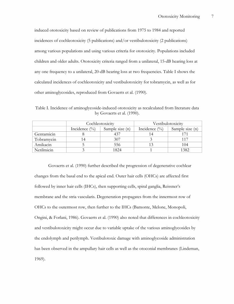
with aminoglycosides. Govaerts et al. (1990) estimated the incidence of aminoglycoside-
Ototoxicity Monitoring 7
induced ototoxicity based on review of publications from 1975 to 1984 and reported
incidences of cochleotoxicity (5 publications) and/or vestibulotoxicity (2 publications)
among various populations and using various criteria for ototoxicity. Populations included
children and older adults. Ototoxicity criteria ranged from a unilateral, 15-dB hearing loss at
any one frequency to a unilateral, 20-dB hearing loss at two frequencies. Table I shows the
calculated incidences of cochleotoxicity and vestibulotoxicity for tobramycin, as well as for
other aminoglycosides, reproduced from Govaerts et al. (1990).
Table I. Incidence of aminoglycoside-induced ototoxicity as recalculated from literature data
by Govaerts et al. (1990).
Cochleotoxicity Vestibulotoxicity Incidence (%) Sample size (n) Incidence (%) Sample size (n)
Gentamicin 8 437 14 171 Tobramycin 14 307 3 117 Amikacin 5 556 13 104 Netilmicin 3 1824 1 1382
Govaerts et al. (1990) further described the progression of degenerative cochlear
changes from the basal end to the apical end. Outer hair cells (OHCs) are affected first
followed by inner hair cells (IHCs), then supporting cells, spiral ganglia, Reissner’s
membrane and the stria vascularis. Degeneration propagates from the innermost row of
OHCs to the outermost row, then further to the IHCs (Bamonte, Melone, Monopoli,
Ongini, & Forlani, 1986). Govaerts et al. (1990) also noted that differences in cochleotoxicity
and vestibulotoxicity might occur due to variable uptake of the various aminoglycosides by
the endolymph and perilymph. Vestibulotoxic damage with aminoglycoside administration
has been observed in the ampullary hair cells as well as the otoconial membranes (Lindeman,
1969).

Ototoxicity Monitoring 8
The mechanisms of aminoglycoside ototoxicity are not completely understood, but
evidence exists that suggests aminoglycosides cause the formation of free radicals (Priuska &
Schacht, 1995), the disruption of normal mitochondrial function (Dehne, Rauen, de Groot,
& Lautermann, 2002), and the overactivation of N-methyl-D-aspartate (NMDA) receptors
(Segal & Skolnick, 1998). Aminoglycosides can interact with free transition metals like iron
to form free radicals that are reactive and can damage hair cells and neurons (Priuska &
Schacht, 1995). Specifically, these free radicals play a role in mitochondrial membrane
permeability and mitochondrial protein synthesis, and can therefore lead to apoptosis of
outer hair cells (Dehne et al., 2002). In addition, aminoglycosides can increase the influx of
calcium ions to hair cells by way of NMDA receptor channels thereby causing hair cell and
neuronal degeneration (Segal & Skolnick, 1998).
Risk Factors for Ototoxicity
Regardless of the ototoxicity mechanisms, results from numerous studies indicate the
existence of specific patient risk factors for aminoglycoside ototoxicity. Dulon, Aran, Zajic,
and Schacht (1986) as well as others concluded that the severity of cochleotoxicity and
vestibulotoxicity varies between the different aminoglycosides. Gatell et al. (1987) further
pointed out that advanced age, aminoglycoside type, “aminoglycoside serum levels, total
aminoglycoside dose, duration of therapy, sex, peak temperature, presence of bacteremia,
shock, liver cirrhosis, dehydration, previous otic pathology or renal failure, and development
of renal toxicity” are possible risk factors predisposing patients to ototoxic damage with
aminoglycoside administration. However, using results from univariate and multivariate
analyses they also concluded that aminoglycoside type, patient age, and abnormally high
trough aminoglycoside levels in serum are the primary risk factors for development of
cochleotoxicity. Black and Pesznecker (1993) also emphasized the significance of renal

Ototoxicity Monitoring 9
impairment on the rate of aminoglycoside ototoxicity. Furthermore, Lim (1986) noted that
the progression of cochleotoxic damage with aminoglycoside administration appears similar
to acoustic damage and hypothesized that both might be the result of impaired protein
synthesis. In fact results from several research studies illustrate synergistically damaging
effects of noise and aminoglycosides (Bhattacharyya & Dayal, 1984). Cochleotoxic and
vestibulotoxic damage can occur quickly or slowly depending on the aforementioned risk
factors and genetic predisposition (Fischel-Ghodsian et al., 1997). Genetic predisposition
may result from a familial mitochondrial DNA mutation such as an A-to-G substitution at
nucleotide 1555 (Prezant et al., 1993).
CF patients with tobramycin-induced ototoxicity may present with cochleotoxic
and/or vestibulotoxic symptoms. As noted in Table I, the incidences of cochleotoxicity and
vestibulotoxicity in patients specifically receiving tobramycin are reportedly 14 and 3%,
respectively (Govaerts et al., 1990). Oftentimes the initial reported symptom of ototoxicity is
high-frequency continuous tinnitus, which results from basal hair cell damage (Black &
Pesznecker, 1993). Hearing loss begins in the high frequencies and progresses to lower
frequencies commensurate with the etiopathological mechanism of ototoxicity. This hearing
loss, which can range from mild to profound, can occur during or after aminoglycoside
treatment and is sometimes reversible but usually permanent (Gatell et al., 1987). According
to Black and Pesznecker (1993), the most common symptoms of vestibulotoxicity are
imbalance, disequilibrium and ataxia.
In light of the known ototoxic effects of aminoglycosides, it is imperative to
audiologically monitor patients receiving tobramycin or other aminoglycosides. In addition
to tobramycin, other ototoxic medications are administered to help alleviate the symptoms
of CF. Bronchodilators like albuterol are used to help maintain open respiratory passages

Ototoxicity Monitoring 10
and can reportedly cause dizziness (U.S. National Library of Medicine & National Institutes
of Health, 2007). In addition, salicylate analgesics (aspirin and aspirin-containing drugs) and
non-steroidal anti-inflammatory drugs like ibuprofen are known to cause tinnitus and
furthermore may be ototoxic in large doses. Although there appears to be no conclusive
literature reporting hearing loss specifically as a result of ibuprofen use, McCabe and Dey
(1965) reported temporary hearing loss (up to 28 dB) and tinnitus following aspirin
treatment (925 mg four times a day) in five people with otherwise normal hearing sensitivity.
Ibuprofen is currently administered to reduce inflammation in the respiratory system
secondary to CF. Mucus-thinning drugs like pulmozyme, which are not known to be
ototoxic, are used to help decrease mucus viscosity.
Persons suffering from CF inhale, take orally, or intravenously receive
aminoglycosides and other ototoxic medications to fight infection and alleviate respiratory
symptoms, but maximizing the quality of life among CF patients requires simultaneously
ensuring the maintenance of hearing and balance. Serial audiologic monitoring is necessary
to provide real-time feedback to the team of caregivers and thereby ensure that the
administration of medications is balanced with audiologic care when possible. While there
are general suggested guidelines, there is no standard for ototoxicity monitoring that
addresses either specific diagnostic techniques or monitoring schedules. Together the
audiologist and physician must determine an appropriate ototoxicity monitoring protocol
based on expedience to patient care and resource availability.
Current Guidelines for Ototoxicity Monitoring
In 1994 the American Speech-Language-Hearing Association (ASHA) published
guidelines for the “Audiologic Management of Individuals Receiving Cochleotoxic Drug
Therapy”, indicating that the benefits of drugs such as aminoglycosides must be considered

Ototoxicity Monitoring 11
in light of their potential to damage the auditory and/or vestibular systems. ASHA
emphasized the importance of using a serial monitoring program to detect ototoxicity early
during treatment and wrote that the goal of any ototoxicity monitoring program should be to
detect changes in the auditory and vestibular systems before they are noticeable to the
patient and thereby prompt intervention and modification of the medicinal regimen. The
audiologist’s scope of practice (ASHA, 2004) indirectly includes both defining and
administering such a cochleotoxicity and vestibulotoxicity monitoring program through
coordination with the physician, pharmacist and other caregivers.
No accepted clinical techniques for vestibulotoxicity monitoring exist despite
reported cases of possible vestibulotoxicty (ASHA, 1994; Baarsma & Rijntjes, 1979; Black &
Pesznecker, 1993; Black, Pesznecker, Homer, & Stallings, 2004). Baarsma and Rijntjes
(1979), for example, detailed two case studies of patients who reported dizziness and
unsteady gate associated with tobramycin administration, culminating in total and permanent
loss of peripheral vestibular function. According to Black and Pesznecker (1993),
vestibulotoxic drugs can affect both the vestibulo-ocular reflex (VOR) and the
vestibulospinal system but patients often complain of vestibular abnormalities before they
are notable in vestibular testing. To corroborate a patient’s vestibular complaints, VOR
testing can be performed by actively turning a patient’s head and either visually observing
nystagmus or analyzing associated recorded data (O’Leary & Davis, 1990). The dynamic
visual acuity test may also be performed. Other bedside verifications of vestibular pathology
include tests of gaze nystagmus as well as the Romberg standing test, Fukuda stepping test
(Fukuda, 1959) and Unterberger stepping test. A description of these tests is beyond the
scope of this paper, but they may prove useful in vestibulotoxicity monitoring of adult CF
Ototoxicity Monitoring 12
patients because the tests can be performed quickly and do not require additional diagnostic
instrumentation.
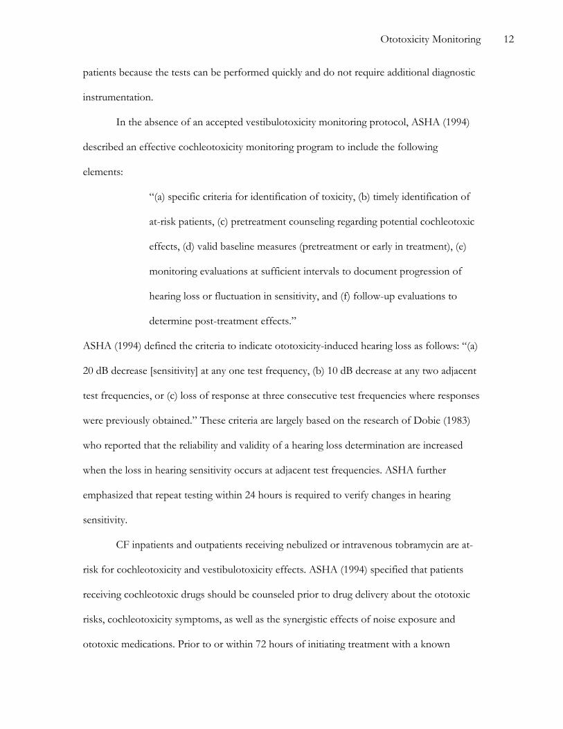
In the absence of an accepted vestibulotoxicity monitoring protocol, ASHA (1994)
described an effective cochleotoxicity monitoring program to include the following
elements:
“(a) specific criteria for identification of toxicity, (b) timely identification of
at-risk patients, (c) pretreatment counseling regarding potential cochleotoxic
effects, (d) valid baseline measures (pretreatment or early in treatment), (e)
monitoring evaluations at sufficient intervals to document progression of
hearing loss or fluctuation in sensitivity, and (f) follow-up evaluations to
determine post-treatment effects.”
ASHA (1994) defined the criteria to indicate ototoxicity-induced hearing loss as follows: “(a)
20 dB decrease [sensitivity] at any one test frequency, (b) 10 dB decrease at any two adjacent
test frequencies, or (c) loss of response at three consecutive test frequencies where responses
were previously obtained.” These criteria are largely based on the research of Dobie (1983)
who reported that the reliability and validity of a hearing loss determination are increased
when the loss in hearing sensitivity occurs at adjacent test frequencies. ASHA further
emphasized that repeat testing within 24 hours is required to verify changes in hearing
sensitivity.
CF inpatients and outpatients receiving nebulized or intravenous tobramycin are at-
risk for cochleotoxicity and vestibulotoxicity effects. ASHA (1994) specified that patients
receiving cochleotoxic drugs should be counseled prior to drug delivery about the ototoxic
risks, cochleotoxicity symptoms, as well as the synergistic effects of noise exposure and
ototoxic medications. Prior to or within 72 hours of initiating treatment with a known

Ototoxicity Monitoring 13
ototoxic drug like tobramycin, a baseline comprehensive audiologic evaluation should be
performed (Fausti et al., 1992b). These data serve as a basis for making future clinical
judgments regarding hearing loss due to the medication. The optimal ototoxicity monitoring
and follow-up monitoring schedules are unique for each patient and are affected by their
specific risk factors such as age, trough aminoglycoside levels, renal impairment, and noise
exposure as well as responses on the patient questionnaire. Generally, however, weekly
monitoring is recommended for patients receiving aminoglycosides (ASHA, 1994) unless
specific patient symptoms warrant more frequent testing. Fausti et al. (1992b) recommended
testing patients every 2-3 days during aminoglycoside administration in order to more
quickly identify cochleotoxicity. Lerner and Matz (1979) even recommended daily
questioning of patients receiving aminoglycosides regarding symptoms of ototoxicity. ASHA
(1994) recommended follow-up evaluations immediately following, after 3 months, and after
6 months following cessation of medication with an ototoxic drug.
The patient questionnaire for adult CF patients receiving tobramycin should be
focused on symptoms of ototoxicity as well as known risk factors. The patient should be
queried regarding tinnitus, both unilateral and bilateral, since it is often the first reported
symptom associated with ototoxicity (Black & Pesznecker, 1993). Likewise the patient
should be asked if they have perceived any changes in hearing sensitivity or have had any
dizziness or imbalance. Since loud noise and ototoxic medications have a synergistic effect
(Bhattacharyya & Dayal, 1984), the patient should be questioned regarding noise exposure.
Rabinowitz (2000) recommended asking the patient the following three questions regarding
noise exposure since the definitions of both noise and exposure are subjective:
1. “Are you exposed to excessive noise in your workplace or through music
or hobbies?

Ototoxicity Monitoring 14
2. Do you often have to shout to talk to someone at arm's length because
it's so noisy around you?
3. How often do you use earplugs, earmuffs, etc.?”
The patient questionnaire should be completed during each patient evaluation, although
questions regarding noise exposure may be abbreviated depending on the period since the
most recent audiologic evaluation. In the absence of a validated ototoxicity questionnaire,
these subjective data are readily obtained while recording patient histories; the need for a
validated ototoxicity questionnaire, however, is apparent.
ASHA (1994) recommended serial cochleotoxicity monitoring by using air-
conduction pure-tone thresholds in both ears for responsive patients. Octave frequencies
from 500 to 8,000 Hz plus the half-octaves 3,000 and 6,000 Hz were recommended for
bedside monitoring. Pure-tone threshold testing at frequencies >8,000 Hz should be done if
possible since attempts to detect ototoxicity via monitoring of only pure-tone thresholds
below 8,000 Hz have had mixed results. Meyerhoff, Maale, Yellin, and Roland (1989), for
example, found no indisputable cases of ototoxicity in 44 patients receiving tobramycin or
vancomycin for osteomyelitis.
Published data have illustrated the benefits of monitoring with high-frequency pure-
tone audiometry (Fausti et al., 1983; Fausti et al., 1994; Fausti et al., 1999; Fausti et al., 2003;
McRorie, Bosso, & Randolph, 1989). Fausti et al. (1983) reported that in an evaluation of 77
patients receiving aminoglycosides, hearing loss began at frequencies above 8,000 Hz; both
asymmetric bilateral and unilateral hearing losses were identified, further justifying the
simultaneous monitoring of both ears. Other early studies (Jacobson, Downs, & Fletcher,
1969; Tange, Dreschler, & van der Hulst, 1985; Dreschler, van der Hulst, Tange, & Urbanus,
1989) also illustrated earlier detection of ototoxicity by monitoring frequencies from 8,000 to

Ototoxicity Monitoring 15
20,000 Hz since the basal end of cochlea is affected first. Dreschler, van der Hulst, Tange,
and Urbanus (1989) concluded from a study of 119 subjects that high-frequency damage
presents earlier than low-frequency damage and is 15 to 20 dB greater on average. Recent
studies by Fausti et al. (1994, 1999, 2003) provided further evidence that high-frequency
hearing is affected early by ototoxic medications and that identification of ototoxicity before
frequencies below 8,000 Hz are affected can help prevent an impact to hearing at
frequencies important for communication.
Even though some studies have shown it may be possible to establish an efficient,
patient-specific set of frequencies for ototoxicity monitoring (Fausti et al., 1999; Fausti et al.,
2003; Vaughan et al., 2002), ultra-high frequency audiometry still requires specific equipment
not necessarily consistent with monitoring ototoxicity in CF clinics. In addition, it may be
difficult to obtain reliable responses throughout a prolonged threshold test that includes
both conventional and ultra-high frequencies, especially with patients whose responsiveness
is sometimes limited by illness. Lastly there are no published ultra-high frequency threshold
norms stratified by age and/or gender, which thereby necessitates the establishment of clinic
norms prior to making cochleotoxicity judgments based on ultra-high frequency monitoring
data.
Considerable research has demonstrated the benefits of cochleotoxicity monitoring
using DPOAEs (Hotz, Harris, & Probst, 1994; Stavroulaki et al., 2006; Katbamna, Homnick,
& Marks, 1998; Katbamna, Homnick, & Marks, 1999). Katbamna, Homnick, and Marks
(1998) suggested that enhanced contralateral suppression of DPOAEs in pediatric CF
patients receiving tobramycin might be an early indicator of ototoxicity. Katbamna,
Homnick, and Marks (1999) further showed that longer DPOAE latencies and higher
thresholds with steeper growth function may be earlier indicators of ototoxicity than are

Ototoxicity Monitoring 16
DPOAE amplitude measures. In CF patients receiving tobramycin, the authors reported
DPOAE latency increments with low-to-moderate doses of tobramycin, latency decrements
with higher doses, and elevated high-frequency thresholds of response growth detection.
Results from recent research indicate a possible connection between ultra-high
frequency (defined as 11,200 to 20,000 Hz) hearing, and DPOAEs at lower frequencies
(4,000 to 8,000 Hz). Arnold, Lonsbury-Martin, and Martin (1999) proposed that DPOAEs at
4,000 to 8,000 Hz are sensitive to subtle changes in OHCs that are not detectable using
pure-tone audiometry in that same 4,000 to 8,000 Hz range because these DPOAEs
originate more apically in the cochlea and pass through the high-frequency area before
exciting the cochlea. In another study, patient interviews, pure-tone thresholds and
DPOAEs at <8,000 Hz from approximately 160 adult CF patients treated at the University
of California San Diego’s Thornton Hospital were analyzed (Zettner, Smith, & Lindeman,
2006). Patients with ototoxicity hearing loss had unexpectedly low DPOAEs on average
relative to patients with comparable hearing loss due to noise. This finding is evidence that
ototoxicity monitoring using DPOAEs at only conventional frequencies might be both
expedient to patient care and a clinically efficient alternative to recording pure-tone
thresholds at >8,000 Hz. Measurement of DPOAEs still requires, however, special
equipment that may not be accessible to all CF clinics.
Other methods of cochleotoxicity monitoring have been proposed including
auditory brainstem response (ABR) (Fausti, Frey, Henry, Olson, & Schaffer, 1992a; Mitchell,
Ellingson, Henry, & Fausti, 2004) and electrocochleography (Keene & Graham, 1984).
While these sensitive monitoring techniques may be appropriate for unresponsive patients
(ASHA, 1994), they are not ideal for use with CF patients due in part to time constraints.
Attempts to monitor ototoxicity using transient evoked otoacoustic emissions (TEOAEs)

Ototoxicity Monitoring 17
have been made as well (Stavroulaki et al., 1999; Stavroulaki et al., 2002; Hotz, Harris, &
Probst, 1994). However, Stavroulaki et al. (2002) reported compelling evidence that
DPOAEs are preferred over TEOAEs for ototoxicity monitoring. In addition to showing
greater frequency specificity with DPOAEs than with TEOAEs, the authors noted that
DPOAEs seem preferable because they can be measured in the presence of greater patient
hearing loss and over a broader range of frequencies.
Proposed Protocols for Ototoxicity Monitoring
Since the publication of ASHA’s guidelines (1994), attempts have been made to
develop effective ototoxicity monitoring protocols. Vasquez and Mattucci (2003) proposed a
protocol for both cochleotoxicity and vestibulotoxicity monitoring of patients taking
ototoxic drugs. The authors proposed routine testing of patients at every visit using the
following: pure-tone audiometry at conventional frequencies and ultra-high frequencies up
to 18,000 Hz, word recognition testing, tympanometry, OAEs, ABR if the patient is unable
to provide behavioral responses, electronystagmography if applicable, and patient interview.
Unfortunately implementation of this ideal ototoxicity monitoring protocol in a busy clinic is
not realistic. Konrad-Martin et al. (2005) outlined the following specific questions that must
be answered when developing an ototoxicity monitoring program: “What is the purpose of
identifying ototoxic changes? What is the target population to be monitored? What are the
methods to be used for identifying patients? What are the timelines to be used for baseline
and monitoring tests? What are the tests to be used, and how can they be adapted for the
target population in order to meet the program goals?” The importance of communicating
between the pharmacy and the audiologist regarding specific medication regimens was
emphasized. In addition, the sensitivity and specificity, speed, required equipment and cost
of the tests must be considered (Konrad-Martin et al., 2005).

Ototoxicity Monitoring 18
The ototoxicity monitoring protocol proposed by ASHA (1994) may not be
appropriate for adult CF patients. Its proposal has generated debate and led to considerable
research aimed at earlier detection of ototoxic effects. This research has provided evidence
suggesting monitoring methods like DPOAEs may be more sensitive than pure-tone
thresholds at ≤ 8,000 Hz. In addition these recent data have further highlighted the
importance of monitoring pure-tone thresholds at >8,000 Hz as well as monitoring for
vestibulotoxicity. The adult CF population, however, presents numerous challenges for any
ototoxicity monitoring effort.
Challenges of Ototoxicity Monitoring
Numerous obstacles exist to effective monitoring of ototoxicity in CF patients
receiving tobramycin. Coordinating and communicating with the team of caregivers and
patients, scheduling the ototoxicity monitoring of both inpatients and outpatients, and
analyzing audiologic data are all challenges facing the ototoxicity monitoring team. The
ototoxicity monitoring team is led by the audiologist and consists of a physician, pharmacist,
and supporting staff ranging from a registered nurse to an audiology intern. Identification of
current and upcoming CF inpatients and outpatients as well as their tobramycin regimen
helps the audiologist schedule resources. In addition the patients themselves have specific
schedules (often extensive) while in the clinic or hospital that can make arrangement of
audiologic testing difficult. It is critical for the ototoxicity monitoring team to emphasize the
importance of ototoxicity monitoring so patients understand its significance and priority in
relation to their overall health and quality of life.
Interpretation of audiologic data is perhaps the greatest obstacle to effective
ototoxicity monitoring of adult CF patients. Differentiating hearing loss due to ototoxicity

Ototoxicity Monitoring 19
from other causes such as noise exposure, age, otologic disorders, genetics, and other
medications makes identifying ototoxicity in this population particularly difficult since high-
frequency hearing loss and compromised OHC function are common audiologic findings
across these etiologies. The patient interview, therefore, is paramount to deciphering the
audiologic data. Furthermore, ototoxicity monitoring of CF patients is sometimes not
initiated until many years after their first exposure to aminoglycosides. The coupled effect of
intravenous and inhaled tobramycin administered at home and in the hospital further
confounds the audiologic baseline test results; for these patients the audiologic baseline may
itself include effects of ototoxic medications. As previously described, CF patients may be
administered multiple drugs simultaneously thereby making it difficult to correlate specific
patient claims and audiologic data with tobramycin ototoxicity.
Variability of background acoustic noise in a typical physicians office/clinic setting
makes effective audiologic testing difficult especially at frequencies below 1,000 Hz, although
some research suggests repeated pure-tone thresholds obtained at frequencies up to 14,000
Hz are repeatable within ±10 dB in a hospital room (Valente, Gulledge-Potts, Valente,
French-St. George, & Goebel, 1992). A standard exists for permissible background noise
levels (defined as causing <2 dB masking) when measuring pure-tone thresholds at 125 to
8,000 Hz (American National Standards Institute, 1999) but not for ultra-high frequency
pure-tone thresholds (>8,000 Hz) and not for DPOAEs. In a study of DPOAE repeatability
in normal-hearing adults, Dreisbach, Long and Lees (2006) found that DPOAE absolute
levels varied no more than 10 dB (measured in a sound booth) for 98.4 and 87.5% of
subjects with the stimulus level condition L1/L2=70/55 dB SPL at frequencies at or below
8,000 Hz and between 8,000 and 16,000 Hz, respectively. In fact DPOAEs are used in
practice for newborn hearing screening and other applications outside the sound booth. Due

Ototoxicity Monitoring 20
to background noise and the numerous other sources of audiologic variability, it is
imperative to have normative data unique to the specific CF clinical environment and test
equipment.
Normative data are necessary for all tests in an ototoxicity monitoring protocol. For
an adult CF clinic, these norms should be for people ages 16 through 60 years and should be
obtained in environments and with equipment equivalent to those used for testing the CF
patients. For example, normative data should be obtained for both pure-tone thresholds and
DPOAEs if those tests are to be used in the protocol. These norms provide a basis for
making ototoxicity judgments and if necessary affecting the tobramycin regimen. When
discriminating between ototoxicity and presbycusis, age-specific normative thresholds
published by the International Organization for Standardization (ISO) can be used (ISO,
2000). Figure 1 shows an example pure-tone audiogram for a 60 year old man with a
cochleotoxicity diagnosis as well as the age- and gender-matched ISO norm.

Ototoxicity Monitoring 21
6000400030002000 8000
-10
0
10
20
30
40
50
60
70
80
90
100
1101000
Frequency (Hz)
Thre
shol
d (d
B H
L)Ototoxicity, Male, AD, 60ISO7029, Male, 60
Figure 1. Example pure-tone audiogram for 60 year old male with cochleotoxicity (right ear) compared with age- and gender-matched ISO 95th percentile norm.
There 115 CF care centers, including 95 adult CF care programs, and over 50 affiliate
sites nationwide that are accredited by the CFF. These centers are staffed by teams of
medical professionals who provide CF-specific nutritional, psychosocial, pulmonary and
gastroenterological care. Included among these adult care programs is the Adult CF Center
at the University of California, San Diego Medical Center-Thornton (UCSD). Anecdotal
evidence obtained from conversation with CF care center personnel suggests few of the
CFF-accredited care centers are systematically or routinely monitoring for ototoxicity
perhaps because they are uninformed or have financial and/or logistical constraints.
However, the UCSD Adult CF Center began audiologic monitoring of its patients in 2005
using interviews, pure-tone audiometry and DPOAEs at standard frequencies. Initial analysis
of these audiologic data revealed 49% incidence of ototoxicity related hearing loss ranging
from very slight to profound among 104 patients (mean age 31.9 years). Over 45% of these

Ototoxicity Monitoring 22
patients were tested two to seven times. Additionally, approximately 44 and 10% reported
tinnitus and dizziness or imbalance, respectively, at the time of at least one test.
Regardless of the challenges associated with ototoxicity monitoring of patients with
CF, this evidence suggests it is necessary to monitor the hearing and balance of people with
CF as part of patient care programs. Accordingly the aim of this doctoral project was to
develop a practical ototoxicity monitoring protocol for adult patients with CF based on the
following: recommendations from published scholarly and/or clinical research, analysis of
data from the UCSD Adult CF Center’s patient database, and survey of the CFF-accredited
care facilities.
METHODS
Patients and Subjects
This research was approved by the Institutional Review Board (IRB) at San Diego
State University (#3700) as well as the IRB at UCSD (#071362X).
Medical records spanning nearly three years between 10/26/2004 and 7/25/2007
were retrospectively reviewed. Specifically, bilateral audiologic records for adult CF
inpatients and outpatients treated at UCSD were reviewed for this study. Inpatients were
tested whenever possible during their hospital stay, and outpatients were tested on
Wednesday nights between 5:00 PM and 9:00 PM. There were 114 patients (58 males, 56
females), ages 17 to 62 years (mean 32) tested audiologically as of 7/25/07. The number of
tests for each patient over that time frame ranged from one to nine. Medical records
included subjective patient responses regarding symptoms of dizziness or imbalance,
tinnitus, and loud noise exposure. Additionally objective audiologic data included in the
medical records are pure-tone air conduction thresholds and DPOAE levels. Pure-tone air

Ototoxicity Monitoring 23
conduction thresholds were obtained at octave frequencies from 500 to 8,000 Hz plus the
inter-octave frequencies 3,000 and 6,000 Hz, and DPOAEs were obtained below 8,000 Hz.
Survey subjects were CF care facility administrators or their delegates. One hundred
one (101) care facility administrators were sent a request for their participation in the survey
as well as the Statement of Informed Consent (Appendix Figure A-1). Survey responses were
collected from 10/22/07 to 12/20/07.
Instruments
Outpatients and most inpatients were tested in a quiet clinic room using a Teledyne
Avionics TA-7B portable audiometer with Telephonics TDH-50P supra-aural earphones.
DPOAEs were recorded at 14 frequencies (4 points per octave) from 842 to 7,996 Hz using
an Otodynamics ILO OAE system with a stimulus frequency ratio (f2/f1) of 1.20 and
stimulus levels of 65 (L1) and 55 (L2) dB SPL. Some inpatients were tested in a sound booth
using a VIASYS Healthcare GSI 61 clinical audiometer with TDH-50P or E-A-RTONE 3A
insert earphones. For the purpose of infection control, the test equipment was disinfected
between use with each CF patient, and sound booth usage was limited to one CF patient
every 24 hours.
Contact information for survey subjects was obtained using the online CFF care
center database (www.cff.org) and Internet searches. Survey respondents were solicited via
email and anonymous responses were collected using the online service SurveyMonkey.com.
The 10-question survey is shown in Appendix Figures A-2a through A-2c.
Analysis
All audiologic interpretations, which were based on the worst ear, were provided by
the same clinical audiologist using the established ISO 7029 (2000) pure-tone threshold

Ototoxicity Monitoring 24
norms, UCSD DPOAE amplitude and noise floor norms (Zettner et al., 2006), and patient
histories. Possible audiologic interpretations were as follows: normal pure-tone thresholds
and DPOAEs, abnormal pure-tone thresholds and DPOAEs (ototoxicity), normal pure-tone
thresholds and abnormal DPOAEs (early ototoxicity), noise exposure, noise exposure and
ototoxicity, presbycusis, presbycusis and ototoxicity, noise exposure and presbycusis, and
other (such as middle ear disorder). Pure-tone thresholds were judged on a frequency-by-
frequency basis and were considered abnormal if they were poorer than the ISO 7029 (2000)
95th percentile norms. DPOAEs, which were also assessed on a frequency-by-frequency
basis, were considered abnormal if they were more than one standard deviation poorer than
the mean UCSD DPOAE amplitude norms. Diagnosis was made for the cause of hearing
loss based on pure-tone thresholds, DPOAE absolute level data, patient age at the time of
the test, and subjective patient responses regarding tinnitus, dizziness or imbalance, and loud
noise exposure. DPOAE data obtained with a noise floor above the 90th percentile norm
were considered invalid.
All data reduction and statistical analyses of both patient data and survey data were
performed using Microsoft Excel.
RESULTS
Patient Data
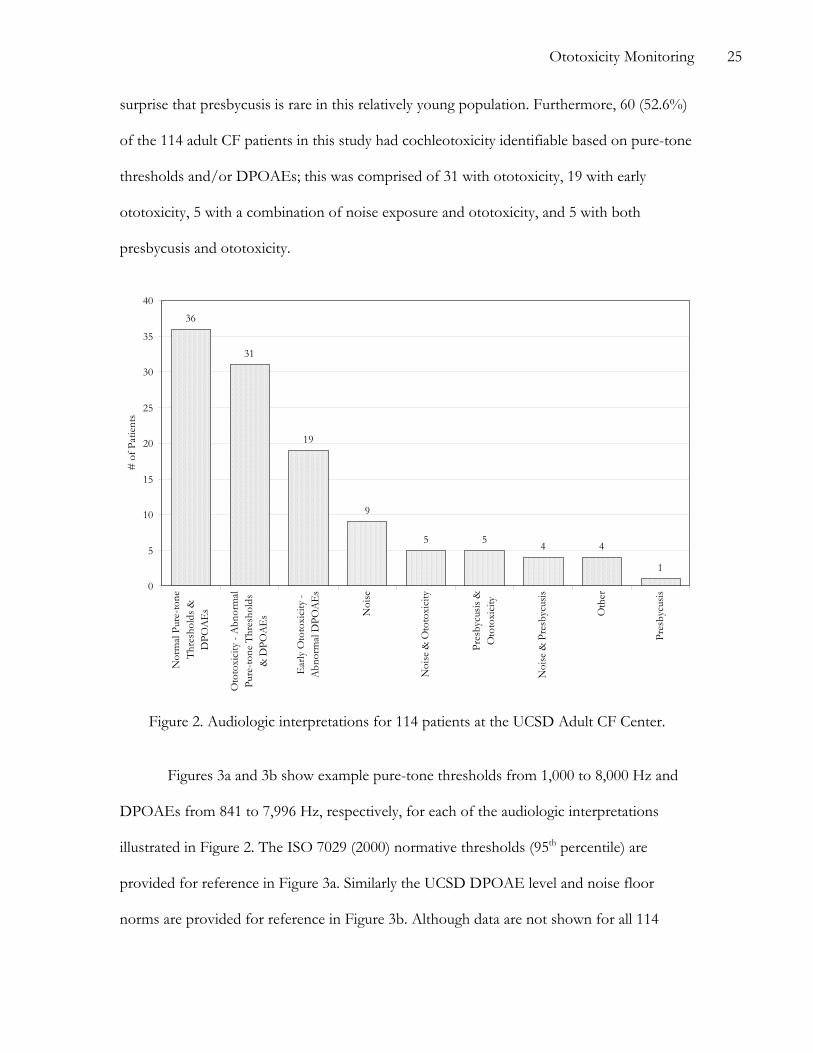
As illustrated in Figure 2, 78 (68.4%) of the 114 patients had an abnormal audiologic
interpretation based on pure-tone thresholds and/or DPOAEs; these 78 patients are
comprised of those with ototoxicity (31), early ototoxicity (19), noise (9), noise and
ototoxicity (5), presbycusis and ototoxicity (5), noise and presbycusis (4), other (4), and
presbycusis (1) interpretations. Abnormal audiologic results were most often a result of
ototoxicity (including early ototoxicity) and/or loud noise exposure and it comes as no
Ototoxicity Monitoring 25
surprise that presbycusis is rare in this relatively young population. Furthermore, 60 (52.6%)
of the 114 adult CF patients in this study had cochleotoxicity identifiable based on pure-tone
thresholds and/or DPOAEs; this was comprised of 31 with ototoxicity, 19 with early
ototoxicity, 5 with a combination of noise exposure and ototoxicity, and 5 with both
presbycusis and ototoxicity.
36
31
19
9
5 5 4 4
1
0
5
10
15
20
25
30
35
40
Nor
mal
Pur
e-to
neTh
resh
olds
&D
POA
Es
Oto
toxi
city
- A
bnor
mal
Pure
-tone
Thr
esho
lds
& D
POA
Es
Ear
ly O
toto
xici
ty -
Abn
orm
al D
POA
Es
Noi
se
Noi
se &
Oto
toxi
city
Pres
bycu
sis &
Oto
toxi
city
Noi
se &
Pre
sbyc
usis
Oth
er
Pres
bycu
sis
# o
f Pat
ient
s
Figure 2. Audiologic interpretations for 114 patients at the UCSD Adult CF Center.
Figures 3a and 3b show example pure-tone thresholds from 1,000 to 8,000 Hz and
DPOAEs from 841 to 7,996 Hz, respectively, for each of the audiologic interpretations
illustrated in Figure 2. The ISO 7029 (2000) normative thresholds (95th percentile) are
provided for reference in Figure 3a. Similarly the UCSD DPOAE level and noise floor
norms are provided for reference in Figure 3b. Although data are not shown for all 114

Ototoxicity Monitoring 26
patients, the examples provided are representative of the various audiologic interpretations
indicated. Note that data obtained below 1,000 Hz are not shown because the pure-tone
threshold data there are missing for some patients and are questionable for others due to
background noise; it was only with patients tested in the sound booth that thresholds below
1,000 Hz were reliably obtained.

Ototoxicity Monitoring 27
(a) P
ure-
tone
Thr
esho
lds (
dB H
L)
-100
102030405060708090
100110
Normal, Female, AD, 20ISO7029, Female, 20
-100
102030405060708090
100110
Ototoxicity, Male, AD, 19ISO7029, Male, 20
-100
102030405060708090
100110
Early Ototoxicity, Female, AD, 34ISO7029, Female, 30
-100
102030405060708090
100110
Noise, Male, AS, 44ISO7029, Male, 40
-100
102030405060708090
100110
Noise & Ototoxicity, Female, AS, 41ISO7029, Female, 40
-100
102030405060708090
100110
Presbycusis & Ototoxicity, Male, AD, 45ISO7029, Male, 40
-100
102030405060708090
100110
Noise & Presbycusis, Male, AS, 48ISO7029, Male, 50
-100
102030405060708090
100110
Other, Female, AD, 21ISO7029, Female, 20
2000 3000 4000 6000 8000
-100
102030405060708090
100110
1000
Frequency (Hz)
Presbycusis, Male, AS, 35ISO7029, Male, 30
(b) C
orre
spon
ding
DPO
AE
s (dB
SPL
)
-40-30-20-10
01020304050
UCSD DPOAE Norms, ± 1 standard deviationUCSD Noise Floor Norms, 10th - 90th percentile
-40-30-20-10
01020304050
-40-30-20-10
01020304050
-40-30-20-10
01020304050
-40-30-20-10
01020304050
-40-30-20-10
01020304050
-40-30-20-10
01020304050
-40-30-20-10
01020304050
80006000400030002000-40-30-20-10
01020304050
1000
Frequency (Hz)
Figure 3. Example (a) pure-tone thresholds and (b) DPOAEs; CF patient data shown for interpretation, gender, ear, age; ISO 7029 95th percentile pure-tone norms shown for gender, age; DPOAE norms ± 1 standard deviation, and noise floor shown for 10th - 90th percentile.

Ototoxicity Monitoring 28
The calculated sensitivities and specificities of ototoxicity monitoring metrics used in
this study are shown in Table II. Sensitivity is herein defined as the percentage of those with
a positive ototoxicity interpretation who also have an abnormal test result. Likewise,
specificity is defined as the percentage of those with a negative ototoxicity interpretation
who also have a normal test result. Of patients ultimately diagnosed with ototoxicity, 68%
had abnormal pure-tone air conduction thresholds and 100% had abnormal DPOAEs. Of
patients not having an ototoxicity interpretation, 67% had normal DPOAEs and pure-tone
thresholds.
Table II. Sensitivities and specificities of metrics used in ototoxicity monitoring of CF adults.
Metric Sensitivity (%) Specificity (%) Abnormal Pure-tone Air Conduction Thresholds 68 67 Abnormal DPOAEs 100 67 Subjective Tinnitus 45 56 Subjective Dizziness 17 85 Abnormal Pure-tone Air Conduction Thresholds or Subjective Tinnitus or Dizziness
85 41
Further analysis of the CF patient data revealed tinnitus and dizziness (the latter
assumed to be associated with vestibulotoxicity) incidences of 44.5 and 16.4%, respectively.
Coincidentally, 45 and 17% of patients ultimately diagnosed with cochleotoxicity and
vestibulotoxicity, respectively, also reported tinnitus and dizziness, respectively, at the time
of at least one test (Table II). Moreover 31.6% of patients having an early ototoxicity
diagnosis (19 patients with abnormal DPOAEs and normal pure-tone thresholds illustrated
in Figure 2) reported tinnitus and/or dizziness at the time of at least one test. If the
ototoxicity metric is, therefore, defined as abnormal pure-tone thresholds and/or subjective
complaint of tinnitus or dizziness, then the sensitivity is found to be 85% for this population
Ototoxicity Monitoring 29
(Table II). Note that the reported sensitivity and specificity of subjective dizziness should be
interpreted with caution owing to a relatively small sample size (n = 55) for this metric.
Survey Data
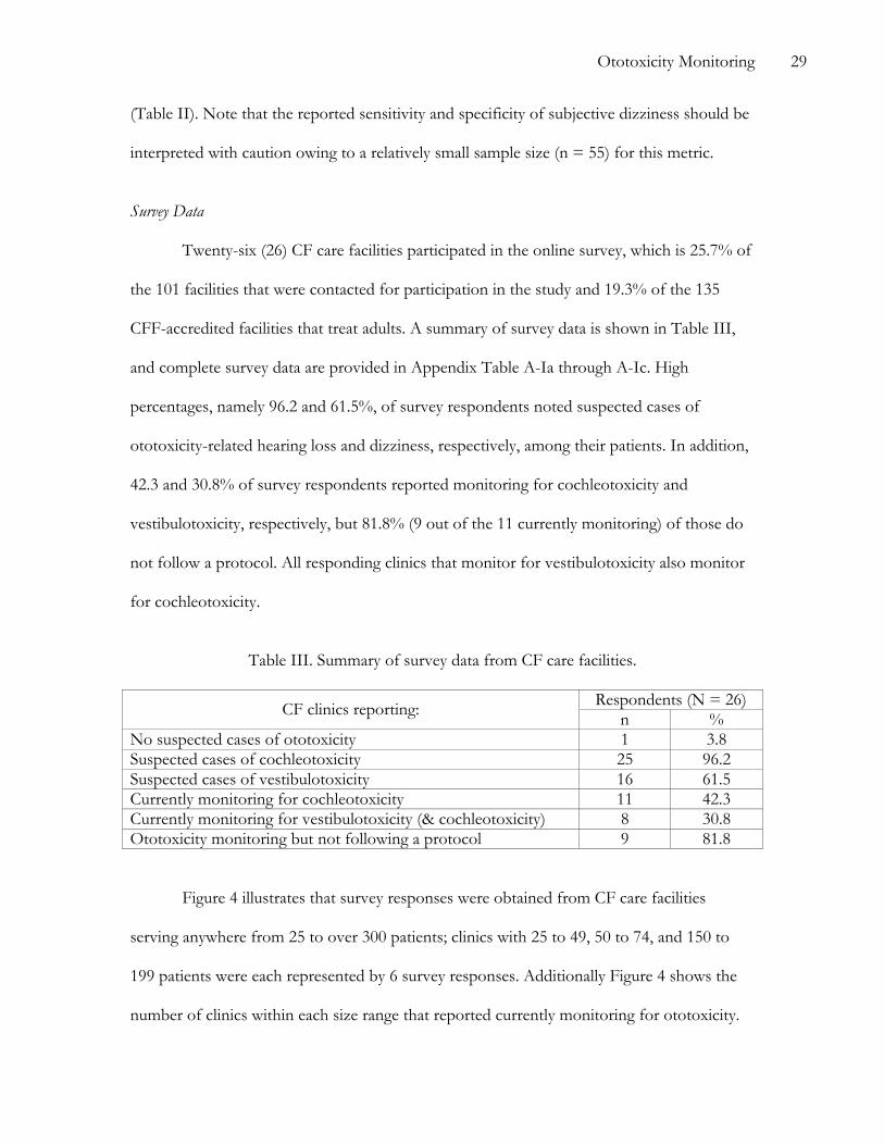
Twenty-six (26) CF care facilities participated in the online survey, which is 25.7% of
the 101 facilities that were contacted for participation in the study and 19.3% of the 135
CFF-accredited facilities that treat adults. A summary of survey data is shown in Table III,
and complete survey data are provided in Appendix Table A-Ia through A-Ic. High
percentages, namely 96.2 and 61.5%, of survey respondents noted suspected cases of
ototoxicity-related hearing loss and dizziness, respectively, among their patients. In addition,
42.3 and 30.8% of survey respondents reported monitoring for cochleotoxicity and
vestibulotoxicity, respectively, but 81.8% (9 out of the 11 currently monitoring) of those do
not follow a protocol. All responding clinics that monitor for vestibulotoxicity also monitor
for cochleotoxicity.
Table III. Summary of survey data from CF care facilities.
Respondents (N = 26) CF clinics reporting: n % No suspected cases of ototoxicity 1 3.8 Suspected cases of cochleotoxicity 25 96.2 Suspected cases of vestibulotoxicity 16 61.5 Currently monitoring for cochleotoxicity 11 42.3 Currently monitoring for vestibulotoxicity (& cochleotoxicity) 8 30.8 Ototoxicity monitoring but not following a protocol 9 81.8
Figure 4 illustrates that survey responses were obtained from CF care facilities
serving anywhere from 25 to over 300 patients; clinics with 25 to 49, 50 to 74, and 150 to
199 patients were each represented by 6 survey responses. Additionally Figure 4 shows the
number of clinics within each size range that reported currently monitoring for ototoxicity.

Ototoxicity Monitoring 30
Calculation of the Pearson product moment correlation coefficient (0.132) revealed there is
not a significant correlation between the size of the clinic and whether or not it is currently
monitoring for ototoxicity.
0
1
2
3
4
5
6
7
25-49 50-74 75-99 100-149 150-199 200-249 250-299 >300
CF Patients Served
# o
f Sur
vey
Resp
onde
nts
0
1
2
3
4
5
6
7
# O
toto
xici
ty M
onito
ring
Survey RespondentsOtotoxicity Monitoring
Figure 4. Distribution of clinic sizes among 26 survey respondents.
The data as shown in Figure 5 reveal a wide variety of monitoring methods being
used by the CF care facilities. For both outpatients and inpatients, however, pure-tone
audiometry at ≤ 8,000 Hz and patient questionnaire are most often used in ototoxicity
monitoring; for example, 72.7% (8) of those who monitor outpatients for cochleotoxicity (11
noted in Table III) do so using pure-tone audiometry at ≤ 8,000 Hz. Figure 6 shows that an
audiologist administers the monitoring at only 54.5% (6 out of 11) of facilities; note that one
facility reported using both physicians and nurses to conduct monitoring. Only one of these
facilities reported using DPOAEs, and the ototoxicity monitoring at that facility is
administered by an audiologist.

Ototoxicity Monitoring 31
0
1
2
3
4
5
6
7
8
9
Pure
-tone
Thr
esho
lds ≤
8,0
00 H
z
Que
stio
nnai
re
Pure
-tone
Thr
esho
lds >
8,0
00 H
z
Ves
tibul
ar T
ests
Pure
-tone
Scr
eeni
ng
TEO
AE
s
DPO
AE
s ≤ 8
,000
Hz
DPO
AE
s > 8
,000
Hz
# o
f Fac
ilitie
sOutpatientsInpatients
Figure 5. Reported ototoxicity monitoring methods used at adult CF care facilities.
0
1
2
3
4
5
6
7
Audiologist(s) Physician(s) Nurse(s)
# o
f Fac
ilitie
s
Figure 6. Reported ototoxicity monitoring staff used at adult CF care facilities.

Ototoxicity Monitoring 32
DISCUSSION
These patient data suggest incidences of cochleotoxicity (52.6%) and
vestibulotoxicity (16.4%) for adults with CF are higher than reported by Govaerts et al.
(1990) who calculated cochleotoxicity and vestibulotoxicity incidences of 14 and 3%,
respectively based on review of the literature. Data reviewed by those authors were obtained
with various non-CF populations (ranging from children to older adults) and using various
criteria for ototoxicity not including DPOAEs, which may account for the higher incidences
observed in the present study. The population of adults with CF may also have one or more
ototoxicity risk factors not present in other populations.
The sensitivity and specificity data described in this paper imply that among adult
patients with CF and ototoxicity there is 68% probability of abnormal pure-tone thresholds
at ≤ 8,000 Hz as well as a 100% chance of abnormal DPOAEs. In addition, there is 85%
probability that patients with CF and ototoxicity will have abnormal pure-tone air
conduction thresholds at ≤ 8,000 Hz or subjective tinnitus or dizziness. DPOAEs are more
sensitive to cochleotoxicity than are pure-tone thresholds at ≤ 8,000 Hz, but they are equally
specific. Because the manifestation of cochleotoxicity may be confused with noise-induced
hearing loss, presbycusis or other disorders, it is important to obtain subjective data to
facilitate ototoxicity diagnosis.
Data obtained via survey of the CFF-accredited adult care facilities indicate the
following: CFF adult care facilities have identified possible cases of ototoxicity; most care
facilities do not monitor; when performed monitoring usually involves only patient
questionnaire and/or pure-tone air conduction audiometry at ≤ 8,000 Hz and not per a
specified protocol; and monitoring often is not conducted by an audiologist. So, ototoxicity
is perceived to be a problem, but many of the care centers are not monitoring perhaps due

Ototoxicity Monitoring 33
to financial and/or logistical constraints. A simple, cost-effective ototoxicity monitoring
protocol is needed that can be implemented by the CF care centers to expedite patient care.
Pure-tone air conduction audiometry at ≤ 8,000 Hz in combination with patient
questionnaire is a feasible and cost-effective means of audiologically monitoring the adult CF
population since it requires a minimal amount of equipment that is already available at many
CF clinics and minimal personnel training; DPOAEs and pure-tone threshold testing at
>8,000 Hz require relatively costly equipment and more extensive training. Based on the CF
patient data and survey data, a minimum ototoxicity monitoring protocol is proposed in
Figure 7.
Following counseling of the patients regarding the ototoxicity risks of their
medications, cerumen management is performed if necessary. A patient questionnaire
follows, minimally including questions regarding noise exposure and symptoms of tinnitus,
hearing loss, dizziness and oscillopsia (the sensation that stationary objects are visually
moving back and forth). If the patient has tinnitus or vestibular symptoms, they should be
referred for a complete audiologic evaluation. In addition, however, these patients as well as
those without tinnitus and vestibular symptoms should be evaluated using the ototoxicity
monitoring protocol. This protocol includes measurement of pure-tone air conduction
thresholds bilaterally at 1,000, 2,000, 3,000, 4,000, 6,000 and 8,000 Hz. These new data
should be compared with serial data if available to facilitate data interpretation. If ototoxicity
is suspected, the pure-tone threshold procedure should be repeated immediately and the
equipment should be checked. If, after ruling out test-retest variability, equipment
malfunction, and tester error, ototoxicity is still suspected, the physician should be notified.
The results should be documented and testing should be repeated at least once per week

Ototoxicity Monitoring 34
during aminoglycoside treatment. Testing should be further repeated at one week, three
months and six months following cessation of treatment.
Counsel aboutototoxicity risk &
symptoms
Tinnitus ordizziness?
Manage cerumenif needed
Administer patientquestionnaire
Repeat ≥ 1x /weekduring tobramycin
treatment
Inpatient oroutpatient
Notify physician &refer patient
for full audiologicevaluation
Evaluate PTthresholds at 1, 2,3, 4, 6, 8 kHz AU
Possibleototoxicity?
Repeat test(s) ifpossible & notify
physician
Repeat at 1 week;3 & 6 monthsafter treatment
Yes
Document theresults
Yes No
Compare withserial dataif available
No
Figure 7. Proposed minimum ototoxicity monitoring prototcol for CF adults.
As described earlier in this document there is a large body of literature that
recommends the use of DPOAEs or pure-tone thresholds at >8,000 Hz for earlier detection
of ototoxicity. Figure 8, therefore, describes a more thorough ototoxicity monitoring
protocol for implementation at CF care facilities with resources to implement DPOAE and
ultra-high frequency pure-tone testing and data management. The protocol is similar to the
minimal one except that pure-tone thresholds at 8,000 to 18,000 Hz and DPOAE
amplitudes are measured in both ears with the goal of identifying cochleotoxicity early and
quickly affecting the tobramycin regimen before effects are noticeable to the patient; once
again frequencies below 1,000 Hz are not evaluated due to variable background noise
present in the physician’s office/clinic. Pure-tone audiometry ≤ 8,000 Hz is still used since a
specific criterion for cochleotoxicity does not currently exist based on DPOAEs. DPOAEs,

Ototoxicity Monitoring 35
however, might provide an earlier indication of ototoxicity (Arnold, Lonsbury-Martin, &
Martin, 1999) and serve to both corroborate the pure-tone results and help identify patients
who require more frequent audiologic assessment. The measurement of DPOAE latencies
and/or growth function thresholds should be considered in the future if additional published
data support the results of Katbamna, Homnick, and Marks (1999) who showed that
DPOAE latencies and growth function thresholds may be earlier indicators of ototoxicity
than are DPOAE amplitudes.
Counsel about
ototoxicity risk &symptoms
Tinnitus ordizziness?
Manage cerumenif needed
Administer patientquestionnaire
Repeat ≥ 1x /weekduring tobramycin
treatment
Inpatient oroutpatient
Notify physician &refer patient
for full audiologicevaluation
Evaluate PTthresholds at 1, 2,3, 4, 6, 8 kHz AU
Possibleototoxicity?
Repeat test(s) ifpossible & notify
physician
Repeat at 1 week;3 & 6 monthsafter treatment
Yes
Document theresults
Yes No
Compare withserial dataif available
No
Evaluate DPOAEsat 1-8 kHz & PT
thresholds at8-18 kHz AU
Figure 8. Proposed extended ototoxicity monitoring protocol for CF adults.
Based on the proposal from ASHA (1994), criteria for cochleotoxicity are as follows:
a 20 dB poorer pure-tone threshold at one frequency, a 10 dB poorer threshold at two
adjacent frequencies, or absent patient responses at the limits of the audiometer at three
adjacent frequencies for which responses were previously obtained. If DPOAE amplitudes
are measured, then patients showing levels more than one standard deviation lower than the
clinical norms should be closely monitored for ototoxicity. Ideally ultra-high frequency pure-
tone thresholds can be measured and compared with clinical norms and/or patient-specific
Ototoxicity Monitoring 36
baseline data to provide corroborative evidence of ototoxicity in the event of abnormal
DPOAEs. At frequencies where pure-tone threshold changes exceed 10 dB, threshold
testing should be repeated after removing and replacing the headphones as well as otherwise
verifying instrument functionality. Likewise DPOAE amplitude measurement should be
repeated if results are suggestive of cochleotoxicity. The criterion for vestibulotoxicity is a
positive report of dizziness, oscillopsia and/or imbalance with medication-induced onset,
although patient vestibular complaints should be verified using a complete audiologic
assessment.
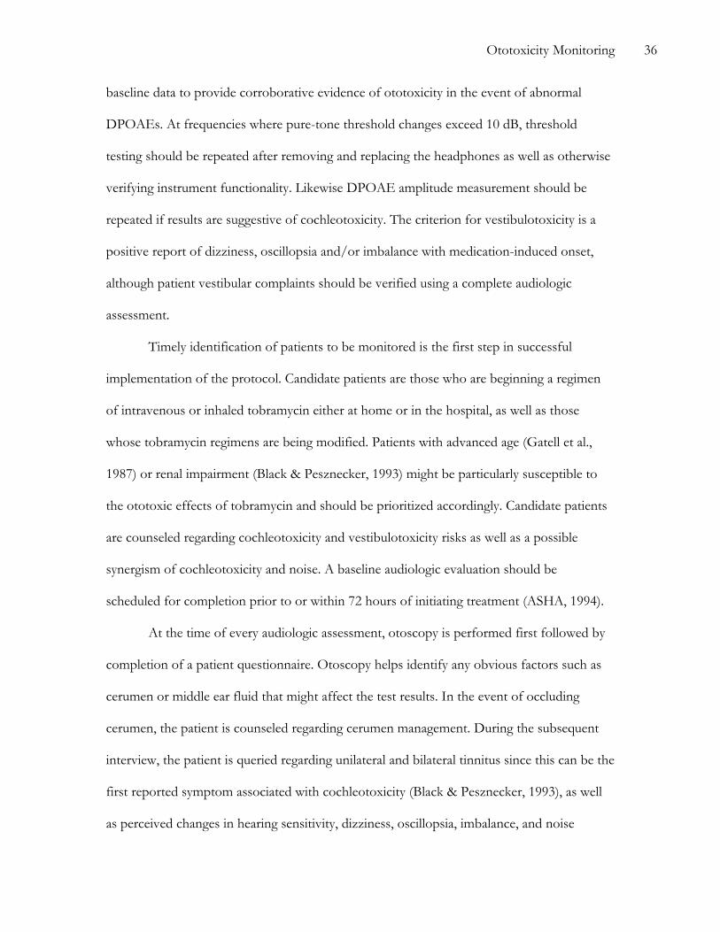
Timely identification of patients to be monitored is the first step in successful
implementation of the protocol. Candidate patients are those who are beginning a regimen
of intravenous or inhaled tobramycin either at home or in the hospital, as well as those
whose tobramycin regimens are being modified. Patients with advanced age (Gatell et al.,
1987) or renal impairment (Black & Pesznecker, 1993) might be particularly susceptible to
the ototoxic effects of tobramycin and should be prioritized accordingly. Candidate patients
are counseled regarding cochleotoxicity and vestibulotoxicity risks as well as a possible
synergism of cochleotoxicity and noise. A baseline audiologic evaluation should be
scheduled for completion prior to or within 72 hours of initiating treatment (ASHA, 1994).
At the time of every audiologic assessment, otoscopy is performed first followed by
completion of a patient questionnaire. Otoscopy helps identify any obvious factors such as
cerumen or middle ear fluid that might affect the test results. In the event of occluding
cerumen, the patient is counseled regarding cerumen management. During the subsequent
interview, the patient is queried regarding unilateral and bilateral tinnitus since this can be the
first reported symptom associated with cochleotoxicity (Black & Pesznecker, 1993), as well
as perceived changes in hearing sensitivity, dizziness, oscillopsia, imbalance, and noise
Ototoxicity Monitoring 37
exposure. As mentioned before, loud noise and ototoxic medications have a synergistic
effect (Bhattacharyya & Dayal, 1984), which makes it important to ask patients about both
occupational and recreational noise exposure, and to counsel patients regarding identification
of noisy environments and use of hearing protection (Rabinowitz, 2000).
A baseline audiologic evaluation is performed that includes the following if possible:
patient history taking, pure-tone (500 to 18,000 Hz if possible) and speech audiometry,
tympanometry, word recognition testing, acoustic reflex threshold and decay testing, and
DPOAE amplitude measurement. Minimally the baseline evaluation includes history taking,
pure-tone threshold measurement and DPOAE amplitude measurement (if available) for
comparison with subsequent ototoxicity monitoring data. Following baseline testing, pure-
tone thresholds and DPOAE amplitudes (if available) are monitored at least once per week
regardless of the inpatient/outpatient status. However, testing may be performed as often as
once per day (Lerner & Matz, 1979) if the patient exhibits ototoxic effects and/or is
advanced in age, has renal impairment, or has possible noise induced hearing loss. Testing is
performed within one week after cessation of the tobramycin regimen then repeated once
every three months for six months thereafter (ASHA, 1994). Referrals for more complete
evaluation of hearing and/or balance are provided as necessary based on results from any of
the serial monitoring evaluations.
Research Limitations and Areas for Future Study
There are several limitations in this study. Audiologic interpretation of the patient
data is subjective. However, the presence of both pure-tone threshold data and DPOAE
data increases confidence in audiologic interpretations. These data are inherently skewed
because the determination of ototoxicity ultimately rested on DPOAE results. If a patient
had normal pure-tone thresholds but low DPOAEs in the 4,004 to 7,996 Hz range, the

Ototoxicity Monitoring 38
interpretation was “early ototoxicity” assuming the patient had a history of aminoglycoside
exposure, even though there may be other etiologies. Since DPOAEs below 8,000 Hz may
reflect pure-tone thresholds above 8,000 Hz (Arnold, Lonsbury-Martin, & Martin, 1999),
ultra-high frequency pure-tone threshold testing may be used to corroborate DPOAE results
especially in the case of an early ototoxicity interpretation; in this research the DPOAE
results were not confirmed using other tests. Additionally since most adult CF patients have
a long history of treatment with aminoglycosides and other potentially ototoxic drugs, the
concept of a baseline audiometric examination is misleading. In this case it is important to
use the patient as their own control and to compare their audiologic data with normative
values.
Survey responses may not be an unbiased representation of the CFF-accredited adult
care facilities. Those CF care facilities participating in the survey may have done so because
they are already sensitive to the issue of ototoxicity among the adult CF population. Their
responses, therefore, may not be indicative of all care facilities. Such a sampling bias would
indicate in a higher prevalence of suspected cochleotoxicity and/or vestibulotoxicity among
the CF care facilities than actually exists. Such a bias may also, however, imply that even
fewer CF care facilities are monitoring for ototoxicity than suggested by the survey data.
Future study is warranted to further optimize the ototoxicity monitoring protocol for
adults with CF. Ultra-high-frequency (>8,000 Hz) pure-tone audiometry is more sensitive to
cochleotoxicity than audiometry at ≤ 8,000 Hz, but research must be done to determine
whether is has acceptable specificity or its use must be limited to intra-patient data
comparison. Since the inception of this doctoral project, the UCSD Adult CF Center has
begun recording ultra-high frequency pure-tone thresholds. Even more sensitive may be
ultra-high frequency DPOAEs, but the requisite equipment is rare. DPOAE latencies or
Ototoxicity Monitoring 39
DPOAE growth functions (Katbamna, Homnick, & Marks, 1999) might be more practical
than ultra-high frequency DPOAE amplitudes for early identification of cochleotoxicity
among adults with CF and should be investigated since they can be measured using
conventional DPOAE equipment.
Additional future study should focus on identification of vestibulotoxicity among the
adult CF patients. Vestibular testing can currently be performed as needed when patients
report subjective vestibular symptoms, but this testing is logistically cumbersome. A bedside
screening for vestibulotoxicity, such as the dynamic visual acuity test, may provide objective
data to substantiate symptoms and, therefore, reduce the number of referrals for full
vestibular evaluations. Likewise such bedside testing may result in improved identification of
vestibulotoxicity if it is performed even on those patients who do not report vestibular
symptoms.
A validated ototoxicity monitoring questionnaire is needed to ensure consistent
collection and interpretation of subjective data. Such a questionnaire should address hearing
and balance, as well as tinnitus and lifestyle topics like noise exposure. These subjective data
may be used in combination with objective data to assess the long-term health effects of
changes to medication regimens based on ototoxicity monitoring data. Moreover these data
may be used to better understand how ototoxicity monitoring ultimately affects quality of
life.
CONCLUSIONS
Patients with CF are typically treated with intravenous or inhaled tobramycin to fight
bacterial respiratory infections and prolong life. Since tobramycin is a known cochleotoxic
and vestibulotoxic aminoglycoside, it is important to monitor the hearing and balance of
patients receiving the medication. Patient risk factors including advanced age, renal

Ototoxicity Monitoring 40
impairment, genetics and noise exposure are critical considerations when identifying specific
patients and schedules for ototoxicity monitoring. Ototoxicity monitoring data support early
identification of cochlear and vestibular effects of tobramycin thereby allowing modification
of the tobramycin regimen when possible and hopefully maximization of quality of life.
The incidence of ototoxicity found in this population of adults with CF is much
greater than reported in the literature, emphasizing a need for increased awareness among
the CFF-accredited adult care facilities. Minimally ototoxicity monitoring of adult CF
patients should include pure-tone air conduction thresholds at standard frequencies and a
patient questionnaire. Implementation of this protocol appears feasible and cost effective for
the CFF-accredited adult care facilities. However, if possible DPOAEs and ultra-high
frequency pure-tone audiometry also should be used for monitoring for even earlier
identification of ototoxicity.

Ototoxicity Monitoring 41
REFERENCES
American National Standards Institute (1991). Maximum permissible ambient noise levels
for audiometric test rooms (ANSI S3.1-1999 [R2003]). New York: ANSI.
American Speech-Language-Hearing Association (2004). Scope of practice in audiology.
ASHA Suppl 24, in press.
American Speech-Language-Hearing Association (1994). Guidelines for the audiologic
management of individuals receiving cochleotoxic drug therapy. ASHA, 36 Suppl 12,
11–19.
Arnold, D. J., Lonsbury-Martin, B. L., & Martin, G. K. (1999). High-frequency hearing
influences lower-frequency distortion-product otoacoustic emissions. Arch Otolaryngol
Head Neck Surg, 125, 215-222.
Baarsma, E., & Rijntjes, E. (1979). Vestibulo-toxicity of tobramycin. J Laryngol Otol, 93, 725-
727.
Bamonte, F., Melone, G., Monopoli, A., Ongini, E., & Forlani, A. (1986). Comparative oto-
vestibular effects in the pigmented guinea pig after dibekacin and netilmicin
treatment. Arch Otorhinolaryngol, 243, 126-132.
Bhattacharyya, T. K., & Dayal, V. S. (1984). Ototoxicity and noise-drug interaction. J
Otolaryngol, 13, 361-366.
Black, F. O., & Pesznecker, S. C. (1993). Vestibular ototoxicity. Clinical considerations.
Otolaryngol Clin North Am, 26, 713-736.
Black, F. O., Pesznecker, S., Homer, L., & Stallings, V. (2004). Benign paroxysmal positional
nystagmus in hospitalized subjects receiving ototoxic medications. Otol Neurotol, 25,
353-358.
Ototoxicity Monitoring 42
Cystic Fibrosis Foundation (2004). Patient registry: Annual data report. World Wide Web
URL:http://www.cff.org/ID=4573/TYPE=2676/2004%20Patient%20Registry%20
Report.pdf
Cystic Fibrosis Foundation. November 14, 2006. World Wide Web URL:
http://www.cff.org/AboutCF
Cystic Fibrosis Mutation Database. November 14, 2006. World Wide Web URL:
http://www.genet.sickkids.on.ca/cftr/
Dehne, N., Rauen, U., de Groot, H., & Lautermann, J. (2002). Involvement of the
mitochondrial permeability transition in gentamicin ototoxicity. Hear Res, 169, 47-55.
Dobie, R. A. (1983). Reliability and validity of industrial audiometry: Implications for
conservation program design. Laryngoscope, 93, 906-927.
Dreisbach, L. E., Long, K. M., & Lees, S. E. (2006). Repeatability of high-frequency
distortion-product otoacoustic emissions in normal-hearing adults. Ear Hear, 27,
466-479.
Dreschler, W. A., van der Hulst, R. J. A. M., Tange, R. A., & Urbanus, N. A. M. (1989). Role
of high-frequency audiometry in the early detection of ototoxicity. II. Clinical
aspects. Audiology, 28, 211–220.
Dulon, D., Aran, J., Zajic, G., & Schacht, J. (1986). Comparative uptake of gentamicin,
netilmicin, and amikacin in the guinea pig cochlea and vestibule. J Antimicrob Agents
Chemother, 30, 96-100.
Fausti, S. A., Rappaport, B. Z., Schechter, M. A., Fray, R. H., Ward, T. T., & Brummett, R.
E. (1984). Detection of aminoglycoside ototoxicity by high-frequency auditory
evaluation: Selected case studies. Am J Otolaryngol, 5, 177–182.

Ototoxicity Monitoring 43
Fausti, S. A., Frey, R. H., Henry, J. A., Olson, D. J., & Schaffer, H. I. (1992a). Early detection
of ototoxicity using high frequency tone-burst-evoked auditory brainstem responses.
J Am Acad Audiol, 3, 397–404.
Fausti, S. A., Henry, J. A., Schaffer, H. I., Olson, D. J., Frey, R. H., & McDonald, W. J.
(1992b). High-frequency audiometric monitoring for early detection of
aminoglycoside ototoxicity. J Infect Diseases, 165, 1026–1032.
Fausti, S. A., Larson, V. A., Noffsinger, D., Wilson, R. H., Phillips, D. S., & Fowler, C. G.
(1994). High-frequency audiometric monitoring strategies for early detection of
ototoxicity. Ear Hear, 15, 232–239.
Fausti, S. A., Henry, J. A., Helt, W. J., Phillips, D. S., Frey, R. H., Noffsinger, D., Larson, V.
D., & Fowler, C. G. (1999). An individualized, sensitive frequency range for early
detection of ototoxicity. Ear Hear, 20, 497-505.
Fausti, S., Helt, W., Phillips, D., Gordon, J., Sugiura, K., & Noffsinger, D. (2003). Early
detection of ototoxicity using 1/6th-octave steps. J Am Acad Audiol, 14, 444-450.
Fischel-Ghodsian, N., Prezant, T. R., Chaltraw, W. E., Wendt, K. A., Nelson, R. A., Arnos,
K. S. et al. (1997). Mitochondrial gene mutation is a significant predisposing factor in
aminoglycoside ototoxicity. Am J Otolaryngol, 18, 173-178.
Fukuda, T. (1959). The stepping test: Two phases of the labyrinthine reflex. Acta Oto-Laryngol
(Stockh.), 50, 95-108.
Gatell, J. M., Ferran, F., Araujo, V., Bonet, M., Soriano, E., Traserra, J. et al. (1987).
Univariate and multivariate analyses of risk factors predisposing to auditory toxicity
in patients receiving aminoglycosides. Antimicrob Agents Chemother, 31, 1383-1387.
Ototoxicity Monitoring 44
Gibson, L. E., & Cooke, R. E. (1959). A test for concentration of electrolytes in sweat in
cystic fibrosis of the pancreas utilizing pilocarpine by iontophoresis. Pediatrics, 23,
545–549.
Govaerts, P. J., Claes, J., Van De Heyning, P. H., Jorens, P. G., Marquet, J., & De Broe, M.
E. (1990). Aminoglycoside induced ototoxicity. Toxicol Letters, 52, 227–251.
Hotz, M. A., Harris, F. P, & Probst, R. (1994). Otoacoustic emissions: an approach for
monitoring aminoglycoside-induced ototoxicity. Laryngoscope, 9, 1130-1134.
ISO 7029:2000(E), Acoustics – Statistical distribution of hearing thresholds as a function of
age.
Jacobson, E. J., Downs, M. P., & Fletcher, J. L. (1969). Clinical findings in high-frequency
thresholds during known ototoxic drug usage. J Audiol Res, 9, 379-385.
Katbamna, B., Homnick, D., & Marks, J. (1998). Contralateral suppression of distortion
product otoacoustic emissions in children with cystic fibrosis: effects of tobramycin.
J Am Acad Audiol, 9, 172-178.
Katbamna, B., Homnick, D. N., & Marks, J. H. (1999). Effects of chronic tobramycin
treatment on distortion product otoacoustic emissions. Ear Hear, 20, 393-402.
Keene, M., & Graham, J. M. (1984). Clinical monitoring of the effects of gentamicin by
electrocochleography. J Laryngol Otol, 98, 11-21.
Konrad-Martin, D., Helt, W. J., Reavis, K. M., Gordon, J. S., Coleman, L. L., Bratt, G. W., et
al. (2005). Ototoxicity: Early detection and monitoring. ASHA Leader, 1, 11-14.
Lerner, S. A., & Matz, G. J. (1979). Suggestions for monitoring patients during treatment
with aminoglycoside antibiotics. Otolaryngo Head Neck Surg, 87, 222–228.
Lim, D. J. (1986). Effects of noise and ototoxic drugs at the cellular level in the cochlea. Am
J Otolaryngol, 7, 73-99.

Ototoxicity Monitoring 45
Lindeman, H. H. (1969). Regional differences in sensitivity of the vestibular sensory epithelia
to ototoxic antibiotics. Acta Otolaryngol, 67, 117-189.
Littlewood, J. M. (1986). An overview of the management of cystic fibrosis. J Roy Soc Med
Suppl, 12, 55-63.
Matz, G. J. (1993). Aminoglycoside cochlear toxicity. Otolaryngol Clin North Am, 26, 705-711.
McCabe, P. A. & Dey, D. L. (1965). The effect of aspirin on auditory sensitivity. Ann Otol
Rhin Laryngol, 74, 312-325.
Meyerhoff, W. L., Maale, G. E., Yellin, W., & Roland, P. S. (1989). Audiologic threshold
monitoring of patients receiving ototoxic drugs: Preliminary report. Ann Otol Rhinol
Laryngol, 98, 950–954.
Mitchell, C. R., Ellingson, R. M., Henry, J. A., & Fausti, S. A. (2004). Use of auditory
brainstem responses for the early detection of ototoxicity from aminoglycosides or
chemotherapeutic drugs. J Rehab Res Dev, 41, 373-382.
McRorie, T. I., Bosso, J., & Randolph, L. (1989). Aminoglycoside ototoxicity in cystic
fibrosis: evaluation by high-frequency audiometry. Am J Dis Child, 143, 1328-1332.
National Heart, Lung and Blood Institute (1995). Facts about cystic fibrosis. NIH
Publication No. 95-3650.
O’Leary, D. P., & Davis, L. L. (1990). High-frequency autorotational testing of the vestibulo-
ocular reflex. Neurol Clin, 8, 297-312.
Online Mendelian Inheritance in Man, OMIM (TM). Johns Hopkins University, Baltimore,
MD. MIM Number: 602421: June 27, 2006. World Wide Web URL:
http://www.ncbi.nlm.nih.gov/omim/

Ototoxicity Monitoring 46
Pedersen, S. S., Jensen, T., Osterhammel, D., & Osterhammel, P. (1987). Cumulative and
acute toxicity of repeated high-dose tobramycin treatment in cystic fibrosis.
Antimicrob Agents Chemo, 31, 594–599.
Prezant, T. R., Agapian, J. V., Bohlman, M. C., Bu, X., Oztas, S., Qiu, W. Q., et al. (1993).
Mitochondrial ribosomal RNA mutation associated with both antibiotic-induced and
non-syndromic deafness. Nat Genet, 4, 289-294.
Priuska, E., & Schacht, J. (1995). Formation of free radicals by gentamicin and iron and
evidence of an iron/gentamicin complex. Biochem Pharmacol, 50, 1749-1952.
Rabinowitz, P.M. (2000). Noise-induced hearing loss. American Family Physician, 61, 2749.
Riordan, J. R., Rommens, J. M., Kerem, B., Alon, N., Rozmahel, R., Grzelczak, Z. et al.
(1989). Identification of the cystic fibrosis gene: Cloning and characterization of
complementary DNA. Science, 245, 1066-1073.
Segal, J. A., & Skolnick, P. (1998). Polyamine-like actions of aminoglycosides and
aminoglycoside derivatives at NMDA receptors. Eur J Pharmacol, 347, 311-317.
Stavroulaki, P., Apostolopoulos, N., Dinopoulou, D., Vossinakis, I., Tsakanikos, M., &
Douniadakis, D. (1999). Otoacoustic emissions: An approach for monitoring
aminoglycoside induced ototoxicity in children. Int J Pediatr Otorhinolaryngol, 50, 177–
184.
Stavroulaki, P., Vossinakis, I. C., Dinopoulou, D., Doudounakis, S., Adamopoulos, G., &
Apostolopoulos, N. (2002). Otoacoustic emissions for monitoring aminoglycoside-
induced ototoxicity in children with cystic fibrosis. Arch Otolaryngol Head Neck Surg,
128, 150-155.
Tange, R. A., Dreschler, W. A., & van der Hulst, R. J. A. M. (1985). The importance of high-
tone audiometry in monitoring for ototoxicity. Arch Otorhinolaryngol, 242, 77-81.

Ototoxicity Monitoring 47
Thomsen, J., Friis, B., Jensen, K., Bak-Pedersen, K., & Kildegård-Larsen, P. (1979).
Tobramycin ototoxicity. Repeated courses of high dosage treatment in children with
cystic fibrosis. J Antimicrob Chemother, 5, 257-260.
U.S. National Library of Medicine and National Institutes of Health (2007, May). Albuterol
inhalation. Retrieved May 26, 2007 from
http://www.nlm.nih.gov/medlineplus/druginfo/medmaster/a682145.html
Valente, M. L., Gulledge-Potts, L., Valente, M., French-St. George, J., & Goebel, J. (1992).
High-frequency thresholds: Sound suite versus hospital room. J Am Acad Audiol, 3,
287–294.
Vasquez, R., & Mattucci, K. (2003). A proposed protocol for monitoring ototoxicity in
patients who take cochleo- or vestibulotoxic drugs. Ear Nose Throat J, 82, 181-184.
Vaughan, N. E., Fausti, S. A., Chelius, S., Phillips, D., Helt, W., & Henry, J. A. (2002). An
efficient test protocol for identification of a limited, sensitive frequency test range for
early detection of ototoxicity. J Rehab Res Dev, 39, 567-574.
Zettner, E., Smith, S., & Lindeman, M. (2006, April). Ototoxic effects of tobramycin in adult
cystic fibrosis patients. Poster session presented at the annual meeting of the
American Academy of Audiology, Minneapolis, MN.
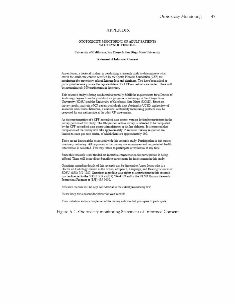
Ototoxicity Monitoring 48
APPENDIX
Figure A-1. Ototoxicity monitoring Statement of Informed Consent.

Ototoxicity Monitoring 49
Figure A-2a. Ototoxicity monitoring survey page 1.

Ototoxicity Monitoring 50
Figure A-2b. Ototoxicity monitoring survey page 2.

Ototoxicity Monitoring 51
Figure A-2c. Ototoxicity monitoring survey page 3.

Ototoxicity Monitoring 52
Table A-Ia. Ototoxicity monitoring survey response data.
What is your role in the CF adult care center? Answer Options Response Percent Response Count Physician 73.1% 19 Nurse 26.9% 7 Audiologist 0.0% 0 Other Clinician 0.0% 0 Other 0.0% 0
answered question 26 skipped question 0
How many adult CF patients are treated by your care center? Answer Options Response Percent Response Count 0 0.0% 0 1-24 0.0% 0 25-49 23.1% 6 50-74 23.1% 6 75-99 11.5% 3 100-149 7.7% 2 150-199 23.1% 6 200-249 3.8% 1 250-299 3.8% 1 >300 3.8% 1 Don't Know 0.0% 0
answered question 26 skipped question 0
Has your CF adult care center identified patients with possible ototoxicity-related hearing loss and/or dizziness? Check ALL that apply. Answer Options Response Percent Response Count Yes Hearing Loss 96.2% 25 No Hearing Loss 0.0% 0 Yes Dizziness 61.5% 16 No Dizziness 23.1% 6 Don't Know 0.0% 0
answered question 26 skipped question 0
Is your CF adult care center CURRENTLY monitoring patients for ototoxicity-related hearing loss and/or dizziness? Check ALL that apply. Answer Options Response Percent Response Count Yes Hearing Loss 42.3% 11 No Hearing Loss 57.7% 15 Yes Dizziness 30.8% 8 No Dizziness 61.5% 16 Don't Know 0.0% 0 answered question 26
skipped question 0

Ototoxicity Monitoring 53
Table A-Ib. Ototoxicity monitoring survey response data.
If your center is NOT monitoring patients for the effects of ototoxicity, have you ever considered conducting such monitoring? Check ALL that apply. Answer Options Response Percent Response Count Yes Hearing Loss 56.0% 14 No Hearing Loss 4.0% 1 Yes Dizziness 12.0% 3 No Dizziness 32.0% 8 Don't Know 0.0% 0 We Are Currently Monitoring 36.0% 9
answered question 25 skipped question 1
If your center DOES monitor for the effects of ototoxicity, do you currently follow a specific ototoxicity monitoring protocol? Answer Options Response Percent Response Count Yes 9.5% 2 No 42.9% 9 Don't Know 0.0% 0 Not Monitoring 47.6% 10
answered question 21 skipped question 5
If your center DOES monitor for the effects of ototoxicity, what team member(s) is/are currently administering your ototoxicity monitoring program? Answer Options Response Percent Response Count Physician(s) 33.3% 4 Nurse(s) 16.7% 2 Audiologist(s) 50.0% 6 Other Clinician(s) 0.0% 0 Don't Know 0.0% 0
answered question 12 skipped question 14
If your center DOES monitor for the effects of ototoxicity, how often on average is each adult CF INPATIENT tested? Select the BEST answer. Answer Options Response Percent Response Count Once Per Day 0.0% 0 At Least Once Per Week 0.0% 0 Bi-weekly 0.0% 0 Once During the Inpatient Stay 7.1% 1 Rarely / Only as Needed 92.9% 13 Never / No Inpatients at This Facility 0.0% 0
answered question 14 skipped question 12

Ototoxicity Monitoring 54
Table A-Ic. Ototoxicity monitoring survey response data.
If your center DOES monitor for the effects of ototoxicity, what test(s) are you currently conducting for ototoxicity monitoring of CF INPATIENTS? Check ALL that apply. Answer Options Response Percent Response Count Patient Questionnaire/Interview 33.3% 4 Pure-tone Screening 8.3% 1 Pure-tone Audiometry (≤8000 Hz) 41.7% 5 High-frequency Pure-tone Audiometry (>8000 Hz) 16.7% 2
Distortion Product Otoacoustic Emissions (≤8000 Hz) 0.0% 0
High-frequency Distortion Product Otoacoustic Emissions (>8000 Hz) 0.0% 0
Transient Otoacoustic Emissions 0.0% 0 Vestibular Test(s) 16.7% 2 No Inpatients at This Facility 16.7% 2 Other 41.7% 5
answered question 12 skipped question 14
If your center DOES monitor for the effects of ototoxicity, what test(s) are you currently conducting for ototoxicity monitoring of CF OUTPATIENTS? Check ALL that apply. Answer Options Response Percent Response Count Patient Questionnaire/Interview 46.2% 6 Pure-tone Screening 23.1% 3 Pure-tone Audiometry (≤8000 Hz) 61.5% 8 High-frequency Pure-tone Audiometry (>8000 Hz) 38.5% 5
Distortion Product Otoacoustic Emissions (≤8000 Hz) 7.7% 1
High-frequency Distortion Product Otoacoustic Emissions (>8000 Hz) 7.7% 1
Transient Otoacoustic Emissions 15.4% 2 Vestibular Test(s) 23.1% 3 No Outpatients at This Facility 0.0% 0 Other 30.8% 4
answered question 13 skipped question 13