original research article morphometric and morphological ... · original research article...
TRANSCRIPT
Int J Anat Res 2017, 5(1):3547-51. ISSN 2321-4287 3547
Original Research Article
MORPHOMETRIC AND MORPHOLOGICAL ANALYSIS OF FORAMENOVALE IN DRY HUMAN SKULLSAshwini. N.S *1, Venkateshu. K.V 2.
ABSTRACT
Address for Correspondence: Dr. Ashwini. N.S, Assistant Professor, Department of Anatomy, SriDevaraj Urs Medical College, Tamaka, Kolar-563101, Karnataka, India. Phone no.: 8123261376E-Mail: [email protected]
Introduction: Foramen ovale is an important foramen in the middle cranial fossa. Foramen ovale is situated inthe greater wing of sphenoid bone, posterior to the foramen rotandum. Through the foramen ovale the mandibularnerve, accessory meningeal artery, lesser petrosal nerve, emissary veins pass .The shape of the foramen ovale isusually oval compared to other foramen of the skull.Materials and Methods: The study was conducted on 55 dry human skulls(110 sides) in Department of Anatomy,Sri Devaraj Urs Medical college, Tamaka, Kolar. Skulls in poor condition or skulls with partially damagedsurroundings around the foramen ovale were excluded from the study. Linear measurements were taken on rightand left sides of foramen ovale using divider and meter rule.Results: The maximum length of foramen ovale was 14 mm on the right side and 17 mm on the left, its maximumbreadth on the right side was 8mm and 10mm on the left . Through the statistical analysis of morphometricmeasurements between right and left foramen ovale which was found to be insignificant , the results of bothsides marks as the evidence of asymmetry in the morphometry of the foramen ovale.Conclusions: Regional variations in morphometric and morphological analysis of foramen ovale are useful inneurosurgical procedures like administration of anaesthesia involving the mandibular nerve, treatment oftrigeminal neuraglia and in cases dealing with tumours of the cavernous sinus.KEY WORDS: Foramen Ovale, Sphenoid bone, Trigeminal neuralgia.
INTRODUCTION
International Journal of Anatomy and Research,Int J Anat Res 2017, Vol 5(1):3547-51. ISSN 2321-4287
DOI: https://dx.doi.org/10.16965/ijar.2017.109
Access this Article online
Quick Response code Web site: International Journal of Anatomy and ResearchISSN 2321-4287
www.ijmhr.org/ijar.htm
DOI: 10.16965/ijar.2017.109
*1 Assistant Professor, Department Of Anatomy,Sri Devaraj Urs Medical College ,Tamaka, Kolar,Karnataka, India.2 Professor And Head, Department Of Anatomy ,Sri Devaraj Urs Medical College, Tamaka, Kolar,India.
Received: 10 Jan 2017Peer Review: 11 Jan 2017Revised: None
Accepted: 13 Feb 2017Published (O): 28 Feb 2017Published (P): 28 Feb 2017
thus forms a wall of an apparent canal, whichopens on the lateral side of the pterygoidprocess[1]. It has has clinical implication intrigeminal neuralgia, diagnosing tumours andabnormal bony growth in this region. Moreoverpercutaneous biopsy of cavernous sinus is alsoperformed through foramen Ovale [2]. Variationsof Foramen Ovale are found to be associated
Foramina ovale is one of the importantopenings on the infratemporal surface of thegreater wing of the sphenoid bone. Variationsin number, size, symmetry leads to vascular com-promise. The region of the foramen ovale isfound to be covered by an osseous lamina andcontinuous with the lateral pterygoid plate and
Int J Anat Res 2017, 5(1):3547-51. ISSN 2321-4287 3548
Ashwini. N.S, Venkateshu. K.V. MORPHOMETRIC AND MORPHOLOGICAL ANALYSIS OF FORAMEN OVALE IN DRY HUMAN SKULLS.
with vascular and nervous malformations asimplicated by clinical symptoms. Knowledge ofvariations of foramen ovale will help in distin-guishing potentially abnormal foramina fromnormal during computed tomography and mag-netic resonance imaging. Moreover percutane-ous biopsy of cavernous sinus is also performedthrough foramen Ovale [1,2].The structures which pass through the foramenovale are the mandibular nerve, accessorymeningeal artery, lesser petrosal nerve, emis-sary veins. The shape of the foramen ovale isusually oval compared to other foramen of theskull[3,4]. This is one of the important foraminawhich is situated in the transition zone betweenthe intracranial and extracranial structures[5].Itis situated posterolaterally to the foramenrotandum and anteromedial to the foramenspinosum. The otic ganglion lies under the fora-men ovale. The mean length of the foramenovale is about 3.85 mm in the newborn andabout 7.2 mm in adults and its width extendsfrom 1.81 mm in the newborn to 3.7 mm in caseof adults.[6,7].Knowledge of topography andvariations of the foramen ovale can preventinjury to the trigeminal nerve during surgicalapproaches.Therefore the study has been under-taken to know the difference in linear measure-ments of right and left foramen ovale in humanskulls of Indian origin.Aim and objectives: The objectives of the studyare; To determine the anteroposterior (length)diameters of right and left foramen ovale, todetermine the transverse(breadth) diameters ofright and left foramen ovale, to determine theshape of foramen ovale.
The study was conducted on 55 dry human skulls(110 sides) in Department of Anatomy, SriDevaraj Urs Medical college, Tamaka, Kolar.Skulls in poor condition or skulls with partiallydamaged surroundings around the foramenovale were excluded from the study. Linear mea-surements were taken on right and left sides offoramen ovale using divider and meter rule.Measurements were taken by placing the divideron the anteroposterior (length) and transverse(width) diameters of right and left foramen ovaleand then carefully transferred to a meter rule
MATERIALS AND METHODS
for the reading to be taken. The measurementswere recorded in millimeters. The symmetry ofthe foramen ovale were also noted. Maximumlength and breadth of the foramen ovale andvariations were determined and the the varia-tions in shape were also recorded. The dataobtained was then statistically analysed.
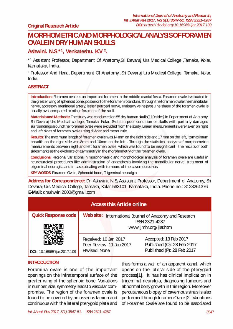
Fig. 1: Showing oval shaped foramen ovale(blackarrow) on left side of the skull.
Fig. 2: Showing almond shaped foramen ovale(blackarrow) on right side of the skull.
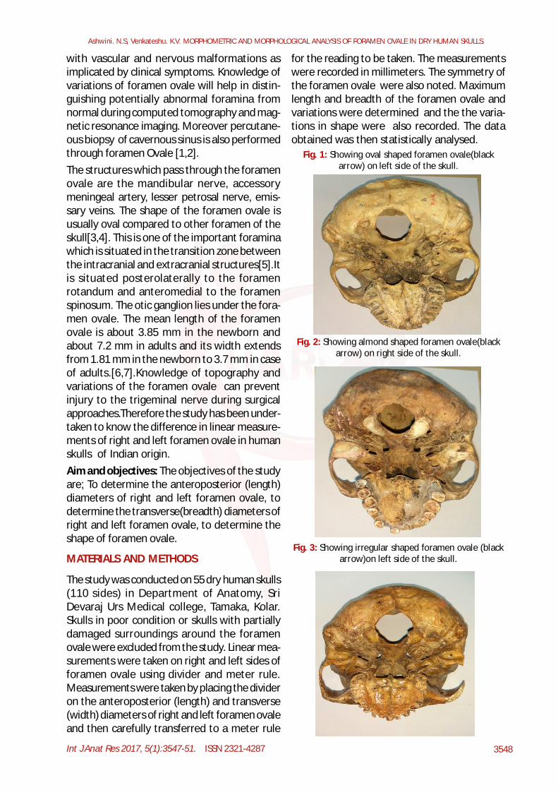
Fig. 3: Showing irregular shaped foramen ovale (blackarrow)on left side of the skull.
Int J Anat Res 2017, 5(1):3547-51. ISSN 2321-4287 3549
Ashwini. N.S, Venkateshu. K.V. MORPHOMETRIC AND MORPHOLOGICAL ANALYSIS OF FORAMEN OVALE IN DRY HUMAN SKULLS.
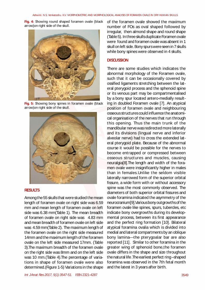
Fig. 4: Showing round shaped foramen ovale (blackarrow)on right side of the skull.
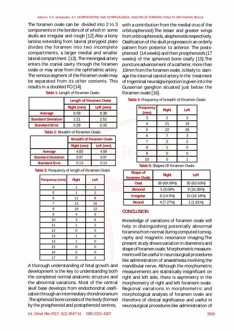
Fig. 5: Showing bony spines in foramen ovale (blackarrow)on right side of the skull.
of the foramen ovale showed the maximumnumber of FOs as oval shaped followed byirregular, then almond shape and round shape(Table 5). In three skulls duplicate Foramen ovalewere found and foramen ovale was absent in 1skull on left side. Bony spurs were seen in 7 skullswhile bony spines were observed in 4 skulls.
RESULTS
Among the 55 skulls that were studied the meanlength of foramen ovale on right side was 6.59mm and mean length of foramen ovale on leftside was 6.38 mm(Table 1). The mean breadthof foramen ovale on right side was 4.83 mmand mean breadth of foramen ovale on left sidewas 4.59 mm(Table 2). The maximum length ofthe foramen ovale on the right side measured14mm and the maximum length of the foramenovale on the left side measured 17mm. (Table3).The maximum breadth of the foramen ovaleon the right side was 8mm and on the left sidewas 10 mm.(Table 4).The percentage of varia-tions in shape of foramen ovale were alsodetermined.(Figure 1-5) Variations in the shape
DISCUSSION
There are some studies which indicates theabnormal morphology of the Foramen ovale,such that it can be occasionally covered byossified ligaments stretching between the lat-eral pterygoid process and the sphenoid spineor its venous part may be compartmentalisedby a bony spur located antero-medially result-ing in doubled Foramen ovale [7]. An atypicalposition of foramen ovale and neighbouringosseous structures could influence the anatomi-cal organisation of the nerves that run throughthis opening. Thus the main trunk of themandibular nerve was redirected more laterallyand its divisions (lingual nerve and inferioralveolar nerve) had to cross the extended lat-eral pterygoid plate. Because of the abnormalcourse it would be possible for the nerves tobecome entrapped or compressed betweenosseous structures and muscles, causingneuralgia[8].The length and width of the fora-men ovale were insignificantly higher in malesthan in females.Unlike the seldom visiblelaterally narrowed form of the superior orbitalfissure, a wide form with or without accessoryspine was the most commonly observed. Thediameters of both superior orbital fissures andovale foramina indicated the asymmetry of theneurocranium[9].Various bony outgrowths of theforamen ovale like spines, spurs, tubercles, etcindicate bony overgrowths during its develop-mental process, between its first appearanceand the perfect ring formation [10]. Bilateralatypical foramina ovalia which is divided intomedial and lateral compartments by an obliquebony lamina—the pterygoalar bar are alsoreported [11]. Similar to other foramina in thegreater wing of sphenoid bone,the foramenovale differs in the shape and size throughoutthe natural life .The earliest perfect ring –shapedforamina was observed in the 7th fetal monthand the latest in 3 years after birth.
Int J Anat Res 2017, 5(1):3547-51. ISSN 2321-4287 3550
Ashwini. N.S, Venkateshu. K.V. MORPHOMETRIC AND MORPHOLOGICAL ANALYSIS OF FORAMEN OVALE IN DRY HUMAN SKULLS.
The foramen ovale can be divided into 2 to 3components in the borders of of which in someskulls are irregular and rough [12].Also a bonylamina extending from lateral pterygoid platedivides the foramen into two incompletecompartments, a larger medial and smallerlateral compartment .[13]. The meningeal arteryenters the cranial cavity through the foramenovale or may arise from the ophthalmic artery.The venous segment of the Foramen ovale maybe separated from its other contents. Thisresults in a doubled FO [14].
Table 1: Length of Foramen Ovale.
Right (mm) Left (mm)Average 6.59 6.38
Standard Deviation 2.21 2.52Standard Error 0.29 0.33
Length of Foramen Ovale
Table 2: Breadth of Foramen Ovale.
Right (mm) Left (mm)Average 4.83 4.59
Standard Deviation 0.97 0.97Standard Error 0.13 0.13
Breadth of Foramen Ovale
Table 3: Frequency of length of Foramen Ovale.
Frequency (mm) Right Left
4 1 15 1 26 11 67 13 158 19 129 4 9
10 3 611 1 212 0 013 1 014 1 015 0 016 0 017 0 1
with a contribution from the medial crus of theorbitosphenoid).The lesser and greater wingsfrom orbitosphenoids, alisphenoids respectively.Ossification of the skull progresses in an orderlypattern from posterior to anterior. The posts-phenoid (14 weeks) and then presphenoids (17weeks) of the sphenoid bone ossify [15].Thepuncture advancement of a catheter, more than10mm from the foramen ovale, is likely to dam-age the internal carotid artery.In the treatmentof trigeminal neuralgia injection is given into theGusserian ganglion situated just below theForamen ovale [16].
A thorough understanding of fetal growth anddevelopment is the key to understanding boththe completed normal anatomic structure andthe abnormal variations. Most of the centralskull base develops from endochondral ossifi-cation through an intermediary chondrocranium.The sphenoid bone consists of the body (formedby the presphenoid and postsphenoid centres,
Table 4: Frequency of breadth of Foramen Ovale.
Frequency (mm)
Right Left
3 2 34 21 195 22 266 7 37 2 28 1 09 0 0
10 0 1
Table 5: Shapes Of Foramen Ovale.
Shape of foramen Ovale
Right Left
Oval 38 (69.09%) 35 (63.63%)
Almond 5 (9.09% 9 (16.36%)
Irregular 8 (14.5%) 10 (18.18%)
Round 4 (7.27%) 1 (1.81%)
CONCLUSION
Knowledge of variations of foramen ovale willhelp in distinguishing potentially abnormalforamina from normal during computed tomog-raphy and magnetic resonance imaging.Thepresent study shows variation in diameters andshape of foramen ovale. Morphometric measure-ments will be useful in neurosurgical procedureslike administration of anaesthesia involving themandibular nerve. Although the morphometricmeasurements are statistically insignificant onright and left side, there is asymmetry in themorphometry of right and left foramem ovale.Regional variations in morphometric andmorphological analysis of foramen ovale aretherefore of clinical significance and useful inneurosurgical procedures like administration of
Int J Anat Res 2017, 5(1):3547-51. ISSN 2321-4287 3551
anaesthesia involving the mandibular nerve,treatment of trigeminal neuraglia and in casesdealing with tumours of the cavernous sinus.
Conflicts of Interests: None
REFERENCES
[8]. J. Skrzat, J. Walocha1, R. Œrodek, A. Ni¿ankowska..Anatypical position of the foramen ovale. FoliaMorphol. 2006;65(4):396-399.
[9]. F.Burdan, W.Umlawska, W.Dworzanski. Anatomicalvariences and dimensions of the superior orbitalfissure and foramen Ovale in adults.FoliaMorphol.,2011;70(4):263-271.
[10]. Karan Bhagwan Khairnar, Prashant Aman RaoBhusari. An anatomical study on the foramen Ovaleand the Foramen spinosum.JCDR,2013;7(3):427-429.
[11]. S Suhani, H Mamatha, K Naveen. Bilateral atypicalforamina ovalia formation due to the presence ofpterygoalar bar: a rare osseous variation.OA Casereports,2013,2(16).
[12]. https://en.wikipedia.org/wiki/foramen Ovale[13]. Ray B,Gupta N, Ghose S. Anatomic variations of Fo-
ramen Ovale.Kathmandu University Medical Jour-nal,2005;3:64-68.
[14]. LangJ.Clinical Anatomy of the Head Neurocranium,Orbit and Craniocervical Region Springer-Vertag,Berlin. 1883.
[15]. Karthiga Devi,Thenmozhi M.S. Study on morphol-ogy, morphomertric and duplication of the Fora-men Ovale in human skulls of South Indian popula-tion. Indian Journal Of Applied Research,2016,6(7).
[16]. Shikha sharma , Chetna Thakur et al. Study of ana-tomic variant of foramen ovale and Spinosum indried human skulls. Int J Anat Res 2016;4(1):2002-06.
[1]. Kulkarni Saurabh P,Nikade Vrushali V.A morphomet-ric study of foramen ovale and foramen spinosumin dried Indian human skulls.International journalof recent trends in science and technology2013;7(2):74-75.
[2]. Nirupama Gupta, Anju Lata Rai. Foramen Ovale-Morphometry and its surgical importance. Innova-tive journal of medical and health science.2013;3(1):4-6.
[3]. Magi Murugan, Shaik Hussain Saheb. Morphomet-ric and morphological study on foramen ovale. Int.J. Anat. Res. 2014,2(4):664-666.
[4]. Jyothsna Patil,Naveen Kumar.The foramen OvaleMorphometry of sphenoid bone in South Indianpopulation.JCDR,2013,7(12):2668-2670.
[5]. Karishma Ravinthar, Thenmozhi. Morphometric studyof size and symmetry of foramen ovale in dryskulls.Journal of pharmaceutical sciences and re-search,2015;7(10):830-833.
[6]. Osunwoke E.A,Mbadugha C.C et al. A morphometricstudy of foramen Ovale and foramen spinosum ofthe human sphenoid bone in he South Nigerianpopulation. J.Appl.Biosci.,2010;1631-1635.
[7]. M.S.Somesh,H.B.Sridevi et al. A morphometric studyof foramen Ovale.Turkish Neurosurgery,2011;2 1 (3): 378-383.
How to cite this article:Ashwini. N.S, Venkateshu. K.V. MORPHOMETRIC ANDMORPHOLOGICAL ANALYSIS OF FORAMEN OVALE IN DRYHUMAN SKULLS. Int J Anat Res 2017;5(1):3547-3551. DOI:10.16965/ijar.2017.109
Ashwini. N.S, Venkateshu. K.V. MORPHOMETRIC AND MORPHOLOGICAL ANALYSIS OF FORAMEN OVALE IN DRY HUMAN SKULLS.