original article open access synthesis, characterization ...original article open access synthesis,...
TRANSCRIPT
Kumar et al. International Nano Letters 2013, 3:30http://www.inl-journal.com/content/3/1/30
ORIGINAL ARTICLE Open Access
Synthesis, characterization and optical propertiesof zinc oxide nanoparticlesSurabhi Siva Kumar1*, Putcha Venkateswarlu2, Vanka Ranga Rao3 and Gollapalli Nageswara Rao4
Abstract
Zinc oxide nanoparticles were synthesized using a simple precipitation method with zinc sulfate and sodiumhydroxide as starting materials. The synthesized sample was calcined at different temperatures for 2 h. The sampleswere characterized by X-ray diffraction (XRD), scanning electron microscopy (SEM), energy dispersive spectroscopy(EDS), and proton-induced X-ray emission (PIXE) analysis. SEM images show various morphological changes of ZnOobtained by the above method. The average crystallite sizes of the samples were calculated from the full width athalf maximum of XRD peaks by using Debye-Scherrer's formula and were found to be in the nanorange. EDSshows that the above route produced highly pure ZnO nanostructures. PIXE technique was used for traceelemental analysis of ZnO. The optical band gaps of various ZnO powders were calculated from UV-visible diffusereflectance spectroscopic studies.
Keywords: Nanostructures, Semiconductors, Chemical synthesis, Catalytic properties, Optical properties
BackgroundNanosized particles of semiconductor materials havegained much more interest in recent years due to theirdesirable properties and applications in different areassuch as catalysts [1], sensors [2], photoelectron devices[3,4], highly functional and effective devices [5]. Thesenanomaterials have novel electronic, structural, andthermal properties which are of high scientific interestsin basic and applied fields. Zinc oxide (ZnO) is a wideband gap semiconductor with an energy gap of 3.37 eVat room temperature. It has been used considerably forits catalytic, electrical, optoelectronic, and photochem-ical properties [6-9]. ZnO nanostructures have a greatadvantage to apply to a catalytic reaction process due totheir large surface area and high catalytic activity [10].Since zinc oxide shows different physical and chemicalproperties depending upon the morphology of nano-structures, not only various synthesis methods but alsothe physical and chemical properties of synthesized zincoxide are to be investigated in terms of its morphology.Many methods have been described in the literature for
the production of ZnO nanostructures such as laser
* Correspondence: [email protected] of Chemistry, Anil Neerukonda Institute of Technology andSciences, Sangivalasa, Visakhapatnam 531 162, IndiaFull list of author information is available at the end of the article
© 2013 Kumar et al.; licensee Springer. This is aAttribution License (http://creativecommons.orin any medium, provided the original work is p
ablation [11], hydrothermal methods [12], electrochemicaldepositions [13], sol–gel method [14], chemical vapordeposition [15], thermal decomposition [16], and combus-tion method [17,18]. Recently, ZnO nanoparticles wereprepared by ultrasound [19], microwave-assisted combus-tion method [20], two-step mechanochemical-thermalsynthesis [21], anodization [22], co-precipitation [23], andelectrophoretic deposition [24].Rodrigues-Paez et al. synthesized zinc oxide nano-
particles with different morphologies by controlling dif-ferent parameters of the precipitation process such assolution concentration, pH, and washing medium [25].In the present study, ZnO nanostructures were synthe-sized using a simple precipitation method. Zinc sulfateheptahydrate and sodium hydroxide were used as pre-cursors to formulate ZnO nanostructures. The preparedsamples were characterized by X-ray diffraction (XRD)and scanning electron microscopy (SEM), and the purityof the sample was tested by energy dispersive spectros-copy (EDS) and proton-induced X-ray emission (PIXE)analysis. The band gap energies of the samples were cal-culated from diffuse reflectance spectroscopy. Themorphology, crystallite size, and optical properties ofZnO nanostructures were investigated, and an attemptwas made to correlate the optical properties of ZnO withmorphology and crystallite size.
n Open Access article distributed under the terms of the Creative Commonsg/licenses/by/2.0), which permits unrestricted use, distribution, and reproductionroperly cited.
20 40 60 80
0
400
800
(202
)
(004
)
(201
)(112
)(200
)(103
)
(110
)
(102
)
(101
)(002
)100 0C
3000C
5000C
7000C
Inte
nsi
ty
2 Theta
9000C
(100
)
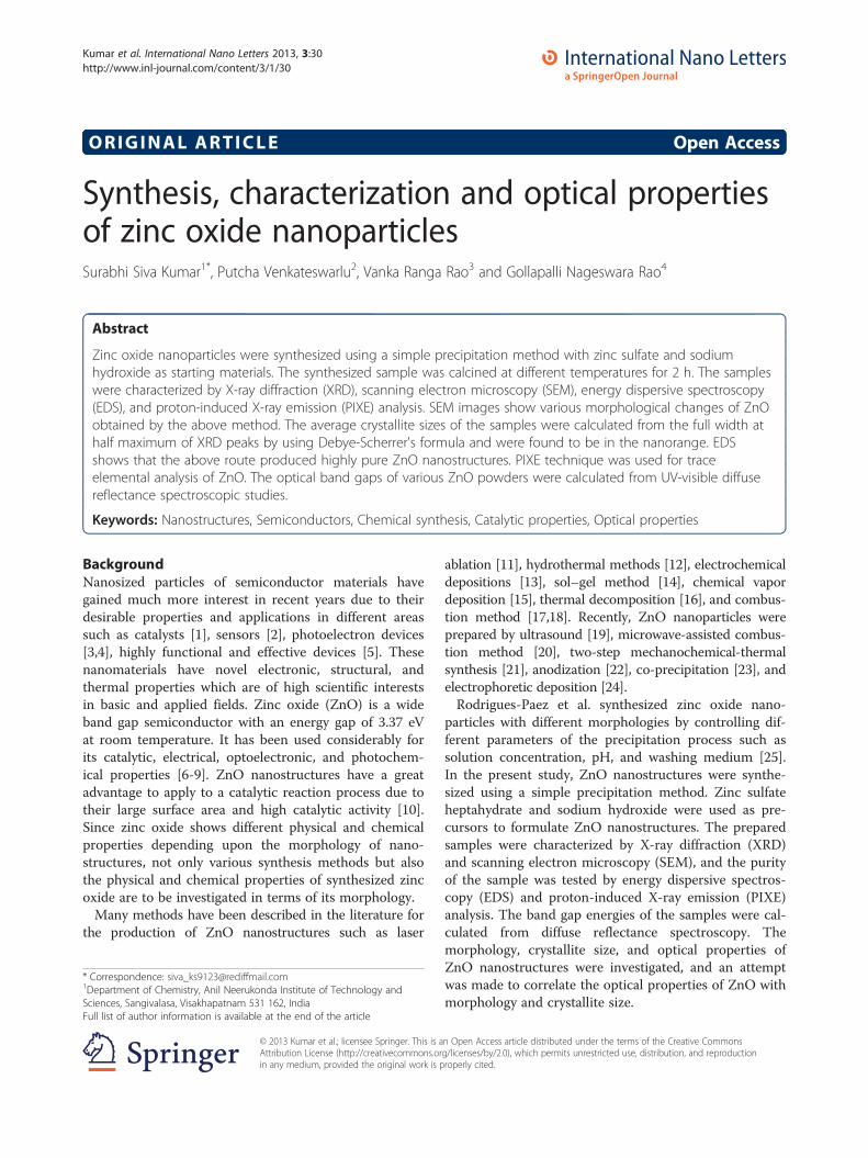
Figure 1 XRD patterns of ZnO-1 calcined at different temperatures.
Kumar et al. International Nano Letters 2013, 3:30 Page 2 of 6http://www.inl-journal.com/content/3/1/30
Methods
MaterialsZinc sulfate heptahydrate and sodium hydroxide wereused in the experiments. All the chemicals used were ofanalytical reagent grade obtained from Merck (Mumbai,India), and deionized water is used for the preparationof solutions.
Synthesis of ZnOTo the aqueous solution of zinc sulfate, sodium hydrox-ide solution was added slowly dropwise in a molar ratioof 1:2 under vigorous stirring, and the stirring was con-tinued for 12 h. The precipitate obtained was filtered andwashed thoroughly with deionized water. The precipitatewas dried in an oven at 100°C and ground to fine powderusing agate mortar [26]. The powder obtained from theabove method was calcined at different temperatures suchas 300°C, 500°C, 700°C, and 900°C for 2 h.
CharacterizationXRD and SEMThe compounds were characterized for their structureand morphology by XRD and SEM. The XRD patterns of
Table 1 Average crystallite size of ZnO obtained fromXRD using Equation 1
Calcination temperature (°C) Crystallite size, D (nm)
300 64
500 74
700 131
900 187
the powdered samples were recorded using a Bruker D8Advanced X-ray diffractometer (Bruker Optik GmbH,Ettlingen, Germany) with CuKα radiation (λ = 1.5418 Å,rated as 1.6 kW), and SEM images of the samples weretaken using a Philips XL 30 ESEM scanning electronmicroscope (FEI-Philips Company, Hillsboro).
UV–vis diffuse reflectance spectroscopyUV–vis spectroscopy was used to characterize theoptical absorption properties of ZnO. The UV–vis ab-sorption spectra of the samples were recorded in thewavelength range of 200 to 800 nm using a ShimadzuUV 3600 UV–vis-NIR spectrometer (Shimadzu Corpor-ation, Kyoto, Japan) in diffuse reflectance mode usingBaSO4 as reference. Spectra were recorded at roomtemperature, and the data were transformed through theKubelka-Munk function [27].
Proton-induced X-ray emissionFinely powdered samples were mixed with pure graphitein the ratio of 1:1 (150 mg each), homogenized, andpressed into a 13-mm-diameter pellet. PIXE measure-ments have been carried out on a 3-MV horizontalpelletron accelerator at the Institute of Physics, Bhuba-neswar [28]. Proton beam was collimated to a diameterof 3 mm on the target. A Si(Li) detector was kept at 90°with respect to the beam direction. The detector has anactive area of 30 mm2 with a beryllium window having athickness of 12 μm and an energy resolution of 170 eV at5.9 keV. Integrated charge on the sample was measuredusing a current integrator, which was connected to the tar-get holder. The X-rays coming out of the chamber
cba
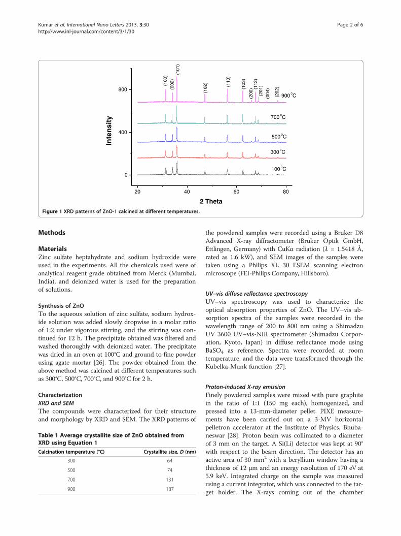
Figure 2 SEM images of ZnO samples calcined at three different temperatures. (a) 300°C, (b) 500°C, and (c) 700°C.
Kumar et al. International Nano Letters 2013, 3:30 Page 3 of 6http://www.inl-journal.com/content/3/1/30
through a 95-μm Mylar window traveled through an airgap of 4 cm before entering the Si(Li) detector.The targets were kept in the PIXE chamber at 45° to the
beam. The Si(Li) detector is placed at 90° to the beam, andthe beam current was kept in the range of 3 to 10 nA. Spec-tra were recorded using a Canberra MCA [29] (CanberraIndustries, Meriden, CT, USA) and were transferred to apersonal computer [30].PIXE spectral analyses were carried out using GUPIX-95
[31] software that provides nonlinear least-square fittingof the spectrum. The thick-target PIXE analysis wasperformed since the target was thick enough to stop theproton beam entirely. To check the adopted analysis pro-cedure and input parameters, external standard methodwas adopted using the macrometer standards and othercertified reference materials, and accordingly, the valueswere normalized. Further details about the analysis proced-ure can be obtained from our earlier references [28,29].
Results and discussionCatalyst characterizationXRD analysisThe XRD patterns of the ZnO powders prepared by theabove method and calcined at different temperatures are
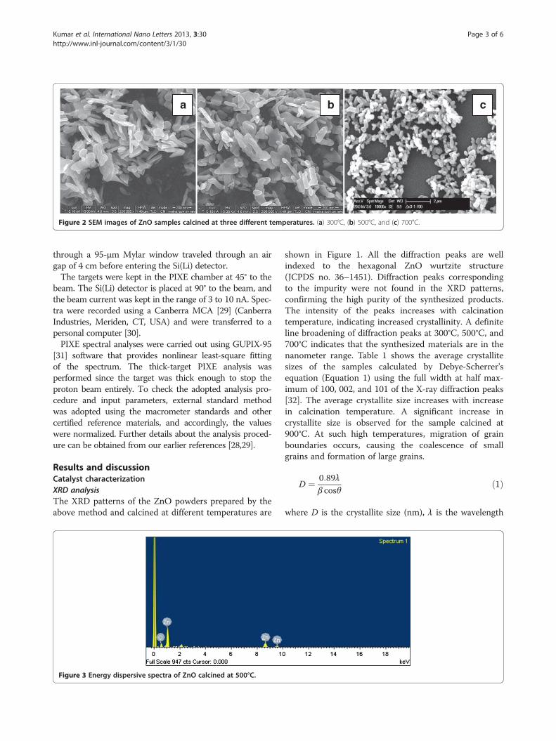
Figure 3 Energy dispersive spectra of ZnO calcined at 500°C.
shown in Figure 1. All the diffraction peaks are wellindexed to the hexagonal ZnO wurtzite structure(JCPDS no. 36–1451). Diffraction peaks correspondingto the impurity were not found in the XRD patterns,confirming the high purity of the synthesized products.The intensity of the peaks increases with calcinationtemperature, indicating increased crystallinity. A definiteline broadening of diffraction peaks at 300°C, 500°C, and700°C indicates that the synthesized materials are in thenanometer range. Table 1 shows the average crystallitesizes of the samples calculated by Debye-Scherrer'sequation (Equation 1) using the full width at half max-imum of 100, 002, and 101 of the X-ray diffraction peaks[32]. The average crystallite size increases with increasein calcination temperature. A significant increase incrystallite size is observed for the sample calcined at900°C. At such high temperatures, migration of grainboundaries occurs, causing the coalescence of smallgrains and formation of large grains.
D ¼ 0:89λβ cosθ
ð1Þ
where D is the crystallite size (nm), λ is the wavelength
350 375 400 425 450
0
1
2
c
d
b
Ab
sorb
ance
Wavelength (nm)
a
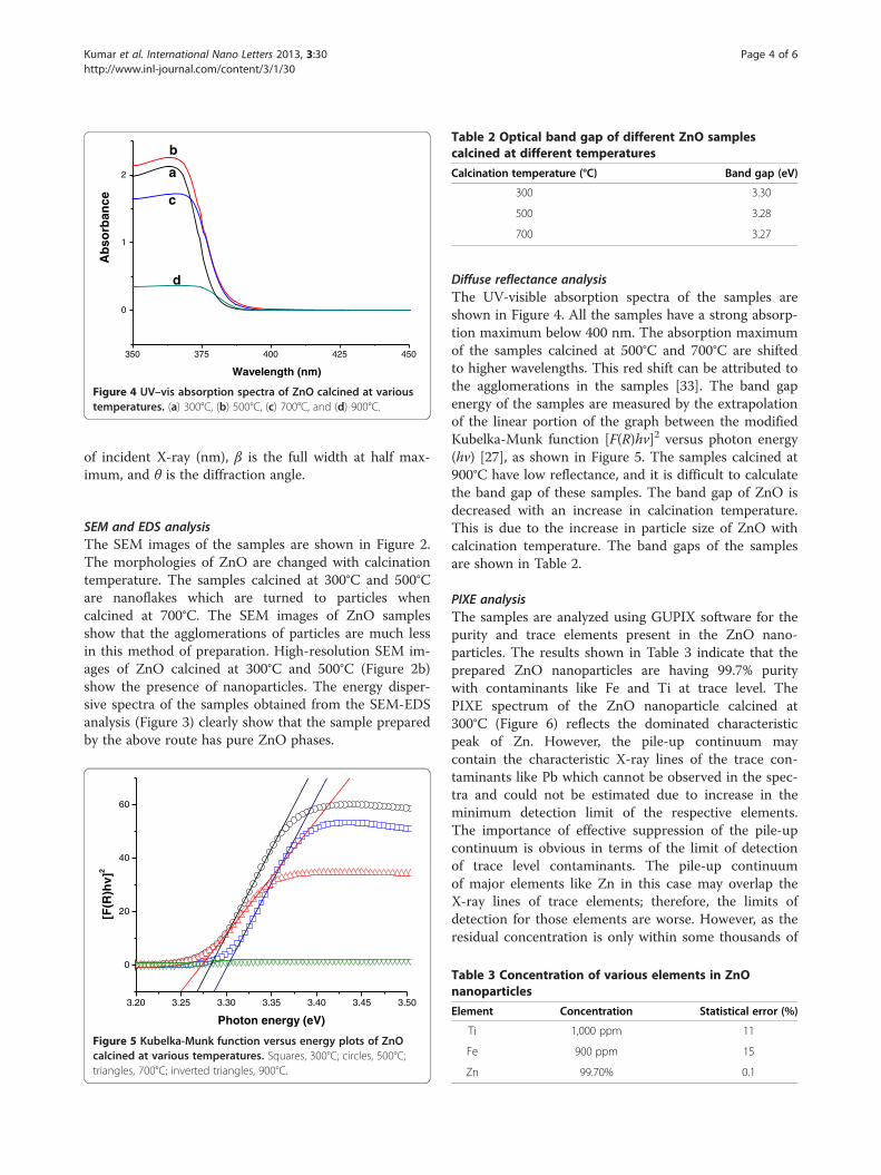
Figure 4 UV–vis absorption spectra of ZnO calcined at varioustemperatures. (a) 300°C, (b) 500°C, (c) 700°C, and (d) 900°C.
Table 2 Optical band gap of different ZnO samplescalcined at different temperatures
Calcination temperature (°C) Band gap (eV)
300 3.30
500 3.28
700 3.27
Kumar et al. International Nano Letters 2013, 3:30 Page 4 of 6http://www.inl-journal.com/content/3/1/30
of incident X-ray (nm), β is the full width at half max-imum, and θ is the diffraction angle.
SEM and EDS analysisThe SEM images of the samples are shown in Figure 2.The morphologies of ZnO are changed with calcinationtemperature. The samples calcined at 300°C and 500°Care nanoflakes which are turned to particles whencalcined at 700°C. The SEM images of ZnO samplesshow that the agglomerations of particles are much lessin this method of preparation. High-resolution SEM im-ages of ZnO calcined at 300°C and 500°C (Figure 2b)show the presence of nanoparticles. The energy disper-sive spectra of the samples obtained from the SEM-EDSanalysis (Figure 3) clearly show that the sample preparedby the above route has pure ZnO phases.
3.20 3.25 3.30 3.35 3.40 3.45 3.50
0
20
40
60
[F(R
)hv]
2
Photon energy (eV)
Figure 5 Kubelka-Munk function versus energy plots of ZnOcalcined at various temperatures. Squares, 300°C; circles, 500°C;triangles, 700°C; inverted triangles, 900°C.
Diffuse reflectance analysisThe UV-visible absorption spectra of the samples areshown in Figure 4. All the samples have a strong absorp-tion maximum below 400 nm. The absorption maximumof the samples calcined at 500°C and 700°C are shiftedto higher wavelengths. This red shift can be attributed tothe agglomerations in the samples [33]. The band gapenergy of the samples are measured by the extrapolationof the linear portion of the graph between the modifiedKubelka-Munk function [F(R)hν]2 versus photon energy(hν) [27], as shown in Figure 5. The samples calcined at900°C have low reflectance, and it is difficult to calculatethe band gap of these samples. The band gap of ZnO isdecreased with an increase in calcination temperature.This is due to the increase in particle size of ZnO withcalcination temperature. The band gaps of the samplesare shown in Table 2.
PIXE analysisThe samples are analyzed using GUPIX software for thepurity and trace elements present in the ZnO nano-particles. The results shown in Table 3 indicate that theprepared ZnO nanoparticles are having 99.7% puritywith contaminants like Fe and Ti at trace level. ThePIXE spectrum of the ZnO nanoparticle calcined at300°C (Figure 6) reflects the dominated characteristicpeak of Zn. However, the pile-up continuum maycontain the characteristic X-ray lines of the trace con-taminants like Pb which cannot be observed in the spec-tra and could not be estimated due to increase in theminimum detection limit of the respective elements.The importance of effective suppression of the pile-upcontinuum is obvious in terms of the limit of detectionof trace level contaminants. The pile-up continuumof major elements like Zn in this case may overlap theX-ray lines of trace elements; therefore, the limits ofdetection for those elements are worse. However, as theresidual concentration is only within some thousands of
Table 3 Concentration of various elements in ZnOnanoparticles
Element Concentration Statistical error (%)
Ti 1,000 ppm 11
Fe 900 ppm 15
Zn 99.70% 0.1
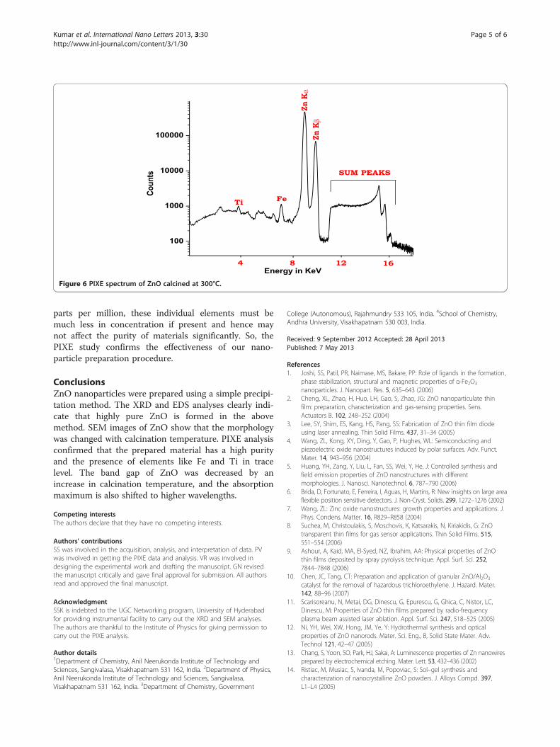
Figure 6 PIXE spectrum of ZnO calcined at 300°C.
Kumar et al. International Nano Letters 2013, 3:30 Page 5 of 6http://www.inl-journal.com/content/3/1/30
parts per million, these individual elements must bemuch less in concentration if present and hence maynot affect the purity of materials significantly. So, thePIXE study confirms the effectiveness of our nano-particle preparation procedure.
ConclusionsZnO nanoparticles were prepared using a simple precipi-tation method. The XRD and EDS analyses clearly indi-cate that highly pure ZnO is formed in the abovemethod. SEM images of ZnO show that the morphologywas changed with calcination temperature. PIXE analysisconfirmed that the prepared material has a high purityand the presence of elements like Fe and Ti in tracelevel. The band gap of ZnO was decreased by anincrease in calcination temperature, and the absorptionmaximum is also shifted to higher wavelengths.
Competing interestsThe authors declare that they have no competing interests.
Authors' contributionsSS was involved in the acquisition, analysis, and interpretation of data. PVwas involved in getting the PIXE data and analysis. VR was involved indesigning the experimental work and drafting the manuscript. GN revisedthe manuscript critically and gave final approval for submission. All authorsread and approved the final manuscript.
AcknowledgmentSSK is indebted to the UGC Networking program, University of Hyderabadfor providing instrumental facility to carry out the XRD and SEM analyses.The authors are thankful to the Institute of Physics for giving permission tocarry out the PIXE analysis.
Author details1Department of Chemistry, Anil Neerukonda Institute of Technology andSciences, Sangivalasa, Visakhapatnam 531 162, India. 2Department of Physics,Anil Neerukonda Institute of Technology and Sciences, Sangivalasa,Visakhapatnam 531 162, India. 3Department of Chemistry, Government
College (Autonomous), Rajahmundry 533 105, India. 4School of Chemistry,Andhra University, Visakhapatnam 530 003, India.
Received: 9 September 2012 Accepted: 28 April 2013Published: 7 May 2013
References1. Joshi, SS, Patil, PR, Naimase, MS, Bakare, PP: Role of ligands in the formation,
phase stabilization, structural and magnetic properties of α-Fe2O3
nanoparticles. J. Nanopart. Res. 5, 635–643 (2006)2. Cheng, XL, Zhao, H, Huo, LH, Gao, S, Zhao, JG: ZnO nanoparticulate thin
film: preparation, characterization and gas-sensing properties. Sens.Actuators B. 102, 248–252 (2004)
3. Lee, SY, Shim, ES, Kang, HS, Pang, SS: Fabrication of ZnO thin film diodeusing laser annealing. Thin Solid Films. 437, 31–34 (2005)
4. Wang, ZL, Kong, XY, Ding, Y, Gao, P, Hughes, WL: Semiconducting andpiezoelectric oxide nanostructures induced by polar surfaces. Adv. Funct.Mater. 14, 943–956 (2004)
5. Huang, YH, Zang, Y, Liu, L, Fan, SS, Wei, Y, He, J: Controlled synthesis andfield emission properties of ZnO nanostructures with differentmorphologies. J. Nanosci. Nanotechnol. 6, 787–790 (2006)
6. Brida, D, Fortunato, E, Ferreira, I, Aguas, H, Martins, R: New insights on large areaflexible position sensitive detectors. J. Non-Cryst. Solids. 299, 1272–1276 (2002)
7. Wang, ZL: Zinc oxide nanostructures: growth properties and applications. J.Phys. Condens. Matter. 16, R829–R858 (2004)
8. Suchea, M, Christoulakis, S, Moschovis, K, Katsarakis, N, Kiriakidis, G: ZnOtransparent thin films for gas sensor applications. Thin Solid Films. 515,551–554 (2006)
9. Ashour, A, Kaid, MA, El-Syed, NZ, Ibrahim, AA: Physical properties of ZnOthin films deposited by spray pyrolysis technique. Appl. Surf. Sci. 252,7844–7848 (2006)
10. Chen, JC, Tang, CT: Preparation and application of granular ZnO/Al2O3
catalyst for the removal of hazardous trichloroethylene. J. Hazard. Mater.142, 88–96 (2007)
11. Scarisoreanu, N, Metai, DG, Dinescu, G, Epurescu, G, Ghica, C, Nistor, LC,Dinescu, M: Properties of ZnO thin films prepared by radio-frequencyplasma beam assisted laser ablation. Appl. Surf. Sci. 247, 518–525 (2005)
12. Ni, YH, Wei, XW, Hong, JM, Ye, Y: Hydrothermal synthesis and opticalproperties of ZnO nanorods. Mater. Sci. Eng., B, Solid State Mater. Adv.Technol 121, 42–47 (2005)
13. Chang, S, Yoon, SO, Park, HJ, Sakai, A: Luminescence properties of Zn nanowiresprepared by electrochemical etching. Mater. Lett. 53, 432–436 (2002)
14. Ristiac, M, Musiac, S, Ivanda, M, Popoviac, S: Sol–gel synthesis andcharacterization of nanocrystalline ZnO powders. J. Alloys Compd. 397,L1–L4 (2005)
Kumar et al. International Nano Letters 2013, 3:30 Page 6 of 6http://www.inl-journal.com/content/3/1/30
15. Wu, JJ, Liu, SC: Low-temperature growth of well-aligned ZnO nanorods bychemical vapor deposition. Adv. Mater. 14, 215–218 (2002)
16. Wang, RC, Tsai, CC: Efficient synthesis of ZnO nanoparticles, nanowalls, andnanowires by thermal decomposition of zinc acetate at a low temperature.Appl. Phys. A. 94, 241–245 (2009)
17. Lamas, DG, Lascalea, GE, Walsoc, NE: Synthesis and characterization ofnanocrystalline powders for partially stabilized zirconia ceramics. J. Eur.Ceram. Soc. 18, 1217–1221 (1998)
18. Badhuri, S, Badhuri, SB: Enhanced low temperature toughness of Al2O3-ZrO2
nano/nano composites. Nanostrct. Mater. 8, 755–763 (1997)19. Khorsand, Z, Abid, A, Majid, WH, Wang, HZ, Yousefi, R, Golsheikh, M, Ren, ZF:
Sonochemical synthesis of hierarchical ZnO nanostructures. UltrasonicSonochemistry 20, 395–400 (2013)
20. Kooti, M, Nagdhi Sedish, A: Microwave-assisted combustion synthesis ofZnO nanoparticles. J. Chem (2013). doi:10.1155/2013/562028
21. Rajesh, D, Vara Lakshmi, B, Sunandana, CS: Two-step synthesis andcharacterization of ZnO nanoparticles. Physica B-Cond. Mater. 407,4537–4539 (2012)
22. Shetty, A, Nanda, K: Synthesis of zinc oxide porous structures by anodisationwith water as an electrolyte. Appl. Phys. A. 109, 151–157 (2012)
23. Singh, O, Kohli, N, Singh, RC: Precursor controlled morphology of zinc oxideand its sensing behavior. Sens. Actuators B. 178, 149–154 (2013)
24. Vazquez, A, Lopez, IA, Gomez, I: Growth mechanism of one-dimensionalzinc sulfide nanostructures through electrophoretic deposition. J. Mater.Sci 48, 2701–2704 (2013)
25. Rodrigues-Paez, J, Caballero, AC, Villegas, M, Moure, C, Duran, P, Fernandz,JF: Controlled precipitation methods: formation mechanism of ZnOnanoparticles. J. Eur. Ceram. Soc. 21, 925–930 (2001)
26. Daneshvar, N, Aber, S, Sayed Dorraji, MS, Khataee, AR, Rasoulifard, MH:Preparation and investigation of photocatalytic properties of ZnOnanocrystals: effect of operational parameters and kinetic study. Int. J.Chem. Biom. Eng 1(1), 24–29 (2008)
27. Cimitan, S, Albonetti, S, Forni, L, Peri, F, Lazzari, D: Solvothermal synthesisand properties control of doped ZnO nanoparticles. J. Colloid. Interface Sci.329, 73–80 (2009)
28. Vijayan, V, Ramamurthy, VS, Behera, SN, Puri, S, Shanti, JS, Singh, N:Elemental composition of fly ash from a coal fired thermal power plant: astudy using PIXE and EDXRF. X-ray spectrometry. 26, 65–68 (1997)
29. Vijayan, V, Nayak, PK, Chakrovortty, V: Proton induced X-ray emission studieson Indian copper coins. Ind. J. Phys. A. 76, 477–479 (2002)
30. Maxwell, JA, Teesdale, WJ, Campbell, JL: The Guelph PIXE software packageII. Nucl. Instrum. Methods B. 95, 407–421 (1995)
31. Gleiter, H: Nanocrystalline materials. Prog. Mater. Sci. 33, 223–315 (1989)32. Chen, CC, Liu, P, Lu, CH: Synthesis and characterization of nano-sized ZnO
powders by direct precipitation method. Chem. Eng. J. 144, 509–513 (2008)33. Babita, B, Kishore Kumar, D, Manorama, SV: Hydrothermal synthesis of highly
crystalline ZnO nanoparticles: a competitive sensor for LPG and EtOH. Sens.Actuators B. 119, 676–682 (2006)
doi:10.1186/2228-5326-3-30Cite this article as: Kumar et al.: Synthesis, characterization and opticalproperties of zinc oxide nanoparticles. International Nano Letters 20133:30.
Submit your manuscript to a journal and benefi t from:
7 Convenient online submission
7 Rigorous peer review
7 Immediate publication on acceptance
7 Open access: articles freely available online
7 High visibility within the fi eld
7 Retaining the copyright to your article
Submit your next manuscript at 7 springeropen.com