original article interleukin-17a promotes paraquat … · original article interleukin-17a promotes...
TRANSCRIPT
Int J Clin Exp Pathol 2017;10(2):2436-2445www.ijcep.com /ISSN:1936-2625/IJCEP0045674
Original Article Interleukin-17A promotes paraquat-induced acute lung injury on mice
Hong-Wei Song, Wei Liu, Chen Yang, Xiao-Wei Liu, Zhi Liu
Department of Emergency, The First Affiliated Hospital of China Medical University, Shenyang, P. R. China
Received December 2, 2016; Accepted December 22, 2016; Epub February 1, 2017; Published February 15, 2017
Abstract: Objective: Interleukin-17A (IL-17A) is a potent proinflammatory factor. Our study aimed to investigate the role of IL-17A on acute lung injury (ALI) induced by paraquat (PQ) and the potential way. Methods: A total of 180 female mice were randomly and evenly divided into 9 groups: Control group (NS), PQ groups (4 groups) and PQ+A b groups (4 groups). NS group mice were treated with equal volume of physiological saline by gastric gavage. Mice in PQ and PQ+A b groups were exposed to PQ solution by gastric gavage (25 mg/kg). PQ+A b groups mice were intraperitoneally (i.p.) injected with IL-17A neutralizing antibody (5 mg/kg) 2 hours after PQ perfusion, all of managements above were one-off. Mice were executed respectively at 8 h, 24 h, 48 h and 72 h, NS group mice at 24 h. Lung pathological change was observed with HE staining, wet-to-dry (W/D) ratio and total protein content in broncho-alveolar lavage fluid (BALF) were determined, changes of cytokines were detected by ELISA, leukocyte and neutrophil in BALF were recorded, lung IL-17A and Nuclear Factor-κB p65 (NF-κB p65) were examined by immuno-histochemical (IHC), q-PCR was used for the expression of IL-17A mRNA in lung tissue. Results: After PQ perfusion, levels and expression of IL-17A in mice increased (P<0.05); While blockade IL-17A with antibody, the ALI induced by PQ was attenuated, activation of NF-κB p65 and the percentage of neutrophil were reduced (P<0.05). Conclusion: IL-17A promotes the process of ALI induced by PQ, and probably by activating NF-κB p65 and recruiting neutrophil.
Keywords: Interleukin-17A, paraquat, acute lung injury, mouse, Nuclear Factor-κB p65, neutrophil
Introduction
Paraquat (PQ) is an organic heterocyclic con-tact defoliant and herbicide, which is widely used in agriculture around the world, especially in the developing countries [1]. PQ is highly toxic to human and animal, and oral is the main way of poisoning. Frequent PQ poisoning and high mortality makes it become a severe public health issue in developing countries since no specific treatment was effective at present, therefore, many countries try to limit the pro-duction and use of PQ. However, it is still widely used due to the effective weed control effect, and the poisoning therefore is staying at a high level.
PQ can cause multiple organ injury or even fail-ure, but lung is the main target organ [2]. In spite of the mechanisms of PQ-induced lung injury stay not been fully elucidated, current research suggests that the generation of oxy-gen radicals is the initiating factor. PQ poison-
ing early produces oxygen radicals [3], which then activates a variety of effector cells and prompts the release of inflammatory mediators to damage lung tissues [4], causes the patho-logical characteristics of pulmonary interstitial edema, hemorrhage [5], clinically for Acute Respiratory Distress Syndrome (ARDS) as the major performance, the severe cases can quickly appear to death for respiratory failure. After the acute injury periods, pulmonary fibro-sis results in respiratory failure to death [6]. Recent studies show that the participation of inflammation medium is an important part of the PQ poisoning lung injury.
IL-17A also known as IL-17, is a pro-inflammato-ry cytokine, which mainly produced by T helper cell 17 (Th17), natural killer cell and neutrophil [7]. Studies show that, since IL-17A could induce respiratory epithelial cells to secrete CXCL8, CXCL1, CXCL5, IL-6, GCSF, and GM-CSF, which in turn recruit neutrophils to the airways [8, 9], the cytokine implicates in a multitude of inflam-
Interleukin-17A in paraquat-induced acute lung injury
2437 Int J Clin Exp Pathol 2017;10(2):2436-2445
(25 mg/kg). Mice in PQ+A b groups were intra-peritoneally (i.p.) injected with IL-17A neutraliz-ing antibody (Catalog Number: 16-7173, eBiosi-cence, USA) (5 mg/kg) 2 hours after PQ perfusion, all of the managements above were one-off.
Specimen harvest
At 8 h, 24 h, 48 h and 72 h after the above treatments, mice from each group were execut-ed after anesthetized with 10% chloral hydrate (0.3 ml/100 g i.p.), NS group mice were execut-ed at 24 h. Peripheral blood, bronchoalveolar lavage fluid (BALF) and lung tissues were obtained respectively.
Peripheral blood was centrifuged for 10 min (1500 rpm, 4°C) to obtain the upper serum. 6 mice tracheas were cannulated, and each mouse was lavaged with phosphate buffered saline (PBS) three times each mouse immedi-ately after blood was collected, BALF was col-lected and the volume was recorded. Then the BALF was mixed well and centrifuged (1500 rpm, 4°C) for 10 min. Supernatants were stor- ed at -80°C for total protein analysis and cyto-kines assay, pellets were prepared for inflam-matory cell counts. Mice lungs which were not lavaged were used to do Wet-to-Dry (W/D) ratio calculation, histological study and q-PCR test.
Lung wet-to-dry (W/D) calculation
Lungs were cleared and weighed immediately after removal to obtain the “wet” weight, and then placed in an oven at 60°C for 72 h to obtain the “dry” weight.
ELISA for cytokines in serum and BALF
IL-17A, IL-6, TNF-α in serum and IL-17A in BALF were detected by Enzyme-linked (ELISA) kits (Bioss, Beijing, CHN) according to the manufac-turer’s instructions.
Inflammatory cell counts in BALF
BALF pellets were resuspended in 0.3 ml saline solution, total number of leukocytes and neu-trophils were calculated with a hemocyto- meter.
Total protein analysis in BALF
Total protein content in BALF was measured by Bradford method using Coomassie brilliant
matory lung diseases, both autoimmune and acquired [10]. Reports revealed that, IL-17AR (IL-17A receptor)-deficient mice or neutralized with IL-17A antibody reduced neutrophil influx, attenuated the early lung response to silica particles or bleomycin (BLM), and then alleviat-ed the lung injury induced by silica particles or BLM [7, 11]. Recent evidences confirmed that elevated IL-17A expression closely correlated to the development of Acute Lung Injury (ALI), IL-17A-deficient mouse was resistant to the induction of ALI, in addition, blockade IL-17A with neutralizing antibody ameliorated the induction of ALI induced by lipopolysaccharide (LPS) or multiple-trauma [12, 13]. The above studies prompted us to speculate whether IL-17A implicates in the development of ALI induced by PQ poisoning.
In our study, we built a mouse model of acute PQ poisoning and blockade IL-17A in vivo with neutralizing antibody to investigate the role of IL-17A in the ALI process, and further to explore the possible ways.
Materials and methods
Animals
Healthy female ICR (Institute of Cancer Rese- arch) mice (SPF grade, 6-8 weeks, 26-30 g in body weight) were purchased from Liaoning Changsheng Biological Technology Company (Animal production license No.: SCXK (l) 2015-0001). The mice were kept under a 12-h light/12-h dark cycle with free access to food and water for 1 week prior to experimental pro-cedures in the Animal Lab of China Medical University Science Experiment Center. Animal experiments were in accordance with ethical policies of the International Journal of Exper- imental Pathology and approved by the local ethical committee.
Animal grouping and model establishment
A total of 180 female mice were randomly and evenly divided into 9 groups: Control group (NS, n=20), PQ groups and PQ+A b groups, then both of PQ and PQ+A b groups divided into 4 subgroups (n=20) respectively according to 8 h, 24 h, 48 h, 72 h. Mice in NS group were treated with equal volume of physiological saline by gastric gavage. Mice in PQ and PQ+A b groups were exposed to PQ solution (Shaanxi Galen Crop Science co., LTD, CHN) by gastric gavage
Interleukin-17A in paraquat-induced acute lung injury
2438 Int J Clin Exp Pathol 2017;10(2):2436-2445
mary antibody (IL-17A for 1:50; NF-κB p65 for 1:100) (IL-17A:130-82-1-AP; NF-κB p65:8242P. Proteintech, Wuhan, CHN) over night at 4°C. The sections were then washed with PBS and incubated with biotin-conjugated secondary antibody (ZB-2301, Beyotime, Shanghai, CHN) (1:200 diluted) for 30 min at 37°C, then, strep-tavidin/HRP (ZB-2301, Beyotime, Shanghai, China) was added and 100 μl diaminobenzidine (DAB) was added successively until reaction was terminated by the tap water, dyeing time was controlled under microscope. Finally, the slides were re-dyed with hematoxylin (Solarbio, Beijing, China), dehydrated, vitrified and mount-ed before observed under light microscope. Negative controls were generated by omitting the primary antibodies. The results were evalu-ated semi-quantitatively according to the per-centage of positive cells in 5 randomly selected fields under 400-fold magnification and then take pictures [15]. For the score of positive cell ratio, 0-1%, 1-10%, 10-50%, 50-80% and 80-100% were scored as 0, 1, 2, 3 and 4, respectively. For intensity score, negative, weakly positive, positive and strongly positive were scored as 0, 1, 2, and 3, respectively. IHC score value = positive cell ratio score × inten-sity score.
All histology procedures were performed by two experienced pathologist who were blinded to the treatment group.
Expression of IL-17A mRNA in lung tissue
Lung tissues used for q-PCR were stored at -80°C, and total RNA was isolated from lung homogenates with Trizol reagent (15596026, Invitrogen, San Diego, CA, USA), then used RT kit (TaKaRa, Dalian, China) perform the reverse transcription (RT) reaction in 20 μl system. q-PCR reaction was performed in 20 μl system containing 10 μl SYBR® Premix Ex TaqTM, 0.8 μl forward primer (10 μM), 0.8 μl reverse primer (10 μM), 1.6 μl cDNA template, and 6.8 μl dis-
blue G-250 kits (Solarbio, Beijing, China) in accordance with the manufacturer’s instru- ctions.
Tissue preparation and histological study
Left upper lung tissues were fixed with 10% neutral formalin, embedded in paraffin, and sliced at thickness of 5 μm.
Histology of lung was examined under the microscope with Hematoxylin-Eosin staining. To grade the lung injury, five visual fields per sec-tion were randomly selected with 200× magni-fication under bright-field viewing, and per-formed by measuring the thickness of the alveolar septa as well as semi-quantitative scoring, as described earlier [14] for 5 different aspects: Pulmonary edema, inflammatory infil-tration, hemorrhage, atelectasis and hyaline membrane formation: 0 for no injury, 1 for inju-ry <25%, 2 for injury ranging from 25% to 50%, 3 for 50% to 75%, and 4 for injury >75%.
Immunohistochemistry (IHC) for IL-17A and NF-κB p65
Lung slides were dewaxed and hydrated. The antigens were repaired in sodium citrate at pH
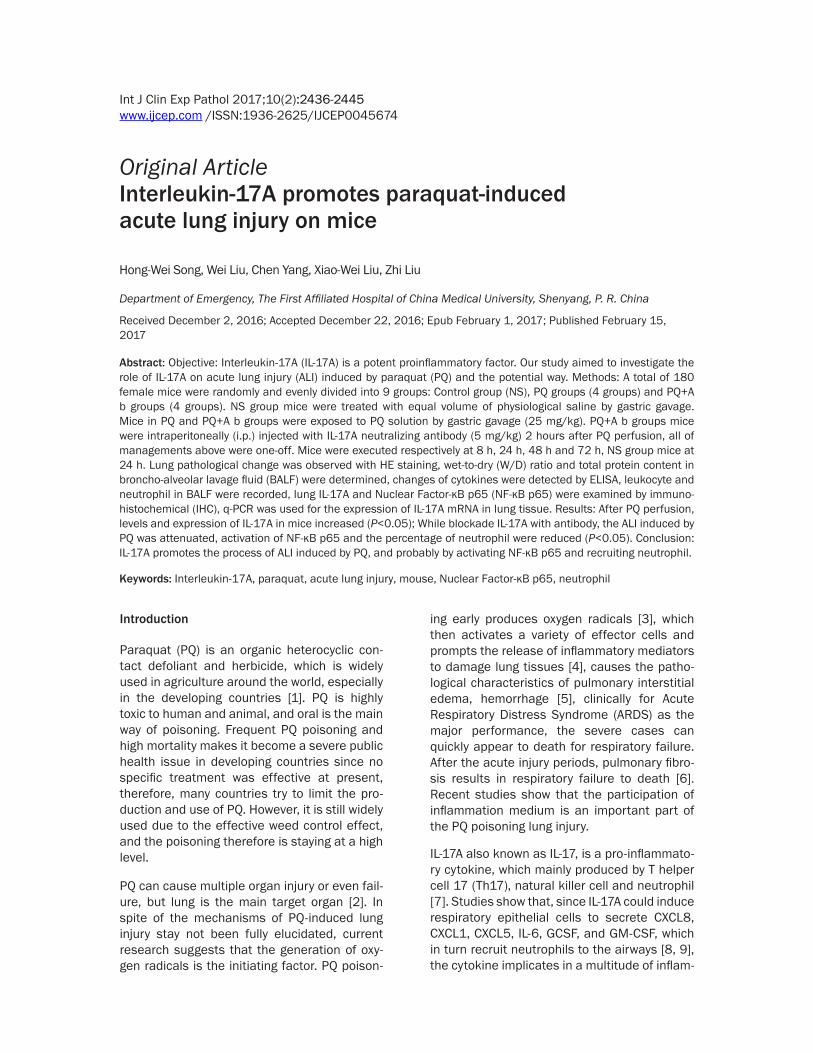
Table 1. Changes of lung W/DGroups n 8 h 24 h 48 h 72 hNS 6 3.86±0.04PQ 6 4.32±0.04a 4.72±0.07a 5.51±0.17a 5.11±0.10a
PQ+A b 6 4.23±0.09a,b 4.32±0.05a,b 4.96±0.14a,b 4.52±0.05a,b
Note: aP<0.05, vs. NS group; bP<0.05, vs. PQ group.
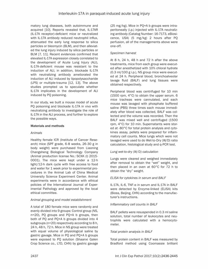
Table 2. Total protein in BALFGroups n Total protein in BALF (μg/ml)NS 6 50.317±7.027PQ 8 h 6 100.208±10.977a
24 h 6 152.392±15.696a
48 h 6 190.278±13.460a
72 h 6 178.822±17.881a
PQ+A b 8 h 6 78.863±9.420a,b
24 h 6 113.107±15.512a,b
48 h 6 156.438±14.661a,b
72 h 6 148.847±6.913a,b
Note: aP<0.001, vs. NS group; bP<0.001, vs. PQ group.
6.0 by microwave for 10 min. Endogenous peroxidase was inac-tivated by 3% H2O2 for 15 min. Goat serum (SL2-10, Solarbio, Beijing, CHN) was used to block non-specific antigen sites for 15 min. Then, the slides were incubat-ed successionally with diluted pri-
Interleukin-17A in paraquat-induced acute lung injury
2439 Int J Clin Exp Pathol 2017;10(2):2436-2445
tive gene expression was calculated using the ratios that IL-17A mRNA/GAPDH mRNA. The sequences were referred from PUBMED data-base, all the primers (Sangon, Shanghai, China) were designed with Primer 5.0. Dissolve curve was drew at the same time to ensure the speci-ficity of fluorescent q-PCR. The primers used were listed as follows: IL-17A (344 bp): forward: 5’-TGTCAATGCGGAGGGAAAG-3’, reverse: 5’-GC- AGTTTGGGACCCCTTTAC-3’; GAPDH (183 bp): forward: 5’-GGTTGTCTCCTGCGACTTCA-3’, reve- rse: 5’-TGGTCCAGGGTTTCTTACTC-3’.
Statistical analysis
Data are expressed as _x (mean) ± s (standard
derivation). Statistical analysis was performed with SPSS 22.0, statistical differences were determined by one-way analysis of variance (one-way ANOVA), followed by Least Significant Difference test (LSD-t) or Games-Howell test for multiple comparis ons, P<0.05 was consid-ered to be statistically significant.
tilled H2O (dH2O). PCR program: 37°C reverse transcription 15 min, 85°C pre-denaturation 2 min, 95°C denaturation 30 s, 40°C annealing 30 s, 40 cycles on 96 Real-Time Quantitative lightcycle 480II (Roche, Switzerland/German). GAPDH was used for an internal control, rela-
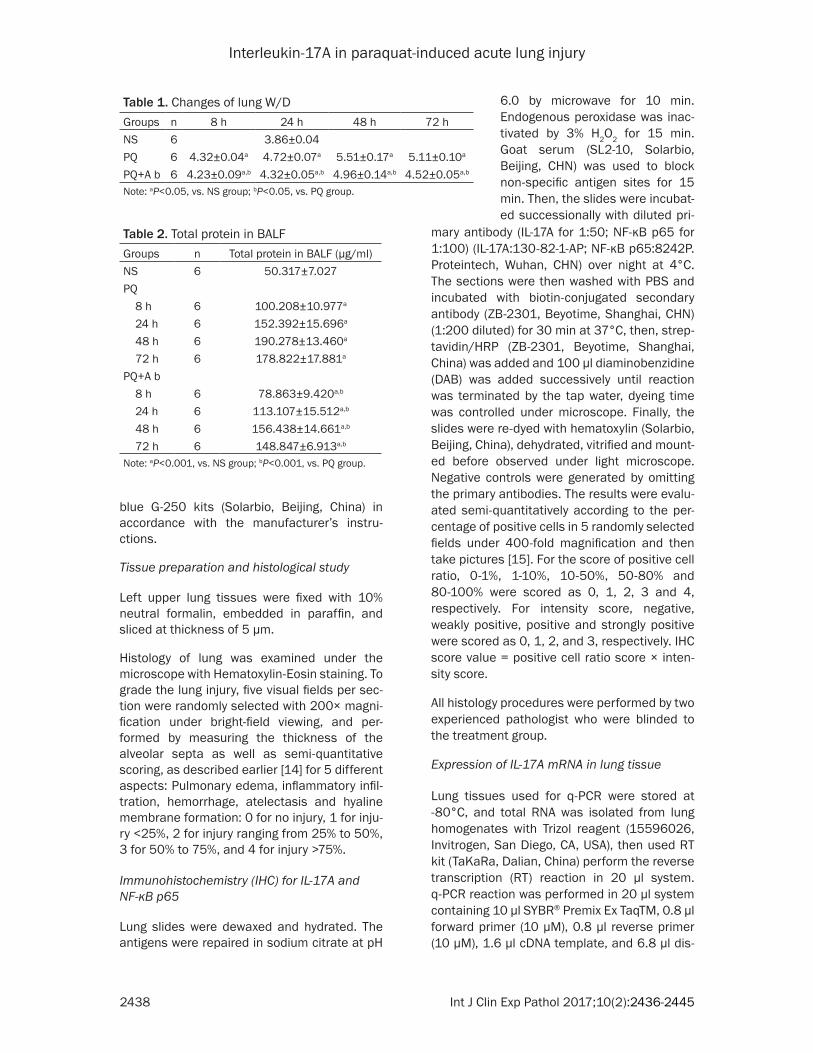
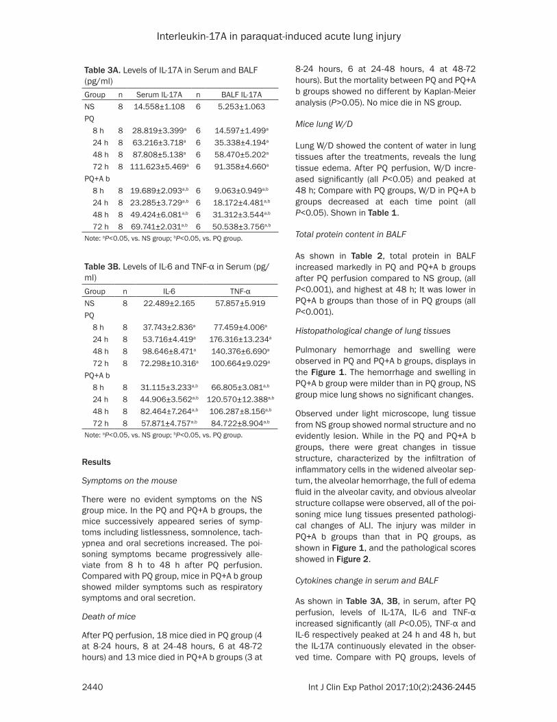
Figure 1. Gross inspection was at 24 h, and pulmonary hemorrhage and swelling were observed in PQ and PQ+A b groups. The hemorrhage and swelling in PQ+A b group were milder than in PQ group, NS group mice lung shows no significant changes. Histology observed with HE staining under light microscope (×200), lungs of mice from NS group showed normal structure and no evident lesion. While in the PQ and PQ+A b groups, there were great changes in tissue structure characterized by the infiltration of inflammatory cells in the widened alveolar septum, alveolar hemorrhage, full of edema fluid in the alveolar cavity, and obvious alveolar structure collapse were observed, all of the poisoning mice lung tissues showed pathological changes of ALI.
Figure 2. Lung injury score increased after PQ perfu-sion, and highest at 48 h, while the scores in PQ+A b groups were lower than those in PQ groups at each time. (aP<0.01, vs. PQ groups).
Interleukin-17A in paraquat-induced acute lung injury
2440 Int J Clin Exp Pathol 2017;10(2):2436-2445
8-24 hours, 6 at 24-48 hours, 4 at 48-72 hours). But the mortality between PQ and PQ+A b groups showed no different by Kaplan-Meier analysis (P>0.05). No mice die in NS group.
Mice lung W/D
Lung W/D showed the content of water in lung tissues after the treatments, reveals the lung tissue edema. After PQ perfusion, W/D incre- ased significantly (all P<0.05) and peaked at 48 h; Compare with PQ groups, W/D in PQ+A b groups decreased at each time point (all P<0.05). Shown in Table 1.
Total protein content in BALF
As shown in Table 2, total protein in BALF increased markedly in PQ and PQ+A b groups after PQ perfusion compared to NS group, (all P<0.001), and highest at 48 h; It was lower in PQ+A b groups than those of in PQ groups (all P<0.001).
Histopathological change of lung tissues
Pulmonary hemorrhage and swelling were observed in PQ and PQ+A b groups, displays in the Figure 1. The hemorrhage and swelling in PQ+A b group were milder than in PQ group, NS group mice lung shows no significant changes.
Observed under light microscope, lung tissue from NS group showed normal structure and no evidently lesion. While in the PQ and PQ+A b groups, there were great changes in tissue structure, characterized by the infiltration of inflammatory cells in the widened alveolar sep-tum, the alveolar hemorrhage, the full of edema fluid in the alveolar cavity, and obvious alveolar structure collapse were observed, all of the poi-soning mice lung tissues presented pathologi-cal changes of ALI. The injury was milder in PQ+A b groups than that in PQ groups, as shown in Figure 1, and the pathological scores showed in Figure 2.
Cytokines change in serum and BALF
As shown in Table 3A, 3B, in serum, after PQ perfusion, levels of IL-17A, IL-6 and TNF-α increased significantly (all P<0.05), TNF-α and IL-6 respectively peaked at 24 h and 48 h, but the IL-17A continuously elevated in the obser- ved time. Compare with PQ groups, levels of
Results
Symptoms on the mouse
There were no evident symptoms on the NS group mice. In the PQ and PQ+A b groups, the mice successively appeared series of symp-toms including listlessness, somnolence, tach- ypnea and oral secretions increased. The poi-soning symptoms became progressively alle- viate from 8 h to 48 h after PQ perfusion. Compared with PQ group, mice in PQ+A b group showed milder symptoms such as respiratory symptoms and oral secretion.
Death of mice
After PQ perfusion, 18 mice died in PQ group (4 at 8-24 hours, 8 at 24-48 hours, 6 at 48-72 hours) and 13 mice died in PQ+A b groups (3 at
Table 3A. Levels of IL-17A in Serum and BALF (pg/ml)Group n Serum IL-17A n BALF IL-17ANS 8 14.558±1.108 6 5.253±1.063PQ 8 h 8 28.819±3.399a 6 14.597±1.499a
24 h 8 63.216±3.718a 6 35.338±4.194a
48 h 8 87.808±5.138a 6 58.470±5.202a
72 h 8 111.623±5.469a 6 91.358±4.660a
PQ+A b 8 h 8 19.689±2.093a,b 6 9.063±0.949a,b
24 h 8 23.285±3.729a,b 6 18.172±4.481a,b
48 h 8 49.424±6.081a,b 6 31.312±3.544a,b
72 h 8 69.741±2.031a,b 6 50.538±3.756a,b
Note: aP<0.05, vs. NS group; bP<0.05, vs. PQ group.
Table 3B. Levels of IL-6 and TNF-α in Serum (pg/ml)Group n IL-6 TNF-αNS 8 22.489±2.165 57.857±5.919PQ 8 h 8 37.743±2.836a 77.459±4.006a
24 h 8 53.716±4.419a 176.316±13.234a
48 h 8 98.646±8.471a 140.376±6.690a
72 h 8 72.298±10.316a 100.664±9.029a
PQ+A b 8 h 8 31.115±3.233a,b 66.805±3.081a,b
24 h 8 44.906±3.562a,b 120.570±12.388a,b
48 h 8 82.464±7.264a,b 106.287±8.156a,b
72 h 8 57.871±4.757a,b 84.722±8.904a,b
Note: aP<0.05, vs. NS group; bP<0.05, vs. PQ group.
Interleukin-17A in paraquat-induced acute lung injury
2441 Int J Clin Exp Pathol 2017;10(2):2436-2445
those in NS groups (all P<0.05), while com-pared to PQ groups, IL-17A decreased signifi-cantly in PQ+A b groups (all P<0.05).
IL-17A, IL-6, TNF-α in PQ+A b groups mice decreased (all P<0.05); In BALF, levels of IL-17A in PQ and PQ+A b groups were higher than
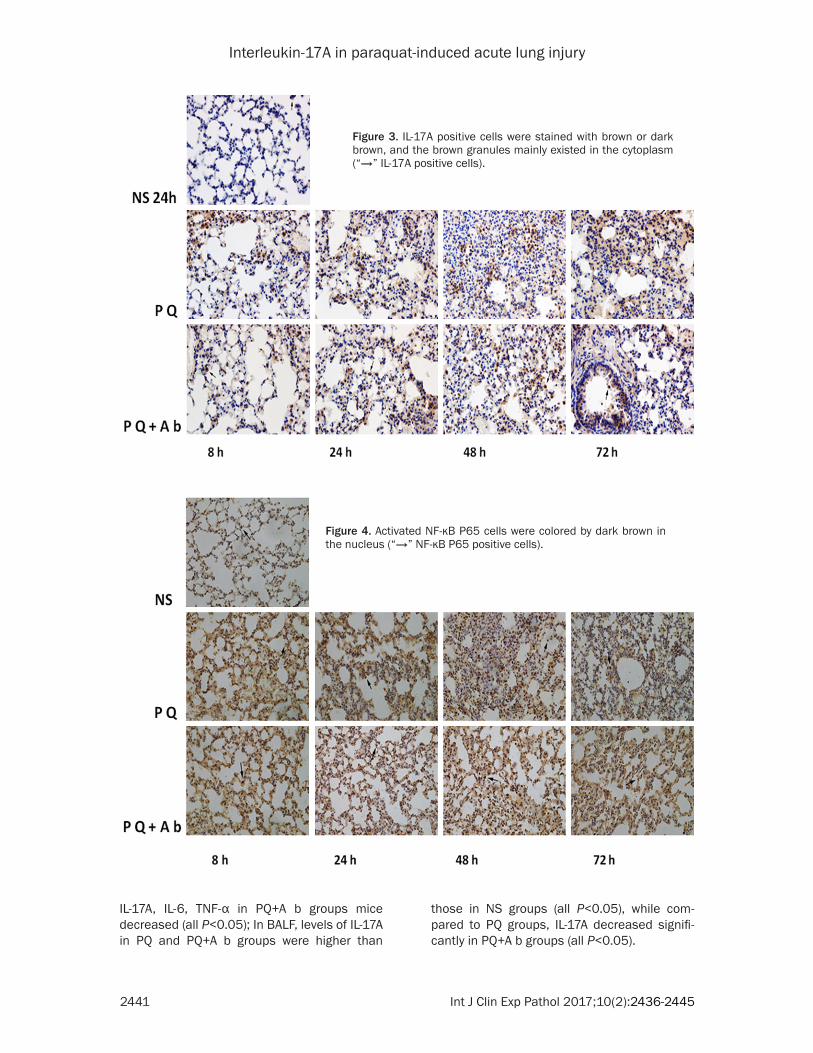
Figure 3. IL-17A positive cells were stained with brown or dark brown, and the brown granules mainly existed in the cytoplasm (“→” IL-17A positive cells).
Figure 4. Activated NF-κB P65 cells were colored by dark brown in the nucleus (“→” NF-κB P65 positive cells).
Interleukin-17A in paraquat-induced acute lung injury
2442 Int J Clin Exp Pathol 2017;10(2):2436-2445
cantly compared to NS groups (all P<0.01); While in PQ+A b groups, leukocytes and the percentage of neutrophils were lower than those of in PQ groups (all P<0.05) (in Table 6).
Discussion
As is well known, intentional or accidental ingestion of commercial liquid formulations of PQ is the leading cause of PQ-intoxication that induced millions of human death every year in the world, it causes multiple organs injury once was poisoned. Previous studies have shown that the major target organ in PQ poisoning is the lung [2], both of ALI and pulmonary fibrosis can lead to respiratory failure and eventually to death. The mechanisms of PQ-induced lung injury are complex. Even though free radicals has been deemed to play a crucial role in the lung injury [16], several inflammatory and che-motactic cytokines have been reported to play a very important role in the course, such inflam-matory cytokines as IL-6, TGF-β1, TNF-α are found to be involved in the pathogenesis of lung injury [17].
IL-17A is a potent pro-inflammatory cytokine that has been verified to play a crucial role in the development of ALI induced by various rea-sons (LPS, Bleomycin, silicon and multiple-trau-ma) [7, 11, 13, 18] and was suggested to be a potential application for IL-17A-based therapy in clinical practice [12]. Herein, we used IL-17A neutralizing antibody to blockade IL-17A in mouse after PQ perfusion to prove if IL-17A plays the same role in the PQ-induced ALI and the possible way.
In our study, toxic symptoms presented on mouse after PQ perfusion, successively as rest-less, somnolence, tachypnea, and oral secre-tions increased. But the symptoms appeared to be milder after IL-17A was blockaded with anti-body, although the mortality showed no decrease between PQ and PQ+A b groups mice by the Kaplan-Meier analysis, but the death in the PQ+A b group tend to be reduced.
Pulmonary edema is a change of lung injury after PQ perfusion, and we measured the wet-to-dry ratios of lung tissues to quantify the pulmonary edema extent. Our experiments showed that treatment with IL-17A antibody inhibited pulmonary edema, as the wet-to-dry ratios in the PQ+A b groups were significantly
Expression of IL-17A and NF-κB p65 examined by IHC
IL-17A positive cells were stained with brown or dark brown and the brown granules mainly existed in the cytoplasm (in Figure 3). As for NF-κB p65, in the NS group, brown granules were mainly in cytoplasm, while in PQ and PQ+A b groups, the brown granules were mainly exist-ed in cell nucleus (in Figure 4).
Compared with NS group, IL-17A IHC scores in PQ and PQ+A b groups were significantly increased (all P<0.05) after PQ perfusion and peaked at the 48 h, while in PQ+A b groups, IL-17A IHC scores were lower than those in PQ groups at 24 h, 48 h and 72 h (all P<0.05). The IHC scores of NF-κB p65 in PQ and PQ+A b groups were higher than those in NS groups (all P<0.05), compared to PQ groups, the scores in PQ+A b groups decreased at each time in the observation period (all P<0.05), (in Table 4).
q-PCR for the expression of IL-17A mRNA in lung tissue
Compared with NS groups, the expression of IL-17A mRNA in PQ and PQ+A b groups was sig-nificantly increased from 8 to 72 hours after PQ perfusion (all P<0.01), and highest at 48 h. While the IL-17A mRNA level in PQ+A b groups was lower than those of in PQ groups at 24 h, 48 h and 72 h (all P <0.05) (in Table 5).
Inflammatory cell counts in BALF
After PQ perfusion, leukocytes and the percent-age of neutrophils in BALF increased signifi-
Table 4. IHC Scores of IL-17A and NF-κB p65 Group n IL-17A NF-κB p65NS 6 2.467±0.767 2.667±0.576PQ 8 h 6 4.333±1.270a 5.733±0.729a
24 h 6 6.033±1.033a 8.567±0.817a
48 h 6 7.733±0.832a 9.467±0.610a
72 h 6 6.633±1.926a 7.433±0.817a
PQ+A b 8 h 6 3.567±0.865a,b 4.567±0.385a,b
24 h 6 4.567±1.033a,b 6.133±1.188a,b
48 h 6 4.867±1.559a,b 7.067±1.228a,b
72 h 6 4.400±0.970a,b 6.467±0.879a,b
Note: aP<0.05, vs. NS group; bP<0.05, vs. PQ group.
Interleukin-17A in paraquat-induced acute lung injury
2443 Int J Clin Exp Pathol 2017;10(2):2436-2445
taneously after PQ perfusion, suggesting that IL-17A implicated in the PQ-induced ALI. However, the IL-17A positive cell numbers and IL-17A mRNA levels decreased at 24 h after administrated with antibody, not fully in line with the levels in serum and BALF, we speculate that there may be some feedback pathways between IL-17A and the IL-17A-derived cells, but we failed to investigate the pathway in the work.
Previous studies suggest that PQ induces alve-olar macrophage and neutrophil infiltrate to lung, recruit and release cytokines to damage lung tissues [4], cytokines as IL-6, TGF-β1, TNF-α play a definite but complex role in lung injury including PQ-induced [5, 23, 24]. These cytokines can initiate, amplify, and perpetuate the inflammatory response during the ALI pro-cess [17]. In our study, levels of IL-6 and TNF-α in serum increased after PQ was perfused, meanwhile, leucocytes and the percentage of neutrophil in BALF increased, which was con-sistent with previous studies. While blockade with IL-17A antibody, levels of above-mentioned cytokines in serum and the leucocytes in BALF were lower. Moreover, the percentage of neu-trophil in BALF significantly decreased after IL-17A was neutralized, therefore, we speculat-ed that IL-17A also recruits neutrophil to exacer-bate the PQ-induced injury as that in LPS/bleo-mycin-induced lung injury [25].
As a nuclear transcription factor, NF-κB plays a crucial role in regulating gene transcription in inflammatory reaction [26]. p65 protein is a main subunit of NF-κB which activates cyto-kines to enlarge the cascade effect in the response by promoting related gene transcrip-tion [27]. In resting state, p65 protein combines with the inhibiting protein IκB formed a com-plexity which existing in the cytoplasm in an inactive form, when stimulated by infection or cytokines, IκB be predominate phosphorylation and separated from IκB p65 predominate, the free p65 quickly transferred to the nucleus and
lower than those in PQ groups. Pathological changes in the lung reflected the injury; we observed that lungs from poisoning mice looked swelling and congestive. Under the light microscope, ALI pathological alterations in lung tissues present as alveolar edema, hemor-rhage, inflammatory cell infiltration, and perfu-sion alveolar collapse accompanied with wall thickening. Administrated PQ poisoning mice with IL-17A antibody significantly alleviated the pathological changes. As an evaluation criteria, total protein content in BALF was also used to assess the injury of PQ poisoning mice [19], in our study, total proteins in BALF increased after PQ perfusion and decreased after IL-17A was blockaded with antibody in mice.
Studies showed that IL-17A plays an important role in triggering an inflammatory response [20], such as IL-17A recruits and/or activates neutrophils to damage lung tissues in LPS-induced pulmonary inflammation and bleomy-cin-induced lung injury [11, 12, 21, 22]. In our experiments, the contents of IL-17A in serum and BALF significantly increased immediately after mice were perfused with PQ, and the lev-
Table 5. Expression of IL-17A mRNA examined by q-PCR (IL-17A mRNA/GAPDH mRNA) (
_x±s)
Group n 8 h 24 h 48 h 72 hNS 6 1.00PQ 6 1.46±0.40a 2.87±0.39a 3.50±0.38a 3.02±0.52a
PQ+A b 6 1.46±0.38a 1.97±0.30a,b 2.51±0.50a,b 2.15±0.37a,b
Note: aP<0.01, vs. NS group; bP<0.05, vs. PQ group.
Table 6. Changes of Inflammatory Cells in BALF (
_x±s)
Group n Leukocytes count (×106/ml)
Neutrophil percentage (%)
NS 6 0.183±0.019 21.500±1.521PQ 8 h 6 0.252±0.016a 35.555±7.096a
24 h 6 0.312±0.028a 44.655±9.827a
48 h 6 0.517±0.017a 52.917±11.109a
72 h 6 0.442±0.018a 45.883±8.912a
PQ+A b 8 h 6 0.207±0.016a,b 27.633±4.812a,b
24 h 6 0.267±0.016a,b 35.672±7.891a,b
48 h 6 0.367±0.030a,b 40.367±9.402a,b
72 h 6 0.345±0.015a,b 34.367±6.173a,b
Note: aP<0.01, vs. NS group; bP<0.05, vs. PQ group.
els of IL-17A in PQ+A b groups mice decreased after neutralized with antibody. We also found that, the expression of IL-17A determined by IHC and q-PCR elevated in the PQ-poisoning mice lung tissues. The numbers of cell which secret IL-17A increased and the expres-sion of IL-17A mRNA elevated simul-
Interleukin-17A in paraquat-induced acute lung injury
2444 Int J Clin Exp Pathol 2017;10(2):2436-2445
[4] Wegener T, Sandhagen B, Chan KW, Saldeen T. N-acetylcysteine in paraquat toxicity: toxico-logical and histological evaluation in rats. Ups J Med Sci 1988; 93: 81-9.
[5] Chen D, Jiao GY, Ma T, Liu XW, Yang C, Liu Z. The mechanism of rapamycin in the interven-tion of paraquat-induced acute lung injury in rats. Xenobiotica 2015; 45: 538-46.
[6] Orito K, Suzuki Y, Matsuda H, Shirai M, Akahori F. Chymase is activated in the pulmonary in-flammation and fibrosis induced by paraquat in hamsters. Tohoku J Exp Med 2004; 203: 287-94.
[7] Lo Re S, Dumoutier L, Couillin I, Van Vyve C, Yakoub Y, Uwambayinema F, Marien B, van den Brûle S, Van Snick J, Uyttenhove C, Ryffel B, Renauld JC, Lison D, Huaux F. IL-17A-producing gammadelta T and Th17 lymphocytes mediate lung inflammation but not fibrosis in experi-mental silicosis. J Immunol 2010; 184: 6367-77.
[8] McAllister F, Henry A, Kreindler JL, Dubin PJ, Ulrich L, Steele C, Finder JD, Pilewski JM, Car-reno BM, Goldman SJ, Pirhonen J, Kolls JK. Role of IL-17A, IL-17F, and the IL-17 receptor in regulating growth-related oncogene-alpha and granulocyte colony-stimulating factor in bron-chial epithelium: implications for airway in-flammation in cystic fibrosis. J Immunol 2005; 175: 404-12.
[9] Nembrini C, Marsland BJ, Kopf M. IL-17-pro-ducing T cells in lung immunity and inflamma-tion. J Allergy Clin Immunol 2009; 123: 986-94.
[10] Holloway TL Schwacha MG. The Th-17 re-sponse and its potential role in post-injury pul-monary complications. Int J Burns Trauma 2012; 2: 11-7.
[11] Braun RK, Ferrick C, Neubauer P, Sjoding M, Sterner-Kock A, Kock M, Putney L, Ferrick DA, Hyde DM, Love RB. IL-17 producing gammad-elta T cells are required for a controlled inflam-matory response after bleomycin-induced lung injury. Inflammation 2008; 31: 167-79.
[12] Li Q, Gu Y, Tu Q, Wang K, Gu X, Ren T. Blockade of interleukin-17 restrains the development of acute lung injury. Scand J Immunol 2016; 83: 203-11.
[13] Dai H, Xu L, Tang Y, Liu Z, Sun T. Treatment with a neutralising anti-rat interleukin-17 antibody after multiple-trauma reduces lung inflamma-tion. Injury 2015; 46: 1465-70.
[14] Smith KM, Mrozek JD, Simonton SC, Bing DR, Meyers PA, Connett JE, Mammel MC. Pro-longed partial liquid ventilation using conven-tional and high-frequency ventilatory tech-niques: gas exchange and lung pathology in an animal model of respiratory distress syndrome. Crit Care Med 1997; 25: 1888-97.
bonded the specificity of gene sequences into the NF-κB, which prompts the cytokines release [26, 28]. IL-17A has been reported to activate NF-κB p65 in mice lung which results the large-ly secretion of TNF-α to damage the lung [29, 30]. In our experiments, thimbleful activated NF-κB in nucleus in NS group mice lung tissue while a mass of activated NF-κB presented in lung tissues after the mice were perfused with PQ, but activated NF-κB in PQ+A b groups was less than that in the PQ groups. The result indi-cated NF-κB p65 involved in the ALI induced by PQ, and blockade IL-17A hindered the activation of NF-κB p65, and was likely to reduce TNF-α.
In conclusion, this study investigated for the first time that IL-17A implicated in the ALI induced by PQ in a mouse model, blockade IL-17A with the neutralizing antibody attenuates the injury and was likely to reduce the activa-tion of NF-κB p65 and recruitment of neutro-phil. Our experiments provide a new idea for the treatment of ALI induced by PQ poisoning.
Acknowledgements
This work was partly supported by Science and Technology Plan Projects of Liaoning Province (2013225303) and National Natural Science Foundation of China (81571882).
Disclosure of conflict of interest
None.
Address correspondence to: Dr. Zhi Liu, Depart- ment of Emergency, The First Affiliated Hospital of China Medical University, 155 Nanjing Street, Heping District, Shenyang 110001, P. R. China. Tel: +86-24-83282011; Fax: +86-24-83282011; E-mail: [email protected]
References
[1] Mainwaring G, Lim LF, Antrobus K, Swain C, Clapp M, Kimber I, Orphanides G, Moggs JG. Identification of early molecular pathways af-fected by paraquat in rat lung. Toxicology 2006; 225: 157-72.
[2] Rose MS, Smith LL, Wyatt I. Evidence for ener-gy-dependent accumulation of paraquat into rat lung. Nature 1974; 252: 314-5.
[3] Frank L. Prolonged survival after paraquat. Role of the lung antioxidant enzyme systems. Biochem Pharmacol 1981; 30: 2319-24.
Interleukin-17A in paraquat-induced acute lung injury
2445 Int J Clin Exp Pathol 2017;10(2):2436-2445
[23] Zhang Z, Ding L, Wu L, Xu L, Zheng L, Huang X. Salidroside alleviates paraquat-induced rat acute lung injury by repressing TGF-β1 expres-sion. Int J Clin Exp Pathol 2014; 7: 8841-7.
[24] Dong XS, Hu XB, Liu W, Sun YQ, Liu Z. Effects of RNA interference-induced Smad3 gene silenc-ing on pulmonary fibrosis caused by paraquat in mice. Exp Biol Med (Maywood) 2012; 237: 548-555.
[25] Miyamoto M, Prause O, Sjöstrand M, Laan M, Lötvall J, Lindén A. Endogenous IL-17 as a me-diator of neutrophil recruitment caused by en-dotoxin exposure in mouse airways. J Immunol 2003; 170: 4665-72.
[26] Brennenstuhl H, Armento A, Braczysnki AK, Mittelbronn M, Naumann U. IκBζ, an atypical member of the inhibitor of nuclear factor kap-pa B family, is induced by γ-irradiation in glio-ma cells, regulating cytokine secretion and as-sociated with poor prognosis. Int J Oncol 2015; 47: 1971-80.
[27] Baeuerle PA, Baltimore D. A 65-kappaD sub-unit of active NF-kappaB is required for inhibi-tion of NF-kappaB by I kappaB. Genes Dev 1989; 3: 1689-98.
[28] Metelev VG, Kubareva EA, Oretskaya TS. Regu-lation of activity of transcription factor NF-κB by synthetic oligonucleotides. Biochemistry (Mosc) 2013; 78: 867-78.
[29] Song X, Qian Y. IL-17 family cytokines mediated signaling in the pathogenesis of inflammatory diseases. Cell Signal 2013; 25: 2335-47.
[30] Mi S, Li Z, Liu H, Hu ZW, Hua F. Blocking IL-17A protects against lung injury-induced pulmo-nary fibrosis through promoting the activation of p50NF-kappaB. Yao Xue Xue Bao 2012; 47: 739-44.
[15] Kaemmerer D, Peter L, Lupp A, Schulz S, Sän-ger J, Baum RP, Prasad V, Hommann M. Com-paring of IRS and Her2 as immunohistochemi-cal scoring schemes in gastroenteropancreatic neuroendocrine tumors. Int J Clin Exp Pathol 2012; 5: 187-94.
[16] Kim JH, Gil HW, Yang JO, Lee EY, Hong SY. Ef-fect of glutathione administration on serum levels of reactive oxygen metabolites in pa-tients with paraquat intoxication: a pilot study. Korean J Intern Med 2010; 25: 282-7.
[17] Hemmati AA, Nazari Z, Motlagh ME, Goldasteh S. The role of sodium cromolyn in treatment of paraquat-induced pulmonary fibrosis in rat. Pharmacol Res 2002; 46: 229-34.
[18] Kim SR, Kim HJ, Kim DI, Lee KB, Park HJ, Jeong JS, Cho SH, Lee YC. Blockade of inter-play between IL-17A and endoplasmic reticu-lum stress attenuates LPS-induced lung injury. Theranostics 2015; 5: 1343-62.
[19] Jin W, Rong L, Liu Y, Song Y, Li Y, Pan J. In-creased claudin-3, -4 and -18 levels in bron-choalveolar lavage fluid reflect severity of acute lung injury. Respirology 2013; 18: 643-51.
[20] Korn T, Bettelli E, Oukka M, Kuchroo VK. IL-17 and Th17 cells. Annu Rev Immunol 2009; 27: 485-517.
[21] Fujie H, Niu K, Ohba M, Tomioka Y, Kitazawa H, Nagashima K, Ohrui T, Numasaki M. A distinct regulatory role of Th17 cytokines IL-17A and IL-17F in chemokine secretion from lung micro-vascular endothelial cells. Inflammation 2012; 35: 1119-31.
[22] You QH, Zhang D, Niu CC, Zhu ZM, Wang N, Yue Y, Sun GY. Expression of IL-17A and IL-17F in li-popolysaccharide-induced acute lung injury and the counteraction of anisodamine or methylprednisolone. Cytokine 2014; 66: 78-86.