original article hypoxia-inducible factor 1α (hif-1α ... · benign prostatic hyperplasia (bph) is...
TRANSCRIPT
Int J Clin Exp Pathol 2019;12(1):295-304www.ijcep.com /ISSN:1936-2625/IJCEP0084969
Original ArticleHypoxia-inducible factor 1α (HIF-1α) mediates the epithelial-mesenchymal transition in benign prostatic hyperplasia
Yanbo Chen*, Huan Xu*, Qiling Shi, Meng Gu, Xiang Wan, Qi Chen#, Zhong Wang#
Department of Urology, Shanghai Ninth People’s Hospital Affiliated to Shanghai Jiaotong University School of Medicine, Shanghai, China. *Equal contributors and co-first authors. #Equal contributors.
Received September 2, 2018; Accepted September 27, 2018; Epub January 1, 2019; Published January 15, 2019
Abstract: Background: Epithelial-mesenchymal transition (EMT) based cancer cell invasion and metastasis has been thoroughly studied in prostate cancer. It was well known that EMT markers which have been found in benign prostatic hyperplasia (BPH) tissues, but system descriptions have not been described. Methods: First, in order to construct the epithelial cells to mesenchymal cell transformation model, BPH-1 cells were cultured with superna-tant of prostate matrix normal prostate stromal WPMY-1 cells, after obtaining the culture medium through a filter. After that, we observed the morphology of cells cultured for a period of time by microscopy, detected cell invasion ability by transwell assay, detected cell proliferation ability by MTT, and detected EMT marker expression by western. Finally, we treated the cells with anti-HIF-1α drugs to study their effects on EMT, and then tested several related pro-teins simultaneously. Results: The results showed that the morphology of BPH-1 cells gradually changed to fusiform after cultured with WSCM. At the same time, E-cadherin and cytokeratin levels were significantly lower than those in normal medium. Simultaneous detection of vimentin (SMA) and Snail was positive compared to normal cultured cells. At the same time, the cells were cultured with WSCM and the invasive ability was up-regulated. After treatment with anti-HIF-1α drug, E-cadherin and CK5/8 protein expression was up-regulated, but vimentin, α-SMA, and Snail expression was down-regulated, and in addition, p-Smad3 protein expression was also down-regulated after anti-HIF-1α drug was added. Conclusion: The above results indicated that WSCM-1 stromal cell supernatant WSCM can induced BPH-1 cell interstitialization, and at the same time, by inducing EMT, secreting HIF-1α activates Smad3 sig-naling. Our study shows that inhibition of HIF-1α expression provides a new reference for clinical treatment of BPH.
Keywords: Hypoxia inducible factor 1, benign prostatic hyperplasia, epithelial-mesenchymal transition
Introduction
Benign prostatic hyperplasia (BPH) is a noncan-cerous enlargement of the prostate gland of- ten resulting in lower urinary tract symptoms (LUTS). Prevalence of BPH increases with age, and BPH is a major disease among older males which significantly reduces the quality of their life [1-3]. It is well known that epithelial-mesen-chymal transition (EMT) is a key inducing factor of aberrant prostate growth. EMT and its reverse mechanism mesenchymal-epithelial transition (MET) play important roles in the embryonic development, especially in the for-mation of the prostate, chronic inflammation, tissue reconstitution and cancer metastasis [4]. When epithelial cells undergo EMT, they
lose their original markers such as E-cadherin and CK8, in addition, acquiring the phenotypes of mesenchymal cells, for instance, expression of vimentin and α-smooth muscle actin [5, 6]. Until now, some clear triggers of EMT have been studied. First, cadherin switching: transition- ing epithelial cells lose epithelial cadherin (E-cadherin) and express neural cadherin (N-cadherin) [7]. Secondly, apoptosis deregula-tion: cells lose apoptosis control and increase proliferation-to-apoptosis ratio [8]. Additionally, epigenetic regulation is also extremely signifi-cant to EMT [9-11]. Finally, the reactive stroma in the prostate microenvironment is a crucial contributor driving the EMT, it regulates andro-gen status and levels of multiple factors includ-ing transforming growth factor-β (TGF-β) via
HIF-1α regulates the EMT of BPH
296 Int J Clin Exp Pathol 2019;12(1):295-304
paracrine mechanisms [12, 13]. Recently, the microenvironment, as an important regulator of EMT has been studied extensively, and plenty of factors have been found that are involved in EMT process.
The hypoxia inducible factor 1 (HIF 1) signaling pathway promotes angiogenesis, and it con-tributes to the progression of numerous hyper-plastic diseases, including BPH. HIF 1 is a cru-cial signaling factor which contributes to the activation of the ‘angiogenic switch’ and results in increasing expression of numerous angio-genic growth factors, including vascular endo-thelial growth factor (VEGF) and basic fibroblast growth factors (bFGF) [14-17]. HIF 1α and HIF 1β form the heterodimeric transcription factor HIF-1, and as a master regulator of oxygen homeostasis, HIF 1 accumulates in the cyto-plasmic matrix [18]. Along with translocation to the nucleus, HIF-1 activates hypoxia-sensitive genes and induces the expression of genes encoding angiogenic cytokines, such as VEGF and bFGF [19, 20]. VEGF or bFGF binds to the specific receptors on the cytomembrane of vas-cular endothelial cells (ECs), which regulates ECs survival, proliferation, migration, and extends the primitive vascular network [21-25]. However, so far, there is no study that shows the role of HIF-1 in the stromal microenviron-ment of prostate.
We hypothesized that HIF 1α may play a signifi-cant role in inducing EMT of prostate epithelial cells through affecting stromal microenviron-ment, which causes BPH. To test our conjec-ture, we used the WPMY-1, which is a prostate stromal cell line, to simulate a stromal microen-vironment for BPH-1 cells in vitro, and then we used this system to explore the influence of HIF-1α on BPH-1 cells’ EMT.
Materials and methods
Cell culture
The human BPH-1 cells were purchased from American Type Culture Collection (ATCC) (USA), the WPMY-1 cell is a human prostatic cell line, which was purchased from The Global Biore- source Center (USA). The BPH-1 cells were cul-tivated in RPMI-1640 fundamental medium (Invitrogen, Canada) with 10% fetal bovine serum (FBS) (Gibco, DC), but the WPMY-1 cells
were maintained culture in RPMI-1640 with 5% FBS (Gibco, USA) medium. Those cells were cultured at 37 degree in temperature incubator with 5% CO2.
Experiment reagents
All of antibody was purchased from Abcam, including polyclone anti-HIF-1α antibody and mouse or ribbit IgG isotype control antibody. And all of antibody was dissolved in PBS con-tain 5% BSA. The antibody HIF-1α was adapt- ed to concentration of 2 ug/ml, and the WS- CM solution was added to anti-HIF-1α (2 ug/ ml).WPMY-1 supernatant-conditioned medium (WSCM) was collected from WPMY-1 cell cul-ture supernatant. When WPMY-1 cells were cul-tured at 80% density, WSCM was obtained, and WPMY-1 cells need replaced to fresh complete medium per two days. In the first place, the supernatant was spun in centrifuge tubes at 2000 rpm and filtered through a 0.45 mm filter for concentration to a proper state. In the end, the fresh WSCM was storage at -80°.
Cell grouping and treatment
The cells were plated per 1×10⁵ in 6 cm dish and maintained for 24 h, then we divided those dishes into seven groups and treatment with different stimulation: a: BPH-1 cells cultured in ordinary complete medium; b: BPH-1 cells cul-tured in WSCM; c: BPH-1 cells cultured in WSCM treatment with anti-HIF-1α; d: BPH-1 cells cul-tured in WSCM treatment with mouse or rabbit IgG control; e: BPH-1 cells cultured in ordinary complete medium treatment with anti-HIF-1α antibody; f: BPH-1 cells cultured in ordinary complete medium treatment with mouse or rabbit IgG; g: WPMY-1 cells cultured in special complete medium of WPMY-1.
Immunofluorescence analysis
BPH-1 cells were collected in a 1.5 ml tube and were washed by phosphate-buffered saline (PBS), and the cells were fixed with 4% parafor-maldehyde; we next used the 0.5% Triton X-100 permeabilized cells for 10 min, and washed with PBS. Finally, the cells were blocked with 5% Bovine Serum Albumin (BSA) or 2% FBS for 20 minutes at RT. We used the primary anti-body rabbit anti-E-cadherin or mouse anti-vimentin (1:100; Abcam); rabbit anti-CK5/8
HIF-1α regulates the EMT of BPH
297 Int J Clin Exp Pathol 2019;12(1):295-304
(1:100; Cell Signaling Technology); and mouse anti-α-SMA and anti-Snail (1:100; Abcam). Cells were incubated overnight at 4°C. Next day, we used PE-labeled secondary antibody incubat- ed with the cells (1:1000; Cell Signaling Te- chnology) for 2 h or the cells were incubated with DAPI (Sigma) for 20 min at RT, and washed twice by PBS. Finally, the cells were observed by fluorescence microscope (PerkinElmer Life Sciences).
Western blotting assay
The cells were harvested, washed twice with cold PBS, and the total proteins were prepared from lysed cells using cell lysis buffer (Amresco, Cochran, GA) (50 mM Tris-HCl [pH 8.0], 150 mM NaCl, 0.5% sodium deoxycholate (SDS), 1% NP-40, 1× protease inhibitor cocktail, 1 mM EDTA) on ice, and supplemented with a prote-ase inhibitor cocktail tablet (Roche, USA). We use the BCA protein assay kit detected protein concentration (Thermo fisher, USA). The cell lysates were sonicated, and resolved by 10%-12% SDS-PAGE Gels were transferred to PVDF membranes (Millipore, USA), blocked with 6% BSA, and probed using Abs directed against anti-E-cadherin and anti-vimentin (1:3,000; Cell Signaling Technology), rabbit anti-CK5/8, mouse anti-alpha-SMA, anti-Snail (1:4,000; Cell Signaling Technology), rabbit anti-p-Smad3 (1:1000; Abcam, USA) and GAPDH (1:1000; Abcam, USA). Secondary Abs conjugated to HRP that were used for detection included anti-rabbit (1:5,000) and anti-mouse (1:5,000; Cell Signaling Technology, Beverly, MA). In the end, the protein signals were detected by chemilu-miniscence systems (GE, USA), and the western blot figure was quantified using PS software.
Real-time PCR
Total RNA of cells was extracted by the RNeasy extraction kit (Qiagen, Valencia, CA), conferring to the manufacturer’s instructions. CDNAs were synthesized using Reverse Transcriptase kit (Thermofisher, USA), the genes of smad3 were detected by qPCR with SYBR® Green RT-PCR Master Mix (Roche, Switzerland) using 7500 Fast (ABI, USA). The PCR amplification process (95°C, 30 s; and 60°C, 1 min) was done for 40 cycles. The levels of gene expression relative to GAPDH were calculated with Delta-Delta-Ct (ddCt) algorithm method. All of the primers were quality checked for a single curve on melt-
ing curve, and efficiencies were 90-110%. Smad3, Forward, 5-GAGTAGAGACGCCAGTTCT- ACC-3, Reverse, 5-GGTTTGGAGAACCTGCGTC- CAT-3; GAPDH, Forward, 5-ATCTTCTCGAACCC- CGAGTGA, Reverse, CGGTT CAGCC ACTGG AGCTT.
Transwell migration assays in vitro
The BPH-1 cells were grown up to ~ 80% den-sity and were treated with WSCM of covering with/without anti-HIF-1α in the 24-well tran-swell and cultured with ordinary but FBS-free medium for 24 h. The BPH-1 cells were collect-ed and carried through cell migration and trans-ference assay after 24 h. In both experiments, BPH-1 cells cultured with/without WSCM were added to the lower chamber and then the cells were migrated the upper chamber. After 24 h of incubation, the migration of cells was mea-sured through the pores using calculating under the microscope (Zeiss, Germany) using a 20× microscope.
The MTT assay
To detect the cell viability/proliferation curves when the cells were treated with inhibitor, BPH-1 cells were plated into 24-well plates with 5000 cells/well and grown in RPMI-1640 media or WSCM Cells. Survival was detected by MTT assay. On this measurement day, medium was carefully changed to MTT and set at 37°C for 3-5 h. After removing MTT mediums, and 200 μl solution of DMSO was added. MTT reduction was detected absorbance at 570 nm using the EThermo Scientific Microplate Reader (BioTek, USA).
Statistical analyses
Two groups data were compared using Stud- ent’s t test, and P < 0.05 was considered sig-nificant. All of the statistical significance calcu-lations were implemented in Graghpad prism 5 software.
Results
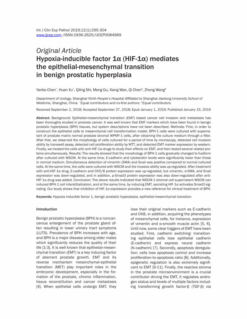
EMT was induced by WSCM in BPH-1 cells
Noncancerous prostatic cell WPMY-1 showed a spindle shaped myofibroblast morphology. As previously researched [26], BPH-1 cells were cultured by the WPMY-1 cell culture superna-
HIF-1α regulates the EMT of BPH
298 Int J Clin Exp Pathol 2019;12(1):295-304
tant in vitro. In Figure 1, BPH-1 cells were cul-tured in ordinary medium (left panel), which show the representative feature of epithelial
cells; however, BPH-1 cells that were main-tained with WSCM medium, which presented a spindle-shaped morphology (right panel), and
Figure 1. EMT was induced by WSCM in BPH-1 cells. BPH-1 was cultured in ordinary RPMI 1640 medium and WSCM-BPH-1 was cultured WPMY-1 supernatant. WPMY-1 cells were photographed as a control. A. Cells were pho-tographed after cultured in different media for 5 days. B, C. Western blot analysis of EMT-related proteins in BPH cells after 7 days’ culture period and relative protein levels to GAPDH. D, E. Migration assay results showed WSCM can promote BPH-1 cell migration. F. MTT assay shows the growth of BPH-1 did not alter cells after 1, 3 and 5 days of WSCM culture.
HIF-1α regulates the EMT of BPH
299 Int J Clin Exp Pathol 2019;12(1):295-304
BPH-1 cells had a typical spindle shaped mor-phology (middle panel) after maintenance in WSCM 5 days, which is similar to the prostate
normal WPMY-1 (Figure 1). To be sure of this phenotype, we detected E-cadherin and CK5/8 of the epithelial markers using western blot,
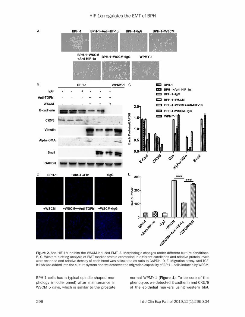
Figure 2. Anti-HIF-1α inhibits the WSCM-induced EMT. A. Morphologic changes under different culture conditions. B, C. Western blotting analysis of EMT marker protein expression in different conditions and relative protein levels were scanned and relative density of each band was calculated as ratio to GAPDH. D, E. Migration assay. Anti-TGF-b1 Ab was added into the culture system and we detected the migration capability of BPH-1 cells induced by WSCM.
HIF-1α regulates the EMT of BPH
300 Int J Clin Exp Pathol 2019;12(1):295-304
and we found the protein levels of E-cadherin and CK5/8 expression were decresedin BPH-1 cells with WSCM medium in comparison to the control medium (Figure 1B, 1C). Notably, we also respectively detected the mesenchymal markers vimentin and α-SMA or the EMT mark-er Snail expression in the BPH-1 cell with WSCM medium. We found that the expression of E-cadherin and CK5/8 were decreased in WSCM cultured BPH-1 cells, but the BPH-1 cells unexpectedly acquired Vimentin, α,-SMA and Snail expression by Immunofluorescent stain-ing (Figure 1D). Migration assay showed that the BPH-1 cells’ migratory capacity was also up-regulated by treatment with WSCM medium (Figure 1E, right panel). Therefore, to further ensure the role of WSCM in BPH-1 cell prolifera-tion, we implemented the MTT assay. As in Figure 1F, we did not detect a difference in growth rates with ordinary medium or WSCM medium cultured the BPH-1 cells, respectively. From the above results, we say that EMT can be induced in BPH-1 cells with WSCM medium.
Anti-HIF-1α inhibit the WSCM-induced EMT
HIF-1α is a transcription factor and belongs to Hypoxia-inducible factor 1 (HIF-1) family, and previous research reported that HIF-1α was an EMT inducer in diverse normal or cancer cell types [27, 28]. To detect whether HIF-1α is referred to changing of the EMT markers expression in BHP-1 cells, we evaluated its transcription factors’ change on the WSCM-induced EMT by adding anti-HIF-1α antibody with WSCM medium. As shown in Figure 2, BPH-1 cells had a spindle form in WSCM media; however, this morphology was reversed by anti- HIF-1α treatment to culture medium. To further determine anti-HIF-1α role in WSCM-induced EMT, we carried out western blotting, and we found the E-cadherin and CK5/8 expression were strongly enhanced after treatment with anti-HIF-1α. Vimentin, α-SMA and Snail protein expression were decreased by western blot. In addition to this, we got similar results from immunofluorescence, when the BPH-1 cells was added to the HIF-1α antibody. Results showed that vimentin, α-SMA and Snail were inhibited, but E-cadherin and CK5/8 were upregulated (Figure 2D). In the end, we also proved that anti-HIF-1α could reverse the WSCM induced the BPH-1 migratory potential by using a migration assay (Figure 2E). These
results with anti-HIF-1α indicated that it may play a role in WSCM cultured BPH-1 cells.
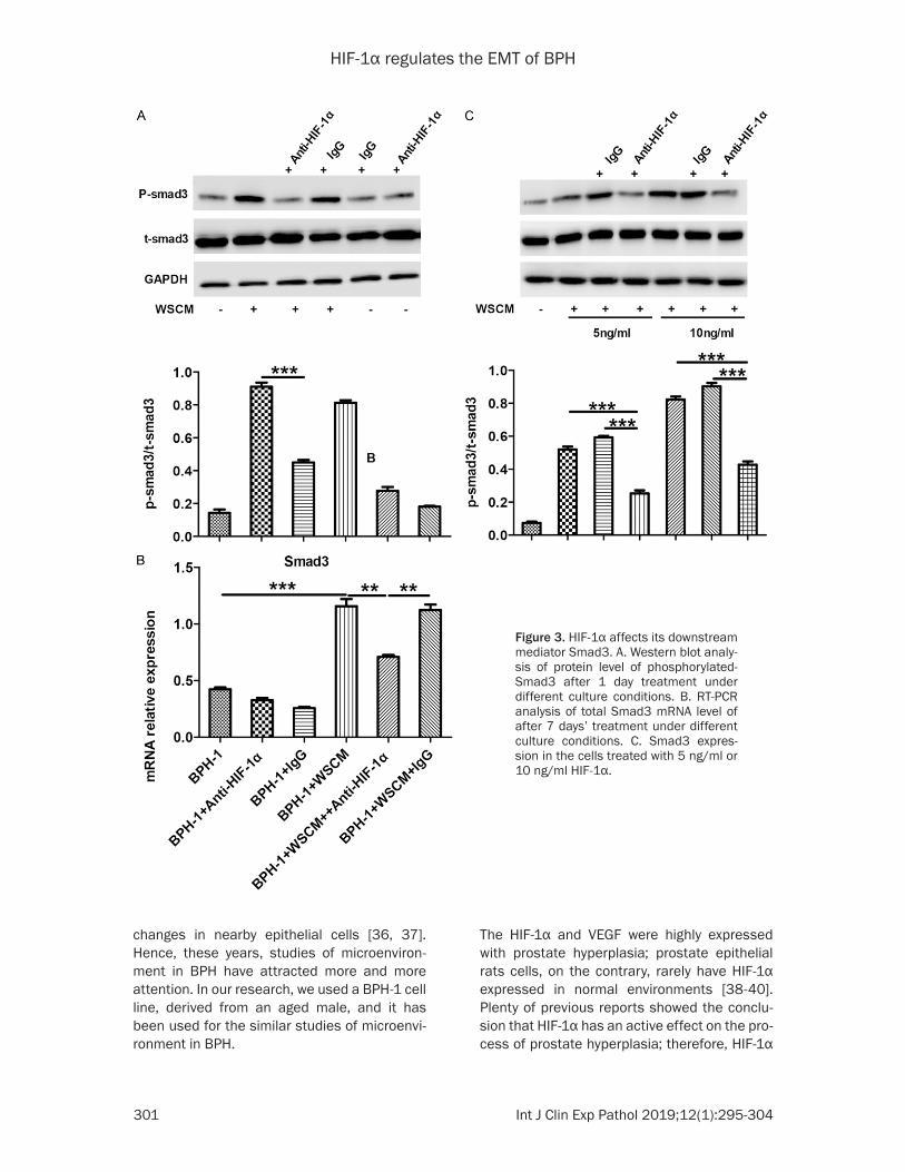
HIF-1α affects its downstream mediator Smad3
Smad3, as a HIF-1α signal-pathway receptor protein, has a significant role in the WSCM-induced EMT in BPH-1 cells. Hence, we investi-gated the function of Smad3 in the BPH-1 cells. To further investigate the interaction between HIF-1α and Smad3, we performed the qPCR and western blotting to determine the expres-sion of Smad3 in Figure 3. As shown in Figure 3A, we found Smad3 was phosphorylated and was up-regulated in WSCM medium cultured BPH-1 cells by comparison to the ordinary con-trol medium. However, when we added HIF-1α antibody into WSCM-cultured BPH-1 cells, we found that the embellishment of p-Smad3 was decreased after 7 days, and the gene of Smad3 expression was increased in mRNA level in WSCM medium cultured BPH-1 cells. We next found that if we add anti- HIF-1α antibody into BPH-1 cells, this phenotype will be partly reversed (Figure 3B). These data further show that HIF-1α may play an important role in WSCM-induced EMT in BPH-1 cells, and HIF-1α may be coordinated by a Smad3 signal pathway.
Discussion
BPH is a well known age-related disease, in men over sixty years old [29, 30]. The main clinical manifestation of BPH is LUTS, which include increased frequency of urination, weak urine stream, urgency, hesitancy and nocturia [31, 32]. Thus, BPH reduces life quality. Several processes are involved in BPH, and the EMT process of prostate epithelial cells plays a very crucial role in BPH [33]. Numerous factors con-tribute to the EMT of prostate epithelial cells, and among these complicated regulators, the microenvironment provided by stromal cells is an extremely important one [7, 34]. It is already known that stromal cells play a key role in age-related tissue remodeling [35]. Compared to young men, older men have more reactive stro-mal cells that are characterized by up-regula-tion of the amount of myofibroblasts and loss of smooth muscle cells. Since myofibroblasts have more active function of secreting aberrant factors than normal fibroblasts this induces
HIF-1α regulates the EMT of BPH
301 Int J Clin Exp Pathol 2019;12(1):295-304
changes in nearby epithelial cells [36, 37]. Hence, these years, studies of microenviron-ment in BPH have attracted more and more attention. In our research, we used a BPH-1 cell line, derived from an aged male, and it has been used for the similar studies of microenvi-ronment in BPH.
The HIF-1α and VEGF were highly expressed with prostate hyperplasia; prostate epithelial rats cells, on the contrary, rarely have HIF-1α expressed in normal environments [38-40]. Plenty of previous reports showed the conclu-sion that HIF-1α has an active effect on the pro-cess of prostate hyperplasia; therefore, HIF-1α
Figure 3. HIF-1α affects its downstream mediator Smad3. A. Western blot analy-sis of protein level of phosphorylated-Smad3 after 1 day treatment under different culture conditions. B. RT-PCR analysis of total Smad3 mRNA level of after 7 days’ treatment under different culture conditions. C. Smad3 expres-sion in the cells treated with 5 ng/ml or 10 ng/ml HIF-1α.
HIF-1α regulates the EMT of BPH
302 Int J Clin Exp Pathol 2019;12(1):295-304
is identified as an underlying therapeutic target for BPH [38, 39]. Until now, numerous mole-cules are prepared for clinical use for inhibiting HIF-1α [41]. However, a large proportion of HIF-1α inhibitors perform their functions by target-ing genes that regulate HIF-1α expression. In addition, a HIF-1α specific inhibitor has not been developed; thus, up to now, targeted ther-apy against HIF-1α has not been established. Therefore, it is meaningful to establish a spe-cific method to impair the function of HIF-1α.
After treatment with WSCM, BPH-1 cells pre-sented a stretched contour and the markers of epithelia, namely E-cadherin and CK5/8, were decreased (Figure 1). Additionally, the WSCM-treated BPH-1 cells expressed α-SMA, and Vimentin, both mesenchymal markers, and the Snail EMT marker (Figure 1). Snail is a zinc-fin-ger transcription factor and is an EMT inducer. Snail can bind to an E-cadherin promoter E-box and represses its transcription; additionally, Snail also suppress endocytosis of transmem-brane E-cadherin. In cancer cells, the high expression of Snail causes impairment of E-cadherin function and often induces EMT. In our results, we found that the microenviron-ment provided by stromal cells could boost EMT in BPH-1 cells by secreting EMT factors. And we discovered that WSCM could boost the BPH-1 cell migratory potential by migration assay (Figure 1). We also found that WSCM influence on the proliferation of BPH-1 cells was unchanged by the MTT results (Figure 1).
To examine the function of HIF-1α in stromal microenvironment-induced EMT in BPH cells, we neutralized HIF-1α by treating WSCM culture medium with HIF-1α antibody. Then we found that anti-HIF-1α treatment partially reversed morphologic changes induced by WSCM in comparison with the control medium. In- addition, CK5/8 and E-cadherin expression were markedly increased; on the contrary, the expression of vimentin, α-SMA and Snail declined. Those results also can be supported by migration assays. Anti-HIF-1α treatment reversed the migratory potential of BPH cells induced by WSCM.
Conclusions
In conclusion, our research shows that HIF-1α may play a significant role in the stromal micro-environment-induced EMT process in BPH-1
cells; in addition, we established a specific method to suppress the function of HIF-1α. Therefore, we found a novel therapeutic target for BPH.
Acknowledgements
This study was supported by Program of Sh- anghai City Committee of Science and Tech- nology (15DZ1941502) and Shanghai Jiaotong University Medical and Industrial Cross Project (YG2017QN06).
Disclosure of conflict of interest
None.
Address correspondence to: Zhong Wang and Qi Chen, Department of Urology, Ninth People’s Hos- pital, School of Medicine, Jiaotong University, Sh- anghai, China. Tel: +86 13301980998; Fax: +86 021 63136856; E-mail: [email protected] (ZW); [email protected] (QC)
References
[1] O’Malley KJ, Dhir R, Nelson JB, Bost J, Lin Y and Wang Z. The expression of androgen-re-sponsive genes is up-regulated in the epithelia of benign prostatic hyperplasia. Prostate 2014; 69: 1716-1723.
[2] Paolone DR. Benign prostatic hyperplasia. Clin Geriatr Med 2014; 26: 223-239.
[3] Khan FU, Ihsan AU, Khan HU, Jana R, Wazir J, Khongorzul P, Waqar M and Zhou X. Comprehensive overview of prostatitis. Biomed Pharmacother 2017; 94: 1064-1076.
[4] van der Pluijm G. Epithelial plasticity, cancer stem cells and bone metastasis formation. Bone 2011; 48: 37-43.
[5] Kalluri R and Weinberg RA. The basics of epi-thelial-mesenchymal transition. J Clin Invest 2014; 119: 1420-1428.
[6] Jie XX, Zhang XY and Xu CJ. Epithelial-to-mesenchymal transition, circulating tumor cells and cancer metastasis: mechanisms and clinical applications. Oncotarget 2017; 8: 81558-81571.
[7] Thiery JP, Acloque H, Huang RY and Nieto MA. Epithelial-mesenchymal transitions in develop-ment and disease. Cell 2014; 139: 871-890.
[8] Kyprianou N and Isaacs JT. Activation of pro-grammed cell death in the rat ventral prostate after castration. Endocrinology 2015; 122: 552-562.
[9] Ding B, Sun Y and Huang J. Overexpression of SKI oncoprotein leads to p53 degradation
HIF-1α regulates the EMT of BPH
303 Int J Clin Exp Pathol 2019;12(1):295-304
through regulation of MDM2 protein sumoyla- tion. J Biol Chem 2012; 287: 14621-14630.
[10] Naidu SR, Lakhter AJ and Androphy EJ. PIASy-mediated Tip60 sumoylation regulates p53-in-duced autophagy. Cell Cycle 2012; 11: 2717-2728.
[11] Sciacovelli M and Frezza C. Metabolic repro-gramming and epithelial-to-mesenchymal tran-sition in cancer. FEBS J 2017; 284: 3132-3144.
[12] De Wever O and Mareel M. Role of tissue stro-ma in cancer cell invasion. J Pathol 2003; 200: 429-447.
[13] Yokobori T and Nishiyama M. TGF-beta signal-ing in gastrointestinal cancers: progress in ba-sic and clinical research. J Clin Med 2017; 6: 23-32.
[14] Lin CM, Chiu JH, Wu IH, Wang BW, Pan CM and Chen YH. Ferulic acid augments angiogenesis via VEGF, PDGF and HIF-1 alpha. J Nutr Biochem 2010; 21: 627-633.
[15] Shi YH, Bingle L, Gong LH, Wang YX, Corke KP and Fang WG. Basic FGF augments hypoxia in-duced HIF-1-alpha expression and VEGF re-lease in T47D breast cancer cells. Pathology 2017; 39: 396-400.
[16] Andrikopoulou E, Zhang X, Sebastian R, Marti G, Liu L, Milner SM and Harmon JW. Current Insights into the role of HIF-1 in cutaneous wound healing. Curr Mol Med 2011; 11: 218-235.
[17] Hong XY, Wang J and Li Z. AGR2 expression is regulated by HIF-1 and contributes to growth and angiogenesis of glioblastoma. Cell Biochem Biophys 2013; 67: 1487-1495.
[18] Huang LE, Gu J, Schau M and Bunn HF. Regulation of hypoxia-inducible factor 1alpha is mediated by an O2-dependent degradation domain via the ubiquitin-proteasome pathway. Proc Natl Acad Sci U S A 2015; 95: 7987-7992.
[19] Semenza GL. Hydroxylation of HIF-1: oxygen sensing at the molecular level. Physiology (Bethesda) 2014; 19: 176-182.
[20] Yang MH, Wu MZ, Chiou SH, Chen PM, Chang SY, Liu CJ, Teng SC and Wu KJ. Direct regula-tion of TWIST by HIF-1alpha promotes metas-tasis. Nat Cell Biol 2008; 10: 295-305.
[21] Risau W. Mechanisms of angiogenesis. Nature 2014; 386: 671-674.
[22] Lin J, Wei L, Xu W, Hong Z, Liu X and Peng J. Effect of hedyotis diffusa willd extract on tumor angiogenesis. Mol Med Rep 2011; 4: 1283-1288.
[23] Goto F, Goto K, Weindel K and Folkman J. Synergistic effects of vascular endothelial growth factor and basic fibroblast growth fac-tor on the proliferation and cord formation of bovine capillary endothelial cells within colla-gen gels. Lab Invest 2017; 69: 508-517.
[24] Prior BM, Yang HT and Terjung RL. What makes vessels grow with exercise training? J Appl Physiol (1985) 2014; 97: 1119-1128.
[25] Cavaco A, Rezaei M, Niland S and Eble JA. Collateral damage intended-cancer-associated fibroblasts and vasculature are potential tar-gets in cancer therapy. Int J Mol Sci 2017; 18: 56-64.
[26] Webber MM, Trakul N, Thraves PS, Bello-DeOcampo D, Chu WW, Storto PD, Huard TK, Rhim JS and Williams DE. A human prostatic stromal myofibroblast cell line WPMY-1: a mod-el for stromal-epithelial interactions in prostat-ic neoplasia. Carcinogenesis 2016; 20: 1185-1192.
[27] Nallamshetty S, Chan SY and Loscalzo J. Hypoxia: a master regulator of microRNA bio-genesis and activity. Free Radic Biol Med 2013; 64: 20-30.
[28] Harris AL. Hypoxia--a key regulatory factor in tumour growth. Nat Rev Cancer 2012; 2: 38-47.
[29] Sausville J and Naslund M. Benign prostatic hyperplasia and prostate cancer: an overview for primary care physicians. Int J Clin Pract 2010; 64: 1740-1745.
[30] Novara G, Giannarini G, Alcaraz A, Cozar-Olmo JM, Descazeaud A, Montorsi F and Ficarra V. Efficacy and safety of hexanic lipidosterolic ex-tract of serenoa repens (Permixon) in the treat-ment of lower urinary tract symptoms due to benign prostatic hyperplasia: systematic re-view and meta-analysis of randomized con-trolled trials. Eur Urol Focus 2016; 2: 553-561.
[31] Roehrborn CG. Male lower urinary tract symp-toms (LUTS) and benign prostatic hyperplasia (BPH). Med Clin North Am 2011; 95: 87-100.
[32] Oh SJ. Unsolved issues in managing benign prostatic hyperplasia. Korean J Urol 2013; 54: 349-350.
[33] Huang X and Lee C. Regulation of stromal pro-liferation, growth arrest, differentiation and apoptosis in benign prostatic hyperplasia by TGF-beta. Front Biosci 2013; 8: s740-749.
[34] Lim J and Thiery JP. Epithelial-mesenchymal transitions: insights from development. Deve- lopment 2012; 139: 3471-3486.
[35] Untergasser G, Madersbacher S and Berger P. Benign prostatic hyperplasia: age-related tissue-remodeling. Exp Gerontol 2015; 40: 121-128.
[36] Niu YN and Xia SJ. Stroma-epithelium crosstalk in prostate cancer. Asian J Androl 2014; 11: 28-35.
[37] Tuxhorn JA, Ayala GE, Smith MJ, Smith VC, Dang TD and Rowley DR. Reactive stroma in human prostate cancer: induction of myofibro-blast phenotype and extracellular matrix re-modeling. Clin Cancer Res 2012; 8: 2912-2923.
HIF-1α regulates the EMT of BPH
304 Int J Clin Exp Pathol 2019;12(1):295-304
[38] Park SE, Park JW, Cho YS, Ryu JH, Paick JS and Chun YS. HIF-1alpha promotes survival of pros-tate cells at a high zinc environment. Prostate 2017; 67: 1514-1523.
[39] Yun YJ, Li SH, Cho YS, Park JW and Chun YS. Survivin mediates prostate cell protection by HIF-1alpha against zinc toxicity. Prostate 2016; 70: 1179-1188.
[40] Li SH, Ryu JH, Park SE, Cho YS, Park JW, Lee WJ and Chun YS. Vitamin C supplementation prevents testosterone-induced hyperplasia of rat prostate by down-regulating HIF-1alpha. J Nutr Biochem 2010; 21: 801-808.
[41] Semenza GL. Hypoxia-inducible factors: medi-ators of cancer progression and targets for cancer therapy. Trends Pharmacol Sci 2012; 33: 207-214.