original article drug resistance, serological study … · 2017-02-13 · original article drug...
TRANSCRIPT
Original Article
Drug Resistance, Serological Study and Plasmid Profile Analysis of Bacterial Isolates from Anorectal Sepsis Sheikh Shahidul Islam1, Md. Abdul Malek1, Kaisar Ali Talukder2, Muhammad Asaduzzaman1, Farhana Akther1 and Marufa Zerin Akhter1
1Department of Microbiology, University of Dhaka, Dhaka 1000, Bangladesh; 2Laboratory of Enteric Microbiology, Laboratory Science Division, International Center for Diarrhoeal Disease Research, Bangladesh (ICDDR,B), Mohakhali, Dhaka 1212, Bangladesh.
ABSTRACT: Anorectal sepsis is a clinical state accompanied by the infection of the anorectal region of the human body and inappropriately treated sepsis may result in increased morbidity and mortality. Common anorectal sepsis cases found in this study were the anorectal abscess, anal fistula, surgical wound infection and fissure in ano. A total of 100 bacterial positive anorectal sepsis cases collected from patients of three large referral hospitals in Dhaka, Bangladesh were studied during the period from May 2006 to December 2007. Of these anorectal specimens, abscess cases were 42%, wound infection 28%, fistulae in ano were 26% and fissure in ano 4%. The microorganisms obtained from the specimens were identified by phenotypical and biochemical tests. Escherichia coli was the most prevalent isolate (61%) followed by Staphylococcus aureus (22%), Proteus spp. (10%) and Pseudomonas spp. (7%). All the E. coli strains isolated were totally resistant to multiple drugs including ampicillin, cotrimoxazole and nalidixic acid. However, 81%, 58% and 36% of the E. coli isolates were sensitive to gentamycin, ceftazidime and ceftriaxone respectively. S. aureus obtained from all types of anorectal sepsis were sensitive to gentamycin (79%), ceftazidime (46%), ceftriaxone (28%), ciprofloxacin (39%), erythromycin (40%), penicillin (19%), tetracycline (14%) and cephalexin (25%). S. aureus showed 100% resistance to cloxacillin. All the Proteus isolates were totally resistant to penicillin, amoxicillin and cotrimoxazole. However, 55%, 44%, 38% and 25% of these Proteus isolates were sensitive to ceftazidime, gentamicin, ceftriaxone and ciprofloxacin respectively. The isolated Pseudomonas spp. showed 67%, 63%, 45% and 25% sensitivity to gentamycin, ceftriaxone, ceftazidime, ciprofloxacin respectively and absolute resistance to penicillin, amoxycillin, cotrimoxazole, cephalexin, cephradine, ampicillin, nalidixic acid and nitrofurantoin. In this study, the most prevalent serotype of E. coli was found to be O25 and O20 and the other isolates of E .coli were untypable. In plasmid profile analysis of 14 randomly selected E. coli isolates, 10 different plasmid patterns ranging from 1 to 140 MDa were observed. However, no correlation could be ascertained between plasmid pattern and drug resistance. KEYWORDS: multi drug resistance, anorectal sepsis, serotype, plasmid
CITATION: Islam, S. S., Malek, M. A., Talukder, K. A., Asaduzzaman, M., Akther, F., Akther, M. Z., 2016. Drug Resistance, Serological Study and Plasmid Profile Analysis of Bacterial Isolates from Anorectal Sepsis. Biores Comm. 2(2),
CORESPONDENCE: Dr. Marufa Zerin Akhter, E-mail: [email protected]
INTRODUCTION
Anorectal sepsis is a common surgical emergency. Severe
bacterial infection of the anorectal region causes anorectal
sepsis. The anorectal region of the human body is the
lower portion of the large intestine between the sigmoid
colon and anal canal. Bacterial infection in this region is
called anorectal sepsis which is associated with disease
conditions involving perianal abscess, fistula, fissure in
ano, hemorrhoid, proctitis, ischiorectal abscess and
surgical wound infection1.
Bacterial pathogens that have been isolated from
anorectal sepsis are E. coli, Enterobacteria,
Staphylococcus aureus, Proteus, Pseudomonas,
Salmonella spp. and gut-specific anaerobes2,3
. E. coli is
Bioresearch Communications Volume 02, issue 02, July 2016.
Journal Homepage: www.bioresearchcommunications.com
Volume 02, Issue 02, July 2016 270
270 - 275.
the most common member of the family
Enterobacteriaceae that accounts for the majority of the
perianal abscesses and wound infectious of anorectal
sepsis4, 5
.
Sepsis may take place in cases of immunosuppressive,
diabetes, Crohn disease and ulcerative colitis. Timely and
appropriate treatment prevents more serious
complications as an extension of the anorectal sepsis or
abscess or serious systemic infection6,7,8
. Antibiotic
resistance is one of the major causes of failure in the
treatment of infectious disease that results in increased
morbidity, mortality and cost of health care9.
Bangladesh is a densely populated developing country
and most of the people suffer from ignorance, illiteracy
and malnutrition10
. Poor knowledge of anal hygiene,
inadequate number of toilets, life style, poor economic
status, food habit, lack of safe disposal of excreta etc.
predispose disease condition of the anorectal region and
the bacteria take the opportunity in infecting the anorectal
region. In addition, lack of proper education and training
of the doctors and mal practice of the concerned
practitioners also play a role in the occurrence of frequent
infection11
.
The continuing emergence of pathogenic microorganisms
that are multidrug resistance (MDR) is a cause of
increasing concern. Systematic study is necessary to
determine the prevalence of MDR E. coli and other
organisms because our recent studies revealed the strong
correlation between MDR E. coli and extended spectrum
beta lactamase (ESBL) producing E. coli in anorectal
sepsis cases12
. The present study was conducted to
determine the pattern of microbial flora present in the
samples from anorectal sepsis patients. After initial
identification by culture, microscopy, and biochemical
tests, the organisms were subjected to antibiogram and
serotyping. Being the most prevalent isolate of multi drug
resistant bacteria, E. coli implicated the necessity to study
the overall characteristics of this bacterium by using both
phenotypic and genotypic techniques. Plasmid profile
analyses of some selected strains of E. coli were
performed. According to the modified Kauffman 13
scheme, E. coli are serotyped on the basis of their O
(somatic), H (flagellar) and K (capsular) surface antigen
profiles14
. E. coli of specific serogroups can be associated
reproducibly with certain clinical syndromes, but it is not
in general the serologic antigens themselves that confer
virulence. Rather, the serotypes and serogroups serve as
readily identifiable chromosomal markers that correlate
with specific virulent clones15
.
The present study also covered the plasmid profile
analysis to correlate the presence of plasmids with
virulence which would allow to comprehend the
molecular mechanism of the isolates by which resistance
gene are acquired or transmitted that might contribute to
the creation of new antimicrobial strategies as well as to
acquire newer preventive measures to stop further
spreading of resistance determinants among the
pathogens16
.
MATERIALS AND METHODS
Collection of Samples
The study was conducted from May 2006 to December
2007 and the samples were collected from Dhaka Medical
College Hospital, Bangabandhu Sheikh Mujib Medical
University and Japan Bangladesh Friendship Hospital
located in Dhaka city. A total of 125 samples comprising
pus, exudate, and rectal swabs were collected from
patients of anal abscess, fistula, post-surgical wound
(after haemorrhoidectomy, incision and drainage of
different origin) and anal fissure following aseptic
technique and transferred to the laboratory using special
transport medium (thioglycollate broth medium) at the
earliest convenience.
Bacterial isolation
Various commercial media were used for the isolation and
characterization of bacterial isolates e.g. blood agar,
MacConkey agar, mannitol salt agar, nutrient agar,
cetrimide Agar, eosin methylene blue agar and Loeffler‟s
serum17,18,19
. The pure culture was transferred to
appropriate media mixed with sterile 80% glycerin and
used as stock culture that were preserved at -20C.
Microscopic examination
Microscopic examinations of the isolates were performed
for the determination of bacterial size, shape,
arrangement, presence of endospore, capsule and staining
properties20
.
Identification of bacteria by conventional biochemical
tests
Isolated bacteria were identified by standard laboratory
biochemical tests according to the methods described
elsewhere21
. The biochemical tests for E. coli were
brilliant green lactose bile broth test, indole test, citrate
utilization test, methyl red test, Voges-Proskaur test,
Kilgler‟s iron agar test and nitrate reduction test.
Biochemical characteristics of S. aureus were determined
by motility, sugar fermentation, urea, catalase, nitrate and
methyl red test. For analysis of the biochemical
characteristics of Proteus, gram staining, motility, lactose,
urea and phenylalanine agar tests were carried out. In case
of Pseudomonas, gram staining, motility, sugar
fermentation tests were performed.
Serotyping
All the E. coli isolates (n=24) were serologically
confirmed by using commercially available antisera kit
(Denka Saikn, Co. Ltd., Japan). Isolates were subcultured
on Tripticase soy agar (Difco, Becton-Dickinson &
Sparks, USA) plates and after about 18h of incubation;
the serological reaction was performed by the glass slide
agglutination test as described by Sakazaki22
.
271 Volume 02, Issue 02, July 2016
Antibiotic Susceptibility test
Bacterial susceptibility to antimicrobial agents was done
in vitro by employing the standardized agar-disc-diffusion
method23
. In this process bacteria were categorized
resistant or susceptible to each antimicrobial agent
following the standard chart24
. Antibiotics (Oxoid Ltd.,
England) and the disc potency used were: Penicillin (10U)
ampicillin (10 μg), amoxycillin (25 μg), tetracycline (30
μg), erythromycin (15 μg), cloxacillin (30 μg),
gentamycin (10 μg), ciprofloxacin (5 μg), nalidixic acid
(30 μg), cephalexin (30 μg), ceftriaxone (30 μg),
ceftazidime (30 μg), cotrimoazole (25 μg), cephradine (30
μg), nitrofurantoin (300 μg).
Plasmid DNA profiling
Plasmid DNA was isolated following the alkaline lysis
method of Kado and Liu25
. Plasmid DNA preparations
were electrophoresed through a horizontal gel apparatus
from MAX Submarine Agarose gel unit, HE 99
(California, USA) in a 0.7% agarose gel (50 V at room
temperature for about 2 hr). The gel containing the
plasmid DNA was first stained with ethidium bromide
and then visualized in Gel-Doc.
RESULTS AND DISCUSSION
Anorectal sepsis has long existed as a problem that results
from the impediment of anal glands with subsequent
retrograde infection7. In Bangladesh, morbidity and
mortality due to anorectal sepsis leading to malignancy
are an increasing burden to the society. However, due to
the lack of microbiological research at the molecular level
on the etiology and changing patterns of antibiogram of
anorectal pathogens, the appropriate intervention,
treatment and preventive measures have become an
increasing problem for the clinicians11,12
. This study had
been designed to investigate the drug resistance pattern
and the frequency of plasmids as well as the relationship
between antibiotic resistance and plasmids carriage of the
MDR E. coli isolates in anorectal sepsis patients. Other
aetiological bacteria and their antibiograms were also
investigated. In the present study, a total of 125 samples
from anorectal sepsis of various types were examined for
identification of the organisms of which 100 (80%) were
positive as to contain bacterial isolates. Out of the one
hundred samples, anorectal sepsis, anorectal abscess, anal
fistula, wound infection, and fissure in ano cases were
42%, 26%, 28% and 4% respectively (Table 1). The
common etiological agents of anorectal sepsis are
Bacteroides, Pseudomonas, E. coli, Proteus,
Streptococcus β hemolytic, Staphylococcus aureus and
rarely tubercle bacilli and gonococci26
. A recent study
showed that E. coli, Enterococcus and Klebsiella
pneumoniae were the leading pathogens5. However, in
contrast to these findings, the present study found E. coli
as the most predominant bacterial isolate which was
(61%) and the second leading pathogen as S. aureus
(22%) followed by Proteus (10%). Pseudomonas
comprised only 7% in this study (Table 1). In two
previous studies carried out by Vanhueverzwyn et al.27
and Barnes and colleagues28
, Pseudomonas was reported
to be the most common organism isolated from samples
of abscess fluid or blood. The present study demonstrated
that aerobic bacteria are the most frequently isolated
organisms in these infections which were similar to some
previous findings29, 30
.
Table 1. Prevalence of bacteria according to the type of anorectal sepsis cases (Total no. of positive cases=100).
Bacterial isolates
Anorectal sepsis cases
Anorectal
abscess
Anal fistula Surgical wound
(Infected)
Fissure in ano Total
(percentage)
E. coli 22 17 20 2 61 (61%)
S. aureus 12 3 5 2 22 (22%)
Proteus 2 5 3 None 10 (10%)
Pseudomonas 6 1 None None 7 (7%)
Total (percentage) 42 (42%) 26 (26%) 28 (28%) 4 (4%)
The use of antibiotics for the treatment of anorectal sepsis
ideally requires the isolation of the bacterium and a
determination of its antibiotic sensitivity. There are two
approaches to antibiotic treatment. A narrow spectrum
antibiotic may be used to treat a known sensitive
infection. Combinations of broad spectrum antibiotics can
be used when the organism is not known or when it is
suspected that more than one bacterium may be
responsible for infection acting in synergy. In the present
study, all the organisms isolated from anorectal sepsis
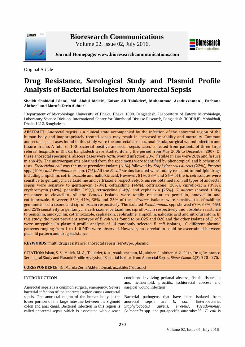
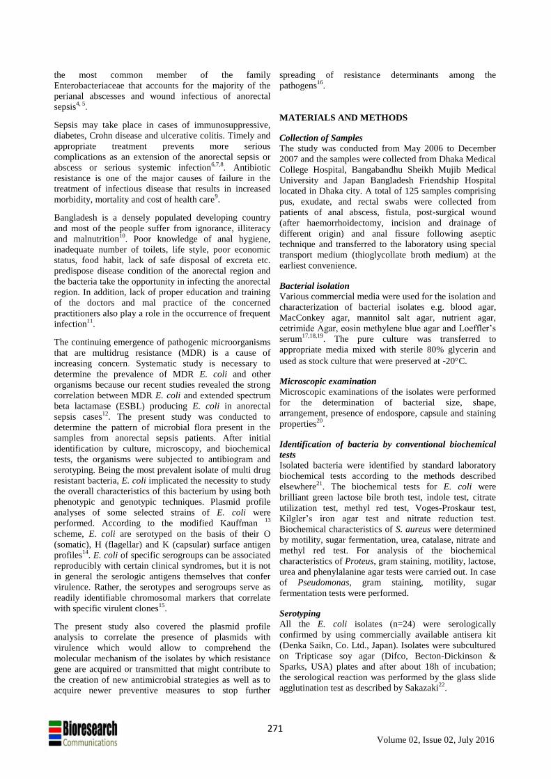
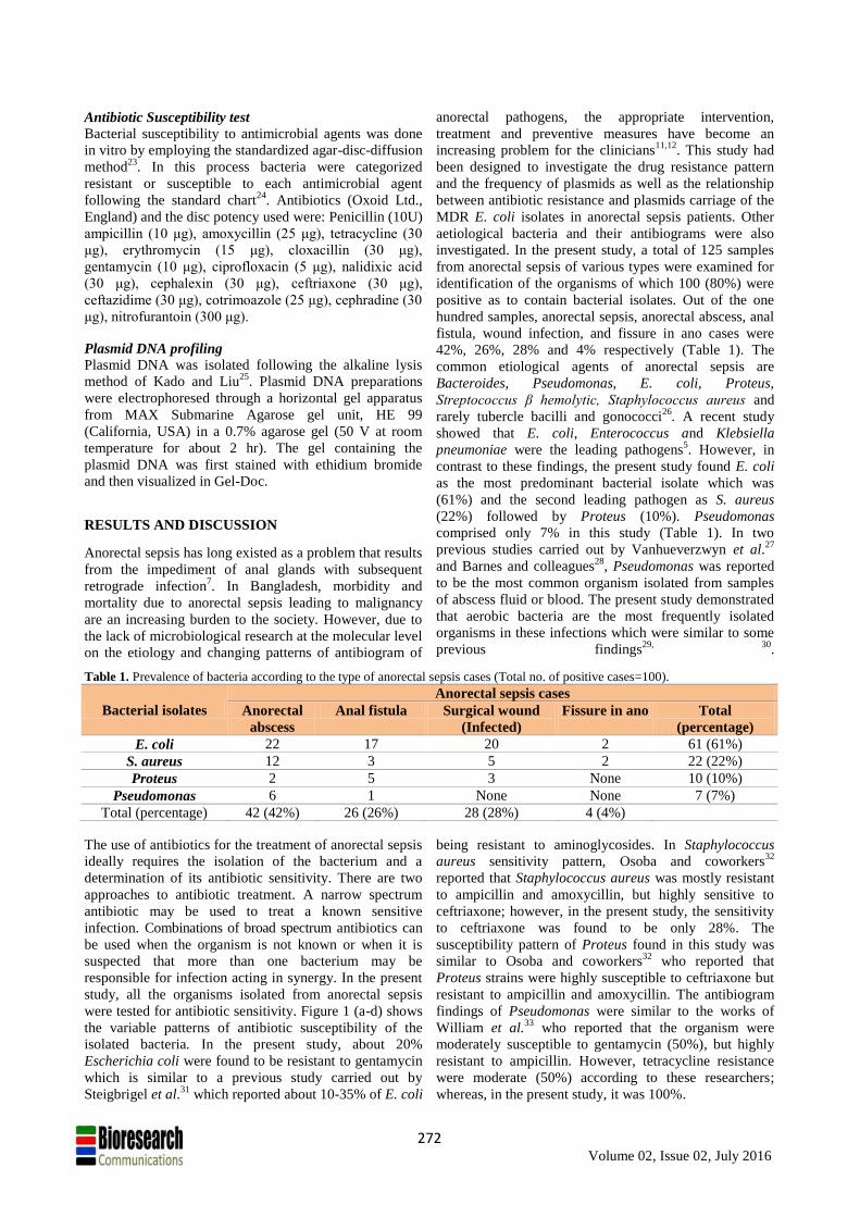
were tested for antibiotic sensitivity. Figure 1 (a-d) shows
the variable patterns of antibiotic susceptibility of the
isolated bacteria. In the present study, about 20%
Escherichia coli were found to be resistant to gentamycin
which is similar to a previous study carried out by
Steigbrigel et al.31
which reported about 10-35% of E. coli
being resistant to aminoglycosides. In Staphylococcus
aureus sensitivity pattern, Osoba and coworkers32
reported that Staphylococcus aureus was mostly resistant
to ampicillin and amoxycillin, but highly sensitive to
ceftriaxone; however, in the present study, the sensitivity
to ceftriaxone was found to be only 28%. The
susceptibility pattern of Proteus found in this study was
similar to Osoba and coworkers32
who reported that
Proteus strains were highly susceptible to ceftriaxone but
resistant to ampicillin and amoxycillin. The antibiogram
findings of Pseudomonas were similar to the works of
William et al.33
who reported that the organism were
moderately susceptible to gentamycin (50%), but highly
resistant to ampicillin. However, tetracycline resistance
were moderate (50%) according to these researchers;
whereas, in the present study, it was 100%.
272 Volume 02, Issue 02, July 2016
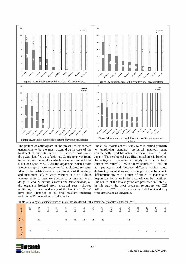
Figure 1a. Antibiotic susceptibility pattern of E. coli isolates.
Figure 1b. Antibiotic susceptibility pattern of S. aureus isolates.
Figure 1c. Antibiotic susceptibility pattern of Proteus spp. isolates.
Figure 1d. Antibiotic susceptibility pattern of Pseudomonas spp.
isolates.
The pattern of antibiogram of the present study showed
gentamycin to be the most potent drug in case of the
treatment of anorectal sepsis. The second most potent
drug was identified as ceftazidime. Ceftriaxone was found
to be the third potent drug which is almost similar to the
result of Osoba et al.32
. All the organisms isolated from
anorectal sepsis were found to be multidrug resistant.
Most of the isolates were resistant to at least three drugs
and maximum isolates were resistant to 6 or 7 drugs
whereas some of them were found to be resistant to all
drugs. E. coli, S. aureus, Proteus and Pseudomonas, all
the organism isolated from anorectal sepsis showed
multidrug resistance and many of the isolates of E. coli
have been identified as all drug resistant including
resistant to 3rd
generation cephalosporins.
The E. coli isolates of this study were identified primarily
by employing standard serological methods using
commercially available antisera (Denka Saiken Co Ltd.,
Japan). The serological classification scheme is based on
the antigenic differences in highly variable bacterial
surface molecules34
. Because most strains of E. coli are
not pathogens and because different strains cause
different types of diseases, it is important to be able to
differentiate strains or groups of strains so that strains
responsible for a particular outbreak can be identified.
The results of the investigation are presented in Table 2.
In this study, the most prevalent serogroup was O25
followed by O20. Other isolates were different and they
were designated as untypable.
Table 2. Serological characteristics of E. coli isolates tested with commercially available antisera (n=24).
Iso
late
s
E 0
2
E 0
3
E 0
4
E 0
7
E 1
2
E 1
4
E 1
5
E 2
0
E 2
2
E 2
4
E 2
5
E 2
6
E 2
8
E 2
9
E 3
0
E 3
5
Po
ly
5
O25 O25 O25 O25 O25 O20 O20
Un
typab
le
273 Volume 02, Issue 02, July 2016
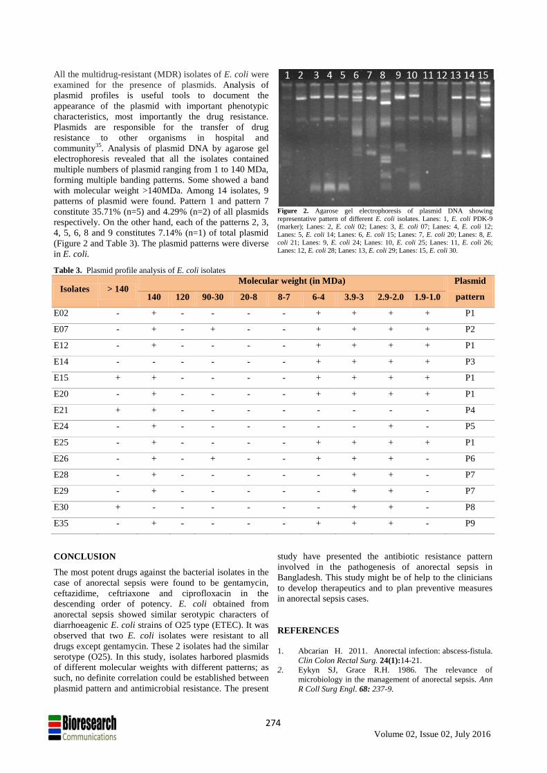
All the multidrug-resistant (MDR) isolates of E. coli were
examined for the presence of plasmids. Analysis of
plasmid profiles is useful tools to document the
appearance of the plasmid with important phenotypic
characteristics, most importantly the drug resistance.
Plasmids are responsible for the transfer of drug
resistance to other organisms in hospital and
community35
. Analysis of plasmid DNA by agarose gel
electrophoresis revealed that all the isolates contained
multiple numbers of plasmid ranging from 1 to 140 MDa,
forming multiple banding patterns. Some showed a band
with molecular weight >140MDa. Among 14 isolates, 9
patterns of plasmid were found. Pattern 1 and pattern 7
constitute 35.71% (n=5) and 4.29% (n=2) of all plasmids
respectively. On the other hand, each of the patterns 2, 3,
4, 5, 6, 8 and 9 constitutes 7.14% (n=1) of total plasmid
(Figure 2 and Table 3). The plasmid patterns were diverse
in E. coli.
Figure 2. Agarose gel electrophoresis of plasmid DNA showing
representative pattern of different E. coli isolates. Lanes: 1, E. coli PDK-9 (marker); Lanes: 2, E. coli 02; Lanes: 3, E. coli 07; Lanes: 4, E. coli 12;
Lanes: 5, E. coli 14; Lanes: 6, E. coli 15; Lanes: 7, E. coli 20; Lanes: 8, E.
coli 21; Lanes: 9, E. coli 24; Lanes: 10, E. coli 25; Lanes: 11, E. coli 26;
Lanes: 12, E. coli 28; Lanes: 13, E. coli 29; Lanes: 15, E. coli 30.
Table 3. Plasmid profile analysis of E. coli isolates
Isolates > 140 Molecular weight (in MDa) Plasmid
pattern 140 120 90-30 20-8 8-7 6-4 3.9-3 2.9-2.0 1.9-1.0
E02 - + - - - - + + + + P1
E07 - + - + - - + + + + P2
E12 - + - - - - + + + + P1
E14 - - - - - - + + + + P3
E15 + + - - - - + + + + P1
E20 - + - - - - + + + + P1
E21 + + - - - - - - - - P4
E24 - + - - - - - - + - P5
E25 - + - - - - + + + + P1
E26 - + - + - - + + + - P6
E28 - + - - - - - + + - P7
E29 - + - - - - - + + - P7
E30 + - - - - - - + + - P8
E35 - + - - - - + + + - P9
CONCLUSION
The most potent drugs against the bacterial isolates in the
case of anorectal sepsis were found to be gentamycin,
ceftazidime, ceftriaxone and ciprofloxacin in the
descending order of potency. E. coli obtained from
anorectal sepsis showed similar serotypic characters of
diarrhoeagenic E. coli strains of O25 type (ETEC). It was
observed that two E. coli isolates were resistant to all
drugs except gentamycin. These 2 isolates had the similar
serotype (O25). In this study, isolates harbored plasmids
of different molecular weights with different patterns; as
such, no definite correlation could be established between
plasmid pattern and antimicrobial resistance. The present
study have presented the antibiotic resistance pattern
involved in the pathogenesis of anorectal sepsis in
Bangladesh. This study might be of help to the clinicians
to develop therapeutics and to plan preventive measures
in anorectal sepsis cases.
REFERENCES
1. Abcarian H. 2011. Anorectal infection: abscess-fistula.
Clin Colon Rectal Surg. 24(1):14-21.
2. Eykyn SJ, Grace R.H. 1986. The relevance of
microbiology in the management of anorectal sepsis. Ann
R Coll Surg Engl. 68: 237-9.
274 Volume 02, Issue 02, July 2016
3. Brook I. and Frazier E.H. 1997. The aerobic and
anaerobic bacteriology of perirectal abscesses. J. Clin.
Microbiol. 35: 2974–2976.
4. Liu C.K., Liu C.P., Leung C.H. and Sun F.J. 2011.
Clinical and microbiological analysis of adult perianal
abscess. J Microbiol Immunol Infect. 44: 204–8.
5. Chen C.Y., Cheng A., Huang S.Y., Sheng W.H., Liu J.H.,
Ko B.S., Yao M., Chou W.C., Lin H.C., Chen Y.C., Tsay
W., Tang J.L., Chang S.C. and Tien H.F. 2013. Clinical
and microbiological characteristics of perianal infections
in adult patients with acute leukemia. PLoS One. 8:
e60624.
6. Genua J.G. and Vivas D.A. 2007. Management of
nonhealing perineal wounds. Clin Colon Rectal Surg. 20:
322–328.
7. Ulug M., Gedik E., Girgin S., Celen M.K. and Ayaz C.
2010. Braz J Infect Dis. 14: 225-229.
8. Pini Prato A., Castagnola E., Micalizzi C., Dufour C.,
Avanzini S., et al. 2012. Early diverting colostomy for
perianal sepsis in children with acute leukemia. J Pediatr
Surg. 47: e23–7.
9. Bouza E. and Cercenado E. 2002. Klebsiella and
Enterobacter: Antibiotic resistance and treatment
implications. Semin. Respir . Infect. 17: 215-230.
10. Haseen F. “Malnutrition among Bangladeshi women in
ultra poor households: prevalence and determinants”,
M.Sc. Thesis in international health, International
maternal and child health. Department of women‟s and
children‟s health, Uppsala University, Uppsala, Sweden,
pp.4, 2010.
11. Sarkar H., Hassan M. and Laila R.N. 2008. Pattern of
Anorectal Disorders in Surgical Practice in Rajshahi.
Journal of Teachers Association. 21: 69-72.
12. Islam S.S., Malek M.A., Haque A.K.M.F., Talukder K.A.
and Akhter M.Z. 2013. Beta lactamase genes of extended
spectrum beta lactamase producing Escherichia coli from
anorectal sepsis cases in Bangladesh. Bangladesh J
Microbiol. 30: 23-29.
13. Kauffmann F. 1944. The basis of O Antigen Serotyping is
the standard tube agglutination 492 tests adapted to use U-
bottomed microtitre trays. Microbiologica Scandinavia.
21: 20–40.
14. Edwards P.R. and Ewing W.H. 1972. Identification of
Enterobacteriaceae, 3rd edn, Burgess Publishing Company,
Minneapolis.
15. Whittam T..S, Wolfe M.L., Wachsmuth I.K., Ørskov F.,
Ørskov I. and Wilson R.A. 1993. Clonal relationships
among Escherichia coli strains that cause hemorrhagic
colitis and infantile diarrhea. Infection and Immunity. 61:
1619-1629.
16. Shapiro J.A. 1999. Views about evolution are evolving.
Am Soc Microbiol News. 65: 201-207.
17. Cheesbrough M. 1984. Medical laboratory manual for
topical countries. 1st edn. Vol. 2, pp. 35: 40-57.
Microbiology, English Language Book Society, London.
18. Carter G.R. 1986. Essentials of Veterinary Bacteriology
and Mycology. 3rd edn. pp. 312-330.
19.
Cowan S.T. 1985. Biochemical behavior of E. coli.
Journal of Genetic Microbiology, 8: 391.
20. Kreig N.R., Holt J.G., Williams and Wilkins. 1984.
Bergeys Manual of Systematic Bacteriology, Vol. 1: 428,
East Preston street, Baltimore, M.D. 21202, USA.
21. Collee J.G., Miles R.S. and Watt B. 1996.Tests for
identification of bacteria. In Mackie and McCartney
Practical Medical Microbiology. 14th edition, Collee JG,
Fraser AG, Marmion BP, Simmons A (Eds.), pp.131-49,
166-7. Churchill Livingstone: New York,
22. Sakazaki R. 1992. Bacteriology of Vibrio and related
organisms. In: Cholera. (Barua D and Greenough WB
Eds.), pp. 37–55. Plenum Medical Book Co., New York.
23. Bauer A.W., Kirby W.M.M., Sheris J.C. and Truck M.
1966. Antibiotic susceptibility testing by a standardized
single disc method. Am J Clin Path. 145: 225-230.
24. Barry A.L. and Thornsberry C. 1985. Susceptibility tests:
Diffusion test procedures. In Lennette EH, Balows A,
Hausler Jr WJ, Shadomy HJ, eds. Manual of Clinical
Microbiology. 4th ed, pp. 978–987. Am Soc Microbiol,
Washington, D.C.
25. Kado C.I. and Liu S.T. 1981. Rapid procedure for
detection and isolation of large and small plasmids. J
Bacteriol. 145:1365-73.
26. Russel R.C., Norman G., William S. and Christopher J.K.
2004. In Bailey and Love‟s Short Practice of Surgery, 24th
edn, pp 1263-65. Hodder Arnold, London.
27. Vanhueverzwyn R., Delannoy A., Michaux J.L. and Dive
C. 1980. Anal lesions in hematologic diseases. Dis Colon
Rectum. 23: 310–2.
28. Barnes S.G., Sattler F.R. and Ballard J.O. 1984. Perirectal
infections in acute leukemia. Improved survival after
incision and debridement. Ann Intern Med. 100: 515–8.
29. Enberg R.N., Cox R.H. and Bury V.F. 1974. Perirectal
abscess in children. Am J Dis Child. 128: 360-1.
30. Krieger R.W. and Chusid M.J. 1979. Perirectal abscess in
childhood: a review of 29 cases. Am J Dis Child. 133:
411-2.
31. Steigbrigel N.H., McCall C.E., Reed C.W. and Finland M.
1967. Antibacterial action of „broad spectrum‟ penicilins,
cephalosporins and other antibiotics against Gram-
negative bacilli isolated from blood cultures. Ann. N.Y.
Acced Sci. 145: 224-236. Australia.
32. Osoba A.O., Al-Rasheed A., Bartlett F., Khizzi N., Al-
Saud A. and Al-Admany A.M. 1991. In-vitro
susceptibilities to ampicillin. Saudi. Med. J. 12: 309-313.
33.
34.
Hernández J.R., Martínez-Martínez L., Cantón R., Coque
T.M. and Pascual A. the Spanish Group for Nosocomial
Infections (GEIH). 2005. Nationwide study of
Escherichia coli and Klebsiella pneumoniae producing
extended-spectrum -lactamases in Spain. Antimicrob.
Agents Chemother. 49: 2122–2125.
Salyers A.A. and Whitt D.D. 2005. Escherichia coli
gastrointestinal infections. In Bacterial Pathogenesis: A
Molecular Approach (Salyers AA and Whitt DD eds.), pp.
191-192. American Society for Microbiology,
Washington DC.
275 Volume 02, Issue 02, July 2016