original article clinicopathologic significance of ... 117 (97.5) 42 (100) 75 (96.1) lvi ... was...
TRANSCRIPT

Int J Clin Exp Pathol 2017;10(7):7929-7939www.ijcep.com /ISSN:1936-2625/IJCEP0053187
Original ArticleClinicopathologic significance of androgen receptor expression and discordant receptor status during progression in breast cancer
Eun Young Kim1, Kwan Ho Lee1, Ji-Sup Yun1, Yong Lai Park1, Chan Heun Park1, Sung-Im Do2, Seoung Wan Chae2
Departments of 1Surgery, 2Pathology, Kangbuk Samsung Hospital, School of Medicine, Sungkyunkwan University, Seoul, Korea
Received March 17, 2017; Accepted May 27, 2017; Epub July 1, 2017; Published July 15, 2017
Abstract: The role of androgen receptor (AR) as a prognostic marker has been proposed in breast cancer. This study investigated AR status and its clinical significance in breast cancer, especially in triple negative breast cancer (TNBC). We also evaluated discordant AR status during the process of lymph node metastasis, locoregional recur-rences (LRR) and distant metastasis. From January 2005 to December 2010, we retrospectively reviewed 120 patients including 55 TNBC patients diagnosed as invasive carcinoma with no special type (NST), who were treated at the Kangbuk Samsung Hospital. Tissue microarray was constructed and immunohistochemical expression of AR was performed for 120 invasive carcinomas, NST specimens and matching samples from 28 lymph node metasta-sis, 2 LRR and 8 distant metastases. AR expression was found in 35.0% (42/120) of the total patients and 14.5% (8/55) of those diagnosed as TNBC. Positive expression of AR was significantly correlated with smaller tumor size, early T stage, fewer lymph node metastases, early AJCC stage, lower histologic grade, estrogen receptor/progester-one receptor positivity, more luminal A type, less TNBC, longer disease-free survival and overall survival, fewer dis-tant metastasis and no deaths from breast cancer (all P < 0.05). AR was a favorable prognostic marker for disease free survival in univariate analysis (P = 0.041). The discordance rate of AR status between primary and recurrent/metastatic disease was 21.6%. AR expression was associated with favorable clinicopathological outcomes in the whole study population. AR status can be altered during tumor progression.
Keywords: Androgen receptor, breast cancer, discordance, triple negative breast cancer
Introduction
Breast cancer (BC) is the most common malig-nancy in women, and invasive carcinoma of no special type (NST) is the most common form of BC [1, 2]. Important parameters with therapeu-tic significance and those that aid in the prog-nosis of BC have been identified. American Joint Committee on Cancer (AJCC) stage, histo-logical grade, estrogen receptor (ER), proges-terone receptor (PR) and human epidermal growth factor receptor 2 (HER2) gene amplifica-tion are important parameters determining the therapeutic options for BC. Androgen receptor (AR) is a steroid receptor expressed in 70-80% of BC cells and is more frequently expressed in ER-positive than in ER-negative tumors [3]. AR signaling pathways play different roles accord-
ing to BC subtypes. In ER-positive BC, AR often inhibits the growth effect of ER signaling. In HER-2 positive BC without ER expression, it plays a proliferative role and in triple-negative breast cancer (TNBC) it induces tumor progres-sion [4]. Identifying the underlying mechanisms of AR in each subtype of BC will allow the design of appropriate target therapies for BC, especial-ly TNBC. No targeted therapies are available yet for TNBC. AR is expressed in 10-43% of TNBCs, and one subset of gene expression profiles in TNBCs is androgen responsive [5, 6]. Therefore, it is important to investigate the prevalence of AR expression in each BC subtype.
Discordance of ER, PR and HER-2 receptor sta-tus between primary tumor and metastatic tis-sue has been observed in several studies [7, 8].

Androgen receptor expression in breast cancer
7930 Int J Clin Exp Pathol 2017;10(7):7929-7939
Possible mechanisms pro-posed for discordance in- clude (a) a genuine switch in the biology of the cancer, (b) sampling error, (c) limit-ed accuracy and reproduc-ibility of receptor assays and (d) intra-tumoral het-erogeneity [8]. Discordance rates are reported in the range of 10% to between 35% and 40% [9]. However, discordance of AR expres-sion are not been report- ed widely in the literature. Therefore, it is important to investigate discordance of AR expression in primary tumor, lymph node metas-tasis, recurrence and dis-tant metastasis. The aim of this study is to evaluate AR expression in BC popula-tion and assess how it cor-relates with patient out-comes. We also aimed to measure AR expression across different subtypes and correlate discordance during tumor progression.
Materials and methods
Patient selection
From January 2005 to De- cember 2010, we retrosp- ectively reviewed 120 pa- tients including 55 TNBC, diagnosed as invasive car-cinoma of NST, who were treated at the Kangbuk Samsung Hospital. Patients were characterized based on clinicopathological char-acteristics of age at diag- nosis, TNM stage, axillary lymph node (LN) status, American Joint Committee on Cancer (AJCC) stage, histologic grade, extensi- ve intraductal component (EIC), skin or chest wall invasion, Paget’s disease,
Table 1. Clinicopathological characteristics of patients
ParametersAR status n (%)
pTotal (n = 120)
Positive (n = 42)
Negative (n = 78)
Age at Diagnosis (years)* 50.8±0.9 51.6±1.8 50.4±1.2 0.566Tumor size (cm)* 2.6±1.6 2.2±1.4 2.9±1.7 0.025T stage† 0.023 1 49 (40.8) 23 (54.7) 26 (33.3) 2 58 (48.4) 17 (40.5) 41 (52.7) 3 12 (10.0) 1 (2.4) 11 (14.1) 4 1 (0.8) 1 (2.4) 0 (0)N stage† 0.366 0 61 (50.8) 26 (61.9) 35 (44.8) 1 26 (21.7) 7 (16.7) 19 (24.4) 2 18 (15.0) 5 (11.9) 13 (16.7) 3 15 (12.5) 4 (9.5) 11 (14.1)Tumor size (cm)† 0.019 ≤2.0 54 (45.0) 25 (59.5) 29 (37.2) >2.0 66 (55.0) 17 (40.5) 49 (62.8)LN metastasis† 0.126 Yes 59 (49.2) 16 (38.1) 43 (55.2) No 61 (50.8) 26 (61.9) 35 (44.8)Number of LN metastases* 3.7±0.6 2.1±0.6 4.5±0.8 0.028AJCC stage† 0.010 I 30 (25.0) 18 (42.9) 12 (15.4) II 51 (42.5) 14 (33.3) 37 (47.4) III 33 (27.5) 9 (21.4) 24 (30.8) IV 6 (5.0) 1 (2.4) 5 (6.4)Histologic grade† 0.000 1 19 (16.1) 12 (28.6) 7 (8.9) 2 45 (38.1) 22 (52.4) 24 (30.7) 3 54 (45.8) 8 (19.0) 47 (60.4)EIC† 0.037 Yes 11 (9.2) 7 (16.7) 4 (5.1) No 109 (90.8) 35 (83.3) 74 (94.9)Skin/chest wall invasion‡ 1.000 Yes 2 (1.7) 1 (2.4) 1 (1.3) No 118 (98.3) 41 (97.6) 77 (98.7)Paget disease‡ 0.551 Yes 3 (2.5) 0 (0) 3 (3.9) No 117 (97.5) 42 (100) 75 (96.1)LVI† 0.221 Yes 37 (30.8) 10 (23.8) 27 (34.6) No 83 (69.2) 32 (76.2) 51 (65.4)ER status† 0.000 Positive 54 (45.0) 32 (76.2) 22 (28.2) Negative 66 (55.0) 10 (23.8) 56 (71.8)PR status† 0.000 Positive 45 (37.5) 29 (69.1) 16 (20.5)
Androgen receptor expression in breast cancer
7931 Int J Clin Exp Pathol 2017;10(7):7929-7939
lymphovascular invasion (LVI), ER positivity, PR positivity, HER-2 overexpression and subtypes of tumor, type of surgery, locoregional recur-rence (LRR), distant metastasis and death from BC. Overall survival (OS) was defined as the time interval between the date of surgical resection and the date of disease specific death or last follow-up. Disease-free survival (DFS) was defined as the time between the date of surgical resection and the date of docu-mented relapse, including LRR and distant metastasis. All studies were conducted with prior approval from the Institutional Review Board of Kangbuk Samsung Hospital (Approval No. 2014-10-027).
Tissue selection and tissue microarray (TMA) construction
We obtained primary invasive ductal carcino-ma, NST tissue, metastatic cancer tissue from LNs, other organ and recurred tissue for TMA construction. Surgical specimens were fixed using 10% buffered formalin, processed and
4-mm punch depth stop device and semi-auto-matic micrometers. The instrument was used to create holes in a recipient block with defined array cores. The fit needle was used to deliver the tissue cores into the recipient block. Taking into account the limitations of the representa-tive areas of the tumor, we used duplicate 2-mm-diameter tissue cores from each donor block. The percentage of tissue cores taken from within the tumor exceeded 70%.
Immunohistochemistry
Immunohistochemical staining was performed on 3 μm-thick TMA block sections. Sections were dehydrated and deparaffinized in xylene and rehydrated in a graded series of alcohol solutions. We used primary antibodies against ER (1:200, clone SP1; Lab Vision Corporation, Fremont, CA, USA), PR (1:200, clone PgR 636; DakoCytomation, Glostrup, Denmark), HER-2 (1:200, clone SP3, Lab Vision Corporation), and AR receptor (1:200, clone AR 441; Abcam, Cambridge, UK). Immunostaining was per-
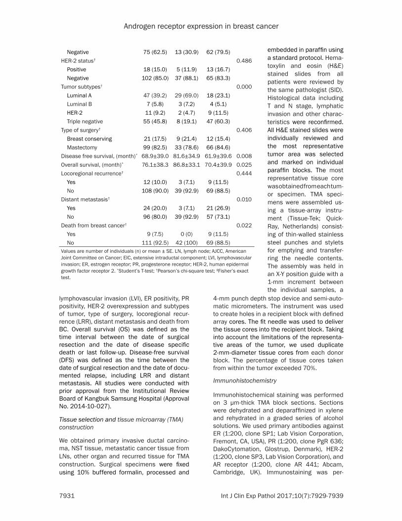
Negative 75 (62.5) 13 (30.9) 62 (79.5)HER-2 status† 0.486 Positive 18 (15.0) 5 (11.9) 13 (16.7) Negative 102 (85.0) 37 (88.1) 65 (83.3)Tumor subtypes† 0.000 Luminal A 47 (39.2) 29 (69.0) 18 (23.1) Luminal B 7 (5.8) 3 (7.2) 4 (5.1) HER-2 11 (9.2) 2 (4.7) 9 (11.5) Triple negative 55 (45.8) 8 (19.1) 47 (60.3)Type of surgery† 0.406 Breast conserving 21 (17.5) 9 (21.4) 12 (15.4) Mastectomy 99 (82.5) 33 (78.6) 66 (84.6)Disease free survival, (month)* 68.9±39.0 81.6±34.9 61.9±39.6 0.008Overall survival, (month)* 76.1±38.3 86.8±33.1 70.4±39.9 0.025Locoregional recurrence† 0.444 Yes 12 (10.0) 3 (7.1) 9 (11.5) No 108 (90.0) 39 (92.9) 69 (88.5)Distant metastasis† 0.010 Yes 24 (20.0) 3 (7.1) 21 (26.9) No 96 (80.0) 39 (92.9) 57 (73.1)Death from breast cancer† 0.022 Yes 9 (7.5) 0 (0) 9 (11.5) No 111 (92.5) 42 (100) 69 (88.5)Values are number of individuals (n) or mean ± SE. LN, lymph node; AJCC, American Joint Committee on Cancer; EIC, extensive intraductal component; LVI, lymphovascular invasion; ER, estrogen receptor; PR, progesterone receptor; HER-2, human epidermal growth factor receptor 2. *Student’s T-test; †Pearson’s chi-square test; ‡Fisher’s exact test.
embedded in paraffin using a standard protocol. Hema- toxylin and eosin (H&E) stained slides from all patients were reviewed by the same pathologist (SID). Histological data including T and N stage, lymphatic invasion and other charac-teristics were reconfirmed. All H&E stained slides were individually reviewed and the most representative tumor area was selected and marked on individual paraffin blocks. The most representative tissue core was obtained from each tum- or specimen. TMA speci-mens were assembled us- ing a tissue-array instru-ment (Tissue-Tek; Quick-Ray, Netherlands) consist-ing of thin-walled stainless steel punches and stylets for emptying and transfer-ring the needle contents. The assembly was held in an X-Y position guide with a 1-mm increment between the individual samples, a

Androgen receptor expression in breast cancer
7932 Int J Clin Exp Pathol 2017;10(7):7929-7939
Figure 1. Androgen receptor expression in invasive carcinoma, NST. A: Strong expression; B: Moderate expression; C: Weak expression; D: Negative.
formed using a compact polymer method (Bond Intense Detection Kit; Leica Biosystems, New- castle upon Tyne, UK). The primary antibodies were detected with an EnVision+ System utiliz-ing horseradish peroxidase (DakoCytoma- tion) according to the manufacturer’s instruc-tions. An EnVision+ Detection System incorpo-rating peroxidase and 3,3’-diaminogenzidine (DakoCytomation) was used to perform chro-mogenic visualization. The slides were counter-stained with hematoxylin and cover slipped. ER, PR and AR status was assessed using the Allred scoring method [10]. HER-2 overexpres-sion was evaluated using American Society of Clinical Oncology/College of American Patho- logists guideline recommendations. In cases with equivocal HER-2 staining (score 2), silver in situ hybridization (Ventana Medical Systems, Tucson, AZ, USA) was performed to determine HER2 gene status. All slides were examined and scored by two board-certified pathologis- ts blinded to the clinicopathological data and pa- tient identity. Disagreements between the two pathologists were resolved by consensus.
Statistical analyses
Student’s t-test for continuous variables and the Pearson’s χ² test for categorical variables were used to evaluate the associations between AR expression and clinicopathologic parameters. Cox proportional hazards regres-sion test was used to calculate the hazard ratio (HR) of data on DFS and OS. Multivariate Cox regression analysis was performed only for variables with significant univariate impact. Survival probability curves were calculated by the Kaplan-Meier method. A p-value < 0.05 (2-tailed) was considered statistically signifi-cant. All statistical analyses were performed with PASW Statistics for Windows, Version 18.0 (SPSS Inc., Chicago, IL, USA).
Results
Patient demographics
We retrospectively reviewed the clinicopatho-logical data of 120 BC patients including 55

Androgen receptor expression in breast cancer
7933 Int J Clin Exp Pathol 2017;10(7):7929-7939
TNBC (Table 1). The mean age was 50.8±10.9 years (range, 27-82 years). Thirty patients (25.0%) were AJCC stage I, 51 (42.5%) were stage II, 33 (27.5%) were stage III and 6 (5.0%) were stage IV. LN metastasis was detected in 59 (49.2%) patients. Twelve patients (10.0%), 24 patients (20.0 %) and 9 patients (7.5%) had LRR, distant metasta-sis and death from BC, respectively, during the fol-low-up period. The mean duration of DFS and OS was 68 months (range, 1- 140 months) and 76 mon- ths (range, 3-192 months), respectively.
AR expression and associ-ation with clinicopathologi-cal characteristics
AR expression was found in 42 (35.0%) of the 120 cases (Figure 1). Positive expression of AR showed significant correlation with smaller tumor size, early T stage, fewer number of LN metastases, early AJCC stage, lower histologic gra- de, ER/PR positivity, more luminal A type, lesser TN- BC, longer DFS and OS, fewer distant metastasis and no death from BC (all P < 0.05) (Table 1). No statis-tically significant differenc-es were observed between AR expression and age at diagnosis, histologic type, skin/chest wall invasion, Paget disease, LVI, HER-2 status, type of surgery and LRR. A summary of the rela-tionship between AR expre- ssion and clinicopathologi-cal parameters is provided in Table 1.
Table 2. Clinicopathological characteristics of 55 TNBC patients
ParametersAR status n (%)
pTotal (n = 55)
Positive (n = 8)
Negative (n = 47)
Age at diagnosis (years)* 20.9±1.5 59.6±2.3 49.4±1.6 0.006Tumor size (cm)* 2.9±0.2 1.8±0.3 3.1±0.3 0.032T stage† 0.059 1 21 (38.2) 6 (75.0) 15 (31.9) 2 26 (47.3) 2 (25.0) 24 (51.1) 3 8 (14.5) 0 (0) 8 (17.0) 4 0 (0) 0 (0) 0 (0)N stage† 0.100 0 25 (45.5) 6 (75.0) 19 (40.4) 1 15 (27.3) 0 (0) 15 (31.9) 2 7 (12.7) 0 (0) 7 (14.9) 3 8 (14.5) 2 (25.0) 6 (12.8)Tumor size (cm)† 0.096 ≤2.0 20 (36.4) 5 (62.5) 15 (31.9) >2.0 35 (63.6) 3 (37.5) 32 (68.1)LN metastasis† 0.089 Yes 26 (47.3) 6 (75.0) 20 (42.5) No 29 (52.7) 2 (25.0) 27 (57.5)Number of LN metastases* 3.6±0.8 3.2±2.2 3.7±0.9 0.576AJCC stage‡ 0.029 I 12 (21.8) 5 (62.5) 7 (14.9) II 24 (43.6) 1 (12.5) 23 (48.9) III 16 (29.1) 2 (25.0) 14 (29.8) IV 3 (5.5) 0 (0) 3 (6.4)Histologic grade‡ 0.002 1 2 (3.7) 1 (12.5) 1 (2.2) 2 17 (31.5) 6 (75.0) 11 (23.9) 3 35 (64.8) 1 (12.5) 34 (73.9)EIC‡ 0.272 Yes 53 (96.4) 7 (87.5) 46 (97.9) No 2 (3.6) 1 (12.5) 1 (2.1)Skin/chest wall invasion‡ 1.000 Yes 1 (1.8) 0 (0) 1 (2.1) No 54 (98.2) 8 (100) 46 (97.9)Paget disease‡ 1.000 Yes 1 (1.8) 0 (0) 1 (2.1) No 54 (98.2) 8 (100) 46 (97.9)LVI‡ 0.696 Yes 21 (38.2) 2 (25.0) 19 (40.4) No 34 (61.8) 6 (75.0) 28 (59.6)Type of surgery† 0.859 Breast conserving 8 (14.5) 1 (12.5) 7 (14.9) Mastectomy 47 (85.5) 7 (87.5) 40 (85.1)Disease free survival, (month)* 68.9±39.0 81.6±34.9 61.9±39.6 0.008Overall survival, (month)* 76.1±38.3 86.8±33.1 70.4±39.9 0.025

Androgen receptor expression in breast cancer
7934 Int J Clin Exp Pathol 2017;10(7):7929-7939
AR expression and clinicopatholgic association in TNBC
AR expression in TNBC occurred in 8 (14.5%) of the 55 patients. AR expression in TNBC was associated with older age at diagnosis (59 vs. 49 years, respectively; P = 0.006), smaller tumor size (P = 0.032), early AJCC stage (P = 0.021) and lower histologic grade (P = 0.003) (Table 2). We could not evaluate association between AR expression in TNBC patients and prognosis, because there was no LRR, distant metastasis or death from BC in AR positive TNBCs.
AR expression and association with prognosis
AR expression was significantly independent favorable prognostic factor with distant metas-tasis (P = 0.010) and death from BC (P = 0.022) (Table 1). In univariate analyses, AR expres-sion, tumor size, LN metastasis, skin/chest wall invasion, LVI, ER/PR positivity and HER-2 enriched subtype were all statistically signifi-cantly associated with DFS (all P < 0.05; Table 3). However, multivariate analyses showed that LN metastasis (HR 2.88, 95% confidence inter-val (CI), 1.19-6.97; P = 0.019) and skin/chest wall invasion (HR 10.562, 95% CI, 1.95-57.17; P = 0.006) were significantly associated with DFS (Table 3). Patients with AR expression had a more favorable DFS than those without expression in Kaplan-Meier curve analyses (χ2 = 4.18; df = 2; P = 0.041) (Figure 2). We could not evaluate association between AR expres-sion and OS, because there was no death from BC in AR positive BCs.
21.6% (8/37) of cases tested. Among 16 AR positive primary BCs, 1 case was negative in matching LN metastasis (Figure 3). The remain-der of the 15 patients stayed AR positive during LN metastasis and distant metastasis. Among 21 AR negative primary tumors, 4, 1 and 2 tumors changed to AR positive on matching LN metastasis, both LN metastasis and LRR, and distant metastasis, respectively (Figure 4). Rest of 14 AR negative BCs stayed negative in LN metastasis, LRR and distant metastasis. The results are summarized in Table 4.
Discussion
AR is a member of the steroid hormone recep-tor family, which also includes ER and PR. Steroid hormone receptor plays significant roles in signaling pathways and as a transcrip-tion factor. ER and PR are well-known prognos-tic and predictive factors of endocrine thera-pies in BC. However, the role of AR in BC and its progression has been less profoundly studied and remains as an unanswered question.
In this study, we assessed how AR serves as a prognostic marker. Consistent with previous studies, our results showed that AR expression was related with favorable prognostic markers such as DFS [11]. AR negativity was associated with larger tumor size, LN metastasis, higher AJCC stage, higher histologic grade, ER/PR negativity, triple negativity and shorter DFS in univariate analysis.
Majority of the literatures described favorable prognostic impact of AR expression in BC [12]. However, others have reported mixed results such as patients with AR expressing ER nega-
Locoregional recurrence† 0.333 Yes 5 (9.1) 0 (0) 5 (10.6) No 50 (90.9) 8 (100) 42 (89.4)Distant metastasis† 0.089 Yes 13 (23.6) 0 (0) 13 (27.7) No 42 (76.4) 8 (100) 34 (72.3)Death from breast cancer† 0.243 Yes 7 (12.7) 0 (0) 7 (14.9) No 48 (87.3) 8 (100) 40 (85.1)Values are number of individuals (n) or mean ± SE. LN, lymph node; AJCC, American Joint Committee on Cancer; EIC, extensive intraductal component; LVI, lymphovascular invasion; ER, estrogen receptor; PR, progesterone receptor; HER-2, human epidermal growth factor receptor 2. *Student’s T-test; †Pearson’s chi-square test; ‡Fisher’s exact test.
AR status in metastases and recurrence
Only 37 primary BCs had available AR status on LN metastasis, LRR, and dis-tant metastasis. AR sta- tus was positive in 43.2% (16/37) of primary tumor, 64.2% (18/28) of LN meta- stasis, 50.0% (1/2) of LRR and 50.0% (4/8) of distant metastases. A discordant AR status between primary BC and matched metastat-ic samples was observed in

Androgen receptor expression in breast cancer
7935 Int J Clin Exp Pathol 2017;10(7):7929-7939
Table 3. Univariate and multivariate analysis of disease-free survival in whole population
ParameterUnivariate Multivariate
HR (95% CI) p HR (95% CI) pAR expression 0.041 0.346 Negative 1 1 Positive 0.37 (0.14-0.99) 0.61 (0.22-1.71)Age at diagnosis 0.272 ≤40 1 >40 0.98 (0.94-1.02)Tumor size (cm) 0.005 0.398 ≤2.0 1 1 >2.0 1.35 (1.09-1.66) 1.46 (0.61-3.50)LN metastasis 0.015 0.019 No 1 1 Yes 2.98 (1.24-7.16) 2.88 (1.19-6.97)Histologic grade 1 1 2 1.21 (0.32-4.56) 0.320 3 2.12 (0.61-7.41) 0.610EIC 0.242 No 1 Yes 0.30 (0.04-2.25)Skin/chest wall invasion 0.001 0.006 No 1 1 Yes 12.41 (2.73-56.48) 10.562 (1.95-57.17)Paget disease 0.075 No 1 Yes 3.77 (0.88-16.19)LVI 0.035 0.334 No 1 1 Yes 2.35 (1.06-5.21) 1.59 (0.62-4.08)ER positive 0.045 0.729 No 1 1 Yes 0.42 (0.18-0.98) 1.25 (0.36-4.32)PR positive 0.019 0.123 No 1 1 Yes 0.31 (0.11-0.83) 0.31 (0.07-1.37)HER-2 overexpression 0.338 No 1 Yes 1.57 (0.62-3.97)Subtype Luminal A 1 Luminal B 0.71 (0.08-5.96) 0.750 HER-2 3.79 (1.20-12.00) 0.023 Triple negative 1.95 (0.76-4.97) 0.163Type of Surgery 0.091 BCS 1 Mastectomy 5.62 (0.76-41.55)AR, androgen receptor; LN, lymph node; EIC, extensive intraductal component; LVI, lymphovascular invasion; ER, estrogen receptor; PR, progesterone receptor; HER-2, human epidermal growth factor receptor 2; BCS, breast conserving surgery.

Androgen receptor expression in breast cancer
7936 Int J Clin Exp Pathol 2017;10(7):7929-7939
tive BC had shorter survival [13]. Presently, AR expression in TNBC was associated with older age at diagnosis, smaller tumor size, early AJCC stage and lower histologic grade. To elucidate these mixed results among the studies, we should clarify unique role of the AR during tumorigenesis of TNBC.
There are currently no targeted therapies avail-able for TNBC. AR is expressed in 10-43% of TNBCs, and one gene expression subset of TNBCs is androgen responsive [6]. The luminal group (LAR) was driven by AR signaling and sensitive to the effects of antiandrogens. The role and prognostic significance of AR expres-sion in TNBC is unclear. AR expression in TNBC was correlated with postmenopausal status, lower histological grade, lack of LN metastasis and better OS [14]. Another study reported that AR expression in 287 TNBC patients was a favorable prognostic factor of DFS and OS [15]. However, contrary to these findings, several reports demonstrated that AR positivity in TNBC was associated with worse clinicopatho-logical parameters and prognosis. AR positive TNBCs reportedly are associated with an 83% increase in overall mortality compared to AR negative tumors [16].
Although, there is disagreement as to the prog-nostic significance of AR expression in TNBC, it should be emphasized because AR positive TNBC patients may benefit from future target-ed therapy. AR blockade could be a potential endocrine therapy for patients with ER negative BCs. Bicalutamide is an oral active nonsteroi-dal antiandrogen agent, in which ongoing clini-cal trials has designed the effect of bicalu-tamide in advanced AR positive and ER/PR negative BC [3].
We also found that AR status tended to be pre-served in metastatic lymph node, recurrence
and distant metastasis. A discordant AR status between primary BC and matched metastatic samples was observed in 21.6% (8/37) of cases tested. Preservation of AR in carcinoma cells between primary and metastatic/recur-rent sites has previously been reported. In a recent study, 23 TNBC patients with matched recurrences (n = 16) and LN metastases (n = 46), AR discrepancies between primary tumors and metastasis did not occur [17]. In another study, AR status was performed on 356 prima-ry BCs, 135 matching metastases and 12 recurrences [18]. A discrepant result was seen in 4.3% (5/117) of primary BC and matching LN metastases. No discrepancies were seen between primary BC and distant metastases or recurrence (n = 17). Compared to these two studies, we observed a higher discordant rate.
Several explanations can be offered for the high discordant rate of AR during tumor pro-gression. First is the true molecular conversion during tumor progression. Conversion for ER-α and PR is mainly confined to the primary tumor and is absent in the metastasized tumors [7]. This finding may be explained by clonal selec-tion of less differentiated receptor negative cells during the metastatic process. Likewise, discordant AR status could be a result from genetic drift during tumor progression. A sec-ond explanation is that the limited accuracy and reproducibility of receptor assays can lead to discordant AR status. Differences in tissue handling, tissue processing, interpretation of immunohistochemistry and different cut-off values that determine whether a tumor is posi-tive or negative may have influenced discor-dance. The third explanation is that premature handling or insufficiently fixed specimens causes impairing of staining [19]. Finally, pri-mary BCs could have exhibited marked intra-tumoral heterogeneity [20]. Remarkable het-erogeneity in the mutational system and copy number alterations between primary tumors, circulating and disseminated tumor cells and metastases has been revealed [21]. Several studies reported discordance in receptor sta-tus between primary BC and synchronous nodal metastases as well as with metastatic sites [22, 23]. Many national and international guide-lines for metastatic BC management recom-mend re-testing of at least one metastatic biop-sy for hormonal and HER2 status [24, 25]. Therefore, it is important to re-biopsy metastat-ic sites during tumor progression for hormonal, HER2 as well as AR status, especially for TNBC
Figure 2. Disease-free survival curve of the whole study population.

Androgen receptor expression in breast cancer
7937 Int J Clin Exp Pathol 2017;10(7):7929-7939
Figure 3. Discordant androgen receptor expression status. A. Primary breast cancer shows positive AR expression. B. Metastatic breast cancer of same patient shows negative AR status.
Figure 4. Discordant androgen receptor expression status. A. Primary breast cancer shows negative AR status. B. Metastatic breast cancer of same patient shows positive AR expression.
patients, in which targeted therapies are not available yet. Further investigations are war-ranted to validate these findings.
was associated with older age at diagnosis, smaller tumor size, early AJCC stage and lower histologic grade. In addition, AR status can be
Table 4. Discordant AR status between primary BC and matched metastases
Number of patients
AR status
Primary BC LN metastasis Locoregional recurrence
Distant metastasis
13 Positive Positive - -2 Positive - - Positive1 Positive Negative - -9 Negative Negative - -1 Negative - Negative -4 Negative - - Negative4 Negative Positive - -1 Negative Positive Positive -2 Negative - - PositiveAR, androgen receptor; BC, breast cancer; LN, lymph node.
Our study has several limitations. First, there were a small number of patients with too few LRR, distant metastasis and no death from BC. Larger sample size with better matched metastatic samples can effectively characterize survival dif-ferences. Second, due to the poor preservation status of tissue sam-ples, immunohistochemical staining for LN metastasis, LRR and distant metastasis could not be performed for the entire population.
In conclusion, the presence of AR was significantly associated with favorable clinicopathologic and pro- gnostic features. AR positive TNBC

Androgen receptor expression in breast cancer
7938 Int J Clin Exp Pathol 2017;10(7):7929-7939
changed during tumor progression such as LN metastasis, recurrent and distant metastatic tumors. Further evaluation will be needed to find out the mechanism of AR expression alteration.
Acknowledgements
This work was supported by a grant from the Samsung Biomedical Research Institute.
Disclosure of conflict of interest
None.
Address correspondence to: Dr. Sung-Im Do, Depart- ment of Pathology, Kangbuk Samsung Hospital, School of Medicine, Sungkyunkwan University, 29 Saemunan-ro, Jongno-gu, Seoul 03181, Korea. Tel: +82-2-2001-2393; Fax: +82-2-2001-2398; E-mail: [email protected]; Dr. Chan Heun Park, Department of Surgery, Kangbuk Samsung Hospital, School of Medicine, Sungkyunkwan University, 29 Saemunan-ro, Jongno-gu, Seoul 03181, Korea. Tel: +82-2-2001-1730; Fax: +82-2-2001-1883; E-mail: [email protected]
References
[1] Isaacs C, Stearns V and Hayes DF. New prog-nostic factors for breast cancer recurrence. Semin Oncol 2001; 28: 53-67.
[2] Duffy MJ and Duggan C. The urokinase plas-minogen activator system: a rich source of tu-mour markers for the individualised manage-ment of patients with cancer. Clin Biochem 2004; 37: 541-548.
[3] Gucalp A and Traina TA. Triple-negative breast cancer: role of the androgen receptor. Cancer J 2010; 16: 62-65.
[4] Pietri E, Conteduca V, Andreis D, Massa I, Mel-egari E, Sarti S, Cecconetto L, Schirone A, Bra-vaccini S, Serra P, Fedeli A, Maltoni R, Amadori D, De Giorgi U and Rocca A. Androgen receptor signaling pathways as a target for breast can-cer treatment. Endocr Relat Cancer 2016; 23: R485-498.
[5] Niemeier LA, Dabbs DJ, Beriwal S, Striebel JM and Bhargava R. Androgen receptor in breast cancer: expression in estrogen receptor-posi-tive tumors and in estrogen receptor-negative tumors with apocrine differentiation. Mod Pathol 2010; 23: 205-212.
[6] Lehmann BD, Bauer JA, Chen X, Sanders ME, Chakravarthy AB, Shyr Y and Pietenpol JA. Identification of human triple-negative breast cancer subtypes and preclinical models for se-
lection of targeted therapies. J Clin Invest 2011; 121: 2750-2767.
[7] Broom RJ, Tang PA, Simmons C, Bordeleau L, Mulligan AM, O’Malley FP, Miller N, Andrulis IL, Brenner DM and Clemons MJ. Changes in es-trogen receptor, progesterone receptor and Her-2/neu status with time: discordance rates between primary and metastatic breast can-cer. Anticancer Res 2009; 29: 1557-1562.
[8] Pusztai L, Viale G, Kelly CM and Hudis CA. Es-trogen and HER-2 receptor discordance be-tween primary breast cancer and metastasis. Oncologist 2010; 15: 1164-1168.
[9] Osborne CK. Heterogeneity in hormone recep-tor status in primary and metastatic breast cancer. Semin Oncol 1985; 12: 317-326.
[10] Harvey JM, Clark GM, Osborne CK and Allred DC. Estrogen receptor status by immunohisto-chemistry is superior to the ligand-binding as-say for predicting response to adjuvant endo-crine therapy in breast cancer. J Clin Oncol 1999; 17: 1474-1481.
[11] Agrawal AK, Jelen M, Grzebieniak Z, Zukrowski P, Rudnicki J and Nienartowicz E. Androgen re-ceptors as a prognostic and predictive factor in breast cancer. Folia Histochem Cytobiol 2008; 46: 269-276.
[12] Qu Q, Mao Y, Fei XC and Shen KW. The impact of androgen receptor expression on breast cancer survival: a retrospective study and me-ta-analysis. PLoS One 2013; 8: e82650.
[13] Farmer P, Bonnefoi H, Anderle P, Cameron D, Wirapati P, Becette V, Andre S, Piccart M, Cam-pone M, Brain E, Macgrogan G, Petit T, Jassem J, Bibeau F, Blot E, Bogaerts J, Aguet M, Bergh J, Iggo R and Delorenzi M. A stroma-related gene signature predicts resistance to neoadju-vant chemotherapy in breast cancer. Nat Med 2009; 15: 68-74.
[14] Luo X, Shi YX, Li ZM and Jiang WQ. Expression and clinical significance of androgen receptor in triple negative breast cancer. Chin J Cancer 2010; 29: 585-590.
[15] Alshenawy HA. Prevalence of androgen recep-tors in invasive breast carcinoma and its rela-tion with estrogen receptor, progesterone re-ceptor and Her2/neu expression. J Egypt Natl Canc Inst 2012; 24: 77-83.
[16] Hu R, Dawood S, Holmes MD, Collins LC, Schnitt SJ, Cole K, Marotti JD, Hankinson SE, Colditz GA and Tamimi RM. Androgen receptor expression and breast cancer survival in post-menopausal women. Clin Cancer Res 2011; 17: 1867-1874.
[17] McNamara KM, Yoda T, Miki Y, Nakamura Y, Suzuki T, Nemoto N, Miyashita M, Nishimura R, Arima N, Tamaki K, Ishida T, Ohuchi N and Sa-sano H. Androgen receptor and enzymes in lymph node metastasis and cancer reoccur-

Androgen receptor expression in breast cancer
7939 Int J Clin Exp Pathol 2017;10(7):7929-7939
rence in triple-negative breast cancer. Int J Biol Markers 2015; 30: e184-189.
[18] Grogg A, Trippel M, Pfaltz K, Ladrach C, Droes-er RA, Cihoric N, Salhia B, Zweifel M and Tapia C. Androgen receptor status is highly con-served during tumor progression of breast can-cer. BMC Cancer 2015; 15: 872.
[19] Neumeister VM, Anagnostou V, Siddiqui S, Eng-land AM, Zarrella ER, Vassilakopoulou M, Pari-si F, Kluger Y, Hicks DG and Rimm DL. Quanti-tative assessment of effect of preanalytic cold ischemic time on protein expression in breast cancer tissues. J Natl Cancer Inst 2012; 104: 1815-1824.
[20] Lyng MB, Laenkholm AV, Pallisgaard N, Vach W, Knoop A, Bak M and Ditzel HJ. Intratumor genetic heterogeneity of breast carcinomas as determined by fine needle aspiration and Taq-Man low density array. Cell Oncol 2007; 29: 361-372.
[21] Kimbung S, Kovacs A, Danielsson A, Bendahl PO, Lovgren K, Frostvik Stolt M, Tobin NP, Lind-strom L, Bergh J, Einbeigi Z, Ferno M, Hatschek T and Hedenfalk I. Contrasting breast cancer molecular subtypes across serial tumor pro-gression stages: biological and prognostic im-plications. Oncotarget 2015; 6: 33306-33318.
[22] Nedergaard L, Haerslev T and Jacobsen GK. Immunohistochemical study of estrogen re-ceptors in primary breast carcinomas and their lymph node metastases including comparison of two monoclonal antibodies. Apmis 1995; 103: 20-24.
[23] Feng Y, Sun B, Li X, Zhang L, Niu Y, Xiao C, Ning L, Fang Z, Wang Y, Zhang L, Cheng J, Zhang W and Hao X. Differentially expressed genes be-tween primary cancer and paired lymph node metastases predict clinical outcome of node-positive breast cancer patients. Breast Cancer Res Treat 2007; 103: 319-329.
[24] Penault-Llorca F, Coudry RA, Hanna WM, Osamura RY, Ruschoff J and Viale G. Experts’ opinion: recommendations for retesting breast cancer metastases for HER2 and hormone re-ceptor status. Breast 2013; 22: 200-202.
[25] Lin NU, Thomssen C, Cardoso F, Cameron D, Cufer T, Fallowfield L, Francis PA, Kyriakides S, Pagani O, Senkus E, Costa A and Winer EP. In-ternational guidelines for management of met-astatic breast cancer (MBC) from the Europe-an School of Oncology (ESO)-MBC task force: surveillance, staging, and evaluation of pa-tients with early-stage and metastatic breast cancer. Breast 2013; 22: 203-210.